Case Report

A Family with a Single LMNA Mutation Illustrates Diversity in Cardiac Phenotypes Associated with Laminopathic Progeroid Syndromes

1
Reference Center for Rare Neurological and Neuromuscular Diseases (CERCA), University Hospital of Martinique, 97200 Fort de France, Martinique, France
2
Department of Cardiology, University Hospital of Martinique, 97200 Fort de France, Martinique, France
3
Research Group n°7525—Cardiovascular Vulnerability in the Caribbean Environment, 97200 Fort de France, Martinique, France
4
Inserm UMR_S938, Saint-Antoine Research Center, Institute of Cardiometabolism and Nutrition, Sorbonne University, 75012 Paris, France
5
National Reference Center for Rare Diseases of Insulin Secretion and Insulin Sensitivity (PRISIS), Department of Endocrinology, Assistance Publique-Hôpitaux de Paris, Saint-Antoine University Hospital, 75012 Paris, France
6
GRC n°22, C2MV-Complications Cardiovasculaires et Métaboliques chez les Patients Vivant avec le Virus de L’immunodéficience Humaine, INSERM UMR_S 938, Centre de Recherche Saint-Antoine, Institut Hospitalo-Universitaire de Cardio-métabolisme et Nutrition (ICAN), Hôpital Saint-Antoine Service de Cardiologie, Assistance Publique-Hôpitaux de Paris, Sorbonne Université, 75012 Paris, France
7
National Institute of Health and Medical Research, Department of Cardiology, AP-HP Saint-Antoine Hospital, 75012 Paris, France
8
Department of Molecular Biology and Genetics—AP-HP Saint-Antoine Hospital, 75571 Paris, France
9
Functional Unit of Molecular and Cellular, Cardiogenetics and Myogenetics, Department of Metabolic Biochemistry, AP-HP Pitié Salpêtrière-Charles Foix University Hospital, 75013 Paris, France
10
Cardiology Department, FILNEMUS, APHP, Cochin Hospital, Paris-Descartes, Sorbonne Paris Cité University, 75006 Paris, France
11
INSERM UMRS970, Paris Cardiovascular Research Centre (PARCC), 75015 Paris, France
*
Authors to whom correspondence should be addressed.
Cardiogenetics 2023, 13(4), 135-144; https://doi.org/10.3390/cardiogenetics13040013
Submission received: 16 June 2023 / Revised: 17 July 2023 / Accepted: 20 September 2023 / Published: 26 September 2023
(This article belongs to the Section Rare Disease-Genetic Syndromes)

Abstract

:
The likely pathogenic variant c.407A>T p.Asp136Val of the LMNA gene has been recently described in a young woman presenting with atypical progeroid syndrome, associated with severe aortic valve stenosis. We further describe the cardiovascular involvement associated with the syndrome in her family. We identified seven members with a general presentation suggestive of progeroid syndrome. All of them presented heart conduction abnormalities: degenerative cardiac diseases such as coronary artery disease (two subjects) and aortic stenosis (three subjects) occurred in the 3rd–5th decade, and a young patient developed a severe dilated cardiomyopathy, leading to death at 15 years of age. The likely pathogenic variant was found in all the patients who consented to carry out the genetic test. This diverse family cardiologic phenotype emphasizes the complex molecular background at play in lamin-involved cardiac diseases, and the need for early and thorough cardiac evaluations in patients with laminopathic progeroid syndromes.

1. Introduction

The LMNA gene consists of approximately 24 kb and 12 exons. The alternative splicing of exon 10 results in two major isoforms, lamin A and lamin C. Lamins A/C belong to the intermediate filament protein family and are components of the nuclear lamina, a fibrous layer located near the inner nuclear membrane. They are present in equal amounts in the lamina of mammals. Their various functions include nuclear structural support, the anchoring of nuclear pores, chromatin and protein binding, and mechanotransduction. They are required for the normal development of the peripheral nervous system, the cardiac and skeletal muscle, and the adipose tissue [1]. Lamins A/C have a N-terminal region, a central α-helical containing three coiled-coil (CC) domains, and a tail harboring a nuclear localization signal (NLS) and an Ig-like domain. Coils domains are keys to the assembly of lamin dimers and tetramers to form the nuclear lamina scaffold.
Pathogenic variants of this gene result in laminopathies, a heterogeneous group of rare diseases ranging from cardiac and neuromuscular dystrophies (e.g., Emery-Dreifuss Muscular Dystrophy-EDMD OMIM#616516, Limb Girdle Muscular Dystrophy Type 1B-LGMD1B OMIM#181350, Charcot-Marie-Tooth type 2B1 axonal neuropathy-CMT2B1 OMIM#605588, Dilated Cardiomyopathy with Conduction Disturbances OMIM#115200) [2,3,4,5,6] to broad systemic disorders such as lipodystrophies or premature aging (e.g., Familial Partial Lipodystrophy type 2-FPLD2 OMIM#151660, Hutchinson-Gilford Progeria Syndrome-HGPS OMIM#176670, Mandibuloacral Dysplasia-MADA OMIM#248370) [7,8,9,10]. Dilated cardiomyopathy typically presents with severe ventricular arrhythmias and conduction defects. Rare cases of restrictive cardiomyopathies have also been described [11]. Degenerative aortic and mitral valve diseases with thickening and calcifications of the leaflets and annular rings have also been observed [4,12,13,14].
More than 210 variants of LMNA have been described. HGPS results from mutations in the tail, whereas mutations associated with FPLD2 are distributed from the last CC domain to the tail of the protein. Mutations distributed along the NLS, the Ig-like domain and the tail of the protein may be involved in the muscle syndromes, FPLD2 or HGPS [15]. Recently, an LMNA A/C c.407A>T, p.Asp136Val variant has been reported in a female patient presenting with a lipodystrophic phenotype associated with severe aortic valve stenosis [16]. Currently, this point substitution is located in the coil1b functional domain of the protein’s central rod. Bioinformatics tools produce conflicting predictions. It is not indexed in ClinVar or GnomAD population databases. It is found in all affected individuals in the family and is absent in unaffected individuals. Another missense variant substituting the Asp for a His at the same position was identified in a family presenting with an atypical progeroid syndrome, associated with generalized lipodystrophy, diabetes mellitus and dyslipidemia [17]. According to the last update of the ACGM recommendations on the classification of genomic variants, our variant is considered to likely be pathogenic [18].
To further describe the clinical characteristics associated with the LMNA A/C c.407A>T, p.Asp136Val variant, we evaluated the family members of this woman, who is the first-reported patient with this variant. All genetic analyses were performed by direct Sanger sequencing of exonic and flanking regions. All patients have consented to the use of their medical data and images for scientific publication. All clinical and biological data were collected retrospectively. Finally, we investigated the pathogenic effect of this variant using “Waggawagga” bioinformatics prediction software (web-based tool, Max-Planck-Institute for Biophysical Chemistry, Goettingen, Germany) [19].

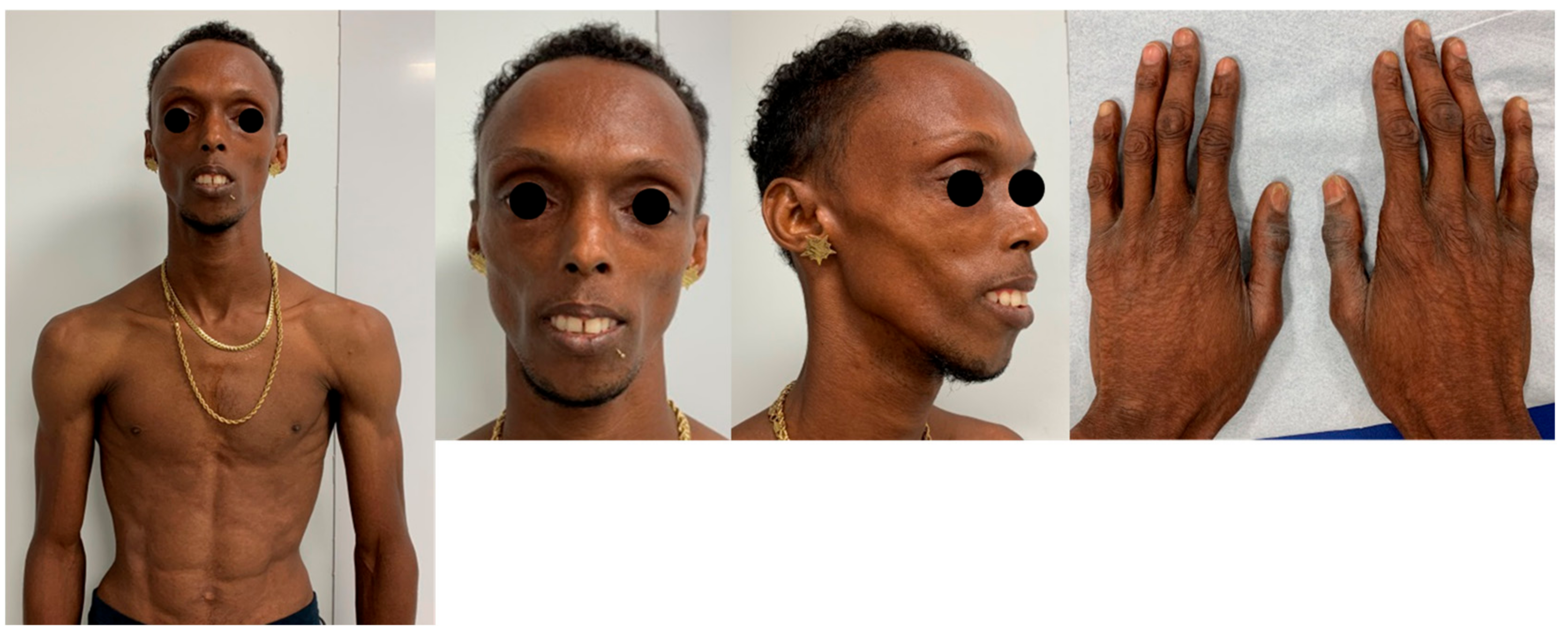
2. Case Presentation

Figure 1 shows the pedigree and Table 1 summarizes the clinical and cardiac phenotypes.
The oldest case (I.1) in the family was born in 1956 and reportedly died at 48 years of age from congestive heart failure. An interview with relatives suggests that she had a lipodystrophic phenotype. An in-hospital check-up for aortic stenosis at 44 years of age found moderate aortic stenosis with a mean gradient of 29 mmHg, thickened mitral valve, and normal left ventricular function. She had a complete Left Bundle Branch Block (LBBB), and ventricular premature beats (VPBs) were observed on various electro-cardiograms (ECGs). She stopped medical follow-ups soon after and died four years later (aged 48 y) reportedly from congestive heart failure. The determinants of heart failure were unknown. Genetic testing was not performed in her lifetime. This woman had five children from five different men.
The first child (II.1) was born from a non-consanguineous union and remained single without children. He was hospitalized at age 44 years for dyspnea and chest pain. He had no previous medical history and engaged in regular soccer practice until then. His body mass index (BMI) was 14.5 kg/m2 (height 1.66 m, weight 40 kg). He had a bird-beaked face, clinical lipoatrophy (generalized absence of adipose tissue) and a high-pitched voice. A 3/6 systolic murmur was found at physical examination. The biological assessment found elevated NT pro-BNP (3401 pg/mL; local upper normal value: 300 pg/mL) and an ultra-sensitive troponin level (67 pg/mL; local upper normal value: 14 pg/mL). His triglyceride level was 1.81 mmol/L (local normal range: 0.6–2.60 mmol/L), total cholesterol level was 5.87 mmol/L, and LDL-cholesterol level was 3.9 mmol/L (normal < 3.5 mmol/L). The ECG showed sinus rhythm, bi-atrial hypertrophy, left ventricular (LV) hypertrophy, and unspecific ventricular conduction delay (Figure 2a). Echocardiography showed a severe aortic stenosis (body surface indexed aortic valve area 0.4 cm2/m2; mean pressure gradient 51 mmHg), with extensive calcification of the aortic and mitral rings (Figure 2b). A routine preoperative coronary angiogram found a severe three-vessel disease (Figure 2c) with a diffusely narrow vascular bed. Cardiac surgery was performed including aortic valve replacement with a mechanical prosthesis and aorto-coronary bypass of the left descending, diagonal, and left marginal arteries. The post-operative course was complicated by a massive cerebral infarction, eventually resulting in death 32 days later. Genetic testing revealed a heterozygous c.407A>T p.Asp136Val variant in the LMNA gene.
The second child (II.2) is a 43-year-old woman. Her height and weight were 1.75 m and 32 kg, respectively, with a BMI of 10.4 kg/m2. She had a facial dysmorphism with atrophy of the bichat balls and a marked superficial venous network. No cognitive assessment was made, even though she was known as an unemployed woman with a low level of education. Her serum lipid levels and echocardiogram were normal. She was the only one of her siblings to have children. She did not consent to genetic analyses. Two of her four children have the typical progeroid dysmorphism, including one who tested positive for the likely pathogenic variant.
The third child (II.3) is a 34-year-old woman whose case was published [16]. Briefly, she presented with a suggestive dysmorphic pattern, lipodystrophy, diabetes, and a BMI of 11.7 kg/m2 (weight 30 kg; height 160 cm). A shortness of breath and systolic murmur led to the diagnosis of three-vessel coronary artery disease associated with an aortic valve stenosis, which was eventually treated with aortocoronary bypass and trans-catheter valve implantation. Sustained ventricular tachycardia occurred during the post-operative course and she was proposed an implantation of an implantable cardiac defibrillator (ICD), which she refused. Her genetic analysis revealed the c.407A>T p.Asp136Val LMNA heterozygous missense variant.
The youngest child (II.4) is a 25-year-old young man. He also had lipodystrophy and dysmorphic features, as his affected sibling (Figure 3). His height, weight and BMI were 1.70 m, 53 kg and 18.3 kg/m2, respectively. No cognitive assessment was made, but his autonomy and integration capacities were limited. His renal, hepatic and cardio-vascular biological parameters were normal. Repeated echocardiograms showed progressive thickening of the mitral and aortic valve leaflet, yet no obstructive disease was found at the more recent examination. The ECG showed a right bundle branch block (RBBB) with frequent ventricular premature beats. He was the index case of this family. The highly suggestive clinical characteristics prompted, at first, the sequencing of the LMNA gene (Sanger technique), leading to the discovery of a new c.407A>T p.Asp136Val heterozygous missense variant.
The last generation of the family comprises two girls and two boys (III-1 to III-4) from the second child, with one girl and one boy presenting with the disease. The affected son (III-2) came from non-inbred parents. He was referred at the 6 years of age for delayed oral language and comprehension, short-term memory difficulties, and disorders in structuring and spatio-temporal orientation. He benefited from an adapted education, with personalized help at school. At a general examination, Bichat ball atrophy, a marked superficial venous network, and lipo-dystrophy identical to that of his mother were found. At 16, he was 1.70 m tall and weighed 40 kg, with a BMI of 14.2 kg/m2. An echocardiography performed at five years of age was normal. At 15 years of age, a routine exam found a moderate left ventricular hypokinesia with left ventricular ejection fraction (LVEF) around 45%, and mild functional mitral regurgitation. The estimated systolic pulmonary arterial pressure was slightly elevated (32 + 5 mmHg). The NT pro-BNP level was increased to a rate of 2619 pg/mL (local upper normal value: 300 pg/mL). Three months after this exam, before any treatment was initiated, he was referred for dyspnea. LBBB was found at an ECG. Marked worsening of the echocardiographic parameters was then found with LV enlargement (the LV diastolic diameter increased from 45 to 63 mm), decreased LV contractility (the LVEF dropped down from 45 to 30%), and an increase in the estimated systolic pulmonary artery pressure (from 32 + 5 to 75 + 10 mmHg). The BNP and troponin levels were, respectively, 818 pg/mL (local upper normal value 100 pg/mL) and 179 pg/mL (local upper normal value 14 pg/mL). The CRP was 11 mg/L (local upper normal value 5 mg/L), creatinine was 109 μmol/L (normal range: 64–104 µmol/L), CPK was 397 IU/L (normal: 30–200 IU/L), and blood urea nitrogen was 4.3 mmol/L (normal range: 3.2–7.4 mmol/L). Blood cell count and electrolytes were otherwise normal. CMR showed thin LV walls with diffuse late gadolinium enhancements (Figure 4). The screening for viral, autoimmune and inflammatory diseases was negative. The coronary angiogram was normal. The hemodynamic condition quickly worsened, prompting extracorporeal life support and referral to a heart transplant unit. Unfortunately, this young patient died from infectious complications before transplantation could be attempted. Genetic testing showed the presence of the LMNA c.407A>T p.Asp136Val variant.
The affected sister (III-1) is about 21 years old. She is the second eldest of her siblings. She has lipodystrophy, a high-pitched voice, and a bird face. She was not reported for learning or cognitive disabilities. Her BMI is 14.2. The electrocardiogram shows an incomplete RBBB. A genetic test is pending.

3. Discussion

The paper describes ten members encompassing three generations of the family of a lady presenting with a newly described c.407A>T p.Asp136Val, likely pathogenic variant of the LMNA gene (16). Variants in this location have been associated with EDMD, LGMD1B, DCM1A, MADA, and progeroid syndromes [15]. However, unlike the previous family described, which has a variant in the same position, none of our patients had diabetes mellitus. Most members of this family (7/10) had a lipodystrophic phenotype. Among the seven patients affected, five were found with a degenerative disease of either the mitral or aortic valves. The valve lesions ranged from marked thickening of a leaflet to a significant obstructive disease. Aortic valve stenosis was found in three patients. A valve replacement was required in two of them. A severe obstructive coronary artery disease was found in two patients aged 44 and 28. Therefore, this new variant is associated with the dysmorphic pattern and degenerative heart valve diseases found in atypical progeroid syndromes. Most patients (6/7) also displayed heart conduction defects, another hallmark of progeroid syndromes [4]. Two patients were diagnosed with a severe coronary artery disease requiring bypass surgery, in accordance with previous reports of severe premature atherosclerosis. [1]. Indeed, the real frequency of this complication in this family may have been underestimated by the lack of a systematic evaluation either by a coronary angiogram or ischemia stress tests.
Thus, beyond evidence of premature cardiovascular aging, such as degenerative valvular and coronary artery disease, the same variant appears to be also associated with myocardial disease. Indeed, dilated cardiomyopathies have been well described in patients with laminopathies [4,17,20]. Also, late gadolinium enhancements have been observed in laminopathy-related dilated cardiomyopathy, suggesting interstitial fibrosis following cardiac tissue injury. In case III-2, although the fast progression and presence of late gadolinium enhancements at MRI could suggest myocarditis, the lack of inflammatory/autoimmune mechanisms, of coronary artery disease or virus involvement, led us to conclude to rather a diagnosis of laminopathy-associated dilated cardiomyopathy. Indeed, dilated cardiomyopathies may occur as a direct consequence of a lamin pathogenic variant. Apart from Case II-3, who presented an episode of sustained ventricular tachycardia during the post-operative course, the risk of life-threatening ventricular arrhythmias remained low in the other patients. This evaluation integrated risk factors identified in a recent cohort study (gender, LV function, the presence of atrio-ventricular conduction defects, type of mutation, presence of non-sustained ventricular tachycardia) [21]. Accordingly, ICD has not been proposed to the other patients still alive pending regular evaluation of their electrophysiological status. Besides therapeutic procedures for severe IEC coronary artery and valvular heart disease, there was no laminopathy-specific medication used in our patients. They had no indication for other conventional therapy, particularly a Renin Angiotensin system inhibitor, which use has been associated with the delayed onset of cardiac disease in animal models of laminopathy [22].
Learning difficulties observed in some family members might not be associated with the LMNA variant, given that the LMNA expression is usually abolished in central nervous system cells due to brain-specific miRNA [21]. Instead, other mechanisms, such as unfavorable socio-economic conditions, could be advocated. The present variant is associated with various clinical presentations, which suggest various underlying mechanisms in this family. The molecular bases of clinical-phenotype-overlapping are not well understood. The variation described may decrease the ability of LMNA site concerned to bind various proteins, and therefore, affect different cell types. Functional analysis studies may be too complex to perform due to the pleiotropism of lamin A protein. To our knowledge, there are very few and discordant in silico and in vitro data. An experimental study aimed at systematically identifying disruptions to peptide interactions generated by deleterious lamin A variants showed that the Asp136 substitution with His amino acid would not affect any of the interactions tested [23]. On another side, using a machine learning approach for in silico predictions provided evidence of a biochemical and pathogenic effect of the p.Asp136Glu LMNA variant [24]. Asp, His and Glu are three polar and charged amino acids, whereas Val is a non-polar hydrophobic AA. Using a bioinformatics prediction tool is probably the reason why we find that changing the Asp in Val to position 136 abolishes the interaction with the Arg133 residue, whereas other interactions are intact. The absence of interactions would weaken the bond between the laminating monomers and the tetramer assembly.
Eventually, such diversity in a clinical presentation needs to translate into adequate genetic counseling and the early screening of cardiomyopathy, valvular and ischemic heart disease, once an LMNA variant or a suggestive dysmorphism is found [25,26].

Author Contributions

Conceptualization, A.-G.G.-V. and J.I.; formal analysis, A.-G.G.-V. and A.M.; investigation, A.-G.G.-V., A.M. and E.S.; resources, H.L., C.V. (Camille Vatier), C.V. (Corinne Vigouroux), P.R.; data curation, J.I.; writing—original draft preparation, A.-G.G.-V.; writing—review and editing, J.I.; H.M., F.B., K.W., R.B., E.S. All authors have read and agreed to the published version of the manuscript.

Funding

This research received no external funding.

Institutional Review Board Statement

The study was conducted in accordance with the Declaration of Helsinki, and approved by the Institutional Review Board of CHU MARTINIQUE (protocol code 2021/155, approved on 17 December 2021).

Informed Consent Statement

Informed consent was obtained from all subjects involved in the study.

Data Availability Statement

Not applicable.

Acknowledgments

We acknowledge all contributors for implication in data curation, formal analysis, writing review and editing. We are thankful to all patients.

Conflicts of Interest

The authors declare no conflict of interest.

References

  1. Schreiber, K.H.; Kennedy, B.K. When Lamins Go Bad: Nuclear Structure and Disease. Cell 2013, 152, 1365–1375. [Google Scholar] [CrossRef] [PubMed]
  2. Bonne, G.; Barletta, M.R.D.; Varnous, S.; Bécane, H.-M.; Hammouda, E.-H.; Merlini, L.; Muntoni, F.; Greenberg, C.R.; Gary, F.; Urtizberea, J.-A.; et al. Mutations in the gene encoding lamin A/C cause autosomal dominant Emery-Dreifuss muscular dystrophy. Nat. Genet. 1999, 21, 285–288. [Google Scholar] [CrossRef] [PubMed]
  3. De Sandre-Giovannoli, A.; Chaouch, M.; Kozlov, S.; Vallat, J.-M.; Tazir, M.; Kassouri, N.; Szepetowski, P.; Hammadouche, T.; Vandenberghe, A.; Stewart, C.L.; et al. Homozygous Defects in LMNA, Encoding Lamin A/C Nuclear-Envelope Proteins, Cause Autosomal Recessive Axonal Neuropathy in Human (Charcot-Marie-Tooth Disorder Type 2) and Mouse. Am. J. Hum. Genet. 2002, 70, 726–736. [Google Scholar] [CrossRef] [PubMed]
  4. Fatkin, D.; MacRae, C.; Sasaki, T.; Wolff, M.R.; Porcu, M.; Frenneaux, M.; Atherton, J.; Vidaillet, H.J.; Spudich, S.; De Girolami, U.; et al. Missense Mutations in the Rod Domain of the Lamin A/C Gene as Causes of Dilated Cardiomyopathy and Conduction-System Disease. N. Engl. J. Med. 1999, 341, 1715–1724. [Google Scholar] [CrossRef] [PubMed]
  5. Muchir, A. Identification of mutations in the gene encoding lamins A/C in autosomal dominant limb girdle muscular dystrophy with atrioventricular conduction disturbances (LGMD1B). Hum. Mol. Genet. 2000, 9, 1453–1459. [Google Scholar] [CrossRef]
  6. Raffaele di Barletta, M.; Ricci, E.; Galluzzi, G.; Tonali, P.; Mora, M.; Morandi, L.; Romorini, A.; Voit, T.; Orstavik, K.H.; Merlini, L.; et al. Different Mutations in the LMNA Gene Cause Autosomal Dominant and Autosomal Recessive Emery-Dreifuss Muscular Dystrophy. Am. J. Hum. Genet. 2000, 66, 1407–1412. [Google Scholar] [CrossRef]
  7. Cao, H. Nuclear lamin A/C R482Q mutation in Canadian kindreds with Dunnigan-type familial partial lipodystrophy. Hum. Mol. Genet. 2000, 9, 109–112. [Google Scholar] [CrossRef]
  8. Eriksson, M.; Brown, W.T.; Gordon, L.B.; Glynn, M.W.; Singer, J.; Scott, L.; Erdos, M.R.; Robbins, C.M.; Moses, T.Y.; Berglund, P.; et al. Recurrent de novo point mutations in lamin A cause Hutchinson–Gilford progeria syndrome. Nature 2003, 423, 293–298. [Google Scholar] [CrossRef]
  9. Novelli, G.; Muchir, A.; Sangiuolo, F.; Helbling-Leclerc, A.; D’Apice, M.R.; Massart, C.; Capon, F.; Sbraccia, P.; Federici, M.; Lauro, R.; et al. Mandibuloacral Dysplasia Is Caused by a Mutation in LMNA-Encoding Lamin A/C. Am. J. Hum. Genet. 2002, 71, 426–431. [Google Scholar] [CrossRef]
  10. De Sandre-Giovannoli, A.; Bernard, R.; Cau, P.; Navarro, C.; Amiel, J.; Boccaccio, I.; Lyonnet, S.; Stewart, C.L.; Munnich, A.; Le Merrer, M.; et al. Lamin a truncation in Hutchinson-Gilford progeria. Science 2003, 300, 2055. [Google Scholar] [CrossRef]
  11. Brodehl, A.; Gerull, B. Genetic Insights into Primary Restrictive Cardiomyopathy. JCM 2022, 11, 2094. [Google Scholar] [CrossRef] [PubMed]
  12. Caux, F.; Dubosclard, E.; Lascols, O.; Buendia, B.; Chazouillères, O.; Cohen, A.; Courvalin, J.-C.; Laroche, L.; Capeau, J.; Vigouroux, C.; et al. A new clinical condition linked to a novel mutation in lamins A and C with generalized lipoatrophy, insulin-resistant diabetes, disseminated leukomelanodermic papules, liver steatosis, and cardiomyopathy. J. Clin. Endocrinol. Metab. 2003, 88, 1006–1013. [Google Scholar] [CrossRef] [PubMed]
  13. Garg, A.; Subramanyam, L.; Agarwal, A.K.; Simha, V.; Levine, B.; D’Apice, M.R.; Novelli, G.; Crow, Y. Atypical Progeroid Syndrome due to Heterozygous Missense LMNA Mutations. J. Clin. Endocrinol. Metab. 2009, 94, 4971–4983. [Google Scholar] [CrossRef]
  14. Hennekam, R.C.M. Hutchinson-Gilford progeria syndrome: Review of the phenotype. Am. J. Med. Genet. A 2006, 140, 2603–2624. [Google Scholar] [CrossRef] [PubMed]
  15. Chiarini, F.; Evangelisti, C.; Cenni, V.; Fazio, A.; Paganelli, F.; Martelli, A.M.; Lattanzi, G. The Cutting Edge: The Role of mTOR Signaling in Laminopathies. Int. J. Mol. Sci. 2019, 20, 847. [Google Scholar] [CrossRef] [PubMed]
  16. Mosbah, H.; Vatier, C.; Boccara, F.; Jéru, I.; Lascols, O.; Vantyghem, M.-C.; Fève, B.; Donadille, B.; Sarrazin, E.; Benabbou, S.; et al. Looking at New Unexpected Disease Targets in LMNA-Linked Lipodystrophies in the Light of Complex Cardiovascular Phenotypes: Implications for Clinical Practice. Cells 2020, 9, 765. [Google Scholar] [CrossRef]
  17. Lee, S.; Park, S.M.; Kim, H.J.; Kim, J.-W.; Yu, D.S.; Lee, Y.B. Genomic diagnosis by whole genome sequencing in a Korean family with atypical progeroid syndrome. J. Dermatol. 2015, 42, 1149–1152. [Google Scholar] [CrossRef]
  18. Masson, E.; Zou, W.-B.; Génin, E.; Cooper, D.N.; Le Gac, G.; Fichou, Y.; Pu, N.; Rebours, V.; Férec, C.; Liao, Z.; et al. Expanding ACMG variant classification guidelines into a general framework. Hum. Genom. 2022, 16, 31. [Google Scholar] [CrossRef]
  19. Simm, D.; Hatje, K.; Kollmar, M. Waggawagga: Comparative visualization of coiled-coil predictions and detection of stable single α-helices (SAH domains). Bioinformatics 2015, 31, 767–769. [Google Scholar] [CrossRef]
  20. Bera, M.; Ainavarapu, S.R.K.; Sengupta, K. Significance of 1B and 2B domains in modulating elastic properties of lamin A. Sci. Rep. 2016, 6, 27879. [Google Scholar] [CrossRef]
  21. Wahbi, K.; Ben Yaou, R.; Gandjbakhch, E.; Anselme, F.; Gossios, T.; Lakdawala, N.K.; Stalens, C.; Sacher, F.; Babuty, D.; Trochu, J.-N.; et al. Development and Validation of a New Risk Prediction Score for Life-Threatening Ventricular Tachyarrhythmias in Laminopathies. Circulation 2019, 140, 293–302. [Google Scholar] [CrossRef] [PubMed]
  22. Muchir, A.; Wu, W.; Sera, F.; Homma, S.; Worman, H.J. Mitogen-activated protein kinase kinase 1/2 inhibition and angiotensin II converting inhibition in mice with cardiomyopathy caused by lamin A/C gene mutation. Biochem. Biophys. Res. Commun. 2014, 452, 958–961. [Google Scholar] [CrossRef] [PubMed]
  23. Dittmer, T.A.; Sahni, N.; Kubben, N.; Hill, D.E.; Vidal, M.; Burgess, R.C.; Roukos, V.; Misteli, T. Systematic identification of pathological lamin A interactors. Mol. Biol. Cell 2014, 25, 1493–1510. [Google Scholar] [CrossRef] [PubMed]
  24. Bennett, J.S.; Gordon, D.M.; Majumdar, U.; Lawrence, P.J.; Matos-Nieves, A.; Myers, K.; Kamp, A.N.; Leonard, J.C.; McBride, K.L.; White, P.; et al. Use of machine learning to classify high-risk variants of uncertain significance in lamin A/C cardiac disease. Heart Rhythm. 2022, 19, 676–685. [Google Scholar] [CrossRef] [PubMed]
  25. Nicolas, H.A.; Akimenko, M.-A.; Tesson, F. Cellular and Animal Models of Striated Muscle Laminopathies. Cells 2019, 8, 291. [Google Scholar] [CrossRef]
  26. Treiber, G.; Flaus Furmaniuk, A.; Guilleux, A.; Medjane, S.; Bonfanti, O.; Schneebeli, S.; Bernard, C.; Le-Moullec, N.; Bakiri, F.; Pholsena, M.; et al. A recurrent familial partial lipodystrophy due to a monoallelic or biallelic LMNA founder variant highlights the multifaceted cardiac manifestations of metabolic laminopathies. Eur. J. Endocrinol. 2021, 185, 453–462. [Google Scholar] [CrossRef]
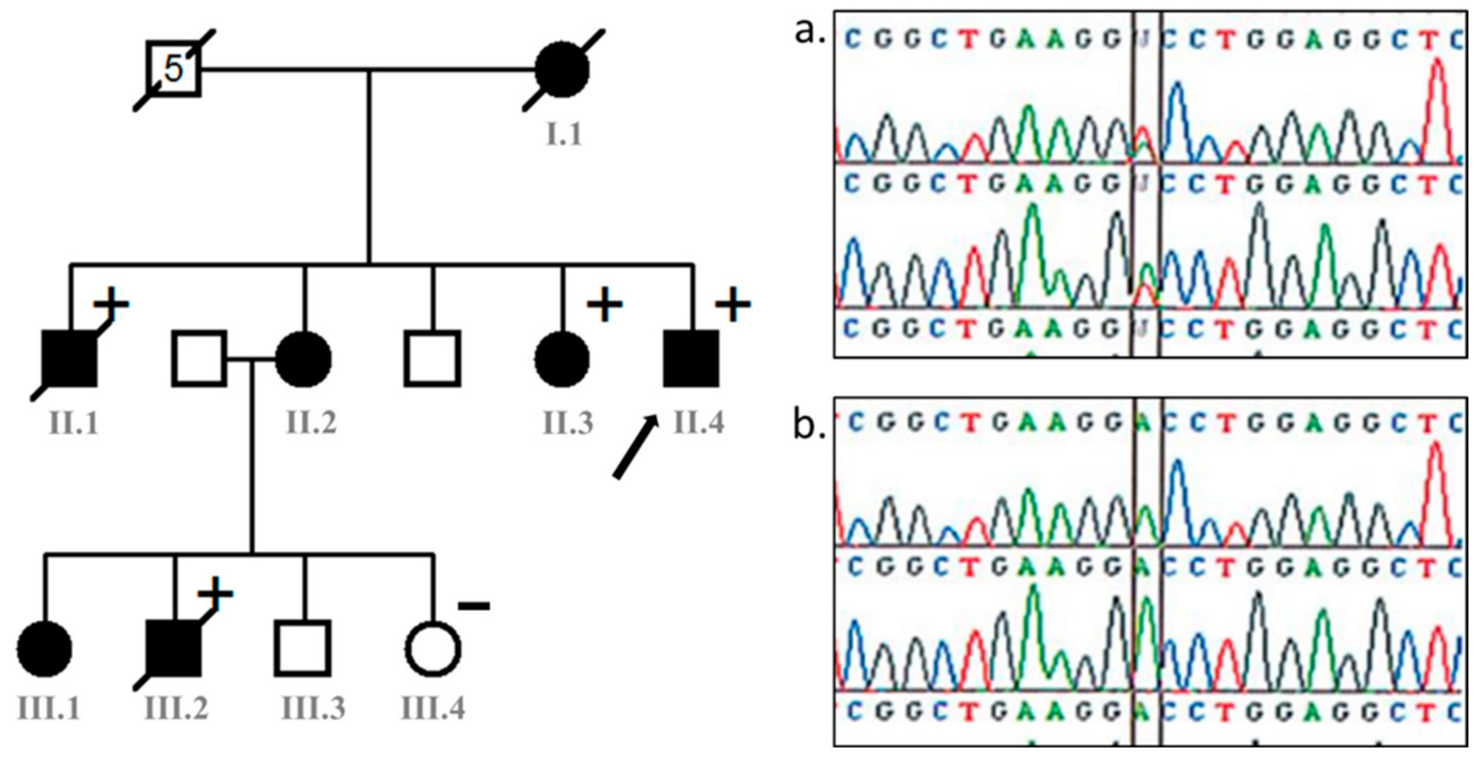
Figure 1. Pedigree Tree of the family and Sanger analysis of every affected person. Round for females, square for males, symbol in black for affected individual; (+) indicates the presence of c.407A>T p.Asp136Val likely pathogenic variant; (-) indicates the absence of the variant; number 5 in the square on the top of the tree means the number of partners. Arrow indicates the index case. Only 4 patients underwent genetic analysis, all with a positive result. Result of the PCR-Sanger sequencing of the exon2 region including the LMNA c.407A>T, p.(Asp136Val) variant for genomic DNA of (a) all affected individuals who have been tested (a) and the control (b).
Cardiogenetics 13 00013 g001
Figure 2. Cardiovascular findings in Case II.1. (a) Electrocardiography shows sinus rhythm with biatrial hypertrophy, left ventricular hypertrophy, unspecific delayed ventricular conduction, and negative T waves in inferior and apical leads; (b) coronary angiogram shows diffuse three-vessel disease; (c) echocardiography: mitral and aortic valvular disease with severe aortic valve stenosis (aortic valve surface: 0.4 cm/m2, mean pressure gradient: 51 mmHg).
Cardiogenetics 13 00013 g002
Figure 3. Dysmophic features of case II:4. Face dysmorphy with mild ptosis, prominent nose bones, thin nose, long philtrum, Bichat ball atrophia, Diffuse lipoatrophia, Arachnodactily, Leucoonichy.
Cardiogenetics 13 00013 g003
Figure 4. CMR findings in patient III-2. Four-chamber view (a,b) and short axis view (c,d) of the left ventricle (LV); reduced thickness (<4 mm) of the LV lateral, anterior and posterior walls (white arrows) with diffuse transmural (yellow arrows) or sub-epicardial (green arrows) late gadolinium enhancements; circumferential pericardial effusion (red arrow).
Cardiogenetics 13 00013 g004
Table 1. Clinical features of affected family members.
Pedigree NumberI.1II.1II.2II.3II.4III.2III.1
SexFMFFMMF
Age of diagnostic (years)44443425181011
Age of death and age (years)4844Alive (46)Alive (37)Alive (28)15Alive (21)
Height/Weight (cm/kg)167/41166/40175/32160/30170/53170/41168/40
BMI (kg/m2)14.714.510.411.718.314.214.2
Dysmorphism pattern+++++++
Lipodystrophy+++++++
High-pitched voice+-+++-+
Bird’s face+++++++
Alopecia--+-+--
Cutaneous atrophyndnd+-+--
Scoliosis---+-+nd
ECG Conduction defectsLBBBUnspecific delayed ventricular conductionIncomplete RBBBRBBBIncomplete RBBBLBBBIncomplete RBBB
Ventricular arrythmiasFrequent VPBFrequent VPBsndSustained VTFrequent VPBs-nd
Valve Thickening+++-+--
Aortic Stenosis+++----
Congestive Heart Failure+----+-
Coronary angiogramndThree-vessel diseasendThree-vessel diseasendNormalnd
Hepatic cell insufficiency/Type 2 diabetis-------
LMNA c.407A>T (p.Asp136Val)nd+nd+++nd
M: Male, F: Female; BMI: Body mass index; LBBB: Left bundle branch block; RBBB: Right bundle branch block; VPB: Ventricular premature beats; VT: Ventricular tachycardia; nd: not defined; +: feature is present; -: feature is absent.
Disclaimer/Publisher’s Note: The statements, opinions and data contained in all publications are solely those of the individual author(s) and contributor(s) and not of MDPI and/or the editor(s). MDPI and/or the editor(s) disclaim responsibility for any injury to people or property resulting from any ideas, methods, instructions or products referred to in the content.

