Review

Clinical and Molecular Characteristics of Patients with PLN R14del Cardiomyopathy: State-of-the-Art Review

Inherited and Rare Cardiovascular Diseases, Department of Translational Medical Sciences, University of Campania “Luigi Vanvitelli”, AORN Ospedali dei Colli–Monaldi Hospital, 80131 Naples, Italy
*
Author to whom correspondence should be addressed.
Cardiogenetics 2022, 12(1), 112-121; https://doi.org/10.3390/cardiogenetics12010012
Submission received: 2 January 2022 / Revised: 23 February 2022 / Accepted: 1 March 2022 / Published: 2 March 2022

Abstract

:
The deletion of the arginine 14 codon (R14del) in the phospholamban (PLN) gene is a rare cause of arrhythmogenic cardiomyopathy (ACM) and is associated with prevalent ventricular arrhythmias, heart failure, and sudden cardiac death. The pathophysiological mechanism which culminates in the ACM phenotype is multifactorial and mainly based on the alteration of the endoplasmic reticulum proteostasis, mitochondrial dysfunction and compromised Ca2+ cytosolic homeostasis. The symptoms of this condition are usually non-specific and consist of arrhythmia-related or heart failure-related manifestation; however, some peculiar diagnostic clues were detected, such as the T-wave inversion in the lateral leads, low QRS complexes voltages, mid-wall or epicardial fibrosis of the inferolateral wall of the left ventricle, and their presence should raise the suspicion of this condition. The risk stratification for sudden cardiac death is mandatory and several predictors were identified in recent years. However, the management of affected patients is often challenging due to the absence of specific prediction tools and therapies. This review aims to provide the current state of the art of PLN R14del cardiomyopathy, focusing on its pathophysiology, clinical manifestation, risk stratification for sudden cardiac death, and management.

1. Introduction

Arrhythmogenic cardiomyopathy (ACM) is a myocardial disease that affects the left ventricle (LV), right ventricle (RV), or both, whose most typical characteristics are the progressive fibrotic or fibrofatty myocardial replacement that predisposes to ventricular arrhythmias and can be responsible for global or regional ventricular dysfunction [1]. In the pre-genetic era, ACM was considered a myocardial disease that exclusively or predominantly involved the RV, the so-called arrhythmogenic RV dysplasia (ARVD) or cardiomyopathy (ARVC), whose clinical features were RV dysfunction and arrhythmias [2,3]. Therefore, the classical diagnostic criteria for ACM were focused on RV involvement [4]. Subsequently, autopsy investigation, cardiac magnetic resonance (CMR) and genotype-phenotype correlation studies showed that the LV was commonly involved by the fibrotic replacement, changing the paradigm of the disease [5,6].
The current classification of ACM includes different clinical variants according to the prevalent ventricular involvement. The classical ARVC phenotype is characterized by isolated RV involvement. On the other hand, the LV phenotype, defined arrhythmogenic left ventricular cardiomyopathy (ALVC), is characterized by predominant LV involvement, and the biventricular phenotype is defined as a disease involving both the ventricles [7].
The genetic basis of ACM is responsible for the clinical phenotype. The classic ARVC and the biventricular phenotype are mainly caused by pathogenetic variants involving desmosomal genes, such as PKP2, JUP, DSC2, DSG2, and DSP [8,9,10]. On the other hand, patients with ALVC phenotype carry non-desmosomal gene pathogenic variants, such as ion channel, sarcomere, cytoskeleton, mitochondrial, and sarcomeric genes [11,12,13]. Therefore, with the increased knowledge of the ALVC phenotype, specific diagnostic criteria for the left-side disease variants were proposed (the “Padua Criteria”) [14], based on the following phenotypic features: electrocardiographic (ECG) abnormalities, such as low QRS voltages and T-wave inversion in the lateral or inferolateral leads; ventricular arrhythmias with a QRS morphology which denotes its origin from the LV; normal or mild hypokinetic LV with no or mild dilation; extensive myocardial fibrosis evidenced by CMR as late gadolinium enhancement (LGE) with a non-ischemic pattern of distribution.
However, in the absence of RV involvement, the diagnosis of ALVC cannot be formulated based on the phenotype criteria due to the extreme overlap with other inherited or acquired conditions, such as dilated cardiomyopathy (DCM), myocarditis, or cardiac sarcoidosis [15]. Thus, in the presence of a phenotype suggestive for ALVC, the demonstration of a pathogenic or likely-pathogenic variant of an ACM-related gene is required for the diagnosis [16].
The identification of the genetic variant underlying the ACM phenotype is essential not only for the diagnosis but also for risk stratification and management. Indeed, several genetic variants were found to be associated with an increased risk of ventricular arrhythmias and sudden cardiac death (SCD) [17,18]. Among these, the pathogenic PLN R14del gene variant, commonly identified in patients fulfilling the diagnostic criteria for ALVC, is generally associated with early-onset arrhythmias and a worse prognosis [19]. Unfortunately, data on the natural history, risk prediction and management of ALVC caused by this pathogenic variant are scant.
This review aims to provide the current state of the art of PLN (phospholamban) R14del cardiomyopathy, focusing on its pathophysiology, clinical manifestation, risk stratification for SCD, and management.

2. Pathophysiology

Physiological cardiac muscle contraction is a finely tuned process mainly regulated by accurately synchronized Ca2+ fluxes in cardiomyocytes [20,21]. When an action potential depolarizes the cell, voltage-dependent L-type Ca2+ channels (LTCCs) open to generate an inward Ca2+ current, which leads to an additional release of Ca2+ from the sarcoplasmic reticulum (SR) through the activation of ryanodine receptor channels 2 (RYR2). The intracellular increase of the Ca2+ concentration is responsible for the myofilament activation and contraction. During diastole, Ca2+ is removed from the cytosol via the sarcolemmal Na+/Ca2+ exchanger (NCX1), which transfers Ca2+ in the extracellular space, and the SR Ca2+ ATPase (SERCA2a), which pumps the Ca2+ in the SR lumen [22]. SERCA2a-dependent diastolic Ca2+ uptake dominates over the extracellular extrusion via the NCX1.
PLN finely regulates SERCA2a activity. PLN is a protein of 52 amino acids, localized into the SR membrane and involved in cardiomyocyte calcium handling. PLN activity is modified by its phosphorylation state [23,24]. Its phosphorylation by protein kinase A (PKA) at Ser-16 and/or by calmodulin-dependent kinase II (CaMKII) at Thr-17 releases its inhibitory effects on SERCA2a [24].
From a theoretical point of view, the mutation of PLN and its subsequent dysfunction results in more significant inhibition of SERCA2a by non-phosphorylated PLN, thereby leading to an impairment of Ca2+ reuptake. Thus, the decrease in SR Ca2+ content is responsible for impaired systolic function, while the diastolic cytosolic overload is responsible for diastolic dysfunction and arrhythmias.
However, the pathophysiological mechanisms which culminate in the ALVC phenotype are more complex and not fully understood. In recent years, several studies have been carried out to elucidate the underlying molecular features of the PLN R14del variant.
This variant was identified for the first time in humans with hereditary cardiomyopathy by Haghighi et al. [19] and then studied in murine models. To understand the molecular mechanisms which link the PLN variant to ACM in human induced pluripotent stem cells (hiPSC-CMs), Feyen et al. [25] used single-cell RNA sequencing. They found the presence of elevated stress of the endoplasmic reticulum ER with an unfolded protein response (UPR), a signaling pathway with a critical role in the keeping of proteostasis in the ER [26], in the PLN R14del mutants compared with controls. These findings suggest that the PLN R14del variant is responsible for an altered ER proteostasis. This observation is of clinical interest since for long-term cell function preservation, the balance among protein production, folding and degradation is required. With the aging of the cells, this ability progressively reduces, and the aggregation of unfolded proteins is typical of different age-related diseases, such as Parkinson’s Diseases and Alzheimer’s Disease [27].
The proteostasis involvement was also identified by Eijgenraam et al. [28], which postulated that this alteration, combined with the aggregation on PLN proteins, are among the first hallmarks of PLN R14del cardiomyopathy.
Furthermore, Cuello et al. [29] reprogrammed dermal fibroblasts to hiPSC-CMs and established isogenic controls using CRISP/Cas9. Then, cardiomyocytes were differentiated. They found that cardiomyocytes that bring the PLN R14del variant showed a Ca2+ load-dependent irregular beating pattern, lower force and a prolonged Ca2+ transient decay time than controls. In addition, the ER, ribosomes, and mitochondria exhibited less protein content when analyzed using proteomic analysis. Finally, large lipid droplets in mitochondria and an ER dilation were observed using electron microscopy. This evidence suggests that the ER and mitochondrial impairment are a novel disease mechanism underlying the PLN R14del cardiomyopathy.
In conclusion, the molecular mechanisms underlying the PLN R14del cardiomyopathy are complex and under investigation (Figure 1). Therefore, a better understanding of its pathophysiology is required to formulate a tailored therapy.

3. Clinical Manifestations

PLN R14del cardiomyopathy presents overlapping clinical features between ALVC and DCM [30]. The identification of specific phenotypic features to distinguish patients with PLN R14del cardiomyopathy and their relatives from those with other forms of ACM or DCM has been investigated in different studies. Many signs of the disease can be identified in the pre-symptomatic phase, in particular repolarization abnormalities, frequent ventricular premature complexes (VPCs), and CMR LGE, as evidenced in the recently published iPHORECAST (PHOspholamban RElated CArdiomyopathy intervention Study) trial [31].

3.1. Signs and Symptoms

The disease onset seems to be age-related, with a slightly higher prevalence in males [32]. The symptoms are usually non-specific and consist of arrhythmia-related (e.g., palpitation, syncope) or heart failure-related symptoms (e.g., dyspnoea, exercise limitation). Symptoms usually appear in the fifth decade of life [30]; however, cases of SCD have been described in patients younger than 30 years old [30,32,33].

3.2. Electrocardiography

The ECG findings reflect the myocardial fibrosis substrate, as proved by histological examination studies, and typically consist of low QRS voltages with reduced R-wave amplitude [19,34]. These abnormalities have not been found in patients without the mutation [35]. Te Rijdt et al. [36] found a median R-wave amplitude of 5.3 mV, with more decreased QRS voltages in older mutation carriers. Negative T-waves are also common in PLN R14del cardiomyopathy. They were identified in the right precordial leads in 11% of carriers and in V4–V6 in 29% of them (80% of index patients) [36]. Moreover, 15% of patients experience ventricular tachycardia episodes.
Van de Leur et al. [37] utilized deep neural networks (DNNs) to detect possible typical ECG abnormalities in PLN R14del cardiomyopathy, useful to identify pre-symptomatic mutation carriers. The elaborated algorithm was capable not only of confirming known features of the disease, such as negative T-waves and low QRS voltages, but also to define their characteristics. Low QRS voltages consisted more properly in R-wave attenuation with normal S-wave, localized in the right precordial leads V2 and V3, and in the lateral leads DI, aVL and V6. Moreover, T-waves attenuation/inversion was situated not only in V2, V3, and V6 but also in DI and aVL. Furthermore, it was also able to find a new distinctive element on surface ECG, the prolonged PR interval, that suggests a possible involvement of atrioventricular conduction.

3.3. Cardiac Magnetic Resonance

The most common CMR pattern of disease is the presence of epicardial or mid-wall fibrosis in the inferolateral LV wall, which usually corresponds with negative T waves in the LV inferolateral leads [33,38]. Functional and structural impairment of LV is common, usually represented by mild LV dilation and dysfunction, as confirmed either by echocardiography or CMR studies [33,38,39].
A recent study showed an extensive LGE in the LV of the affected patients, even in those with preserved or mildly reduced LV ejection fraction (LVEF) (>45%) and was found to be independently associated with ventricular arrhythmias [36]. However, LGE was more significant in older and in those with reduced LVEF. RV was involved by LGE only in 5% of patients, and it was associated with reduced RV ejection fraction.

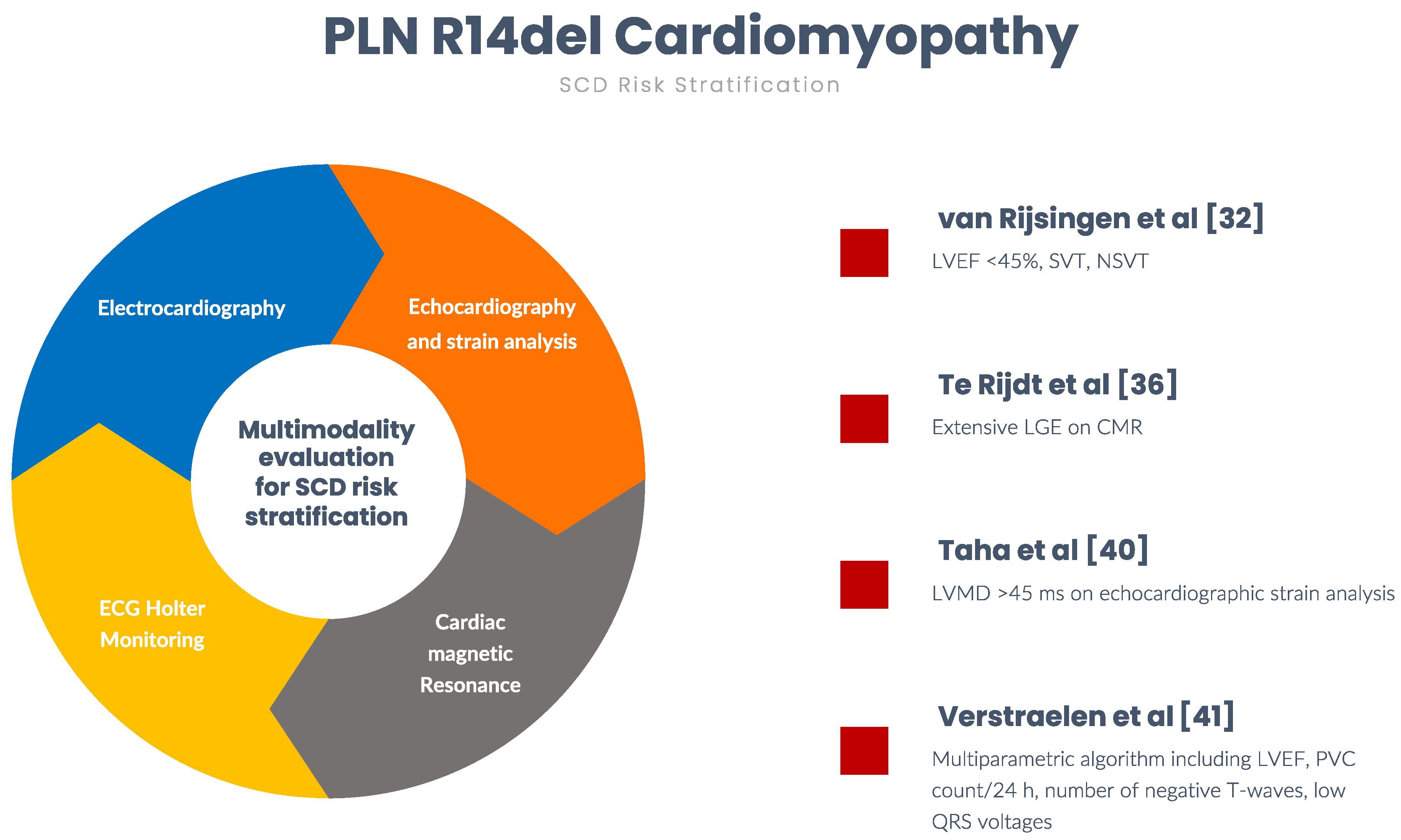
4. Risk Stratification for Sudden Cardiac Death

PLN R14del variant carriers can experience early-onset ventricular arrhythmias, ranging from frequent PVCs to ventricular fibrillation and SCD, which in rare cases may be the first clinical presentation of the cardiomyopathy [30,32,33]. Due to the lack of specific recommendations for PLN R14del cardiomyopathy, the indication for the implantable cardioverter–defibrillator (ICD) implantation for SCD prevention follows the current ACM and DCM guidelines and consensus documents [7,40].
However, while there is clear evidence that ICD implantation is recommended for secondary prevention in those patients who experienced sudden cardiac arrest or ventricular arrhythmias with hemodynamic instability, more difficult is the identification of subjects at high risk for SCD who require an ICD for primary prevention. In recent years, several studies investigated the SCD predictors in patients with PLN R14del cardiomyopathy.
Firstly, van Rijsingen et al. [32] identified LVEF < 45%, and sustained and non-sustained ventricular tachycardia (SVT and NSVT) as independent risk factors for malignant ventricular arrhythmias.
Subsequently, Te Rijdt et al. [36] investigated the extent and localization of myocardial fibrosis and its association with ECG features and ventricular arrhythmias in PLN R14del mutation carriers. They found that LGE in the LV, but not attenuated R-waves and inverted T-waves, was independently associated with ventricular arrhythmias. Of importance, 30% of patients with preserved LVEF showed a significant LGE in the LV. However, in this study, the occurrence of ventricular arrhythmias was determined on ambulatory 24 h ECG Holter or exercise ECG, which were not available for every patient, leading to a possible selection bias.
Furthermore, the incremental value of the LV mechanical dispersion (LVMD) by echocardiographic deformation imaging for sustained ventricular arrhythmias prediction was recently investigated. Taha et al. [40] evaluated 243 PLN R14del mutation carriers, which were classified into three groups according to the “45/45” rule. Patients with overt LV dysfunction (LVEF < 45%) had the worst prognosis in terms of ventricular arrhythmic events and were considered to be at high risk, similar to a previous study [32]. In contrast, patients with normal LV function (LVEF > 45% and LVMD < 45 ms) showed a low risk of developing sustained ventricular arrhythmias, and those with mechanical LV dysfunction (LVEF > 45% and LVMD > 45 ms) exhibited an intermediate risk, falling into a “grey zone” where a multiparametric assessment for SCD risk prediction is required.
Finally, a multiparametric algorithm to identify patients who may benefit from ICD implantation for primary prevention was developed [41] (https://plnriskcalculator.shinyapps.io/final_shiny/, accessed on 1 March 2022). The multivariable model, including LVEF, PVC count in 24 h, number of negative T-waves, and presence of low QRS voltages on ECG, showed an excellent discriminative ability (C-statistic 0.83 (95% CI 0.78–0.88)). However, the study suffers from some limitations, such as the endpoint used for the assessment of the arrhythmic outcome, the lack of an external validation cohort, and the insufficient amount of data on the presence and extent of LGE. In detail, the use of a combined endpoint, consisting of SVT, appropriated ICD intervention, and SCD, may overestimate the true risk of SCD. Indeed, ICD intervention is a poor surrogate of SCD since most ventricular tachycardia episodes treated by ICD are expected to be self-terminating. Moreover, the lack of data on LGE may affect the power of the prediction model and can be responsible for the identification of ECG abnormalities (i.e., low QRS voltages and T-waves inversion) as SCD predictors, in contrast with the previous study.
In conclusion, two predictors (LVEF < 45% and extensive LGE in the LV) were found to be strongly associated with major arrhythmic events and, in their presence, an ICD implantation in primary prevention should be considered. However, in the remaining patients, risk stratification should be based on a multiparametric approach, including family and clinical history, ECG, ECG-Holter monitoring, echocardiography and CMR, and discussed case by case in the context of a multidisciplinary team of experts (Figure 2).

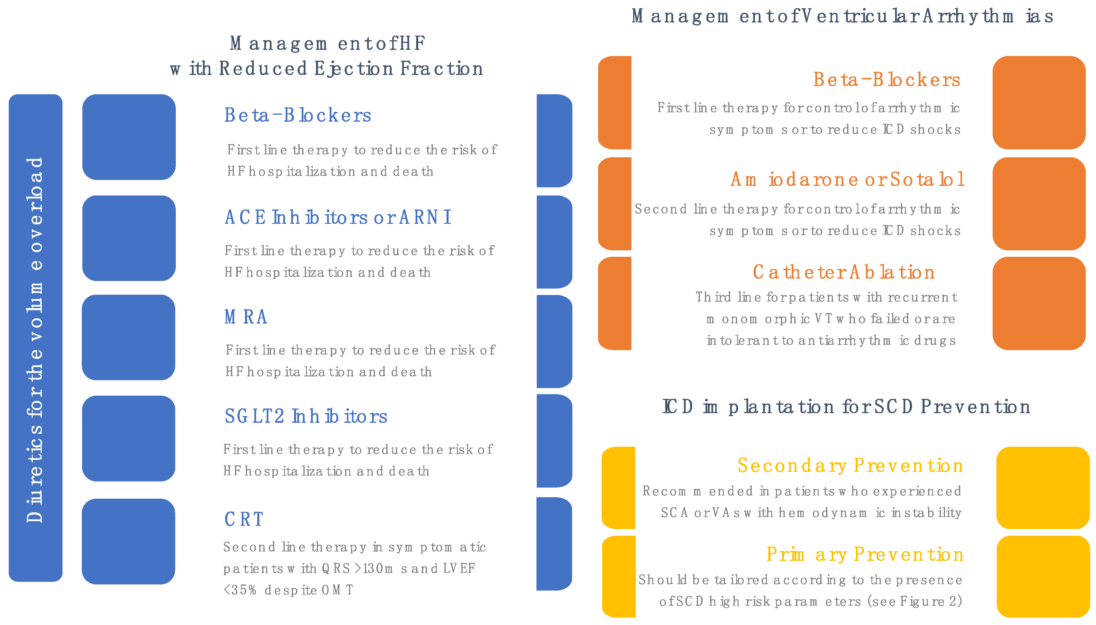
5. Medical Treatment

As previously reported, the PLN R14del cardiomyopathy is associated with a high prevalence of ventricular arrhythmias, heart failure (HF), and SCD. Unfortunately, no specific treatments for this condition are currently available. For this reason, the primary efforts should be oriented toward the prevention and treatment of life-threatening arrhythmias and HF (Figure 3).
The PLN R14del cardiomyopathy phenotype is typically associated with a reduction (<40%) or a mild reduction (LVEF 40–50%) of the LVEF, while the RV is rarely involved. Therefore, the treatment of HF in these patients is based on disease-modifying drugs, such as angiotensin-converting enzyme inhibitors (ACEi) or angiotensin receptor blockers (ARBs), beta-blockers, mineralocorticoids antagonists (MRAs), angiotensin receptor–neprilysin inhibitor (ARNI), and sodium–glucose cotransporter 2 (SGLT2) inhibitor, and diuretics for the treatment of congestion, according to the current guidelines [42]. Furthermore, in patients with more severely reduced LVEF, cardiac resynchronization therapy should be considered. In addition, anticoagulant therapy is recommended if there are atrial fibrillation, intracavitary thrombosis and venous or systemic thromboembolism [7,43]. Moreover, it may be considered in individuals with LV or RV aneurysms.
Next to its use in the setting of HF, beta-blockers are used for the management of arrhythmias. In particular, the use of beta-blockers is recommended in patients with ACM receiving inappropriate ICD interventions due to arrhythmias such as sinus tachycardia, supraventricular tachycardia, atrial fibrillation, or atrial flutter causing a high ventricular rate [44,45,46]. Moreover, in patients with ACM and ventricular arrhythmias, antiarrhythmic drugs such as amiodarone and sotalol may be used to control symptoms and reduce ICD shocks [47,48]. Finally, in patients with recurrent sustained monomorphic VT despite antiarrhythmic drug therapy (or intolerant to such therapy), catheter ablation can be opted to reduce arrhythmic events and ICD shocks [49].
As stated before, no evidence-based treatment is available for pre-symptomatic carriers. The i-PHORECAST trial, aiming to address whether pre-emptive treatment of PLN R14del mutation carriers with eplerenone can prevent or delay the onset of cardiomyopathy, is still ongoing [31].

6. Conclusions

PLN R14del cardiomyopathy is a rare cause of ACM and is associated with prevalent ventricular arrhythmias, HF, and SCD. In the spectrum of ACM, the identification of this condition is mandatory to approach a tailored risk stratification and management.
However, several gaps in knowledge still exist in this field (e.g., pathophysiology is still poorly understood, no tailored therapy available, etc.). A better understating of the molecular mechanisms responsible for the cardiomyopathy phenotype is required to develop an aetiological therapy.

Author Contributions

Conceptualization, E.M. and G.L.; writing—original draft preparation, E.B., A.D.P., A.D.V., M.L.M., O.M. and A.V.; writing—review and editing, E.M., F.A., M.C., G.D., M.L., A.P., F.V. and G.L. All authors have read and agreed to the published version of the manuscript.

Funding

This research received no external funding.

Conflicts of Interest

The authors declare no conflict of interest.

References

  1. Elliott, P.M.; Anastasakis, A.; Asimaki, A.; Basso, C.; Bauce, B.; Brooke, M.A.; Calkins, H.; Corrado, D.; Duru, F.; Green, K.J.; et al. Definition and treatment of arrhythmogenic cardiomyopathy: An updated expert panel report. Eur. J. Heart Fail. 2019, 21, 955–964. [Google Scholar] [CrossRef] [Green Version]
  2. Marcus, F.I.; Fontaine, G.H.; Guiraudon, G.; Frank, R.; Laurenceau, J.L.; Malergue, C.; Grosgogeat, Y. Right ventricular dysplasia: A report of 24 adult cases. Circulation 1982, 65, 384–398. [Google Scholar] [CrossRef] [Green Version]
  3. Corrado, D.; Basso, C.; Thiene, G.; McKenna, W.J.; Davies, M.J.; Fontaliran, F.; Nava, A.; Silvestri, F.; Blomstrom-Lundqvist, C.; Wlodarska, E.K.; et al. Spectrum of clinicopathologic manifestations of arrhythmogenic right ventricular cardiomyopathy/dysplasia: A multicenter study. J. Am. Coll. Cardiol. 1997, 30, 1512–1520. [Google Scholar] [CrossRef] [Green Version]
  4. Marcus, F.I.; McKenna, W.J.; Sherrill, D.; Basso, C.; Bauce, B.; Bluemke, D.A.; Calkins, H.; Corrado, D.; Cox, M.G.; Daubert, J.P.; et al. Diagnosis of arrhythmogenic right ventricular cardiomyopathy/dysplasia: Proposed modification of the task force criteria. Circulation 2010, 121, 1533–1541. [Google Scholar] [CrossRef]
  5. Sen-Chowdhry, S.; Syrris, P.; Ward, D.; Asimaki, A.; Sevdalis, E.; McKenna, W.J. Clinical and genetic characterization of families with arrhythmogenic right ventricular dysplasia/cardiomyopathy provides novel insights into patterns of disease expression. Circulation 2007, 115, 1710–1720. [Google Scholar] [CrossRef] [Green Version]
  6. Corrado, D.; Basso, C.; Judge, D.P. Arrhythmogenic Cardiomyopathy. Circ. Res. 2017, 121, 784–802. [Google Scholar] [CrossRef] [Green Version]
  7. Towbin, J.A.; McKenna, W.J.; Abrams, D.; Ackerman, M.J.; Calkins, H.; Darrieux, F.; Daubert, J.P.; De Chillou, C.; DePasquale, E.C.; Desai, M.Y.; et al. 2019 HRS expert consensus statement on evaluation, risk stratification, and management of arrhythmogenic cardiomyopathy. Heart Rhythm 2019, 16, e301–e372. [Google Scholar] [CrossRef] [Green Version]
  8. McKoy, G.; Protonotarios, N.; Crosby, A.; Tsatsopoulou, A.; Anastasakis, A.; Coonar, A.; Norman, M.; Baboonian, C.; Jeffery, S.; McKenna, W.J. Identification of a deletion in plakoglobin in arrhythmogenic right ventricular cardiomyopathy with palmoplantar keratoderma and woolly hair (Naxos disease). Lancet 2000, 355, 2119–2124. [Google Scholar] [CrossRef]
  9. Gerull, B.; Heuser, A.; Wichter, T.; Paul, M.; Basson, C.T.; McDermott, D.A.; Lerman, B.B.; Markowitz, S.M.; Ellinor, P.; Macrae, C.A.; et al. Mutations in the desmosomal protein plakophilin-2 are common in arrhythmogenic right ventricular cardiomyopathy. Nat. Genet. 2004, 36, 1162–1164. [Google Scholar] [CrossRef]
  10. Awad, M.M.; Dalal, D.; Cho, E.; Amat-Alarcon, N.; James, C.; Tichnell, C.; Tucker, A.; Russell, S.D.; Bluemke, D.; Dietz, H.C.; et al. DSG2 mutations contribute to arrhythmogenic right ventricular dysplasia/cardiomyopathy. Am. J. Hum. Genet. 2006, 79, 136–142. [Google Scholar] [CrossRef] [Green Version]
  11. Quarta, G.; Syrris, P.; Ashworth, M.; Jenkins, S.; Alapi, K.Z.; Morgan, J.; Muir, A.; Pantazis, A.; McKenna, W.J.; Elliott, P.M. Mutations in the Lamin A/C gene mimic arrhythmogenic right ventricular cardiomyopathy. Eur. Heart J. 2012, 33, 1128–1136. [Google Scholar] [CrossRef] [PubMed] [Green Version]
  12. Ortiz-Genga, M.; Cuenca, S.; Ferro, M.D.; Zorio, E.; Aranda, R.S.; Climent, V.; Padron-Barthe, L.; Duro-Aguado, I.; Jiménez-Jáimez, J.; Hidalgo-Olivares, V.M.; et al. Truncating FLNC Mutations Are Associated With High-Risk Dilated and Arrhythmogenic Cardiomyopathies. J. Am. Coll. Cardiol. 2016, 68, 2440–2451. [Google Scholar] [CrossRef]
  13. Te Riele, A.S.J.; Agullo-Pascual, E.; James, C.A.; Leo-Macias, A.; Cerrone, M.; Zhang, M.; Lin, X.; Lin, B.; Rothenberg, E.; Sobreira, N.L.; et al. Multilevel analyses of SCN5A mutations in arrhythmogenic right ventricular dysplasia/cardiomyopathy suggest non-canonical mechanisms for disease pathogenesis. Cardiovasc. Res. 2017, 113, 102–111. [Google Scholar] [CrossRef] [PubMed]
  14. Corrado, D.; Perazzolo Marra, M.; Zorzi, A.; Beffagna, G.; Cipriani, A.; Lazzari, M.; Migliore, F.; Pilichou, K.; Rampazzo, A.; Rigato, I.; et al. Diagnosis of arrhythmogenic cardiomyopathy: The Padua criteria. Int. J. Cardiol. 2020, 319, 106–114. [Google Scholar] [CrossRef] [PubMed]
  15. Corrado, D.; Van Tintelen, P.J.; McKenna, W.J.; Hauer, R.N.W.; Anastastakis, A.; Asimaki, A.; Basso, C.; Bauce, B.; Brunckhorst, C.; Bucciarelli-Ducci, C.; et al. International Experts. Arrhythmogenic right ventricular cardiomyopathy: Evaluation of the current diagnostic criteria and differential diagnosis. Eur. Heart J. 2020, 41, 1414–1429. [Google Scholar] [CrossRef] [PubMed] [Green Version]
  16. Corrado, D.; Zorzi, A.; Cipriani, A.; Bauce, B.; Bariani, R.; Beffagna, G.; De Lazzari, M.; Migliore, F.; Pilichou, K.; Rampazzo, A.; et al. Evolving Diagnostic Criteria for Arrhythmogenic Cardiomyopathy. J. Am. Heart Assoc. 2021, 10, e021987. [Google Scholar] [CrossRef]
  17. van Rijsingen, I.A.W.; Arbustini, E.; Elliott, P.M.; Mogensen, J.; Ast, J.F.H.-V.; van der Kooi, A.J.; van Tintelen, J.P.; van den Berg, M.P.; Pilotto, A.; Pasotti, M.; et al. Risk factors for malignant ventricular arrhythmias in lamin a/c mutation carriers a European cohort study. J. Am. Coll. Cardiol. 2012, 59, 493–500. [Google Scholar] [CrossRef]
  18. Gigli, M.; Stolfo, D.; Graw, S.L.; Merlo, M.; Gregorio, C.; Chen, S.N.; Ferro, M.D.; PaldinoMD, A.; De Angelis, G.; Brun, F.; et al. Phenotypic Expression, Natural History, and Risk Stratification of Cardiomyopathy Caused by Filamin C Truncating Variants. Circulation 2021, 144, 1600–1611. [Google Scholar] [CrossRef]
  19. Haghighi, K.; Kolokathis, F.; Gramolini, A.O.; Waggoner, J.R.; Pater, L.; Lynch, R.A.; Fan, G.-C.; Tsiapras, D.; Parekh, R.R.; Dorn, G.W., 2nd; et al. A mutation in the human phospholamban gene, deleting arginine 14, results in lethal, hereditary cardiomyopathy. Proc. Natl. Acad. Sci. USA 2006, 103, 1388–1393. [Google Scholar] [CrossRef] [PubMed] [Green Version]
  20. MacLennan, D.H.; Kranias, E.G. Phospholamban: A crucial regulator of cardiac contractility. Nat. Rev. Mol. Cell Biol. 2003, 4, 566–577. [Google Scholar] [CrossRef] [PubMed]
  21. Bers, D.M. Cardiac excitation-contraction coupling. Nature 2002, 415, 198–205. [Google Scholar] [CrossRef] [PubMed]
  22. Van Opbergen, C.J.M.; Delmar, M.; Van Veen, T.A.B. Potential new mechanisms of pro-arrhythmia in arrhythmogenic cardiomyopathy: Focus on calcium sensitive pathways. Neth. Heart J. 2017, 25, 157–169. [Google Scholar] [CrossRef] [Green Version]
  23. Eijgenraam, T.R.; Boukens, B.J.; Boogerd, C.J.; Schouten, E.M.; Van De Kolk, C.W.A.; Stege, N.M.; Rijdt, W.P.T.; Hoorntje, E.T.; Van Der Zwaag, P.A.; Van Rooij, E.; et al. The phospholamban p.(Arg14del) pathogenic variant leads to cardiomyopathy with heart failure and is unreponsive to standard heart failure therapy. Sci. Rep. 2020, 10, 9819. [Google Scholar] [CrossRef]
  24. Maier, L.S.; Bers, D. Role of Ca2+/calmodulin-dependent protein kinase (CaMK) in excitation-contraction coupling in the heart. Cardiovasc. Res. 2007, 73, 631–640. [Google Scholar] [CrossRef] [PubMed] [Green Version]
  25. Feyen, D.A.; Perea-Gil, I.; Maas, R.G.; Harakalova, M.; Gavidia, A.A.; Ataam, J.A.; Wu, T.-H.; Vink, A.; Pei, J.; Vadgama, N.; et al. Unfolded Protein Response as a Compensatory Mechanism and Potential Therapeutic Target in PLN R14del Cardiomyopathy. Circulation 2021, 144, 382–392. [Google Scholar] [CrossRef]
  26. Walter, P.; Ron, D. The unfolded protein response: From stress pathway to homeostatic regulation. Science 2011, 334, 1081–1086. [Google Scholar] [CrossRef] [PubMed] [Green Version]
  27. Hetz, C.; Saxena, S. ER stress and the unfolded protein response in neurodegeneration. Nat. Rev. Neurol. 2017, 13, 477–491. [Google Scholar] [CrossRef] [PubMed]
  28. Eijgenraam, T.R.; Boogerd, C.J.; Stege, N.M.; Teixeira, V.O.N.; Dokter, M.M.; Schmidt, L.E.; Yin, X.; Theofilatos, K.; Mayr, M.; van der Meer, P.; et al. Protein Aggregation Is an Early Manifestation of Phospholamban p.(Arg14del)-Related Cardiomyopathy: Development of PLN-R14del-Related Cardiomyopathy. Circ. Heart Fail. 2021, 14, e008532. [Google Scholar] [CrossRef]
  29. Cuello, F.; Knaust, A.E.; Saleem, U.; Loos, M.; Raabe, J.; Mosqueira, D.; Laufer, S.; Schweizer, M.; van der Kraak, P.; Flenner, F.; et al. Impairment of the ER/mitochondria compartment in human cardiomyocytes with PLN p.Arg14del mutation. EMBO Mol. Med. 2021, 13, e13074. [Google Scholar] [CrossRef]
  30. Van Der Zwaag, P.A.; Van Rijsingen, I.A.; Asimaki, A.; Jongbloed, J.D.; Van Veldhuisen, D.J.; Wiesfeld, A.C.; Cox, M.G.; Van Lochem, L.T.; De Boer, R.A.; Hofstra, R.; et al. Phospholamban R14del mutation in patients diagnosed with dilated cardiomyopathy or arrhythmogenic right ventricular cardiomyopathy: Evidence supporting the concept of arrhythmogenic cardiomyopathy. Eur. J. Heart Fail. 2012, 14, 1199–1207. [Google Scholar] [CrossRef]
  31. Rijdt, W.P.T.; Hoorntje, E.T.; de Brouwer, R.; Oomen, A.; Amin, A.; van der Heijden, J.F.; Karper, J.C.; Westenbrink, B.D.; Silljé, H.H.W.; Riele, A.S.J.M.T.; et al. Rationale and design of the PHOspholamban RElated CArdiomyopathy intervention STudy (i-PHORECAST). Neth. Heart J. 2021, 30, 84–95. [Google Scholar] [CrossRef]
  32. van Rijsingen, I.A.; van der Zwaag, P.A.; Groeneweg, J.A.; Nannenberg, E.A.; Jongbloed, J.D.; Zwinderman, A.H.; Pinto, Y.M.; Deprez, R.H.L.D.; Post, J.G.; Tan, H.L.; et al. Outcome in phospholamban R14del carriers: Results of a large multicentre cohort study. Circ. Cardiovasc. Genet. 2014, 7, 455–465. [Google Scholar] [CrossRef] [PubMed] [Green Version]
  33. Bhonsale, A.; Groeneweg, J.A.; James, C.A.; Dooijes, D.; Tichnell, C.; Jongbloed, J.D.H.; Murray, B.; Te Riele, A.S.J.M.; Van Den Berg, M.P.; Bikker, H.; et al. Impact of genotype on clinical course in arrhythmogenic right ventricular dysplasia/cardiomyopathy-associated mutation carriers. Eur. Heart J. 2015, 36, 847–855. [Google Scholar] [CrossRef]
  34. Sepehrkhouy, S.; Gho, J.M.; van Es, R.; Harakalova, M.; de Jonge, N.; Dooijes, D.; van der Smagt, J.J.; Buijsrogge, M.P.; Hauer, R.N.; Goldschmeding, R.; et al. Distinct fibrosis pattern in desmosomal and phospholamban mutation carriers in hereditary cardiomyopathies. Heart Rhythm 2017, 14, 1024–1032. [Google Scholar] [CrossRef] [PubMed]
  35. Posch, M.G.; Perrot, A.; Geier, C.; Boldt, L.-H.; Schmidt, G.; Lehmkuhl, H.B.; Hetzer, R.; Dietz, R.; Gutberlet, M.; Haverkamp, W.; et al. Genetic deletion of arginine 14 in phospholamban causes dilated cardiomyopathy with attenuated electrocardiographic R amplitudes. Heart Rhythm 2009, 6, 480–486. [Google Scholar] [CrossRef] [PubMed]
  36. Rijdt, W.P.T.; Sande, J.N.T.; Gorter, T.M.; Van Der Zwaag, P.A.; Van Rijsingen, I.A.; Boekholdt, M.; Van Tintelen, J.P.; Van Haelst, P.L.; Planken, R.N.; De Boer, R.A.; et al. Myocardial fibrosis as an early feature in phospholamban p.Arg14del mutation carriers: Phenotypic insights from cardiovascular magnetic resonance imaging. Eur. Heart J. Cardiovasc. Imaging 2019, 20, 92–100. [Google Scholar] [CrossRef] [PubMed]
  37. van de Leur, R.R.; Taha, K.; Bos, M.N.; van der Heijden, J.F.; Gupta, D.; Cramer, M.J.; Hassink, R.J.; van der Harst, P.; Doevendans, P.A.; Asselbergs, F.W.; et al. Discovering and Visualizing Disease-Specific Electrocardiogram Features Using Deep Learning: Proof-of-Concept in Phospholamban Gene Mutation Carriers. Circ. Arrhythmia Electrophysiol. 2021, 14, e009056. [Google Scholar] [CrossRef] [PubMed]
  38. Bourfiss, M.; Riele, A.S.T.; Mast, T.P.; Cramer, M.J.; Van Der Heijden, J.F.; Van Veen, T.A.; Loh, P.; Dooijes, D.; Hauer, R.N.; Velthuis, B.K. Influence of Genotype on Structural Atrial Abnormalities and Atrial Fibrillation or Flutter in Arrhythmogenic Right Ventricular Dysplasia/Cardiomyopathy. J. Cardiovasc. Electrophysiol. 2016, 27, 1420–1428. [Google Scholar] [CrossRef]
  39. Groeneweg, J.A.; van der Zwaag, P.A.; Olde Nordkamp, L.R.; Bikker, H.; Jongbloed, J.D.; Jongbloed, R.; Wiesfeld, A.C.; Cox, M.G.; van der Heijden, J.F.; Atsma, D.E.; et al. Arrhythmogenic right ventricular dysplasia/cardiomyopathy according to revised 2010 task force criteria with inclusion of non-desmosomal phospholamban mutation carriers. Am. J. Cardiol. 2013, 112, 1197–1206. [Google Scholar] [CrossRef]
  40. Taha, K.; Verstraelen, T.E.; de Brouwer, R.; de Bruin-Bon, R.H.A.C.M.; Cramer, M.J.; Te Rijdt, W.P.; Bouma, B.J.; de Boer, R.A.; Doevendans, P.A.; Asselbergs, F.W.; et al. Optimal echocardiographic assessment of myocardial dysfunction for arrhythmic risk stratification in phospholamban mutation carriers. Eur. Heart J. Cardiovasc. Imaging 2021. [Google Scholar] [CrossRef] [PubMed]
  41. Verstraelen, T.E.; van Lint, F.H.M.; Bosman, L.P.; de Brouwer, R.; Proost, V.M.; Abeln, B.G.S.; Taha, K.; Zwinderman, A.H.; Dickhoff, C.; Oomen, T.; et al. Prediction of ventricular arrhythmia in phospholamban p.Arg14del mutation carriers-reaching the frontiers of individual risk prediction. Eur. Heart J. 2021, 42, 2842–2850. [Google Scholar] [CrossRef]
  42. McDonagh, T.A.; Metra, M.; Adamo, M.; Gardner, R.S.; Baumbach, A.; Böhm, M.; Burri, H.; Butler, J.; Čelutkienė, J.; Chioncel, O.; et al. ESC Scientific Document Group. 2021 ESC Guidelines for the diagnosis and treatment of acute and chronic heart failure. Eur. Heart J. 2021, 42, 3599–3726. [Google Scholar] [CrossRef] [PubMed]
  43. Wlodarska, E.K.; Wozniak, O.; Konka, M.; Rydlewska-Sadowska, W.; Biederman, A.; Hoffman, P. Thromboembolic complications in patients with arrhythmogenic right ventricular dysplasia/cardiomyopathy. Europace 2006, 8, 596–600. [Google Scholar] [CrossRef]
  44. Moss, A.J.; Schuger, C.; Beck, C.A.; Brown, M.W.; Cannom, D.S.; Daubert, J.P.; Estes, N.M.; Greenberg, H.; Hall, W.J.; Huang, D.T.; et al. Reduction in inappropriate therapy and mortality through ICD programming. N. Engl. J. Med. 2012, 367, 2275–2283. [Google Scholar] [CrossRef] [PubMed] [Green Version]
  45. Gasparini, M.; Proclemer, A.; Klersy, C.; Kloppe, A.; Lunati, M.; Ferrer, J.B.M.; Hersi, A.; Gulaj, M.; Wijfels, M.C.E.F.; Santi, E.; et al. Effect of long-detection interval vs standard-detection interval for implantable cardioverter-defibrillators on antitachycardia pacing and shock delivery: The ADVANCE III randomized clinical trial. JAMA 2013, 309, 1903–1911. [Google Scholar] [CrossRef]
  46. Ruwald, M.H.; Abu-Zeitone, A.; Jons, C.; Ruwald, A.-C.; McNitt, S.; Kutyifa, V.; Zareba, W.; Moss, A.J. Impact of carvedilol and metoprolol on inappropriate implantable cardioverter-defibrillator therapy: The MADIT-CRT trial (Multicenter Automatic Defibrillator Implantation with Cardiac Resynchronization Therapy). J. Am. Coll. Cardiol. 2013, 62, 1343–1350. [Google Scholar] [CrossRef] [PubMed] [Green Version]
  47. Marcus, G.M.; Glidden, D.; Polonsky, B.; Zareba, W.; Smith, L.M.; Cannom, D.S.; Estes, N.M., III; Marcus, F.; Scheinman, M.M. Multidisciplinary Study of Right Ventricular Dysplasia Investigators. Efficacy of antiarrhythmic drugs in arrhythmogenic right ventricular cardiomyopathy: A report from the North American ARVC Registry. J. Am. Coll. Cardiol. 2009, 54, 609–615. [Google Scholar] [CrossRef] [Green Version]
  48. Connolly, S.J.; Dorian, P.; Roberts, R.S.; Gent, M.; Bailin, S.; Fain, E.S.; Thorpe, K.; Champagne, J.; Talajic, M.; Coutu, B.; et al. Optimal Pharmacological Therapy in Cardioverter Defibrillator Patients (OPTIC) Investigators. Comparison of beta-blockers, amiodarone plus beta-blockers, or sotalol for prevention of shocks from implantable cardioverter defibrillators: The OPTIC Study: A randomized trial. JAMA 2006, 295, 165–171. [Google Scholar] [CrossRef] [Green Version]
  49. Tung, R.; Vaseghi, M.; Frankel, D.S.; Vergara, P.; Di Biase, L.; Nagashima, K.; Yu, R.; Vangala, S.; Tseng, C.-H.; Choi, E.-K.; et al. Freedom from recurrent ventricular tachycardia after catheter ablation is associated with improved survival in patients with structural heart disease: An International VT Ablation Center Collaborative Group study. Heart Rhythm 2015, 12, 1997–2007. [Google Scholar] [CrossRef] [PubMed] [Green Version]
Figure 1. Pathophysiology of PLN R14del Cardiomyopathy. ACM, arrhythmogenic cardiomyopathy; ER, endoplasmic reticulum; PLN, phospholamban; SERCA2a, sarcoplasmic reticulum Ca2+ ATPase.
Cardiogenetics 12 00012 g001
Figure 2. Risk stratification for sudden cardiac death in PLN R14del cardiomyopathy. CMR, cardiac magnetic resonance; ECG, electrocardiography; LGE, late gadolinium enhancement; LVEF, left ventricular ejection fraction; LVMD, left ventricular mechanical dispersion; NSVT, non-sustained ventricular tachycardia; PVC, premature ventricular contraction; SCD, sudden cardiac death; SVT, sustained ventricular tachycardia.
Cardiogenetics 12 00012 g002
Figure 3. Medical treatment of PLN R14del cardiomyopathy manifestations. ACE, angiotensin converting enzyme; ARNI, angiotensin receptor neprilysin inhibitor; CRT, cardiac resynchronization therapy; HF, heart failure; ICD, implantable cardioverter defibrillator; LVEF, left ventricular ejection fraction; MRA, mineralocorticoid receptor antagonist; OMT, optimal medical treatment; SCA, sudden cardiac arrest; SGLT2. sodium-glucose co-transporter-2; VA, ventricular arrhythmia; VT, ventricular tachycardia.
Cardiogenetics 12 00012 g003
Publisher’s Note: MDPI stays neutral with regard to jurisdictional claims in published maps and institutional affiliations.

