1. Text Correction
There was an error in the original publication [
1]. In the abstract, there is a mistake in the element name.
A correction has been made to “Abstract”:
Nowadays, cancer poses a significant hazard to humans. Limitations in early diagnosis techniques not only result in a waste of healthcare resources but can even lead to delays in diagnosis and treatment, consequently reducing cure rates. Therefore, it is crucial to develop an imaging probe that can provide diagnostic information precisely and rapidly. Here, we used a simple hydrothermal method to design a multimodal imaging probe based on the excellent properties of rare-earth ions. Calcium fluoride co-doped with yttrium, gadolinium, and neodymium (CaF2:Y,Gd,Nd) nanoparticles (NPs) is highly crystalline, homogeneous in morphology, and displays a high biosafety profile. In addition, in vitro and ex vivo experiments explored the multimodal imaging capability of CaF2:Y,Gd,Nd and demonstrated the efficient performance of CaF2:Y,Gd,Nd during NIR-II fluorescence/photoacoustic/magnetic resonance imaging. Collectively, our novel diagnosis nanoparticle will generate new ideas for the development of multifunctional nanoplatforms for disease diagnosis and treatment.
There was an error in the original publication [
1]. There is a need to correct some details.
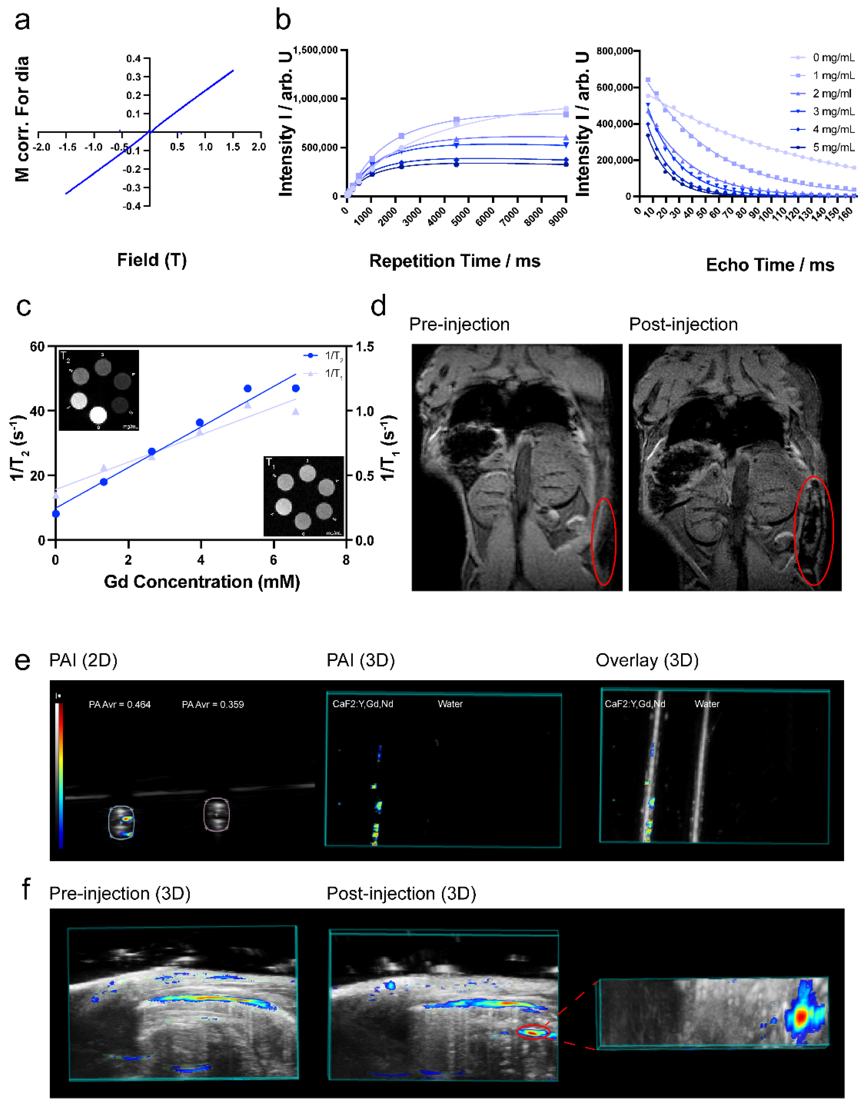
A correction has been made to “2.9.3 MRI Studies”:
To determine that the CaF2:Y,Gd,Nd NPs were magnetic, MRI measurements were performed on a 7T Bruker BioSpec (Ettlingen, Germany) with a 38 mm transmit/receive birdcage coil. CaF2:Y,Gd,Nd gels were configured using 1% agarose solution at different concentrations (0 mg/mL, 1 mg/mL, 2 mg/mL, 3 mg/mL, 4 mg/mL, 5 mg/mL). Then, the samples were placed in a circular test tube for testing. T1 relaxation was measured using a saturation recovery sequence with the following parameters: 9 repetition times (TR) of 18, 35, 70, 125, 250, 500, 1050, 2250, 4500, 9000 ms, echo time (TE) 6 ms, field of view (FoV) 30 × 30 mm2, matrix 64 × 64, slice thickness 2 mm. T2 relaxation was measured using a multi spin echo sequence with the following parameters: TR 2200 ms, TE and echo spacing 6.5 ms, 25 echoes, FoV 30 × 30 mm2, matrix 64 × 64, slice thickness 2 mm. Next, to test the MRI properties of NPs in a biological environment, we injected 100 µL of CaF2:Y,Gd,Nd NPs (10 mg/mL) into C57BL/6J mouse cadavers via subcutaneous injection. Images were obtained before and after injections. The relevant parameters were as follows: gradient echo sequence with TR/TE = 10/2.8 ms, FoV = 40 × 40 mm2, matrix = 256 × 256. The attenuation images and results were analyzed with ParaVision 360 (Version 2.0. pl.1, Bruker, Germany) software. Relaxation times were determined using standard mono-exponential functions.
There was an error in the original publication [
1]. The figure numbers are incorrectly labelled.
A correction has been made to “3. Results and Discussion, Paragraph 8”:
CaF2:Y,Gd,Nd NPs’ optical characteristics partially overlap with the excitation and emission spectra of dyes used in flow cytometry and confocal microscopy, such as Alexa Fluor 647. Thus, we explored whether CaF2:Y,Gd,Nd NPs could be used directly to measure the cellular uptake of NPs using flow cytometry and confocal microscopy (Figure S2a,b). Rare-earth NPs represent a promising tool for the early detection and treatment of cancer. For breast cancer in particular, the development of new imaging probes is desperately needed, because mammography, the standard diagnosis method, often leads to false-negative results and therefore to therapeutic delays [67]. In order to ensure that CaF2:Y,Gd,Nd NPs could be taken up efficiently by breast cancer cells to achieve high-contrast bioimaging, we first analyzed the uptake of CaF2:Y,Gd,Nd NPs by 4T1 cells at different time points using flow cytometry. As shown in Figure 6a,b, compared to the control group, the uptake efficiency of NPs was low during a short period of time, but gradually increased with time. To verify this conclusion, we used confocal microscopy to visualize the uptake of CaF2:Y,Gd,Nd NPs. As shown in Figure 6c, the signal of CaF2:Y,Gd,Nd NPs (magenta) was barely observed after 1 h of incubation, while a larger amount of signal could be observed as the incubation time increased. This result was consistent with the results obtained by flow cytometry. Therefore, we can conclude that CaF2:Y,Gd,Nd NPs were effectively taken up by 4T1 cells. Moreover, CaF2:Y,Gd,Nd NPs were intrinsically monitorable using flow cytometry and confocal imaging.
There was an error in the original publication [
1]. We have discovered errors in the reporting and interpretation of the MRI properties of the nanoparticles.
A correction has been made to “3. Results and Discussion, Paragraph 10”:
The presence of the element Gd3+, which possesses a high atomic number, makes it possible for CaF2:Y,Gd,Nd NPs to be used as a contrast agent in MRI [69–71]. To verify the magnetic properties of CaF2:Y,Gd,Nd NPs, VSM measurement is required. As we expected, the NPs exhibited typical paramagnetic behavior at room temperature (300 K) in an applied magnetic field of 1.5 T, indicating that the NPs are paramagnetically responsive to external magnetic fields (Figure 8a). This is consistent with previous studies, offering the possibility of CaF2:Y,Gd,Nd NPs as MR probes [72,73]. Then, MRI scans were performed on agarose-gel-embedded samples with different concentrations of CaF2:Y,Gd,Nd NPs. As the results in Figure 8b,c show, the signal intensity of CaF2:Y,Gd,Nd NPs show a faster decay of the T2 relaxation, resulting in lower signal intensities (dark contrast) in the T2-weighted MR image with increasing sample concentration. T1 contrast was less pronounced; for this concentration range, only subtle changes could be observed in the T1-weighted MR image. Both the T1 relaxation rate and T2 relaxation rate increased linearly with Gd concentration (R12 = 0.9388, R22 = 0.9677, respectively). The r1 and r2 relaxivity values are 0.1059 mM Gd−1·s−1 and 6.275 mM Gd−1·s−1; the r2/r1 ratio for CaF2:Y,Gd,Nd NPs is ~59. These indicate that CaF2:Y,Gd,Nd NPs is superparamagnetic and functions as a potential T2 MRI agent [74]. Subsequently, to test the MRI performance of CaF2:Y,Gd,Nd NPs within a complex environment, CaF2:Y,Gd,Nd NPs (10 mg/mL) were injected subcutaneously into a mouse cadaver and a significant signal could be observed at the site of injection by means of MRI (Figure 8d). Therefore, when used as a contrast agent, paramagnetic CaF2:Y,Gd,Nd NPs can effectively improve MRI efficiency and sensitivity. Currently, lanthanide-doped NPs are gradually applied to high-contrast PA imaging [75,76]. To investigate this property, we first performed in vitro PA measurements on CaF2:Y,Gd,Nd NPs and found that the presence of CaF2:Y,Gd,Nd NPs significantly enhanced the PA signal compared to water, with PA amplitudes up to 0.464 AU (arbitrary unit; Figure 8e). On this basis, we further evaluated the potential of CaF2:Y,Gd,Nd NPs as a PAI agent by intraperitoneal injection of CaF2:Y,Gd,Nd NPs in mouse cadavers. When compared to the PAI signal before injection of the NPs, a distinct PAI signal could be observed at 808 nm at the injection site after injection. The PAI signal of the NPs could be well distinguished from the surrounding tissue (Figure 8f). The success of our ex vivo PAI experiments illustrates that CaF2:Y,Gd,Nd NPs can be used as excellent PAI contrast agents for tissue imaging and diagnosis. In addition to their application as NIR/PAI contrast agent, our CaF2:Y,Gd,Nd NPs can also be used as an MRI contrast agent.
There was an error in the original publication. The conclusion of the MRI performance of nanoparticles is not accurate.
A correction has been made to “4. Conclusions”:
Small-sized CaF2:Y,Gd,Nd NPs synthesized using a facile hydrothermal method can be used as multimodal NIR-II fluorescence/photoacoustic/magnetic resonance imaging probes. The excellent morphology and NIR-II optical properties provide the basis for NIR-II diagnostic applications of our CaF2:Y,Gd,Nd NPs. Biotoxicity and stability analyses have shown that our NPs are biocompatible as well as safe in biological settings. In addition, the fact that immune cells were not activated in response to CaF2:Y,Gd,Nd NPs provides favorable leverage for their application in vivo. The doping of Gd3+ shows a stronger increase of transverse relaxation rate (1/T2) than the longitudinal relaxation rate (1/T1) in CaF2:Y,Gd,Nd NPs, thereby enabling accurate T2-MR diagnostic properties, which are similar to the previous study [77]. In addition, the apparent PAI signal offers a wide range of applications for CaF2:Y,Gd,Nd NPs in PAI-based diagnostics. Thus, our work not only demonstrates a multifunctional NP with excellent three-mode imaging capability, but also provides a potential solution to the medical system dilemma. A hot trend in current research is to equip rare-earth NPs with therapeutic functions through surface modifications (antibodies, chemotherapeutic agents, or photosensitizers, etc.), while achieving multimodal imaging of tumor sites [78–81]. In particular, surface modification with tumor-targeting ligands can enhance the potential of rare-earth NPs for cancer diagnosis and treatment [19]. We strongly believe that this study provides an important insight into the design of novel multifunctional nanomaterials as potential therapeutic agents for the treatment and diagnosis of diseases in the future, and helps to drive the clinical translation of novel therapeutic agents.
The authors state that the scientific conclusions are unaffected. This correction was approved by the Academic Editor. The original publication has also been updated.

 ,
, 
