Review

Recent Advances in Cell Membrane Coated-Nanoparticles as Drug Delivery Systems for Tackling Urological Diseases

1
Urology & Nephrology Center, Department of Urology, Zhejiang Provincial People’s Hospital, Affiliated People’s Hospital, Hangzhou Medical College, Hangzhou 310014, China
2
The Second Clinical Medical College, Zhejiang Chinese Medical University, Hangzhou 310053, China
*
Authors to whom correspondence should be addressed.
Pharmaceutics 2023, 15(7), 1899; https://doi.org/10.3390/pharmaceutics15071899
Submission received: 28 May 2023 / Revised: 28 June 2023 / Accepted: 3 July 2023 / Published: 6 July 2023

Abstract

:
Recent studies have revealed the functional roles of cell membrane coated-nanoparticles (CMNPs) in tackling urological diseases, including cancers, inflammation, and acute kidney injury. Cells are a fundamental part of pathology to regulate nearly all urological diseases, and, therefore, naturally derived cell membranes inherit the functional role to enhance the biopharmaceutical performance of their encapsulated nanoparticles on drug delivery. In this review, methods for CMNP synthesis and surface engineering are summarized. The application of different types of CMNPs for tackling urological diseases is updated, including cancer cell membrane, stem cell membrane, immune cell membrane, erythrocytes cell membranes, and extracellular vesicles, and their potential for clinical use is discussed.

Graphical Abstract

1. Introduction

Urological diseases encompass many complex disorders, including tumors, inflammation, infection, acute kidney injury, etc., and have become a global public health problem that collectively affects more than 1 in 10 individuals worldwide, resulting in high healthcare costs [1,2,3,4]. Moreover, it tends to develop at a younger age [5]. Current strategies for tackling urological diseases are ineffective due to physiological barriers in urological systems [6,7]. The application of nanomedicine is becoming more widespread [8] in urological disease treatment because it improves the pharmaceutical performance of drugs. Nevertheless, nanoparticles will be recognized and fast removed by the reticuloendothelial system (RES) as foreign elements in the body, causing immune responses and toxic effects, which will be the major hurdles that almost all platforms must overcome [9,10]. Based on recent research findings, it has been observed that over 85% of the re-ported stealth nanomaterials experience a rapid decline in blood concentration, reducing to half of the administered dose within 1 h after administration. This indicates that the nanoparticles have not achieved a satisfactory stealth effect [11]. PEGylation is a widely employed technique for prolonging blood circulation. However, membrane coating aligns more closely with the concept of holism due to the intricate structural hierarchy of biological systems.
Cell membrane coated-nanoparticles (CMNPs) form a biomimetic structure that inherits the surface properties and functions of the source cells and imparts additional biological capabilities to the encapsulated nanoparticles, such as immune evasion, cell-specific targeting, and extension of the systemic circulation [12,13,14,15,16]. Meanwhile, natural cell membranes will preserve the integrity and bioactivity of nanoparticles in vivo. Recent years have witnessed the increasing interest in the use of CMNPs as drug delivery systems to treat urological diseases, which is rarely reviewed elsewhere [17]. In this review, the current state of nanomedicine for treating urological diseases is briefly summarized, and the methods for CMNPs synthesis and surface engineering are updated. The application of different types of CMNPs for tackling urological diseases, especially tumors, inflammation, and acute kidney injury, is reviewed.

2. Current State of Nanomedicine for Treating Urological Diseases

According to the etiology, urological diseases are mainly classified into urological cancers, stones, infections, inflammation, and injuries [18]. The incidence of these diseases is increasing annually, leading to significant social burdens and high healthcare costs. Although surgery is the primary treatment for urological cancers, stones, and injuries, its clinical outcomes often fall short of expectations. For example, 25% to 30% of patients with renal cancer present with distant metastatic disease at the time of diagnosis [19], and approximately 40% of patients who receive surgical treatment eventually develop recurrence [20]. For infections, inflammation, and the remaining part of urological diseases which do not need surgical intervention, drug treatment is most widely used, but not that effective. There remain obstacles to drugs reaching and remaining in their targeted lesions. (1) Inherent physiological permeability barriers impede the delivery of therapeutic agents to diseased tissues and cells, such as the urothelial barrier [21], glomerular filtration barriers (GFB) [22], and blood–prostate barrier (BPB) [23]. (2) Off-targeting occurs in normal tissues frequently, leading to a high risk of side effects [24]. (3) Another challenge is the lack of subcellular targeted therapy, via which a specific type of drug can directly interact with its subcellular target to maximize therapeutic effects [25]. (4) Short-term retention of drugs in their targeted lesions determines their short-acting effects.
Nanoparticles, composed of materials such as lipids, metals, synthetic and biopolymeric polymers, proteins, and inorganic and organometallic compounds, have shown promise in improving the in vivo performance of loaded drugs [26,27,28,29]. (1) Nanomaterials have been long pursued as drug delivery systems to conquer urological physiological barriers [30,31]. For example, surface modification of drugs with mucus-penetrating moieties or hydrophobic ligands could accelerate the translocation of drugs across the mucus barrier or the epithelial barrier, respectively [32,33,34,35]. (2) Nanomaterials can reduce the off-target effect of drugs via passive targeting or active targeting [36]. In the past, nanoparticles with a hydrodynamic diameter (HD) within the range of 100 to 400 nm were considered ideal for passive tumor targeting due to the enhanced permeability and retention (EPR) effect. However, a recent study conducted by Cabral et al. has highlighted that micelles measuring 30 nm in size could potentially achieve much more effective tumor penetration when compared to micelles with a size of 100 nm [37]. (3) Nanomaterials correct the intracellular trafficking of drugs into the expected subcellular compartments [38]. For instance, nuclear localization signals (NLS)-labeled nanoparticles could facilitate the active nuclear transport of drugs [39,40]. Nanoparticles can also enter mitochondria through a variety of pathways. In one study, Tritylphosphine (PPh3)-coupled nanoparticles could send doxorubicin to mitochondria [41]. The application of organelle-targeted nanoparticles has also expanded to drug delivery to the endoplasmic reticulum [42] and Golgi apparatus [43]. (4) The prolonged exposure of drugs to their targeted lesions can be achieved via the combined use of nanomaterials. Mucoadhesive nanomaterials could adhere to the mucus barrier to allow the sustained release of drugs [30]. At the same time, mechanisms also exist within urological diseases to facilitate the targeted delivery of nanoparticles. These mechanisms include the EPR effect [44], the presence of specific receptors on the surface of tumor cells [45], alterations in the tissue microenvironment [46], and the involvement of inflammatory factors.
Among all types of nanoparticles, CMNPs have gained significant attention as drug delivery vehicles. CMNPs exhibit a biomimetic structure that retains the surface characteristics of the source cells and imparts bioactivities to the encapsulated nanoparticles, including immune evasion, cell-specific targeting, and extension of systemic circulation [12,13]. Apart from these inherent functionalities, the lipid layers on the outer surface of CMNPs are easily amenable to surface engineering. Furthermore, CMNPs are highly biocompatible drug delivery systems. From a translational nanomedicine perspective, CMNPs offer a promising alternative.

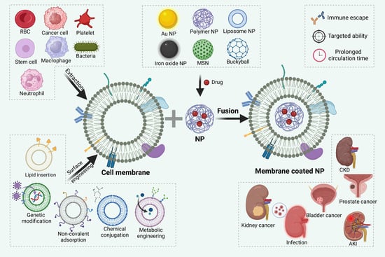
3. Synthesis and Surface Engineering of CMNPs

Hu et al. [47] pioneered the study of CMNP fabrication in 2011. They applied red blood cell membranes to shroud poly (lactic-co-glycolic acid) (PLGA) nanoparticles and prolong the circulation time of the CMNPs up to 72 h. Building on their work, a general consensus has been established regarding the synthesis procedure of CMNPs, which can be broadly divided into three steps: extraction of cell membranes, fusion of cell membranes with core nanoparticles, and surface engineering of CMNPs.

3.1. Extraction of Cell Membrane

Cell lysis and membrane purification constitute two critical steps in the extraction of cell membranes [48]. It is important to note that the extraction procedures vary depending on whether the source cells are nucleus-free.
For nucleus-free cells such as erythrocytes and platelets, the cells are lysed through hypotonic treatment or repeated freeze-thaw cycles. Subsequently, the purified cell membrane is obtained by removing soluble cytoplasmic components using centrifugation [49]. In order to maintain the biological activity of membrane proteins, protease inhibitors should be added to the extracted cell membranes and stored at 4 °C.
The extraction and purification of eukaryotic cell membranes are more complex compared to nucleus-free cells, mainly because the removal of cell nuclei is required [50]. First of all, similar to the extraction of nucleus-free cell membranes, a sufficient number of cells must be collected for concentrating and purifying cell membranes, which are disrupted by incubation in hypotonic lysate or repeated freeze-thaw treatments. Afterward, the nucleus and intracellular biomolecules were removed by discontinuous sucrose gradient centrifugation or differential centrifugation. The membrane-rich fraction obtained from the previous step is washed with a plasma buffer. To obtain cell membrane vesicles, the fraction can be further processed through sonication or extrusion using porous membranes [51].

3.2. Fusion of Cell Membranes with Core Nanoparticles

The most commonly used method of CMNPs fusion is physical extrusion [47], during which core nanoparticles and the extracted purified cell membranes are co-extruded through a porous membrane using a micro-extruder. The mechanical force of extrusion can disrupt the integrity of cell membranes and reorganizes them to surround core nanoparticles. Although this method is convenient to perform and effective in producing CMNPs, it is difficult to achieve large-scale production due to the material loss caused by material deposition onto filter membranes [52].
Another method, ultrasound treatment, utilizes ultrasound waves in the range of 20 to 50 kHz to disrupt the integrity of cell membranes. The attractive force between the nanoparticles and the cell membrane fragments facilitates the formation of cell membrane vesicles with a spherical shape, which then surround the core nanoparticles [53]. Ultrasound treatment is more efficient for scaled-up production of CMNPs, but it may not be suitable for certain nanoparticles as sonication can affect the size and stability of the core nanoparticles [54].
Under the stimuli of the external electric field, the electroporation can increase the semi-permeability of cell membranes and induce the formation of a large number of micropores on the cell membrane, through which nanoparticles can diffuse into the cell membrane [55]. The electroporation avoids the destruction of nanoparticles and is less time-consuming and labor-intensive nature than the co-extrusion method [55]. However, its operating procedure is more complex. Additionally, the electroporation method can be combined with microfluidic devices to promote the productivity of CMNPs. Rao et al. injected a mixture of RBC membrane-derived vesicles and Fe3O4 nanoparticles into a microfluidic device, where Fe3O4 magnetic nanoparticles (MNs) and RBC membrane vesicles flow through the electroporation zone and eventually fused with the assistance of electrical pulses [56]. As a result, microfluidic electroporation can produce CMNPs with uniform sizes. Its high reproducibility also guarantees the potential for the large scale-up production of CMNPs with enhanced colloidal stability and in vivo bioactivity [57].
In situ polymerization is a technique employed to coat isolated cell membranes onto nanocomposites, enabling the growth of nanoparticle cores inside cell membrane-derived vesicles. This process involves the use of a monomer, an initiator, and a cross-linker, which, when exposed to radiation or heat, initiates polymerization. In situ polymerization offers several advantages, including the assurance of coating integrity, simple encapsulation of the payload, and easy control over the size and stiffness of the resulting CMNPs [58]. Zhang et al. utilized RBC membrane-derived vesicles as nanoreactors to synthesize polymeric cores through in situ polymerization, resulting in cell membrane-coated hydrogel nanoparticles [59]. By selectively polymerizing monomers upon ultraviolet exposure, a stable structure is formed. This approach effectively eliminates potential risks of content leakage during the nano gel preparation process [60].
A comparison of different fusion methods is summarized in Table 1.
Noteworthy, the orientation of cell membrane coating on the surface of core nanoparticles regulates the bioactivity of CMNPs [61]. The right-side-out oriented cell membranes can inherit the biological function from their parent cells, therefore providing core nanoparticles with immune-evasive stealth and prolonging the in vivo circulation. The inside-out cell membranes expose binding domains to membrane-impermeable drugs, such as small-molecule tyrosine kinase inhibitors, and, thus, they act as sponges to adsorb these drugs [61]. During the fusion procedure, correctly orientated cell membrane coating cannot be achieved via random cell membrane-core pairs. A more reliable strategy relies on the specific interaction between ligands anchoring at the surface of core nanoparticles and the intracellular domain of their corresponding receptors [62], thus connecting the surface of core nanoparticles with the inner side of the cell membrane. By this principle, the right-side-out oriented assembly of CMNPs can be achieved via the cytoplasmic domain of band 3- the P4.2-derived peptide pairs [62] and inside-out oriented assembly of CMNPs can be achieved via azide analogues in the glycan-alkynyl pairs [63], biotin-streptavidin pairs [61], and sucrose density gradient centrifugation [64]. To verify the corrected assembly of CMNPs, the presence of two cell membrane markers at the outer surface of CMNPs is tested. CD47 is a transmembrane protein that contains a variable extracellular N-terminal domain, a presenilin domain with five transmembrane regions, and a C-terminal intracellular region [65]. Under the view of the transmission electron microscope (TEM), immunogold staining of different domains of CD47 can reveal the spatial relationship of different layers of cell membranes with core nanoparticles [66]. Considering that inside-out cell membrane patches and right-side-out cell membrane patches may assemble onto core nanoparticles together, immune electron microscopy is not effective in quantifying the exact proportion of differently oriented cell membrane patches on the surface of core nanoparticles. Sialic acid exclusively appears as a terminal residue of extracellular surface glycans [67]. Sialidase can dissociate the sialic acid from the outside surface of CMNPs, which can be further quantified via commercial kits [63]. The equivalent density of sialic acid from the outside surface of CMNPs to that from the outside surface of natural cells is solid evidence on the right-side-out cell membrane coating.
Loss of membrane integrity in CMNPs may cause drug leakage from core nanoparticles during drug delivery, unwanted biomolecule adsorption in physiological fluids, altered mechanical properties of nanoparticles, and changed molecules affinity of the membrane. The ratio of full cell membrane coated-nanoparticles in CMNPs is as low as 1.8 ± 0.1%, 6.2 ± 0.3%, and 6.5 ± 0.3% via sonication and extrusion [68]. To further improve the cell membrane integrity, red blood cell membrane ghosts and negatively charged core nanoparticles with small size are preferred as sources of CMNPs due to the well-preserved cell membrane structure of RBC membrane ghosts and their mild binding affinities to small and negatively charged core nanoparticles [68]. TEM or a confocal laser scanning microscope (CLSM) is used to validate the cell membrane integrity in CMNPs; TEM offers a vertical view, and CLSM is diffraction limited and not available for nanoparticle observation [63]. Liu et al. developed a fluorescence quenching assay to observe the full coverage of cell membranes on the surface of core nanoparticles. Fluorescent nitro-2,1,3-benzoxadiazol-4-yl (NBD) was covalently coupled to the core nanoparticles, and integrated cell membrane coating could shield NBD from the reduction by dithionite (DT), which was membrane-impermeable and could quench the fluorescence of NBD-labelled nanoparticles. The NBD fluorescence could remain only in CMNPs with full cell membrane coating in the presence of DT [68]. However, such a fluorescence-quenching assay failed to distinguish partially cell membrane coated-nanoparticles with different surface coverage.

3.3. Surface Engineering

Surface engineering of CMNPs plays a crucial role in enhancing their active targeting, improving their pharmacokinetics, and modifying their pharmacodynamics to enhance bioactivity [69]. Surface engineering is generally completed after cell membrane extraction and the fusion of cell membranes with core nanoparticles. In this way, functional moieties can reside at the outer surface of CMNPs. However, the purification operation can be labor-intensive and may alter the physiochemical properties of CMNPs. Another strategy is to directly functionalize the cell membrane of live cells before the cell membrane extraction and the fusion of cell membranes with core nanoparticles [70], which makes the purification operation convenient but imposes uncertainty on the orientation of the engineered cell membrane. Moreover, every step should be cell-friendly during the surface engineering of live cells [71].
Lipid insertion is a non-disruptive approach to equipping CMNPs with functional moieties via lipid anchors [72]. In this strategy, a functional moiety is attached to a lipid molecule, which then guides the functional moiety, such as small molecules or antibodies, to adhere to the cell membrane. However, compared to small molecules, antibodies are larger in size and their geometric orientation is more challenging to control due to the random distribution of functional groups on the protein surface [73]. Lipid insertion can be performed on live cells and cell membrane vesicles [74]. In addition to serving as anchors, the inserted lipids can also function as stimuli-responsive sensors to achieve photothermal conversion [75].
Chemical conjugation is a method of surface functionalization that involves the addition of functional moieties to the surface through strong covalent bonds [76]. Compared to other direct modification methods, which can damage cell membranes and reduce their plasticity, chemical conjugation offers advantages such as high yield synthesis, a wide range of applicability, and easy product separation. Several techniques are commonly employed, including thiol-maleimide coupling, EDC/NHS (1-ethyl-3-(3-dimethyl aminopropyl)-carbodiimide/N-hydroxysuccinimide) coupling, azide-alkyne cycloaddition, and amidation chemistry. Smyth et al. utilized EDC/NHS coupling chemistry to attach alkynyl groups to the surface of extracellular vesicles (EVs), where the amine groups of EV proteins or lipids were cross-linked to 4-pentenyl acids through carbodiimide-activated cross-linking [77]. Microscopy, flow cytometry, and nanoparticle tracking analysis demonstrated that surface functionalization had minimal impact on EV functionality. Chemical conjugation offers the advantage of being easy, quick, and compatible with biomolecules, making it widely utilized for functionalizing biomolecules [78].
Non-covalent adsorption facilitates the binding of functional moieties to CMNPs through hydrogen bonding (dipole-dipole interactions), electrostatic interactions (charge-charge interactions), van der Waals forces, or hydrophobic interactions [70]. Zheng et al. decorated cell membranes with hendeca-arginine peptide (R11) in live bladder cancer cells, and afterward, R11-coated cell membrane vesicles were generated. R11 permeated but did not remain on the cell membrane; therefore, R11 was polymerized with DNA to form nanoparticles so that R11 could be internalized via slow endocytosis rather than fast transduction. Consequently, large amounts of R11 were deposited onto the cell membrane with a right-outside orientation [70].
Genetic modification can present functional moieties on the cell membrane via gene transfection [79]. In the process of genetic modification, robust cell lines can be utilized to permanently express unique surface ligands via viral transfection. These transfected cells can replicate, allowing for maximized population growth, which is beneficial for large-scale manufacturing at a reduced cost compared to other surface engineering techniques [80,81,82,83]. HepG2 cells were genetically modified to express the hepatitis B virus (HBV) preS1 ligand, which was then extracted from the cell membrane to coat oncolytic adenoviruses (OAs). This genetic surface engineering approach has the advantage of decreasing the immunogenicity of OAs and facilitating their targeting of preS1 receptor overexpressed tumors [84]. Genetic modification causes less damage to cell membranes compared to alternative methods, ensuring a more precise surface engineering process.
Metabolic engineering is a technique that manipulates the natural biosynthetic pathways within cells to transport functional moieties onto the cell membrane [85]. More specifically, metabolic substrates are covalently linked with functional moieties, forming conjugates that can be introduced into cells and recycled within the intracellular metabolism [86], and one of their destinations belongs to the cell membrane. Metabolic engineering primarily relies on glycoengineering and lipid engineering. Glycoengineering involves the production of oligosaccharides and glycoconjugates, employing pathways such as the Sialic acid pathway, GalNAc salvage pathway, and Fucose salvage pathway. On the other hand, lipid engineering utilizes natural lipid synthesis pathways, such as the cytidine 5′-diphosphocholine (CDP-choline) pathway [87].
Different methods for cell membrane surface engineering are summarized in Table 2.

4. Application of CMNPs in Tackling Urological Diseases

To date, the main types of urological diseases that have been reported to be treated with CMNPs include the following. (1) Cancers: kidney cancer, bladder cancer, and prostate cancer. (2) Inflammations and infections: sepsis and fibrosis. (3) Injury: acute kidney injury, especially ischemia-reperfusion injury. (4) Nephrotoxicity induced by some medicines. (5) Erectile dysfunction.

4.1. Cancer Cell Membranes (CCMs)

CCMs have emerged as the ideal candidates for encapsulating nanoparticles in oncological treatment applications for the following reasons. Firstly, CCMs possess remarkable robustness and the ability to proliferate indefinitely [88], making it convenient to obtain and culture their membrane material in vitro. Secondly, CCMs are enriched with a wide array of functional proteins, including membrane proteins that facilitate homologous binding (such as selectins and integrins), biomarkers involved in self-recognition and immune evasion (such as CD47), and tumor antigens associated with immune activation (such as tumor-associated Thomsen–Friedenreich glycoantigens). Notably, when used as a coating, CCMs retain the inherent targeting abilities conferred by a diverse range of cell adhesion molecules, thereby endowing CCM-coated nanoparticles with enhanced cancer-homing features through a homotypic targeting mechanism [89,90]. A study demonstrated that the targeting efficiency of CCMs-coated nanoparticles was 20 times and 40 times higher than that of erythrocyte membrane-camouflaged nanoparticles and bare nanoparticles, respectively [91].
Doxorubicin (DOX) is a common chemotherapeutic drug that can bind and hinder the replication of DNA, thereby inhibiting the cell cycle of cancers. To address its severe side effects, such as impaired bone marrow hematopoietic function, cardiotoxicity, high fever, and so on, which limit its clinical application, Liu et al. cloaked CCM and capped CaCO3 on mesoporous silica NPs (DOX/MSN/CaCO3@CM) [92]. Benefiting from the homotypic targeting abilities of CCMs, DOX/MSN@CaCO3@CM was easily internalized by LNCaP-AI cells (a prostate cancer cell line) and accumulated into the prostate cancer site. The fluorescence intensity of DOX/MSN@CaCO3@CM in the corresponding source cells of LNCaP-AI was much higher than those in the heterotypic cells of MCF-7 (human breast cancer) cells, suggesting the highly specific self-recognition affinity of DOX/MSN@CaCO3@CM to the source cells. CLSM was used to track the DOX red fluorescence in the cells; with the time increased to 8 h, the red fluorescence became dominant in the nuclei rather than in the cytoplasm, which enables DOX to better exert therapeutic effects. A major obstacle in prostate cancer treatment is that most patients eventually develop castration-resistant prostate cancer (CRPC) and have intrinsic or acquired resistance to androgen deprivation therapy (ADT) and other hormone therapy [93]. Therefore, a suitable drug delivery mode is needed for the treatment of CRPC. Lu et al. used CRPC cell membranes as bionic carriers for encapsulating PLGA containing the chemotherapeutic drug docetaxel (DTX) [94]. In addition to properties such as evading early clearance by the immune system and circulatory system and penetrating the extracellular barrier, CRPC cell membranes contain a highly specific library of isotypic molecules that can be used to recognize the same cancer cell types and increase targeted drug delivery by DTX. Measured by flow cytometric analysis to quantify the differences, the cellular uptake of CRPC membrane-coated NPs was approximately 40 times higher than PLGA NPs. Apart from chemotherapy drug delivery, CCMs can also deliver cancer vaccines to fight cancer by activating the body’s immune system. In a recent study [95], Li et al. synthesized PMBEOx-COOH [thioglycolic-acid-grafted poly(2-methyl-2-oxazoline)-block-poly(2-butyl-2-oxazoline-co2-butenyl-2-oxazoline)] to load imiquimod (R837). The surgically harvested CCMs were then coated onto R837-loaded PMBEOx-COOH NPs to obtain surgically derived CCM-coated POxTA NPs (SCNPs/R837). SCNPs/R837 efficiently traveled to the draining lymph nodes and then activated plasmacytoid dendritic cells, triggered a massive release of inflammation-related factors, recruited and activated NK and cytotoxic T lymphocytes cells in prostate cancer lesions, bypassing the tumor immunosuppressive microenvironment and killing tumor cells.
In a study of CMNPs for bladder cancer treatment, a cancer cell membrane-decorated zeolitic-imidazolate framework hybrid nanoparticle (HP) was successfully constructed by Chen et al. [96] co-delivering cisplatin (DDP) and oleanolic acid (OLA). It showed positive results of the platform (HP/DDP/OLA) for the treatment of bladder cancer (BCa) (SW780 cells). HP/DDP/OLA could enhance apoptosis, in detail, after 72 h of incubation at the same drug concentration, the HP/DDP group and HP/OLA group showed 43.6% and 31.4% cell apoptosis, respectively; on the contrary, the HP/DDP/OLA group showed a significant increase to 72.3%. Meanwhile, it can reverse multidrug resistance in SW780 cells more than free drugs alone or mono-delivery systems. Alternatively, to overcome the side effects caused by the permeability barrier and off-targeting of normal urothelial cells and to enhance the efficacy of chemotherapy within BCa. Zheng et al. [70] disguised PLGA nanoparticles containing gemcitabine (PLGA-G) into BCa cell-derived membrane (TM) that were surface-modified with hendeca-arginine peptide (R11). The surface functionalization with R11 endowed TM with the dual-targeting capacity, which originated from the intrinsic BCa targeting capacity of R11 and the homologous tumor targeting capacity of TM. The intravesical drug delivery system comprising R11@TM-camouflaged PLGA-G (R11@TM@PLGA-G) exhibited excellent BCa-targeting capacity and mucus-penetrating efficiency, and even chemo-resected most tumors in murine orthotopic BCa models. It has been acknowledged that the process of endocytosis, specifically through the caveolin-mediated pathway, plays a crucial role in determining the intracellular trafficking of nanoparticles to regions outside of lysosomes. As a result, drugs loaded within CMNPs have the ability to avoid degradation within lysosomes [97].
In conclusion, CCM-coated NPs have been researched for urological cancers targeting to achieve higher on-target payload delivery and lower off-target side effects.

4.2. Immune Cell Membranes (ICMs)

Immune cell membrane-coated nanoparticles (ICMCNPs) are emerging, nature-inspired approaches that leverage biocompatibility, prolonged blood circulation time, and enhanced specificity of immune cells to target inflamed tissues and tumors [98]. Each type of immune cell membrane (such as macrophages, dendritic cells, granulocytes, mast cells, and lymphocytes) possesses unique characteristics that can be utilized in membrane-coated nanoparticle-based drug delivery systems [99]. For instance, dendritic cells exhibit specific co-stimulatory/inhibitory molecules on their membranes and play a crucial role in antigen presentation, thus holding promise for DC-based vaccine delivery [100]. T lymphocytes, on the other hand, present T cell receptors (TCRs) on their cell membranes, allowing them to bind specifically and strongly to antigenic determinants found in pathogens, microbes, and tumor cells. Therefore, the membranes of T lymphocytes can be utilized to create efficient nanocarriers for targeted drug delivery [101]. Macrophage derived ICMCNPs can avoid phagocytosis by immune cells via cellular self-recognition mechanisms, and the surface ligands inherited from macrophages can drive ICMCNPs to target diseased sites [102]. Macrophage membrane coated-nanoparticles can respond to multiple abnormal signals, including bacterial toxins, viruses, inflammatory cytokines, cancerous antigens, and so forth [103]. Until now, ICMCNPs that are involved in the treatment of urological diseases have been mainly derived from neutrophils.
Neutrophils are an integral part of the innate immune defense system and are among the early cell types recruited to sites of injury or inflammation [104,105]. This recruitment process involves interactions between P-/E-selectin and their glycosylated ligands, such as P-selectin glycoprotein ligand 1, as well as integrin-mediated adhesion. Leveraging the properties of neutrophils, Liu et al. utilized neutrophil-derived cell membranes to encapsulate Coenzyme Q10 (CoQ10) nanoparticles, creating a formulation known as N-NP CoQ10. This approach aimed to target Ischemia-reperfusion (I/R) injury for therapeutic intervention. Moreover, the functionalization of CoQ10 with neutrophil-derived cell membranes led to synergistic effects in inhibiting oxidative damage and neutralizing pro-inflammatory cytokines at I/R injured sites [106]. In in vitro experiments, the N-NP CoQ10-treated group exhibited 5.32% apoptotic HK-2 cells, while the CoQ10-treated group and NP CoQ10-treated group had 24.9% and 10.3% apoptotic HK-2 cells, respectively. In vivo, N-NP CoQ10 treatment resulted in significantly reduced release of inflammatory cytokines (IL-1β, TNF-α, and IL-6) in I/R injured kidneys, leading to better preservation of kidney function compared to CoQ10 treatment or NP CoQ10 treatment alone.

4.3. Stem Cell Membranes (SCMs)

Stem cells possess unique characteristics, including their ability to self-renew and differentiate into specialized cell types [107]. When injured tissues release chemokines, adhesion molecules, and growth factors, stem cells can detect and respond to these signals, migrating toward the sites of injury [108]. Amongst stem cells, mesenchymal stem cells (MSCs) have demonstrated inherent capabilities for homing to tumors and specifically migrating to inflamed tissues. This is facilitated by the interactions between surface receptors on MSCs, such as CXC motif chemokine receptor (CXCR) and CD74, and the corresponding cytokines expressed in cancerous or inflamed tissues [109,110]. Stem cell membrane-coated nanoparticles (SCM-NPs) preserve the surface molecules present on the source stem cell membrane, thereby retaining their intrinsic targeting ability. Additionally, this coating provides protection to the cargo within the nanoparticles, preventing it from being captured by the immune system. By utilizing stem cell membranes as coatings for nanoparticles, SCM-NPs can take advantage of the inherent homing and targeting capabilities of stem cells. This approach offers the potential for targeted drug delivery and therapy, enabling precise delivery to specific tissues or regions of interest.
A novel platform, PDA-Fe3O4@MSC, was developed for the delivery of small interfering RNA (siRNA) across cell membranes [111]. This platform utilized polydopamine-coated iron oxide nanoparticles (PDA-Fe3O4) that were coated with membranes derived from mesenchymal stem cells (MSCs). The incorporated iron oxide nanoparticles served as photothermal agents that could be activated by laser therapy and also acted as magnetic resonance imaging (MRI) trackers. PDA-Fe3O4@MSC combined the therapeutic abilities of gene silencing, photothermal activity, and MRI tracing into a single system for the non-invasive treatment of prostate cancer. In an in vivo antitumor test, the combination of Fe3O4@PDA-siRNA@MSC nanoparticles with laser irradiation resulted in a 60% reduction in tumor volume after a 15-day therapy period. Another multifunctional platform (PDA-DOX/siRNA@SCM NPs) is introduced by Mu et al. to combine chemotherapy and gene therapy for prostate cancer treatment. Considering the adaptive upregulation of programmed cell death ligand 1 (PD-L1) during Dox-based chemotherapy, this platform co-delivered PD-L1 siRNA and Dox into prostate cancer cells. The goal was to eliminate the expression of the PD-L1 protein, thereby restoring the immune anti-tumor activity of T cells while maximizing the anti-cancer effects of Dox. The SCM coating on the nanoparticles reduced the clearance of Dox from the bloodstream, resulting in higher Dox accumulation in tumors compared to the Dox group and PDA-DOX group. Additionally, the SCM coating alleviated the acute toxicity of Dox without causing a significant decrease in body weight over time [112].
The value of SCM-coated nanoparticles for drug delivery is discounted by the relatively shorter circulation time of SCM-coated nanoparticles, compared with red blood cell membrane-coated nanoparticles. In addition, it is inconvenient to obtain large numbers of stem cells for large-scale production of SCM-coated nanoparticles [113].

4.4. Red Blood Cell Membranes (RBCMs)

Red blood cells (RBCs) have an average lifespan of about 120 days, making them an ideal choice for the preparation of long-circulating nanoparticles [114]. RBCs were the first type of cells used to create membrane biomimetic carriers, as demonstrated by Hu et al. in 2011 [47]. In their study, encapsulating nanoparticles with RBC membranes (RBCMs) extended the blood circulation time from 24 h to 72 h. RBCM-coated nanoparticles retain the natural surface structure of RBCs and preserve the presence of CD47, a protein that interacts with the SIRP-α receptor to inhibit phagocytosis by macrophages. This interaction releases a “don’t eat me” signal, contributing to the nanoparticles’ ability to evade macrophage uptake [69,115]. It has been observed that the expression level of CD47 is down-regulated on aging RBC membranes, which ultimately leads to their phagocytosis by macrophages and accumulation in the liver. Leveraging the natural properties of aging erythrocytes, aging RBCMs can serve as carriers for drug delivery targeting detoxification and liver-specific therapeutic purposes [116].
Although there have been limited advances in the study of RBCM-coated nanoparticles for treating urological diseases, they hold the potential for mitigating or even eliminating drug-induced nephrotoxicity. Antibiotics are the first choice for the treatment of sepsis and septicemia, but high doses of antibiotics can lead to serious adverse toxic reactions and side effects [117]. To address this issue, Liu et al. synthesized RBCM-coated nanoparticles (RBCNPs) modified with γ3 peptide (γ3-RBCNPs) for targeted therapy of Klebsiella pneumonia-induced sepsis. The γ3 peptide specifically binds to ICAM-1 at the infection sites, allowing γ3-RBCNPs to transport ciprofloxacin to the infection regions rather than accumulating in the kidneys. In another study, Su et al. introduced RBCM-coated gelatin nanoparticles loaded with berberine hydrochloride (RBGPs) to achieve sustained release of the drug and ensure biosafety [118]. Human embryonic kidney (HEK 293T), a standard cell model for the nephrotoxicity evaluation in vivo, was incubated with different concentrations of RBGP (30, 60, 120, 240, 300, 480, 600, and 750 μg/mL) for 24 h. The RBGPs with the highest concentration (750 μg/mL) did not hamper the cell viability of HEK 293T cells, while free BH will cause cytotoxic effects on liver and kidney cells at 50 ug/mL [119]. This indicated that RBGPs improved the biocompatibility of their loading drugs to reduce nephrotoxicity. Similarly, Malhotra et al. derived amphiphilic fluorophore-labeled nanovesicles (NVEs) from RBCMs [120] to load the hydrophobic drug camptothecin (CPT). Compared with free CPT, NVEs-coated CPT showed higher retention in the circulation over 48 h and insignificant accumulation in kidneys.
Due to the deformability of RBCNPs, they are excellent fits for the delivery drugs across physiological barriers. However, dense tumor stroma contributes to the impermeability of cancerous tissues [30], thus blocking the penetration of drugs, nanoparticles, or even highly deformable RBCNPs. Zhou et al. anchored recombinant human hyaluronidase, PH20 (rHuPH20) onto the surface of RBCNPs to degrade hyaluronic acid (HA) [121,122], which is highly expressed in the extracellular matrix (ECM) of approximately 80% of prostate cancer [122]. The diffusion co-efficiency of rHuPH20-Anchored RBCNPs nearly doubled over that of RBCNPs, and they were preferably internalized by PC3 prostate cancer cells.

4.5. Extracellular Vesicles (EVs)

EVs are structures surrounded by a lipid bilayer and are released by both prokaryotic and eukaryotic cells [123,124]. According to guideline of International Society for Extracellular Vesicles, EVs is a collective term covering various subtypes of membranous structures released by cells, including exosomes, microvesicles, microparticles, ectoderm, epithelium, apoptotic bodies, and many others [125]. Currently, the most relevant to the treatment of urological diseases are exosomes (Exos), microvesicles (MVs) [126]. Exosomal formation is regulated by endosomal sorting complexes required for transport (ESCRT) proteins; therefore, these proteins and their accessory proteins (Alix, TSG101, HSC70, and HSP90β) can be found in exosomes regardless of the type of cell from which they originate. Thus, this set of proteins is often regarded as “exosomal markers”. Exosomes not only participate in cell–cell communication, tumor progression, and stimulation of immune responses by acting as antigen-presenting vesicles [127], but also act as carriers of biomarkers for diseases [128,129,130], such as Parkinson, glioblastoma, acute kidney injury, etc. The marker proteins associated with MVs mainly include cytosolic proteins and proteins associated with the plasma membrane [131], such as tetraspanins, cytoskeletal proteins, heat shock proteins, integrins, glycan-binding proteins, etc. MVs also play a crucial role in cell–cell communication, facilitating interactions between local cells as well as between local and distant cells. They have the ability to package various cargoes such as proteins, nucleic acids, and lipids and transport them to recipient cells [132]. In contrast to exosomes and MVs, apoptotic bodies contain intact organelles, chromatin, and small amounts of glycosylated proteins. Apoptotic bodies are involved in important biological processes, including the clearance of apoptotic cells and intercellular communication [133]. It is worth noting that the biggest difference between EVs and other membrane vesicles is that EVs accommodate ample intracellular components with multiple biological functions [134], including lipids, proteins, sugars, and RNAs. Unique composition (more enriched lipids compared to the plasma membrane, e.g., cholesterol, phosphatidylserine, glycosphingolipids, sphingomyelin, and unsaturated lipids) of EVs membrane endows EVs with high deformability to cross physiological barriers [135] with stability both in circulation and in vitro [136,137].
The fusion process of nanoparticles to EVs mainly includes the passive loading of nanoparticles into EVs via sonication, repeated mechanical extrusion and electroporation, and active excretion of nanoparticles into EVs after the endocytosis of nanoparticles into parental cells [138,139,140]. Huang et al. reported an effective transportation system utilizing both mesenchymal stem cells and their secreted MVs to contain gold nanostars (GNS) and intracellularly assemble GNS into clusters for targeted photothermal therapy of prostate cancer. Under the irradiation of a near-infrared ray, MSCs mobilized GNS-loaded EVs excretion to infiltrate tumors. In vivo intratumoral distribution assessment revealed that localized GNS-generated signal spots within free GNS-treated tumors covered an area of 0.022 cm2, while those within the transportation system-treated tumors were uniformly distributed throughout the tumor with an area of 0.073 cm2. The anti-cancer effect of the transportation system was the strongest among all types of therapy applied in this study including GNS-treated group and PBS control group [141]. Saari et al. demonstrated [142] that EVs are capable of delivering loaded drugs to parental cells via an endocytic pathway. While empty EVs increased tumor cell viability, EVs loaded with paclitaxel still exerted enhanced cytotoxicity. Pan et al. utilized urine-derived exosomes to encapsulate nano-sized Fe3O4 integrated with DOX (Exo/Fe3O4@Dox) as a chemo/chemodynamic therapeutic nanoplatform for targeted treatment of prostate cancer. In 3D cell spheroid assays, Exo/Fe3O4@Dox exhibited deeper penetration compared to Fe3O4@Dox and free Dox [140]. Zhou et al. created EVs from macrophages that co-deliver CD73 inhibitor (AB680) and antibodies targeting PD-L1 (aPDL1) [143]. AB680 inhibits extracellular cytidine production and allows activation of cytotoxic T lymphocytes, providing synergistic efficacy with aPDLl in the fight against bladder tumors. Extracellular vesicles from urine stem cells (USC-EVs) deliver Hyaluronic acid (HA) and have been shown to be effective in erectile dysfunction (ED) in type 2 diabetic treatment [144]. Compared to the HA group, ED was improved in the USC-EVs-HA group by promoting the proliferation of endothelial cells and smooth muscle in the corpus cavernosum.
The dose-dependent nature of glucocorticoids can produce serious side effects (osteoporosis, concentric obesity, infection, etc.) in clinical applications of treatment for renal inflammation [145,146]; therefore, Tang et al. used macrophage-derived MVs delivered with dexamethasone (DEX) to treat renal inflammation and demonstrated superior ability to suppress renal inflammation compared to DEX treatment alone, without obvious adverse effects. LFA-1 and VLA-4 present on MV-DEX were responsible for their homing to the inflamed kidney. In addition, adjunct proteins in the MV-DEX could compensate for the loss of the receptor in kidneys and improve susceptibility to glucocorticoids, which would benefit many steroid-resistant patients [147]. Moreover, glucocorticoid receptors encapsulated in EVs were also delivered to the receptor cells, thereby enhancing cellular sensitivity to dexamethasone treatment [147]. In addition to glucocorticoids, RNA interference therapeutics, such as small interfering RNA (siRNA) and microRNA (miRNA), offer a more specific and potent approach to modulating inflammation-associated genes. EVs have been harnessed as delivery vehicles for siRNAs to injured tubules, resulting in the dual suppression of transcription factors and attenuating renal inflammation, fibrosis, and the transition from acute kidney injury (AKI) to chronic kidney disease (CKD) [148]. RVG peptide-modified EVs loaded with miR-29 demonstrated tropism for the injured kidney in mice and improved renal fibrosis by downregulating YY1 and TGF-β3 [149]. Diao et al. developed a novel strategy by using the polycationic membrane-penetrating peptide TAT to encapsulate siRNAs into EVs. Simultaneous knockdown of FLOH1, NKX3, and DHRS7 genes using siRNA showed potential for improving treatment in CRPC [150]. Kurniawati et al. utilized MSC-derived exosomes as exogenous vectors to deliver microRNA-let-7c, attenuating CRPC aggressiveness and significantly reducing cell proliferation and migration in CRPC cells [151]. Zhupanyn et al. combined polyethyleneimine (PEI) nanoparticles with EVs, harnessing the beneficial properties of both systems for the delivery of siRNAs and antimiRs, resulting in a significant inhibition in loaded PC3 cells [152]. EVs enhance the performance of PEI nanoparticles and show significant inhibition in loaded PC3 cells. In addition to synthetic therapeutics, natural anti-inflammatory materials present in the body can be exploited for disease therapy [153]. For instance, inhibitors of NF-κB (IκB) proteins, when engineered into a nondegradable super-repressor form (srIκB), can sequester NF-κB in the cytoplasm. Using EVs as carriers, srIκB was efficiently packaged and delivered to neutrophils and macrophages, ameliorating inflammation by inhibiting NF-κB signaling in sepsis and ischemia-injured kidneys [154].
Besides acting as nanoparticles and therapeutic materials delivery vehicles, EVs derived from certain cell types (e.g., stem cells, immune cells, tubular cells, cancer cells) possess intrinsic self-therapeutic efficacy and can be utilized directly as therapeutic biomolecules [155,156,157,158,159]. Through the presence of natural surface proteins (e.g., integrins, L-selectin, CD44, CXCR4) or engineered targeting moieties (e.g., Kim-1-binding peptide, RVG peptide), EVs exhibit efficient localization to the diseased kidney, leading to improvements in renal function and injury by inhibiting apoptosis, inflammation, and fibrosis, while promoting cell proliferation, angiogenesis, and autophagy. For example, EVs derived from mesenchymal stem cells exert protective immunomodulatory effects through the release of IL-10, a crucial anti-inflammatory mediator, both in vitro and in mouse models of bacterial-induced sepsis [160], so that sepsis-associated acute kidney injury (SA-AKI) can be alleviated and treated.
However, there are still many unresolved issues, for example, cancer cell-derived EVs have been suggested to promote cancer survival and proliferation [161], and EVs may amplify urological damage and contribute to the progression of urological diseases due to their roles in cell-to-cell and organ-to-organ crosstalk. In addition, distinguishing from other cell membranes, EVs actually cannot evade the own immune system effectively [162]. Meanwhile, there is a lack of sufficient preclinical experiments in the application of disease treatment, and manufacturing and engineering are indeed complex and challenging. However, they are by no means insurmountable, and we can expect rapid expansion in the realm of EV-based therapy in urological diseases.

4.6. Other Membranes

Bacterial outer membrane vesicles (OMVs) have emerged as a novel approach in the design of CMNPs. Both Gram-negative and Gram-positive bacteria can release OMVs, which are enriched with bioactive proteins, toxins, virulence factors, and immunogenic substances that play important roles in bacterial-host interactions [163]. The non-replicative nature of OMVs contributes to their generally safe use in vivo [164]. OMVs possess selective permeability and enable cellular communication, making them advantageous as nanocarriers in biomedical applications. The OMV-coated nanoparticles exhibit excellent drug-loading capacity and selective permeability to bacterial cell membranes [165] because bacteria do not attack or block OMVs owing to their self-recognition functions and intercellular communication functions [166]. Taking advantage of this, Gao et al. developed an active targeting delivery system by coating antibiotic-preloaded nanoparticles (NP-Antibiotic) with the membrane of EVs secreted by Staphylococcus aureus. The resulting NP-Antibiotic@EV particles mimicked S. aureus and actively targeted S. aureus-infected macrophages in vitro. These antibiotic-loaded CMNPs exhibited potent intracellular elimination of S. aureus, comparable to or even better than the effects of their individual antibiotic cargoes, despite the slow release of the antibiotics [167]. Importantly, NP@EV showed improved efficacy in mitigating metastatic foci of infection in major organs, particularly in the kidney, which is the organ with the highest bacterial burden and the highest risk of Staphylococcus aureus infection.
Platelets (PLTs) are small cytoplasmic fragments that are released from mature macrophages in the bone marrow of mammals. One unique feature of platelets is the expression of CD47, which allows platelet-derived cell membrane-coated nanoparticles to evade uptake by macrophages. The surface of platelet-derived cell membranes is enriched with various proteins, including P-selectin, CD40, CD55, and CD59, which play important roles in modulating disease progression and inhibiting the activation of the immune complement system [54,168]. P-selectin, in particular, can bind to CD44, which is highly expressed on the membranes of tumor cells. This property enables platelet-derived cell membrane-coated nanoparticles to target tumors effectively. Thanks to the interactions between specific receptors and glycoproteins that mediate their strong adhesion to damaged arteries [53], PLTs-coated NPs can target atherosclerosis and bacterial infections.
Examples of CMNPs as drug carriers for the treatment of urological diseases are summarized in Table 3.

5. Prospects and Challenges

CMNPs, this biomimetic structure preserves the surface properties and functions of the source cells. Cell membranes with biological properties can well compensate for the instability of nanoparticles and the disadvantages of adverse effects on the organism, as mentioned in this review, which can prolong the circulation time of nanoparticles, evade clearance by the immune system and target the drug transport and release to the corresponding organ tissues and lesions. Benefiting from these properties, it provides an efficient and biocompatible drug delivery strategy for the treatment of various urinary system diseases such as urinary system tumors, inflammation, and acute kidney injury.
After our study, we found that single-cell membrane coating still has limitations for enhancing the utilization of nanoparticles (Table 4). For instance, RBCM-coated nanoparticles prolong blood circulation, but they lack targeting capabilities [169]. As mentioned earlier, surface engineering of CMNPs can enhance their active targeting, pharmacokinetics, and biological activity. Hybrid cell membrane coating represents a promising approach [170], as it ideally integrates diverse biological functions. In 2018, Zhang and colleagues reported an innovative bionanotechnology for membrane hybridization [171]. They developed an erythrocyte-platelet hybrid membrane-camouflaged nanoparticle, incorporating surface membrane protein markers derived from both cell types. When compared to single-membrane-coated formulations using only erythrocytes or platelets, the resulting dual membrane-coated nanoparticles exhibited remarkable long circulation and distribution in mouse models. Subsequently, hybrid membranes combining erythrocytes with cancer cells, platelets with neutrophils, and cancer cells with platelets were successfully fabricated for applications in fields such as individualized cancer therapy [172,173]. The potential for exploring countless different combinations is vast, and this may eventually lead to the development of novel multifilm nanoparticle platforms.
Although much has been achieved with CMNPs, there remain some challenges to be overcome. First and foremost, the primary concern when utilizing cell membranes, such as CCMs or EVs, is ensuring biosafety. These membranes themselves possess the potential to facilitate tumor growth or disease progression. Inappropriate development of immunogenicity can induce a harmful immune response when using bacterial membranes [161]. Additionally, the application of RBCMs can lead to hemolysis during transfusion if there is a blood group mismatch, activating the host immune system [174]. Even in the case of normal cell membranes, it is essential to consider their long-term safety during clinical implementation, as there may exist a biological disparity between the native cell membrane and the extracted formulation.
Furthermore, in relation to operational techniques, additional research and improvements are required regarding the integrity and orientation of cell membrane coatings [62,68]. This is crucial since the functionality of cell membranes is largely determined by their intactness and proper orientation. Additionally, functional surface proteins are frequently inactivated when treated with lysis buffer and hypotonic solution under in vitro conditions [175]. Challenges may also arise in terms of sourcing and culturing cell membranes for larger-scale production. For instance, certain cell membranes such as SCMs may not be easily accessible, and the cultivation of red blood cells and platelets necessitates a blood supply.
However, despite the promising results obtained in animal experiments, particularly in mice, there remain numerous areas that require further investigation and exploration before the technology can be effectively applied in clinical settings for the benefit of patients. Currently, CMNPs have not been utilized in clinical practice, indicating that the field is still in its early stages and has yet to mature to the level of translational research, encompassing the transition from laboratory experiments to clinical trials. To facilitate their clinical translation, it is crucial to establish scalable and easily reproducible manufacturing practices that can expedite the process.

6. Conclusions

Overall, naturally derived cell membranes possess inherent functional properties that enhance the biopharmaceutical performance of encapsulated nanoparticles. This makes Cell Membrane Coated-Nanoparticles (CMNPs) a promising avenue for various applications, including drug delivery, disease prevention, and treatment. The potential of CMNPs in the medical field is extensive, and despite some existing limitations, their remarkable advantages pave the way for targeted treatments of urological diseases. In the future, further innovative strategies will be explored to unlock new possibilities for CMNPs in the treatment of kidney and urological diseases.

Author Contributions

Conceptualization, C.Y. and H.W.; validation, C.Y.; resources, C.Y. and P.Z.; data curation, P.Z.; writing—original draft preparation, C.Y.; writing—review and editing, C.Y., D.Z. and P.Z.; visualization, P.Z.; supervision, D.Z., H.W. and P.Z.; project administration, H.W.; funding acquisition, D.Z. All authors have read and agreed to the published version of the manuscript.

Funding

This research received no external funding.

Institutional Review Board Statement

Not applicable.

Informed Consent Statement

Not applicable.

Data Availability Statement

Data sharing not applicable—no new data generated.

Conflicts of Interest

The authors declare no conflict of interest.

References

  1. Luyckx, V.A.; Tonelli, M.; Stanifer, J.W. The global burden of kidney disease and the sustainable development goals. Bull. World Health Organ. 2018, 96, 414–422d. [Google Scholar] [CrossRef] [PubMed]
  2. Stevens, P.E.; Levin, A. Evaluation and management of chronic kidney disease: Synopsis of the kidney disease: Improving global outcomes 2012 clinical practice guideline. Ann. Intern. Med. 2013, 158, 825–830. [Google Scholar] [CrossRef] [PubMed] [Green Version]
  3. Levin, A.; Tonelli, M.; Bonventre, J.; Coresh, J.; Donner, J.A.; Fogo, A.B.; Fox, C.S.; Gansevoort, R.T.; Heerspink, H.J.L.; Jardine, M.; et al. Global kidney health 2017 and beyond: A roadmap for closing gaps in care, research, and policy. Lancet 2017, 390, 1888–1917. [Google Scholar] [CrossRef] [PubMed]
  4. Sung, H.; Ferlay, J.; Siegel, R.L.; Laversanne, M.; Soerjomataram, I.; Jemal, A.; Bray, F. Global Cancer Statistics 2020: GLOBOCAN Estimates of Incidence and Mortality Worldwide for 36 Cancers in 185 Countries. CA Cancer J. Clin. 2021, 71, 209–249. [Google Scholar] [CrossRef]
  5. Wang, Z.; Luo, Y.; Yang, S.; Zou, K.; Pei, R.; He, J.; Deng, Y.; Zhou, M.; Zhao, L.; Guo, H. Premature deaths caused by smoking in Sichuan, Southwest China, 2015–2030. Sci. Rep. 2021, 11, 171. [Google Scholar] [CrossRef]
  6. Wang, J.; Masehi-Lano, J.J.; Chung, E.J. Peptide and antibody ligands for renal targeting: Nanomedicine strategies for kidney disease. Biomater. Sci. 2017, 5, 1450–1459. [Google Scholar] [CrossRef] [Green Version]
  7. Oroojalian, F.; Charbgoo, F.; Hashemi, M.; Amani, A.; Yazdian-Robati, R.; Mokhtarzadeh, A.; Ramezani, M.; Hamblin, M.R. Recent advances in nanotechnology-based drug delivery systems for the kidney. J. Control. Release Off. J. Control. Release Soc. 2020, 321, 442–462. [Google Scholar] [CrossRef]
  8. Williams, R.M.; Jaimes, E.A.; Heller, D.A. Nanomedicines for kidney diseases. Kidney Int. 2016, 90, 740–745. [Google Scholar] [CrossRef] [Green Version]
  9. Blanco, E.; Shen, H.; Ferrari, M. Principles of nanoparticle design for overcoming biological barriers to drug delivery. Nat. Biotechnol. 2015, 33, 941–951. [Google Scholar] [CrossRef]
  10. Poste, G.; Bucana, C.; Raz, A.; Bugelski, P.; Kirsh, R.; Fidler, I.J. Analysis of the fate of systemically administered liposomes and implications for their use in drug delivery. Cancer Res. 1982, 42, 1412–1422. [Google Scholar]
  11. Wen, P.; Ke, W.; Dirisala, A.; Toh, K.; Tanaka, M.; Li, J. Stealth and pseudo-stealth nanocarriers. Adv. Drug Deliv. Rev. 2023, 198, 114895. [Google Scholar] [CrossRef] [PubMed]
  12. Fang, R.H.; Kroll, A.V.; Gao, W.; Zhang, L. Cell Membrane Coating Nanotechnology. Adv. Mater. 2018, 30, e1706759. [Google Scholar] [CrossRef] [PubMed]
  13. Fang, R.H.; Gao, W.; Zhang, L. Targeting drugs to tumours using cell membrane-coated nanoparticles. Nat. Rev. Clin. Oncol. 2023, 20, 33–48. [Google Scholar] [CrossRef] [PubMed]
  14. Wang, H.; Liu, Y.; He, R.; Xu, D.; Zang, J.; Weeranoppanant, N.; Dong, H.; Li, Y. Cell membrane biomimetic nanoparticles for inflammation and cancer targeting in drug delivery. Biomater. Sci. 2020, 8, 552–568. [Google Scholar] [CrossRef] [PubMed]
  15. Wang, Y.; Zhang, P.; Wei, Y.; Shen, K.; Xiao, L.; Miron, R.J.; Zhang, Y. Cell-Membrane-Display Nanotechnology. Adv. Healthc. Mater. 2021, 10, e2001014. [Google Scholar] [CrossRef]
  16. Lee, N.H.; You, S.; Taghizadeh, A.; Taghizadeh, M.; Kim, H.S. Cell Membrane-Cloaked Nanotherapeutics for Targeted Drug Delivery. Int. J. Mol. Sci. 2022, 23, 2223. [Google Scholar] [CrossRef]
  17. Zou, S.; Wang, B.; Wang, C.; Wang, Q.; Zhang, L. Cell membrane-coated nanoparticles: Research advances. Nanomedicine 2020, 15, 625–641. [Google Scholar] [CrossRef]
  18. Atala, A. What’s new in urology. J. Am. Coll. Surg. 2004, 199, 446–461. [Google Scholar] [CrossRef]
  19. Gupta, K.; Miller, J.D.; Li, J.Z.; Russell, M.W.; Charbonneau, C. Epidemiologic and socioeconomic burden of metastatic renal cell carcinoma (mRCC): A literature review. Cancer Treat. Rev. 2008, 34, 193–205. [Google Scholar] [CrossRef]
  20. Kim, S.H.; Park, B.; Hwang, E.C.; Hong, S.H.; Jeong, C.W.; Kwak, C.; Byun, S.S.; Chung, J. Retrospective Multicenter Long-Term Follow-up Analysis of Prognostic Risk Factors for Recurrence-Free, Metastasis-Free, Cancer-Specific, and Overall Survival after Curative Nephrectomy in Non-metastatic Renal Cell Carcinoma. Front. Oncol. 2019, 9, 859. [Google Scholar] [CrossRef] [Green Version]
  21. Tyagi, P.; Wu, P.C.; Chancellor, M.; Yoshimura, N.; Huang, L. Recent advances in intravesical drug/gene delivery. Mol. Pharm. 2006, 3, 369–379. [Google Scholar] [CrossRef] [PubMed] [Green Version]
  22. Liu, C.P.; Hu, Y.; Lin, J.C.; Fu, H.L.; Lim, L.Y.; Yuan, Z.X. Targeting strategies for drug delivery to the kidney: From renal glomeruli to tubules. Med. Res. Rev. 2019, 39, 561–578. [Google Scholar] [CrossRef] [PubMed]
  23. Xia, H.; Yang, D.; He, W.; Zhu, X.; Yan, Y.; Liu, Z.; Liu, T.; Yang, J.; Tan, S.; Jiang, J.; et al. Ultrasound-mediated microbubbles cavitation enhanced chemotherapy of advanced prostate cancer by increasing the permeability of blood-prostate barrier. Transl. Oncol. 2021, 14, 101177. [Google Scholar] [CrossRef]
  24. Doench, J.G.; Fusi, N.; Sullender, M.; Hegde, M.; Vaimberg, E.W.; Donovan, K.F.; Smith, I.; Tothova, Z.; Wilen, C.; Orchard, R.; et al. Optimized sgRNA design to maximize activity and minimize off-target effects of CRISPR-Cas9. Nat. Biotechnol. 2016, 34, 184–191. [Google Scholar] [CrossRef] [Green Version]
  25. Shao, J.; Zaro, J.; Shen, Y. Advances in Exosome-Based Drug Delivery and Tumor Targeting: From Tissue Distribution to Intracellular Fate. Int. J. Nanomed. 2020, 15, 9355–9371. [Google Scholar] [CrossRef]
  26. Patra, J.K.; Das, G.; Fraceto, L.F.; Campos, E.V.R.; Rodriguez-Torres, M.D.P.; Acosta-Torres, L.S.; Diaz-Torres, L.A.; Grillo, R.; Swamy, M.K.; Sharma, S.; et al. Nano based drug delivery systems: Recent developments and future prospects. J. Nanobiotechnol. 2018, 16, 71. [Google Scholar] [CrossRef] [Green Version]
  27. Markman, J.L.; Rekechenetskiy, A.; Holler, E.; Ljubimova, J.Y. Nanomedicine therapeutic approaches to overcome cancer drug resistance. Adv. Drug Deliv. Rev. 2013, 65, 1866–1879. [Google Scholar] [CrossRef] [PubMed] [Green Version]
  28. Paluszkiewicz, P.; Martuszewski, A.; Zaręba, N.; Wala, K.; Banasik, M.; Kepinska, M. The Application of Nanoparticles in Diagnosis and Treatment of Kidney Diseases. Int. J. Mol. Sci. 2021, 23, 131. [Google Scholar] [CrossRef] [PubMed]
  29. Xu, Y.; Luo, C.; Wang, J.; Chen, L.; Chen, J.; Chen, T.; Zeng, Q. Application of nanotechnology in the diagnosis and treatment of bladder cancer. J. Nanobiotechnol. 2021, 19, 393. [Google Scholar] [CrossRef]
  30. Zhang, P.; Wu, G.; Zhang, D.; Lai, W.F. Mechanisms and strategies to enhance penetration during intravesical drug therapy for bladder cancer. J. Control. Release Off. J. Control. Release Soc. 2023, 354, 69–79. [Google Scholar] [CrossRef]
  31. Zhang, P.; Zhang, H.; Zheng, B.; Wang, H.; Qi, X.; Wang, S.; Liu, Z.; Sun, L.; Liu, Y.; Qin, X.; et al. Combined Self-Assembled Hendeca-Arginine Nanocarriers for Effective Targeted Gene Delivery to Bladder Cancer. Int. J. Nanomed. 2022, 17, 4433–4448. [Google Scholar] [CrossRef] [PubMed]
  32. Yin, L.; Ding, J.; He, C.; Cui, L.; Tang, C.; Yin, C. Drug permeability and mucoadhesion properties of thiolated trimethyl chitosan nanoparticles in oral insulin delivery. Biomaterials 2009, 30, 5691–5700. [Google Scholar] [CrossRef]
  33. Miklavžin, A.; Cegnar, M.; Kerč, J.; Kristl, J. Effect of surface hydrophobicity of therapeutic protein loaded in polyelectrolyte nanoparticles on transepithelial permeability. Acta Pharm. 2018, 68, 275–293. [Google Scholar] [CrossRef] [Green Version]
  34. Patel, A.J.; Garde, S. Efficient method to characterize the context-dependent hydrophobicity of proteins. J. Phys. Chem. B 2014, 118, 1564–1573. [Google Scholar] [CrossRef]
  35. Vaage, J.; Barberá-Guillem, E.; Abra, R.; Huang, A.; Working, P. Tissue distribution and therapeutic effect of intravenous free or encapsulated liposomal doxorubicin on human prostate carcinoma xenografts. Cancer 1994, 73, 1478–1484. [Google Scholar] [CrossRef] [PubMed]
  36. Mendes, B.B.; Sousa, D.P.; Conniot, J.; Conde, J. Nanomedicine-based strategies to target and modulate the tumor microenvironment. Trends Cancer 2021, 7, 847–862. [Google Scholar] [CrossRef]
  37. Cabral, H.; Matsumoto, Y.; Mizuno, K.; Chen, Q.; Murakami, M.; Kimura, M.; Terada, Y.; Kano, M.R.; Miyazono, K.; Uesaka, M.; et al. Accumulation of sub-100 nm polymeric micelles in poorly permeable tumours depends on size. Nat. Nanotechnol. 2011, 6, 815–823. [Google Scholar] [CrossRef] [PubMed]
  38. Donahue, N.D.; Acar, H.; Wilhelm, S. Concepts of nanoparticle cellular uptake, intracellular trafficking, and kinetics in nanomedicine. Adv. Drug Deliv. Rev. 2019, 143, 68–96. [Google Scholar] [CrossRef]
  39. Panté, N.; Kann, M. Nuclear pore complex is able to transport macromolecules with diameters of about 39 nm. Mol. Biol. Cell 2002, 13, 425–434. [Google Scholar] [CrossRef] [Green Version]
  40. Yang, C.; Uertz, J.; Yohan, D.; Chithrani, B.D. Peptide modified gold nanoparticles for improved cellular uptake, nuclear transport, and intracellular retention. Nanoscale 2014, 6, 12026–12033. [Google Scholar] [CrossRef]
  41. Qu, Q.; Ma, X.; Zhao, Y. Targeted delivery of doxorubicin to mitochondria using mesoporous silica nanoparticle nanocarriers. Nanoscale 2015, 7, 16677–16686. [Google Scholar] [CrossRef]
  42. Cubillos-Ruiz, J.R.; Silberman, P.C.; Rutkowski, M.R.; Chopra, S.; Perales-Puchalt, A.; Song, M.; Zhang, S.; Bettigole, S.E.; Gupta, D.; Holcomb, K.; et al. ER Stress Sensor XBP1 Controls Anti-tumor Immunity by Disrupting Dendritic Cell Homeostasis. Cell 2015, 161, 1527–1538. [Google Scholar] [CrossRef] [Green Version]
  43. Yu, R.Y.; Xing, L.; Cui, P.F.; Qiao, J.B.; He, Y.J.; Chang, X.; Zhou, T.J.; Jin, Q.R.; Jiang, H.L.; Xiao, Y. Regulating the Golgi apparatus by co-delivery of a COX-2 inhibitor and Brefeldin A for suppression of tumor metastasis. Biomater. Sci. 2018, 6, 2144–2155. [Google Scholar] [CrossRef]
  44. Li, J.; Kataoka, K. Chemo-physical Strategies to Advance the in Vivo Functionality of Targeted Nanomedicine: The Next Generation. J. Am. Chem. Soc. 2021, 143, 538–559. [Google Scholar] [CrossRef] [PubMed]
  45. Kang, H.; Rho, S.; Stiles, W.R.; Hu, S.; Baek, Y.; Hwang, D.W.; Kashiwagi, S.; Kim, M.S.; Choi, H.S. Size-Dependent EPR Effect of Polymeric Nanoparticles on Tumor Targeting. Adv. Healthc. Mater. 2020, 9, e1901223. [Google Scholar] [CrossRef]
  46. Patel, V.G.; Oh, W.K.; Galsky, M.D. Treatment of muscle-invasive and advanced bladder cancer in 2020. CA Cancer J. Clin. 2020, 70, 404–423. [Google Scholar] [CrossRef] [PubMed]
  47. Hu, C.M.; Zhang, L.; Aryal, S.; Cheung, C.; Fang, R.H.; Zhang, L. Erythrocyte membrane-camouflaged polymeric nanoparticles as a biomimetic delivery platform. Proc. Natl. Acad. Sci. USA 2011, 108, 10980–10985. [Google Scholar] [CrossRef] [PubMed]
  48. Zhai, Y.; Su, J.; Ran, W.; Zhang, P.; Yin, Q.; Zhang, Z.; Yu, H.; Li, Y. Preparation and Application of Cell Membrane-Camouflaged Nanoparticles for Cancer Therapy. Theranostics 2017, 7, 2575–2592. [Google Scholar] [CrossRef]
  49. Copp, J.A.; Fang, R.H.; Luk, B.T.; Hu, C.M.; Gao, W.; Zhang, K.; Zhang, L. Clearance of pathological antibodies using biomimetic nanoparticles. Proc. Natl. Acad. Sci. USA 2014, 111, 13481–13486. [Google Scholar] [CrossRef]
  50. Gao, C.; Lin, Z.; Jurado-Sánchez, B.; Lin, X.; Wu, Z.; He, Q. Stem Cell Membrane-Coated Nanogels for Highly Efficient In Vivo Tumor Targeted Drug Delivery. Small 2016, 12, 4056–4062. [Google Scholar] [CrossRef]
  51. Meng, Q.F.; Rao, L.; Zan, M.; Chen, M.; Yu, G.T.; Wei, X.; Wu, Z.; Sun, Y.; Guo, S.S.; Zhao, X.Z.; et al. Macrophage membrane-coated iron oxide nanoparticles for enhanced photothermal tumor therapy. Nanotechnology 2018, 29, 134004. [Google Scholar] [CrossRef] [PubMed]
  52. Rao, L.; Wu, L.; Liu, Z.; Tian, R.; Yu, G.; Zhou, Z.; Yang, K.; Xiong, H.G.; Zhang, A.; Yu, G.T.; et al. Hybrid cellular membrane nanovesicles amplify macrophage immune responses against cancer recurrence and metastasis. Nat. Commun. 2020, 11, 4909. [Google Scholar] [CrossRef] [PubMed]
  53. Choi, B.; Park, W.; Park, S.B.; Rhim, W.K.; Han, D.K. Recent trends in cell membrane-cloaked nanoparticles for therapeutic applications. Methods 2020, 177, 2–14. [Google Scholar] [CrossRef] [PubMed]
  54. Wang, F.; Fang, R.H.; Luk, B.T.; Hu, C.J.; Thamphiwatana, S.; Dehaini, D.; Angsantikul, P.; Kroll, A.V.; Pang, Z.; Gao, W.; et al. Nanoparticle-Based Antivirulence Vaccine for the Management of Methicillin-Resistant Staphylococcus aureus Skin Infection. Adv. Funct. Mater. 2016, 26, 1628–1635. [Google Scholar] [CrossRef] [Green Version]
  55. Wei, X.; Zhang, G.; Ran, D.; Krishnan, N.; Fang, R.H.; Gao, W.; Spector, S.A.; Zhang, L. T-Cell-Mimicking Nanoparticles Can Neutralize HIV Infectivity. Adv. Mater. 2018, 30, e1802233. [Google Scholar] [CrossRef]
  56. Rao, L.; Cai, B.; Bu, L.L.; Liao, Q.Q.; Guo, S.S.; Zhao, X.Z.; Dong, W.F.; Liu, W. Microfluidic Electroporation-Facilitated Synthesis of Erythrocyte Membrane-Coated Magnetic Nanoparticles for Enhanced Imaging-Guided Cancer Therapy. ACS Nano 2017, 11, 3496–3505. [Google Scholar] [CrossRef]
  57. Molinaro, R.; Evangelopoulos, M.; Hoffman, J.R.; Corbo, C.; Taraballi, F.; Martinez, J.O.; Hartman, K.A.; Cosco, D.; Costa, G.; Romeo, I.; et al. Design and Development of Biomimetic Nanovesicles Using a Microfluidic Approach. Adv. Mater. 2018, 30, e1702749. [Google Scholar] [CrossRef]
  58. Jain, N.; Shahrukh, S.; Famta, P.; Shah, S.; Vambhurkar, G.; Khatri, D.K.; Singh, S.B.; Srivastava, S. Immune cell-camouflaged surface-engineered nanotherapeutics for cancer management. Acta Biomater. 2023, 155, 57–79. [Google Scholar] [CrossRef]
  59. Zhang, J.; Gao, W.; Fang, R.H.; Dong, A.; Zhang, L. Synthesis of Nanogels via Cell Membrane-Templated Polymerization. Small 2015, 11, 4309–4313. [Google Scholar] [CrossRef] [Green Version]
  60. Chugh, V.; Vijaya Krishna, K.; Pandit, A. Cell Membrane-Coated Mimics: A Methodological Approach for Fabrication, Characterization for Therapeutic Applications, and Challenges for Clinical Translation. ACS Nano 2021, 15, 17080–17123. [Google Scholar] [CrossRef]
  61. Bu, Y.; Zhang, X.; Zhu, A.; Li, L.; Xie, X.; Wang, S. Inside-Out-Oriented Cell Membrane Biomimetic Magnetic Nanoparticles for High-Performance Drug Lead Discovery. Anal. Chem. 2021, 93, 7898–7907. [Google Scholar] [CrossRef] [PubMed]
  62. Xie, J.; Shen, Q.; Huang, K.; Zheng, T.; Cheng, L.; Zhang, Z.; Yu, Y.; Liao, G.; Wang, X.; Li, C. Oriented Assembly of Cell-Mimicking Nanoparticles via a Molecular Affinity Strategy for Targeted Drug Delivery. ACS Nano 2019, 13, 5268–5277. [Google Scholar] [CrossRef] [PubMed]
  63. Zhang, X.; Zhen, X.; Yang, Y.; Feng, Q.; Yuan, W.; Xie, X. Precise assembly of inside-out cell membrane camouflaged nanoparticles via bioorthogonal reactions for improving drug leads capturing. Acta Pharm. Sin. B 2023, 13, 852–862. [Google Scholar] [CrossRef] [PubMed]
  64. Zhang, X.; Jia, L.; Liu, N.; Zhao, Y.; Zhang, T.; Xie, X. Inside-out extracellular vesicles-like biomimetic magnetic nanoparticles for efficient screening P-Glycoprotein inhibitors to overcome cancer multidrug resistance. Colloids Surf. B Biointerfaces 2023, 222, 113134. [Google Scholar] [CrossRef] [PubMed]
  65. Feng, M.; Jiang, W.; Kim, B.Y.S.; Zhang, C.C.; Fu, Y.X.; Weissman, I.L. Phagocytosis checkpoints as new targets for cancer immunotherapy. Nat. Rev. Cancer 2019, 19, 568–586. [Google Scholar] [CrossRef]
  66. Hu, C.M.; Fang, R.H.; Luk, B.T.; Chen, K.N.; Carpenter, C.; Gao, W.; Zhang, K.; Zhang, L. ‘Marker-of-self’ functionalization of nanoscale particles through a top-down cellular membrane coating approach. Nanoscale 2013, 5, 2664–2668. [Google Scholar] [CrossRef] [Green Version]
  67. Klaus, C.; Liao, H.; Allendorf, D.H.; Brown, G.C.; Neumann, H. Sialylation acts as a checkpoint for innate immune responses in the central nervous system. Glia 2021, 69, 1619–1636. [Google Scholar] [CrossRef]
  68. Liu, L.; Bai, X.; Martikainen, M.V.; Kårlund, A.; Roponen, M.; Xu, W.; Hu, G.; Tasciotti, E.; Lehto, V.P. Cell membrane coating integrity affects the internalization mechanism of biomimetic nanoparticles. Nat. Commun. 2021, 12, 5726. [Google Scholar] [CrossRef]
  69. Sun, D.; Chen, J.; Wang, Y.; Ji, H.; Peng, R.; Jin, L.; Wu, W. Advances in refunctionalization of erythrocyte-based nanomedicine for enhancing cancer-targeted drug delivery. Theranostics 2019, 9, 6885–6900. [Google Scholar] [CrossRef]
  70. Zheng, B.; Liu, Z.; Wang, H.; Sun, L.; Lai, W.F.; Zhang, H.; Wang, J.; Liu, Y.; Qin, X.; Qi, X.; et al. R11 modified tumor cell membrane nanovesicle-camouflaged nanoparticles with enhanced targeting and mucus-penetrating efficiency for intravesical chemotherapy for bladder cancer. J. Control. Release Off. J. Control. Release Soc. 2022, 351, 834–846. [Google Scholar] [CrossRef]
  71. Guo, M.; Xia, C.; Wu, Y.; Zhou, N.; Chen, Z.; Li, W. Research Progress on Cell Membrane-Coated Biomimetic Delivery Systems. Front. Bioeng. Biotechnol. 2021, 9, 772522. [Google Scholar] [CrossRef]
  72. Escribá, P.V.; Busquets, X.; Inokuchi, J.; Balogh, G.; Török, Z.; Horváth, I.; Harwood, J.L.; Vígh, L. Membrane lipid therapy: Modulation of the cell membrane composition and structure as a molecular base for drug discovery and new disease treatment. Prog. Lipid Res. 2015, 59, 38–53. [Google Scholar] [CrossRef] [Green Version]
  73. Greene, M.K.; Richards, D.A.; Nogueira, J.C.F.; Campbell, K.; Smyth, P.; Fernández, M.; Scott, C.J.; Chudasama, V. Forming next-generation antibody-nanoparticle conjugates through the oriented installation of non-engineered antibody fragments. Chem. Sci. 2018, 9, 79–87. [Google Scholar] [CrossRef] [Green Version]
  74. Luk, B.T.; Zhang, L. Cell membrane-camouflaged nanoparticles for drug delivery. J. Control. Release Off. J. Control. Release Soc. 2015, 220, 600–607. [Google Scholar] [CrossRef] [PubMed] [Green Version]
  75. Chen, H.; Guo, Q.; Chu, Y.; Li, C.; Zhang, Y.; Liu, P.; Zhao, Z.; Wang, Y.; Luo, Y.; Zhou, Z.; et al. Smart hypoxia-responsive transformable and charge-reversible nanoparticles for the deep penetration and tumor microenvironment modulation of pancreatic cancer. Biomaterials 2022, 287, 121599. [Google Scholar] [CrossRef] [PubMed]
  76. Rayamajhi, S.; Aryal, S. Surface functionalization strategies of extracellular vesicles. J. Mater. Chem. B 2020, 8, 4552–4569. [Google Scholar] [CrossRef] [PubMed]
  77. Smyth, T.; Petrova, K.; Payton, N.M.; Persaud, I.; Redzic, J.S.; Graner, M.W.; Smith-Jones, P.; Anchordoquy, T.J. Surface functionalization of exosomes using click chemistry. Bioconjugate Chem. 2014, 25, 1777–1784. [Google Scholar] [CrossRef] [Green Version]
  78. Shen, H.; Jawaid, A.M.; Snee, P.T. Poly(ethylene glycol) carbodiimide coupling reagents for the biological and chemical functionalization of water-soluble nanoparticles. ACS Nano 2009, 3, 915–923. [Google Scholar] [CrossRef]
  79. Levy, O.; Zhao, W.; Mortensen, L.J.; Leblanc, S.; Tsang, K.; Fu, M.; Phillips, J.A.; Sagar, V.; Anandakumaran, P.; Ngai, J.; et al. mRNA-engineered mesenchymal stem cells for targeted delivery of interleukin-10 to sites of inflammation. Blood 2013, 122, e23–e32. [Google Scholar] [CrossRef] [Green Version]
  80. Stephan, M.T.; Irvine, D.J. Enhancing Cell therapies from the Outside In: Cell Surface Engineering Using Synthetic Nanomaterials. Nano Today 2011, 6, 309–325. [Google Scholar] [CrossRef] [Green Version]
  81. Perez, E.E.; Wang, J.; Miller, J.C.; Jouvenot, Y.; Kim, K.A.; Liu, O.; Wang, N.; Lee, G.; Bartsevich, V.V.; Lee, Y.L.; et al. Establishment of HIV-1 resistance in CD4+ T cells by genome editing using zinc-finger nucleases. Nat. Biotechnol. 2008, 26, 808–816. [Google Scholar] [CrossRef] [Green Version]
  82. Pack, D.W.; Hoffman, A.S.; Pun, S.; Stayton, P.S. Design and development of polymers for gene delivery. Nat. Rev. Drug Discov. 2005, 4, 581–593. [Google Scholar] [CrossRef]
  83. Kim, T.K.; Eberwine, J.H. Mammalian cell transfection: The present and the future. Anal. Bioanal. Chem. 2010, 397, 3173–3178. [Google Scholar] [CrossRef] [PubMed] [Green Version]
  84. Lv, P.; Liu, X.; Chen, X.; Liu, C.; Zhang, Y.; Chu, C.; Wang, J.; Wang, X.; Chen, X.; Liu, G. Genetically Engineered Cell Membrane Nanovesicles for Oncolytic Adenovirus Delivery: A Versatile Platform for Cancer Virotherapy. Nano Lett. 2019, 19, 2993–3001. [Google Scholar] [CrossRef]
  85. García-Granados, R.; Lerma-Escalera, J.A.; Morones-Ramírez, J.R. Metabolic Engineering and Synthetic Biology: Synergies, Future, and Challenges. Front. Bioeng. Biotechnol. 2019, 7, 36. [Google Scholar] [CrossRef]
  86. Han, Y.; Pan, H.; Li, W.; Chen, Z.; Ma, A.; Yin, T.; Liang, R.; Chen, F.; Ma, Y.; Jin, Y.; et al. T Cell Membrane Mimicking Nanoparticles with Bioorthogonal Targeting and Immune Recognition for Enhanced Photothermal Therapy. Adv. Sci. 2019, 6, 1900251. [Google Scholar] [CrossRef] [PubMed]
  87. Biz, A.; Proulx, S.; Xu, Z.; Siddartha, K.; Mulet Indrayanti, A.; Mahadevan, R. Systems biology based metabolic engineering for non-natural chemicals. Biotechnol. Adv. 2019, 37, 107379. [Google Scholar] [CrossRef] [PubMed]
  88. Li, A.; Zhao, Y.; Li, Y.; Jiang, L.; Gu, Y.; Liu, J. Cell-derived biomimetic nanocarriers for targeted cancer therapy: Cell membranes and extracellular vesicles. Drug Deliv. 2021, 28, 1237–1255. [Google Scholar] [CrossRef]
  89. Harris, J.C.; Scully, M.A.; Day, E.S. Cancer Cell Membrane-Coated Nanoparticles for Cancer Management. Cancers 2019, 11, 1836. [Google Scholar] [CrossRef] [Green Version]
  90. Sun, H.; Su, J.; Meng, Q.; Yin, Q.; Chen, L.; Gu, W.; Zhang, P.; Zhang, Z.; Yu, H.; Wang, S.; et al. Cancer-Cell-Biomimetic Nanoparticles for Targeted Therapy of Homotypic Tumors. Adv. Mater. 2016, 28, 9581–9588. [Google Scholar] [CrossRef]
  91. Fang, R.H.; Hu, C.M.; Luk, B.T.; Gao, W.; Copp, J.A.; Tai, Y.; O’Connor, D.E.; Zhang, L. Cancer cell membrane-coated nanoparticles for anticancer vaccination and drug delivery. Nano Lett. 2014, 14, 2181–2188. [Google Scholar] [CrossRef] [PubMed]
  92. Liu, C.M.; Chen, G.B.; Chen, H.H.; Zhang, J.B.; Li, H.Z.; Sheng, M.X.; Weng, W.B.; Guo, S.M. Cancer cell membrane-cloaked mesoporous silica nanoparticles with a pH-sensitive gatekeeper for cancer treatment. Colloids Surf. B Biointerfaces 2019, 175, 477–486. [Google Scholar] [CrossRef] [PubMed]
  93. Rasool, R.U.; Natesan, R.; Deng, Q.; Aras, S.; Lal, P.; Sander Effron, S.; Mitchell-Velasquez, E.; Posimo, J.M.; Carskadon, S.; Baca, S.C.; et al. CDK7 Inhibition Suppresses Castration-Resistant Prostate Cancer through MED1 Inactivation. Cancer Discov. 2019, 9, 1538–1555. [Google Scholar] [CrossRef] [PubMed] [Green Version]
  94. Lu, K.; Li, Z.; Hu, Q.; Sun, J.; Chen, M. CRPC Membrane-Camouflaged, Biomimetic Nanosystem for Overcoming Castration-Resistant Prostate Cancer by Cellular Vehicle-Aided Tumor Targeting. Int. J. Mol. Sci. 2022, 23, 3623. [Google Scholar] [CrossRef]
  95. Li, S.; Dong, S.; Wu, J.; Lv, X.; Yang, N.; Wei, Q.; Wang, C.; Chen, J. Surgically Derived Cancer Cell Membrane-Coated R837-Loaded Poly(2-Oxazoline) Nanoparticles for Prostate Cancer Immunotherapy. ACS Appl. Mater. Interfaces 2023, 15, 7878–7886. [Google Scholar] [CrossRef]
  96. Chen, D.; Cai, L.; Guo, Y.; Chen, J.; Gao, Q.; Yang, J.; Li, Y. Cancer Cell Membrane-Decorated Zeolitic-Imidazolate Frameworks Codelivering Cisplatin and Oleanolic Acid Induce Apoptosis and Reversed Multidrug Resistance on Bladder Carcinoma Cells. ACS Omega 2020, 5, 995–1002. [Google Scholar] [CrossRef] [Green Version]
  97. Chen, S.; Zhong, Y.; Fan, W.; Xiang, J.; Wang, G.; Zhou, Q.; Wang, J.; Geng, Y.; Sun, R.; Zhang, Z.; et al. Enhanced tumour penetration and prolonged circulation in blood of polyzwitterion-drug conjugates with cell-membrane affinity. Nat. Biomed. Eng. 2021, 5, 1019–1037. [Google Scholar] [CrossRef]
  98. Oroojalian, F.; Beygi, M.; Baradaran, B.; Mokhtarzadeh, A.; Shahbazi, M.A. Immune Cell Membrane-Coated Biomimetic Nanoparticles for Targeted Cancer Therapy. Small 2021, 17, e2006484. [Google Scholar] [CrossRef]
  99. Wang, D.; Wang, S.; Zhou, Z.; Bai, D.; Zhang, Q.; Ai, X.; Gao, W.; Zhang, L. White Blood Cell Membrane-Coated Nanoparticles: Recent Development and Medical Applications. Adv. Healthc. Mater. 2022, 11, e2101349. [Google Scholar] [CrossRef]
  100. Hansen, M.; Andersen, M.H. The role of dendritic cells in cancer. Semin. Immunopathol. 2017, 39, 307–316. [Google Scholar] [CrossRef]
  101. Wang, J.; Li, P.; Yu, Y.; Fu, Y.; Jiang, H.; Lu, M.; Sun, Z.; Jiang, S.; Lu, L.; Wu, M.X. Pulmonary surfactant-biomimetic nanoparticles potentiate heterosubtypic influenza immunity. Science 2020, 367, eaau0810. [Google Scholar] [CrossRef]
  102. Zhang, Y.; Cai, K.; Li, C.; Guo, Q.; Chen, Q.; He, X.; Liu, L.; Zhang, Y.; Lu, Y.; Chen, X.; et al. Macrophage-Membrane-Coated Nanoparticles for Tumor-Targeted Chemotherapy. Nano Lett. 2018, 18, 1908–1915. [Google Scholar] [CrossRef]
  103. Pei, W.; Li, X.; Bi, R.; Zhang, X.; Zhong, M.; Yang, H.; Zhang, Y.; Lv, K. Exosome membrane-modified M2 macrophages targeted nanomedicine: Treatment for allergic asthma. J. Control. Release Off. J. Control. Release Soc. 2021, 338, 253–267. [Google Scholar] [CrossRef] [PubMed]
  104. Kolaczkowska, E.; Kubes, P. Neutrophil recruitment and function in health and inflammation. Nat. Rev. Immunol. 2013, 13, 159–175. [Google Scholar] [CrossRef]
  105. Petri, B.; Phillipson, M.; Kubes, P. The physiology of leukocyte recruitment: An in vivo perspective. J. Immunol. 2008, 180, 6439–6446. [Google Scholar] [CrossRef] [PubMed] [Green Version]
  106. Liu, Z.; Liu, X.; Yang, Q.; Yu, L.; Chang, Y.; Qu, M. Neutrophil membrane-enveloped nanoparticles for the amelioration of renal ischemia-reperfusion injury in mice. Acta Biomater. 2020, 104, 158–166. [Google Scholar] [CrossRef] [PubMed]
  107. Lagarkova, M.A. Such Various Stem Cells. Biochem. Biokhimiia 2019, 84, 187–189. [Google Scholar] [CrossRef] [PubMed]
  108. Cui, Y.; Madeddu, P. The role of chemokines, cytokines and adhesion molecules in stem cell trafficking and homing. Curr. Pharm. Des. 2011, 17, 3271–3279. [Google Scholar] [CrossRef] [PubMed]
  109. Ackova, D.G.; Kanjevac, T.; Rimondini, L.; Bosnakovski, D. Perspectives in Engineered Mesenchymal Stem/Stromal Cells Based Anti- Cancer Drug Delivery Systems. Recent Pat. Anti-Cancer Drug Discov. 2016, 11, 98–111. [Google Scholar] [CrossRef] [Green Version]
  110. Zhang, W.; Gong, C.; Chen, Z.; Li, M.; Li, Y.; Gao, J. Tumor microenvironment-activated cancer cell membrane-liposome hybrid nanoparticle-mediated synergistic metabolic therapy and chemotherapy for non-small cell lung cancer. J. Nanobiotechnol. 2021, 19, 339. [Google Scholar] [CrossRef]
  111. Mu, X.; Li, J.; Yan, S.; Zhang, H.; Zhang, W.; Zhang, F.; Jiang, J. siRNA Delivery with Stem Cell Membrane-Coated Magnetic Nanoparticles for Imaging-Guided Photothermal Therapy and Gene Therapy. ACS Biomater. Sci. Eng. 2018, 4, 3895–3905. [Google Scholar] [CrossRef] [PubMed]
  112. Mu, X.; Zhang, M.; Wei, A.; Yin, F.; Wang, Y.; Hu, K.; Jiang, J. Doxorubicin and PD-L1 siRNA co-delivery with stem cell membrane-coated polydopamine nanoparticles for the targeted chemoimmunotherapy of PCa bone metastases. Nanoscale 2021, 13, 8998–9008. [Google Scholar] [CrossRef] [PubMed]
  113. Liang, H.; Huang, K.; Su, T.; Li, Z.; Hu, S.; Dinh, P.U.; Wrona, E.A.; Shao, C.; Qiao, L.; Vandergriff, A.C.; et al. Mesenchymal Stem Cell/Red Blood Cell-Inspired Nanoparticle Therapy in Mice with Carbon Tetrachloride-Induced Acute Liver Failure. ACS Nano 2018, 12, 6536–6544. [Google Scholar] [CrossRef] [PubMed]
  114. Han, X.; Shen, S.; Fan, Q.; Chen, G.; Archibong, E.; Dotti, G.; Liu, Z.; Gu, Z.; Wang, C. Red blood cell-derived nanoerythrosome for antigen delivery with enhanced cancer immunotherapy. Sci. Adv. 2019, 5, eaaw6870. [Google Scholar] [CrossRef] [Green Version]
  115. Xia, Q.; Zhang, Y.; Li, Z.; Hou, X.; Feng, N. Red blood cell membrane-camouflaged nanoparticles: A novel drug delivery system for antitumor application. Acta Pharm. Sin. B 2019, 9, 675–689. [Google Scholar] [CrossRef]
  116. Yao, Q.; Yang, G.; Wang, H.; Liu, J.; Zheng, J.; Lv, B.; Yang, M.; Yang, Y.; Gao, C.; Guo, Y. Aging erythrocyte membranes as biomimetic nanometer carriers of liver-targeting chromium poisoning treatment. Drug Deliv. 2021, 28, 1455–1465. [Google Scholar] [CrossRef]
  117. Fritzenwanker, M.; Imirzalioglu, C.; Herold, S.; Wagenlehner, F.M.; Zimmer, K.P.; Chakraborty, T. Treatment Options for Carbapenem- Resistant Gram-Negative Infections. Dtsch. Arztebl. Int. 2018, 115, 345–352. [Google Scholar] [CrossRef]
  118. Su, J.; Zhang, R.; Lian, Y.; Kamal, Z.; Cheng, Z.; Qiu, Y.; Qiu, M. Preparation and Characterization of Erythrocyte Membrane-Camouflaged Berberine Hydrochloride-Loaded Gelatin Nanoparticles. Pharmaceutics 2019, 11, 93. [Google Scholar] [CrossRef] [Green Version]
  119. Ge, Y.; Zhang, Y.; Li, R.; Chen, W.; Li, Y.; Chen, G. Berberine regulated Gck, G6pc, Pck1 and Srebp-1c expression and activated AMP-activated protein kinase in primary rat hepatocytes. Int. J. Biol. Sci. 2011, 7, 673–684. [Google Scholar] [CrossRef] [Green Version]
  120. Malhotra, S.; Dumoga, S.; Sirohi, P.; Singh, N. Red Blood Cells-Derived Vesicles for Delivery of Lipophilic Drug Camptothecin. ACS Appl. Mater. Interfaces 2019, 11, 22141–22151. [Google Scholar] [CrossRef]
  121. Zhou, H.; Fan, Z.; Lemons, P.K.; Cheng, H. A Facile Approach to Functionalize Cell Membrane-Coated Nanoparticles. Theranostics 2016, 6, 1012–1022. [Google Scholar] [CrossRef]
  122. Aaltomaa, S.; Lipponen, P.; Tammi, R.; Tammi, M.; Viitanen, J.; Kankkunen, J.P.; Kosma, V.M. Strong Stromal Hyaluronan Expression Is Associated with PSA Recurrence in Local Prostate Cancer. Urol. Int. 2002, 69, 266–272. [Google Scholar] [CrossRef]
  123. Kalluri, R.; LeBleu, V.S. The biology, function, and biomedical applications of exosomes. Science 2020, 367, eaau6977. [Google Scholar] [CrossRef]
  124. van Niel, G.; D’Angelo, G.; Raposo, G. Shedding light on the cell biology of extracellular vesicles. Nat. Rev. Mol. Cell Biol. 2018, 19, 213–228. [Google Scholar] [CrossRef] [PubMed]
  125. Théry, C.; Witwer, K.W.; Aikawa, E.; Alcaraz, M.J.; Anderson, J.D.; Andriantsitohaina, R.; Antoniou, A.; Arab, T.; Archer, F.; Atkin-Smith, G.K.; et al. Minimal information for studies of extracellular vesicles 2018 (MISEV2018): A position statement of the International Society for Extracellular Vesicles and update of the MISEV2014 guidelines. J. Extracell. Vesicles 2018, 7, 1535750. [Google Scholar] [CrossRef] [PubMed] [Green Version]
  126. Zhang, W.; Zhou, X.; Zhang, H.; Yao, Q.; Liu, Y.; Dong, Z. Extracellular vesicles in diagnosis and therapy of kidney diseases. Am. J. Physiol. Ren. Physiol. 2016, 311, F844–F851. [Google Scholar] [CrossRef] [Green Version]
  127. Bobrie, A.; Colombo, M.; Raposo, G.; Théry, C. Exosome secretion: Molecular mechanisms and roles in immune responses. Traffic 2011, 12, 1659–1668. [Google Scholar] [CrossRef] [PubMed]
  128. Giusti, I.; Di Francesco, M.; Dolo, V. Extracellular Vesicles in Glioblastoma: Role in Biological Processes and in Therapeutic Applications. Curr. Cancer Drug Targets 2017, 17, 221–235. [Google Scholar] [CrossRef] [PubMed]
  129. Simpson, R.J.; Lim, J.W.; Moritz, R.L.; Mathivanan, S. Exosomes: Proteomic insights and diagnostic potential. Expert Rev. Proteom. 2009, 6, 267–283. [Google Scholar] [CrossRef]
  130. Zhou, H.; Pisitkun, T.; Aponte, A.; Yuen, P.S.; Hoffert, J.D.; Yasuda, H.; Hu, X.; Chawla, L.; Shen, R.F.; Knepper, M.A.; et al. Exosomal Fetuin-A identified by proteomics: A novel urinary biomarker for detecting acute kidney injury. Kidney Int. 2006, 70, 1847–1857. [Google Scholar] [CrossRef] [Green Version]
  131. Ratajczak, M.Z.; Ratajczak, J. Extracellular microvesicles/exosomes: Discovery, disbelief, acceptance, and the future? Leukemia 2020, 34, 3126–3135. [Google Scholar] [CrossRef]
  132. Stahl, P.D.; Raposo, G. Extracellular Vesicles: Exosomes and Microvesicles, Integrators of Homeostasis. Physiology 2019, 34, 169–177. [Google Scholar] [CrossRef] [PubMed]
  133. Xu, X.; Lai, Y.; Hua, Z.C. Apoptosis and apoptotic body: Disease message and therapeutic target potentials. Biosci. Rep. 2019, 39, BSR20180992. [Google Scholar] [CrossRef] [Green Version]
  134. O’Brien, K.; Breyne, K.; Ughetto, S.; Laurent, L.C.; Breakefield, X.O. RNA delivery by extracellular vesicles in mammalian cells and its applications. Nat. Rev. Mol. Cell Biol. 2020, 21, 585–606. [Google Scholar] [CrossRef] [PubMed]
  135. Liang, Q.; Bie, N.; Yong, T.; Tang, K.; Shi, X.; Wei, Z.; Jia, H.; Zhang, X.; Zhao, H.; Huang, W.; et al. The softness of tumour-cell-derived microparticles regulates their drug-delivery efficiency. Nat. Biomed. Eng. 2019, 3, 729–740. [Google Scholar] [CrossRef]
  136. Wiklander, O.P.B.; Brennan, M.; Lötvall, J.; Breakefield, X.O.; El Andaloussi, S. Advances in therapeutic applications of extracellular vesicles. Sci. Transl. Med. 2019, 11, 492. [Google Scholar] [CrossRef] [PubMed]
  137. Herrmann, I.K.; Wood, M.J.A.; Fuhrmann, G. Extracellular vesicles as a next-generation drug delivery platform. Nat. Nanotechnol. 2021, 16, 748–759. [Google Scholar] [CrossRef] [PubMed]
  138. Cheng, G.; Li, W.; Ha, L.; Han, X.; Hao, S.; Wan, Y.; Wang, Z.; Dong, F.; Zou, X.; Mao, Y.; et al. Self-Assembly of Extracellular Vesicle-like Metal-Organic Framework Nanoparticles for Protection and Intracellular Delivery of Biofunctional Proteins. J. Am. Chem. Soc. 2018, 140, 7282–7291. [Google Scholar] [CrossRef]
  139. Liu, C.; Zhang, W.; Li, Y.; Chang, J.; Tian, F.; Zhao, F.; Ma, Y.; Sun, J. Microfluidic Sonication To Assemble Exosome Membrane-Coated Nanoparticles for Immune Evasion-Mediated Targeting. Nano Lett. 2019, 19, 7836–7844. [Google Scholar] [CrossRef]
  140. Pan, S.; Zhang, Y.; Huang, M.; Deng, Z.; Zhang, A.; Pei, L.; Wang, L.; Zhao, W.; Ma, L.; Zhang, Q.; et al. Urinary exosomes-based Engineered Nanovectors for Homologously Targeted Chemo-Chemodynamic Prostate Cancer Therapy via abrogating EGFR/AKT/NF-kB/IkB signaling. Biomaterials 2021, 275, 120946. [Google Scholar] [CrossRef]
  141. Huang, L.; Xu, C.; Xu, P.; Qin, Y.; Chen, M.; Feng, Q.; Pan, J.; Cheng, Q.; Liang, F.; Wen, X.; et al. Intelligent Photosensitive Mesenchymal Stem Cells and Cell-Derived Microvesicles for Photothermal Therapy of Prostate Cancer. Nanotheranostics 2019, 3, 41–53. [Google Scholar] [CrossRef] [PubMed]
  142. Saari, H.; Lázaro-Ibáñez, E.; Viitala, T.; Vuorimaa-Laukkanen, E.; Siljander, P.; Yliperttula, M. Microvesicle- and exosome-mediated drug delivery enhances the cytotoxicity of Paclitaxel in autologous prostate cancer cells. J. Control. Release Off. J. Control. Release Soc. 2015, 220, 727–737. [Google Scholar] [CrossRef] [Green Version]
  143. Zhou, Q.; Ding, W.; Qian, Z.; Zhu, Q.; Sun, C.; Yu, Q.; Tai, Z.; Xu, K. Immunotherapy Strategy Targeting Programmed Cell Death Ligand 1 and CD73 with Macrophage-Derived Mimetic Nanovesicles to Treat Bladder Cancer. Mol. Pharm. 2021, 18, 4015–4028. [Google Scholar] [CrossRef]
  144. Zhuang, J.; Gao, P.; Chen, H.; Fang, Z.; Zheng, J.; Zhu, D.; Hou, J. Extracellular vesicles from human urine-derived stem cells merged in hyaluronic acid ameliorate erectile dysfunction in type 2 diabetic rats by glans administration. Andrology 2022, 10, 1673–1686. [Google Scholar] [CrossRef] [PubMed]
  145. Hardy, R.S.; Raza, K.; Cooper, M.S. Therapeutic glucocorticoids: Mechanisms of actions in rheumatic diseases. Nat. Rev. Rheumatol. 2020, 16, 133–144. [Google Scholar] [CrossRef] [PubMed] [Green Version]
  146. Goodwin, J.E. Role of the glucocorticoid receptor in glomerular disease. Am. J. Physiol. Ren. Physiol. 2019, 317, F133–F136. [Google Scholar] [CrossRef]
  147. Tang, T.T.; Lv, L.L.; Wang, B.; Cao, J.Y.; Feng, Y.; Li, Z.L.; Wu, M.; Wang, F.M.; Wen, Y.; Zhou, L.T.; et al. Employing Macrophage-Derived Microvesicle for Kidney-Targeted Delivery of Dexamethasone: An Efficient Therapeutic Strategy against Renal Inflammation and Fibrosis. Theranostics 2019, 9, 4740–4755. [Google Scholar] [CrossRef]
  148. Tang, T.T.; Wang, B.; Li, Z.L.; Wen, Y.; Feng, S.T.; Wu, M.; Liu, D.; Cao, J.Y.; Yin, Q.; Yin, D.; et al. Kim-1 Targeted Extracellular Vesicles: A New Therapeutic Platform for RNAi to Treat AKI. J. Am. Soc. Nephrol. JASN 2021, 32, 2467–2483. [Google Scholar] [CrossRef]
  149. Wang, H.; Wang, B.; Zhang, A.; Hassounah, F.; Seow, Y.; Wood, M.; Ma, F.; Klein, J.D.; Price, S.R.; Wang, X.H. Exosome-Mediated miR-29 Transfer Reduces Muscle Atrophy and Kidney Fibrosis in Mice. Mol. Ther. J. Am. Soc. Gene Ther. 2019, 27, 571–583. [Google Scholar] [CrossRef] [Green Version]
  150. Diao, Y.; Wang, G.; Zhu, B.; Li, Z.; Wang, S.; Yu, L.; Li, R.; Fan, W.; Zhang, Y.; Zhou, L.; et al. Loading of “cocktail siRNAs” into extracellular vesicles via TAT-DRBD peptide for the treatment of castration-resistant prostate cancer. Cancer Biol. Ther. 2022, 23, 163–172. [Google Scholar] [CrossRef]
  151. Kurniawati, I.; Liu, M.C.; Hsieh, C.L.; Do, A.D.; Sung, S.Y. Targeting Castration-Resistant Prostate Cancer Using Mesenchymal Stem Cell Exosomes for Therapeutic MicroRNA-let-7c Delivery. Front. Biosci. (Landmark Ed.) 2022, 27, 256. [Google Scholar] [CrossRef]
  152. Zhupanyn, P.; Ewe, A.; Büch, T.; Malek, A.; Rademacher, P.; Müller, C.; Reinert, A.; Jaimes, Y.; Aigner, A. Extracellular vesicle (ECV)-modified polyethylenimine (PEI) complexes for enhanced siRNA delivery in vitro and in vivo. J. Control. Release Off. J. Control. Release Soc. 2020, 319, 63–76. [Google Scholar] [CrossRef] [PubMed]
  153. Jobin, C.; Panja, A.; Hellerbrand, C.; Iimuro, Y.; Didonato, J.; Brenner, D.A.; Sartor, R.B. Inhibition of proinflammatory molecule production by adenovirus-mediated expression of a nuclear factor kappaB super-repressor in human intestinal epithelial cells. J. Immunol. 1998, 160, 410–418. [Google Scholar] [CrossRef] [PubMed]
  154. Choi, H.; Kim, Y.; Mirzaaghasi, A.; Heo, J.; Kim, Y.N.; Shin, J.H.; Kim, S.; Kim, N.H.; Cho, E.S.; In Yook, J.; et al. Exosome-based delivery of super-repressor IκBα relieves sepsis-associated organ damage and mortality. Sci. Adv. 2020, 6, eaaz6980. [Google Scholar] [CrossRef] [Green Version]
  155. Shen, B.; Liu, J.; Zhang, F.; Wang, Y.; Qin, Y.; Zhou, Z.; Qiu, J.; Fan, Y. CCR2 Positive Exosome Released by Mesenchymal Stem Cells Suppresses Macrophage Functions and Alleviates Ischemia/Reperfusion-Induced Renal Injury. Stem Cells Int. 2016, 2016, 1240301. [Google Scholar] [CrossRef] [PubMed] [Green Version]
  156. Cao, H.; Cheng, Y.; Gao, H.; Zhuang, J.; Zhang, W.; Bian, Q.; Wang, F.; Du, Y.; Li, Z.; Kong, D.; et al. In Vivo Tracking of Mesenchymal Stem Cell-Derived Extracellular Vesicles Improving Mitochondrial Function in Renal Ischemia-Reperfusion Injury. ACS Nano 2020, 14, 4014–4026. [Google Scholar] [CrossRef]
  157. Zhao, M.; Liu, S.; Wang, C.; Wang, Y.; Wan, M.; Liu, F.; Gong, M.; Yuan, Y.; Chen, Y.; Cheng, J.; et al. Mesenchymal Stem Cell-Derived Extracellular Vesicles Attenuate Mitochondrial Damage and Inflammation by Stabilizing Mitochondrial DNA. ACS Nano 2021, 15, 1519–1538. [Google Scholar] [CrossRef]
  158. Marar, C.; Starich, B.; Wirtz, D. Extracellular vesicles in immunomodulation and tumor progression. Nat. Immunol. 2021, 22, 560–570. [Google Scholar] [CrossRef]
  159. Dominguez, J.M., 2nd; Dominguez, J.H.; Xie, D.; Kelly, K.J. Human extracellular microvesicles from renal tubules reverse kidney ischemia-reperfusion injury in rats. PLoS ONE 2018, 13, e0202550. [Google Scholar] [CrossRef]
  160. Park, K.S.; Svennerholm, K.; Shelke, G.V.; Bandeira, E.; Lässer, C.; Jang, S.C.; Chandode, R.; Gribonika, I.; Lötvall, J. Mesenchymal stromal cell-derived nanovesicles ameliorate bacterial outer membrane vesicle-induced sepsis via IL-10. Stem Cell Res. Ther. 2019, 10, 231. [Google Scholar] [CrossRef] [Green Version]
  161. Qu, J.L.; Qu, X.J.; Zhao, M.F.; Teng, Y.E.; Zhang, Y.; Hou, K.Z.; Jiang, Y.H.; Yang, X.H.; Liu, Y.P. Gastric cancer exosomes promote tumour cell proliferation through PI3K/Akt and MAPK/ERK activation. Dig. Liver Dis. Off. J. Ital. Soc. Gastroenterol. Ital. Assoc. Study Liver 2009, 41, 875–880. [Google Scholar] [CrossRef] [PubMed]
  162. Kooijmans, S.A.A.; Fliervoet, L.A.L.; van der Meel, R.; Fens, M.; Heijnen, H.F.G.; van Bergen En Henegouwen, P.M.P.; Vader, P.; Schiffelers, R.M. PEGylated and targeted extracellular vesicles display enhanced cell specificity and circulation time. J. Control. Release Off. J. Control. Release Soc. 2016, 224, 77–85. [Google Scholar] [CrossRef] [PubMed]
  163. Feng, Q.; Ma, X.; Cheng, K.; Liu, G.; Li, Y.; Yue, Y.; Liang, J.; Zhang, L.; Zhang, T.; Wang, X.; et al. Engineered Bacterial Outer Membrane Vesicles as Controllable Two-Way Adaptors to Activate Macrophage Phagocytosis for Improved Tumor Immunotherapy. Adv. Mater. 2022, 34, e2206200. [Google Scholar] [CrossRef]
  164. Kaparakis-Liaskos, M.; Ferrero, R.L. Immune modulation by bacterial outer membrane vesicles. Nat. Rev. Immunol. 2015, 15, 375–387. [Google Scholar] [CrossRef]
  165. Barua, S.; Mitragotri, S. Challenges associated with Penetration of Nanoparticles across Cell and Tissue Barriers: A Review of Current Status and Future Prospects. Nano Today 2014, 9, 223–243. [Google Scholar] [CrossRef] [PubMed] [Green Version]
  166. Gao, W.; Fang, R.H.; Thamphiwatana, S.; Luk, B.T.; Li, J.; Angsantikul, P.; Zhang, Q.; Hu, C.M.; Zhang, L. Modulating antibacterial immunity via bacterial membrane-coated nanoparticles. Nano Lett. 2015, 15, 1403–1409. [Google Scholar] [CrossRef] [PubMed] [Green Version]
  167. Gao, F.; Xu, L.; Yang, B.; Fan, F.; Yang, L. Kill the Real with the Fake: Eliminate Intracellular Staphylococcus aureus Using Nanoparticle Coated with Its Extracellular Vesicle Membrane as Active-Targeting Drug Carrier. ACS Infect. Dis. 2019, 5, 218–227. [Google Scholar] [CrossRef]
  168. Hu, Q.; Sun, W.; Qian, C.; Wang, C.; Bomba, H.N.; Gu, Z. Anticancer Platelet-Mimicking Nanovehicles. Adv. Mater. 2015, 27, 7043–7050. [Google Scholar] [CrossRef]
  169. Xiong, J.; Wu, M.; Chen, J.; Liu, Y.; Chen, Y.; Fan, G.; Liu, Y.; Cheng, J.; Wang, Z.; Wang, S.; et al. Cancer-Erythrocyte Hybrid Membrane-Camouflaged Magnetic Nanoparticles with Enhanced Photothermal-Immunotherapy for Ovarian Cancer. ACS Nano 2021, 15, 19756–19770. [Google Scholar] [CrossRef]
  170. Chen, H.Y.; Deng, J.; Wang, Y.; Wu, C.Q.; Li, X.; Dai, H.W. Hybrid cell membrane-coated nanoparticles: A multifunctional biomimetic platform for cancer diagnosis and therapy. Acta Biomater. 2020, 112, 1–13. [Google Scholar] [CrossRef]
  171. Dehaini, D.; Wei, X.; Fang, R.H.; Masson, S.; Angsantikul, P.; Luk, B.T.; Zhang, Y.; Ying, M.; Jiang, Y.; Kroll, A.V.; et al. Erythrocyte-Platelet Hybrid Membrane Coating for Enhanced Nanoparticle Functionalization. Adv. Mater. 2017, 29, 1606209. [Google Scholar] [CrossRef] [PubMed] [Green Version]
  172. Xie, W.; Liu, P.; Gao, F.; Gu, Y.; Xiao, Y.; Wu, P.; Chen, B.; Liu, W.; Liu, Q. Platelet-neutrophil hybrid membrane-coated gelatin nanoparticles for enhanced targeting ability and intelligent release in the treatment of non-alcoholic steatohepatitis. Nanomed. Nanotechnol. Biol. Med. 2022, 42, 102538. [Google Scholar] [CrossRef] [PubMed]
  173. Wu, G.; Liu, Z.; Mu, C.; Song, D.; Wang, J.; Meng, X.; Li, Z.; Qing, H.; Dong, Y.; Xie, H.Y.; et al. Enhanced Proliferation of Visualizable Mesenchymal Stem Cell-Platelet Hybrid Cell for Versatile Intracerebral Hemorrhage Treatment. ACS Nano 2023, 17, 7352–7365. [Google Scholar] [CrossRef]
  174. Delaney, M.; Wendel, S.; Bercovitz, R.S.; Cid, J.; Cohn, C.; Dunbar, N.M.; Apelseth, T.O.; Popovsky, M.; Stanworth, S.J.; Tinmouth, A.; et al. Transfusion reactions: Prevention, diagnosis, and treatment. Lancet 2016, 388, 2825–2836. [Google Scholar] [CrossRef]
  175. Gao, J.; Chu, D.; Wang, Z. Cell membrane-formed nanovesicles for disease-targeted delivery. J. Control. Release Off. J. Control. Release Soc. 2016, 224, 208–216. [Google Scholar] [CrossRef] [PubMed] [Green Version]
Table 1. Comparison of different fusion methods of CMNPs.
Fusion MethodsProcessAdvantagesLimitationsReferences
ExtrusionNPs and the extracted purified cell membranes are co-extruded through a porous membrane using extrudersSimple and practical operation stepsInefficient synthesis
Low rate of synthesis
[46,51]
UltrasoundThe mixture formed by mixing cell membranes and NPs is sonicated at a certain frequency for a certain period of timeReducing loss of raw materials for mass productionAffecting the size and stability of core NPs
Potentially disrupting NPs
[52]
Microfluidic electroporationMix cell membranes and NPs in a microfluidic chip, then flow through the electroporation zone, inducing the formation of micropores on the cell membraneAvoiding the destruction of NPs
Less time-consuming and labor-intensive
Complex operating procedures[53]
In situ polymerizationUsing cell membranes to template inner core NPs, polymerize NPs by the action of initiatorsEnsuring the integrity of the coating
Easy control of the size and stiffness of CMNPs with no easy leakage of contents
Small application range
High selectivity for core NPs
[57]
Table 2. Methods for cell membrane surface engineering.
MethodMechanismSuperiorityDeficiencyReferences
Lipid insertionLigands or therapeutic molecules are anchored to the cell membrane via lipid–lipid interactionsMaintaining the integrity of content while avoiding complex stepsNot suitable for large transmembrane protein receptors or ligands[71]
Chemical conjugationAdding functional moieties by strong covalent connection on the surfaceHigh yield, wide in scope, and easy product separationAffecting protein integrity and function potentially by the use of chemical reagents[75]
Non-covalent adsorptionNon-covalent and weaker binding of functional moiety to CMNPsEnhancing the surface function without affecting the orientation of cell membrane coatingPoor adhesion and unable to maintain long-term stability[69]
Genetic modificationPresenting functional moieties on the cell membrane via gene transfectionFunctionalizing cell membranes precisely in a non-invasive strategyNot suitable for small molecule therapeutic factors or ligands and operation is challenging[78]
Metabolic engineeringManipulating cellular natural biosynthetic pathways to transport functional moieties onto the cell membraneAllowing for straightforward cell membrane functionalization by endogenous processes in cellsDifficult to control splice site specificity and efficiency[84]
Table 3. Applications of diverse sources of cell membrane in urological diseases.
Sources of MembraneCargoes/NanoparticleDiseases/EffectPropertiesReferences
LNCaP-AI cellDOX/MSNPCaAdhesion to targeted tumor sites
Proteins that mediate homologous binding
Promotes tumor-specific immunity
[91]
DU145 cellDTX/PLGACRPC[93]
Surgically derived cancer cellImiquimod/PMBEOx-COOHPca[94]
BCa cellCisplatin and oleanolic acid/Hybrid nanoparticleBCa[95]
BCa cellGemcitabine/PLGABCa[69]
NeutrophilCoenzyme Q10/PEG-PLARenal ischemia-reperfusion injuryGood biocompatibility
Mitigates the inflammatory conditions and tumors specifically
Produces toxic molecules to quickly eradicate the phagocytosed pathogen
[105]
RBCBH/GelatinAchieve sustained release and
reduce the nephrotoxicity of BH
Prolonged blood circulation
CD47 expression
Immune evasion
[117]
RBCCiprofloxacin/PLGAKlebsiella pneumoniae-Induced sepsis[116]
RBC nanovesiclesCamptothecin/-Reduce the accumulation of camptothecin in the kidneys[119]
RBC-/PLGAPCa[121]
Mesenchymal stem cellsiRNA/Fe3O4@PDAPCaPenetrates across the endothelium
May target particular tumor
Homing ability
[110]
Stem cellDOX and PD-L1 siRNA/PolydopaminePCa bone metastases[111]
MSC-Derived MVs-/Gold nanostarsPhotothermal Therapy of PCaPossess functional intracellular components
Inheritance of parent cell characteristics
High deformability to cross physiological barriers
Therapeutic biomolecules directly
[140]
Cancer cell-derived MVs and EXOsPaclitaxel/-PCa[141]
Urinary exosomesDOX/Fe3O4PCa[139]
MSC-derived NanovesiclesIL-10/-Alleviate and treat sepsis-associated acute kidney injury[159]
Macrophage-Derived exosome-mimetic nanovesiclesMonoclonal antibody to PD-L1 and CD73 inhibitorImmunotherapy strategy for bladder cancer[142]
Macrophage-derived MVsDexamethasone/-Renal inflammation and fibrosis[146]
RBC-derived-EVsTranscription factors P65 and snai1 siRNAAcute kidney injury[145]
EXOsmiRNA-29Kidney Fibrosis[147]
EVssiRNACRPC[148]
MSC-exosomemiRNA-lethal 7cCRPC[150]
EVssiRNA/PolyethyleniminesPCa[151]
EXOsinhibitor of NF-κBSepsis and ischemia-injured kidneys[153]
Urine-derived stem cells EVsHAErectile dysfunction[143]
BacteriumGold nanoparticlesMitigate metastatic foci of infection in kidneysImmune stimulation[166]
Abbreviations: Prostate cancer (PCa); Bladder cancer (BCa); Red blood cell (RBC); Doxorubicin (DOX); Mesoporous silica nanoparticles (MSN); Docetaxel (DTX); Castration-Resistant Prostate Cancer (CRPC); Polylactic-glycolic acid (PLGA); poly(2-methyl-2-oxazoline)-block-poly(2-butyl-2-oxazoline-co-2-butenyl-2-oxazoline(PMBEOx-COOH); Berberine Hydrochloride (BH); polydopamine (PDA)-coated hydrophobic Fe3O4 NPs (Fe3O4@PDA); Small interfering RNA (siRNA); Micro RNA (miRNA); Programmed cell death ligand 1 (PD-L1); Extracellular vesicles (EVs); Microvesicles (MVs); Exosomes (EXOs).
Table 4. Summary of different features of cell membranes in CMNPs.
Cell Membranes Typical Biomarkers Advantages Limitations References
CCMsSelectins, Integrins, CD47, and TAGHomologous targeting and culture conveniently in vitroPotential causes of tumor metastasis[87,88,89,90]
ICMsCD45, CD47, TCRs, Co-stimulatory/inhibitory moleculesImmune escape and good biocompatibilityComplexity of extraction, immunogenicity[97,98,99,100,101,102]
SCMsCD74, CXCR, and Other chemokineParticular tumor
homing ability and inflammatory migratory
High cost of preparation and low specificity[106,107,108,109,112]
RBCMsCD47Long circulation time and simple for surface engineeringLack of targeting capabilities[46,68,114]
EVsESCRT protein and Accessory proteinsHigh deformability and inheritance of parent cellLack of immune evasion and may promote disease progression[125,126,127,128,129,130,131,132,133,134,135,136]
OMVsVirulence actorsImmune activationInsecurity in vivo[162,163,164,165]
PlateletsP-selectin, CD47, CD55 and CD59Tumor and inflammation targetingInstability[52,53,167]
Disclaimer/Publisher’s Note: The statements, opinions and data contained in all publications are solely those of the individual author(s) and contributor(s) and not of MDPI and/or the editor(s). MDPI and/or the editor(s) disclaim responsibility for any injury to people or property resulting from any ideas, methods, instructions or products referred to in the content.

