
Biomimetic Cell-Derived Nanoparticles: Emerging Platforms for Cancer Immunotherapy


Abstract
:1. Introduction
2. Biomimetic Cell-Derived Nanoparticles
2.1. Unique Function of Biomimetic Cell-Derived Nanoparticles
2.1.1. Erythrocyte Membranes
2.1.2. Immune Cell Membranes
2.1.3. Platelet Membranes
2.1.4. Cancer Cell Membranes
2.1.5. Stem Cell Membranes
2.1.6. Bacteria Membranes
2.1.7. Extracellular Vesicles
2.1.8. Hybrid Cell Membranes
2.2. Fabrication of Biomimetic Cell-Derived Nanoparticles
2.2.1. Isolation and Preparation of Parent Cell Membrane-Derived Vesicles
2.2.2. Fusion of Parent Cell Membrane-Derived Vesicles with Nanoparticle Cores
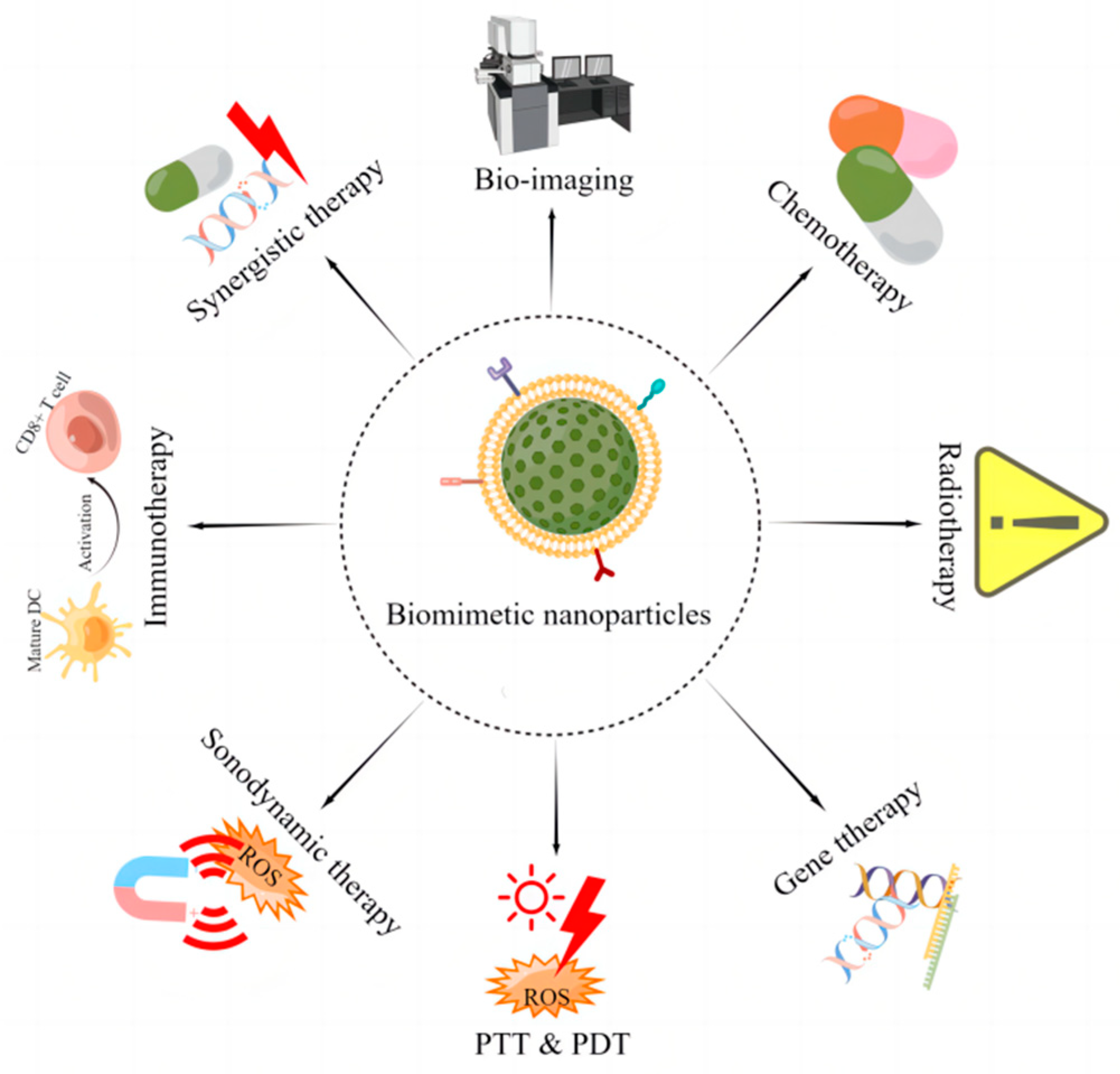
3. Application of Biomimetic Cell-Derived Nanoparticles in Cancer Immunotherapy
3.1. Immune Checkpoint Inhibitors
3.2. Adoptive Cellular Immunotherapy
3.3. Cancer Vaccines
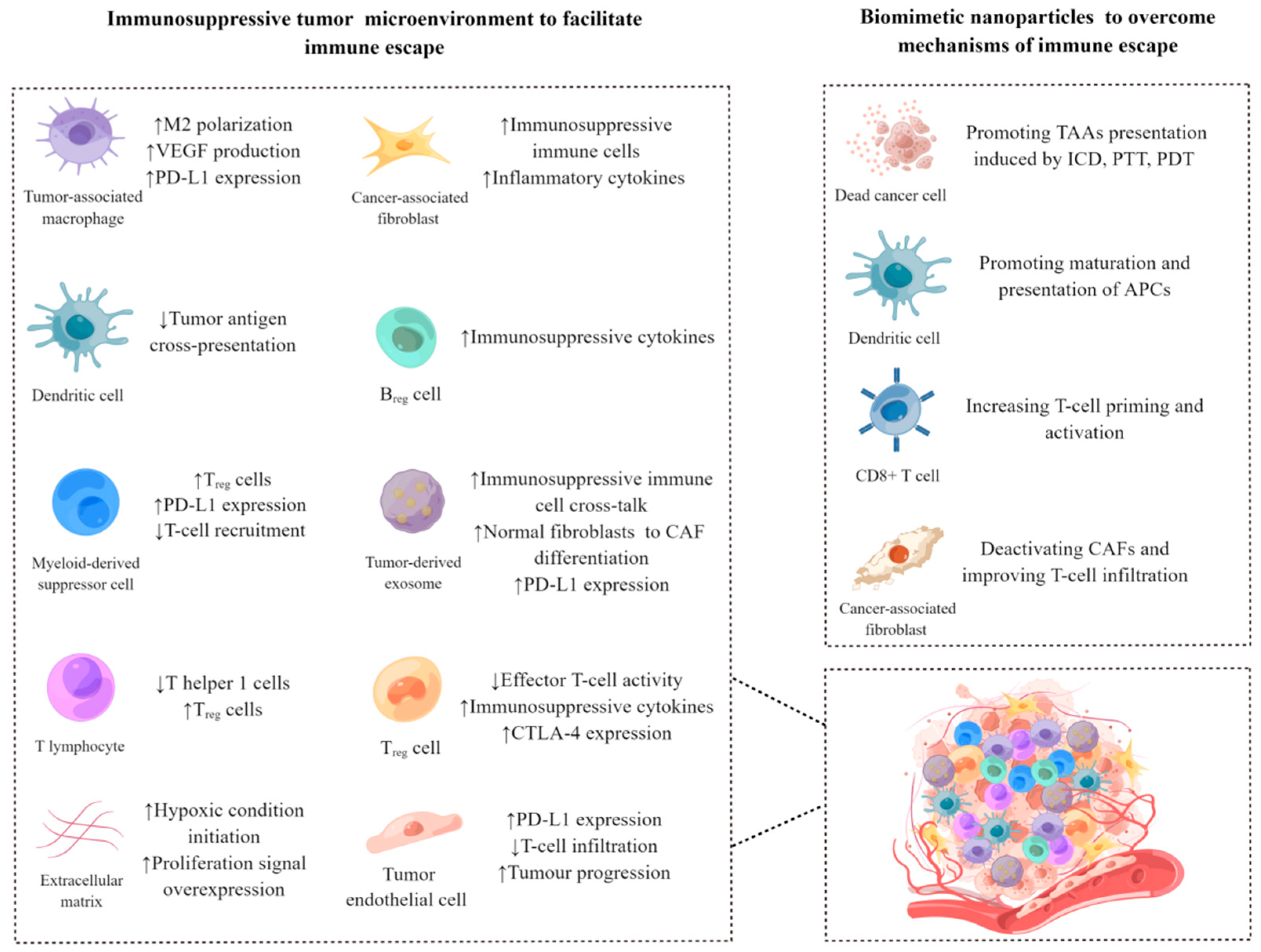
3.4. Modulating the Immunosuppressive Tumor Microenvironment
3.5. Combination Therapy
4. Conclusions and Challenges
Author Contributions
Funding
Institutional Review Board Statement
Informed Consent Statement
Data Availability Statement
Acknowledgments
Conflicts of Interest
References
- Yang, Y. Cancer immunotherapy: Harnessing the immune system to battle cancer. J. Clin. Investig. 2015, 125, 3335–3337. [Google Scholar] [CrossRef] [Green Version]
- Zeng, Y.; Li, S.; Zhang, S.; Wang, L.; Yuan, H.; Hu, F. Cell membrane coated-nanoparticles for cancer immunotherapy. Acta Pharm. Sin. B 2022, 12, 3233–3254. [Google Scholar] [CrossRef]
- Peterson, C.; Denlinger, N.; Yang, Y. Recent Advances and Challenges in Cancer Immunotherapy. Cancers 2022, 14, 3972. [Google Scholar] [CrossRef]
- Alhaj-Suliman, S.O.; Wafa, E.I.; Salem, A.K. Engineering nanosystems to overcome barriers to cancer diagnosis and treatment. Adv. Drug Deliv. Rev. 2022, 189, 114482. [Google Scholar] [CrossRef]
- Yang, K.; Yang, Z.; Yu, G.; Nie, Z.; Wang, R.; Chen, X. Polyprodrug Nanomedicines: An Emerging Paradigm for Cancer Therapy. Adv. Mater. 2021, 34, e2107434. [Google Scholar] [CrossRef]
- Hegde, M.; Naliyadhara, N.; Unnikrishnan, J.; Alqahtani, M.S.; Abbas, M.; Girisa, S.; Sethi, G.; Kunnumakkara, A.B. Nanoparticles in the diagnosis and treatment of cancer metastases: Current and future perspectives. Cancer Lett. 2023, 556, 216066. [Google Scholar] [CrossRef]
- Shukla, A.; Maiti, P. Nanomedicine and versatile therapies for cancer treatment. MedComm 2022, 3, e163. [Google Scholar] [CrossRef]
- Llop, J.; Lammers, T. Nanoparticles for Cancer Diagnosis, Radionuclide Therapy and Theranostics. ACS Nano 2021, 15, 16974–16981. [Google Scholar] [CrossRef]
- Zhou, Y.; Dai, Z. New Strategies in the Design of Nanomedicines to Oppose Uptake by the Mononuclear Phagocyte System and Enhance Cancer Therapeutic Efficacy. Chem. Asian J. 2018, 13, 3333–3340. [Google Scholar] [CrossRef]
- Ibrahim, M.; Ramadan, E.; Elsadek, N.E.; Emam, S.E.; Shimizu, T.; Ando, H.; Ishima, Y.; Elgarhy, O.H.; Sarhan, H.A.; Hussein, A.K.; et al. Polyethylene glycol (PEG): The nature, immunogenicity, and role in the hypersensitivity of PEGylated products. J. Control. Release 2022, 351, 215–230. [Google Scholar] [CrossRef]
- Shi, D.; Beasock, D.; Fessler, A.; Szebeni, J.; Ljubimova, J.Y.; Afonin, K.A.; Dobrovolskaia, M.A. To PEGylate or not to PEGylate: Immunological properties of nanomedicine’s most popular component, polyethylene glycol and its alternatives. Adv. Drug Deliv. Rev. 2022, 180, 114079. [Google Scholar] [CrossRef]
- Kozma, G.T.; Shimizu, T.; Ishida, T.; Szebeni, J. Anti-PEG antibodies: Properties, formation, testing and role in adverse immune reactions to PEGylated nano-biopharmaceuticals. Adv. Drug Deliv. Rev. 2020, 154–155, 163–175. [Google Scholar] [CrossRef]
- Estape Senti, M.; de Jongh, C.A.; Dijkxhoorn, K.; Verhoef, J.J.F.; Szebeni, J.; Storm, G.; Hack, C.E.; Schiffelers, R.M.; Fens, M.H.; Boross, P. Anti-PEG antibodies compromise the integrity of PEGylated lipid-based nanoparticles via complement. J. Control. Release 2022, 341, 475–486. [Google Scholar] [CrossRef]
- Hussain, Z.; Rahim, M.A.; Jan, N.; Shah, H.; Rawas-Qalaji, M.; Khan, S.; Sohail, M.; Thu, H.E.; Ramli, N.A.; Sarfraz, R.M.; et al. Cell membrane cloaked nanomedicines for bio-imaging and immunotherapy of cancer: Improved pharmacokinetics, cell internalization and anticancer efficacy. J. Control. Release 2021, 335, 130–157. [Google Scholar] [CrossRef]
- Dhas, N.; Garcia, M.C.; Kudarha, R.; Pandey, A.; Nikam, A.N.; Gopalan, D.; Fernandes, G.; Soman, S.; Kulkarni, S.; Seetharam, R.N.; et al. Advancements in cell membrane camouflaged nanoparticles: A bioinspired platform for cancer therapy. J. Control. Release 2022, 346, 71–97. [Google Scholar] [CrossRef]
- Raza, F.; Zafar, H.; Zhang, S.; Kamal, Z.; Su, J.; Yuan, W.E.; Mingfeng, Q. Recent Advances in Cell Membrane-Derived Biomimetic Nanotechnology for Cancer Immunotherapy. Adv. Healthc. Mater. 2021, 10, e2002081. [Google Scholar] [CrossRef]
- Zhang, F.; Mundaca-Uribe, R.; Askarinam, N.; Li, Z.; Gao, W.; Zhang, L.; Wang, J. Biomembrane-Functionalized Micromotors: Biocompatible Active Devices for Diverse Biomedical Applications. Adv. Mater. 2022, 34, e2107177. [Google Scholar] [CrossRef]
- Chen, X.; Liu, B.; Tong, R.; Zhan, L.; Yin, X.; Luo, X.; Huang, Y.; Zhang, J.; He, W.; Wang, Y. Orchestration of biomimetic membrane coating and nanotherapeutics in personalized anticancer therapy. Biomater. Sci. 2021, 9, 590–625. [Google Scholar] [CrossRef]
- Shen, M.; Wu, X.; Zhu, M.; Yi, X. Recent advances in biological membrane-based nanomaterials for cancer therapy. Biomater. Sci. 2022, 10, 5756–5785. [Google Scholar] [CrossRef]
- Jha, A.; Nikam, A.N.; Kulkarni, S.; Mutalik, S.P.; Pandey, A.; Hegde, M.; Rao, B.S.S.; Mutalik, S. Biomimetic nanoarchitecturing: A disguised attack on cancer cells. J. Control. Release 2021, 329, 413–433. [Google Scholar] [CrossRef]
- Zhao, J.; Ruan, J.; Lv, G.; Shan, Q.; Fan, Z.; Wang, H.; Du, Y.; Ling, L. Cell membrane-based biomimetic nanosystems for advanced drug delivery in cancer therapy: A comprehensive review. Colloids Surf. B Biointerfaces 2022, 215, 112503. [Google Scholar] [CrossRef]
- Fang, R.H.; Gao, W.; Zhang, L. Targeting drugs to tumours using cell membrane-coated nanoparticles. Nat. Rev. Clin. Oncol. 2022, 20, 33–48. [Google Scholar] [CrossRef]
- Hu, C.M.; Zhang, L.; Aryal, S.; Cheung, C.; Fang, R.H.; Zhang, L. Erythrocyte membrane-camouflaged polymeric nanoparticles as a biomimetic delivery platform. Proc. Natl. Acad. Sci. USA 2011, 108, 10980–10985. [Google Scholar] [CrossRef] [Green Version]
- Liu, H.; Su, Y.Y.; Jiang, X.C.; Gao, J.Q. Cell membrane-coated nanoparticles: A novel multifunctional biomimetic drug delivery system. Drug Deliv. Transl. Res. 2023, 13, 716–737. [Google Scholar] [CrossRef]
- Chen, L.; Hong, W.; Ren, W.; Xu, T.; Qian, Z.; He, Z. Recent progress in targeted delivery vectors based on biomimetic nanoparticles. Signal Transduct. Target. Ther. 2021, 6, 225. [Google Scholar] [CrossRef]
- Zhang, M.; Cheng, S.; Jin, Y.; Zhang, N.; Wang, Y. Membrane engineering of cell membrane biomimetic nanoparticles for nanoscale therapeutics. Clin. Transl. Med. 2021, 11, e292. [Google Scholar] [CrossRef]
- Krishnan, N.; Peng, F.X.; Mohapatra, A.; Fang, R.H.; Zhang, L. Genetically engineered cellular nanoparticles for biomedical applications. Biomaterials 2023, 296, 122065. [Google Scholar] [CrossRef]
- Meng, Z.; Zhang, Y.; Zhou, X.; Ji, J.; Liu, Z. Nanovaccines with cell-derived components for cancer immunotherapy. Adv. Drug Deliv. Rev. 2022, 182, 114107. [Google Scholar] [CrossRef]
- Shi, Y.; Qian, H.; Rao, P.; Mu, D.; Liu, Y.; Liu, G.; Lin, Z. Bioinspired membrane-based nanomodulators for immunotherapy of autoimmune and infectious diseases. Acta Pharm. Sin. B 2022, 12, 1126–1147. [Google Scholar] [CrossRef]
- Wu, Z.; Zhang, H.; Yan, J.; Wei, Y.; Su, J. Engineered biomembrane-derived nanoparticles for nanoscale theranostics. Theranostics 2023, 13, 20–39. [Google Scholar] [CrossRef]
- Shi, Y.; Lu, A.; Wang, X.; Belhadj, Z.; Wang, J.; Zhang, Q. A review of existing strategies for designing long-acting parenteral formulations: Focus on underlying mechanisms, and future perspectives. Acta Pharm. Sin. B 2021, 11, 2396–2415. [Google Scholar] [CrossRef]
- Castro, F.; Martins, C.; Silveira, M.J.; Moura, R.P.; Pereira, C.L.; Sarmento, B. Advances on erythrocyte-mimicking nanovehicles to overcome barriers in biological microenvironments. Adv. Drug Deliv. Rev. 2021, 170, 312–339. [Google Scholar] [CrossRef]
- Xia, Q.; Zhang, Y.; Li, Z.; Hou, X.; Feng, N. Red blood cell membrane-camouflaged nanoparticles: A novel drug delivery system for antitumor application. Acta Pharm. Sin. B 2019, 9, 675–689. [Google Scholar] [CrossRef]
- Bidkar, A.P.; Sanpui, P.; Ghosh, S.S. Transferrin-Conjugated Red Blood Cell Membrane-Coated Poly(lactic-co-glycolic acid) Nanoparticles for the Delivery of Doxorubicin and Methylene Blue. ACS Appl. Nano Mater. 2020, 3, 3807–3819. [Google Scholar] [CrossRef]
- Choi, M.J.; Lee, Y.K.; Choi, K.C.; Lee, D.H.; Jeong, H.Y.; Kang, S.J.; Kim, M.W.; You, Y.M.; Im, C.S.; Lee, T.S.; et al. Tumor-Targeted Erythrocyte Membrane Nanoparticles for Theranostics of Triple-Negative Breast Cancer. Pharmaceutics 2023, 15, 350. [Google Scholar] [CrossRef]
- Long, Y.; Wang, Z.; Fan, J.; Yuan, L.; Tong, C.; Zhao, Y.; Liu, B. A hybrid membrane coating nanodrug system against gastric cancer via the VEGFR2/STAT3 signaling pathway. J. Mater. Chem. B 2021, 9, 3838–3855. [Google Scholar] [CrossRef]
- Huo, Y.Y.; Song, X.; Zhang, W.X.; Zhou, Z.L.; Lv, Q.Y.; Cui, H.F. Thermosensitive Biomimetic Hybrid Membrane Camouflaged Hollow Gold Nanoparticles for NIR-Responsive Mild-Hyperthermia Chemo-/Photothermal Combined Tumor Therapy. ACS Appl. Bio Mater. 2022, 5, 5113–5125. [Google Scholar] [CrossRef]
- Coussens, L.M.; Werb, Z. Inflammation and cancer. Nature 2002, 420, 860–867. [Google Scholar] [CrossRef]
- Li, R.; He, Y.; Zhang, S.; Qin, J.; Wang, J. Cell membrane-based nanoparticles: A new biomimetic platform for tumor diagnosis and treatment. Acta Pharm. Sin. B 2018, 8, 14–22. [Google Scholar] [CrossRef]
- Oroojalian, F.; Beygi, M.; Baradaran, B.; Mokhtarzadeh, A.; Shahbazi, M.A. Immune Cell Membrane-Coated Biomimetic Nanoparticles for Targeted Cancer Therapy. Small 2021, 17, e2006484. [Google Scholar] [CrossRef]
- Cheng, S.; Xu, C.; Jin, Y.; Li, Y.; Zhong, C.; Ma, J.; Yang, J.; Zhang, N.; Li, Y.; Wang, C.; et al. Artificial Mini Dendritic Cells Boost T Cell-Based Immunotherapy for Ovarian Cancer. Adv. Sci. 2020, 7, 1903301. [Google Scholar] [CrossRef]
- Xu, C.; Jiang, Y.; Han, Y.; Pu, K.; Zhang, R. A Polymer Multicellular Nanoengager for Synergistic NIR-II Photothermal Immunotherapy. Adv. Mater. 2021, 33, e2008061. [Google Scholar] [CrossRef]
- Gong, P.; Wang, Y.; Zhang, P.; Yang, Z.; Deng, W.; Sun, Z.; Yang, M.; Li, X.; Ma, G.; Deng, G.; et al. Immunocyte Membrane-Coated Nanoparticles for Cancer Immunotherapy. Cancers 2020, 13, 77. [Google Scholar] [CrossRef]
- Khatoon, N.; Zhang, Z.; Zhou, C.; Chu, M. Macrophage membrane coated nanoparticles: A biomimetic approach for enhanced and targeted delivery. Biomater. Sci. 2022, 10, 1193–1208. [Google Scholar] [CrossRef]
- Gaber, T.; Chen, Y.; Krauss, P.L.; Buttgereit, F. Metabolism of T Lymphocytes in Health and Disease. Int. Rev. Cell Mol. Biol. 2019, 342, 95–148. [Google Scholar] [CrossRef]
- Notarbartolo, S.; Abrignani, S. Human T lymphocytes at tumor sites. Semin. Immunopathol. 2022, 44, 883–901. [Google Scholar] [CrossRef]
- Farhood, B.; Najafi, M.; Mortezaee, K. CD8(+) cytotoxic T lymphocytes in cancer immunotherapy: A review. J. Cell Physiol. 2019, 234, 8509–8521. [Google Scholar] [CrossRef]
- Yaman, S.; Ramachandramoorthy, H.; Oter, G.; Zhukova, D.; Nguyen, T.; Sabnani, M.K.; Weidanz, J.A.; Nguyen, K.T. Melanoma Peptide MHC Specific TCR Expressing T-Cell Membrane Camouflaged PLGA Nanoparticles for Treatment of Melanoma Skin Cancer. Front. Bioeng. Biotechnol. 2020, 8, 943. [Google Scholar] [CrossRef]
- Raskov, H.; Orhan, A.; Christensen, J.P.; Gogenur, I. Cytotoxic CD8(+) T cells in cancer and cancer immunotherapy. Br. J. Cancer 2021, 124, 359–367. [Google Scholar] [CrossRef]
- Chen, B.Q.; Zhao, Y.; Zhang, Y.; Pan, Y.J.; Xia, H.Y.; Kankala, R.K.; Wang, S.B.; Liu, G.; Chen, A.Z. Immune-regulating camouflaged nanoplatforms: A promising strategy to improve cancer nano-immunotherapy. Bioact. Mater. 2023, 21, 1–19. [Google Scholar] [CrossRef]
- Han, B.; Mao, F.Y.; Zhao, Y.L.; Lv, Y.P.; Teng, Y.S.; Duan, M.; Chen, W.; Cheng, P.; Wang, T.T.; Liang, Z.Y.; et al. Altered NKp30, NKp46, NKG2D, and DNAM-1 Expression on Circulating NK Cells Is Associated with Tumor Progression in Human Gastric Cancer. J. Immunol. Res. 2018, 2018, 6248590. [Google Scholar] [CrossRef] [Green Version]
- Jain, N.; Shahrukh, S.; Famta, P.; Shah, S.; Vambhurkar, G.; Khatri, D.K.; Singh, S.B.; Srivastava, S. Immune cell-camouflaged surface-engineered nanotherapeutics for cancer management. Acta Biomater. 2023, 155, 57–79. [Google Scholar] [CrossRef]
- Han, H.; Bartolo, R.; Li, J.; Shahbazi, M.A.; Santos, H.A. Biomimetic platelet membrane-coated nanoparticles for targeted therapy. Eur. J. Pharm. Biopharm. 2022, 172, 1–15. [Google Scholar] [CrossRef]
- Hu, C.M.; Fang, R.H.; Wang, K.C.; Luk, B.T.; Thamphiwatana, S.; Dehaini, D.; Nguyen, P.; Angsantikul, P.; Wen, C.H.; Kroll, A.V.; et al. Nanoparticle biointerfacing by platelet membrane cloaking. Nature 2015, 526, 118–121. [Google Scholar] [CrossRef] [Green Version]
- Hu, Q.; Sun, W.; Qian, C.; Wang, C.; Bomba, H.N.; Gu, Z. Anticancer Platelet-Mimicking Nanovehicles. Adv. Mater. 2015, 27, 7043–7050. [Google Scholar] [CrossRef]
- Fabricius, H.A.; Starzonek, S.; Lange, T. The Role of Platelet Cell Surface P-Selectin for the Direct Platelet-Tumor Cell Contact During Metastasis Formation in Human Tumors. Front. Oncol. 2021, 11, 642761. [Google Scholar] [CrossRef]
- Chen, Z.; Zhao, P.; Luo, Z.; Zheng, M.; Tian, H.; Gong, P.; Gao, G.; Pan, H.; Liu, L.; Ma, A.; et al. Cancer Cell Membrane-Biomimetic Nanoparticles for Homologous-Targeting Dual-Modal Imaging and Photothermal Therapy. ACS Nano 2016, 10, 10049–10057. [Google Scholar] [CrossRef]
- Harris, J.C.; Scully, M.A.; Day, E.S. Cancer Cell Membrane-Coated Nanoparticles for Cancer Management. Cancers 2019, 11, 1836. [Google Scholar] [CrossRef] [Green Version]
- Tian, H.; Luo, Z.; Liu, L.; Zheng, M.; Chen, Z.; Ma, A.; Liang, R.; Han, Z.; Lu, C.; Cai, L. Cancer Cell Membrane-Biomimetic Oxygen Nanocarrier for Breaking Hypoxia-Induced Chemoresistance. Adv. Funct. Mater. 2017, 27, 1703197. [Google Scholar] [CrossRef]
- Zhu, J.Y.; Zheng, D.W.; Zhang, M.K.; Yu, W.Y.; Qiu, W.X.; Hu, J.J.; Feng, J.; Zhang, X.Z. Preferential Cancer Cell Self-Recognition and Tumor Self-Targeting by Coating Nanoparticles with Homotypic Cancer Cell Membranes. Nano Lett. 2016, 16, 5895–5901. [Google Scholar] [CrossRef]
- Sun, H.; Su, J.; Meng, Q.; Yin, Q.; Chen, L.; Gu, W.; Zhang, P.; Zhang, Z.; Yu, H.; Wang, S.; et al. Cancer-Cell-Biomimetic Nanoparticles for Targeted Therapy of Homotypic Tumors. Adv. Mater. 2016, 28, 9581–9588. [Google Scholar] [CrossRef]
- Pei, X.; Pan, X.; Xu, X.; Xu, X.; Huang, H.; Wu, Z.; Qi, X. 4T1 cell membrane fragment reunited PAMAM polymer units disguised as tumor cell clusters for tumor homotypic targeting and anti-metastasis treatment. Biomater. Sci. 2021, 9, 1325–1333. [Google Scholar] [CrossRef]
- Yue, X.S.; Murakami, Y.; Tamai, T.; Nagaoka, M.; Cho, C.S.; Ito, Y.; Akaike, T. A fusion protein N-cadherin-Fc as an artificial extracellular matrix surface for maintenance of stem cell features. Biomaterials 2010, 31, 5287–5296. [Google Scholar] [CrossRef]
- Iurisci, I.; Cumashi, A.; Sherman, A.A.; Tsvetkov, Y.E.; Tinari, N.; Piccolo, E.; D’Egidio, M.; Adamo, V.; Natoli, C.; Rabinovich, G.A.; et al. Synthetic inhibitors of galectin-1 and -3 selectively modulate homotypic cell aggregation and tumor cell apoptosis. Anticancer Res. 2009, 29, 403–410. [Google Scholar]
- Osta, W.A.; Chen, Y.; Mikhitarian, K.; Mitas, M.; Salem, M.; Hannun, Y.A.; Cole, D.J.; Gillanders, W.E. EpCAM is overexpressed in breast cancer and is a potential target for breast cancer gene therapy. Cancer Res. 2004, 64, 5818–5824. [Google Scholar] [CrossRef] [Green Version]
- Wu, H.H.; Zhou, Y.; Tabata, Y.; Gao, J.Q. Mesenchymal stem cell-based drug delivery strategy: From cells to biomimetic. J. Control. Release 2019, 294, 102–113. [Google Scholar] [CrossRef]
- Khosravi, N.; Pishavar, E.; Baradaran, B.; Oroojalian, F.; Mokhtarzadeh, A. Stem cell membrane, stem cell-derived exosomes and hybrid stem cell camouflaged nanoparticles: A promising biomimetic nanoplatforms for cancer theranostics. J. Control. Release 2022, 348, 706–722. [Google Scholar] [CrossRef]
- Wang, M.; Xin, Y.; Cao, H.; Li, W.; Hua, Y.; Webster, T.J.; Zhang, C.; Tang, W.; Liu, Z. Recent advances in mesenchymal stem cell membrane-coated nanoparticles for enhanced drug delivery. Biomater. Sci. 2021, 9, 1088–1103. [Google Scholar] [CrossRef]
- Takayama, Y.; Kusamori, K.; Nishikawa, M. Mesenchymal stem/stromal cells as next-generation drug delivery vehicles for cancer therapeutics. Expert Opin. Drug Deliv. 2021, 18, 1627–1642. [Google Scholar] [CrossRef]
- Starnes, C.O. Coley’s toxins. Nature 1992, 360, 23. [Google Scholar] [CrossRef]
- Sartorio, M.G.; Pardue, E.J.; Feldman, M.F.; Haurat, M.F. Bacterial Outer Membrane Vesicles: From Discovery to Applications. Annu. Rev. Microbiol. 2021, 75, 609–630. [Google Scholar] [CrossRef]
- Liu, J.; Liew, S.S.; Wang, J.; Pu, K. Bioinspired and Biomimetic Delivery Platforms for Cancer Vaccines. Adv. Mater. 2022, 34, e2103790. [Google Scholar] [CrossRef]
- Li, M.; Zhou, H.; Yang, C.; Wu, Y.; Zhou, X.; Liu, H.; Wang, Y. Bacterial outer membrane vesicles as a platform for biomedical applications: An update. J. Control. Release 2020, 323, 253–268. [Google Scholar] [CrossRef]
- Holay, M.; Guo, Z.; Pihl, J.; Heo, J.; Park, J.H.; Fang, R.H.; Zhang, L. Bacteria-Inspired Nanomedicine. ACS Appl. Bio Mater. 2021, 4, 3830–3848. [Google Scholar] [CrossRef]
- Krishnan, N.; Kubiatowicz, L.J.; Holay, M.; Zhou, J.; Fang, R.H.; Zhang, L. Bacterial membrane vesicles for vaccine applications. Adv. Drug Deliv. Rev. 2022, 185, 114294. [Google Scholar] [CrossRef]
- Long, Q.; Zheng, P.; Zheng, X.; Li, W.; Hua, L.; Yang, Z.; Huang, W.; Ma, Y. Engineered bacterial membrane vesicles are promising carriers for vaccine design and tumor immunotherapy. Adv. Drug Deliv. Rev. 2022, 186, 114321. [Google Scholar] [CrossRef]
- Herrmann, I.K.; Wood, M.J.A.; Fuhrmann, G. Extracellular vesicles as a next-generation drug delivery platform. Nat. Nanotechnol. 2021, 16, 748–759. [Google Scholar] [CrossRef]
- Najafi, S.; Majidpoor, J.; Mortezaee, K. Extracellular vesicle-based drug delivery in cancer immunotherapy. Drug Deliv. Transl. Res. 2023, 1–17. [Google Scholar] [CrossRef]
- Wu, P.; Zhang, B.; Ocansey, D.K.W.; Xu, W.; Qian, H. Extracellular vesicles: A bright star of nanomedicine. Biomaterials 2021, 269, 120467. [Google Scholar] [CrossRef]
- Liu, S.; Wu, X.; Chandra, S.; Lyon, C.; Ning, B.; Jiang, L.; Fan, J.; Hu, T.Y. Extracellular vesicles: Emerging tools as therapeutic agent carriers. Acta Pharm. Sin. B 2022, 12, 3822–3842. [Google Scholar] [CrossRef]
- Liu, C.; Wang, Y.; Li, L.; He, D.; Chi, J.; Li, Q.; Wu, Y.; Zhao, Y.; Zhang, S.; Wang, L.; et al. Engineered extracellular vesicles and their mimetics for cancer immunotherapy. J. Control. Release 2022, 349, 679–698. [Google Scholar] [CrossRef]
- Ruan, S.; Greenberg, Z.; Pan, X.; Zhuang, P.; Erwin, N.; He, M. Extracellular Vesicles as an Advanced Delivery Biomaterial for Precision Cancer Immunotherapy. Adv. Healthc. Mater. 2022, 11, e2100650. [Google Scholar] [CrossRef]
- Chen, H.Y.; Deng, J.; Wang, Y.; Wu, C.Q.; Li, X.; Dai, H.W. Hybrid cell membrane-coated nanoparticles: A multifunctional biomimetic platform for cancer diagnosis and therapy. Acta Biomater. 2020, 112, 1–13. [Google Scholar] [CrossRef]
- Wang, D.; Liu, C.; You, S.; Zhang, K.; Li, M.; Cao, Y.; Wang, C.; Dong, H.; Zhang, X. Bacterial Vesicle-Cancer Cell Hybrid Membrane-Coated Nanoparticles for Tumor Specific Immune Activation and Photothermal Therapy. ACS Appl. Mater. Interfaces 2020, 12, 41138–41147. [Google Scholar] [CrossRef]
- Liu, Y.; Sukumar, U.K.; Kanada, M.; Krishnan, A.; Massoud, T.F.; Paulmurugan, R. Camouflaged Hybrid Cancer Cell-Platelet Fusion Membrane Nanovesicles Deliver Therapeutic MicroRNAs to Presensitize Triple-Negative Breast Cancer to Doxorubicin. Adv. Funct. Mater. 2021, 31, 2103600. [Google Scholar] [CrossRef]
- Zhang, T.; Liu, H.; Li, L.; Guo, Z.; Song, J.; Yang, X.; Wan, G.; Li, R.; Wang, Y. Leukocyte/platelet hybrid membrane-camouflaged dendritic large pore mesoporous silica nanoparticles co-loaded with photo/chemotherapeutic agents for triple negative breast cancer combination treatment. Bioact. Mater. 2021, 6, 3865–3878. [Google Scholar] [CrossRef]
- Liu, W.L.; Zou, M.Z.; Liu, T.; Zeng, J.Y.; Li, X.; Yu, W.Y.; Li, C.X.; Ye, J.J.; Song, W.; Feng, J.; et al. Cytomembrane nanovaccines show therapeutic effects by mimicking tumor cells and antigen presenting cells. Nat. Commun. 2019, 10, 3199. [Google Scholar] [CrossRef] [Green Version]
- Gao, W.; Fang, R.H.; Thamphiwatana, S.; Luk, B.T.; Li, J.; Angsantikul, P.; Zhang, Q.; Hu, C.M.; Zhang, L. Modulating antibacterial immunity via bacterial membrane-coated nanoparticles. Nano Lett. 2015, 15, 1403–1409. [Google Scholar] [CrossRef] [Green Version]
- Patel, R.B.; Ye, M.; Carlson, P.M.; Jaquish, A.; Zangl, L.; Ma, B.; Wang, Y.; Arthur, I.; Xie, R.; Brown, R.J.; et al. Development of an In Situ Cancer Vaccine via Combinational Radiation and Bacterial-Membrane-Coated Nanoparticles. Adv. Mater. 2019, 31, e1902626. [Google Scholar] [CrossRef]
- Dash, P.; Piras, A.M.; Dash, M. Cell membrane coated nanocarriers—An efficient biomimetic platform for targeted therapy. J. Control. Release 2020, 327, 546–570. [Google Scholar] [CrossRef]
- Le, Q.V.; Lee, J.; Lee, H.; Shim, G.; Oh, Y.K. Cell membrane-derived vesicles for delivery of therapeutic agents. Acta Pharm. Sin. B 2021, 11, 2096–2113. [Google Scholar] [CrossRef]
- Luk, B.T.; Hu, C.M.; Fang, R.H.; Dehaini, D.; Carpenter, C.; Gao, W.; Zhang, L. Interfacial interactions between natural RBC membranes and synthetic polymeric nanoparticles. Nanoscale 2014, 6, 2730–2737. [Google Scholar] [CrossRef] [Green Version]
- Xu, C.H.; Ye, P.J.; Zhou, Y.C.; He, D.X.; Wei, H.; Yu, C.Y. Cell membrane-camouflaged nanoparticles as drug carriers for cancer therapy. Acta Biomater. 2020, 105, 1–14. [Google Scholar] [CrossRef]
- Rao, L.; Cai, B.; Bu, L.L.; Liao, Q.Q.; Guo, S.S.; Zhao, X.Z.; Dong, W.F.; Liu, W. Microfluidic Electroporation-Facilitated Synthesis of Erythrocyte Membrane-Coated Magnetic Nanoparticles for Enhanced Imaging-Guided Cancer Therapy. ACS Nano 2017, 11, 3496–3505. [Google Scholar] [CrossRef]
- Zhang, J.; Gao, W.; Fang, R.H.; Dong, A.; Zhang, L. Synthesis of Nanogels via Cell Membrane-Templated Polymerization. Small 2015, 11, 4309–4313. [Google Scholar] [CrossRef] [Green Version]
- Lee, J.B.; Kim, H.R.; Ha, S.J. Immune Checkpoint Inhibitors in 10 Years: Contribution of Basic Research and Clinical Application in Cancer Immunotherapy. Immune. Netw. 2022, 22, e2. [Google Scholar] [CrossRef]
- Pisibon, C.; Ouertani, A.; Bertolotto, C.; Ballotti, R.; Cheli, Y. Immune Checkpoints in Cancers: From Signaling to the Clinic. Cancers 2021, 13, 4573. [Google Scholar] [CrossRef]
- Akbay, E.A.; Koyama, S.; Carretero, J.; Altabef, A.; Tchaicha, J.H.; Christensen, C.L.; Mikse, O.R.; Cherniack, A.D.; Beauchamp, E.M.; Pugh, T.J.; et al. Activation of the PD-1 pathway contributes to immune escape in EGFR-driven lung tumors. Cancer Discov. 2013, 3, 1355–1363. [Google Scholar] [CrossRef] [Green Version]
- Daassi, D.; Mahoney, K.M.; Freeman, G.J. The importance of exosomal PDL1 in tumour immune evasion. Nat. Rev. Immunol. 2020, 20, 209–215. [Google Scholar] [CrossRef]
- Chen, G.; Huang, A.C.; Zhang, W.; Zhang, G.; Wu, M.; Xu, W.; Yu, Z.; Yang, J.; Wang, B.; Sun, H.; et al. Exosomal PD-L1 contributes to immunosuppression and is associated with anti-PD-1 response. Nature 2018, 560, 382–386. [Google Scholar] [CrossRef]
- Ye, L.; Zhu, Z.; Chen, X.; Zhang, H.; Huang, J.; Gu, S.; Zhao, X. The Importance of Exosomal PD-L1 in Cancer Progression and Its Potential as a Therapeutic Target. Cells 2021, 10, 3247. [Google Scholar] [CrossRef]
- Yan, J.; Liu, X.; Wu, F.; Ge, C.; Ye, H.; Chen, X.; Wei, Y.; Zhou, R.; Duan, S.; Zhu, R.; et al. Platelet Pharmacytes for the Hierarchical Amplification of Antitumor Immunity in Response to Self-Generated Immune Signals. Adv. Mater. 2022, 34, e2109517. [Google Scholar] [CrossRef]
- Yan, W.; Jiang, S. Immune Cell-Derived Exosomes in the Cancer-Immunity Cycle. Trends Cancer 2020, 6, 506–517. [Google Scholar] [CrossRef]
- Marar, C.; Starich, B.; Wirtz, D. Extracellular vesicles in immunomodulation and tumor progression. Nat. Immunol. 2021, 22, 560–570. [Google Scholar] [CrossRef]
- Kugeratski, F.G.; Kalluri, R. Exosomes as mediators of immune regulation and immunotherapy in cancer. FEBS J. 2021, 288, 10–35. [Google Scholar] [CrossRef]
- Mittal, S.; Gupta, P.; Chaluvally-Raghavan, P.; Pradeep, S. Emerging Role of Extracellular Vesicles in Immune Regulation and Cancer Progression. Cancers 2020, 12, 3563. [Google Scholar] [CrossRef]
- Jung, D.; Shin, S.; Kang, S.M.; Jung, I.; Ryu, S.; Noh, S.; Choi, S.J.; Jeong, J.; Lee, B.Y.; Kim, K.S.; et al. Reprogramming of T cell-derived small extracellular vesicles using IL2 surface engineering induces potent anti-cancer effects through miRNA delivery. J. Extracell. Vesicles 2022, 11, e12287. [Google Scholar] [CrossRef]
- Masjedi, A.; Hassannia, H.; Atyabi, F.; Rastegari, A.; Hojjat-Farsangi, M.; Namdar, A.; Soleimanpour, H.; Azizi, G.; Nikkhoo, A.; Ghalamfarsa, G.; et al. Downregulation of A2AR by siRNA loaded PEG-chitosan-lactate nanoparticles restores the T cell mediated anti-tumor responses through blockage of PKA/CREB signaling pathway. Int. J. Biol. Macromol. 2019, 133, 436–445. [Google Scholar] [CrossRef]
- Pezzuto, A.; Carico, E. Role of HIF-1 in Cancer Progression: Novel Insights. A Review. Curr. Mol. Med. 2018, 18, 343–351. [Google Scholar] [CrossRef]
- Yuan, C.S.; Teng, Z.; Yang, S.; He, Z.; Meng, L.Y.; Chen, X.G.; Liu, Y. Reshaping hypoxia and silencing CD73 via biomimetic gelatin nanotherapeutics to boost immunotherapy. J. Control. Release 2022, 351, 255–271. [Google Scholar] [CrossRef]
- Li, Z.; Cai, H.; Li, Z.; Ren, L.; Ma, X.; Zhu, H.; Gong, Q.; Zhang, H.; Gu, Z.; Luo, K. A tumor cell membrane-coated self-amplified nanosystem as a nanovaccine to boost the therapeutic effect of anti-PD-L1 antibody. Bioact. Mater. 2023, 21, 299–312. [Google Scholar] [CrossRef]
- Tan, Y.N.; Huang, J.D.; Li, Y.P.; Li, S.S.; Luo, M.; Luo, J.; Lee, A.W.; Fu, L.; Hu, F.Q.; Guan, X.Y. Near-Infrared Responsive Membrane Nanovesicles Amplify Homologous Targeting Delivery of Anti-PD Immunotherapy against Metastatic Tumors. Adv. Healthc. Mater. 2022, 11, e2101496. [Google Scholar] [CrossRef]
- Li, Y.; Zhao, R.; Cheng, K.; Zhang, K.; Wang, Y.; Zhang, Y.; Li, Y.; Liu, G.; Xu, J.; Xu, J.; et al. Bacterial Outer Membrane Vesicles Presenting Programmed Death 1 for Improved Cancer Immunotherapy via Immune Activation and Checkpoint Inhibition. ACS Nano 2020, 14, 16698–16711. [Google Scholar] [CrossRef]
- Bai, S.; Lu, Z.; Jiang, Y.; Shi, X.; Xu, D.; Shi, Y.; Lin, G.; Liu, C.; Zhang, Y.; Liu, G. Nanotransferrin-Based Programmable Catalysis Mediates Three-Pronged Induction of Oxidative Stress to Enhance Cancer Immunotherapy. ACS Nano 2022, 16, 997–1012. [Google Scholar] [CrossRef]
- Li, J.; Tang, W.; Yang, Y.; Shen, Q.; Yu, Y.; Wang, X.; Fu, Y.; Li, C. A Programmed Cell-Mimicking Nanoparticle Driven by Potato Alkaloid for Targeted Cancer Chemoimmunotherapy. Adv. Healthc. Mater. 2021, 10, e2100311. [Google Scholar] [CrossRef]
- Shao, D.; Zhang, F.; Chen, F.; Zheng, X.; Hu, H.; Yang, C.; Tu, Z.; Wang, Z.; Chang, Z.; Lu, J.; et al. Biomimetic Diselenide-Bridged Mesoporous Organosilica Nanoparticles as an X-ray-Responsive Biodegradable Carrier for Chemo-Immunotherapy. Adv. Mater. 2020, 32, e2004385. [Google Scholar] [CrossRef]
- Guo, Y.; Fan, Y.; Wang, Z.; Li, G.; Zhan, M.; Gong, J.; Majoral, J.P.; Shi, X.; Shen, M. Chemotherapy Mediated by Biomimetic Polymeric Nanoparticles Potentiates Enhanced Tumor Immunotherapy via Amplification of Endoplasmic Reticulum Stress and Mitochondrial Dysfunction. Adv. Mater. 2022, 34, e2206861. [Google Scholar] [CrossRef]
- Tang, Q.; Sun, S.; Wang, P.; Sun, L.; Wang, Y.; Zhang, L.; Xu, M.; Chen, J.; Wu, R.; Zhang, J.; et al. Genetically Engineering Cell Membrane-Coated BTO Nanoparticles for MMP2-Activated Piezocatalysis-Immunotherapy. Adv. Mater. 2023, 35, e2300964. [Google Scholar] [CrossRef]
- Sun, M.; Shi, W.; Wu, Y.; He, Z.; Sun, J.; Cai, S.; Luo, Q. Immunogenic Nanovesicle-Tandem-Augmented Chemoimmunotherapy via Efficient Cancer-Homing Delivery and Optimized Ordinal-Interval Regime. Adv. Sci. 2022, 10, e2205247. [Google Scholar] [CrossRef]
- Chen, X.; Ling, X.; Xia, J.; Zhu, Y.; Zhang, L.; He, Y.; Wang, A.; Gu, G.; Yin, B.; Wang, J. Mature dendritic cell-derived dendrosomes swallow oxaliplatin-loaded nanoparticles to boost immunogenic chemotherapy and tumor antigen-specific immunotherapy. Bioact. Mater. 2022, 15, 15–28. [Google Scholar] [CrossRef]
- Jiang, Q.; Wang, K.; Zhang, X.; Ouyang, B.; Liu, H.; Pang, Z.; Yang, W. Platelet Membrane-Camouflaged Magnetic Nanoparticles for Ferroptosis-Enhanced Cancer Immunotherapy. Small 2020, 16, e2001704. [Google Scholar] [CrossRef]
- Zheng, X.; Yan, J.; You, W.; Li, F.; Diao, J.; He, W.; Yao, Y. De Novo Nano-Erythrocyte Structurally Braced by Biomimetic Au(I)-peptide Skeleton for MDM2/MDMX Predation toward Augmented Pulmonary Adenocarcinoma Immunotherapy. Small 2021, 17, e2100394. [Google Scholar] [CrossRef]
- Li, L.; Miao, Q.; Meng, F.; Li, B.; Xue, T.; Fang, T.; Zhang, Z.; Zhang, J.; Ye, X.; Kang, Y.; et al. Genetic engineering cellular vesicles expressing CD64 as checkpoint antibody carrier for cancer immunotherapy. Theranostics 2021, 11, 6033–6043. [Google Scholar] [CrossRef]
- DeFrancesco, L. CAR-T cell therapy seeks strategies to harness cytokine storm. Nat. Biotechnol. 2014, 32, 604. [Google Scholar] [CrossRef]
- Biederstadt, A.; Rezvani, K. Engineering the next generation of CAR-NK immunotherapies. Int. J. Hematol. 2021, 114, 554–571. [Google Scholar] [CrossRef]
- Gauthier, J.; Turtle, C.J. Insights into cytokine release syndrome and neurotoxicity after CD19-specific CAR-T cell therapy. Curr. Res. Transl. Med. 2018, 66, 50–52. [Google Scholar] [CrossRef]
- Kang, M.; Hong, J.; Jung, M.; Kwon, S.P.; Song, S.Y.; Kim, H.Y.; Lee, J.R.; Kang, S.; Han, J.; Koo, J.H.; et al. T-Cell-Mimicking Nanoparticles for Cancer Immunotherapy. Adv. Mater. 2020, 32, e2003368. [Google Scholar] [CrossRef]
- Hong, J.; Kang, M.; Jung, M.; Lee, Y.Y.; Cho, Y.; Kim, C.; Song, S.Y.; Park, C.G.; Doh, J.; Kim, B.S. T-Cell-Derived Nanovesicles for Cancer Immunotherapy. Adv. Mater. 2021, 33, e2101110. [Google Scholar] [CrossRef]
- Spits, H.; Bernink, J.H.; Lanier, L. NK cells and type 1 innate lymphoid cells: Partners in host defense. Nat. Immunol. 2016, 17, 758–764. [Google Scholar] [CrossRef]
- Raza, A.; Rossi, G.R.; Janjua, T.I.; Souza-Fonseca-Guimaraes, F.; Popat, A. Nanobiomaterials to modulate natural killer cell responses for effective cancer immunotherapy. Trends Biotechnol. 2023, 41, 77–92. [Google Scholar] [CrossRef]
- Wu, D.; Shou, X.; Zhang, Y.; Li, Z.; Wu, G.; Wu, D.; Wu, J.; Shi, S.; Wang, S. Cell membrane-encapsulated magnetic nanoparticles for enhancing natural killer cell-mediated cancer immunotherapy. Nanomedicine 2021, 32, 102333. [Google Scholar] [CrossRef]
- Gong, L.; Li, Y.; Cui, K.; Chen, Y.; Hong, H.; Li, J.; Li, D.; Yin, Y.; Wu, Z.; Huang, Z. Nanobody-Engineered Natural Killer Cell Conjugates for Solid Tumor Adoptive Immunotherapy. Small 2021, 17, e2103463. [Google Scholar] [CrossRef]
- Saxena, M.; Balan, S.; Roudko, V.; Bhardwaj, N. Towards superior dendritic-cell vaccines for cancer therapy. Nat. Biomed. Eng. 2018, 2, 341–346. [Google Scholar] [CrossRef]
- Fucikova, J.; Palova-Jelinkova, L.; Bartunkova, J.; Spisek, R. Induction of Tolerance and Immunity by Dendritic Cells: Mechanisms and Clinical Applications. Front. Immunol. 2019, 10, 2393. [Google Scholar] [CrossRef] [Green Version]
- Sun, Z.; Deng, G.; Peng, X.; Xu, X.; Liu, L.; Peng, J.; Ma, Y.; Zhang, P.; Wen, A.; Wang, Y.; et al. Intelligent photothermal dendritic cells restart the cancer immunity cycle through enhanced immunogenic cell death. Biomaterials 2021, 279, 121228. [Google Scholar] [CrossRef]
- Lu, Y.; Li, L.; Du, J.; Chen, J.; Xu, X.; Yang, X.; Ding, C.; Mao, C. Immunotherapy for Tumor Metastasis by Artificial Antigen-Presenting Cells via Targeted Microenvironment Regulation and T-Cell Activation. ACS Appl. Mater. Interfaces 2021, 13, 55890–55901. [Google Scholar] [CrossRef]
- Zhao, S.; Duan, J.; Lou, Y.; Gao, R.; Yang, S.; Wang, P.; Wang, C.; Han, L.; Li, M.; Ma, C.; et al. Surface specifically modified NK-92 cells with CD56 antibody conjugated superparamagnetic Fe(3)O(4) nanoparticles for magnetic targeting immunotherapy of solid tumors. Nanoscale 2021, 13, 19109–19122. [Google Scholar] [CrossRef]
- Chen, F.; Geng, Z.; Wang, L.; Zhou, Y.; Liu, J. Biomimetic Nanoparticles Enabled by Cascade Cell Membrane Coating for Direct Cross-Priming of T Cells. Small 2022, 18, e2104402. [Google Scholar] [CrossRef]
- Hu, Z.; Ott, P.A.; Wu, C.J. Towards personalized, tumour-specific, therapeutic vaccines for cancer. Nat. Rev. Immunol. 2018, 18, 168–182. [Google Scholar] [CrossRef]
- Banchereau, J.; Palucka, K. Immunotherapy: Cancer vaccines on the move. Nat. Rev. Clin. Oncol. 2018, 15, 9–10. [Google Scholar] [CrossRef]
- Hollingsworth, R.E.; Jansen, K. Turning the corner on therapeutic cancer vaccines. NPJ Vaccines 2019, 4, 7. [Google Scholar] [CrossRef] [Green Version]
- Zhang, R.; Billingsley, M.M.; Mitchell, M.J. Biomaterials for vaccine-based cancer immunotherapy. J. Control. Release 2018, 292, 256–276. [Google Scholar] [CrossRef]
- Viswanath, D.I.; Liu, H.C.; Huston, D.P.; Chua, C.Y.X.; Grattoni, A. Emerging biomaterial-based strategies for personalized therapeutic in situ cancer vaccines. Biomaterials 2022, 280, 121297. [Google Scholar] [CrossRef]
- Wu, Y.; Feng, L. Biomaterials-assisted construction of neoantigen vaccines for personalized cancer immunotherapy. Expert Opin. Drug. Deliv. 2023, 20, 323–333. [Google Scholar] [CrossRef]
- Xiong, X.; Zhao, J.; Pan, J.; Liu, C.; Guo, X.; Zhou, S. Personalized Nanovaccine Coated with Calcinetin-Expressed Cancer Cell Membrane Antigen for Cancer Immunotherapy. Nano Lett. 2021, 21, 8418–8425. [Google Scholar] [CrossRef]
- Li, J.; Huang, D.; Cheng, R.; Figueiredo, P.; Fontana, F.; Correia, A.; Wang, S.; Liu, Z.; Kemell, M.; Torrieri, G.; et al. Multifunctional Biomimetic Nanovaccines Based on Photothermal and Weak-Immunostimulatory Nanoparticulate Cores for the Immunotherapy of Solid Tumors. Adv. Mater. 2022, 34, e2108012. [Google Scholar] [CrossRef]
- Jiang, T.; Shi, T.; Zhang, H.; Hu, J.; Song, Y.; Wei, J.; Ren, S.; Zhou, C. Tumor neoantigens: From basic research to clinical applications. J. Hematol. Oncol. 2019, 12, 93. [Google Scholar] [CrossRef] [Green Version]
- Blass, E.; Ott, P.A. Advances in the development of personalized neoantigen-based therapeutic cancer vaccines. Nat. Rev. Clin. Oncol. 2021, 18, 215–229. [Google Scholar] [CrossRef]
- Xie, N.; Shen, G.; Gao, W.; Huang, Z.; Huang, C.; Fu, L. Neoantigens: Promising targets for cancer therapy. Signal Transduct. Target. Ther. 2023, 8, 9. [Google Scholar] [CrossRef]
- Meng, F.; Li, L.; Zhang, Z.; Lin, Z.; Zhang, J.; Song, X.; Xue, T.; Xing, C.; Liang, X.; Zhang, X. Biosynthetic neoantigen displayed on bacteria derived vesicles elicit systemic antitumour immunity. J. Extracell. Vesicles 2022, 11, e12289. [Google Scholar] [CrossRef]
- West, A.P.; Khoury-Hanold, W.; Staron, M.; Tal, M.C.; Pineda, C.M.; Lang, S.M.; Bestwick, M.; Duguay, B.A.; Raimundo, N.; MacDuff, D.A.; et al. Mitochondrial DNA stress primes the antiviral innate immune response. Nature 2015, 520, 553–557. [Google Scholar] [CrossRef] [Green Version]
- Lv, M.; Chen, M.; Zhang, R.; Zhang, W.; Wang, C.; Zhang, Y.; Wei, X.; Guan, Y.; Liu, J.; Feng, K.; et al. Manganese is critical for antitumor immune responses via cGAS-STING and improves the efficacy of clinical immunotherapy. Cell Res. 2020, 30, 966–979. [Google Scholar] [CrossRef]
- Chen, C.; Tong, Y.; Zheng, Y.; Shi, Y.; Chen, Z.; Li, J.; Liu, X.; Zhang, D.; Yang, H. Cytosolic Delivery of Thiolated Mn-cGAMP Nanovaccine to Enhance the Antitumor Immune Responses. Small 2021, 17, e2006970. [Google Scholar] [CrossRef]
- Zhao, X.; Wang, Y.; Jiang, W.; Wang, Q.; Li, J.; Wen, Z.; Li, A.; Zhang, K.; Zhang, Z.; Shi, J.; et al. Herpesvirus-Mimicking DNAzyme-Loaded Nanoparticles as a Mitochondrial DNA Stress Inducer to Activate Innate Immunity for Tumor Therapy. Adv. Mater. 2022, 34, e2204585. [Google Scholar] [CrossRef]
- Li, T.; Chen, G.; Xiao, Z.; Li, B.; Zhong, H.; Lin, M.; Cai, Y.; Huang, J.; Xie, X.; Shuai, X. Surgical Tumor-Derived Photothermal Nanovaccine for Personalized Cancer Therapy and Prevention. Nano Lett. 2022, 22, 3095–3103. [Google Scholar] [CrossRef]
- Zhou, H.; He, H.; Liang, R.; Pan, H.; Chen, Z.; Deng, G.; Zhang, S.; Ma, Y.; Liu, L.; Cai, L. In situ poly I:C released from living cell drug nanocarriers for macrophage-mediated antitumor immunotherapy. Biomaterials 2021, 269, 120670. [Google Scholar] [CrossRef]
- Ma, X.; Kuang, L.; Yin, Y.; Tang, L.; Zhang, Y.; Fan, Q.; Wang, B.; Dong, Z.; Wang, W.; Yin, T.; et al. Tumor-Antigen Activated Dendritic Cell Membrane-Coated Biomimetic Nanoparticles with Orchestrating Immune Responses Promote Therapeutic Efficacy against Glioma. ACS Nano 2023, 17, 2341–2355. [Google Scholar] [CrossRef]
- Jiang, Y.; Krishnan, N.; Zhou, J.; Chekuri, S.; Wei, X.; Kroll, A.V.; Yu, C.L.; Duan, Y.; Gao, W.; Fang, R.H.; et al. Engineered Cell-Membrane-Coated Nanoparticles Directly Present Tumor Antigens to Promote Anticancer Immunity. Adv. Mater. 2020, 32, e2001808. [Google Scholar] [CrossRef]
- Li, Y.; Zhang, H.; Wang, R.; Wang, Y.; Li, R.; Zhu, M.; Zhang, X.; Zhao, Z.; Wan, Y.; Zhuang, J.; et al. Tumor Cell Nanovaccines Based on Genetically Engineered Antibody-Anchored Membrane. Adv. Mater. 2023, 35, e2208923. [Google Scholar] [CrossRef]
- Liu, S.; Wu, J.; Feng, Y.; Guo, X.; Li, T.; Meng, M.; Chen, J.; Chen, D.; Tian, H. CD47KO/CRT dual-bioengineered cell membrane-coated nanovaccine combined with anti-PD-L1 antibody for boosting tumor immunotherapy. Bioact. Mater. 2023, 22, 211–224. [Google Scholar] [CrossRef]
- Liu, C.; Liu, X.; Xiang, X.; Pang, X.; Chen, S.; Zhang, Y.; Ren, E.; Zhang, L.; Liu, X.; Lv, P.; et al. A nanovaccine for antigen self-presentation and immunosuppression reversal as a personalized cancer immunotherapy strategy. Nat. Nanotechnol. 2022, 17, 531–540. [Google Scholar] [CrossRef]
- Park, W.; Seong, K.Y.; Han, H.H.; Yang, S.Y.; Hahn, S.K. Dissolving microneedles delivering cancer cell membrane coated nanoparticles for cancer immunotherapy. RSC Adv. 2021, 11, 10393–10399. [Google Scholar] [CrossRef]
- Li, W.; Fan, J.X.; Zheng, D.W.; Zhang, X.Z. Tumor Antigen Loaded Nanovaccine Induced NIR-Activated Inflammation for Enhanced Antigen Presentation During Immunotherapy of Tumors. Small 2022, 18, e2205193. [Google Scholar] [CrossRef]
- Hu, S.; Ma, J.; Su, C.; Chen, Y.; Shu, Y.; Qi, Z.; Zhang, B.; Shi, G.; Zhang, Y.; Zhang, Y.; et al. Engineered exosome-like nanovesicles suppress tumor growth by reprogramming tumor microenvironment and promoting tumor ferroptosis. Acta Biomater. 2021, 135, 567–581. [Google Scholar] [CrossRef]
- Yan, W.L.; Lang, T.Q.; Yuan, W.H.; Yin, Q.; Li, Y.P. Nanosized drug delivery systems modulate the immunosuppressive microenvironment to improve cancer immunotherapy. Acta Pharmacol. Sin. 2022, 43, 3045–3054. [Google Scholar] [CrossRef]
- Kim, J.; Hong, J.; Lee, J.; Fakhraei Lahiji, S.; Kim, Y.H. Recent advances in tumor microenvironment-targeted nanomedicine delivery approaches to overcome limitations of immune checkpoint blockade-based immunotherapy. J. Control. Release 2021, 332, 109–126. [Google Scholar] [CrossRef]
- Westcott, P.M.K.; Sacks, N.J.; Schenkel, J.M.; Ely, Z.A.; Smith, O.; Hauck, H.; Jaeger, A.M.; Zhang, D.; Backlund, C.M.; Beytagh, M.C.; et al. Low neoantigen expression and poor T-cell priming underlie early immune escape in colorectal cancer. Nat. Cancer 2021, 2, 1071–1085. [Google Scholar] [CrossRef]
- Zhu, Y.; Yu, X.; Thamphiwatana, S.D.; Zheng, Y.; Pang, Z. Nanomedicines modulating tumor immunosuppressive cells to enhance cancer immunotherapy. Acta Pharm. Sin. B 2020, 10, 2054–2074. [Google Scholar] [CrossRef]
- Huang, J.; Zhang, L.; Wan, D.; Zhou, L.; Zheng, S.; Lin, S.; Qiao, Y. Extracellular matrix and its therapeutic potential for cancer treatment. Signal Transduct. Target. Ther. 2021, 6, 153. [Google Scholar] [CrossRef]
- Wu, Q.; You, L.; Nepovimova, E.; Heger, Z.; Wu, W.; Kuca, K.; Adam, V. Hypoxia-inducible factors: Master regulators of hypoxic tumor immune escape. J. Hematol. Oncol. 2022, 15, 77. [Google Scholar] [CrossRef]
- Barsoum, I.B.; Smallwood, C.A.; Siemens, D.R.; Graham, C.H. A mechanism of hypoxia-mediated escape from adaptive immunity in cancer cells. Cancer Res. 2014, 74, 665–674. [Google Scholar] [CrossRef] [Green Version]
- Leone, R.D.; Emens, L.A. Targeting adenosine for cancer immunotherapy. J. Immunother. Cancer 2018, 6, 57. [Google Scholar] [CrossRef] [Green Version]
- Qiu, S.Q.; Waaijer, S.J.H.; Zwager, M.C.; de Vries, E.G.E.; van der Vegt, B.; Schroder, C.P. Tumor-associated macrophages in breast cancer: Innocent bystander or important player? Cancer Treat. Rev. 2018, 70, 178–189. [Google Scholar] [CrossRef] [Green Version]
- Fu, L.Q.; Du, W.L.; Cai, M.H.; Yao, J.Y.; Zhao, Y.Y.; Mou, X.Z. The roles of tumor-associated macrophages in tumor angiogenesis and metastasis. Cell Immunol. 2020, 353, 104119. [Google Scholar] [CrossRef]
- Funes, S.C.; Rios, M.; Escobar-Vera, J.; Kalergis, A.M. Implications of macrophage polarization in autoimmunity. Immunology 2018, 154, 186–195. [Google Scholar] [CrossRef] [Green Version]
- Wei, B.; Pan, J.; Yuan, R.; Shao, B.; Wang, Y.; Guo, X.; Zhou, S. Polarization of Tumor-Associated Macrophages by Nanoparticle-Loaded Escherichia coli Combined with Immunogenic Cell Death for Cancer Immunotherapy. Nano Lett. 2021, 21, 4231–4240. [Google Scholar] [CrossRef]
- Rao, L.; Zhao, S.K.; Wen, C.; Tian, R.; Lin, L.; Cai, B.; Sun, Y.; Kang, F.; Yang, Z.; He, L.; et al. Activating Macrophage-Mediated Cancer Immunotherapy by Genetically Edited Nanoparticles. Adv. Mater. 2020, 32, e2004853. [Google Scholar] [CrossRef]
- Sitkovsky, M.V.; Hatfield, S.; Abbott, R.; Belikoff, B.; Lukashev, D.; Ohta, A. Hostile, hypoxia-A2-adenosinergic tumor biology as the next barrier to overcome for tumor immunologists. Cancer Immunol. Res. 2014, 2, 598–605. [Google Scholar] [CrossRef] [Green Version]
- Yang, G.; Xu, L.; Chao, Y.; Xu, J.; Sun, X.; Wu, Y.; Peng, R.; Liu, Z. Hollow MnO2 as a tumor-microenvironment-responsive biodegradable nano-platform for combination therapy favoring antitumor immune responses. Nat Commun. 2017, 8, 902. [Google Scholar] [CrossRef] [Green Version]
- Chen, Q.; Chen, J.; Yang, Z.; Xu, J.; Xu, L.; Liang, C.; Han, X.; Liu, Z. Nanoparticle-Enhanced Radiotherapy to Trigger Robust Cancer Immunotherapy. Adv. Mater. 2019, 31, e1802228. [Google Scholar] [CrossRef]
- Wang, Y.; Yu, J.; Luo, Z.; Shi, Q.; Liu, G.; Wu, F.; Wang, Z.; Huang, Y.; Zhou, D. Engineering Endogenous Tumor-Associated Macrophage-Targeted Biomimetic Nano-RBC to Reprogram Tumor Immunosuppressive Microenvironment for Enhanced Chemo-Immunotherapy. Adv. Mater. 2021, 33, e2103497. [Google Scholar] [CrossRef]
- Chen, X.; Song, E. Turning foes to friends: Targeting cancer-associated fibroblasts. Nat. Rev. Drug Discov. 2019, 18, 99–115. [Google Scholar] [CrossRef]
- Martinez-Outschoorn, U.E.; Lisanti, M.P.; Sotgia, F. Catabolic cancer-associated fibroblasts transfer energy and biomass to anabolic cancer cells, fueling tumor growth. Semin. Cancer Biol. 2014, 25, 47–60. [Google Scholar] [CrossRef]
- Lyssiotis, C.A.; Kimmelman, A.C. Metabolic Interactions in the Tumor Microenvironment. Trends Cell Biol. 2017, 27, 863–875. [Google Scholar] [CrossRef] [Green Version]
- Zang, S.; Huang, K.; Li, J.; Ren, K.; Li, T.; He, X.; Tao, Y.; He, J.; Dong, Z.; Li, M.; et al. Metabolic reprogramming by dual-targeting biomimetic nanoparticles for enhanced tumor chemo-immunotherapy. Acta Biomater. 2022, 148, 181–193. [Google Scholar] [CrossRef]
- Shi, J.; Gao, W.; Shao, F. Pyroptosis: Gasdermin-Mediated Programmed Necrotic Cell Death. Trends Biochem. Sci. 2017, 42, 245–254. [Google Scholar] [CrossRef]
- Wang, Y.; Gao, W.; Shi, X.; Ding, J.; Liu, W.; He, H.; Wang, K.; Shao, F. Chemotherapy drugs induce pyroptosis through caspase-3 cleavage of a gasdermin. Nature 2017, 547, 99–103. [Google Scholar] [CrossRef]
- Zhao, P.; Wang, M.; Chen, M.; Chen, Z.; Peng, X.; Zhou, F.; Song, J.; Qu, J. Programming cell pyroptosis with biomimetic nanoparticles for solid tumor immunotherapy. Biomaterials 2020, 254, 120142. [Google Scholar] [CrossRef]
- You, Q.; Wang, F.; Du, R.; Pi, J.; Wang, H.; Huo, Y.; Liu, J.; Wang, C.; Yu, J.; Yang, Y.; et al. m(6) A Reader YTHDF1-Targeting Engineered Small Extracellular Vesicles for Gastric Cancer Therapy via Epigenetic and Immune Regulation. Adv. Mater. 2023, 35, e2204910. [Google Scholar] [CrossRef]
- Bahmani, B.; Gong, H.; Luk, B.T.; Haushalter, K.J.; DeTeresa, E.; Previti, M.; Zhou, J.; Gao, W.; Bui, J.D.; Zhang, L.; et al. Intratumoral immunotherapy using platelet-cloaked nanoparticles enhances antitumor immunity in solid tumors. Nat. Commun. 2021, 12, 1999. [Google Scholar] [CrossRef]
- Xia, C.; Bai, W.; Deng, T.; Li, T.; Zhang, L.; Lu, Z.; Zhang, Z.; Li, M.; He, Q. Sponge-like nano-system suppresses tumor recurrence and metastasis by restraining myeloid-derived suppressor cells-mediated immunosuppression and formation of pre-metastatic niche. Acta Biomater. 2023, 158, 708–724. [Google Scholar] [CrossRef]
- Chen, C.; Song, M.; Du, Y.; Yu, Y.; Li, C.; Han, Y.; Yan, F.; Shi, Z.; Feng, S. Tumor-Associated-Macrophage-Membrane-Coated Nanoparticles for Improved Photodynamic Immunotherapy. Nano Lett. 2021, 21, 5522–5531. [Google Scholar] [CrossRef]
- Chen, A.; Yang, F.; Kuang, J.; Xiong, Y.; Mi, B.B.; Zhou, Y.; Hu, J.J.; Song, S.J.; Wan, T.; Wan, Z.Z.; et al. A Versatile Nanoplatform for Broad-Spectrum Immunotherapy by Reversing the Tumor Microenvironment. ACS Appl. Mater. Interfaces 2021, 13, 45335–45345. [Google Scholar] [CrossRef]
- Yue, Y.; Li, F.; Li, Y.; Wang, Y.; Guo, X.; Cheng, Z.; Li, N.; Ma, X.; Nie, G.; Zhao, X. Biomimetic Nanoparticles Carrying a Repolarization Agent of Tumor-Associated Macrophages for Remodeling of the Inflammatory Microenvironment Following Photothermal Therapy. ACS Nano 2021, 15, 15166–15179. [Google Scholar] [CrossRef]
- Wang, T.; Zhang, H.; Qiu, W.; Han, Y.; Liu, H.; Li, Z. Biomimetic nanoparticles directly remodel immunosuppressive microenvironment for boosting glioblastoma immunotherapy. Bioact. Mater. 2022, 16, 418–432. [Google Scholar] [CrossRef]
- Wang, Y.; Chen, B.; He, Z.; Tu, B.; Zhao, P.; Wang, H.; Asrorov, A.; Muhitdinov, B.; Jiang, J.; Huang, Y. Nanotherapeutic macrophage-based immunotherapy for the peritoneal carcinomatosis of lung cancer. Nanoscale 2022, 14, 2304–2315. [Google Scholar] [CrossRef]
- Feng, Q.; Li, Y.; Wang, N.; Hao, Y.; Chang, J.; Wang, Z.; Zhang, X.; Zhang, Z.; Wang, L. A Biomimetic Nanogenerator of Reactive Nitrogen Species Based on Battlefield Transfer Strategy for Enhanced Immunotherapy. Small 2020, 16, e2002138. [Google Scholar] [CrossRef]
- Liu, L.; Wang, Y.; Guo, X.; Zhao, J.; Zhou, S. A Biomimetic Polymer Magnetic Nanocarrier Polarizing Tumor-Associated Macrophages for Potentiating Immunotherapy. Small 2020, 16, e2003543. [Google Scholar] [CrossRef]
- Jiang, Q.; Qiao, B.; Lin, X.; Cao, J.; Zhang, N.; Guo, H.; Liu, W.; Zhu, L.; Xie, X.; Wan, L.; et al. A hydrogen peroxide economizer for on-demand oxygen production-assisted robust sonodynamic immunotherapy. Theranostics 2022, 12, 59–75. [Google Scholar] [CrossRef]
- Drost, J.; van Jaarsveld, R.H.; Ponsioen, B.; Zimberlin, C.; van Boxtel, R.; Buijs, A.; Sachs, N.; Overmeer, R.M.; Offerhaus, G.J.; Begthel, H.; et al. Sequential cancer mutations in cultured human intestinal stem cells. Nature 2015, 521, 43–47. [Google Scholar] [CrossRef]
- Jiang, Q.; Xie, M.; Chen, R.; Yan, F.; Ye, C.; Li, Q.; Xu, S.; Wu, W.; Jia, Y.; Shen, P.; et al. Cancer cell membrane-wrapped nanoparticles for cancer immunotherapy: A review of current developments. Front. Immunol. 2022, 13, 973601. [Google Scholar] [CrossRef]
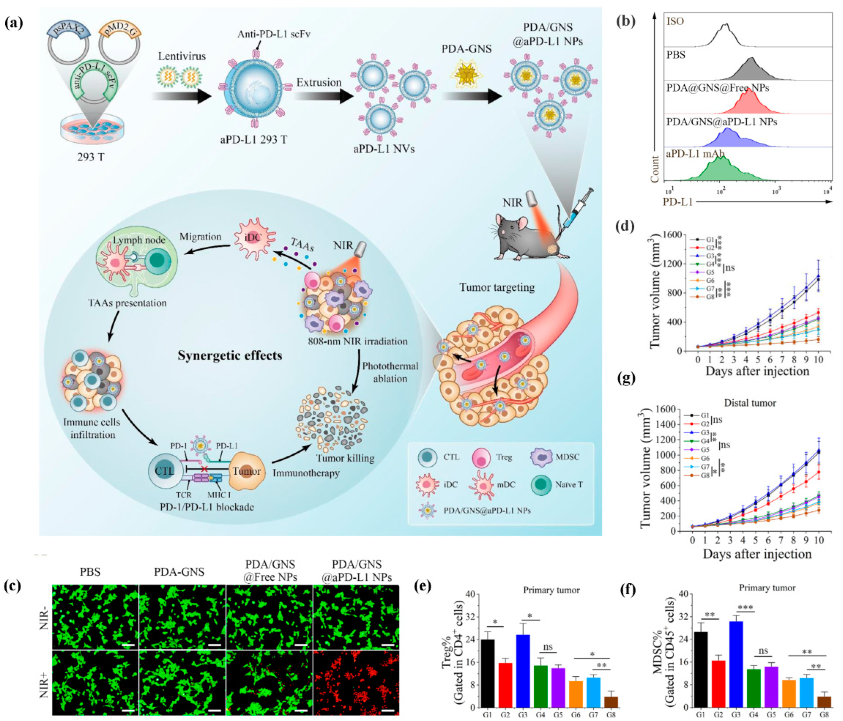
- Xiao, Y.; Zhu, T.; Zeng, Q.; Tan, Q.; Jiang, G.; Huang, X. Functionalized biomimetic nanoparticles combining programmed death-1/programmed death-ligand 1 blockade with photothermal ablation for enhanced colorectal cancer immunotherapy. Acta Biomater. 2023, 157, 451–466. [Google Scholar] [CrossRef]
- Lu, Z.; Bai, S.; Jiang, Y.; Wu, S.; Xu, D.; Zhang, J.; Peng, X.; Zhang, H.; Shi, Y.; Liu, G. Amplifying Dendritic Cell Activation by Bioinspired Nanometal Organic Frameworks for Synergistic Sonoimmunotherapy. Small 2022, 18, e2203952. [Google Scholar] [CrossRef]
- Wang, X.; Zhu, X.; Li, B.; Wei, X.; Chen, Y.; Zhang, Y.; Wang, Y.; Zhang, W.; Liu, S.; Liu, Z.; et al. Intelligent Biomimetic Nanoplatform for Systemic Treatment of Metastatic Triple-Negative Breast Cancer via Enhanced EGFR-Targeted Therapy and Immunotherapy. ACS Appl. Mater. Interfaces 2022, 14, 23152–23163. [Google Scholar] [CrossRef]
- Li, Z.; Rong, L. A Homotypic Membrane-Camouflaged Biomimetic Nanoplatform with Gold Nanocrystals for Synergistic Photothermal/Starvation/Immunotherapy. ACS Appl. Mater. Interfaces 2021, 13, 23469–23480. [Google Scholar] [CrossRef]
- Li, Y.; Wu, J.; Qiu, X.; Dong, S.; He, J.; Liu, J.; Xu, W.; Huang, S.; Hu, X.; Xiang, D.X. Bacterial outer membrane vesicles-based therapeutic platform eradicates triple-negative breast tumor by combinational photodynamic/chemo-/immunotherapy. Bioact. Mater. 2023, 20, 548–560. [Google Scholar] [CrossRef]
- Pan, P.; Dong, X.; Chen, Y.; Ye, J.J.; Sun, Y.X.; Zhang, X.Z. A heterogenic membrane-based biomimetic hybrid nanoplatform for combining radiotherapy and immunotherapy against breast cancer. Biomaterials 2022, 289, 121810. [Google Scholar] [CrossRef]
- Cao, H.; Gao, H.; Wang, L.; Cheng, Y.; Wu, X.; Shen, X.; Wang, H.; Wang, Z.; Zhan, P.; Liu, J.; et al. Biosynthetic Dendritic Cell-Exocytosed Aggregation-Induced Emission Nanoparticles for Synergistic Photodynamic Immunotherapy. ACS Nano 2022, 16, 13992–14006. [Google Scholar] [CrossRef]
- Fu, L.; Zhang, W.; Zhou, X.; Fu, J.; He, C. Tumor cell membrane-camouflaged responsive nanoparticles enable MRI-guided immuno-chemodynamic therapy of orthotopic osteosarcoma. Bioact. Mater. 2022, 17, 221–233. [Google Scholar] [CrossRef]
- Mu, X.; Zhang, M.; Wei, A.; Yin, F.; Wang, Y.; Hu, K.; Jiang, J. Doxorubicin and PD-L1 siRNA co-delivery with stem cell membrane-coated polydopamine nanoparticles for the targeted chemoimmunotherapy of PCa bone metastases. Nanoscale 2021, 13, 8998–9008. [Google Scholar] [CrossRef]
- Yang, C.; Ming, Y.; Zhou, K.; Hao, Y.; Hu, D.; Chu, B.; He, X.; Yang, Y.; Qian, Z. Macrophage Membrane-Camouflaged shRNA and Doxorubicin: A pH-Dependent Release System for Melanoma Chemo-Immunotherapy. Research 2022, 2022, 9768687. [Google Scholar] [CrossRef]
- Song, X.; Qian, R.; Li, T.; Fu, W.; Fang, L.; Cai, Y.; Guo, H.; Xi, L.; Cheang, U.K. Imaging-Guided Biomimetic M1 Macrophage Membrane-Camouflaged Magnetic Nanorobots for Photothermal Immunotargeting Cancer Therapy. ACS Appl. Mater. Interfaces 2022, 14, 56548–56559. [Google Scholar] [CrossRef]
- Huang, X.; Chen, L.; Lin, Y.; Tou, K.I.; Cai, H.; Jin, H.; Lin, W.; Zhang, J.; Cai, J.; Zhou, H.; et al. Tumor targeting and penetrating biomimetic mesoporous polydopamine nanoparticles facilitate photothermal killing and autophagy blocking for synergistic tumor ablation. Acta Biomater. 2021, 136, 456–472. [Google Scholar] [CrossRef]
- Cheng, Y.; Chen, Q.; Guo, Z.; Li, M.; Yang, X.; Wan, G.; Chen, H.; Zhang, Q.; Wang, Y. An Intelligent Biomimetic Nanoplatform for Holistic Treatment of Metastatic Triple-Negative Breast Cancer via Photothermal Ablation and Immune Remodeling. ACS Nano 2020, 14, 15161–15181. [Google Scholar] [CrossRef]
- Sun, Z.; Liu, J.; Li, Y.; Lin, X.; Chu, Y.; Wang, W.; Huang, S.; Li, W.; Peng, J.; Liu, C.; et al. Aggregation-Induced-Emission Photosensitizer-Loaded Nano-Superartificial Dendritic Cells with Directly Presenting Tumor Antigens and Reversed Immunosuppression for Photodynamically Boosted Immunotherapy. Adv. Mater. 2023, 35, e2208555. [Google Scholar] [CrossRef]
- Yao, Y.; Chen, H.; Tan, N. Cancer-cell-biomimetic nanoparticles systemically eliminate hypoxia tumors by synergistic chemotherapy and checkpoint blockade immunotherapy. Acta Pharm. Sin. B 2022, 12, 2103–2119. [Google Scholar] [CrossRef]
- Wang, H.; Wang, K.; He, L.; Liu, Y.; Dong, H.; Li, Y. Engineering antigen as photosensitiser nanocarrier to facilitate ROS triggered immune cascade for photodynamic immunotherapy. Biomaterials 2020, 244, 119964. [Google Scholar] [CrossRef]
- Hu, C.; Lei, T.; Wang, Y.; Cao, J.; Yang, X.; Qin, L.; Liu, R.; Zhou, Y.; Tong, F.; Umeshappa, C.S.; et al. Phagocyte-membrane-coated and laser-responsive nanoparticles control primary and metastatic cancer by inducing anti-tumor immunity. Biomaterials 2020, 255, 120159. [Google Scholar] [CrossRef]
- Dai, J.; Wu, M.; Wang, Q.; Ding, S.; Dong, X.; Xue, L.; Zhu, Q.; Zhou, J.; Xia, F.; Wang, S.; et al. Red blood cell membrane-camouflaged nanoparticles loaded with AIEgen and Poly(I:C) for enhanced tumoral photodynamic-immunotherapy. Natl. Sci. Rev. 2021, 8, nwab039. [Google Scholar] [CrossRef]


| Cell Membrane | Core Nanoparticle | Characteristics of the Core Nanoparticle | Drug/Imaging Agent Encapsulated | Cancer Models and the Reason for Selecting Each Cancer Cell Line | Biomedical Application | Ref. |
|---|---|---|---|---|---|---|
| Platelet membrane | Rab27 siRNA (siRab) polycationic nanocomplexes and aPD-L1 nanogels | Desired siRNA encapsulation and cell-transfection abilities, good biocompatibility, rapid clearance by macrophages | siRab and aPD-L1 | B16F10 cells, leading tumor | siRab silenced Rab27a, inhibited exosome secretion, relieved immunosuppression, and sensitized ICI. | [102] |
| T cell-derived sEVs | -- | -- | Membrane-bound IL2 | B16F10 and B16F10-luc-g5 cells, leading tumor | IL2-sEVs inhibited sEV secretion and PD-L1 expression by cancer cells through altering the miRNA profile of T cell-derived sEVs, and, consequently, sensitized ICI. | [107] |
| Cancer cell membrane | Gelatin nanoparticles | Good biocompatibility, matrix metallopeptidase (MMP)-sensitive, low passive targeting efficiency, rapid clearance by macrophages | CAT and CD73 siRNA | B16F10 cells, homologous targeting | CAT relieved tumor hypoxia, while CD73 siRNA silenced CD73 protein and promoted T cell-specific immune response, which synergically sensitized ICI. | [110] |
| Calreticulin over-expressed cancer cell membrane | Zeolitic imidazolate framework (ZIF-8) nanoparticles | Large surface area, pH-induced biodegradability, low passive targeting efficiency | Epirubicin (EPI), glucose oxidase (Gox) and hemin | 4T1 cells, homologous targeting | The biomimetic delivery system displayed an amplified ICD and activated the tumor immune microenvironment, boosting the therapeutic efficacy of ICI. | [111] |
| Cancer cell membrane | Thermosensitive lipid nanoparticles | Thermosensitive, controlled drug release, good biocompatibility, low passive targeting efficiency | PD-1/PD-L1 inhibitor BMS202 and IR780 | 4T1 and MCF-7 cells, homologous targeting, and multiple mouse models | The biomimetic delivery system inhibited cancer-associated fibroblasts (CAFs), increased the penetration of TILs, blocked the pathway of PD-1/PD-L1, and then sensitized ICI. | [112] |
| Bacterial outer membrane | -- | -- | Membrane-bound PD1 ectodomain | B16F10 and CT26 cells, multiple mouse models | The biomimetic delivery system could effectively block the PD-1/PD-L1 pathway, protect T cells, and sensitize ICI. | [113] |
| HEK 293T cell membrane | IR820-dihydroartemisinin (DHA) complexes | pH-sensitive, excellent biodegradability, sonodynamic therapy (SDT) nanoplatform, low passive targeting efficiency, rapid clearance by macrophages | IR820 and DHA | Hep1–6 cells, one of the major tumors | DHA-mediated chemo-dynamic therapy and IR820-induced sonodynamic therapy synergistically achieved a robust ICD effect and sensitized ICI. | [114] |
| Erythrocyte membrane | Liposomes | Good biocompatibility and biodegradability, pH-sensitive, low passive targeting efficiency, rapid clearance by macrophages | Paclitaxel (PTX) | PC-3 cells, one of the major tumors | The biomimetic delivery system reinforced active tumor-targeting behavior and sensitized ICI. | [115] |
| Cancer cell membrane | Mesoporous organosilica nanoparticles (MONs) | Good biocompatibility, large surface area, X-ray-induced biodegradability, low passive targeting efficiency | Doxorubicin (DOX) | 4T1 cells, homologous targeting | The biomimetic delivery system exhibited enhanced DOX-mediated ICD and sensitized ICI. | [116] |
| Cancer cell membrane | Polymeric nanoparticles | Redox-responsive, good biocompatibility, low passive targeting efficiency | Toyocamycin (Toy) and generation 3 phosphorus dendrimer-copper (II) complexes (1G3-Cu) | B16F10 cells, homologous targeting | Toy-triggered amplification of ER stress and 1G3-Cu-mediated mitochondrial dysfunction synergistically induced significant ICD, which thereby sensitized ICI. | [117] |
| aPD-L1 over-expressed HEK 293T cell membrane anchored with a cleavable peptide by matrix metallopeptidase 2 (MMP2) | Barium titanate (BTO) nanoparticles | Piezocatalysis materials, generating a cascade of redox reactions under US, rapid clearance by macrophages | BTO and membrane-bound aPD-L1 | B16F10 cells, leading tumor | The membrane-bound aPD-L1 could effectively block the PD-1/PD-L1 pathway, while the encapsulated BTO could achieve a piezocatalysis effect and induce tumor antigen release, which synergistically improved ICI therapy. | [118] |
| Cancer cell-derived EVs with PD-L1 knockout | Liposomes | Good biocompatibility and biodegradability, low passive targeting efficiency, rapid clearance by macrophages | DOX | 4T1 cells, homologous targeting | The biomimetic delivery system up-regulated the expression of PD-L1 in the tumor and amplified the ICD mediated by DOX, which thereby sensitized ICI. | [119] |
| Mature dendritic cell membrane | Redox-responsive nanoparticles | Redox-responsive, low passive targeting efficiency, rapid clearance by macrophages | Oxaliplatin (OXA) prodrugs | CT26 cells, homologous targeting | The biomimetic delivery system effectively sensitized the TME to ICI by immunogenic chemotherapy and tumor antigen-specific immunotherapy. | [120] |
| Platelet membrane | Magnetic nanoparticles | Good biocompatibility, effective ferroptosis, magnetic targeting, rapid clearance by macrophages | Sulfasalazine (SAS) | 4T1 cells, classic tumor model | The biomimetic delivery system-mediated ferroptosis elicited an effective immune response and sensitized ICI. | [121] |
| Erythrocyte membrane | Oligomeric Au (I)-PMIV complexes | Good biocompatibility, effective, self-assembling, low passive targeting efficiency, rapid clearance by macrophages | PMIV (a peptide that can degrade MDM2/MDMX) | NCI-H1650 cells, LUAD-patient-derived xenograft mice model, and LUAD-PDX mice model, multiple mouse models | The biomimetic delivery system potently restored p53 and p73, and sensitized ICI. | [122] |
| HEK 293T cell membrane expressing CD64 | -- | -- | Membrane-bound aPD-L1 and cyclophosphamide | B16F10-luci cells, leading tumor | The biomimetic delivery system restrained the Tregs and invigorated Ki67+CD8+ T cells, then improved ICI therapy. | [123] |
| Cell Membrane | Core Nanoparticle | Characteristics of the Core Nanoparticle | Drug/Imaging Agent Encapsulated | Cancer Models and the Reason for Selecting Each Cancer Cell Line | Biomedical Application | Ref. |
|---|---|---|---|---|---|---|
| T cell membrane | Poly(lactic-co-glycolic) acid (PLGA) nanoparticles | Good biocompatibility and biodegradability, low passive targeting efficiency, rapid clearance by macrophages | Dacarbazine (DTIC) | B16F10 cells, DTIC for metastatic melanoma treatment | The biomimetic delivery system could induce Fas-ligand-mediated apoptosis and scavenge TGF-β1 and PD-L1 to restore the function of CTLs. | [127] |
| T cell membrane over-expressing PD-1, TGF-β receptor, and granzyme B | -- | -- | Granzyme B | LLC cancer cells, immunotherapy for lung carcinoma | The biomimetic delivery system prevented CD8+ T cell exhaustion mediated by PD-L1/TGF-β, and promoted tumor cell apoptosis via granzyme B. | [128] |
| Cancer cell membrane | Magnetic nanoparticles | Good biocompatibility, magnetic targeting, rapid clearance by macrophages | -- | HepG2 and A375 cells, homologous targeting, and multiple mouse models | The biomimetic delivery system effectively activated NK cells and enhanced NK cell-based ACT through the enhanced secretion of soluble cytotoxic effectors. | [131] |
| NK92MI cell membrane equipped with the anti-EGFR nanobody 7D12 | -- | -- | -- | EGFRpositive cancer cell lines, including LoVo, MDA-MB-468, A549, and A431 cells, 7D12 for specifically recognizing EGFRpositive cancer cells | The biomimetic delivery system exhibited high targeting for EGFR-positive cancer cells, caused enhanced secretion of cytotoxic effectors, and improved NK cell-based ACT. | [132] |
| Mature DC membrane | Polymeric nanoparticles | Good biocompatibility and biodegradability, low passive targeting efficiency, rapid clearance by macrophages | IR-797 | 4T1 cells, specific recognition between tumor specific antigens and tumor cells | Mature DC membrane facilitated effective cross-priming of T cells, recruited T cells, and produced immunostimulatory cytokines, while mild PTT mediated by IR-797 induced a greater degree of tumoricidal effect, which consequently amplified ICD and reinitiated the self-sustaining cycle of cancer immune responses. | [135] |
| Cancer cell membrane | MnOx nanoparticles functionalized with anti-CD3/CD28 mAbs | Good biocompatibility, low passive targeting efficiency, rapid clearance by macrophages | anti-CD3/CD28 mAbs | B16F10 cells, homologous targeting | The biomimetic delivery system not only efficiently promoted the expansion and activation of CD8+ T cells and DCs but also reversed the immunosuppressive microenvironment to promote T cell survival. | [136] |
| NK92MI cell membrane | Anti-CD56 antibodies modified Fe3O4 nanoparticles | Good biocompatibility, magnetic targeting, rapid clearance by macrophages | -- | K562, B16F10, and H22 cells, NK92 cell possessing a broader-spectrum anti-cancer activity | The biomimetic delivery system up-regulated the secretion of granzyme B and IFN-γ at the tumor site, which thereby significantly improved the antitumor effect. | [137] |
| Mature DC membrane | Cancer cell membrane coated PLGA nanoparticles | Good biocompatibility and biodegradability, high active targeting efficiency | -- | HPV E6- and E7-expressing TC-1, B16-OVA, and Hepa 1–6 cells, multiple mouse models | The biomimetic delivery system could directly cross-prime T cells without the help of APCs and elicit robust antigen-specific T cell immunotherapy. | [138] |
| Cell Membrane | Core Nanoparticle | Characteristics of the Core Nanoparticle | Drug/Imaging Agent Encapsulated | Cancer Models and the Reason for Selecting Each Cancer Cell Line | Biomedical Application | Ref. |
|---|---|---|---|---|---|---|
| CRT-expressed cancer cell membrane | PLGA nanoparticles | Good biocompatibility and biodegradability, low passive targeting efficiency, rapid clearance by macrophages | Adjuvant R837 | Luc-4T1 cells, homologous targeting | The CRT expressed on the surface of the nanovaccine facilitated the internalization of the nanovaccine by DCs. R837 released from the nanovaccine excited TLR7 and further activated DCs. | [145] |
| Cancer cell membrane | Silicon@Au nanocomposites | Good biocompatibility, low passive targeting efficiency, rapid clearance by macrophages | Au | 4T1 cells, homologous targeting | The biomimetic delivery system based on nanocomposites with weak immune stimulation could still induce DCs maturation to activate the downstream anti-tumor immune responses. | [146] |
| BDVs presenting the neoantigens | -- | -- | Adjuvant GM-CSF | B16F10-luc cells, leading tumor | The biomimetic delivery system efficiently recruited DCs and promoted the proliferation of TILs and memory T cells. | [150] |
| Erythrocyte membrane | Manganese-doped imidazolate frameworks-90 nanoparticles | Good biocompatibility, low passive targeting efficiency, rapid clearance by macrophages | DNAzyme | 4T1 cells, classic tumor model | The biomimetic delivery system closely mimicked the process of herpesvirus infection, ultimately initiating cGAS-STING pathway-mediated innate immunotherapy. | [154] |
| Cancer cell membrane | Mesoporous polydopamine nanoparticles | Good biocompatibility and biodegradability, PTT nanoplatform, low passive targeting efficiency, rapid clearance by macrophages | Adjuvant R848 | 4T1 cells, homologous targeting | The biomimetic delivery system effectively stimulated the immune response and demonstrated excellent photothermal immunotherapy, which significantly promoted the activation and maturation of lymph node DCs, and stimulated CD8+ T cells and memory T cells. | [155] |
| Bone marrow-derived macrophage (BMDM) membrane | Poly (lactic-co-glycolic acid) nanoparticles (PLP NPs) | Good biocompatibility and biodegradability, low passive targeting efficiency, rapid clearance by macrophages | Adjuvant Poly I: C (PIC) | 4T1 cells, classic tumor model | The PIC released from the biomimetic nanoparticles could not only directly induce tumor cell apoptosis, but also polarize exogenous BMDMs into killing M1 macrophages and activate endogenous APCs to trigger robust anti-tumor immune responses. | [156] |
| Tumor-antigen activated DC membrane | PLGA nanoparticles | Good biocompatibility and biodegradability, low passive targeting efficiency, rapid clearance by macrophages | Rapamycin (RAPA) | C6-LUC cells, biomimetic nanoparticles possessing the potential to cross the BBB | The biomimetic delivery system could activate CD8+ T cells directly or indirectly to reconstitute the glioma tumor immune microenvironment and increase the proportion of NK cells to reduce the exhausted T cells, resulting in a significant anti-glioma efficacy. | [157] |
| CD80 engineered cancer cell-membrane | -- | -- | -- | B16-OVA cells, homologous targeting | The biomimetic nanoparticle platform could directly activate T cells without the help of professional APCs. | [158] |
| Anti-CD40 scFv-anchored cancer cell membrane | PLGA nanoparticles | Good biocompatibility and biodegradability, low passive targeting efficiency, rapid clearance by macrophages | -- | MC38 and Panc02 cells, homologous targeting, and multiple mouse models | The biomimetic nanoparticle platform effectively promoted DCs maturation in CD40-humanized transgenic mice, improved the engagement and expansion of cognate T-cell immune responses, and facilitated subsequent adaptive immune responses. | [159] |
| CD47KO/CRT dual-bioengineered cancer cell membrane | Hyperbranched PEI25k nanoparticles | Good biocompatibility, large surface area, low passive targeting efficiency, rapid clearance by macrophages | Unmethylated cytosine-phosphate-guanine (CpG) adjuvant | B16F10 cells, homologous targeting | The biomimetic nanoparticle platform significantly stimulated APCs, resulting in the activation of CD8+ T cells and intense anti-tumor immune responses. | [160] |
| MHC-I-Ag-anchored mature DC membrane | -- | -- | aPD-1 | B16F10 cells, leading tumor | The biomimetic nanoparticle platform could present neoantigens to CD8+ T cells directly, resulting in strong CTL responses. In addition, the immunosuppressive reversal function of anti-PD-1 antibody was enhanced by CD28/B7 co-stimulation. | [161] |
| Cancer cell membrane | Poloxamer 407 nanoparticles | Good biocompatibility, low passive targeting efficiency, rapid clearance by macrophages | Adjuvant R837 | HCT116 cells, homologous targeting | The biomimetic nanoparticle platform was presented with APCs to secret immunofactors and to activate the lymphatic immune network, resulting in significant tumor regression. | [162] |
| Cancer cell membrane | Manganese dioxide (MnO2) nanoparticles | Good biocompatibility, low passive targeting efficiency, rapid clearance by macrophages | Polythiophene | B16F10 cells, homologous targeting | The biomimetic nanoparticle platform could stimulate a specific T cell anti-tumor immune response by promoting APCs maturation, autologous tumor antigen presentation, as well as generating local microinflammation. | [163] |
| Cancer cell membrane expressing fibroblast activation protein-α (FAP) | -- | -- | -- | CT26, B16F10, LLC, and 4T1 cells, 90% of human tumor tissues overexpressing FAP and multiple mouse models | The biomimetic nanoparticle platform suppressed tumor growth by triggering robust and specific T cell anti-tumor immune responses. Moreover, the biomimetic nanoparticle platform facilitated tumor ferroptosis by releasing IFN-γ from CTLs and eliminating FAP+CAFs. | [164] |
| Cell Membrane | Core Nanoparticle | Characteristics of the Core Nanoparticle | Drug/Imaging Agent Encapsulated | Cancer Models and the Reason for Selecting Each Cancer Cell Line | Biomedical Application | Ref. |
|---|---|---|---|---|---|---|
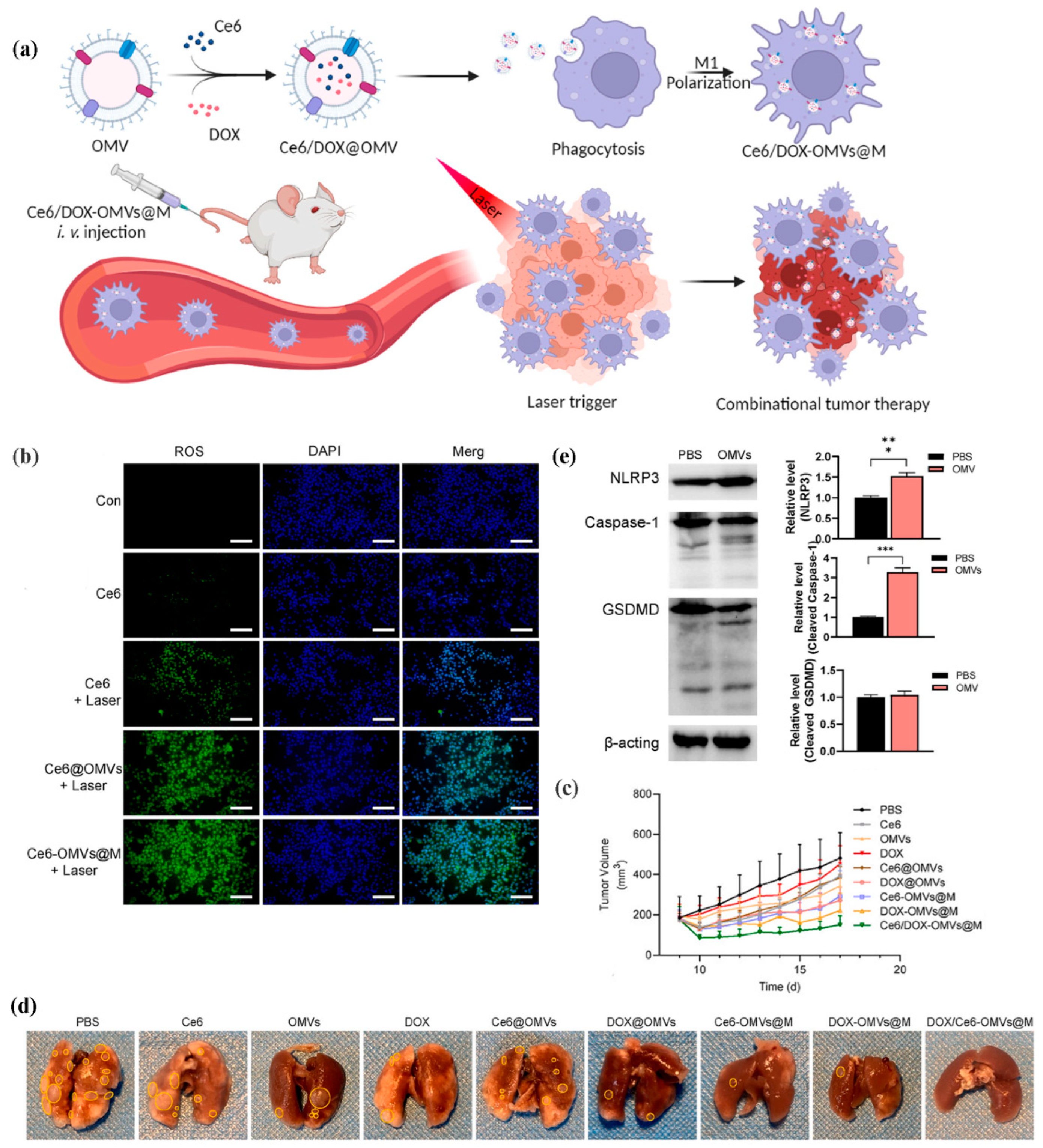
| Bacterial outer membrane | PLGA nanoparticles | Good biocompatibility and biodegradability, low passive targeting efficiency, rapid clearance by macrophages | DOX and R848 | 4T1 cells, classic tumor model | The biomimetic nanoparticle platform could polarize M2 macrophages into M1 macrophages, promote the secretion of TNF-α and IL-6, which thereby activated the anti-tumor immune responses. | [176] |
| Cancer cell membrane overexpressing SIRPα variants | Magnetic nanoparticles | Good biocompatibility, magnetic targeting, rapid clearance by macrophages | -- | B16F10 and 4T1 cells, homologous targeting, and multiple mouse models | In this system, the gCM shell genetically overexpressing SIRPα variants efficiently blocked the CD47-SIRPα pathway, while the nanoparticle cores polarized M2 macrophages into M1 macrophages, which thereby synergistically facilitated macrophage phagocytosis of tumor cells and triggered T-cell immune response. | [177] |
| Erythrocyte membrane | Amphiphilic PCL nanoparticles | Good biocompatibility and biodegradability, low passive targeting efficiency, rapid clearance by macrophages | DOX | 4T1 cells, classic tumor model | The Hb moiety of the biomimetic nanoparticle platform could effectively kill the tumor cells. In addition, the O2 released by Hb alleviated cancer hypoxia and further augmented the anti-tumor immune response. | [181] |
| 4T1 and 3T3 hybrid cell membrane | Solid lipid nanoparticles | Good biocompatibility and biodegradability, low passive targeting efficiency, rapid clearance by macrophages | PTX and glycolysis inhibitor PFK15 | 4T1 cells, homologous targeting | The biomimetic nanoparticle platform possessed outstanding dual-targeting ability. The encapsulated PFK15 could inhibit glycolysis in both CAFs and cancer cells, resulting in elevated immune responses. | [185] |
| Cancer cell membrane | PLGA nanoparticles | Good biocompatibility and biodegradability, low passive targeting efficiency, rapid clearance by macrophages | ICG and DCT | 4T1 cells, homologous targeting | ICG induced a sharp increase in cytosolic Ca2+ concentration, promoted cytochrome c release and subsequent caspase-3 activation. DCT synergistically up-regulated the expression of gasdermin E by inhibiting DNA methylation, enhancing the cleavage of gasdermin E by caspase-3, and causing robust cancer pyroptosis. | [188] |
| Macrophage cell-derived sEVs | -- | -- | siRNA against YTHDF1 | MGC-803 and HGC-27 cells, YTHDF1 promoting tumor progression in a variety of cancers and multiple mouse models | This nanosystem efficiently depleted YTHDF1 expression and stimulated strong cytotoxic T lymphocytes responses through hampering frizzled7 translation and inactivating the Wnt/β-catenin pathway in an m6A dependent manner. | [189] |
| Platelet membrane | PLA nanoparticles | Good biocompatibility and biodegradability, low passive targeting efficiency, rapid clearance by macrophages | R848 | MC38 and 4T1 cells, multiple mouse models | The biomimetic nanoparticle platform promoted the robust activation of APCs and increased immune infiltration, resulting in complete tumor regression. | [190] |
| Activated neutrophils membrane | Redox-responsive polymer nanoparticles | Good biocompatibility and biodegradability, low passive targeting efficiency, rapid clearance by macrophages | DOX | 4T1 cells, classic tumor model | The biomimetic nanoparticle platform could prevent the recruitment and functions of MDSCs, thereby inhibiting tumor recurrence and metastasis. | [191] |
| Tumor-associated macrophage membrane with high macrophage colony-stimulating factor 1 receptor (CSF1R) | Rare-earth-upconversion nanoparticles | Good biocompatibility, “nanotransducer”, theranostic nanoplatform, low passive targeting efficiency, rapid clearance by macrophages | Rose Bengal | 4T1 cells, classic tumor model | The biomimetic nanoparticle platform could block the interaction between TAMs and cancer cells via depleting the CSF1 secreted by cancer cells. In addition, the biomimetic nanoparticle platform-mediated photodynamic therapy (PDT) could polarize M2 macrophages into M1 macrophages. | [192] |
| Erythrocyte membrane | Copper peroxide (CP) nanoparticles | Good biocompatibility, PDT nanoplatform, low passive targeting efficiency, rapid clearance by macrophages | H2O2 and protoporphyrin (PpIX) | B16F10 cells, leading tumor | The biomimetic nanoparticle platform could relieve tumor hypoxia and polarize M2 macrophages into M1 macrophages. Furthermore, the ROS generated by photosensitizers could cause the ICD of tumor cells. | [193] |
| Macrophage cell membrane | Polydopamine nanoparticles | Good biocompatibility and biodegradability, PTT nanoplatform, low passive targeting efficiency, rapid clearance by macrophages | TMP195 (a TAMs repolarization agent) | 4T1 cells, classic tumor model | The loaded TMP195 significantly polarized M2 macrophages into M1 macrophages and subsequently remodeled the ITME in residual tumor after PTT, which thereby rescued CTLs. | [194] |
| Cancer cell membrane | Cu2-xSe nanoparticles | Good biocompatibility, effective ferroptosis, low passive targeting efficiency, rapid clearance by macrophages | Indoximod (IND) and JQ1 | GL261 cells, homologous targeting | The smart nanoparticle platform could remodel the ITME by increasing M1 macrophages and preventing Tregs cell infiltration. In addition, it also served as a checkpoint inhibitor to inhibit the expression of PD-L1 on cancer cells. | [195] |
| M1-like macrophage cell membrane | -- | -- | Celastrol | LLC and GL261 cells, multiple mouse models | M1 macrophages not only acted as drug delivery carriers for celastrol but also as therapeutic agents. In turn, celastrol could maintain the anticancer polarization of M1 macrophages. | [196] |
| Erythrocyte membrane | TiO2 nanoparticles | Good biocompatibility, SDT nanoplatform, low passive targeting efficiency, rapid clearance by macrophages | Hb and RRx-001 | 4T1 cells, classic tumor model | Upon reaching the hypoxic TME, the Hb in this smart nanoparticle platform was deoxygenated, further initiating a series of reactions that generated a large number of RNS in cascade fashion. Subsequently, oxygen compensation by Hb enhanced TiO2-mediated ROS for SDT, and the resulting highly active RNS triggered ICD in cancer cells. | [197] |
| M1-like macrophage cell membrane | Magnetic polymer nanoparticles | Good biocompatibility and biodegradability, magnetic targeting, rapid clearance by macrophages | R837 | 4T1 cells, classic tumor model | The intracellular uptake of this smart nanoparticle platform could greatly polarize M2 macrophages into M1 macrophages with the synergy of R837 and Fe3O4, alleviating the immunosuppression of the TME for immune recovery. | [198] |
| Cancer cell membrane | Fe-doped polydiaminopyridine nanoparticles | Good biocompatibility, SDT nanoplatform, low passive targeting efficiency, rapid clearance by macrophages | Ce6 | 4T1 cells, homologous targeting | The prepared smart nanoparticle platform could significantly reduce the unnecessary consumption of H2O2 in the TME, while US irradiation could promote the exposure of Fe-PDAP and provide more adequate O2 supply, enabling efficient ROS production and DCs maturation. | [199] |
| Cell Membrane | Core Nanoparticle | Characteristics of the Core Nanoparticle | Drug/Imaging Agent Encapsulated | Cancer Models and the Reason for Selecting Each Cancer Cell Line | Biomedical Application | Ref. |
|---|---|---|---|---|---|---|
| Anti-PD-L1 scFv-anchored HEK 293T cell membrane | Polydopamine-modified gold nanoparticles | Good biocompatibility and biodegradability, PTT nanoplatform, low passive targeting efficiency, rapid clearance by macrophages | Au and membrane-bound aPD-L1 scFv | MC38 cells, immunotherapy for colorectal cancer | The developed smart nanoparticle platform combined PD-1/PD-L1 blockade with PTT, which triggered the immune stimulatory responses and remodeled the ITME. | [202] |
| Anti-DEC205 anchored cancer cell membrane | Porphyrin-based metal organic frameworks (MOFs) | Good biocompatibility and biodegradability, SDT nanoplatform, low passive targeting efficiency, rapid clearance by macrophages | R848 and AuPt | Hep1-6 cells, homologous targeting | AMR-MOF@AuPt-mediated multimodal therapies based on sonoimmunotherapy induced systemic immune responses and long-term memory immunity. | [203] |
| Cancer cell membrane anchored with a cleavable peptide by MMP2 | PLGA nanoparticles | Good biocompatibility and biodegradability, low passive targeting efficiency, rapid clearance by macrophages | Afatinib (AFT) and 2-bromopalmitate (2-BP) | 4T1 cells, homologous targeting | The developed smart nanoparticle platform combined targeted therapy with immunotherapy, which remarkably prevented tumor growth and metastasis. | [204] |
| Hybrid pH-sensitive membrane | Mesoporous silica nanoparticles | Good biocompatibility and biodegradability, large surface area, low passive targeting efficiency, rapid clearance by macrophages | R837 and Au | 4T1 cells, homologous targeting | The developed smart nanoparticle platform realized the synergistic effect of starvation/PTT/immunotherapy and induced a long-term immune memory effect. | [205] |
| Macrophage cell membrane | Bacterial outer membrane vesicles | Good biocompatibility and biodegradability, potent anti-cancer potential, low targeting efficiency | Ce6 and DOX | 4T1 cells, classic tumor model | The developed smart nanoparticle platform realized the synergism of PDT/chemo-/immunotherapy. | [206] |
| Hybrid cell membrane | Glutathione (GSH) decorated Te nanoparticles | Good biocompatibility, radiotherapy sensitization, radiotherapy nanoplatform, low passive targeting efficiency, rapid clearance by macrophages | GTe | 4T1 cells, homologous targeting | The developed smart nanoparticle platform realized the synergism of radiotherapy and immunotherapy. | [207] |
| DC-derived sEVs | MBPN-TCyP nanoparticle complexes | Good biocompatibility, PDT nanoplatform, mitochondrion-targeting, rapid clearance by macrophages | MBPN-TCyP (an AIE photosensitizer) | 4T1 and CT26 cells, multiple mouse models | The developed smart nanoparticle platform realized the synergism of PDT/immunotherapy and inhibited the self-renewal of cancer stem cells for tumor suppression. | [208] |
| Cancer cell membrane | Hollow manganese dioxide (HMnO2) nanoparticles | Good biocompatibility, GSH-sensitive, excellent loading capacity, low passive targeting efficiency, rapid clearance by macrophages | Ginsenoside Rh2 (Rh2) | K7M2 cells, homologous targeting | The developed smart nanoparticle platform realized attractive immuno-chemo-dynamic combined therapeutic efficiency. | [209] |
| Stem cell membrane | Polydopamine nanoparticles | Good biocompatibility and biodegradability, PTT nanoplatform, low passive targeting efficiency, rapid clearance by macrophages | DOX and PD-L1 siRNA | PC-3 cells, one of the major tumors | The developed smart nanoparticle platform showed attractive immuno-chemo-dynamic combined therapeutic efficiency for PCa bone metastases. | [210] |
| M1-like macrophage cell membrane | Polyethylenimine (PEI) nanoparticles | Good biocompatibility, large surface area, rapid clearance by macrophages | DOX and short-hairpin RNA (shRNA) targeting Ptpn2 | B16F10 cells, leading tumor | The developed smart nanoparticle platform realized the synergism of chemotherapy and gene immunotherapies. | [211] |
| M1-like macrophage cell membrane | Magnetic photothermal nanocomplexes | Good biocompatibility, PTT nanoplatform, low passive targeting efficiency, rapid clearance by macrophages | DOX | 4T1 cells, classic tumor model | The developed smart nanoparticle platform enhanced ROS generation and exhibited outstanding chemo-phototherapy efficacy. | [212] |
| Cancer cell membrane | Mesoporous polydopamine nanoparticles | Good biocompatibility and biodegradability, PTT nanoplatform, low passive targeting efficiency, rapid clearance by macrophages | Chloroquine (CQ, an autophagy inhibitor) | RM-1 cells, homologous targeting | The developed smart nanoparticle platform facilitated PTT and autophagy blockade for synergistic tumor elimination. | [213] |
| Hybrid cell membrane | Polydopamine nanoparticles | Good biocompatibility and biodegradability, PTT nanoplatform, low passive targeting efficiency, rapid clearance by macrophages | -- | B16F10 cells, homologous targeting | The developed smart nanoparticle platform realized the synergism of OMV-mediated immunotherapy and hollow polydopamine-mediated PTT. | [84] |
| Anti-PD-1 peptide AUNP-12 modified cancer cell membrane | Dendritic large-pore mesoporous silica nanoparticles | Good biocompatibility and biodegradability, low passive targeting efficiency, rapid clearance by macrophages | Copper sulfide (CuS) and R848 | 4T1 cells, homologous targeting | The developed smart nanoparticle platform realized the synergism of PDT and immunotherapy. | [214] |
| Cancer cell membrane expressing CD86 and anti-LAG3 | Polymeric nanoparticles | Good biocompatibility, PDT nanoplatform, mitochondrion-targeting, rapid clearance by macrophages | Fs (an AIE photosensitizer) | 4T1 cells, homologous targeting | The developed smart nanoparticle platform realized the synergism of PDT and immunotherapy by directly presenting tumor antigens and reversed immunosuppression. | [215] |
| Cancer cell membrane | Liposomes | Good biocompatibility and biodegradability, pH-sensitive, low passive targeting efficiency, rapid clearance by macrophages | RA-V (a chemotherapeutic drug) and BMS202 | CT26 cells, homologous targeting | The developed smart nanoparticle platform systemically eliminated hypoxia tumors by synergistic chemotherapy and checkpoint blockade immunotherapy. | [216] |
| Cancer cell membrane | Ovalbumin antigen (OVA) nanoparticles | Good biocompatibility and biodegradability, rapid clearance by macrophages | Ce6 | B16-OVA cells, homologous targeting | The developed smart nanoparticle platform facilitated ROS-triggered immune cascade for photodynamic immunotherapy. | [217] |
| M1-like macrophage cell membrane | Nanoparticle complexes | Good biocompatibility, laser-sensitive, size-changeable, rapid clearance by macrophages | Ce6, DOX, and d indoleamine 2,3-dioxygenase 1 inhibitor | 4T1 and B16F10 cells, multiple mouse models | The developed smart nanoparticle platform realized the synergism of PDT/chemo-/immunotherapy. | [218] |
| Erythrocyte membrane | PLGA nanoparticles | Good biocompatibility and biodegradability, low passive targeting efficiency, rapid clearance by macrophages | P2-PPh3 (an AIE photosensitizer) and Poly (I:C) | B16F10 cells, leading tumor | The developed smart nanoparticle platform combined the PDT properties of P2-PPh3 with the immunotherapy properties of Poly (I:C). | [219] |
Disclaimer/Publisher’s Note: The statements, opinions and data contained in all publications are solely those of the individual author(s) and contributor(s) and not of MDPI and/or the editor(s). MDPI and/or the editor(s) disclaim responsibility for any injury to people or property resulting from any ideas, methods, instructions or products referred to in the content. |
© 2023 by the authors. Licensee MDPI, Basel, Switzerland. This article is an open access article distributed under the terms and conditions of the Creative Commons Attribution (CC BY) license (https://creativecommons.org/licenses/by/4.0/).
Share and Cite
Hu, T.; Huang, Y.; Liu, J.; Shen, C.; Wu, F.; He, Z. Biomimetic Cell-Derived Nanoparticles: Emerging Platforms for Cancer Immunotherapy. Pharmaceutics 2023, 15, 1821. https://doi.org/10.3390/pharmaceutics15071821
Hu T, Huang Y, Liu J, Shen C, Wu F, He Z. Biomimetic Cell-Derived Nanoparticles: Emerging Platforms for Cancer Immunotherapy. Pharmaceutics. 2023; 15(7):1821. https://doi.org/10.3390/pharmaceutics15071821
Chicago/Turabian StyleHu, Tingting, Yuezhou Huang, Jing Liu, Chao Shen, Fengbo Wu, and Zhiyao He. 2023. "Biomimetic Cell-Derived Nanoparticles: Emerging Platforms for Cancer Immunotherapy" Pharmaceutics 15, no. 7: 1821. https://doi.org/10.3390/pharmaceutics15071821