Hybrid Membranes of the Ureasil-Polyether Containing Glucose for Future Application in Bone Regeneration

 and
and 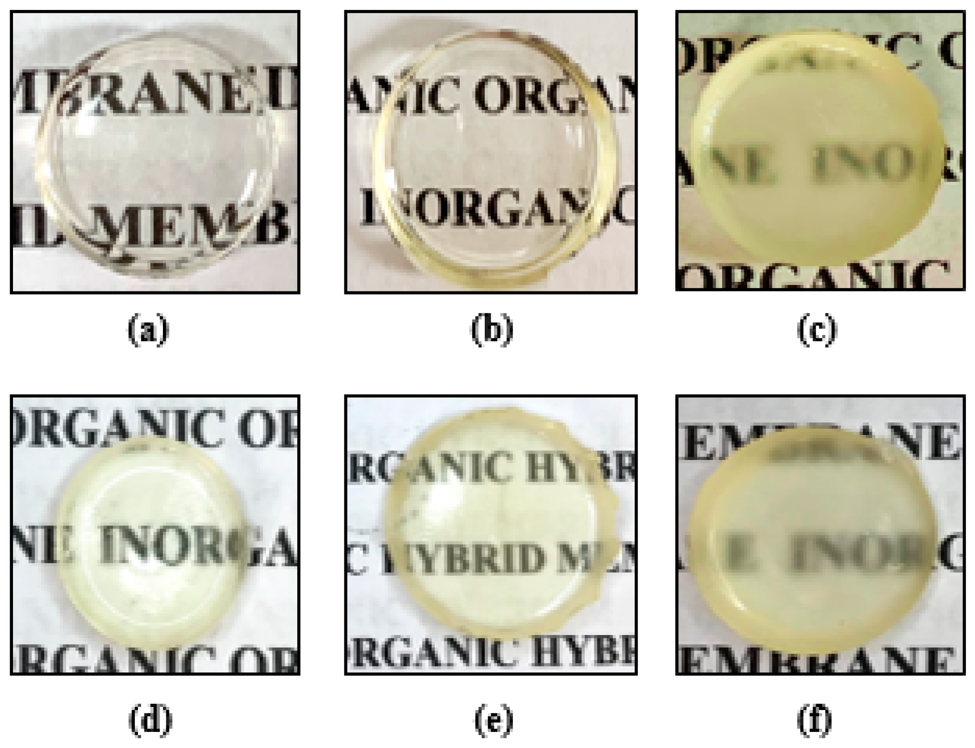
Abstract
:1. Introduction
2. Materials and Methods
2.1. Preparation of the Ureasil-Polyether Hybrid Materials and Glucose Incorporation
2.2. Assessment of Swelling
2.3. Dynamic Mechanical Analysis (DMA)
2.4. X-ray Diffraction (XRD)
2.5. Differential Scanning Calorimetry (DSC)
2.6. Contact Angle Assessment (Wetability)
2.7. In Vitro Bioactivity of Ureasil-Polyether Membranes
2.8. Assessment of Hemolysis Potential
2.9. In Vitro Glucose Release from Ureasil-Polyether Membranes
3. Results and Discussion
3.1. Preparation of the Ureasil-Polyether Hybrid Materials and Glucose Incorporation
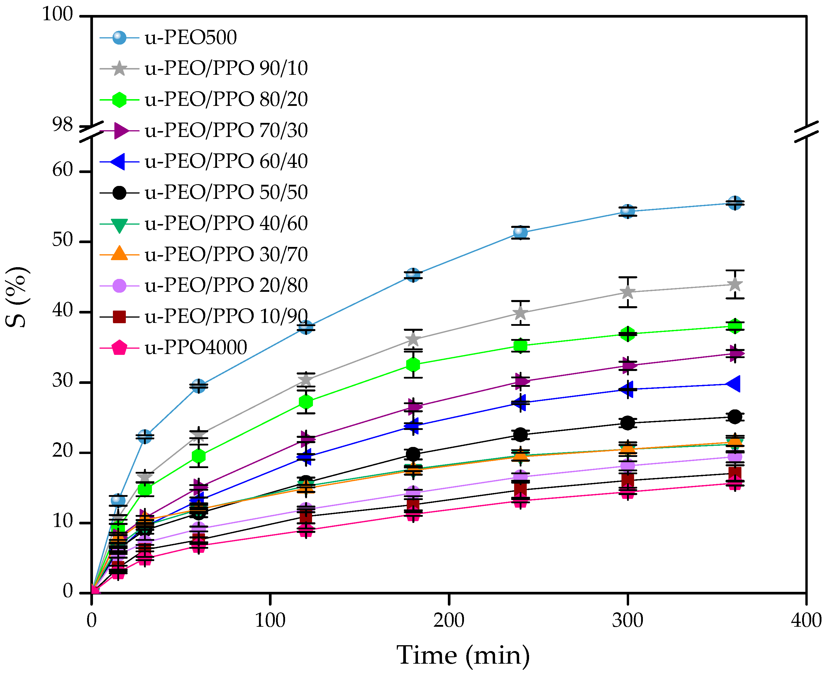
3.2. Assessment of Swelling
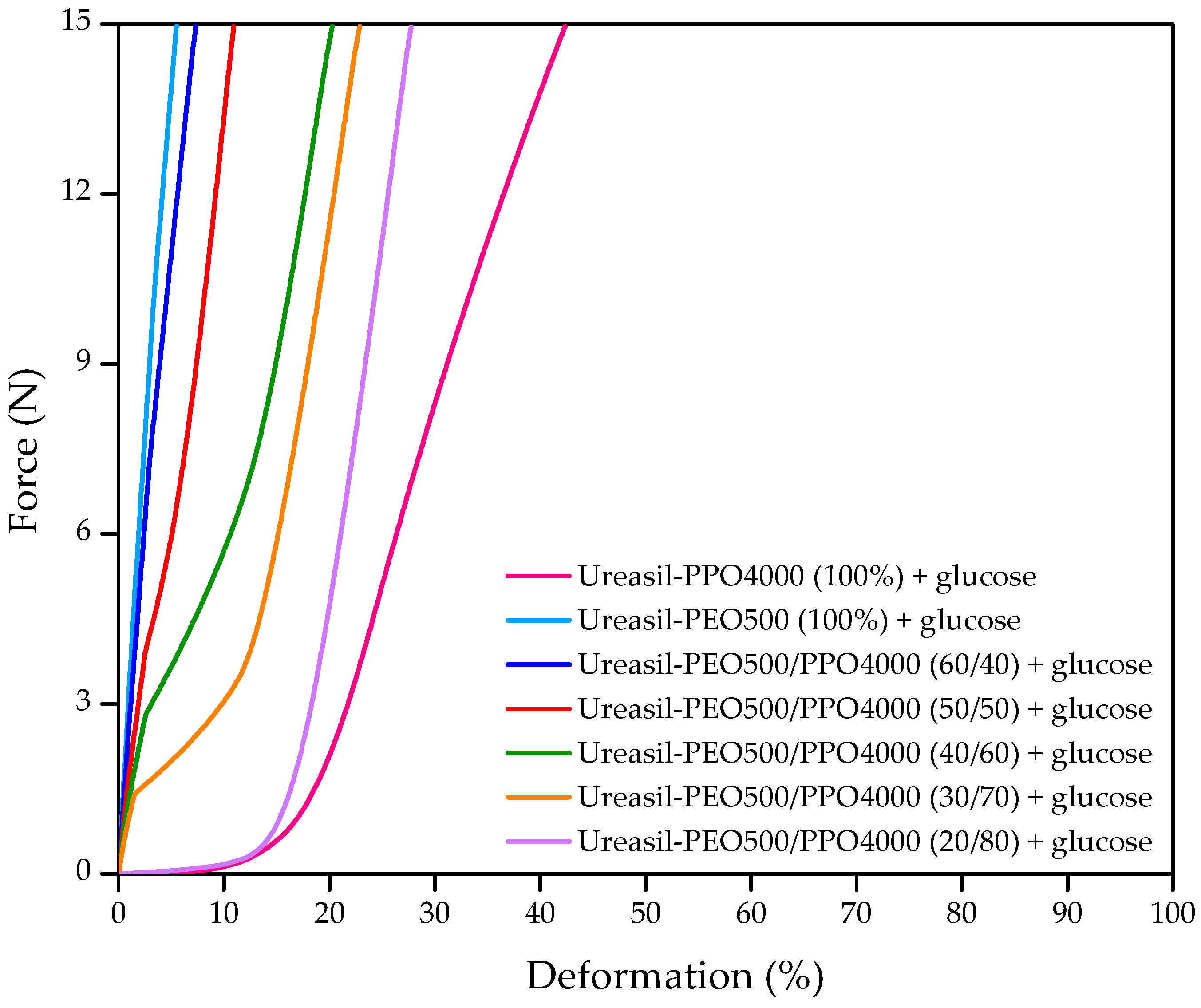
3.3. Dynamic Mechanical Analysis (DMA)
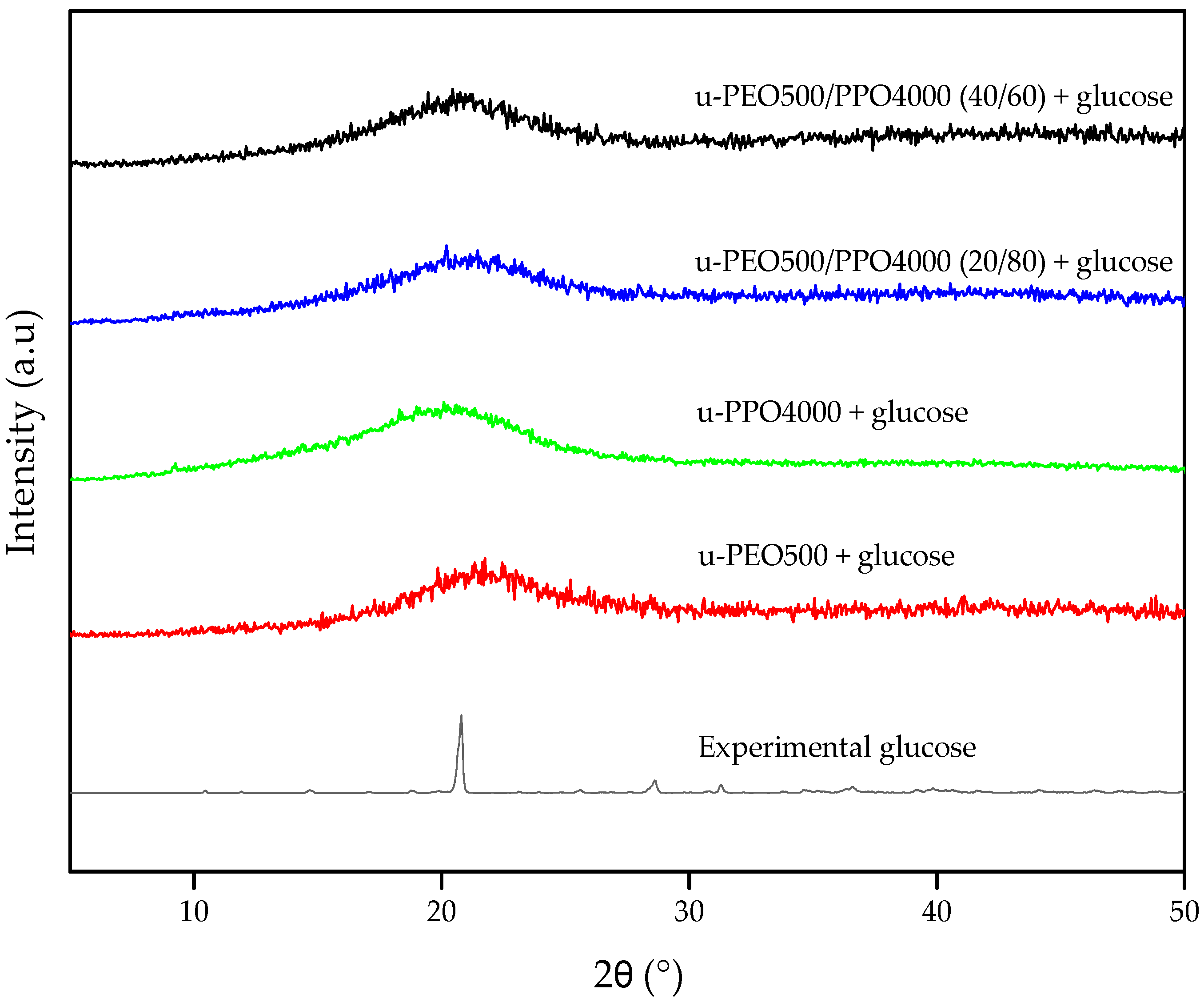
3.4. X-Ray Diffraction (XRD)
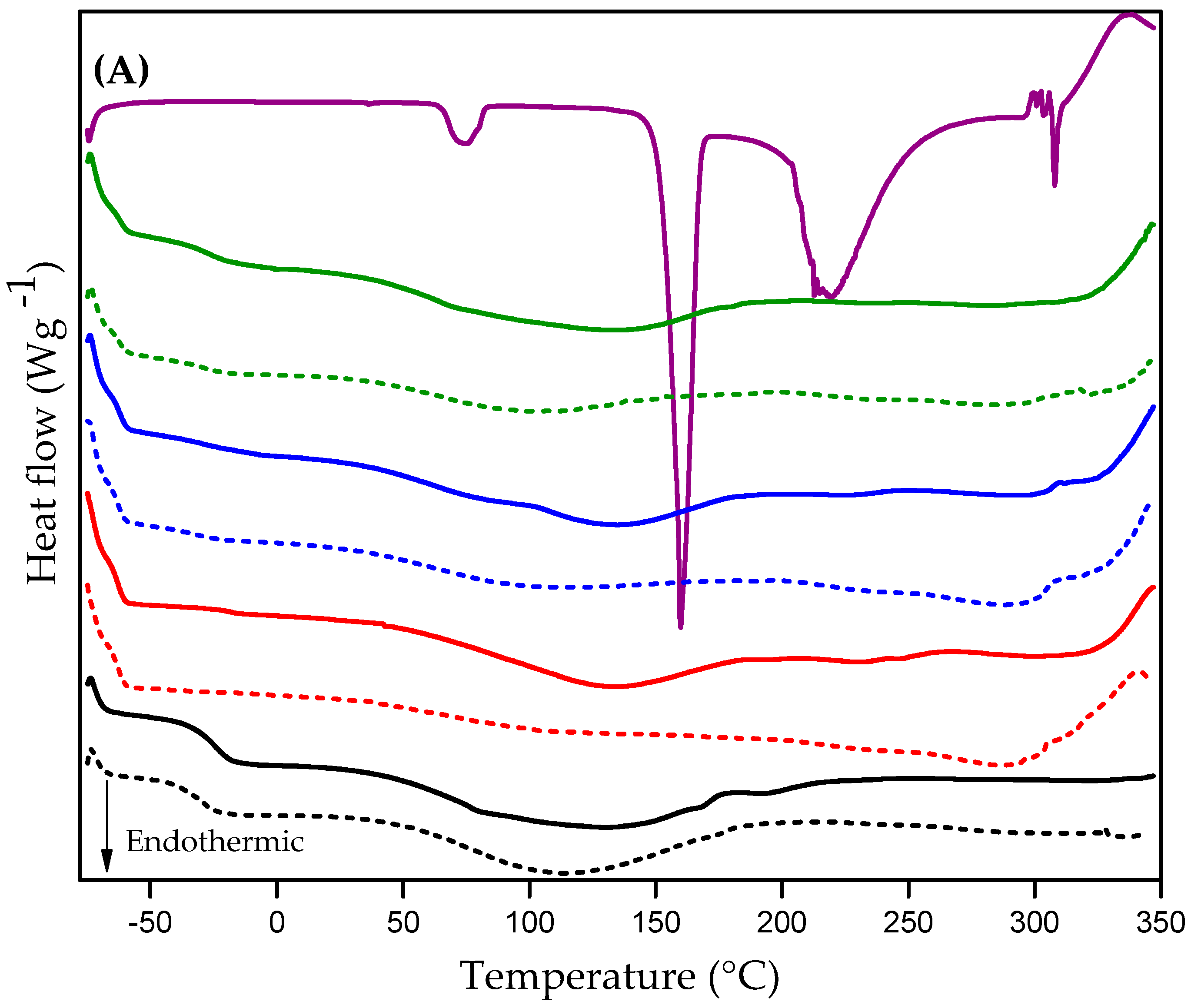
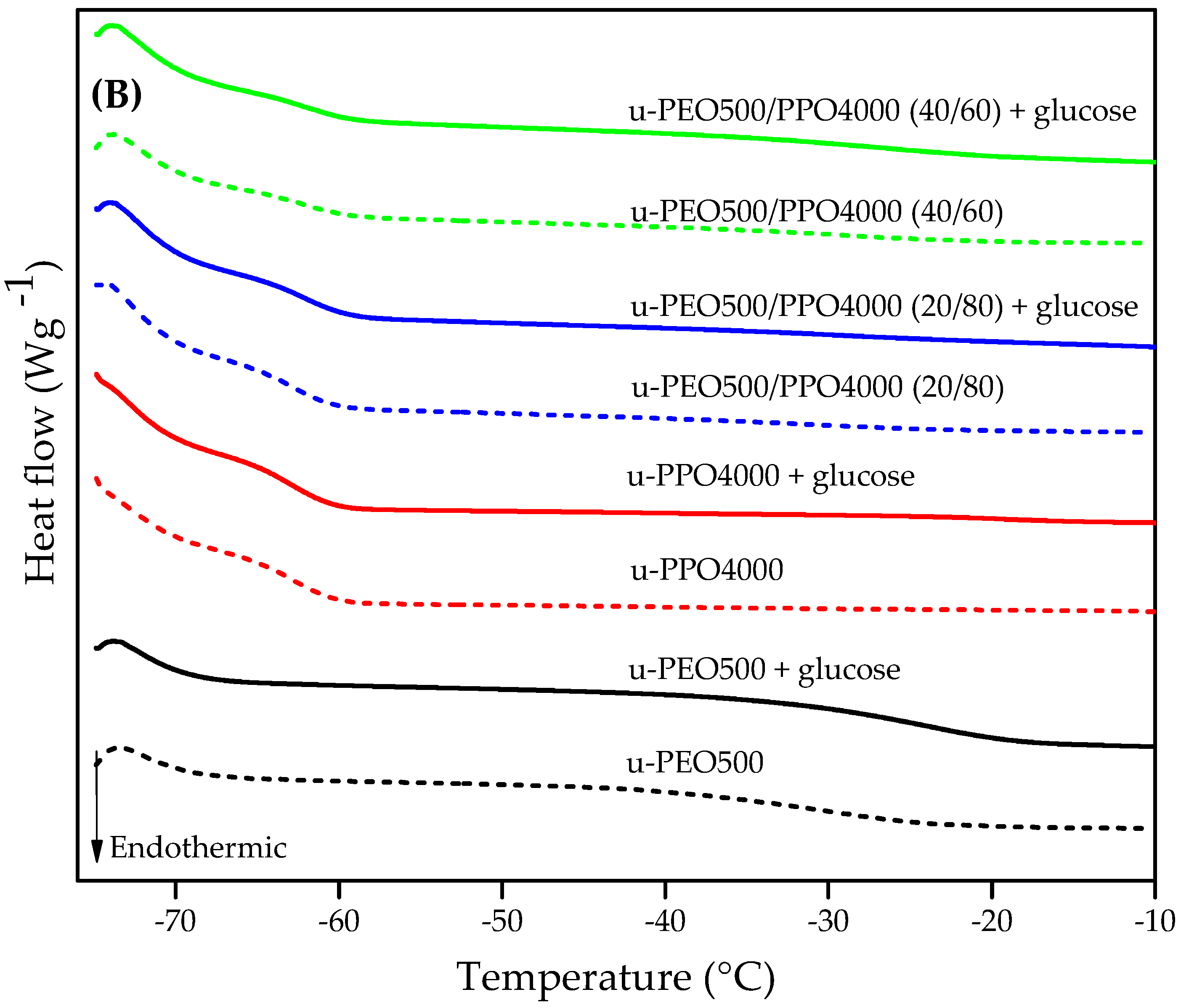
3.5. Differential Scanning Calorimetry (DSC)
3.6. Contact Angle Assessment (Wetability)
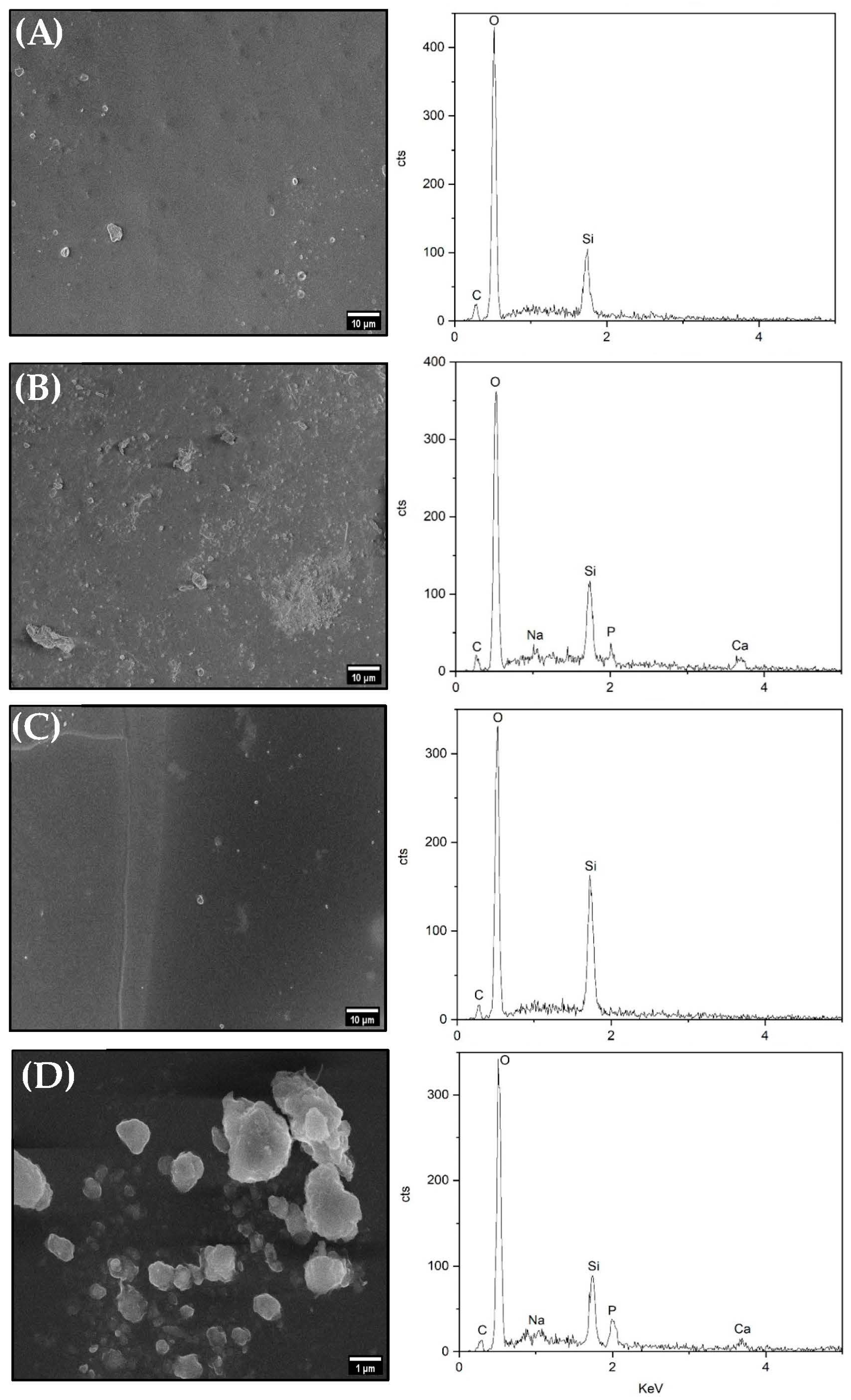
3.7. In Vitro Bioactivity of Ureasil-Polyether Membranes
3.8. Assessment of Hemolysis Potential
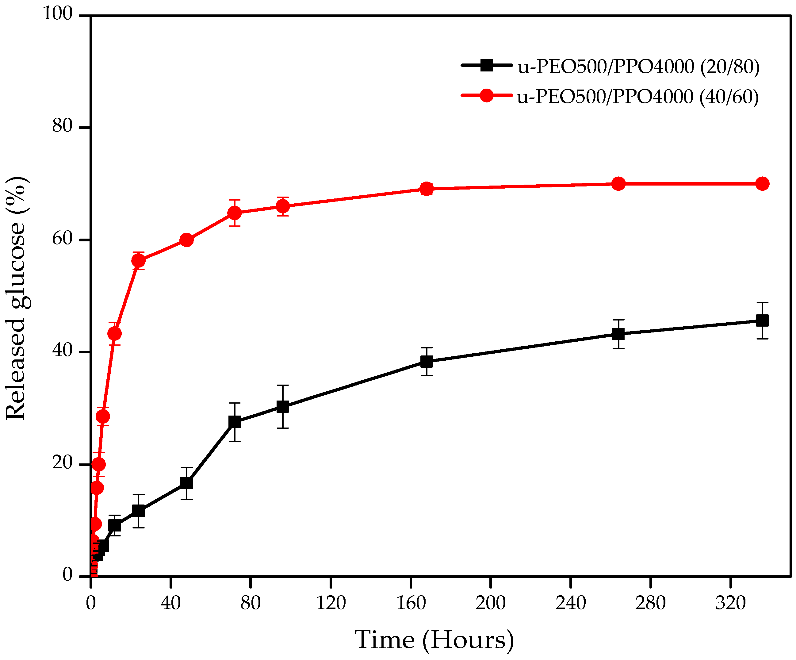
3.9. In Vitro Glucose Release from Ureasil-Polyether Membranes
4. Conclusions
Supplementary Materials
Author Contributions
Funding
Institutional Review Board Statement
Informed Consent Statement
Data Availability Statement
Acknowledgments
Conflicts of Interest
References
- Majidinia, M.; Sadeghpour, A.; Yousefi, B. The Roles of Signaling Pathways in Bone Repair and Regeneration. J. Cell. Physiol. 2018, 233, 2937–2948. [Google Scholar] [CrossRef]
- Schindeler, A.; McDonald, M.M.; Bokko, P.; Little, D.G. Bone Remodeling during Fracture Repair: The Cellular Picture. Semin. Cell Dev. Biol. 2008, 19, 459–466. [Google Scholar] [CrossRef] [PubMed]
- Oshiro, J.A.; Scardueli, C.R.; de Oliveira, G.J.P.L.; Marcantonio, R.A.C.; Chiavacci, L.A. Development of Ureasil–Polyether Membranes for Guided Bone Regeneration. Biomed. Phys. Eng. Express 2017, 3, 015019. [Google Scholar] [CrossRef]
- Petite, H.; Viateau, V.; Bensaid, W.; Meunier, A.; de Pollak, C.; Bourguignon, M.; Oudina, K.; Sedel, L.; Guillemin, G. Tissue-Engineered Bone Regeneration. Nat. Biotechnol. 2000, 18, 959–963. [Google Scholar] [CrossRef]
- Casagrande, S.; Tiribuzi, R.; Cassetti, E.; Selmin, F.; Gervasi, G.L.; Barberini, L.; Freddolini, M.; Ricci, M.; Schoubben, A.; Cerulli, G.G.; et al. Biodegradable Composite Porous Poly(Dl-Lactide-Co-Glycolide) Scaffold Supports Mesenchymal Stem Cell Differentiation and Calcium Phosphate Deposition. Artif. Cells Nanomed. Biotechnol. 2018, 46, 219–229. [Google Scholar] [CrossRef] [PubMed]
- García-Gareta, E.; Coathup, M.J.; Blunn, G.W. Osteoinduction of Bone Grafting Materials for Bone Repair and Regeneration. Bone 2015, 81, 112–121. [Google Scholar] [CrossRef]
- Gómez-Barrena, E.; Rosset, P.; Gebhard, F.; Hernigou, P.; Baldini, N.; Rouard, H.; Sensebé, L.; Gonzalo-Daganzo, R.M.; Giordano, R.; Padilla-Eguiluz, N.; et al. Feasibility and Safety of Treating Non-Unions in Tibia, Femur and Humerus with Autologous, Expanded, Bone Marrow-Derived Mesenchymal Stromal Cells Associated with Biphasic Calcium Phosphate Biomaterials in a Multicentric, Non-Comparative Trial. Biomaterials 2019, 196, 100–108. [Google Scholar] [CrossRef] [PubMed]
- Kawecki, F.; Clafshenkel, W.P.; Fortin, M.; Auger, F.A.; Fradette, J. Biomimetic Tissue-Engineered Bone Substitutes for Maxillofacial and Craniofacial Repair: The Potential of Cell Sheet Technologies. Adv. Healthc. Mater. 2018, 7, e1700919. [Google Scholar] [CrossRef]
- Manassero, M.; Viateau, V.; Deschepper, M.; Oudina, K.; Logeart-Avramoglou, D.; Petite, H.; Bensidhoum, M. Bone Regeneration in Sheep Using Acropora Coral, a Natural Resorbable Scaffold, and Autologous Mesenchymal Stem Cells. Tissue Eng. Part A 2013, 19, 1554–1563. [Google Scholar] [CrossRef]
- Oryan, A.; Baghaban Eslaminejad, M.; Kamali, A.; Hosseini, S.; Moshiri, A.; Baharvand, H. Mesenchymal Stem Cells Seeded onto Tissue-Engineered Osteoinductive Scaffolds Enhance the Healing Process of Critical-Sized Radial Bone Defects in Rat. Cell Tissue Res. 2018, 374, 63–81. [Google Scholar] [CrossRef]
- Anitua, E.; Tejero, R.; Zalduendo, M.M.; Orive, G. Plasma Rich in Growth Factors Promotes Bone Tissue Regeneration by Stimulating Proliferation, Migration, and Autocrine Secretion in Primary Human Osteoblasts. J. Periodontol. 2013, 84, 1180–1190. [Google Scholar] [CrossRef]
- Black, C.R.M.; Goriainov, V.; Gibbs, D.; Kanczler, J.; Tare, R.S.; Oreffo, R.O.C. Bone Tissue Engineering. Bone Tissue Eng. 2015, 1, 132–140. [Google Scholar] [CrossRef] [PubMed]
- Gómez-Barrena, E.; Rosset, P.; Lozano, D.; Stanovici, J.; Ermthaller, C.; Gerbhard, F. Bone Fracture Healing: Cell Therapy in Delayed Unions and Nonunions. Bone 2015, 70, 93–101. [Google Scholar] [CrossRef]
- Pereira, N.S.; Raquel, L.; Souza, D.B.; Manoela, I.; Portela, S. Regeneração Óssea Guiada Utilizando Membrana Reabsorvível Fixada Com Etilcianoacrilato. Rev. Bras. Odontol. 2012, 69, 39–42. [Google Scholar]
- Logeart-Avramoglou, D.; Anagnostou, F.; Bizios, R.; Petite, H. Engineering Bone: Challenges and Obstacles. J. Cell. Mol. Med. 2005, 9, 72–84. [Google Scholar] [CrossRef]
- Potier, E.; Ferreira, E.; Meunier, A.; Sedel, L.; Logeart-Avramoglou, D.; Petite, H. Prolonged Hypoxia Concomitant with Serum Deprivation Induces Massive Human Mesenchymal Stem Cell Death. Tissue Eng. 2007, 13, 1325–1331. [Google Scholar] [CrossRef] [PubMed]
- Becquart, P.; Cambon-Binder, A.; Monfoulet, L.E.; Bourguignon, M.; Vandamme, K.; Bensidhoum, M.; Petite, H.; Logeart-Avramoglou, D. Ischemia Is the Prime but Not the Only Cause of Human Multipotent Stromal Cell Death in Tissue-Engineered Constructs in Vivo. Tissue Eng. Part A 2012, 18, 2084–2094. [Google Scholar] [CrossRef]
- Deschepper, M.; Oudina, K.; David, B.; Myrtil, V.; Collet, C.; Bensidhoum, M.; Logeart-Avramoglou, D.; Petite, H. Survival and Function of Mesenchymal Stem Cells (MSCs) Depend on Glucose to Overcome Exposure to Long-Term, Severe and Continuous Hypoxia. J. Cell. Mol. Med. 2011, 15, 1505–1514. [Google Scholar] [CrossRef]
- Moya, A.; Larochette, N.; Paquet, J.; Deschepper, M.; Bensidhoum, M.; Izzo, V.; Kroemer, G.; Petite, H.; Logeart-Avramoglou, D. Quiescence Preconditioned Human Multipotent Stromal Cells Adopt a Metabolic Profile Favorable for Enhanced Survival under Ischemia. Stem Cells 2017, 35, 181–196. [Google Scholar] [CrossRef]
- Moya, A.; Paquet, J.; Deschepper, M.; Larochette, N.; Oudina, K.; Denoeud, C.; Bensidhoum, M.; Logeart-Avramoglou, D.; Petite, H. Human Mesenchymal Stem Cell Failure to Adapt to Glucose Shortage and Rapidly Use Intracellular Energy Reserves Through Glycolysis Explains Poor Cell Survival After Implantation. Stem Cells 2018, 36, 363–376. [Google Scholar] [CrossRef]
- Deschepper, M.; Manassero, M.; Oudina, K.; Paquet, J.; Monfoulet, L.-E.; Bensidhoum, M.; Logeart-Avramoglou, D.; Petite, H. Proangiogenic and Prosurvival Functions of Glucose in Human Mesenchymal Stem Cells Upon Transplantation. Stem Cells 2013, 31, 526–535. [Google Scholar] [CrossRef] [PubMed]
- Das, S.C.; Prakash, A. A Systematic Review on Drug Delivery Systems Based on Their Mechanism of Drug Release and Their Applications. Int. J. Res. Appl. Sci. Biotechnol. 2021, 8, 55–63. [Google Scholar] [CrossRef]
- Davoodi, P.; Lee, L.Y.; Xu, Q.; Sunil, V.; Sun, Y.; Soh, S.; Wang, C.H. Drug Delivery Systems for Programmed and On-Demand Release. Adv. Drug Deliv. Rev. 2018, 132, 104–138. [Google Scholar] [CrossRef] [PubMed]
- Jacob, J.; Haponiuk, J.T.; Thomas, S.; Gopi, S. Biopolymer Based Nanomaterials in Drug Delivery Systems: A Review. Mater. Today Chem. 2018, 9, 43–55. [Google Scholar] [CrossRef]
- Sharma, D.; Dev, D.; Prasad, D.N.; Hans, M. Sustained Release Drug Delivery System with the Role of Natural Polymers: A Review. J. Drug Deliv. Ther. 2019, 9, 913–923. [Google Scholar]
- Gauer, L.; Takemoto, M.; Zago, C.D.; Tagliari, D.; de Andrade, M.R. Regeneração Óssea Guiada Associada a Membrana de Politetrafluoretileno Expandido (PTFE-E). Tecnológica 2015, 3, 60–67. [Google Scholar]
- Judeinstein, P.; Sanchez, C. Hybrid Organic-Inorganic Materials: A Land of Multidisciplinarity. J. Mater. Chem. 1996, 6, 511–525. [Google Scholar] [CrossRef]
- Oshiro, J.A.; Abuçafy, M.P.; Manaia, E.B.; Da Silva, B.L.; Chiari-Andréo, B.G.; Chiavacci, L.A. Drug Delivery Systems Obtained from Silica Based Organic-Inorganic Hybrids. Polymers 2016, 8, 91. [Google Scholar] [CrossRef]
- Sanchez, C.; Julián, B.; Belleville, P.; Popall, M. Applications of Hybrid Organic-Inorganic Nanocomposites. J. Mater. Chem. 2005, 15, 3559–3592. [Google Scholar] [CrossRef]
- Dahmouche, K.; Santilli, C.V.; Pulcinelli, S.H.; Craievich, A.F. Small-Angle X-Ray Scattering Study of Sol-Gel-Derived Siloxane-PEG and Siloxane-PPG Hybrid Materials. J. Phys. Chem. B 1999, 103, 4937–4942. [Google Scholar] [CrossRef]
- Oshiro-Junior, J.A.; Moreno, R.M.; Silva, B.G.; Souza, C.C.; Scardueli, C.R.; Marcantonio, C.C.; Sanches, P.R.S.; Mendes, L.; Cilli, E.M.; Marcantonio, R.A.C.; et al. In vivo effectiveness of hybrid membranes with osteogenic growth peptide for bone regeneration. J. Tissue Eng. Regen. Med. 2021, 15, 722–731. [Google Scholar] [CrossRef] [PubMed]
- Sanchez, C.; Ribot, F.; Rozes, L.; Alonso, B. Design of Hybrid Organic-Inorganic Nanocomposites Synthesized via Sol-Gel Chemistry. Mol. Cryst. Liq. Cryst. 2000, 354, 143–158. [Google Scholar] [CrossRef]
- Nagandran, S.; Goh, P.S.; Ismail, A.F.; Wong, T.; Dagang, W.R.Z.B.W. The Recent Progress in Modification of Polymeric Membranes Using Organic Macromolecules for Water Treatment. Symmetry 2020, 12, 239. [Google Scholar] [CrossRef]
- José, N.M.; Prado, L.A.S.D.A. Hybrid Organic-Inorganic Materials: Preparation and Some Applications. Quim. Nova 2005, 28, 281–288. [Google Scholar] [CrossRef]
- Molina, E.F.; Pulcinelli, S.H.; Briois, V.; Santilli, C. V. Fine-Tuning of a Nanostructure, Swelling, and Drug Delivery Profile by Blending Ureasil-PEO and Ureasil-PPO Hybrids. Polym. Chem. 2014, 5, 1897–1904. [Google Scholar] [CrossRef]
- Truffault, L.; Magnani, M.; Hammer, P.; Santilli, C.V.; Pulcinelli, S.H. Structural and Optical Features of Ureasiloxane-Polyethylene Oxide Hybrids Containing CeO2 Nanoparticles. Colloids Surf. A Physicochem. Eng. Asp. 2015, 471, 73–80. [Google Scholar] [CrossRef]
- Zaldivar, M.P.; Santilli, C.V.; Peniche Covas, C.A.; Pulcinelli, S.H. Thermal Properties, Nanoscopic Structure and Swelling Behavior of Chitosan/(Ureasil–Polyethylene Oxide Hybrid) Blends. J. Therm. Anal. Calorim. 2017, 130, 791–798. [Google Scholar] [CrossRef]
- Sarker, B.; Lyer, S.; Arkudas, A.; Boccaccini, A.R. Collagen/Silica Nanocomposites and Hybrids for Bone Tissue Engineering. Nanotechnol. Rev. 2013, 2, 427–447. [Google Scholar] [CrossRef]
- Molina, E.F.; Jesus, C.R.N.; Chiavacci, L.A.; Pulcinelli, S.H.; Briois, V.; Santilli, C. V. Ureasil-Polyether Hybrid Blend with Tuneable Hydrophilic/Hydrophobic Features Based on U-PEO1900 and U-PPO400 Mixtures. J. Sol-Gel Sci. Technol. 2014, 70, 317–328. [Google Scholar] [CrossRef]
- Oshiro Junior, J.A.; Mortari, G.R.; de Freitas, R.M.; Marcantonio-Junior, E.; Lopes, L.; Spolidorio, L.C.; Marcantonio, R.A.; Chiavacci, L.A. Assessment of Biocompatibility of Ureasil-Polyether Hybrid Membranes for Future Use in Implantodontology. Int. J. Polym. Mater. Polym. Biomater. 2016, 65, 647–652. [Google Scholar] [CrossRef]
- Oshiro Junior, J.A.; Carvalho, F.C.; Soares, C.P.; Chorilli, M.; Chiavacci, L.A. Development of Cutaneous Bioadhesive Ureasil-Polyether Hybrid Films. Int. J. Polym. Sci. 2015, 2015, 727324. [Google Scholar] [CrossRef]
- ISO 23317; Implants for Surgery—In Vitro Evaluation for Apatite-Forming Ability of Implant Materials Implants. International Organization for Standardization: London, UK, 2007.
- De Souza, D.C.; de Luca Vahia de Abreu, H.; de Oliveira, P.V.; Capelo, L.P.; Passos-Bueno, M.R.; Catalani, L.H. A Fast Degrading PLLA Composite with a High Content of Functionalized Octacalcium Phosphate Mineral Phase Induces Stem Cells Differentiation. J. Mech. Behav. Biomed. Mater. 2019, 93, 93–104. [Google Scholar] [CrossRef] [PubMed]
- Bajpai, S.K.; Chand, N.; Ahuja, S.; Roy, M.K. Curcumin/Cellulose Micro Crystals/Chitosan Films: Water Absorption Behavior and in Vitro Cytotoxicity. Int. J. Biol. Macromol. 2015, 75, 239–247. [Google Scholar] [CrossRef] [PubMed]
- Chhatri, A.; Bajpai, J.; Bajpai, A.K.; Sandhu, S.S.; Jain, N.; Biswas, J. Cryogenic Fabrication of Savlon Loaded Macroporous Blends of Alginate and Polyvinyl Alcohol (PVA). Swelling, Deswelling and Antibacterial Behaviors. Carbohydr. Polym. 2011, 83, 876–882. [Google Scholar] [CrossRef]
- Liquiform, G. Glicose, Labtest; Labtest Diagnóstica: Lagoa Santa, Brazil, 2011; pp. 1–6. [Google Scholar]
- Costa, P.; Lobo, J.M.S. Modeling and Comparison of Dissolution Profiles Paulo. Eur. J. Pharm. Sci. 2001, 13, 123–133. [Google Scholar] [CrossRef]
- Salome, A.C.; Godswill, C.O.; Ikechukwu, I.O. Kinetics and Mechanisms of Drug Release from Swellable and Non Swellable Matrices: A Review. Res. J. Pharm. Biol. Chem. Sci. 2013, 4, 97–103. [Google Scholar]
- Lopes, L.; Molina, E.F.; Chiavacci, L.A.; Santilli, C.V.; Briois, V.; Pulcinelli, S.H. Drug-Matrix Interaction of Sodium Diclofenac Incorporated into Ureasil-Poly(Ethylene Oxide) Hybrid Materials. RSC Adv. 2012, 2, 5629–5636. [Google Scholar] [CrossRef]
- Moreno, R.M.; Silva, B.G.; Souza, C.C.; Silva-Junior, A.; Scardueli, C.R.; Marcantonio, R.A.C.; Chiavacii, L.A.; Oshiro-Junior, J.A. Dexamethasone-Loaded Ureasil Hydrophobic Membrane for Bone Guided Regeneration. Pharmaceutics 2022, 14, 1027. [Google Scholar] [CrossRef]
- Santilli, C.V.; Chiavacci, L.A.; Lopes, L.; Pulcinelli, S.H.; Oliveira, A.G. Controlled Drug Release from Ureasil-Polyether Hybrid Materials. Chem. Mater. 2009, 21, 463–467. [Google Scholar] [CrossRef]
- Liu, H.; Zhang, L.; Zuo, Y.; Wang, L.; Huang, D.; Shen, J.; Shi, P.; Li, Y. Preparation and Characterization of Aliphatic Polyurethane and Hydroxyapatite Composite Scaffold. J. Appl. Polym. Sci. 2009, 112, 2968–2975. [Google Scholar] [CrossRef]
- Gaharwar, A.K.; Dammu, S.A.; Canter, J.M.; Wu, C.J.; Schmidt, G. Highly Extensible, Tough, and Elastomeric Nanocomposite Hydrogels from Poly(Ethylene Glycol) and Hydroxyapatite Nanoparticles. Biomacromolecules 2011, 12, 1641–1650. [Google Scholar] [CrossRef]
- Gaharwar, A.K.; Schexnailder, P.J.; Kline, B.P.; Schmidt, G. Assessment of Using Laponite® Cross-Linked Poly(Ethylene Oxide) for Controlled Cell Adhesion and Mineralization. Acta Biomater. 2011, 7, 568–577. [Google Scholar] [CrossRef] [PubMed]
- Gaharwar, A.K.; Rivera, C.P.; Wu, C.J.; Schmidt, G. Transparent, Elastomeric and Tough Hydrogels from Poly(Ethylene Glycol) and Silicate Nanoparticles. Acta Biomater. 2011, 7, 4139–4148. [Google Scholar] [CrossRef] [PubMed]
- Patyk, E.; Katrusiak, A. Transformable H-Bonds and Conformation in Compressed Glucose. Chem. Sci. 2015, 6, 1991–1995. [Google Scholar] [CrossRef]
- Chiavacci, L.A.; Dahmouche, K.; Silva, N.J.O.; Carlos, L.D.; Amaral, V.S.; De Zea Bermudez, V.; Pulcinelli, S.H.; Santilli, C.V.; Briois, V.; Craievich, A.F. Effect of Presence of an Acid Catalyst on Structure and Properties of Iron-Doped Siloxane-Polyoxyethylene Nanocomposites Prepared by Sol-Gel. J. Non-Cryst. Solids 2004, 345–346, 585–590. [Google Scholar] [CrossRef]
- Carlos, L.D.; De Zea Bermudez, V.; Sá Ferreira, R.A.; Marques, L.; Assunção, M. Sol-Gel Derived Urea Cross-Linked Organically Modified Silicates. 2. Blue-Light Emission. Chem. Mater. 1999, 11, 581–588. [Google Scholar] [CrossRef]
- Hurtta, M.; Pitkänen, I.; Knuutinen, J. Melting Behaviour of D-Sucrose, D-Glucose and D-Fructose. Carbohydr. Res. 2004, 339, 2267–2273. [Google Scholar] [CrossRef] [PubMed]
- Rojek, B.; Wesolowski, M. DSC Supported by Factor Analysis as a Reliable Tool for Compatibility Study in Pharmaceutical Mixtures. J. Therm. Anal. Calorim. 2019, 138, 4531–4539. [Google Scholar] [CrossRef]
- Wang, Y.; Truong, T.; Li, H.; Bhandari, B. Co-Melting Behaviour of Sucrose, Glucose & Fructose. Food Chem. 2019, 275, 292–298. [Google Scholar] [CrossRef]
- Mendes, J.F.; Oshiro Junior, J.A.; Da Silva, C.G.; Chiavacci, L.A. Synthesis of Ureasil-Polyether Film Forming Materials by Using Environmentally Friendly Solvent. Rev. De Cienc. Farm. Basica E Apl. 2021, 42, 1–10. [Google Scholar] [CrossRef]
- Molina, E.F.; Marçal, L.; Pereira De Carvalho, H.W.; Nassar, E.J.; Ciuffi, K.J. Tri-Ureasil Gel as a Multifunctional Organic-Inorganic Hybrid Matrix. Polym. Chem. 2013, 4, 1575–1582. [Google Scholar] [CrossRef]
- Zaldivar, M.P. Materiais Híbridos Ureasil-Polióxido de Etileno/Quitosana Para Aplicação Na Liberação Controlada de Fármacos, Tese de Doutorado em Química; Universidade Estadual Paulista: São Paulo, Brazil, 2015. [Google Scholar]
- Oshiro, J.A.; Lusuardi, A.; Beamud, E.M.; Chiavacci, L.A.; Cuberes, M.T. Nanostructural Arrangements and Surface Morphology on Ureasil-Polyether Films Loaded with Dexamethasone Acetate. Nanomaterials 2021, 11, 1362. [Google Scholar] [CrossRef]
- Grundke, K.; Pöschel, K.; Synytska, A.; Frenzel, R.; Drechsler, A.; Nitschke, M.; Cordeiro, A.L.; Uhlmann, P.; Welzel, P.B. Experimental Studies of Contact Angle Hysteresis Phenomena on Polymer Surfaces—Toward the Understanding and Control of Wettability for Different Applications. Adv. Colloid Interface Sci. 2015, 222, 350–376. [Google Scholar] [CrossRef]
- Pavlenko, V.I.; Cherkashina, N.I. Synthesis of Hydrophobic Filler for Polymer Composites. Int. J. Eng. Technol. 2018, 7, 493–495. [Google Scholar] [CrossRef]
- Francisco Junior, W.E. Carboidratos: Estrutura, Propriedades e Funções. Quimica Nova na Escola. 2008, pp. 8–13. Available online: http://qnesc.sbq.org.br/online/qnesc29/03-CCD-2907.pdf (accessed on 28 December 2022).
- Macêdo, M.O.C.; Macêdo, H.R.A.; Silva, G.C.; Silva, M.A.M.; Júnior, C.A. Estudo Comparativo Da Modificação Superficial de Membranas de Quitosana Tratadas Por Plasma de Oxigênio, Nitrogênio e Hidrogênio. Revista eletrônica de Materiais e Processos 2012, 7, 95–103. [Google Scholar]
- Kokubo, T.; Yamaguchi, S. Simulated Body Fluid and the Novel Bioactive Materials Derived from It. J. Biomed. Mater. Res. Part A 2019, 107, 968–977. [Google Scholar] [CrossRef]
- Kokubo, T.; Takadama, H. How Useful Is SBF in Predicting in Vivo Bone Bioactivity? Biomaterials 2006, 27, 2907–2915. [Google Scholar] [CrossRef]
- Catauro, M.; Bollino, F.; Papale, F.; Gallicchio, M.; Pacifico, S. Influence of the Polymer Amount on Bioactivity and Biocompatibility of SiO2/PEG Hybrid Materials Synthesized by Sol-Gel Technique. Mater. Sci. Eng. C 2015, 48, 548–555. [Google Scholar] [CrossRef]
- Kokubo, T.; Kushitani, H.; Sakka, S.; Kitsugi, T.; Yamamum, T. Solutions Able to Reproduce in Vivo Surface-Structure Changes in Bioactive Glass-Ceramic A-W3. J. Biomed. Mater. Res. 1990, 24, 721–734. [Google Scholar] [CrossRef]
- Sossa, P.A.F.; Giraldo, B.S.; Garcia, B.C.G.; Parra, E.R.; Arango, P.J.A. Comparative Study between Natural and Synthetic Hydroxyapatite: Structural, Morphological and Bioactivity Properties. Rev. Mater. 2018, 23, 1–17. [Google Scholar] [CrossRef]
- Bigham, A.; Kermani, S.; Saudi, A.; Aghajanian, A.H.; Rafienia, M. On the Bioactivity and Mechanical Properties of Gehlenite Nanobioceramic: A Comparative Study. J. Med. Signals Sens. 2020, 10, 105–112. [Google Scholar]
- Saber-Samandari, S.; Saber-Samandari, S.; Ghonjizade-Samani, F.; Aghazadeh, J.; Sadeghi, A. Bioactivity Evaluation of Novel Nanocomposite Scaffolds for Bone Tissue Engineering: The Impact of Hydroxyapatite. Ceram. Int. 2016, 42, 11055–11062. [Google Scholar] [CrossRef]
- Sutha, S.; Kavitha, K.; Karunakaran, G.; Rajendran, V. In-Vitro Bioactivity, Biocorrosion and Antibacterial Activity of Silicon Integrated Hydroxyapatite/Chitosan Composite Coating on 316 L Stainless Steel Implants. Mater. Sci. Eng. C 2013, 33, 4046–4054. [Google Scholar] [CrossRef]
- Mary, M.C.S.; Chatterjee, A.; Abraham, J.; Sasikumar, S. A Study of the In-Vitro Bioactivity, Dissolution and Antibacterial Activity of Larnite Prepared by a Novel Sol–Gel Combustion Method Using Sucrose as a Fuel. Bull. Mater. Sci. 2020, 43, 237. [Google Scholar] [CrossRef]
- Takadama, H.; Kokubo, T. In Vitro Evaluation of Bone Bioactivity. In Bioceramics and Their Clinical Applications; Woodhead Publishing Limited: Toyko, Japan, 2008; pp. 165–182. ISBN 9781845692049. [Google Scholar]
- Mundstock, K.B.; De Oliveira, A.P.N.; Hotza, D.; Rogero, S.O. Avaliação Da Biocompatibilidade de Vidro e Vitrocerâmica Do Sistema SNCP (SiO2-Na2O-CaO-P2O5). Quim. Nova 2012, 35, 665–670. [Google Scholar] [CrossRef]
- Padilla, S.; Román, J.; Sánchez-Salcedo, S.; Vallet-Regí, M. Hydroxyapatite/SiO2-CaO-P2O5 Glass Materials: In Vitro Bioactivity and Biocompatibility. Acta Biomater. 2006, 2, 331–342. [Google Scholar] [CrossRef]
- Toskas, G.; Cherif, C.; Hund, R.D.; Laourine, E.; Mahltig, B.; Fahmi, A.; Heinemann, C.; Hanke, T. Chitosan(PEO)/Silica Hybrid Nanofibers as a Potential Biomaterial for Bone Regeneration. Carbohydr. Polym. 2013, 94, 713–722. [Google Scholar] [CrossRef]
- Bellucci, D.; Cannillo, V.; Sola, A.; Chiellini, F.; Gazzarri, M.; Migone, C. Macroporous Bioglass®-Derived Scaffolds for Bone Tissue Regeneration. Ceram. Int. 2011, 37, 1575–1585. [Google Scholar] [CrossRef]
- Greenbaum, J.; Nirmalan, M. Acid-Base Balance: The Traditional Approach. Curr. Anaesth. Crit. Care 2005, 16, 137–142. [Google Scholar] [CrossRef]
- ISO 10993-4; Biological Evaluation of Medical Devices—Part 4: Selection of Tests for Interactions with Blood. International Organization for Standardization: London, UK, 2002; p. 34.
- Singh, N.; Sahoo, S.K.; Kumar, R. Hemolysis Tendency of Anticancer Nanoparticles Changes with Type of Blood Group Antigen: An Insight into Blood Nanoparticle Interactions. Mater. Sci. Eng. C 2020, 109, 110645. [Google Scholar] [CrossRef]
- Gawlikowski, M.; El Fray, M.; Janiczak, K.; Zawidlak-Węgrzyńska, B.; Kustosz, R. In-Vitro Biocompatibility and Hemocompatibility Study of New Pet Copolyesters Intended for Heart Assist Devices. Polymers 2020, 12, 2857. [Google Scholar] [CrossRef] [PubMed]
- Luo, P.; Nie, M.; Wen, H.; Xu, W.; Fan, L.; Cao, Q. Preparation and Characterization of Carboxymethyl Chitosan Sulfate/Oxidized Konjac Glucomannan Hydrogels. Int. J. Biol. Macromol. 2018, 113, 1024–1031. [Google Scholar] [CrossRef] [PubMed]
- Weber, M.; Steinle, H.; Golombek, S.; Hann, L.; Schlensak, C.; Wendel, H.P.; Avci-Adali, M. Blood-Contacting Biomaterials: In Vitro Evaluation of the Hemocompatibility. Front. Bioeng. Biotechnol. 2018, 6, 99. [Google Scholar] [CrossRef]
- Paredes, M.; Pulcinelli, S.H.; Peniche, C.; Gonçalves, V.; Santilli, C. V. Chitosan/(Ureasil–PEO Hybrid) Blend for Drug Delivery. J. Sol-Gel Sci. Technol. 2014, 72, 233–238. [Google Scholar] [CrossRef]
- Molina, E.F.; Pulcinelli, S.H.; Santilli, C.V.; Blanchandin, S.; Briois, V. Controlled Cisplatin Delivery from Ureasil-PEO1900 Hybrid Matrix. J. Phys. Chem. B 2010, 114, 3461–3466. [Google Scholar] [CrossRef] [PubMed]
- Ritger, P.L.; Peppas, N.A. A simple equation for description of solute release I. Fickian and non-fickian release from non-swellable devices in the form of slabs, spheres, cylinders or DISCS. J. Control. Release 1986, 5, 23–26. [Google Scholar] [CrossRef]
- Ritger, P.L.; Peppas, N.A. A Simple Equation for Description of Solute Release II. Fickian and Anomalous Release from Swellable Devices. J. Control. Release 1987, 5, 37–42. [Google Scholar] [CrossRef]
- Peppas, N.A.; Sahlin, J.J. A Simple Equation for the Description of Solute Release. III. Coupling of Diffusion and Relaxation. Int. J. Pharm. 1989, 57, 169–172. [Google Scholar] [CrossRef]


Disclaimer/Publisher’s Note: The statements, opinions and data contained in all publications are solely those of the individual author(s) and contributor(s) and not of MDPI and/or the editor(s). MDPI and/or the editor(s) disclaim responsibility for any injury to people or property resulting from any ideas, methods, instructions or products referred to in the content. |
© 2023 by the authors. Licensee MDPI, Basel, Switzerland. This article is an open access article distributed under the terms and conditions of the Creative Commons Attribution (CC BY) license (https://creativecommons.org/licenses/by/4.0/).
Share and Cite
da Silva, C.G.; Monteiro, J.R.; Oshiro-Júnior, J.A.; Chiavacci, L.A. Hybrid Membranes of the Ureasil-Polyether Containing Glucose for Future Application in Bone Regeneration. Pharmaceutics 2023, 15, 1474. https://doi.org/10.3390/pharmaceutics15051474
da Silva CG, Monteiro JR, Oshiro-Júnior JA, Chiavacci LA. Hybrid Membranes of the Ureasil-Polyether Containing Glucose for Future Application in Bone Regeneration. Pharmaceutics. 2023; 15(5):1474. https://doi.org/10.3390/pharmaceutics15051474
Chicago/Turabian Styleda Silva, Camila Garcia, João Rodrigues Monteiro, João Augusto Oshiro-Júnior, and Leila Aparecida Chiavacci. 2023. "Hybrid Membranes of the Ureasil-Polyether Containing Glucose for Future Application in Bone Regeneration" Pharmaceutics 15, no. 5: 1474. https://doi.org/10.3390/pharmaceutics15051474