
Hydrodynamic Delivery: Characteristics, Applications, and Technological Advances

 , , ,
, , ,
Abstract
:1. Introduction
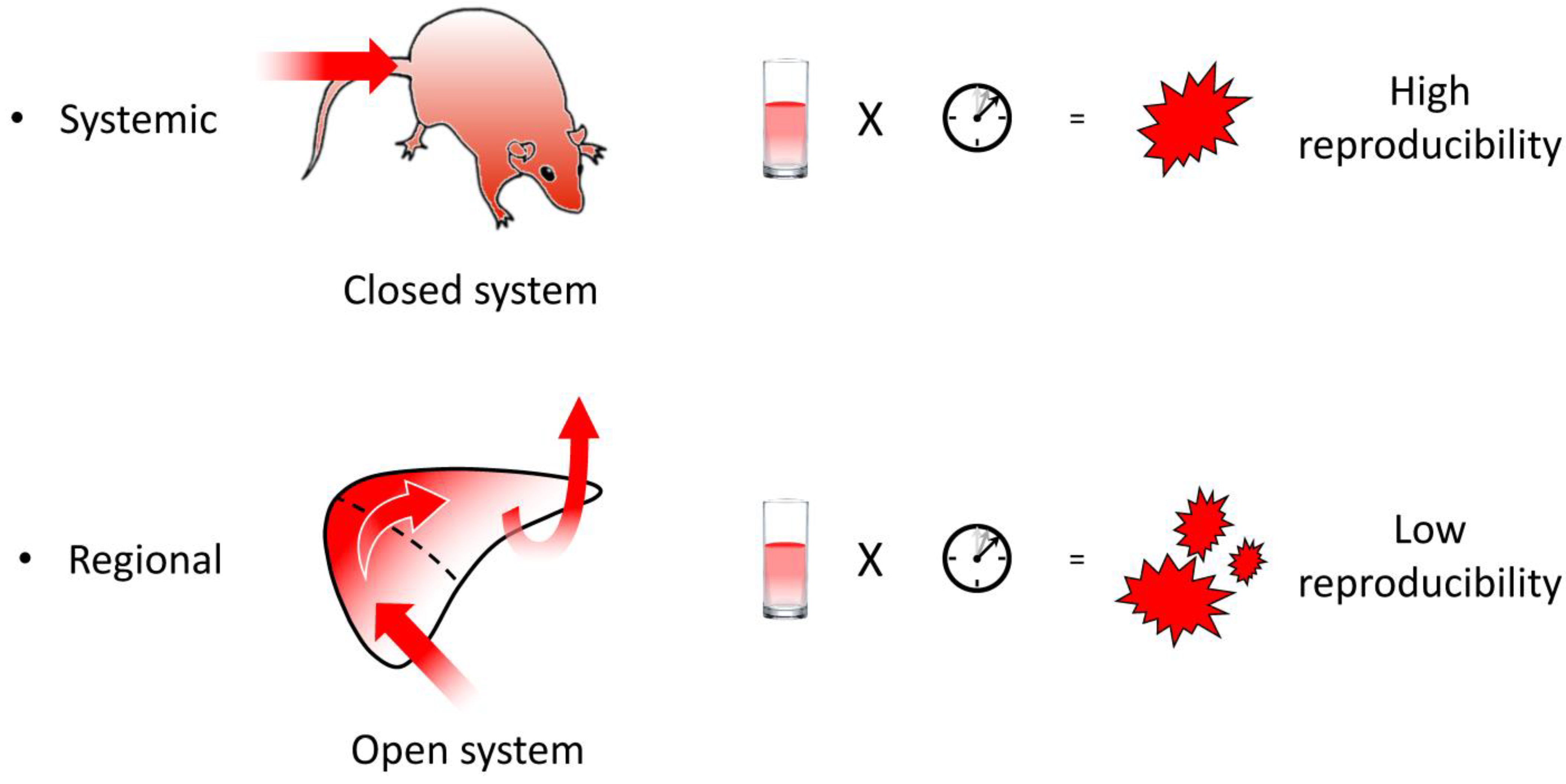
2. Characteristics of Hydrodynamic Delivery
3. Applications of Hydrodynamic Delivery
3.1. Target Animals, Organs, and Routes of Injection

| Target\Animal | Mouse | Rat | Treeshrew | Chicken | Rabbit | Pig | Dog | Monkey | Baboon | Human | |
|---|---|---|---|---|---|---|---|---|---|---|---|
| Systemic | LVR | TV [1,2] | TV [19] | ROS [20] | JV [21] | ||||||
| KDNY | JV [21] | ||||||||||
| BCEC | TV [22] | ||||||||||
| FTS | TV [25] | ||||||||||
| IST HCC | TV [23,24,52] | ||||||||||
| Regional | LVR | IVC, PV [2,12,18] | IVC, PV, BD, ex vivo [12,54,57,59] | IVC, HV [27] | IVC, HV, PV, BD [12,28,29,30,31,32,33,34,35,55,56,58] | HV [37,38] | HV under prep. | ex vivo [41] | |||
| KDNY | RV, RP [43] | RV [12,44,45,60] | RV [12] | ||||||||
| MSL | TA, LV, TV [46,47,48,61,62] | LV, LA * [12,49,63,64,65,66,67] | LV [26] | LV, LA [36,67] | LV, LA [39,40] | ||||||
| PCAS | SMV [50] | ||||||||||
| GND | LA, GV, GA [39] | ||||||||||
| HCC | HA [51] | ||||||||||
| BT | CA [22,53] | ||||||||||
| MCD | ex vivo [42,68] | ||||||||||
| SV | ex vivo [42] | ||||||||||
3.2. Targeted Diseases
| Infectious | Cancer | Hereditary | Liver |
|---|---|---|---|
| Hepatitis B virus (HBV) [72,73,84,85] | Hepatocellular carcinoma [51,92,93,94,95] | Hemophilia A and B [70,107,127,128,129,130] | Liver fibrosis [82,103,104,149,150,151] |
| Hepatitis C virus [71,75,76,87,88,89,90,91] | Hepatoblastoma [96,97,98] | Pseudoxanthoma elasticum [113] | Nonalcoholic fatty liver diseases [103,105,106,152,153] |
| Hepatitis D virus [74] | Cholangiocellular carcinoma [99,100,101,102] | von Willebrand disease [108,109,131,132] | Alcoholic liver injury [154,155] |
| Influenza virus [77,78] | Colorectal cancer [23,24,120,121,122] | Thrombotic thrombocytopenic purpura [110,111,133,134] | Portal hypertension [156] |
| Enterovirus 71 [112] | Lung cancer [123] | Mucopolysaccharidosis I and VII [135,136] | Fulminant hepatitis & regeneration [157,158,159] |
| Vaccination (HBV, Malaria, Influenza) [77,79,80,84,85,86] | Brain tumor [53] | Phenylketonuria [137] | Acute liver injury [160,161] |
| Malaria parasite [79,80] | Lymphoma [124] | Tyrosinemia [138,139,140] | Others |
| Streptococcus [162] | Melanoma [24,125,126] | Leber congenital amaurosis [141] | Atopic skin & cutaneous diseases [114,117,164,165] |
| Sepsis [81] | Metastasis (melanoma, breast cancer, RCC * (lungs, liver, kidneys)) [118,119,121] | Sickle cell disease [142] | Cardiovascular & ischemic diseases [115,166,167,168,169,170,171] |
| Trypanosome [163] | *, renal cell carcinoma | Cystathionine β-synthase deficiency [143] | Kidney diseases & hyperparathyroidism [172,173,174,175] |
| Fabry disease [144] | Diabetes mellitus & obesity [176,177,178,179] | ||
| α-1 antitrypsin deficiency [69] | Hypertriglyceridemia [83] | ||
| Growth hormone deficiency [145] | Inflammatory diseases [180,181,182,183,184] | ||
| Metachromatic leukodystrophy [146] | Osteoporosis [185] | ||
| Short-chain acyl-CoA dehydrogen. def. [147] | Transplantation & intoxication [59,186,187] | ||
| Muscular dystrophy [148] | Humanized immune system [116,117] |
3.3. Technological Issues
3.3.1. Delivery of Materials
3.3.2. Genome Editing (Somatic Gene Editing)
3.3.3. Regional Hydrodynamic Delivery
3.3.4. Miscellaneous
4. Conclusions
Author Contributions
Funding
Institutional Review Board Statement
Informed Consent Statement
Data Availability Statement
Conflicts of Interest
References
- Liu, F.; Song, Y.; Liu, D. Hydrodynamics-Based Transfection in Animals by Systemic Administration of Plasmid DNA. Gene Ther. 1999, 6, 1258–1266. [Google Scholar] [CrossRef] [PubMed]
- Zhang, G.; Budker, V.; Wolff, J.A. High Levels of Foreign Gene Expression in Hepatocytes after Tail Vein Injections of Naked Plasmid DNA. Hum. Gene Ther. 1999, 10, 1735–1737. [Google Scholar] [CrossRef]
- Raper, S.E.; Chirmule, N.; Lee, F.S.; Wivel, N.A.; Bagg, A.; Gao, G.; Wilson, J.M.; Batshaw, M.L. Fatal Systemic Inflammatory Response Syndrome in a Ornithine Transcarbamylase Deficient Patient Following Adenoviral Gene Transfer. Mol. Genet. Metab. 2003, 80, 148–158. [Google Scholar] [CrossRef] [PubMed]
- Manno, C.S.; Pierce, G.F.; Arruda, V.R.; Glader, B.; Ragni, M.; Rasko, J.J.; Ozelo, M.C.; Hoots, K.; Blatt, P.; Konkle, B.; et al. Successful Transduction of Liver in Hemophilia by AAV-Factor IX and Limitations Imposed by the Host Immune Response. Nat. Med. 2006, 12, 342–347. [Google Scholar] [CrossRef] [PubMed]
- Mingozzi, F.; Maus, M.V.; Hui, D.J.; Sabatino, D.E.; Murphy, S.L.; Rasko, J.E.J.; Ragni, M.V.; Manno, C.S.; Sommer, J.; Jiang, H.; et al. CD8(+) T-Cell Responses to Adeno-Associated Virus Capsid in Humans. Nat. Med. 2007, 13, 419–422. [Google Scholar] [CrossRef]
- Suda, T.; Gao, X.; Stolz, D.B.; Liu, D. Structural Impact of Hydrodynamic Injection on Mouse Liver. Gene Ther. 2007, 14, 129–137. [Google Scholar] [CrossRef]
- Kanefuji, T.; Yokoo, T.; Suda, T.; Abe, H.; Kamimura, K.; Liu, D. Hemodynamics of a Hydrodynamic Injection. Mol. Ther. Methods Clin. Dev. 2014, 1, 14029. [Google Scholar] [CrossRef]
- Zhang, G.; Gao, X.; Song, Y.K.; Vollmer, R.; Stolz, D.B.; Gasiorowski, J.Z.; Dean, D.A.; Liu, D. Hydroporation as the Mechanism of Hydrodynamic Delivery. Gene Ther. 2004, 11, 675–682. [Google Scholar] [CrossRef]
- Kobayashi, N.; Kuramoto, T.; Yamaoka, K.; Hashida, M.; Takakura, Y. Hepatic Uptake and Gene Expression Mechanisms Following Intravenous Administration of Plasmid DNA by Conventional and Hydrodynamics-Based Procedures. J. Pharmacol. Exp. Ther. 2001, 297, 853–860. [Google Scholar]
- Magin-Lachmann, C.; Kotzamanis, G.; D’Aiuto, L.; Cooke, H.; Huxley, C.; Wagner, E. In Vitro and in Vivo Delivery of Intact BAC DNA—Comparison of Different Methods. J. Gene Med. 2004, 6, 195–209. [Google Scholar] [CrossRef]
- Kobayashi, N.; Nishikawa, M.; Hirata, K.; Takakura, Y. Hydrodynamics-Based Procedure Involves Transient Hyperpermeability in the Hepatic Cellular Membrane: Implication of a Nonspecific Process in Efficient Intracellular Gene Delivery. J. Gene Med. 2004, 6, 584–592. [Google Scholar] [CrossRef]
- Suda, T.; Suda, K.; Liu, D. Computer-Assisted Hydrodynamic Gene Delivery. Mol. Ther. J. Am. Soc. Gene Ther. 2008, 16, 1098–1104. [Google Scholar] [CrossRef] [PubMed]
- Andrianaivo, F.; Lecocq, M.; Wattiaux-De Coninck, S.; Wattiaux, R.; Jadot, M. Hydrodynamics-Based Transfection of the Liver: Entrance into Hepatocytes of DNA That Causes Expression Takes Place Very Early after Injection. J. Gene Med. 2004, 6, 877–883. [Google Scholar] [CrossRef] [PubMed]
- Maynard, R.L.; Downes, N. Chapter 8—Histology of the Vascular System. In Anatomy and Histology of the Laboratory Rat in Toxicology and Biomedical Research; Maynard, R.L., Downes, N., Eds.; Academic Press: Cambridge, MA, USA, 2019; pp. 91–95. ISBN 978-0-12-811837-5. [Google Scholar]
- Yeikilis, R.; Gal, S.; Kopeiko, N.; Paizi, M.; Pines, M.; Braet, F.; Spira, G. Hydrodynamics Based Transfection in Normal and Fibrotic Rats. World J. Gastroenterol. 2006, 12, 6149–6155. [Google Scholar] [CrossRef] [PubMed]
- Kobayashi, Y.; Kamimura, K.; Abe, H.; Yokoo, T.; Ogawa, K.; Shinagawa-Kobayashi, Y.; Goto, R.; Inoue, R.; Ohtsuka, M.; Miura, H.; et al. Effects of Fibrotic Tissue on Liver-Targeted Hydrodynamic Gene Delivery. Mol. Ther. Nucleic Acids 2016, 5, e359. [Google Scholar] [CrossRef] [PubMed]
- Budker, V.G.; Subbotin, V.M.; Budker, T.; Sebestyén, M.G.; Zhang, G.; Wolff, J.A. Mechanism of Plasmid Delivery by Hydrodynamic Tail Vein Injection. II. Morphological Studies. J. Gene Med. 2006, 8, 874–888. [Google Scholar] [CrossRef]
- Suda, T.; Liu, D. Hydrodynamic Delivery. Adv. Genet. 2015, 89, 89–111. [Google Scholar] [CrossRef]
- Kameda, S.; Maruyama, H.; Higuchi, N.; Nakamura, G.; Iino, N.; Nishikawa, Y.; Miyazaki, J.; Gejyo, F. Hydrodynamics-Based Transfer of PCR-Amplified DNA Fragments into Rat Liver. Biochem. Biophys. Res. Commun. 2003, 309, 929–936. [Google Scholar] [CrossRef]
- Yan, S.; Fu, Q.; Zhou, Y.; Wang, J.; Liu, Y.; Duan, X.; Jia, S.; Peng, J.; Gao, B.; Du, J.; et al. High Levels of Gene Expression in the Hepatocytes of Adult Mice, Neonatal Mice and Tree Shrews via Retro-Orbital Sinus Hydrodynamic Injections of Naked Plasmid DNA. J. Control. Release Off. J. Control. Release Soc. 2012, 161, 763–771. [Google Scholar] [CrossRef]
- Hen, G.; Bor, A.; Simchaev, V.; Druyan, S.; Yahav, S.; Miao, C.H.; Friedman-Einat, M. Expression of Foreign Genes in Chicks by Hydrodynamics-Based Naked Plasmid Transfer in Vivo. Domest. Anim. Endocrinol. 2006, 30, 135–143. [Google Scholar] [CrossRef]
- Hino, T.; Yokota, T.; Ito, S.; Nishina, K.; Kang, Y.-S.; Mori, S.; Hori, S.; Kanda, T.; Terasaki, T.; Mizusawa, H. In Vivo Delivery of Small Interfering RNA Targeting Brain Capillary Endothelial Cells. Biochem. Biophys. Res. Commun. 2006, 340, 263–267. [Google Scholar] [CrossRef] [PubMed]
- Hamana, A.; Takahashi, Y.; Nishikawa, M.; Takakura, Y. Interferon-Inducible Mx Promoter-Driven, Long-Term Transgene Expression System of Interferon-β for Cancer Gene Therapy. Hum. Gene Ther. 2016, 27, 936–945. [Google Scholar] [CrossRef]
- Ochoa, M.C.; Fioravanti, J.; Rodriguez, I.; Hervas-Stubbs, S.; Azpilikueta, A.; Mazzolini, G.; Gúrpide, A.; Prieto, J.; Pardo, J.; Berraondo, P.; et al. Antitumor Immunotherapeutic and Toxic Properties of an HDL-Conjugated Chimeric IL-15 Fusion Protein. Cancer Res. 2013, 73, 139–149. [Google Scholar] [CrossRef] [PubMed]
- Nakamura, S.; Ando, N.; Watanabe, S.; Akasaka, E.; Ishihara, M.; Sato, M. Hydrodynamics-Based Transplacental Delivery as a Useful Noninvasive Tool for Manipulating Fetal Genome. Cells 2020, 9, 1744. [Google Scholar] [CrossRef]
- Bates, M.K.; Zhang, G.; Sebestyén, M.G.; Neal, Z.C.; Wolff, J.A.; Herweijer, H. Genetic Immunization for Antibody Generation in Research Animals by Intravenous Delivery of Plasmid DNA. BioTechniques 2006, 40, 199–208. [Google Scholar] [CrossRef]
- Eastman, S.J.; Baskin, K.M.; Hodges, B.L.; Chu, Q.; Gates, A.; Dreusicke, R.; Anderson, S.; Scheule, R.K. Development of Catheter-Based Procedures for Transducing the Isolated Rabbit Liver with Plasmid DNA. Hum. Gene Ther. 2002, 13, 2065–2077. [Google Scholar] [CrossRef]
- Chan, T.; Grisch-Chan, H.M.; Schmierer, P.; Subotic, U.; Rimann, N.; Scherer, T.; Hetzel, U.; Bozza, M.; Harbottle, R.; Williams, J.A.; et al. Delivery of Non-Viral Naked DNA Vectors to Liver in Small Weaned Pigs by Hydrodynamic Retrograde Intrabiliary Injection. Mol. Ther. Methods Clin. Dev. 2022, 24, 268–279. [Google Scholar] [CrossRef]
- Kamimura, K.; Suda, T.; Zhang, G.; Aoyagi, Y.; Liu, D. Parameters Affecting Image-Guided, Hydrodynamic Gene Delivery to Swine Liver. Mol. Ther. Nucleic Acids 2013, 2, e128. [Google Scholar] [CrossRef] [PubMed]
- Kumbhari, V.; Li, L.; Piontek, K.; Ishida, M.; Fu, R.; Khalil, B.; Garrett, C.M.; Liapi, E.; Kalloo, A.N.; Selaru, F.M. Successful Liver-Directed Gene Delivery by ERCP-Guided Hydrodynamic Injection (with Videos). Gastrointest. Endosc. 2018, 88, 755–763.e5. [Google Scholar] [CrossRef]
- Sendra, L.; Carreño, O.; Miguel, A.; Montalvá, E.; Herrero, M.J.; Orbis, F.; Noguera, I.; Barettino, D.; López-Andújar, R.; Aliño, S.F. Low RNA Translation Activit Limits the Efficacy of Hydrodynamic Gene Transfer to Pig Liver in Vivo. J. Gene Med. 2014, 16, 179–192. [Google Scholar] [CrossRef]
- Sendra, L.; Herrero, M.J.; Montalvá, E.M.; Noguera, I.; Orbis, F.; Díaz, A.; Fernández-Delgado, R.; López-Andújar, R.; Aliño, S.F. Efficacy of Interleukin 10 Gene Hydrofection in Pig Liver Vascular Isolated “in Vivo” by Surgical Procedure with Interest in Liver Transplantation. PLoS ONE 2019, 14, e0224568. [Google Scholar] [CrossRef] [PubMed]
- Stoller, F.; Schlegel, A.; Viecelli, H.M.; Rüfenacht, V.; Cesarovic, N.; Viecelli, C.; Deplazes, S.; Bettschart, R.; Hurter, K.; Schmierer, P.; et al. Hepatocyte Transfection in Small Pigs After Weaning by Hydrodynamic Intraportal Injection of Naked DNA/Minicircle Vectors. Hum. Gene Ther. Methods 2015, 26, 181–192. [Google Scholar] [CrossRef] [PubMed]
- Zacharoulis, D.; Rountas, C.; Katsimpoulas, M.; Morianos, J.; Chatziandreou, I.; Vassilopoulos, G. Efficient Liver Gene Transfer with Foamy Virus Vectors. Med. Sci. Monit. Basic Res. 2013, 19, 214–220. [Google Scholar] [CrossRef] [PubMed]
- Kamimura, K.; Suda, T.; Xu, W.; Zhang, G.; Liu, D. Image-Guided, Lobe-Specific Hydrodynamic Gene Delivery to Swine Liver. Mol. Ther. J. Am. Soc. Gene Ther. 2009, 17, 491–499. [Google Scholar] [CrossRef]
- Kamimura, K.; Zhang, G.; Liu, D. Image-Guided, Intravascular Hydrodynamic Gene Delivery to Skeletal Muscle in Pigs. Mol. Ther. J. Am. Soc. Gene Ther. 2010, 18, 93–100. [Google Scholar] [CrossRef]
- Kamimura, K.; Kanefuji, T.; Yokoo, T.; Abe, H.; Suda, T.; Kobayashi, Y.; Zhang, G.; Aoyagi, Y.; Liu, D. Safety Assessment of Liver-Targeted Hydrodynamic Gene Delivery in Dogs. PLoS ONE 2014, 9, e107203. [Google Scholar] [CrossRef]
- Noda, M.; Tatsumi, K.; Matsui, H.; Matsunari, Y.; Sato, T.; Fukuoka, Y.; Hotta, A.; Okano, T.; Kichikawa, K.; Sugimoto, M.; et al. Development of Alternative Gene Transfer Techniques for Ex Vivo and in Vivo Gene Therapy in a Canine Model. Regen. Ther. 2021, 18, 347–354. [Google Scholar] [CrossRef]
- Zhang, G.; Budker, V.; Williams, P.; Subbotin, V.; Wolff, J.A. Efficient Expression of Naked Dna Delivered Intraarterially to Limb Muscles of Nonhuman Primates. Hum. Gene Ther. 2001, 12, 427–438. [Google Scholar] [CrossRef]
- Hagstrom, J.E.; Hegge, J.; Zhang, G.; Noble, M.; Budker, V.; Lewis, D.L.; Herweijer, H.; Wolff, J.A. A Facile Nonviral Method for Delivering Genes and SiRNAs to Skeletal Muscle of Mammalian Limbs. Mol. Ther. J. Am. Soc. Gene Ther. 2004, 10, 386–398. [Google Scholar] [CrossRef]
- Herrero, M.J.; Sabater, L.; Guenechea, G.; Sendra, L.; Montilla, A.I.; Abargues, R.; Navarro, V.; Aliño, S.F. DNA Delivery to “ex Vivo” Human Liver Segments. Gene Ther. 2012, 19, 504–512. [Google Scholar] [CrossRef]
- Mann, M.J.; Gibbons, G.H.; Hutchinson, H.; Poston, R.S.; Hoyt, E.G.; Robbins, R.C.; Dzau, V.J. Pressure-Mediated Oligonucleotide Transfection of Rat and Human Cardiovascular Tissues. Proc. Natl. Acad. Sci. USA 1999, 96, 6411–6416. [Google Scholar] [CrossRef] [PubMed]
- Woodard, L.E.; Welch, R.C.; Williams, F.M.; Luo, W.; Cheng, J.; Wilson, M.H. Hydrodynamic Renal Pelvis Injection for Non-Viral Expression of Proteins in the Kidney. J. Vis. Exp. JoVE 2018, 8, 56324. [Google Scholar] [CrossRef]
- Corridon, P.R.; Rhodes, G.J.; Leonard, E.C.; Basile, D.P.; Gattone, V.H.; Bacallao, R.L.; Atkinson, S.J. A Method to Facilitate and Monitor Expression of Exogenous Genes in the Rat Kidney Using Plasmid and Viral Vectors. Am. J. Physiol. Renal Physiol. 2013, 304, F1217–F1229. [Google Scholar] [CrossRef]
- Maruyama, H.; Higuchi, N.; Kameda, S.; Nakamura, G.; Iguchi, S.; Miyazaki, J.-I.; Gejyo, F. Rat Kidney-Targeted Naked Plasmid DNA Transfer by Retrograde Injection into the Renal Vein. Mol. Biotechnol. 2004, 27, 23–31. [Google Scholar] [CrossRef] [PubMed]
- Girardin, C.; Maze, D.; Gonçalves, C.; Le Guen, Y.T.; Pluchon, K.; Pichon, C.; Montier, T.; Midoux, P. Selective Attachment of a Microtubule Interacting Peptide to Plasmid DNA via a Triplex Forming Oligonucleotide for Transfection Improvement. Gene Ther. 2022; Online ahead of print. [Google Scholar] [CrossRef] [PubMed]
- Guess, M.G.; Barthel, K.K.; Pugach, E.K.; Leinwand, L.A. Measuring MicroRNA Reporter Activity in Skeletal Muscle Using Hydrodynamic Limb Vein Injection of Plasmid DNA Combined with in Vivo Imaging. Skelet. Muscle 2013, 3, 19. [Google Scholar] [CrossRef]
- Le Guen, Y.T.; Le Gall, T.; Midoux, P.; Guégan, P.; Braun, S.; Montier, T. Gene Transfer to Skeletal Muscle Using Hydrodynamic Limb Vein Injection: Current Applications, Hurdles and Possible Optimizations. J. Gene Med. 2020, 22, e3150. [Google Scholar] [CrossRef]
- Yasuzaki, Y.; Yamada, Y.; Ishikawa, T.; Harashima, H. Validation of Mitochondrial Gene Delivery in Liver and Skeletal Muscle via Hydrodynamic Injection Using an Artificial Mitochondrial Reporter DNA Vector. Mol. Pharm. 2015, 12, 4311–4320. [Google Scholar] [CrossRef]
- Ogawa, K.; Kamimura, K.; Kobayashi, Y.; Abe, H.; Yokoo, T.; Sakai, N.; Nagoya, T.; Sakamaki, A.; Abe, S.; Hayashi, K.; et al. Efficacy and Safety of Pancreas-Targeted Hydrodynamic Gene Delivery in Rats. Mol. Ther. Nucleic Acids 2017, 9, 80–88. [Google Scholar] [CrossRef]
- Tada, M.; Hatano, E.; Taura, K.; Nitta, T.; Koizumi, N.; Ikai, I.; Shimahara, Y. High Volume Hydrodynamic Injection of Plasmid DNA via the Hepatic Artery Results in a High Level of Gene Expression in Rat Hepatocellular Carcinoma Induced by Diethylnitrosamine. J. Gene Med. 2006, 8, 1018–1026. [Google Scholar] [CrossRef]
- Kamimura, K.; Yokoo, T.; Abe, H.; Sakai, N.; Nagoya, T.; Kobayashi, Y.; Ohtsuka, M.; Miura, H.; Sakamaki, A.; Kamimura, H.; et al. Effect of Diphtheria Toxin-Based Gene Therapy for Hepatocellular Carcinoma. Cancers 2020, 12, 472. [Google Scholar] [CrossRef] [PubMed]
- Barnett, F.H.; Scharer-Schuksz, M.; Wood, M.; Yu, X.; Wagner, T.E.; Friedlander, M. Intra-Arterial Delivery of Endostatin Gene to Brain Tumors Prolongs Survival and Alters Tumor Vessel Ultrastructure. Gene Ther. 2004, 11, 1283–1289. [Google Scholar] [CrossRef] [PubMed]
- Zhang, X.; Dong, X.; Sawyer, G.J.; Collins, L.; Fabre, J.W. Regional Hydrodynamic Gene Delivery to the Rat Liver with Physiological Volumes of DNA Solution. J. Gene Med. 2004, 6, 693–703. [Google Scholar] [CrossRef]
- Huang, Y.; Kruse, R.L.; Ding, H.; Itani, M.I.; Morrison, J.; Wang, Z.Z.; Selaru, F.M.; Kumbhari, V. Parameters of Biliary Hydrodynamic Injection during Endoscopic Retrograde Cholangio-Pancreatography in Pigs for Applications in Gene Delivery. PLoS ONE 2021, 16, e0249931. [Google Scholar] [CrossRef] [PubMed]
- Kruse, R.L.; Huang, Y.; Shum, T.; Bai, L.; Ding, H.; Wang, Z.Z.; Selaru, F.M.; Kumbhari, V. Endoscopic-Mediated, Biliary Hydrodynamic Injection Mediating Clinically Relevant Levels of Gene Delivery in Pig Liver. Gastrointest. Endosc. 2021, 94, 1119–1130.e4. [Google Scholar] [CrossRef]
- Jiang, X.; Ren, Y.; Williford, J.-M.; Li, Z.; Mao, H.-Q. Liver-Targeted Gene Delivery through Retrograde Intrabiliary Infusion. Methods Mol. Biol. 2013, 948, 275–284. [Google Scholar] [CrossRef]
- Sendra, L.; Miguel, A.; Pérez-Enguix, D.; Herrero, M.J.; Montalvá, E.; García-Gimeno, M.A.; Noguera, I.; Díaz, A.; Pérez, J.; Sanz, P.; et al. Studying Closed Hydrodynamic Models of “In Vivo” DNA Perfusion in Pig Liver for Gene Therapy Translation to Humans. PLoS ONE 2016, 11, e0163898. [Google Scholar] [CrossRef]
- Tsoulfas, G.; Takahashi, Y.; Liu, D.; Yagnik, G.; Wu, T.; Murase, N.; Geller, D.A. Hydrodynamic Plasmid DNA Gene Therapy Model in Liver Transplantation. J. Surg. Res. 2006, 135, 242–249. [Google Scholar] [CrossRef]
- Woodard, L.E.; Cheng, J.; Welch, R.C.; Williams, F.M.; Luo, W.; Gewin, L.S.; Wilson, M.H. Kidney-Specific Transposon-Mediated Gene Transfer in Vivo. Sci. Rep. 2017, 7, 44904. [Google Scholar] [CrossRef]
- Liang, K.W.; Nishikawa, M.; Liu, F.; Sun, B.; Ye, Q.; Huang, L. Restoration of Dystrophin Expression in Mdx Mice by Intravascular Injection of Naked DNA Containing Full-Length Dystrophin CDNA. Gene Ther. 2004, 11, 901–908. [Google Scholar] [CrossRef]
- Nagata, K.; Itaka, K.; Baba, M.; Uchida, S.; Ishii, T.; Kataoka, K. Muscle-Targeted Hydrodynamic Gene Introduction of Insulin-like Growth Factor-1 Using Polyplex Nanomicelle to Treat Peripheral Nerve Injury. J. Control. Release Off. J. Control. Release Soc. 2014, 183, 27–34. [Google Scholar] [CrossRef] [PubMed]
- Yasuzaki, Y.; Yamada, Y.; Fukuda, Y.; Harashima, H. Condensation of Plasmid DNA Enhances Mitochondrial Association in Skeletal Muscle Following Hydrodynamic Limb Vein Injection. Pharmaceuticals 2014, 7, 881–893. [Google Scholar] [CrossRef]
- Yasuzaki, Y.; Yamada, Y.; Kanefuji, T.; Harashima, H. Localization of Exogenous DNA to Mitochondria in Skeletal Muscle Following Hydrodynamic Limb Vein Injection. J. Control. Release Off. J. Control. Release Soc. 2013, 172, 805–811. [Google Scholar] [CrossRef] [PubMed]
- Sato, Y.; Ajiki, T.; Inoue, S.; Hakamata, Y.; Murakami, T.; Kaneko, T.; Takahashi, M.; Kobayashi, E. A Novel Gene Therapy to the Graft Organ by a Rapid Injection of Naked DNA I: Long-Lasting Gene Expression in a Rat Model of Limb Transplantation. Transplantation 2003, 76, 1294–1298. [Google Scholar] [CrossRef] [PubMed]
- Budker, V.; Zhang, G.; Danko, I.; Williams, P.; Wolff, J. The Efficient Expression of Intravascularly Delivered DNA in Rat Muscle. Gene Ther. 1998, 5, 272–276. [Google Scholar] [CrossRef] [PubMed]
- Danialou, G.; Comtois, A.S.; Matecki, S.; Nalbantoglu, J.; Karpati, G.; Gilbert, R.; Geoffroy, P.; Gilligan, S.; Tanguay, J.-F.; Petrof, B.J. Optimization of Regional Intraarterial Naked DNA-Mediated Transgene Delivery to Skeletal Muscles in a Large Animal Model. Mol. Ther. J. Am. Soc. Gene Ther. 2005, 11, 257–266. [Google Scholar] [CrossRef]
- Su, L.T.; Gopal, K.; Wang, Z.; Yin, X.; Nelson, A.; Kozyak, B.W.; Burkman, J.M.; Mitchell, M.A.; Low, D.W.; Bridges, C.R.; et al. Uniform Scale-Independent Gene Transfer to Striated Muscle after Transvenular Extravasation of Vector. Circulation 2005, 112, 1780–1788. [Google Scholar] [CrossRef]
- Zhang, G.; Song, Y.K.; Liu, D. Long-Term Expression of Human Alpha1-Antitrypsin Gene in Mouse Liver Achieved by Intravenous Administration of Plasmid DNA Using a Hydrodynamics-Based Procedure. Gene Ther. 2000, 7, 1344–1349. [Google Scholar] [CrossRef]
- Miao, C.H.; Thompson, A.R.; Loeb, K.; Ye, X. Long-Term and Therapeutic-Level Hepatic Gene Expression of Human Factor IX after Naked Plasmid Transfer In Vivo. Mol. Ther. J. Am. Soc. Gene Ther. 2001, 3, 947–957. [Google Scholar] [CrossRef]
- McCaffrey, A.P.; Meuse, L.; Karimi, M.; Contag, C.H.; Kay, M.A. A Potent and Specific Morpholino Antisense Inhibitor of Hepatitis C Translation in Mice. Hepatology 2003, 38, 503–508. [Google Scholar] [CrossRef]
- McCaffrey, A.P.; Nakai, H.; Pandey, K.; Huang, Z.; Salazar, F.H.; Xu, H.; Wieland, S.F.; Marion, P.L.; Kay, M.A. Inhibition of Hepatitis B Virus in Mice by RNA Interference. Nat. Biotechnol. 2003, 21, 639–644. [Google Scholar] [CrossRef] [PubMed]
- Yang, P.L.; Althage, A.; Chung, J.; Chisari, F.V. Hydrodynamic Injection of Viral DNA: A Mouse Model of Acute Hepatitis B Virus Infection. Proc. Natl. Acad. Sci. USA 2002, 99, 13825–13830. [Google Scholar] [CrossRef] [PubMed]
- Chang, J.; Sigal, L.J.; Lerro, A.; Taylor, J. Replication of the Human Hepatitis Delta Virus Genome Is Initiated in Mouse Hepatocytes Following Intravenous Injection of Naked DNA or RNA Sequences. J. Virol. 2001, 75, 3469–3473. [Google Scholar] [CrossRef]
- Zhu, W.; Wu, C.; Deng, W.; Pei, R.; Wang, Y.; Cao, L.; Qin, B.; Lu, M.; Chen, X. Inhibition of the HCV Core Protein on the Immune Response to HBV Surface Antigen and on HBV Gene Expression and Replication in Vivo. PLoS ONE 2012, 7, e45146. [Google Scholar] [CrossRef] [PubMed]
- Yu, W.; Grubor-Bauk, B.; Gargett, T.; Garrod, T.; Gowans, E.J. A Novel Challenge Model to Evaluate the Efficacy of Hepatitis C Virus Vaccines in Mice. Vaccine 2014, 32, 3409–3416. [Google Scholar] [CrossRef]
- Yamazaki, T.; Nagashima, M.; Ninomiya, D.; Ainai, A.; Fujimoto, A.; Ichimonji, I.; Takagi, H.; Morita, N.; Murotani, K.; Hasegawa, H.; et al. Neutralizing Antibodies Induced by Gene-Based Hydrodynamic Injection Have a Therapeutic Effect in Lethal Influenza Infection. Front. Immunol. 2018, 9, 47. [Google Scholar] [CrossRef] [PubMed]
- Lin, Y.; Wu, C.; Wang, X.; Liu, S.; Zhao, K.; Kemper, T.; Yu, H.; Li, M.; Zhang, J.; Chen, M.; et al. Glucosamine Promotes Hepatitis B Virus Replication through Its Dual Effects in Suppressing Autophagic Degradation and Inhibiting MTORC1 Signaling. Autophagy 2020, 16, 548–561. [Google Scholar] [CrossRef]
- Chen, L.; Keitany, G.J.; Peng, X.; Gibson, C.; Mohar, I.; Vignali, M.; Crispe, I.N.; Huang, F.; Wang, R. Identification of Pre-Erythrocytic Malaria Antigens That Target Hepatocytes for Killing in Vivo and Contribute to Protection Elicited by Whole-Parasite Vaccination. PLoS ONE 2014, 9, e102225. [Google Scholar] [CrossRef]
- Rai, U.; Huang, J.; Mishra, S.; Li, X.; Shiratsuchi, T.; Tsuji, M. A New Method to Determine Antigen-Specific CD8+ T Cell Activity in Vivo by Hydrodynamic Injection. Biomolecules 2012, 2, 23–33. [Google Scholar] [CrossRef]
- Wesche-Soldato, D.E.; Lomas-Neira, J.; Perl, M.; Chung, C.-S.; Ayala, A. Hydrodynamic Delivery of SiRNA in a Mouse Model of Sepsis. Methods Mol. Biol. 2008, 442, 67–73. [Google Scholar] [CrossRef]
- Yang, K.-L.; Hung, K.-C.; Chang, W.-T.; Li, E.I.C. Establishment of an Early Liver Fibrosis Model by the Hydrodynamics-Based Transfer of TGF-Beta1 Gene. Comp. Hepatol. 2007, 6, 9. [Google Scholar] [CrossRef] [PubMed]
- Sun, K.; Yang, W.; Huang, Y.; Wang, Y.; Xiang, L.; Qi, J. Leu452His Mutation in Lipoprotein Lipase Gene Transfer Associated with Hypertriglyceridemia in Mice in Vivo. PLoS ONE 2013, 8, e75462. [Google Scholar] [CrossRef] [PubMed]
- Zhang, E.; Kosinska, A.D.; Ma, Z.; Dietze, K.K.; Xu, Y.; Meng, Z.; Zhang, X.; Wang, J.; Wang, B.; Dittmer, U.; et al. Woodchuck Hepatitis Virus Core Antigen-Based DNA and Protein Vaccines Induce Qualitatively Different Immune Responses That Affect T Cell Recall Responses and Antiviral Effects. Virology 2015, 475, 56–65. [Google Scholar] [CrossRef] [PubMed]
- Huang, H.; Rückborn, M.; Le-Trilling, V.T.K.; Zhu, D.; Yang, S.; Zhou, W.; Yang, X.; Feng, X.; Lu, Y.; Lu, M.; et al. Prophylactic and Therapeutic HBV Vaccination by an HBs-Expressing Cytomegalovirus Vector Lacking an Interferon Antagonist in Mice. Eur. J. Immunol. 2021, 51, 393–407. [Google Scholar] [CrossRef] [PubMed]
- Yamazaki, T.; Chiba, J.; Akashi-Takamura, S. Neutralizing Anti-Hemagglutinin Monoclonal Antibodies Induced by Gene-Based Transfer Have Prophylactic and Therapeutic Effects on Influenza Virus Infection. Vaccines 2018, 6, 35. [Google Scholar] [CrossRef] [PubMed]
- Duan, L.; Yan, Y.; Liu, J.; Wang, B.; Li, P.; Hu, Q.; Chen, W. Target Delivery of Small Interfering RNAs with Vitamin E-Coupled Nanoparticles for Treating Hepatitis C. Sci. Rep. 2016, 6, 24867. [Google Scholar] [CrossRef] [PubMed]
- Ahlén, G.; Sällberg, M.; Frelin, L. Methods for Monitoring Gene Gun-Induced HBV- and HCV-Specific Immune Responses in Mouse Models. Methods Mol. Biol. 2013, 940, 239–267. [Google Scholar] [CrossRef]
- Nakai, M.; Oshiumi, H.; Funami, K.; Okamoto, M.; Matsumoto, M.; Seya, T.; Sakamoto, N. Interferon (IFN) and Cellular Immune Response Evoked in RNA-Pattern Sensing During Infection with Hepatitis C Virus (HCV). Sensors 2015, 15, 27160–27173. [Google Scholar] [CrossRef]
- Fu, Q.; Yan, S.; Wang, L.; Duan, X.; Wang, L.; Wang, Y.; Wu, T.; Wang, X.; An, J.; Zhang, Y.; et al. Hepatic NK Cell-Mediated Hypersensitivity to ConA-Induced Liver Injury in Mouse Liver Expressing Hepatitis C Virus Polyprotein. Oncotarget 2017, 8, 52178–52192. [Google Scholar] [CrossRef]
- Moyo, B.; Bloom, K.; Scott, T.; Ely, A.; Arbuthnot, P. Advances with Using CRISPR/Cas-Mediated Gene Editing to Treat Infections with Hepatitis B Virus and Hepatitis C Virus. Virus Res. 2018, 244, 311–320. [Google Scholar] [CrossRef]
- Ho, C.; Wang, C.; Mattu, S.; Destefanis, G.; Ladu, S.; Delogu, S.; Armbruster, J.; Fan, L.; Lee, S.A.; Jiang, L.; et al. AKT (v-Akt Murine Thymoma Viral Oncogene Homolog 1) and N-Ras (Neuroblastoma Ras Viral Oncogene Homolog) Coactivation in the Mouse Liver Promotes Rapid Carcinogenesis by Way of MTOR (Mammalian Target of Rapamycin Complex 1), FOXM1 (Forkhead Box M1)/SKP2, and c-Myc Pathways. Hepatol. Baltim. Md. 2012, 55, 833–845. [Google Scholar] [CrossRef]
- Engelholm, L.H.; Riaz, A.; Serra, D.; Dagnæs-Hansen, F.; Johansen, J.V.; Santoni-Rugiu, E.; Hansen, S.H.; Niola, F.; Frödin, M. CRISPR/Cas9 Engineering of Adult Mouse Liver Demonstrates That the Dnajb1-Prkaca Gene Fusion Is Sufficient to Induce Tumors Resembling Fibrolamellar Hepatocellular Carcinoma. Gastroenterology 2017, 153, 1662–1673.e10. [Google Scholar] [CrossRef] [PubMed]
- Moon, H.; Ju, H.-L.; Chung, S.I.; Cho, K.J.; Eun, J.W.; Nam, S.W.; Han, K.-H.; Calvisi, D.F.; Ro, S.W. Transforming Growth Factor-β Promotes Liver Tumorigenesis in Mice via Up-Regulation of Snail. Gastroenterology 2017, 153, 1378–1391.e6. [Google Scholar] [CrossRef]
- Gao, M.; Liu, D. CRISPR/Cas9-Based Pten Knock-out and Sleeping Beauty Transposon-Mediated Nras Knock-in Induces Hepatocellular Carcinoma and Hepatic Lipid Accumulation in Mice. Cancer Biol. Ther. 2017, 18, 505–512. [Google Scholar] [CrossRef] [PubMed]
- Zhang, W.; Meyfeldt, J.; Wang, H.; Kulkarni, S.; Lu, J.; Mandel, J.A.; Marburger, B.; Liu, Y.; Gorka, J.E.; Ranganathan, S.; et al. β-Catenin Mutations as Determinants of Hepatoblastoma Phenotypes in Mice. J. Biol. Chem. 2019, 294, 17524–17542. [Google Scholar] [CrossRef] [PubMed]
- Smith, J.L.; Rodríguez, T.C.; Mou, H.; Kwan, S.-Y.; Pratt, H.; Zhang, X.-O.; Cao, Y.; Liang, S.; Ozata, D.M.; Yu, T.; et al. YAP1 Withdrawal in Hepatoblastoma Drives Therapeutic Differentiation of Tumor Cells to Functional Hepatocyte-Like Cells. Hepatol. Baltim. Md. 2021, 73, 1011–1027. [Google Scholar] [CrossRef]
- Wang, D.; Tian, J.; Yan, Z.; Yuan, Q.; Wu, D.; Liu, X.; Yang, S.; Guo, S.; Wang, J.; Yang, Y.; et al. Mitochondrial Fragmentation Is Crucial for C-Myc-Driven Hepatoblastoma-like Liver Tumors. Mol. Ther. J. Am. Soc. Gene Ther. 2022, 30, 1645–1660. [Google Scholar] [CrossRef]
- Chen, X.; Calvisi, D.F. Hydrodynamic Transfection for Generation of Novel Mouse Models for Liver Cancer Research. Am. J. Pathol. 2014, 184, 912–923. [Google Scholar] [CrossRef]
- Wang, J.; Wang, H.; Peters, M.; Ding, N.; Ribback, S.; Utpatel, K.; Cigliano, A.; Dombrowski, F.; Xu, M.; Chen, X.; et al. Loss of Fbxw7 Synergizes with Activated Akt Signaling to Promote C-Myc Dependent Cholangiocarcinogenesis. J. Hepatol. 2019, 71, 742–752. [Google Scholar] [CrossRef]
- Zhang, J.; Chen, W.; Ma, W.; Song, K.; Lee, S.; Han, C.; Wu, T. Epigenetic Silencing of 15-Hydroxyprostaglandin Dehydrogenase by Histone Methyltransferase EHMT2/G9a in Cholangiocarcinoma. Mol. Cancer Res. MCR 2022, 20, 350–360. [Google Scholar] [CrossRef]
- Zhang, Y.; Xu, H.; Cui, G.; Liang, B.; Chen, X.; Ko, S.; Affo, S.; Song, X.; Liao, Y.; Feng, J.; et al. β-Catenin Sustains and Is Required for YES-Associated Protein Oncogenic Activity in Cholangiocarcinoma. Gastroenterology 2022, 163, 481–494. [Google Scholar] [CrossRef] [PubMed]
- Zhang, Z.; Yuan, Y.; Hu, L.; Tang, J.; Meng, Z.; Dai, L.; Gao, Y.; Ma, S.; Wang, X.; Yuan, Y.; et al. ANGPTL8 Accelerates Liver Fibrosis Mediated by HFD-Induced Inflammatory Activity via LILRB2/ERK Signaling Pathways. J. Adv. Res. 2022; Online ahead of print. [Google Scholar] [CrossRef] [PubMed]
- Matsuda, M.; Tsurusaki, S.; Miyata, N.; Saijou, E.; Okochi, H.; Miyajima, A.; Tanaka, M. Oncostatin M Causes Liver Fibrosis by Regulating Cooperation between Hepatic Stellate Cells and Macrophages in Mice. Hepatol. Baltim. Md. 2018, 67, 296–312. [Google Scholar] [CrossRef]
- Wang, H.; Chen, X. Hydrodynamic Injection for Developing NASH Model. Methods Mol. Biol. 2022, 2455, 31–39. [Google Scholar] [CrossRef]
- Cheng, Y.-W.; Chen, K.-W.; Kuo, H.-C.; Kuo, C.-H.; Lin, W.-H.; Chen, P.-J.; Yeh, S.-H. Specific Diacylglycerols Generated by Hepatic Lipogenesis Stimulate the Oncogenic Androgen Receptor Activity in Male Hepatocytes. Int. J. Obes. 2019, 43, 2469–2479. [Google Scholar] [CrossRef]
- Balestra, D.; Scalet, D.; Pagani, F.; Rogalska, M.E.; Mari, R.; Bernardi, F.; Pinotti, M. An Exon-Specific U1snRNA Induces a Robust Factor IX Activity in Mice Expressing Multiple Human FIX Splicing Mutants. Mol. Ther. Nucleic Acids 2016, 5, e370. [Google Scholar] [CrossRef]
- Navarrete, A.-M.; Casari, C.; Legendre, P.; Marx, I.; Hu, J.-R.; Lenting, P.J.; Christophe, O.D.; Denis, C.V. A Murine Model to Characterize the Antithrombotic Effect of Molecules Targeting Human von Willebrand Factor. Blood 2012, 120, 2723–2732. [Google Scholar] [CrossRef]
- Legendre, P.; Navarrete, A.-M.; Rayes, J.; Casari, C.; Boisseau, P.; Ternisien, C.; Caron, C.; Fressinaud, E.; Goudemand, J.; Veyradier, A.; et al. Mutations in the A3 Domain of von Willebrand Factor Inducing Combined Qualitative and Quantitative Defects in the Protein. Blood 2013, 121, 2135–2143. [Google Scholar] [CrossRef]
- Desch, K.C.; Kretz, C.; Yee, A.; Gildersleeve, R.; Metzger, K.; Agrawal, N.; Cheng, J.; Ginsburg, D. Probing ADAMTS13 Substrate Specificity Using Phage Display. PLoS ONE 2015, 10, e0122931. [Google Scholar] [CrossRef]
- Verhenne, S.; Vandeputte, N.; Pareyn, I.; Izsvák, Z.; Rottensteiner, H.; Deckmyn, H.; De Meyer, S.F.; Vanhoorelbeke, K. Long-Term Prevention of Congenital Thrombotic Thrombocytopenic Purpura in ADAMTS13 Knockout Mice by Sleeping Beauty Transposon-Mediated Gene Therapy. Arterioscler. Thromb. Vasc. Biol. 2017, 37, 836–844. [Google Scholar] [CrossRef]
- Qin, B.; Yan, Q.; Chen, X.; Xu, X.; Wang, Y.; Chen, J.; Dong, X. Enterovirus 71 Infection Impairs the Reproductive Capacity of Female Mice. Exp. Ther. Med. 2017, 14, 403–409. [Google Scholar] [CrossRef] [PubMed]
- Jin, L.; Jiang, Q.; Wu, Z.; Shao, C.; Zhou, Y.; Yang, L.; Uitto, J.; Wang, G. Genetic Heterogeneity of Pseudoxanthoma Elasticum: The Chinese Signature Profile of ABCC6 and ENPP1 Mutations. J. Investig. Dermatol. 2015, 135, 1294–1302. [Google Scholar] [CrossRef] [PubMed]
- Leys, L.; Wang, Y.; Paulsboe, S.; Edelmayer, R.; Salte, K.; Wetter, J.; Namovic, M.; Phillips, L.; Dunstan, R.; Gauvin, D.; et al. Characterization of Psoriasiform Dermatitis Induced by Systemic Injection of Interleukin-23 Minicircles in Mice. J. Dermatol. 2019, 46, 482–497. [Google Scholar] [CrossRef] [PubMed]
- Louet, E.R.; Glavan, M.; Orset, C.; Parcq, J.; Hanley, D.F.; Vivien, D. TPA-NMDAR Signaling Blockade Reduces the Incidence of Intracerebral Aneurysms. Transl. Stroke Res. 2022, 13, 1005–1016. [Google Scholar] [CrossRef]
- Ren, D.; Liu, W.; Ding, S.; Li, Y. Protocol for Generating Human Immune System Mice and Hydrodynamic Injection to Analyze Human Hematopoiesis in Vivo. STAR Protoc. 2022, 3, 101217. [Google Scholar] [CrossRef]
- Mencarelli, A.; Gunawan, M.; Yong, K.S.M.; Bist, P.; Tan, W.W.S.; Tan, S.Y.; Liu, M.; Huang, E.K.; Fan, Y.; Chan, J.K.Y.; et al. A Humanized Mouse Model to Study Mast Cells Mediated Cutaneous Adverse Drug Reactions. J. Leukoc. Biol. 2020, 107, 797–807. [Google Scholar] [CrossRef]
- Alhamhoom, Y.; Zhang, G.; Gao, M.; Cai, H.; Liu, D. In Vivo Growth and Responses to Treatment of Renal Cell Carcinoma in Different Environments. Am. J. Cancer Res. 2017, 7, 301–311. [Google Scholar]
- Li, J.; Yao, Q.; Liu, D. Hydrodynamic Cell Delivery for Simultaneous Establishment of Tumor Growth in Mouse Lung, Liver and Kidney. Cancer Biol. Ther. 2011, 12, 737–741. [Google Scholar] [CrossRef]
- Ochoa, M.C.; Fioravanti, J.; Duitman, E.H.; Medina-Echeverz, J.; Palazon, A.; Arina, A.; Dubrot, J.; Alfaro, C.; Morales-Kastresana, A.; Murillo, O.; et al. Liver Gene Transfer of Interkeukin-15 Constructs That Become Part of Circulating High Density Lipoproteins for Immunotherapy. PLoS ONE 2012, 7, e52370. [Google Scholar] [CrossRef]
- Cheng, L.; Du, X.; Wang, Z.; Ju, J.; Jia, M.; Huang, Q.; Xing, Q.; Xu, M.; Tan, Y.; Liu, M.; et al. Hyper-IL-15 Suppresses Metastatic and Autochthonous Liver Cancer by Promoting Tumour-Specific CD8+ T Cell Responses. J. Hepatol. 2014, 61, 1297–1303. [Google Scholar] [CrossRef]
- Miyakawa, N.; Nishikawa, M.; Takahashi, Y.; Ando, M.; Misaka, M.; Watanabe, Y.; Takakura, Y. Gene Delivery of Albumin Binding Peptide-Interferon-Gamma Fusion Protein with Improved Pharmacokinetic Properties and Sustained Biological Activity. J. Pharm. Sci. 2013, 102, 3110–3118. [Google Scholar] [CrossRef] [PubMed]
- Sun, H.; Liu, D. IL-15/SIL-15Rα Gene Transfer Suppresses Lewis Lung Cancer Growth in the Lungs, Liver and Kidneys. Cancer Gene Ther. 2016, 23, 54–60. [Google Scholar] [CrossRef] [PubMed]
- Pang, X.; Ma, F.; Zhang, P.; Zhong, Y.; Zhang, J.; Wang, T.; Zheng, G.; Hou, X.; Zhao, J.; He, C.; et al. Treatment of Human B-Cell Lymphomas Using Minicircle DNA Vector Expressing Anti-CD3/CD20 in a Mouse Model. Hum. Gene Ther. 2017, 28, 216–225. [Google Scholar] [CrossRef] [PubMed]
- Cueto, F.J.; Del Fresno, C.; Brandi, P.; Combes, A.J.; Hernández-García, E.; Sánchez-Paulete, A.R.; Enamorado, M.; Bromley, C.P.; Gomez, M.J.; Conde-Garrosa, R.; et al. DNGR-1 Limits Flt3L-Mediated Antitumor Immunity by Restraining Tumor-Infiltrating Type I Conventional Dendritic Cells. J. Immunother. Cancer 2021, 9, e002054. [Google Scholar] [CrossRef]
- Mecozzi, N.; Nenci, A.; Vera, O.; Bok, I.; Falzone, A.; DeNicola, G.M.; Karreth, F.A. Genetic Tools for the Stable Overexpression of Circular RNAs. RNA Biol. 2022, 19, 353–363. [Google Scholar] [CrossRef]
- Mashausi, D.S.; Roy, D.; Mangukiya, H.B.; Merugu, S.B.; Raza, G.; Yunus, F.-U.-N.; Liu, G.-S.; Negi, H.; Li, D. A High Efficient FVIII Variant Corrects Bleeding in Hemophilia A Mouse Model. Biochem. Biophys. Res. Commun. 2022, 637, 358–364. [Google Scholar] [CrossRef]
- Fu, R.Y.; Chen, A.C.; Lyle, M.J.; Chen, C.-Y.; Liu, C.L.; Miao, C.H. CD4+ T Cells Engineered with FVIII-CAR and Murine Foxp3 Suppress Anti-Factor VIII Immune Responses in Hemophilia a Mice. Cell. Immunol. 2020, 358, 104216. [Google Scholar] [CrossRef]
- Huai, C.; Jia, C.; Sun, R.; Xu, P.; Min, T.; Wang, Q.; Zheng, C.; Chen, H.; Lu, D. CRISPR/Cas9-Mediated Somatic and Germline Gene Correction to Restore Hemostasis in Hemophilia B Mice. Hum. Genet. 2017, 136, 875–883. [Google Scholar] [CrossRef]
- Mohammed, B.M.; Cheng, Q.; Matafonov, A.; Monroe, D.M.; Meijers, J.C.M.; Gailani, D. Factor XI Promotes Hemostasis in Factor IX-Deficient Mice. J. Thromb. Haemost. JTH 2018, 16, 2044–2049. [Google Scholar] [CrossRef]
- Pruss, C.M.; Golder, M.; Bryant, A.; Hegadorn, C.; Haberichter, S.; Lillicrap, D. Use of a Mouse Model to Elucidate the Phenotypic Effects of the von Willebrand Factor Cleavage Mutants, Y1605A/M1606A and R1597W. J. Thromb. Haemost. JTH 2012, 10, 940–950. [Google Scholar] [CrossRef]
- Swystun, L.L.; Georgescu, I.; Mewburn, J.; Deforest, M.; Nesbitt, K.; Hebert, K.; Dwyer, C.; Brown, C.; Notley, C.; Lillicrap, D. Abnormal von Willebrand Factor Secretion, Factor VIII Stabilization and Thrombus Dynamics in Type 2N von Willebrand Disease Mice. J. Thromb. Haemost. JTH 2017, 15, 1607–1619. [Google Scholar] [CrossRef] [PubMed]
- De Cock, E.; Hermans, C.; De Raeymaecker, J.; De Ceunynck, K.; De Maeyer, B.; Vandeputte, N.; Vandenbulcke, A.; Deckmyn, H.; Rottensteiner, H.; De Maeyer, M.; et al. The Novel ADAMTS13-p.D187H Mutation Impairs ADAMTS13 Activity and Secretion and Contributes to Thrombotic Thrombocytopenic Purpura in Mice. J. Thromb. Haemost. JTH 2015, 13, 283–292. [Google Scholar] [CrossRef] [PubMed]
- Ostertag, E.M.; Bdeir, K.; Kacir, S.; Thiboutot, M.; Gulendran, G.; Yunk, L.; Hayes, V.M.; Motto, D.G.; Poncz, M.; Zheng, X.L.; et al. ADAMTS13 Autoantibodies Cloned from Patients with Acquired Thrombotic Thrombocytopenic Purpura: 2. Pathogenicity in an Animal Model. Transfusion 2016, 56, 1775–1785. [Google Scholar] [CrossRef]
- Schuh, R.S.; Poletto, É.; Pasqualim, G.; Tavares, A.M.V.; Meyer, F.S.; Gonzalez, E.A.; Giugliani, R.; Matte, U.; Teixeira, H.F.; Baldo, G. In Vivo Genome Editing of Mucopolysaccharidosis I Mice Using the CRISPR/Cas9 System. J. Control. Release Off. J. Control. Release Soc. 2018, 288, 23–33. [Google Scholar] [CrossRef] [PubMed]
- Richard, M.; Arfi, A.; Seguin, J.; Gandolphe, C.; Scherman, D. Widespread Biochemical Correction of Murine Mucopolysaccharidosis Type VII Pathology by Liver Hydrodynamic Plasmid Delivery. Gene Ther. 2009, 16, 746–756. [Google Scholar] [CrossRef]
- Viecelli, H.M.; Harbottle, R.P.; Wong, S.P.; Schlegel, A.; Chuah, M.K.; VandenDriessche, T.; Harding, C.O.; Thöny, B. Treatment of Phenylketonuria Using Minicircle-Based Naked-DNA Gene Transfer to Murine Liver. Hepatol. Baltim. Md 2014, 60, 1035–1043. [Google Scholar] [CrossRef] [PubMed]
- Yin, H.; Xue, W.; Chen, S.; Bogorad, R.L.; Benedetti, E.; Grompe, M.; Koteliansky, V.; Sharp, P.A.; Jacks, T.; Anderson, D.G. Genome Editing with Cas9 in Adult Mice Corrects a Disease Mutation and Phenotype. Nat. Biotechnol. 2014, 32, 551–553. [Google Scholar] [CrossRef]
- Song, C.-Q.; Jiang, T.; Richter, M.; Rhym, L.H.; Koblan, L.W.; Zafra, M.P.; Schatoff, E.M.; Doman, J.L.; Cao, Y.; Dow, L.E.; et al. Adenine Base Editing in an Adult Mouse Model of Tyrosinaemia. Nat. Biomed. Eng. 2020, 4, 125–130. [Google Scholar] [CrossRef]
- Liu, B.; Dong, X.; Cheng, H.; Zheng, C.; Chen, Z.; Rodríguez, T.C.; Liang, S.-Q.; Xue, W.; Sontheimer, E.J. A Split Prime Editor with Untethered Reverse Transcriptase and Circular RNA Template. Nat. Biotechnol. 2022, 40, 1388–1393. [Google Scholar] [CrossRef]
- Jang, H.; Jo, D.H.; Cho, C.S.; Shin, J.H.; Seo, J.H.; Yu, G.; Gopalappa, R.; Kim, D.; Cho, S.-R.; Kim, J.H.; et al. Application of Prime Editing to the Correction of Mutations and Phenotypes in Adult Mice with Liver and Eye Diseases. Nat. Biomed. Eng. 2022, 6, 181–194. [Google Scholar] [CrossRef]
- Vercellotti, G.M.; Khan, F.B.; Nguyen, J.; Chen, C.; Bruzzone, C.M.; Bechtel, H.; Brown, G.; Nath, K.A.; Steer, C.J.; Hebbel, R.P.; et al. H-Ferritin Ferroxidase Induces Cytoprotective Pathways and Inhibits Microvascular Stasis in Transgenic Sickle Mice. Front. Pharmacol. 2014, 5, 79. [Google Scholar] [CrossRef] [PubMed]
- Lee, H.-O.; Gallego-Villar, L.; Grisch-Chan, H.M.; Häberle, J.; Thöny, B.; Kruger, W.D. Treatment of Cystathionine β-Synthase Deficiency in Mice Using a Minicircle-Based Naked DNA Vector. Hum. Gene Ther. 2019, 30, 1093–1100. [Google Scholar] [CrossRef] [PubMed]
- Nakamura, G.; Maruyama, H.; Ishii, S.; Shimotori, M.; Kameda, S.; Kono, T.; Miyazaki, J.; Kulkarni, A.B.; Gejyo, F. Naked Plasmid DNA-Based Alpha-Galactosidase A Gene Transfer Partially Reduces Systemic Accumulation of Globotriaosylceramide in Fabry Mice. Mol. Biotechnol. 2008, 38, 109–119. [Google Scholar] [CrossRef]
- Sondergaard, M.; Dagnaes-Hansen, F.; Flyvbjerg, A.; Jensen, T.G. Normalization of Growth in Hypophysectomized Mice Using Hydrodynamic Transfer of the Human Growth Hormone Gene. Am. J. Physiol. Endocrinol. Metab. 2003, 285, E427–E432. [Google Scholar] [CrossRef]
- Takakusaki, Y.; Hisayasu, S.; Hirai, Y.; Shimada, T. Coexpression of Formylglycine-Generating Enzyme Is Essential for Synthesis and Secretion of Functional Arylsulfatase A in a Mouse Model of Metachromatic Leukodystrophy. Hum. Gene Ther. 2005, 16, 929–936. [Google Scholar] [CrossRef] [PubMed]
- Holm, D.A.; Dagnaes-Hansen, F.; Simonsen, H.; Gregersen, N.; Bolund, L.; Jensen, T.G.; Corydon, T.J. Expression of Short-Chain Acyl-CoA Dehydrogenase (SCAD) Proteins in the Liver of SCAD Deficient Mice after Hydrodynamic Gene Transfer. Mol. Genet. Metab. 2003, 78, 250–258. [Google Scholar] [CrossRef] [PubMed]
- Zhang, G.; Ludtke, J.J.; Thioudellet, C.; Kleinpeter, P.; Antoniou, M.; Herweijer, H.; Braun, S.; Wolff, J.A. Intraarterial Delivery of Naked Plasmid DNA Expressing Full-Length Mouse Dystrophin in the Mdx Mouse Model of Duchenne Muscular Dystrophy. Hum. Gene Ther. 2004, 15, 770–782. [Google Scholar] [CrossRef] [PubMed]
- Guo, Q.; Chen, M.; Chen, Q.; Xiao, G.; Chen, Z.; Wang, X.; Huang, Y. Silencing P53 Inhibits Interleukin 10-Induced Activated Hepatic Stellate Cell Senescence and Fibrotic Degradation in Vivo. Exp. Biol. Med. 2021, 246, 447–458. [Google Scholar] [CrossRef]
- Liu, L.; Wang, P.; Wang, Y.-S.; Zhang, Y.-N.; Li, C.; Yang, Z.-Y.; Liu, Z.-H.; Zhan, T.-Z.; Xu, J.; Xia, C.-M. MiR-130a-3p Alleviates Liver Fibrosis by Suppressing HSCs Activation and Skewing Macrophage to Ly6Clo Phenotype. Front. Immunol. 2021, 12, 696069. [Google Scholar] [CrossRef]
- Abe, H.; Kamimura, K.; Kobayashi, Y.; Ohtsuka, M.; Miura, H.; Ohashi, R.; Yokoo, T.; Kanefuji, T.; Suda, T.; Tsuchida, M.; et al. Effective Prevention of Liver Fibrosis by Liver-Targeted Hydrodynamic Gene Delivery of Matrix Metalloproteinase-13 in a Rat Liver Fibrosis Model. Mol. Ther. Nucleic Acids 2016, 5, e276. [Google Scholar] [CrossRef]
- Chai, C.; Cox, B.; Yaish, D.; Gross, D.; Rosenberg, N.; Amblard, F.; Shemuelian, Z.; Gefen, M.; Korach, A.; Tirosh, O.; et al. Agonist of RORA Attenuates Nonalcoholic Fatty Liver Progression in Mice via Up-Regulation of MicroRNA 122. Gastroenterology 2020, 159, 999–1014.e9. [Google Scholar] [CrossRef]
- Yano, K.; Yamaguchi, K.; Seko, Y.; Okishio, S.; Ishiba, H.; Tochiki, N.; Takahashi, A.; Kataoka, S.; Okuda, K.; Liu, Y.; et al. Hepatocyte-Specific Fibroblast Growth Factor 21 Overexpression Ameliorates High-Fat Diet-Induced Obesity and Liver Steatosis in Mice. Lab. Investig. J. Tech. Methods Pathol. 2022, 102, 281–289. [Google Scholar] [CrossRef]
- Wang, Y.; Wu, T.; Hu, D.; Weng, X.; Wang, X.; Chen, P.-J.; Luo, X.; Wang, H.; Ning, Q. Intracellular Hepatitis B Virus Increases Hepatic Cholesterol Deposition in Alcoholic Fatty Liver via Hepatitis B Core Protein. J. Lipid Res. 2018, 59, 58–68. [Google Scholar] [CrossRef] [PubMed]
- Zhang, Y.-Y.; Li, S.-Q.; Song, Y.; Wang, P.; Song, X.-G.; Zhu, W.-F.; Wang, D.-M. Silencing the ADAM9 Gene through CRISPR/Cas9 Protects Mice from Alcohol-Induced Acute Liver Injury. BioMed Res. Int. 2022, 2022, 5110161. [Google Scholar] [CrossRef] [PubMed]
- Mookerjee, R.P.; Mehta, G.; Balasubramaniyan, V.; Mohamed, F.E.Z.; Davies, N.; Sharma, V.; Iwakiri, Y.; Jalan, R. Hepatic Dimethylarginine-Dimethylaminohydrolase1 Is Reduced in Cirrhosis and Is a Target for Therapy in Portal Hypertension. J. Hepatol. 2015, 62, 325–331. [Google Scholar] [CrossRef] [PubMed]
- Huang, M.; Sun, R.; Wei, H.; Tian, Z. Simultaneous Knockdown of Multiple Ligands of Innate Receptor NKG2D Prevents Natural Killer Cell-Mediated Fulminant Hepatitis in Mice. Hepatol. Baltim. Md 2013, 57, 277–288. [Google Scholar] [CrossRef] [PubMed]
- Saito, Y.; Kon, S.; Fujiwara, Y.; Nakayama, Y.; Kurotaki, D.; Fukuda, N.; Kimura, C.; Kanayama, M.; Ito, K.; Diao, H.; et al. Osteopontin Small Interfering RNA Protects Mice from Fulminant Hepatitis. Hum. Gene Ther. 2007, 18, 1205–1214. [Google Scholar] [CrossRef]
- Tsai, S.-M.; Wang, W.-P. Expression and Function of Fibroblast Growth Factor (FGF) 7 during Liver Regeneration. Cell. Physiol. Biochem. Int. J. Exp. Cell. Physiol. Biochem. Pharmacol. 2011, 27, 641–652. [Google Scholar] [CrossRef]
- Geng, J.; Wang, X.; Wei, H.; Sun, R.; Tian, Z. Efficient Attenuation of NK Cell-Mediated Liver Injury through Genetically Manipulating Multiple Immunogenes by Using a Liver-Directed Vector. J. Immunol. Baltim. Md 2013, 190, 4821–4829. [Google Scholar] [CrossRef]
- Sun, Z.; Wang, Q.; Sun, L.; Wu, M.; Li, S.; Hua, H.; Sun, Y.; Ni, T.; Zhou, C.; Huang, S.; et al. Acetaminophen-Induced Reduction of NIMA-Related Kinase 7 Expression Exacerbates Acute Liver Injury. JHEP Rep. Innov. Hepatol. 2022, 4, 100545. [Google Scholar] [CrossRef]
- Lu, S.-L.; Tsai, C.-Y.; Luo, Y.-H.; Kuo, C.-F.; Lin, W.-C.; Chang, Y.-T.; Wu, J.-J.; Chuang, W.-J.; Liu, C.-C.; Chao, L.; et al. Kallistatin Modulates Immune Cells and Confers Anti-Inflammatory Response to Protect Mice from Group A Streptococcal Infection. Antimicrob. Agents Chemother. 2013, 57, 5366–5372. [Google Scholar] [CrossRef]
- Thomson, R.; Molina-Portela, P.; Mott, H.; Carrington, M.; Raper, J. Hydrodynamic Gene Delivery of Baboon Trypanosome Lytic Factor Eliminates Both Animal and Human-Infective African Trypanosomes. Proc. Natl. Acad. Sci. USA 2009, 106, 19509–19514. [Google Scholar] [CrossRef] [PubMed]
- Noti, M.; Kim, B.S.; Siracusa, M.C.; Rak, G.D.; Kubo, M.; Moghaddam, A.E.; Sattentau, Q.A.; Comeau, M.R.; Spergel, J.M.; Artis, D. Exposure to Food Allergens through Inflamed Skin Promotes Intestinal Food Allergy through the Thymic Stromal Lymphopoietin-Basophil Axis. J. Allergy Clin. Immunol. 2014, 133, 1390–1399.e1–e6. [Google Scholar] [CrossRef] [PubMed]
- Watcharanurak, K.; Nishikawa, M.; Takahashi, Y.; Kabashima, K.; Takahashi, R.; Takakura, Y. Regulation of Immunological Balance by Sustained Interferon-γ Gene Transfer for Acute Phase of Atopic Dermatitis in Mice. Gene Ther. 2013, 20, 538–544. [Google Scholar] [CrossRef] [PubMed]
- Zhang, G.; Marshall, A.L.; Thomas, A.L.; Kernan, K.A.; Su, Y.; LeBoeuf, R.C.; Dong, X.R.; Tchao, B.N.A. In Vivo Knockdown of Nicotinic Acetylcholine Receptor A1 Diminishes Aortic Atherosclerosis. Atherosclerosis 2011, 215, 34–42. [Google Scholar] [CrossRef]
- Brampton, C.; Aherrahrou, Z.; Chen, L.-H.; Martin, L.; Bergen, A.A.B.; Gorgels, T.G.M.F.; Erdmann, J.; Erdfdi, J.; Schunkert, H.; Szabó, Z.; et al. The Level of Hepatic ABCC6 Expression Determines the Severity of Calcification after Cardiac Injury. Am. J. Pathol. 2014, 184, 159–170. [Google Scholar] [CrossRef]
- Chen, I.Y.; Paulmurugan, R.; Nielsen, C.H.; Wang, D.S.; Chow, V.; Robbins, R.C.; Gambhir, S.S. A Titratable Two-Step Transcriptional Amplification Strategy for Targeted Gene Therapy Based on Ligand-Induced Intramolecular Folding of a Mutant Human Estrogen Receptor. Mol. Imaging Biol. 2014, 16, 224–234. [Google Scholar] [CrossRef]
- Wu, S.; Zhang, J.; Huang, C.; Jia, H.; Wang, Y.; Xu, Z.; Yang, L.; Miyagishi, M.; Kasim, V. Prolyl Hydroxylase Domain-2 Silencing Induced by Hydrodynamic Limb Vein Injection Enhances Vascular Regeneration in Critical Limb Ischemia Mice through Activation of Multiple Genes. Curr. Gene Ther. 2015, 15, 313–325. [Google Scholar] [CrossRef]
- Wang, C.-H.; Liang, C.-L.; Huang, L.-T.; Liu, J.-K.; Hung, P.-H.; Sun, A.; Hung, K.-S. Single Intravenous Injection of Naked Plasmid DNA Encoding Erythropoietin Provides Neuroprotection in Hypoxia-Ischemia Rats. Biochem. Biophys. Res. Commun. 2004, 314, 1064–1071. [Google Scholar] [CrossRef]
- Shin, Y.J.; Luo, K.; Quan, Y.; Ko, E.J.; Chung, B.H.; Lim, S.W.; Yang, C.W. Therapeutic Challenge of Minicircle Vector Encoding Klotho in Animal Model. Am. J. Nephrol. 2019, 49, 413–424. [Google Scholar] [CrossRef]
- Higuchi, N.; Maruyama, H.; Kuroda, T.; Kameda, S.; Iino, N.; Kawachi, H.; Nishikawa, Y.; Hanawa, H.; Tahara, H.; Miyazaki, J.; et al. Hydrodynamics-Based Delivery of the Viral Interleukin-10 Gene Suppresses Experimental Crescentic Glomerulonephritis in Wistar-Kyoto Rats. Gene Ther. 2003, 10, 1297–1310. [Google Scholar] [CrossRef] [PubMed]
- Bu, X.; Zhou, Y.; Zhang, H.; Qiu, W.; Chen, L.; Cao, H.; Fang, L.; Wen, P.; Tan, R.; Yang, J. Systemic Administration of Naked Plasmid Encoding HGF Attenuates Puromycin Aminonucleoside-Induced Damage of Murine Glomerular Podocytes. Am. J. Physiol. Renal Physiol. 2011, 301, F784–F792. [Google Scholar] [CrossRef]
- Yokoi, H.; Mukoyama, M.; Nagae, T.; Mori, K.; Suganami, T.; Sawai, K.; Yoshioka, T.; Koshikawa, M.; Nishida, T.; Takigawa, M.; et al. Reduction in Connective Tissue Growth Factor by Antisense Treatment Ameliorates Renal Tubulointerstitial Fibrosis. J. Am. Soc. Nephrol. JASN 2004, 15, 1430–1440. [Google Scholar] [CrossRef]
- Lee, S.; Hong, S.W.; Choi, H.S.; Lee, L.Y.; Nam, C.; Rhee, Y.; Chung, U.; Lim, S.-K. Experimental Parathyroid Hormone Gene Therapy Using ØC31 Integrase. Endocr. J. 2008, 55, 1033–1041. [Google Scholar] [CrossRef] [PubMed]
- Jiang, J.; Yamato, E.; Miyazaki, J. Long-Term Control of Food Intake and Body Weight by Hydrodynamics-Based Delivery of Plasmid DNA Encoding Leptin or CNTF. J. Gene Med. 2003, 5, 977–983. [Google Scholar] [CrossRef] [PubMed]
- Fukushima, M.; Hattori, Y.; Tsukada, H.; Koga, K.; Kajiwara, E.; Kawano, K.; Kobayashi, T.; Kamata, K.; Maitani, Y. Adiponectin Gene Therapy of Streptozotocin-Induced Diabetic Mice Using Hydrodynamic Injection. J. Gene Med. 2007, 9, 976–985. [Google Scholar] [CrossRef]
- Yang, X.-F.; Wang, H.-Y.; Lu, W.-L.; Ma, W.; Zhang, H.; Li, F.-R. Direct Reprogramming of Hepatocytes into Insulin-Producing Cells for Anti-Diabetic Treatment by Ultrasound-Targeted Microbubble Destruction Enhanced Hydrodynamic Gene Delivery. Am. J. Transl. Res. 2020, 12, 7275–7286. [Google Scholar]
- Peng, W.; Zhou, X.; Xu, T.; Mao, Y.; Zhang, X.; Liu, H.; Liang, L.; Liu, L.; Liu, L.; Xiao, Y.; et al. BMP-7 Ameliorates Partial Epithelial-Mesenchymal Transition by Restoring SnoN Protein Level via Smad1/5 Pathway in Diabetic Kidney Disease. Cell Death Dis. 2022, 13, 254. [Google Scholar] [CrossRef]
- Elnaggar, R.; Hanawa, H.; Liu, H.; Yoshida, T.; Hayashi, M.; Watanabe, R.; Abe, S.; Toba, K.; Yoshida, K.; Chang, H.; et al. The Effect of Hydrodynamics-Based Delivery of an IL-13-Ig Fusion Gene for Experimental Autoimmune Myocarditis in Rats and Its Possible Mechanism. Eur. J. Immunol. 2005, 35, 1995–2005. [Google Scholar] [CrossRef]
- Effect of Hydrodynamics-Based Gene Delivery of Plasmid DNA Encoding Interleukin-1 Receptor Antagonist-Ig for Treatment of Rat Autoimmune Myocarditis: Possible Mechanism for Lymphocytes and Noncardiac Cells—PubMed. Available online: https://pubmed.ncbi.nlm.nih.gov/15795329/ (accessed on 20 December 2022).
- Shigekawa, M.; Hikita, H.; Kodama, T.; Shimizu, S.; Li, W.; Uemura, A.; Miyagi, T.; Hosui, A.; Kanto, T.; Hiramatsu, N.; et al. Pancreatic STAT3 Protects Mice against Caerulein-Induced Pancreatitis via PAP1 Induction. Am. J. Pathol. 2012, 181, 2105–2113. [Google Scholar] [CrossRef]
- Shashidharamurthy, R.; Machiah, D.; Bozeman, E.N.; Srivatsan, S.; Patel, J.; Cho, A.; Jacob, J.; Selvaraj, P. Hydrodynamic Delivery of Plasmid DNA Encoding Human FcγR-Ig Dimers Blocks Immune-Complex Mediated Inflammation in Mice. Gene Ther. 2012, 19, 877–885. [Google Scholar] [CrossRef] [PubMed]
- Okumura, A.; Saito, T.; Otani, I.; Kojima, K.; Yamada, Y.; Ishida-Okawara, A.; Nakazato, K.; Asano, M.; Kanayama, K.; Iwakura, Y.; et al. Suppressive Role of Leukocyte Cell-Derived Chemotaxin 2 in Mouse Anti-Type II Collagen Antibody-Induced Arthritis. Arthritis Rheum. 2008, 58, 413–421. [Google Scholar] [CrossRef] [PubMed]
- Liu, H.C.; Zhao, H.; Chen, J.; Wu, W.L.; Wang, H.L.; Jiao, G.J.; Chen, Y.Z. Role of Recombinant Plasmid PEGFP-N1-IGF-1 Transfection in Alleviating Osteoporosis in Ovariectomized Rats. J. Mol. Histol. 2013, 44, 535–544. [Google Scholar] [CrossRef]
- Miki, Y.; Maruyama, S.; Liu, D.; Kobayashi, T.; Sato, F.; Shimizu, H.; Kato, S.; Sato, W.; Morita, Y.; Yuzawa, Y.; et al. In Vivo Gene Transfer of Endo-Beta-Galactosidase C Removes AlphaGal Antigen on Erythrocytes and Endothelial Cells of the Organs. Xenotransplantation 2004, 11, 444–451. [Google Scholar] [CrossRef] [PubMed]
- Fu, A.L.; Wang, Y.X.; Sun, M.J. Naked DNA Prevents Soman Intoxication. Biochem. Biophys. Res. Commun. 2005, 328, 901–905. [Google Scholar] [CrossRef] [PubMed]
- Li, Y.; Xie, J.; Xu, X.; Liu, L.; Wan, Y.; Liu, Y.; Zhu, C.; Zhu, Y. Inducible Interleukin 32 (IL-32) Exerts Extensive Antiviral Function via Selective Stimulation of Interferon Λ1 (IFN-Λ1). J. Biol. Chem. 2013, 288, 20927–20941. [Google Scholar] [CrossRef]
- Zahm, A.M.; Wang, A.W.; Wang, Y.J.; Schug, J.; Wangensteen, K.J.; Kaestner, K.H. A High-Content Screen Identifies MicroRNAs That Regulate Liver Repopulation After Injury in Mice. Gastroenterology 2020, 158, 1044–1057.e17. [Google Scholar] [CrossRef]
- Liu, N.; Chang, C.W.; Steer, C.J.; Wang, X.W.; Song, G. MicroRNA-15a/16-1 Prevents Hepatocellular Carcinoma by Disrupting the Communication Between Kupffer Cells and Regulatory T Cells. Gastroenterology 2022, 162, 575–589. [Google Scholar] [CrossRef]
- Liu, N.; Wang, X.; Steer, C.J.; Song, G. MicroRNA-206 Promotes the Recruitment of CD8+ T Cells by Driving M1 Polarisation of Kupffer Cells. Gut 2022, 71, 1642–1655. [Google Scholar] [CrossRef]
- Díaz-Rivera, A.; Meza-Ríos, A.; Chagoya de Sánchez, V.; Velasco-Loyden, G.; García-Benavides, L.; Jave-Suarez, L.F.; Monroy-Ramirez, H.C.; Santos-García, A.; Armendáriz-Borunda, J.; Sandoval-Rodríguez, A. Hydrodynamics-Based Liver Transfection Achieves Gene Silencing of CB1 Using Short Hairpin RNA Plasmid in Cirrhotic Rats. PLoS ONE 2020, 15, e0228729. [Google Scholar] [CrossRef]
- Wang, Z.; Xiao, H.; Dong, J.; Li, Y.; Wang, B.; Chen, Z.; Zeng, X.; Liu, J.; Dong, Y.; Ma, L.; et al. Sexual Dimorphism in Gut Microbiota Dictates Therapeutic Efficacy of Intravenous Immunoglobulin on Radiotherapy Complications. J. Adv. Res. 2022; in press. [Google Scholar] [CrossRef] [PubMed]
- Guo, J.; Fu, W.; Xiang, M.; Zhang, Y.; Zhou, K.; Xu, C.-R.; Li, L.; Kuang, D.; Ye, F. Notch1 Drives the Formation and Proliferation of Intrahepatic Cholangiocarcinoma. Curr. Med. Sci. 2019, 39, 929–937. [Google Scholar] [CrossRef] [PubMed]
- Zhang, X.; Liu, Y.; Zhang, G.; Shi, J.; Zhang, X.; Zheng, X.; Jiang, A.T.; Zhang, Z.-X.; Johnston, N.; Siu, K.S.; et al. Synergic Silencing of Costimulatory Molecules Prevents Cardiac Allograft Rejection. J. Transl. Med. 2014, 12, 142. [Google Scholar] [CrossRef] [PubMed]
- Li, L.; Shen, H.; Li, A.; Zhang, Z.; Wang, B.; Wang, J.; Zheng, X.; Wu, J.; Yang, D.; Lu, M.; et al. Inhibition of Hepatitis B Virus (HBV) Gene Expression and Replication by HBx Gene Silencing in a Hydrodynamic Injection Mouse Model with a New Clone of HBV Genotype B. Virol. J. 2013, 10, 214. [Google Scholar] [CrossRef]
- Umemoto, Y.; Kawakami, S.; Otani, Y.; Higuchi, Y.; Yamashita, F.; Hashida, M. Evaluation of Long-Term Gene Expression in Mouse Liver Using PhiC31 Integrase and Hydrodynamic Injection. Biol. Pharm. Bull. 2012, 35, 1182–1186. [Google Scholar] [CrossRef]
- Yokoo, T.; Kamimura, K.; Suda, T.; Kanefuji, T.; Oda, M.; Zhang, G.; Liu, D.; Aoyagi, Y. Novel Electric Power-Driven Hydrodynamic Injection System for Gene Delivery: Safety and Efficacy of Human Factor IX Delivery in Rats. Gene Ther. 2013, 20, 816–823. [Google Scholar] [CrossRef]
- Molina, L.; Yang, H.; Adebayo Michael, A.O.; Oertel, M.; Bell, A.; Singh, S.; Chen, X.; Tao, J.; Monga, S.P.S. MTOR Inhibition Affects Yap1-β-Catenin-Induced Hepatoblastoma Growth and Development. Oncotarget 2019, 10, 1475–1490. [Google Scholar] [CrossRef]
- Ding, N.; Che, L.; Li, X.-L.; Liu, Y.; Jiang, L.-J.; Fan, B.; Tao, J.-Y.; Chen, X.; Ji, J.-F. Oncogenic Potential of IDH1R132C Mutant in Cholangiocarcinoma Development in Mice. World J. Gastroenterol. 2016, 22, 2071–2080. [Google Scholar] [CrossRef]
- Hubner, E.K.; Lechler, C.; Rösner, T.N.; Kohnke-Ertel, B.; Schmid, R.M.; Ehmer, U. Constitutive and Inducible Systems for Genetic In Vivo Modification of Mouse Hepatocytes Using Hydrodynamic Tail Vein Injection. J. Vis. Exp. JoVE 2018, 132, e56613. [Google Scholar] [CrossRef]
- Hu, S.; Molina, L.; Tao, J.; Liu, S.; Hassan, M.; Singh, S.; Poddar, M.; Bell, A.; Sia, D.; Oertel, M.; et al. NOTCH-YAP1/TEAD-DNMT1 Axis Drives Hepatocyte Reprogramming Into Intrahepatic Cholangiocarcinoma. Gastroenterology 2022, 163, 449–465. [Google Scholar] [CrossRef]
- Cao, X.; Zhang, Y.; Zhou, Q.; Sun, S.; He, M.; Wang, X.; Ma, P.; Yang, X.; Lv, L.; Zhan, L. Establishment of a Novel Mouse Hepatocellular Carcinoma Model for Dynamic Monitoring of Tumor Development by Bioluminescence Imaging. Front. Oncol. 2022, 12, 794101. [Google Scholar] [CrossRef] [PubMed]
- Ju, H.-L.; Ahn, S.H.; Kim, D.Y.; Baek, S.; Chung, S.I.; Seong, J.; Han, K.-H.; Ro, S.W. Investigation of Oncogenic Cooperation in Simple Liver-Specific Transgenic Mouse Models Using Noninvasive in Vivo Imaging. PLoS ONE 2013, 8, e59869. [Google Scholar] [CrossRef] [PubMed]
- Mezzanotte, L.; Blankevoort, V.; Löwik, C.W.G.M.; Kaijzel, E.L. A Novel Luciferase Fusion Protein for Highly Sensitive Optical Imaging: From Single-Cell Analysis to in Vivo Whole-Body Bioluminescence Imaging. Anal. Bioanal. Chem. 2014, 406, 5727–5734. [Google Scholar] [CrossRef] [PubMed]
- Fumoto, S.; Nishimura, K.; Nishida, K.; Kawakami, S. Three-Dimensional Imaging of the Intracellular Fate of Plasmid DNA and Transgene Expression: ZsGreen1 and Tissue Clearing Method CUBIC Are an Optimal Combination for Multicolor Deep Imaging in Murine Tissues. PLoS ONE 2016, 11, e0148233. [Google Scholar] [CrossRef]
- Staber, J.M.; Pollpeter, M.J.; Arensdorf, A.; Sinn, P.L.; Rutkowski, D.T.; McCray, P.B. PiggyBac-Mediated Phenotypic Correction of Factor VIII Deficiency. Mol. Ther. Methods Clin. Dev. 2014, 1, 14042. [Google Scholar] [CrossRef]
- Nakamura, S.; Maehara, T.; Watanabe, S.; Ishihara, M.; Sato, M. Improvement of Hydrodynamics-Based Gene Transfer of Nonviral DNA Targeted to Murine Hepatocytes. BioMed Res. Int. 2013, 2013, 928790. [Google Scholar] [CrossRef]
- Takao, T.; Hiraoka, Y.; Kawabe, K.; Yamada, D.; Ming, L.; Tanaka, K.; Sato, M.; Takarada, T. Establishment of a TTA-Dependent Photoactivatable Cre Recombinase Knock-in Mouse Model for Optogenetic Genome Engineering. Biochem. Biophys. Res. Commun. 2020, 526, 213–217. [Google Scholar] [CrossRef]
- Kruse, R.L.; Legras, X.; Barzi, M. Cre/LoxP-HBV Plasmids Generating Recombinant Covalently Closed Circular DNA Genome upon Transfection. Virus Res. 2021, 292, 198224. [Google Scholar] [CrossRef]
- Johnson, C.G.; Chen, T.; Furey, N.; Hemmingsen, M.G.; Bissig, K.-D. Somatic Liver Knockout (SLiK): A Quick and Efficient Way to Generate Liver-Specific Knockout Mice Using Multiplex CRISPR/Cas9 Gene Editing. Curr. Protoc. Mol. Biol. 2020, 130, e117. [Google Scholar] [CrossRef]
- Hubner, E.K.; Lechler, C.; Kohnke-Ertel, B.; Zmoos, A.-F.; Sage, J.; Schmid, R.M.; Ehmer, U. An in Vivo Transfection System for Inducible Gene Expression and Gene Silencing in Murine Hepatocytes. J. Gene Med. 2017, 19, e2940. [Google Scholar] [CrossRef]
- Yang, X.-F.; Ren, L.-W.; Yang, L.; Deng, C.-Y.; Li, F.-R. In Vivo Direct Reprogramming of Liver Cells to Insulin Producing Cells by Virus-Free Overexpression of Defined Factors. Endocr. J. 2017, 64, 291–302. [Google Scholar] [CrossRef] [PubMed]
- Cim, A.; Sawyer, G.J.; Zhang, X.; Su, H.; Collins, L.; Jones, P.; Antoniou, M.; Reynes, J.-P.; Lipps, H.-J.; Fabre, J.W. In Vivo Studies on Non-Viral Transdifferentiation of Liver Cells towards Pancreatic β Cells. J. Endocrinol. 2012, 214, 277–288. [Google Scholar] [CrossRef] [PubMed]
- Yilmazer, A.; de Lázaro, I.; Bussy, C.; Kostarelos, K. In Vivo Reprogramming of Adult Somatic Cells to Pluripotency by Overexpression of Yamanaka Factors. J. Vis. Exp. JoVE 2013, 17, e50837. [Google Scholar] [CrossRef]
- Yilmazer, A.; de Lázaro, I.; Bussy, C.; Kostarelos, K. In Vivo Cell Reprogramming towards Pluripotency by Virus-Free Overexpression of Defined Factors. PLoS ONE 2013, 8, e54754. [Google Scholar] [CrossRef] [PubMed]
- Matsui, A.; Uchida, S.; Ishii, T.; Itaka, K.; Kataoka, K. Messenger RNA-Based Therapeutics for the Treatment of Apoptosis-Associated Diseases. Sci. Rep. 2015, 5, 15810. [Google Scholar] [CrossRef]
- Liang, W.-C.; Liang, P.-P.; Wong, C.-W.; Ng, T.-B.; Huang, J.-J.; Zhang, J.-F.; Waye, M.M.-Y.; Fu, W.-M. CRISPR/Cas9 Technology Targeting Fas Gene Protects Mice From Concanavalin-A Induced Fulminant Hepatic Failure. J. Cell. Biochem. 2017, 118, 530–536. [Google Scholar] [CrossRef]
- Pankowicz, F.P.; Barzi, M.; Kim, K.H.; Legras, X.; Martins, C.S.; Wooton-Kee, C.R.; Lagor, W.R.; Marini, J.C.; Elsea, S.H.; Bissig-Choisat, B.; et al. Rapid Disruption of Genes Specifically in Livers of Mice Using Multiplex CRISPR/Cas9 Editing. Gastroenterology 2018, 155, 1967–1970.e6. [Google Scholar] [CrossRef]
- Mao, J.; Wang, Y.; Zhang, W.; Shen, Y.; Zhang, G.; Xi, W.; Wang, Q.; Ruan, Z.; Wang, J.; Xi, X. Long-Term Correction of Hemorrhagic Diathesis in Hemophilia A Mice by an AAV-Delivered Hybrid FVIII Composed of the Human Heavy Chain and the Rat Light Chain. Front. Med. 2022, 16, 584–595. [Google Scholar] [CrossRef]
- Zhang, W.; Mao, J.; Shen, Y.; Zhang, G.; Shao, Y.; Ruan, Z.; Wang, Y.; Wu, W.; Wang, X.; Zhu, J.; et al. Evaluation of the Activity Levels of Rat FVIII and Human FVIII Delivered by Adeno-Associated Viral Vectors Both in Vitro and in Vivo. Blood Cells. Mol. Dis. 2018, 73, 47–54. [Google Scholar] [CrossRef]
- Xu, Z.; Ye, J.; Zhang, A.; Xie, L.; Shen, Q.; Xue, J.; Chen, J. Gene Therapy for Hemophilia B with Liver-Specific Element Mediated by Rep-RBE Site-Specific Integration System. J. Cardiovasc. Pharmacol. 2015, 65, 153–159. [Google Scholar] [CrossRef]
- Lu, W.; Zhou, Q.; Yang, H.; Wang, H.; Gu, Y.; Shen, Q.; Xue, J.; Dong, X.; Chen, J. Gene Therapy for Hemophilia B Mice with ScAAV8-LP1-HFIX. Front. Med. 2016, 10, 212–218. [Google Scholar] [CrossRef] [PubMed]
- Kim, S.M.; Choi, J.E.; Hur, W.; Kim, J.-H.; Hong, S.W.; Lee, E.B.; Lee, J.H.; Li, T.Z.; Sung, P.S.; Yoon, S.K. RAR-Related Orphan Receptor Gamma (ROR-γ) Mediates Epithelial-Mesenchymal Transition Of Hepatocytes During Hepatic Fibrosis. J. Cell. Biochem. 2017, 118, 2026–2036. [Google Scholar] [CrossRef] [PubMed]
- Chuai, X.; Wang, W.; Chen, H.; Deng, Y.; Wen, B.; Tan, W. Lentiviral Backbone-Based Hepatitis B Virus Replicon-Mediated Transfer Favours the Establishment of Persistent Hepatitis B Virus Infection in Mice after Hydrodynamic Injection. Antiviral Res. 2014, 101, 68–74. [Google Scholar] [CrossRef]
- Reautschnig, P.; Wahn, N.; Wettengel, J.; Schulz, A.E.; Latifi, N.; Vogel, P.; Kang, T.-W.; Pfeiffer, L.S.; Zarges, C.; Naumann, U.; et al. CLUSTER Guide RNAs Enable Precise and Efficient RNA Editing with Endogenous ADAR Enzymes in Vivo. Nat. Biotechnol. 2022, 40, 759–768. [Google Scholar] [CrossRef]
- Zhang, C.; Zhang, G.; Liu, D. Histone Deacetylase Inhibitors Reactivate Silenced Transgene in Vivo. Gene Ther. 2019, 26, 75–85. [Google Scholar] [CrossRef]
- Yokoo, T.; Kanefuji, T.; Suda, T.; Kamimura, K.; Liu, D.; Terai, S. Site-Specific Impact of a Regional Hydrodynamic Injection: Computed Tomography Study during Hydrodynamic Injection Targeting the Swine Liver. Pharmaceutics 2015, 7, 334–343. [Google Scholar] [CrossRef] [PubMed]


| Delivery Materials | Technological Developments | Gene Editing |
|---|---|---|
| Minicircle DNA [33,114,124,132,136,137,143,171] | US-targeted microbubble destruction [178] | PhiC31 Integrase [197] |
| microRNA [47,150,152,188,189,190,191] | Computer-assisted hydrodynamic delivery [12,198] | Sleeping Beauty [30,96,111,142,191,199,200,201,202] |
| Circular RNA [126] | Bioluminescence imaging [47,203,204,205,206] | piggyBac [126,207] |
| shRNA [94,192,193] | Tissue clearing [206] | Cre-loxP [208,209,210] |
| siRNA [87,194,195,196] | Repopulation [189,211] | CreER [201,212] |
| Cell [118] | Reprogramming [178,202,213,214,215,216] | Optogenetic genome engineering [209] |
| Polyplex [57,62,217] | CRISPR-Cas9 [25,95,203,218,219] | |
| Cationic liposome [135] | Prime editor [141] | |
| Adeno-associated virus [86,103,220,221,222,223] | Split prime editor [140] | |
| Lentivirus [38,224,225] | adenosine deaminase acting on RNA [226] | |
| Foamy virus vector [34] | Adenine base editor [139] |
Disclaimer/Publisher’s Note: The statements, opinions and data contained in all publications are solely those of the individual author(s) and contributor(s) and not of MDPI and/or the editor(s). MDPI and/or the editor(s) disclaim responsibility for any injury to people or property resulting from any ideas, methods, instructions or products referred to in the content. |
© 2023 by the authors. Licensee MDPI, Basel, Switzerland. This article is an open access article distributed under the terms and conditions of the Creative Commons Attribution (CC BY) license (https://creativecommons.org/licenses/by/4.0/).
Share and Cite
Suda, T.; Yokoo, T.; Kanefuji, T.; Kamimura, K.; Zhang, G.; Liu, D. Hydrodynamic Delivery: Characteristics, Applications, and Technological Advances. Pharmaceutics 2023, 15, 1111. https://doi.org/10.3390/pharmaceutics15041111
Suda T, Yokoo T, Kanefuji T, Kamimura K, Zhang G, Liu D. Hydrodynamic Delivery: Characteristics, Applications, and Technological Advances. Pharmaceutics. 2023; 15(4):1111. https://doi.org/10.3390/pharmaceutics15041111
Chicago/Turabian StyleSuda, Takeshi, Takeshi Yokoo, Tsutomu Kanefuji, Kenya Kamimura, Guisheng Zhang, and Dexi Liu. 2023. "Hydrodynamic Delivery: Characteristics, Applications, and Technological Advances" Pharmaceutics 15, no. 4: 1111. https://doi.org/10.3390/pharmaceutics15041111