
Topical Drug Delivery in the Treatment of Skin Wounds and Ocular Trauma Using the Platform Wound Device

Abstract
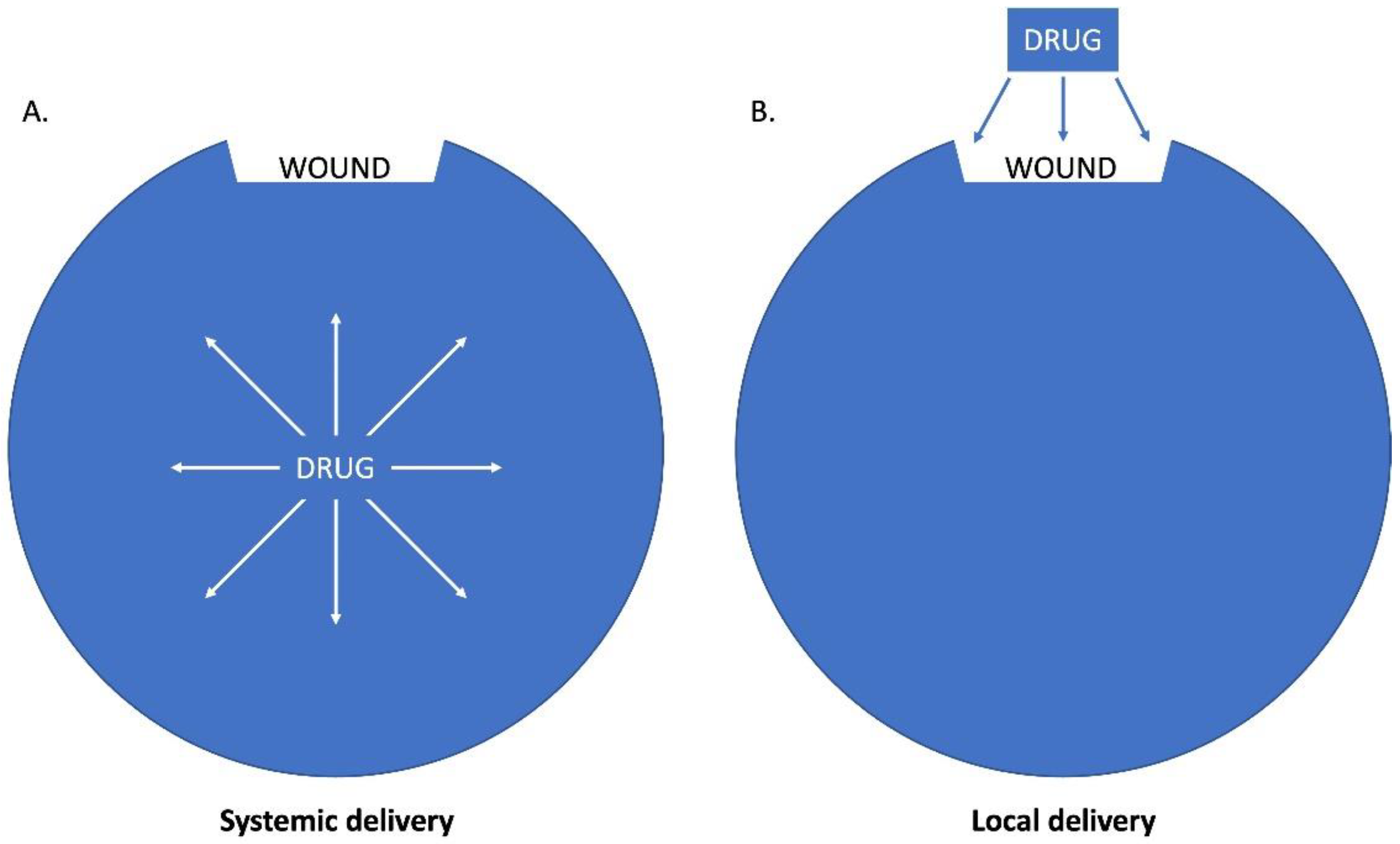
:1. Introduction
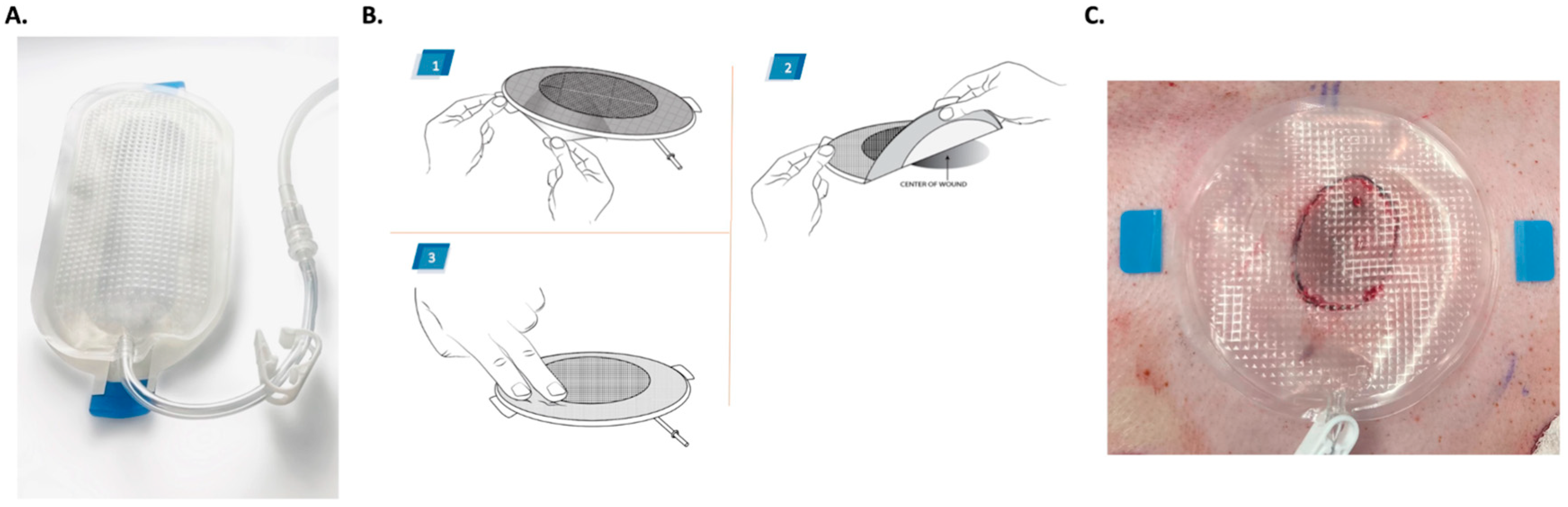
2. The Platform Wound Device in the Treatment of Skin Wounds
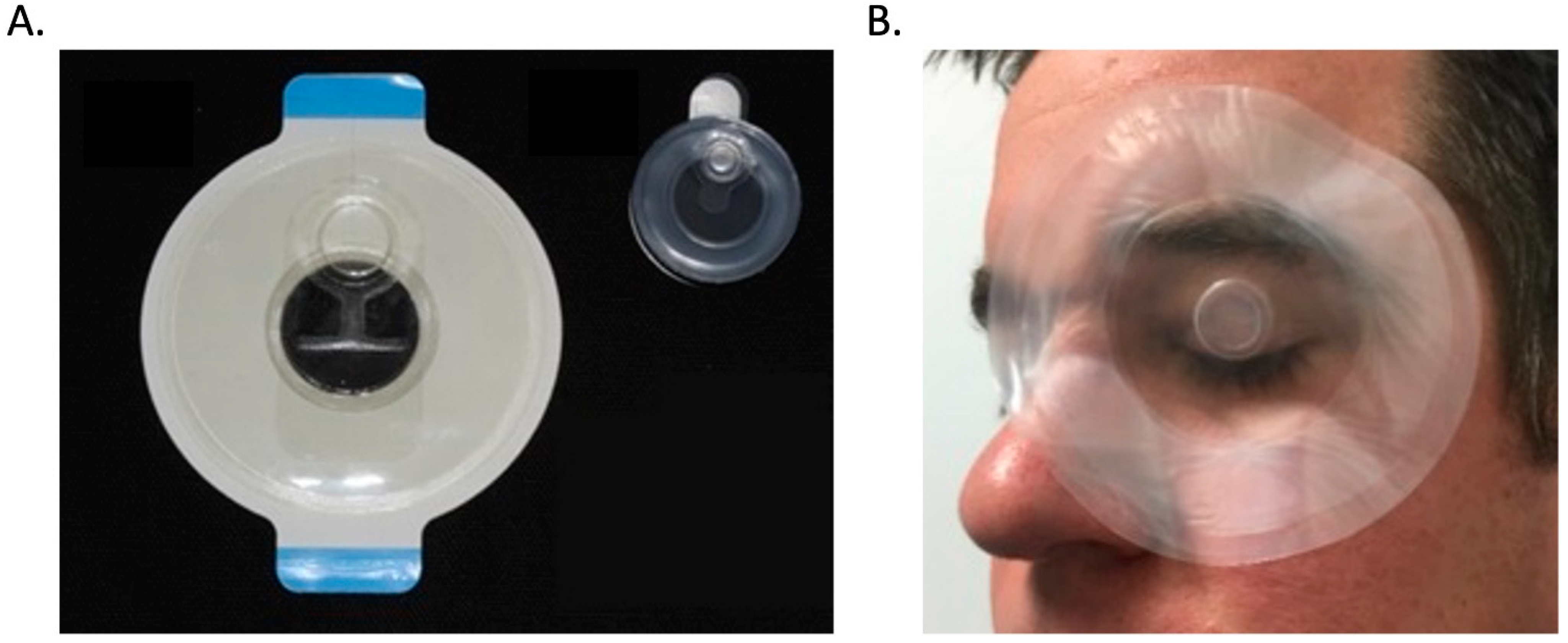
3. The Platform Wound Device in the Treatment of Ocular Trauma
4. Discussion
5. Conclusions
Author Contributions
Funding
Institutional Review Board Statement
Informed Consent Statement
Acknowledgments
Conflicts of Interest
References
- Smith, R.; Russo, J.; Fiegel, J.; Brogden, N. Antibiotic Delivery Strategies to Treat Skin Infections When Innate Antimicrobial Defense Fails. Antibiotics 2020, 9, 56. [Google Scholar] [CrossRef] [Green Version]
- Junker, J.P.; Kamel, R.A.; Caterson, E.; Eriksson, E. Clinical Impact Upon Wound Healing and Inflammation in Moist, Wet, and Dry Environments. Adv. Wound Care 2013, 2, 348–356. [Google Scholar] [CrossRef] [Green Version]
- Wen, H.; Jung, H.; Li, X. Drug Delivery Approaches in Addressing Clinical Pharmacology-Related Issues: Opportunities and Challenges. AAPS J. 2015, 17, 1327–1340. [Google Scholar] [CrossRef]
- Jaffrin, M.Y.; Morel, H. Body fluid volumes measurements by impedance: A review of bioimpedance spectroscopy (BIS) and bioimpedance analysis (BIA) methods. Med. Eng. Phys. 2008, 30, 1257–1269. [Google Scholar] [CrossRef]
- Doherty, M.M.; Pang, K.S. First-Pass Effect: Significance of the Intestine for Absorption and Metabolism. Drug Chem. Toxicol. 1997, 20, 329–344. [Google Scholar] [CrossRef]
- Garau, J.; Bouza, E.; Chastre, J.; Gudiol, F.; Harbarth, S. Management of methicillin-resistant Staphylococcus aureus infections. Clin. Microbiol. Infect. 2009, 15, 125–136. [Google Scholar] [CrossRef]
- Klugman, K.P.; Garau, J.; PACE (Pneumococcal Advisory Council of Experts); ESCMID (European Society of Clinical Microbiology and Infection). A preventable killer: Pneumonia. Clin. Microbiol. Infect. 2009, 15, 989–990. [Google Scholar] [CrossRef] [Green Version]
- Lentino, J.R.; Narita, M.; Yu, V.L. New antimicrobial agents as therapy for resistant gram-positive cocci. Eur. J. Clin. Microbiol. Infect. Dis. 2008, 27, 3–15. [Google Scholar] [CrossRef]
- Neely, A.N.; Gardner, J.; Durkee, P.; Warden, G.D.; Greenhalgh, D.G.; Gallagher, J.J.; Herndon, D.N.; Tompkins, R.G.; Kagan, R.J. Are topical antimicrobials effective against bacteria that are highly resistant to systemic antibiotics? J. Burn Care Res. 2009, 30, 19–29. [Google Scholar] [CrossRef]
- Gerhardt, R.T.; Matthews, J.M.; Sullivan, S.G. The Effect of Systemic Antibiotic Prophylaxis and Wound Irrigation on Penetrating Combat Wounds in a Return-to-Duty Population. Prehospital Emerg. Care 2009, 13, 500–504. [Google Scholar] [CrossRef]
- Nuutila, K.; Yang, L.; Broomhead, M.; Proppe, K.; Eriksson, E. PWD: Treatment Platform for Both Prolonged Field Care and Definitive Treatment of Burn-Injured Warfighters. Mil. Med. 2019, 184, e373–e380. [Google Scholar] [CrossRef]
- Sen, C.K. Human Wound and Its Burden: Updated 2020 Compendium of Estimates. Adv. Wound Care 2021, 10, 281–292. [Google Scholar] [CrossRef]
- Sen, C.K.; Gordillo, G.M.; Roy, S.; Kirsner, R.; Lambert, L.; Hunt, T.K.; Gottrup, F.; Gurtner, G.C.; Longaker, M.T. Human skin wounds: A major and snowballing threat to public health and the economy. Wound Repair Regen. 2009, 17, 763–771. [Google Scholar] [CrossRef] [Green Version]
- Kirkland, K.B.; Briggs, J.P.; Trivette, S.L.; Wilkinson, W.E.; Sexton, D.J. The Impact of Surgical-Site Infections in the 1990s: Attributable Mortality, Excess Length of Hospitalization, And Extra Costs. Infect. Control. Hosp. Epidemiol. 1999, 20, 725–730. [Google Scholar] [CrossRef] [Green Version]
- Saghazadeh, S.; Rinoldi, C.; Schot, M.; Kashaf, S.S.; Sharifi, F.; Jalilian, E.; Nuutila, K.; Giatsidis, G.; Mostafalu, P.; Derakhshandeh, H.; et al. Drug delivery systems and materials for wound healing applications. Adv. Drug Deliv. Rev. 2018, 127, 138–166. [Google Scholar] [CrossRef]
- Xue, M.; Jackson, C.J. Extracellular Matrix Reorganization During Wound Healing and Its Impact on Abnormal Scarring. Adv. Wound Care 2015, 4, 119–136. [Google Scholar] [CrossRef] [Green Version]
- Nuutila, K.; Laukkanen, A.; Lindford, A.; Juteau, S.; Nuopponen, M.; Vuola, J.; Kankuri, E. Inhibition of Skin Wound Contraction by Nanofibrillar Cellulose Hydrogel. Plast. Reconstr. Surg. 2018, 141, 357e–366e. [Google Scholar] [CrossRef]
- Sarrazy, V.; Billet, F.; Micallef, L.; Coulomb, B.; Desmoulière, A. Mechanisms of pathological scarring: Role of myofibroblasts and current developments. Wound Repair. Regen. 2011, 19, s10–s15. [Google Scholar] [CrossRef]
- Wound infection in clinical practice. An international consensus. Int. Wound J. 2008, 5 (Suppl. 3), iii-11. [CrossRef]
- Junker, J.P.E.; Lee, C.C.Y.; Samaan, S.; Hackl, F.; Kiwanuka, E.; Minasian, R.A.; Tsai, D.M.; Tracy, L.E.; Onderdonk, A.B.; Eriksson, E.; et al. Topical Delivery of Ultrahigh Concentrations of Gentamicin Is Highly Effective in Reducing Bacterial Levels in Infected Porcine Full-Thickness Wounds. Plast. Reconstr. Surg. 2015, 135, 151–159. [Google Scholar] [CrossRef]
- Tsai, D.M.; Tracy, L.E.; Lee, C.C.; Hackl, F.; Kiwanuka, E.; Minasian, R.A.; Onderdonk, A.; Junker, J.P.; Eriksson, E.; Caterson, E.J. Full-thickness porcine burns infected with Staphylococcus aureus or Pseudomonas aeruginosa can be effectively treated with topical antibiotics. Wound Repair Regen. 2016, 24, 356–365. [Google Scholar] [CrossRef] [Green Version]
- Daly, L.; Tsai, D.M.; Singh, M.; Nuutila, K.; Minasian, R.A.; Lee, C.C.Y.; Kiwanuka, E.; Hackl, F.; Onderdonk, A.; Junker, J.; et al. Topical Minocycline Effectively Decontaminates and Reduces Inflammation in Infected Porcine Wounds. Plast. Reconstr. Surg. 2016, 138, 856e–868e. [Google Scholar] [CrossRef]
- Yang, L.; Broomhead, M.; Nuutila, K.; Proppe, K.; Eriksson, E. Topically Delivered Minocycline Penetrates a Full-Thickness Burn Eschar and Reduces Tissue Bacterial Counts. J. Burn. Care Res. 2017, 39, 790–797. [Google Scholar] [CrossRef]
- Grolman, J.M.; Singh, M.; Mooney, D.; Eriksson, E.; Nuutila, K. Antibiotic-Containing Agarose Hydrogel for Wound and Burn Care. J. Burn. Care Res. 2019, 40, 900–906. [Google Scholar] [CrossRef]
- Nuutila, K.; Grolman, J.; Yang, L.; Broomhead, M.; Lipsitz, S.; Onderdonk, A.; Mooney, D.; Eriksson, E. Immediate Treatment of Burn Wounds with High Concentrations of Topical Antibiotics in an Alginate Hydrogel Using a Platform Wound Device. Adv. Wound Care 2020, 9, 48–60. [Google Scholar] [CrossRef]
- Eriksson, E.; Perez, N.; Slama, J.; Page, C.P.; Andree, C.; Maguire, J.H. Treatment of chronic, nonhealing abdominal wound in a liq-uid environment. Ann. Plast. Surg. 1996, 36, 80–83. [Google Scholar] [CrossRef]
- Vranckx, J.J.; Slama, J.; Preuss, S.; Perez, N.; Svensjö, T.; Visovatti, S.; Breuing, K.; Bartlett, R.; Pribaz, J.; Weiss, D. Wet wound healing. Plast. Reconstr. Surg. 2002, 110, 1680–1687. [Google Scholar] [CrossRef]
- Cooley, J.; Obaidi, N.; Diaz, V.; Anselmo, K.; Eriksson, E.; Carlsson, A.H.; Chan, R.K.; Nuutila, K. Delivery of topical gentamicin cream via platform wound device to reduce wound infection—A prospective, controlled, randomised, clinical study. Int. Wound J. 2022; Epub ahead of printing. [Google Scholar] [CrossRef]
- McDaniel, J.S.; Holt, A.W.; Por, E.D.; Eriksson, E.; Johnson, A.J.; Griffith, G.L. The utilization of an ocular wound chamber on corneal epithelial wounds. Clin. Ophthalmol. 2018, 12, 903–911. [Google Scholar] [CrossRef] [Green Version]
- Holt, A.W.; McDaniel, J.S.; Bramblett, G.T.; Eriksson, E.; Johnson, A.J.; Griffith, G.L. Use of an ocular wound chamber for the prevention of exposure keratopathy in a guinea pig model. Wound Repair Regen. 2018, 26, 351–358. [Google Scholar] [CrossRef] [Green Version]
- Griffith, G.L.; Holt, A.W.; Eriksson, E.; Johnson, A.J.; McDaniel, J.S. Human platelet lysate delivered via an ocular wound chamber for the treatment of corneal epithelial injuries. Exp. Eye Res. 2021, 206, 108493. [Google Scholar] [CrossRef]
- McDaniel, J.S.; Scott, L.L.F.; Rebeles, J.; Bramblett, G.T.; Eriksson, E.; Johnson, A.J.; Griffith, G.L. Treatment of Corneal Infections Utilizing an Ocular Wound Chamber. Transl. Vis. Sci. Technol. 2020, 9, 4. [Google Scholar] [CrossRef] [PubMed]
- Griffith, G.L.; Kasus-Jacobi, A.; Pereira, H.A. Bioactive Antimicrobial Peptides as Therapeutics for Corneal Wounds and Infections. Adv. Wound Care 2017, 6, 175–190. [Google Scholar] [CrossRef]
- Wilson, S.L.; El Haj, A.J.; Yang, Y. Control of Scar Tissue Formation in the Cornea: Strategies in Clinical and Corneal Tissue Engineering. J. Funct. Biomater. 2012, 3, 642–687. [Google Scholar] [CrossRef] [PubMed] [Green Version]
- Singh Malik, D.; Mital, N.; Kaur, G. Topical drug delivery systems: A patent review. Expert Opin. Ther. Pat. 2016, 26, 213–228. [Google Scholar] [CrossRef]
- Nuutila, K.; Eriksson, E. Moist Wound Healing with Commonly Available Dressings. Adv. Wound Care 2021, 10, 685–698. [Google Scholar] [CrossRef]
- Prausnitz, M.R.; Langer, R. Transdermal drug delivery. Nat. Biotechnol. 2008, 26, 1261–1268. [Google Scholar] [CrossRef]
- Kruse, C.R.; Nuutila, K.; Lee, C.C.; Kiwanuka, E.; Singh, M.; Caterson, E.J.; Eriksson, E.; Sørensen, J.A. The external microenvironment of healing skin wounds. Wound Repair Regen. 2015, 23, 456–464. [Google Scholar] [CrossRef]
- Petrie, N.C.; Yao, F.; Eriksson, E. Gene therapy in wound healing. Surg. Clin. N. Am. 2003, 83, 597–616. [Google Scholar] [CrossRef]
- Eriksson, E.; Vranckx, J. Wet wound healing: From laboratory to patients to gene therapy. Am. J. Surg. 2004, 188, 36–41. [Google Scholar] [CrossRef] [PubMed] [Green Version]
- Hirsch, T.; Spielmann, M.; Yao, F.; Eriksson, E. Gene therapy in cutaneous wound healing. Front. Biosci. 2007, 12, 2507–2518. [Google Scholar] [CrossRef] [Green Version]
- Petrie, N.C.; Vranckx, J.J.; Hoeller, D.; Yao, F.; Eriksson, E. Gene delivery of PDGF for wound healing therapy. J. Tissue Viability 2005, 15, 16–21. [Google Scholar] [CrossRef] [PubMed]
- Hackl, F.; Bergmann, J.; Granter, S.R.; Koyama, T.; Kiwanuka, E.; Zuhaili, B.; Pomahac, B.; Caterson, E.J.; Junker, J.P.E.; Eriksson, E. Epi-dermal regeneration by micrograft transplantation with immediate 100-fold expansion. Plast. Reconstr. Surg. 2012, 129, 443e–452e. [Google Scholar] [CrossRef] [Green Version]
- Nuutila, K.; Singh, M.; Kruse, C.; Eriksson, E. Wound Healing from Dermal Grafts Containing CD34+ Cells Is Comparable to Wound Healing with Split-Thickness Skin Micrografts. Plast. Reconstr. Surg. 2017, 140, 306–314. [Google Scholar] [CrossRef] [PubMed]
- Singh, M.; Nuutila, K.; Kruse, C.; Dermietzel, A.; Caterson, E.J.; Eriksson, E. Pixel Grafting: An Evolution of Mincing for Transplan-tation of Full-Thickness Wounds. Plast. Reconstr. Surg. 2016, 137, 92e–99e. [Google Scholar] [CrossRef] [PubMed]
- Nuutila, K.; Yang, L.; Broomhead, M.; Proppe, K.; Eriksson, E. Novel negative pressure wound therapy device without foam or gauze is effective at −50 mmHg. Wound Repair Regen. 2018, 27, 162–169. [Google Scholar] [CrossRef]
- Nuutila, K.; Broomhead, M.; Proppe, K.; Eriksson, E. Study Comparing Platform Wound DressingTM, a Negative Pressure Device without a Filler, with Three Conventional Negative Pressure Wound Therapy Systems in the Treatment of Excisional and Incisional Wounds. J. Plast. Reconstr. Surg. 2021, 147, 76–86. [Google Scholar] [CrossRef] [PubMed]
- Cooper, L.E.; O'Toole, M.C.; Fields, K.L.; Eriksson, E.K.; Chan, R.K. Utilization of a Novel Negative Pressure Platform Wound Dress-ing on Surgical Incisions: A Case Series. Plast. Reconstr. Surg. Glob. Open 2021, 9, e3455. [Google Scholar] [CrossRef]
- Graves, N. Economics and Preventing Hospital-acquired Infection. Emerg. Infect. Dis. 2004, 10, 561–566. [Google Scholar] [CrossRef] [Green Version]
- Klevens, R.M.; Edwards, J.R.; Richards, C.L.; Horan, T.C., Jr.; Gaynes, R.P.; Pollock, D.A.; Cardo, D.M. Estimating health care-associated infections and deaths in U.S. hospitals, 2002. Public Health Rep. 2007, 122, 160–166. [Google Scholar] [CrossRef] [PubMed]


| Study | Species | Type of Injury | Pathogen | Treatment | |
|---|---|---|---|---|---|
| Skin Wounds | Junker et al. (2015) [20] | Pig | Full-thickness wound | S. aureus | Single dose of gentamicin powder [2 mg/mL] in saline |
| Tsai et al. (2016) [21] | Pig | Full-thickness burn | S. aureus; P. aeruginosa | Single dose of gentamicin powder [2 mg/mL] or minocycline powder [1 mg/mL] in saline | |
| Daly et al. (2016) [22] | Pig | Full-thickness wound | S. aureus | Single dose of minocycline powder [0.1 mg/mL; 1 mg/mL] in saline | |
| Yang et al. (2018) [23] | Pig | Full-thickness burn | MRSA | Single dose of minocycline powder [1 mg/mL] in saline and 5% lidocaine cream | |
| Grolman et al. (2019) [24] | Pig | Deep partial-thickness burn | - | Single dose of minocycline powder [1 mg/mL] in agarose hydrogel | |
| Nuutila et al. (2018) [11] | Pig | Full-thickness burn | - | Single dose of minocycline powder [1 mg/mL] and lidocaine powder [0.5 mg/mL] in saline | |
| Nuutila et al. (2020) [25] | Pig | Deep partial-thickness burn | S. aureus; P. aeruginosa; A. baumannii | Single dose of gentamicin powder [2 mg/mL] or minocycline powder [8 mg/mL] or vancomycin powder [1 mg/mL] in alginate hydrogel | |
| Eriksson et al. (1996) [26] | Human | Abdominal wound | Multiple pathogens | Multiple doses of gentamicin [1 mg/mL], clindamycin [1 mg/mL], vancomycin [1 mg/mL], and amphotericin B (25 ug/mL) in saline | |
| Vranckx et al. (2002) [27] | Human | Infected skin wounds | Multiple pathogens | Multiple doses of high concentrations of various antibiotics in saline (such as amphotericin B, cephalexin, ceftazidime, pentamicin, vancomycin, streptomycin, and penicillin) | |
| Cooley et al. (2022) [28] | Human | Infected skin wounds | Multiple pathogens | Single dose of gentamicin cream [0.1%] | |
| Ocular trauma | McDaniel et al. (2018) [29] | Guinea pig | Corneal epithelial wound | - | Single dose of hydroxypropyl methylcellulose gel or liquid |
| Holt et al. (2018) [30] | Guinea pig | Keratopathy | - | Single dose of hydroxypropyl methylcellulose gel or balanced salt solution | |
| Griffith et al. (2021) [31] | Guinea pig | Corneal epithelial wound | - | Single dose of human platelet lysate [20%; 100%] in balanced salt solution | |
| McDaniel et al. (2020) [32] | Guinea pig | Keratopathy | P. aeruginosa | Single dose of moxifloxacin hydrochloride drops [0.5%] |
Disclaimer/Publisher’s Note: The statements, opinions and data contained in all publications are solely those of the individual author(s) and contributor(s) and not of MDPI and/or the editor(s). MDPI and/or the editor(s) disclaim responsibility for any injury to people or property resulting from any ideas, methods, instructions or products referred to in the content. |
© 2023 by the authors. Licensee MDPI, Basel, Switzerland. This article is an open access article distributed under the terms and conditions of the Creative Commons Attribution (CC BY) license (https://creativecommons.org/licenses/by/4.0/).
Share and Cite
Eriksson, E.; Griffith, G.L.; Nuutila, K. Topical Drug Delivery in the Treatment of Skin Wounds and Ocular Trauma Using the Platform Wound Device. Pharmaceutics 2023, 15, 1060. https://doi.org/10.3390/pharmaceutics15041060
Eriksson E, Griffith GL, Nuutila K. Topical Drug Delivery in the Treatment of Skin Wounds and Ocular Trauma Using the Platform Wound Device. Pharmaceutics. 2023; 15(4):1060. https://doi.org/10.3390/pharmaceutics15041060
Chicago/Turabian StyleEriksson, Elof, Gina L Griffith, and Kristo Nuutila. 2023. "Topical Drug Delivery in the Treatment of Skin Wounds and Ocular Trauma Using the Platform Wound Device" Pharmaceutics 15, no. 4: 1060. https://doi.org/10.3390/pharmaceutics15041060