.jpg)

Radiolabeled Dendrimer Coated Nanoparticles for Radionuclide Imaging and Therapy: A Systematic Review
 , and
, and 
Abstract
:1. Introduction
2. Materials and Methods
2.1. Search Strategy and Study Selection
2.2. Quality of the Selected Studies
3. Results
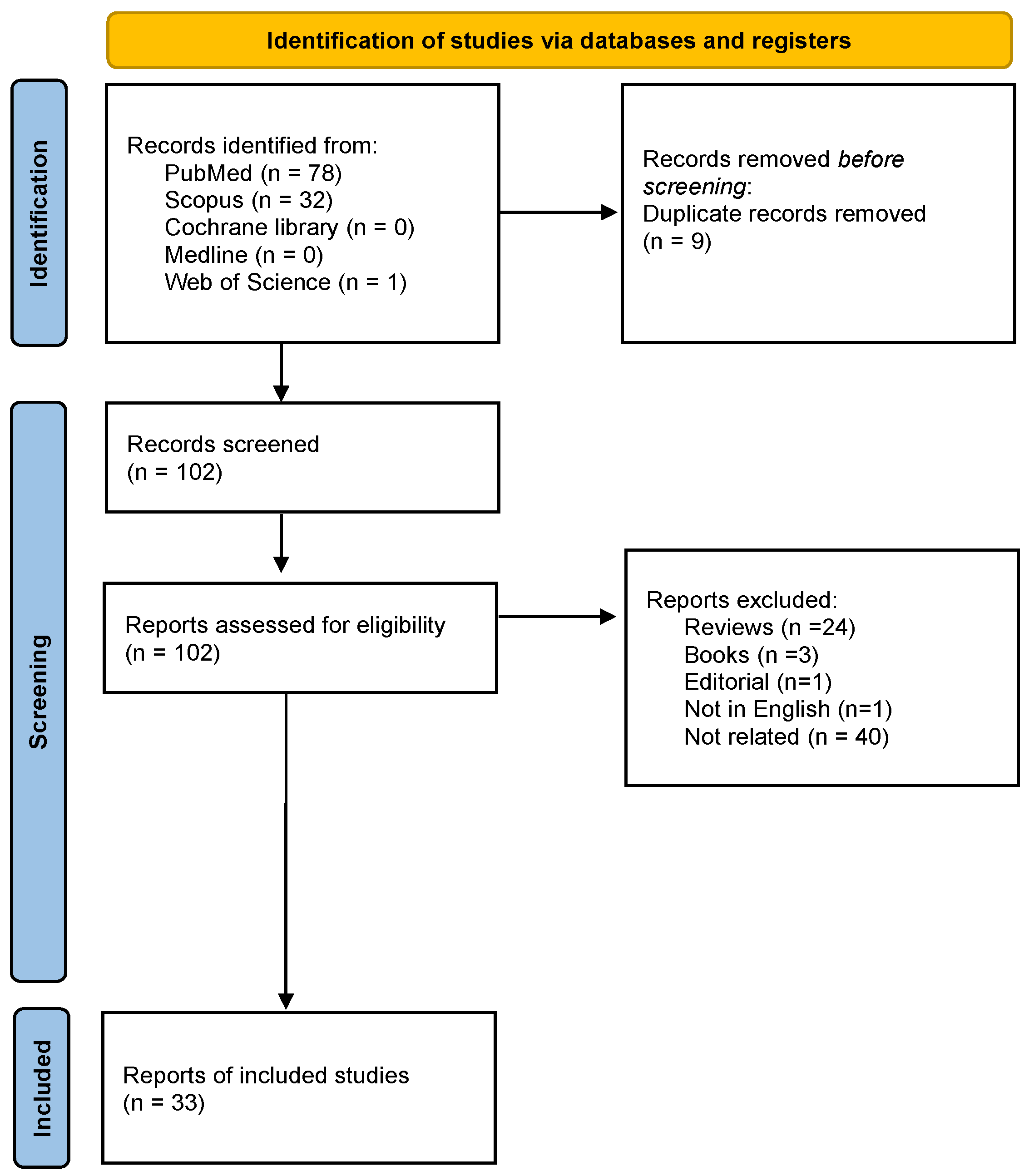
3.1. Search Results
3.2. Study Characteristics
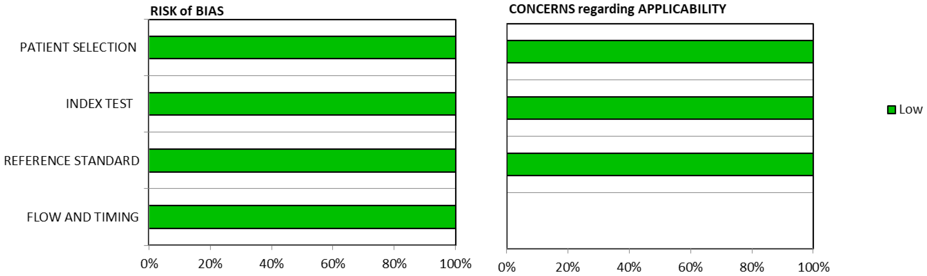
3.3. Methodological Quality Assessment
4. Discussion
4.1. Synthesis Studies
4.2. Oncological Applications
4.2.1. Dendrimers and Melanoma
4.2.2. Breast Cancer
4.2.3. Glioma Imaging
4.2.4. Prostate Cancer
4.2.5. Dendrimer and Apoptotic Cells
4.2.6. Folate Expressing Malignancies and Dendrimers
KB Cell Line and Dendrimer
Lung Adenocarcinoma
Breast Cancer
Generation Five Dendrimer Folic Acid
4.2.7. Chlorambucil Complexed Dendrimers
4.2.8. Neuroendocrine Cancer
4.2.9. Application of Dendrimer in Lymph Node Detection
4.2.10. Ehrlich’s Ascites Tumor
4.2.11. Colon Carcinoma
4.2.12. HeLa Cells In Vitro Study
4.2.13. Fibrosarcoma
4.2.14. Ovarian Cancer
4.2.15. Dendrimers and Tumor Angiogenesis and Metastasis
4.3. Therapeutic Applications of Dendrimers
5. Conclusions
Author Contributions
Funding
Institutional Review Board Statement
Informed Consent Statement
Data Availability Statement
Conflicts of Interest
References
- Parlanti:, P.; Boni, A.; Signore, G.; Santi, M. Targeted Dendrimer-Coated Magnetic Nanoparticles for Selective Delivery of Therapeutics in Living Cells. Molecules 2020, 25, 2252. [Google Scholar] [CrossRef]
- Filippi, L.; Bagni, O.; Nervi, C. Aptamer-based technology for radionuclide targeted imaging and therapy: A promising weapon against cancer. Expert Rev. Med. Devices 2020, 17, 751–758. [Google Scholar] [CrossRef]
- Hossen, S.; Hossain, M.K.; Basher, M.K.; Mia, M.N.H.; Rahman, M.T.; Uddin, M.J. Smart nanocarrier-based drug delivery systems for cancer therapy and toxicity studies: A review. J. Adv. Res. 2019, 15, 1–18. [Google Scholar] [CrossRef]
- Jain, K.; Kesharwani, P.; Gupta, U.; Jain, N.K. Dendrimer toxicity: Let’s meet the challenge. Int. J. Pharm. 2010, 394, 122–142. [Google Scholar] [CrossRef]
- Dykes, G.M.; Brierley, L.J.; Smith, D.K.; McGrail, P.T.; Seeley, G.J. Supramolecular solubilisation of hydrophilic dyes by using individual dendritic branches. Chemistry 2001, 7, 4730–4739. [Google Scholar] [CrossRef]
- Fréchet, J.M. Dendrimers and supramolecular chemistry. Proc. Natl. Acad. Sci. USA 2002, 99, 4782–4787. [Google Scholar] [CrossRef] [Green Version]
- Bugno, J.; Hsu, H.J.; Hong, S. Tweaking dendrimers and dendritic nanoparticles for controlled nano-bio interactions: Potential nanocarriers for improved cancer targeting. J. Drug Target. 2015, 23, 642–650. [Google Scholar] [CrossRef]
- Frantellizzi, V.; Conte, M.; Pontico, M.; Pani, A.; Pani, R.; De Vincentis, G. New Frontiers in Molecular Imaging with Superparamagnetic Iron Oxide Nanoparticles (SPIONs): Efficacy, Toxicity, and Future Applications. Nucl. Med. Mol. Imaging 2020, 54, 65–80. [Google Scholar] [CrossRef]
- Page, M.J.; McKenzie, J.E.; Bossuyt, P.M.; Boutron, I.; Hoffmann, T.C.; Mulrow, C.D.; Shamseer, L.; Tetzlaff, J.M.; Akl, E.A.; Brennan, S.E.; et al. The PRISMA 2020 statement: An updated guideline for reporting systematic reviews. Bmj 2021, 372, n71. [Google Scholar] [CrossRef]
- Lee, C.C.; MacKay, J.A.; Fréchet, J.M.; Szoka, F.C. Designing dendrimers for biological applications. Nat. Biotechnol. 2005, 23, 1517–1526. [Google Scholar] [CrossRef]
- de Brabander-van den Berg, E.M.M.; Meijer, E.W. Poly(propylene imine) Dendrimers: Large-Scale Synthesis by Hetereogeneously Catalyzed Hydrogenations. Angew. Chem. Int. Ed. Engl. 1993, 32, 1308–1311. [Google Scholar] [CrossRef] [Green Version]
- Sadler, K.; Tam, J.P. Peptide dendrimers: Applications and synthesis. J. Biotechnol. 2002, 90, 195–229. [Google Scholar] [CrossRef]
- Hawker, C.J.; Frechet, J.M.J. Preparation of polymers with controlled molecular architecture. A new convergent approach to dendritic macromolecules. J. Am. Chem. Soc. 1990, 112, 7638–7647. [Google Scholar] [CrossRef]
- Grinstaff, M.W. Biodendrimers: New polymeric biomaterials for tissue engineering. Chemistry 2002, 8, 2839–2846. [Google Scholar] [CrossRef]
- Turnbull, W.B.; Stoddart, J.F. Design and synthesis of glycodendrimers. J. Biotechnol. 2002, 90, 231–255. [Google Scholar] [CrossRef]
- Li, Y.; Tseng, Y.D.; Kwon, S.Y.; D’Espaux, L.; Bunch, J.S.; McEuen, P.L.; Luo, D. Controlled assembly of dendrimer-like DNA. Nat. Mater. 2004, 3, 38–42. [Google Scholar] [CrossRef]
- McNelles, S.A.; Knight, S.D.; Janzen, N.; Valliant, J.F.; Adronov, A. Synthesis, Radiolabeling, and In Vivo Imaging of PEGylated High-Generation Polyester Dendrimers. Biomacromolecules 2015, 16, 3033–3041. [Google Scholar] [CrossRef]
- Aarthi, T.; Shaama, M.S.; Madras, G. Degradation of Water Soluble Polymers under Combined Ultrasonic and Ultraviolet Radiation. Ind. Eng. Chem. Res. 2007, 46, 6204–6210. [Google Scholar] [CrossRef]
- Hlídková, H.; Kotelnikov, I.; Pop-Georgievski, O.; Proks, V.; Horák, D. Antifouling Peptide Dendrimer Surface of Monodisperse Magnetic Poly(glycidyl methacrylate) Microspheres. Macromolecules 2017, 50, 1302–1311. [Google Scholar] [CrossRef]
- Sung, H.; Ferlay, J.; Siegel, R.L.; Laversanne, M.; Soerjomataram, I.; Jemal, A.; Bray, F. Global Cancer Statistics 2020: GLOBOCAN Estimates of Incidence and Mortality Worldwide for 36 Cancers in 185 Countries. CA Cancer J. Clin. 2021, 71, 209–249. [Google Scholar] [CrossRef]
- Li, C.; Zhao, L.; Jia, L.; Ouyang, Z.; Gao, Y.; Guo, R.; Song, S.; Shi, X.; Cao, X. (68)Ga-labeled dendrimer-entrapped gold nanoparticles for PET/CT dual-modality imaging and immunotherapy of tumors. J. Mater. Chem. B 2022, 10, 3648–3656. [Google Scholar] [CrossRef]
- Frantellizzi, V.; Conte, M.; De Vincentis, G. Hybrid Imaging of Vascular Cognitive Impairment. Semin. Nucl. Med. 2021, 51, 286–295. [Google Scholar] [CrossRef]
- Tassano, M.R.; Audicio, P.F.; Gambini, J.P.; Fernandez, M.; Damian, J.P.; Moreno, M.; Chabalgoity, J.A.; Alonso, O.; Benech, J.C.; Cabral, P. Development of 99mTc(CO)₃-dendrimer-FITC for cancer imaging. Bioorg. Med. Chem. Lett. 2011, 21, 5598–5601. [Google Scholar] [CrossRef]
- Tanaka, T.; Sano, K.; Munekane, M.; Yamasaki, T.; Sasaki, H.; Mukai, T. A Radiolabeled Self-assembled Nanoparticle Probe for Diagnosis of Lung-Metastatic Melanoma. Biol. Pharm. Bull. 2021, 44, 410–415. [Google Scholar] [CrossRef]
- Ebrahimi, A.; Pirali Hamedani, M.; Mohammadzadeh, P.; Safari, M.; Esmaeil Sadat Ebrahimi, S.; Seyed Hamzeh, M.; Shafiee Ardestani, M.; Masoumeh Ghoreishi, S. (99m)Tc- Anionic dendrimer targeted vascular endothelial growth factor as a novel nano-radiotracer for in-vivo breast cancer imaging. Bioorg. Chem. 2022, 128, 106085. [Google Scholar] [CrossRef]
- Nabi, P.N.; Vahidfar, N.; Tohidkia, M.R.; Hamidi, A.A.; Omidi, Y.; Aghanejad, A. Mucin-1 conjugated polyamidoamine-based nanoparticles for image-guided delivery of gefitinib to breast cancer. Int. J. Biol. Macromol. 2021, 174, 185–197. [Google Scholar] [CrossRef]
- Zamani, S.; Shafeie-Ardestani, M.; Bitarafan-Rajabi, A.; Khalaj, A.; Sabzevari, O. Synthesis, radiolabelling, and biological assessment of folic acid-conjugated G-3 (99m)Tc-dendrimer as the breast cancer molecular imaging agent. IET Nanobiotechnol. 2020, 14, 628–634. [Google Scholar] [CrossRef]
- Song, N.; Zhao, L.; Xu, X.; Zhu, M.; Liu, C.; Sun, N.; Yang, J.; Shi, X.; Zhao, J. LyP-1-Modified Multifunctional Dendrimers for Targeted Antitumor and Antimetastasis Therapy. ACS Appl. Mater. Interfaces 2020, 12, 12395–12406. [Google Scholar] [CrossRef]
- Gibbens-Bandala, B.; Morales-Avila, E.; Ferro-Flores, G.; Santos-Cuevas, C.; Luna-Gutiérrez, M.; Ramírez-Nava, G.; Ocampo-García, B. Synthesis and Evaluation of (177)Lu-DOTA-DN(PTX)-BN for Selective and Concomitant Radio and Drug-Therapeutic Effect on Breast Cancer Cells. Polymers 2019, 11, 1572. [Google Scholar] [CrossRef] [Green Version]
- Gorica, J.; De Feo, M.S.; Filippi, L.; Frantellizzi, V.; Schillaci, O.; De Vincentis, G. Gastrin-releasing peptide receptor agonists and antagonists for molecular imaging of breast and prostate cancer: From pre-clinical studies to translational perspectives. Expert Rev. Mol. Diagn. 2022, 22, 991–996. [Google Scholar] [CrossRef]
- Li, Y.; Zhao, L.; Xu, X.; Sun, N.; Qiao, W.; Xing, Y.; Shen, M.; Zhu, M.; Shi, X.; Zhao, J. Design of (99m)Tc-Labeled Low Generation Dendrimer-Entrapped Gold Nanoparticles for Targeted Single Photon Emission Computed Tomography/Computed Tomography Imaging of Gliomas. J. Biomed. Nanotechnol. 2019, 15, 1201–1212. [Google Scholar] [CrossRef]
- Zhao, L.; Zhu, J.; Cheng, Y.; Xiong, Z.; Tang, Y.; Guo, L.; Shi, X.; Zhao, J. Chlorotoxin-Conjugated Multifunctional Dendrimers Labeled with Radionuclide 131I for Single Photon Emission Computed Tomography Imaging and Radiotherapy of Gliomas. ACS Appl. Mater. Interfaces 2015, 7, 19798–19808. [Google Scholar] [CrossRef]
- Lesniak, W.G.; Boinapally, S.; Banerjee, S.R.; Behnam Azad, B.; Foss, C.A.; Shen, C.; Lisok, A.; Wharram, B.; Nimmagadda, S.; Pomper, M.G. Evaluation of PSMA-Targeted PAMAM Dendrimer Nanoparticles in a Murine Model of Prostate Cancer. Mol. Pharm. 2019, 16, 2590–2604. [Google Scholar] [CrossRef]
- Giannopoulou, C. The role of SPET and PET in monitoring tumour response to therapy. Eur. J. Nucl. Med. Mol. Imaging 2003, 30, 1173–1200. [Google Scholar] [CrossRef]
- Xing, Y.; Zhu, J.; Zhao, L.; Xiong, Z.; Li, Y.; Wu, S.; Chand, G.; Shi, X.; Zhao, J. SPECT/CT imaging of chemotherapy-induced tumor apoptosis using (99m)Tc-labeled dendrimer-entrapped gold nanoparticles. Drug Deliv. 2018, 25, 1384–1393. [Google Scholar] [CrossRef] [Green Version]
- Frantellizzi, V.; Verrina, V.; Raso, C.; Pontico, M.; Petronella, F.; Bertana, V.; Ballesio, A.; Marasso, S.L.; Miglietta, S.; Rosa, P.; et al. 99mTc-labeled keratin gold-nanoparticles in a nephron-like microfluidic chip for photo-thermal therapy applications. Mater. Today Adv. 2022, 16, 100286. [Google Scholar] [CrossRef]
- Bourguignon, L.Y.; Zhu, H.; Shao, L.; Chen, Y.W. CD44 interaction with tiam1 promotes Rac1 signaling and hyaluronic acid-mediated breast tumor cell migration. J. Biol. Chem. 2000, 275, 1829–1838. [Google Scholar] [CrossRef] [Green Version]
- Huang, X.; Li, D.; Li, T.; Zhao, B.O.; Chen, X. Prognostic value of the expression of phosphatase and tensin homolog and CD44 in elderly patients with refractory acute myeloid leukemia. Oncol. Lett. 2015, 10, 103–110. [Google Scholar] [CrossRef] [Green Version]
- Naor, D.; Nedvetzki, S.; Golan, I.; Melnik, L.; Faitelson, Y. CD44 in cancer. Crit. Rev. Clin. Lab. Sci. 2002, 39, 527–579. [Google Scholar] [CrossRef]
- Kim, J.H.; Moon, M.J.; Kim, D.Y.; Heo, S.H.; Jeong, Y.Y. Hyaluronic Acid-Based Nanomaterials for Cancer Therapy. Polymers 2018, 10, 1133. [Google Scholar] [CrossRef] [Green Version]
- Tanaka, T.; Sano, K.; Munemura, M.; Hagimori, M.; Moriyama, R.; Yamamoto, A.; Ozaki, K.I.; Munekane, M.; Yamasaki, T.; Mukai, T. A radiolabeled nanoparticle probe coated with hyaluronic acid via electrostatic interaction to diagnose CD44-positive tumors. J. Drug Deliv. Sci. Technol. 2022, 73, 103473. [Google Scholar] [CrossRef]
- Song, M.; Guo, Z.; Gao, M.; Shi, C.; Xu, D.; You, L.; Wu, X.; Su, X.; Zhuang, R.; Pan, W.; et al. Synthesis and preliminary evaluation of a (99m) Tc-labeled folate-PAMAM dendrimer for FR imaging. Chem. Biol. Drug Des. 2017, 89, 755–761. [Google Scholar] [CrossRef]
- Zhang, Y.; Sun, Y.; Xu, X.; Zhang, X.; Zhu, H.; Huang, L.; Qi, Y.; Shen, Y.M. Synthesis, biodistribution, and microsingle photon emission computed tomography (SPECT) imaging study of technetium-99m labeled PEGylated dendrimer poly(amidoamine) (PAMAM)-folic acid conjugates. J. Med. Chem. 2010, 53, 3262–3272. [Google Scholar] [CrossRef]
- Ma, W.; Fu, F.; Zhu, J.; Huang, R.; Zhu, Y.; Liu, Z.; Wang, J.; Conti, P.S.; Shi, X.; Chen, K. (64)Cu-Labeled multifunctional dendrimers for targeted tumor PET imaging. Nanoscale 2018, 10, 6113–6124. [Google Scholar] [CrossRef]
- Mendoza-Nava, H.; Ferro-Flores, G.; Ramírez, F.M.; Ocampo-García, B.; Santos-Cuevas, C.; Azorín-Vega, E.; Jiménez-Mancilla, N.; Luna-Gutiérrez, M.; Isaac-Olivé, K. Fluorescent, Plasmonic, and Radiotherapeutic Properties of the (177)Lu-Dendrimer-AuNP-Folate-Bombesin Nanoprobe Located Inside Cancer Cells. Mol. Imaging 2017, 16, 1536012117704768. [Google Scholar] [CrossRef] [Green Version]
- Mendoza-Nava, H.; Ferro-Flores, G.; Ramírez, F.D.M.; Ocampo-García, B.; Santos-Cuevas, C.; Aranda-Lara, L.; Azorín-Vega, E.; Morales-Avila, E.; Isaac-Olivé, K. 177Lu-Dendrimer Conjugated to Folate and Bombesin with Gold Nanoparticles in the Dendritic Cavity: A Potential Theranostic Radiopharmaceutical. J. Nanomater. 2016, 2016, 1039258. [Google Scholar] [CrossRef] [Green Version]
- Cui, W.; Zhang, Y.; Xu, X.; Shen, Y.M. Synthesis and 188Re radiolabelling of dendrimer polyamide amine (PAMAM) folic acid conjugate. Med. Chem. 2012, 8, 727–731. [Google Scholar] [CrossRef]
- Ghoreishi, S.M.; Khalaj, A.; Bitarafan-Rajabi, A.; Azar, A.D.; Ardestani, M.S.; Assadi, A. Novel 99mTc-Radiolabeled Anionic Linear Globular PEG-Based Dendrimer-Chlorambucil: Non-Invasive Method for In-Vivo Biodistribution. Drug Res. 2017, 67, 149–155. [Google Scholar] [CrossRef]
- Naeini, A.T.; Adeli, M.; Vossoughi, M. Poly(citric acid)-block-poly(ethylene glycol) copolymers--new biocompatible hybrid materials for nanomedicine. Nanomedicine 2010, 6, 556–562. [Google Scholar] [CrossRef]
- Giray, S.; Bal, T.; Kartal, A.M.; Kızılel, S.; Erkey, C. Controlled drug delivery through a novel PEG hydrogel encapsulated silica aerogel system. J. Biomed. Mater. Res. A 2012, 100, 1307–1315. [Google Scholar] [CrossRef]
- Altiparmak, B.; Lambrecht, F.Y.; Bayrak, E.; Durkan, K. Design and synthesis of 99mTc-citro-folate for use as a tumor-targeted radiopharmaceutical. Int. J. Pharm. 2010, 400, 8–14. [Google Scholar] [CrossRef]
- Orocio-Rodríguez, E.; Ferro-Flores, G.; Santos-Cuevas, C.L.; Ramírez Fde, M.; Ocampo-García, B.E.; Azorín-Vega, E.; Sánchez-García, F.M. Two Novel Nanosized Radiolabeled Analogues of Somatostatin for Neuroendocrine Tumor Imaging. J. Nanosci. Nanotechnol. 2015, 15, 4159–4169. [Google Scholar] [CrossRef]
- Niki, Y.; Ogawa, M.; Makiura, R.; Magata, Y.; Kojima, C. Optimization of dendrimer structure for sentinel lymph node imaging: Effects of generation and terminal group. Nanomedicine 2015, 11, 2119–2127. [Google Scholar] [CrossRef]
- Sano, K.; Iwamiya, Y.; Kurosaki, T.; Ogawa, M.; Magata, Y.; Sasaki, H.; Ohshima, T.; Maeda, M.; Mukai, T. Radiolabeled γ-polyglutamic acid complex as a nano-platform for sentinel lymph node imaging. J. Control. Release 2014, 194, 310–315. [Google Scholar] [CrossRef]
- Ghai, A.; Singh, B.; Panwar Hazari, P.; Schultz, M.K.; Parmar, A.; Kumar, P.; Sharma, S.; Dhawan, D.; Kumar Mishra, A. Radiolabeling optimization and characterization of (68)Ga labeled DOTA-polyamido-amine dendrimer conjugate-Animal biodistribution and PET imaging results. Appl. Radiat. Isot. 2015, 105, 40–46. [Google Scholar] [CrossRef] [Green Version]
- Khosroshahi, A.G.; Amanlou, M.; Sabzevari, O.; Daha, F.J.; Aghasadeghi, M.R.; Ghorbani, M.; Ardestani, M.S.; Alavidjeh, M.S.; Sadat, S.M.; Pouriayevali, M.H.; et al. A comparative study of two novel nanosized radiolabeled analogues of methionine for SPECT tumor imaging. Curr. Med. Chem. 2013, 20, 123–133. [Google Scholar] [CrossRef]
- Guillaudeu, S.J.; Fox, M.E.; Haidar, Y.M.; Dy, E.E.; Szoka, F.C.; Fréchet, J.M. PEGylated dendrimers with core functionality for biological applications. Bioconjug. Chem. 2008, 19, 461–469. [Google Scholar] [CrossRef]
- Xu, X.; Zhang, Y.; Wang, X.; Guo, X.; Zhang, X.; Qi, Y.; Shen, Y.M. Radiosynthesis, biodistribution and micro-SPECT imaging study of dendrimer-avidin conjugate. Bioorg. Med. Chem. 2011, 19, 1643–1648. [Google Scholar] [CrossRef]
- Kobayashi, H.; Sakahara, H.; Hosono, M.; Yao, Z.S.; Toyama, S.; Endo, K.; Konishi, J. Improved clearance of radiolabeled biotinylated monoclonal antibody following the infusion of avidin as a “chase” without decreased accumulation in the target tumor. J. Nucl. Med. 1994, 35, 1677–1684. [Google Scholar]
- Almasi, T.; Gholipour, N.; Akhlaghi, M.; Mokhtari Kheirabadi, A.; Mazidi, S.M.; Hosseini, S.H.; Geramifar, P.; Beiki, D.; Rostampour, N.; Shahbazi Gahrouei, D. Development of Ga-68 radiolabeled DOTA functionalized and acetylated PAMAM dendrimer-coated iron oxide nanoparticles as PET/MR dual-modal imaging agent. Int. J. Polym. Mater. Polym. Biomater. 2021, 70, 1077–1089. [Google Scholar] [CrossRef]
- Sadekar, S.; Ray, A.; Janàt-Amsbury, M.; Peterson, C.M.; Ghandehari, H. Comparative biodistribution of PAMAM dendrimers and HPMA copolymers in ovarian-tumor-bearing mice. Biomacromolecules 2011, 12, 88–96. [Google Scholar] [CrossRef] [Green Version]
- Dijkgraaf, I.; Rijnders, A.Y.; Soede, A.; Dechesne, A.C.; van Esse, G.W.; Brouwer, A.J.; Corstens, F.H.; Boerman, O.C.; Rijkers, D.T.; Liskamp, R.M. Synthesis of DOTA-conjugated multivalent cyclic-RGD peptide dendrimers via 1,3-dipolar cycloaddition and their biological evaluation: Implications for tumor targeting and tumor imaging purposes. Org. Biomol. Chem. 2007, 5, 935–944. [Google Scholar] [CrossRef] [Green Version]
- Mamede, M.; Saga, T.; Kobayashi, H.; Ishimori, T.; Higashi, T.; Sato, N.; Brechbiel, M.W.; Konishi, J. Radiolabeling of avidin with very high specific activity for internal radiation therapy of intraperitoneally disseminated tumors. Clin. Cancer Res. 2003, 9, 3756–3762. [Google Scholar]


Disclaimer/Publisher’s Note: The statements, opinions and data contained in all publications are solely those of the individual author(s) and contributor(s) and not of MDPI and/or the editor(s). MDPI and/or the editor(s) disclaim responsibility for any injury to people or property resulting from any ideas, methods, instructions or products referred to in the content. |
© 2023 by the authors. Licensee MDPI, Basel, Switzerland. This article is an open access article distributed under the terms and conditions of the Creative Commons Attribution (CC BY) license (https://creativecommons.org/licenses/by/4.0/).
Share and Cite
Conte, M.; De Feo, M.S.; Sidrak, M.M.A.; Corica, F.; Gorica, J.; Filippi, L.; Schillaci, O.; De Vincentis, G.; Frantellizzi, V. Radiolabeled Dendrimer Coated Nanoparticles for Radionuclide Imaging and Therapy: A Systematic Review. Pharmaceutics 2023, 15, 867. https://doi.org/10.3390/pharmaceutics15030867
Conte M, De Feo MS, Sidrak MMA, Corica F, Gorica J, Filippi L, Schillaci O, De Vincentis G, Frantellizzi V. Radiolabeled Dendrimer Coated Nanoparticles for Radionuclide Imaging and Therapy: A Systematic Review. Pharmaceutics. 2023; 15(3):867. https://doi.org/10.3390/pharmaceutics15030867
Chicago/Turabian StyleConte, Miriam, Maria Silvia De Feo, Marko Magdi Abdou Sidrak, Ferdinando Corica, Joana Gorica, Luca Filippi, Orazio Schillaci, Giuseppe De Vincentis, and Viviana Frantellizzi. 2023. "Radiolabeled Dendrimer Coated Nanoparticles for Radionuclide Imaging and Therapy: A Systematic Review" Pharmaceutics 15, no. 3: 867. https://doi.org/10.3390/pharmaceutics15030867