
Naringin: Nanotechnological Strategies for Potential Pharmaceutical Applications
Abstract
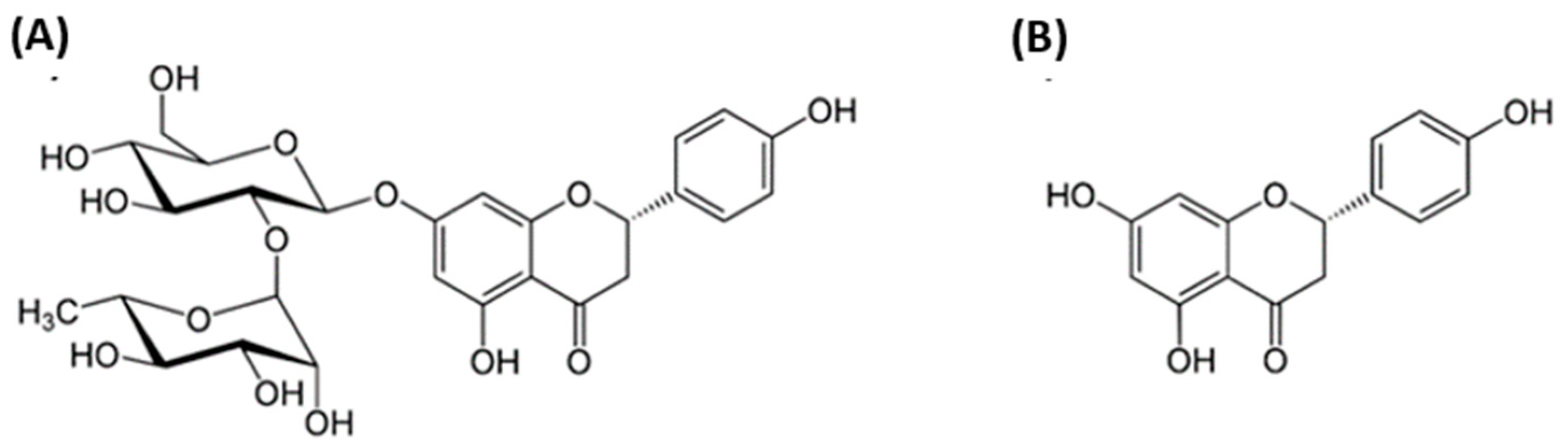
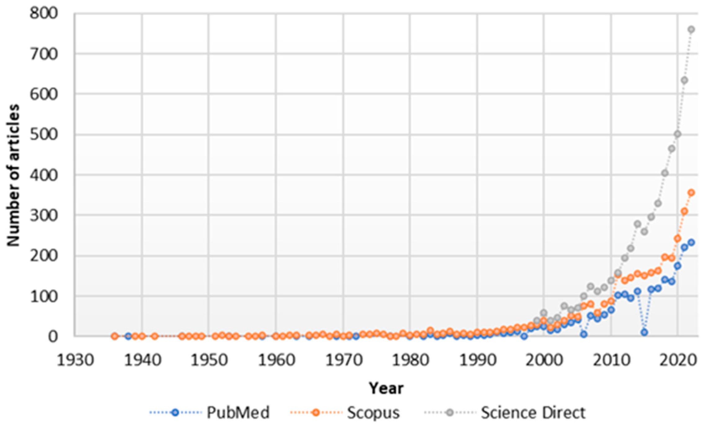
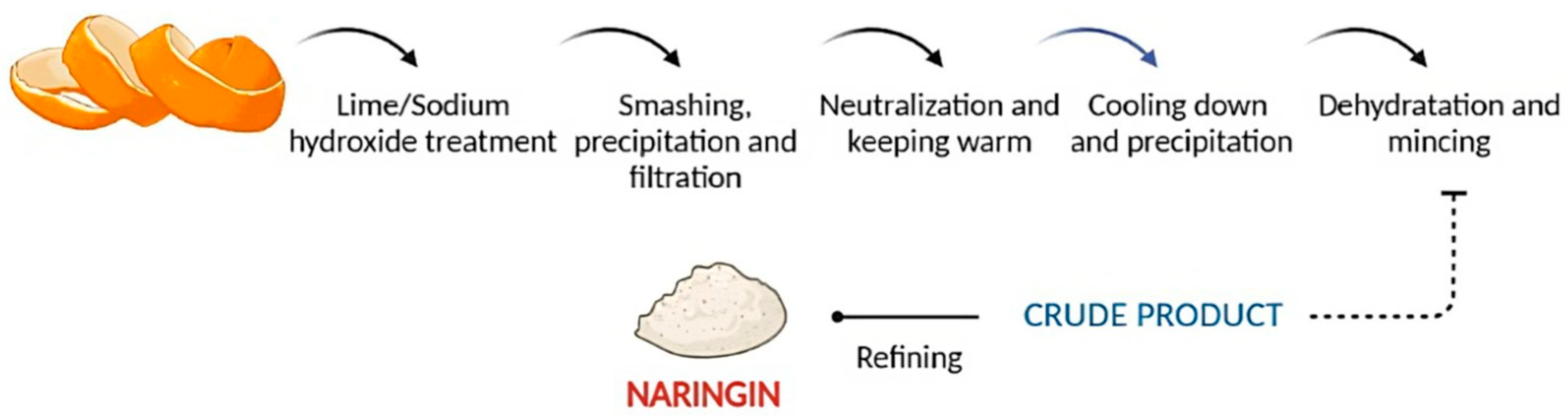
:1. Introduction
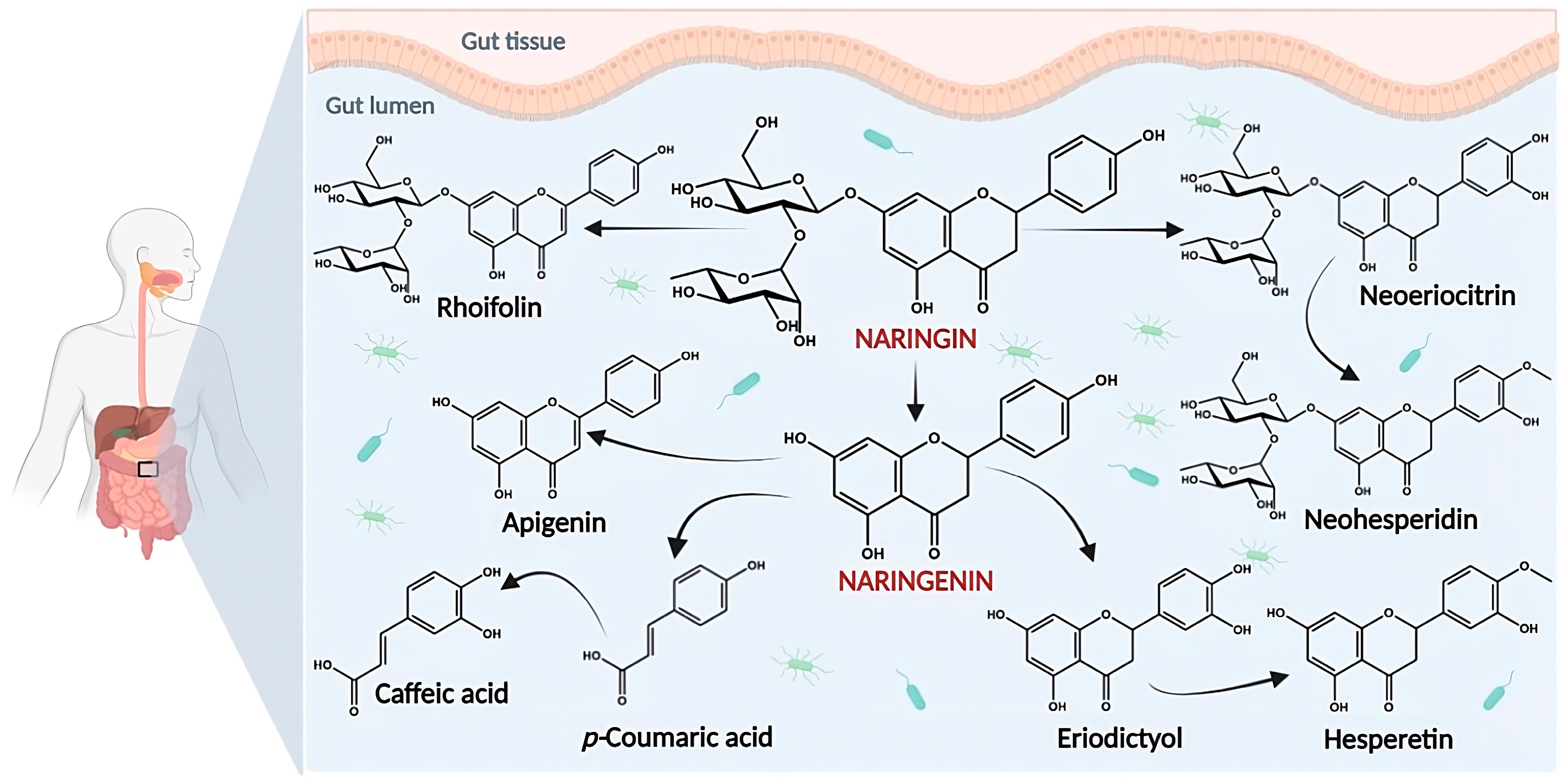
2. Bioavailability and Pharmacokinetic Properties of Naringin
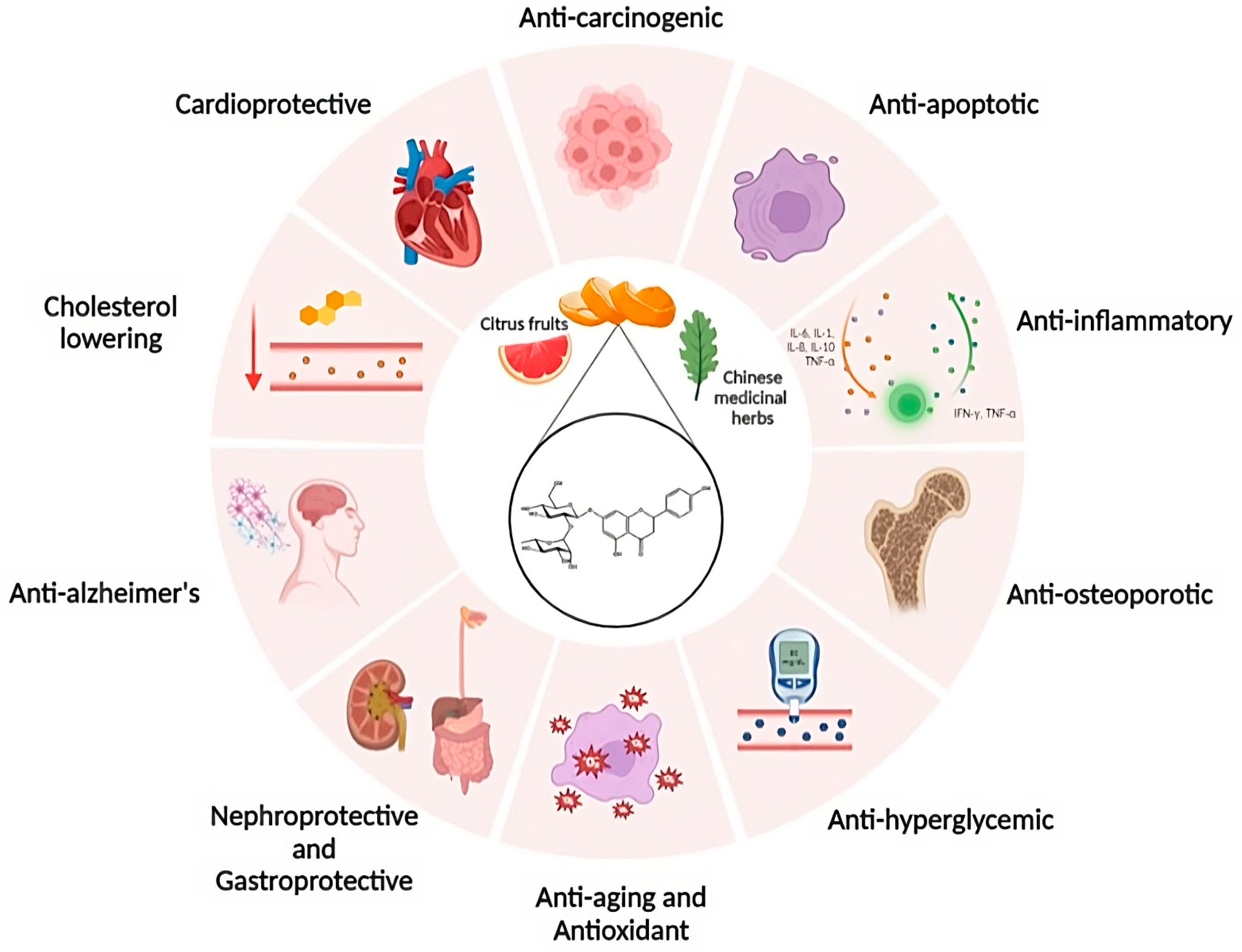
3. Biological Activities of Naringin
4. Clinical Translation and Challenges for Its Therapeutic Application
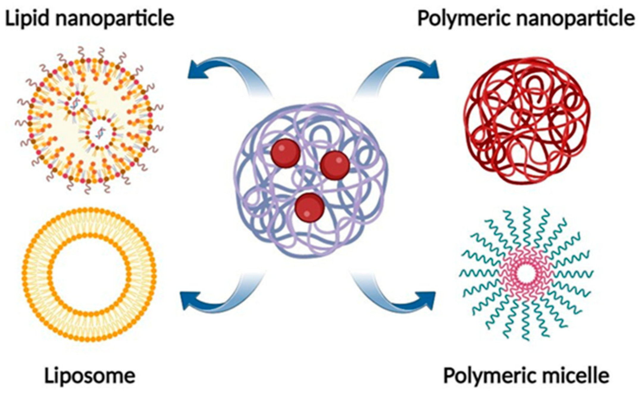
5. Naringin Nanoformulations
5.1. Liposomes
5.2. Polymeric Micelles
5.3. Polymer-Based Nanoparticles
5.4. Lipid Nanoparticles
5.5. Other Formulations
6. Conclusions and Future Perspectives
Funding
Institutional Review Board Statement
Informed Consent Statement
Data Availability Statement
Acknowledgments
Conflicts of Interest
References
- Chen, R.; Qi, Q.L.; Wang, M.T.; Li, Q.Y. Therapeutic potential of naringin: An overview. Pharm. Biol. 2016, 54, 3203–3210. [Google Scholar] [CrossRef] [PubMed] [Green Version]
- Rangaswami, S.; Seshadri, T.R.; Veeraraghaviah, J. Constitution of naringin. The position of the sugar group. J. Proc. Ind. Acad. Sci. 1939, 9, 328–332. [Google Scholar] [CrossRef]
- Zhao, B.T.; Kim, E.J.; Son, K.H.; Son, J.K.; Min, B.S.; Woo, M.H. Quality evaluation and pattern recognition analyses of marker compounds from five medicinal drugs of Rutaceae family by HPLC/PDA. Arch. Pharm. Res. 2015, 38, 1512–1520. [Google Scholar] [CrossRef]
- Stabrauskiene, J.; Marksa, M.; Ivanauskas, L.; Bernatoniene, J. Optimization of naringin and naringenin extraction from Citrus × paradisi L. using hydrolysis and excipients as adsorbent. Pharmaceutics 2022, 14, 890. [Google Scholar] [CrossRef] [PubMed]
- Kanokorn, S.; Surachai, P.; Supason, W. An efficient method for the large scale isolation of naringin from pomelo (Citrus grandis) peel. Int. J. Food Sci. Technol. 2009, 44, 1737–1742. [Google Scholar] [CrossRef]
- Pereira, R.M.; Andrades, N.E.; Paulino, N.; Sawaya, A.C.; Eberlin, M.N.; Marcucci, M.C.; Favero, G.M.; Novak, E.M.; Bydlowski, S.P. Synthesis and characterization of a metal complex containing naringin and Cu, and its antioxidant, antimicrobial, antiinflammatory and tumor cell cytotoxicity. Molecules. 2007, 12, 1352–1366. [Google Scholar] [CrossRef] [Green Version]
- PubChem [Internet]. Bethesda (MD): National Library of Medicine (US), National Center for Biotechnology Information; 2004. PubChem Compound Summary for CID 442428, Naringin. Available online: https://pubchem.ncbi.nlm.nih.gov/compound/Naringin (accessed on 11 January 2023).
- Zhang, L.; Song, L.; Zhang, P.; Liu, T.; Zhou, L.; Yang, G.; Lin, R.; Zhang, J. Solubilities of naringin and naringenin in different solvents and dissociation constants of naringenin. J. Chem. Eng. Data 2015, 60, 932–940. [Google Scholar] [CrossRef]
- Budel, R.G.; da Silva, D.A.; Moreira, M.P.; Dalcin, A.J.F.; da Silva, A.F.; Nazario, L.R.; Majolo, J.H.; Lopes, L.Q.S.; Santos, R.C.V.; Antunes Soares, F.A.; et al. Toxicological evaluation of naringin-loaded nanocapsules in vitro and in vivo. Colloids Surf. B Biointerfaces 2020, 188, 110754. [Google Scholar] [CrossRef] [PubMed]
- Carrier, R.L.; Miller, L.A.; Ahmed, I. The utility of cyclodextrins for enhancing oral bioavailability. J. Control. Release 2007, 123, 78–99. [Google Scholar] [CrossRef] [PubMed]
- Ilangumaran, S.; Hoessli, D.C. Effects of cholesterol depletion by cyclodextrin on the sphingolipid microdomains of the plasma membrane. Biochem. J. 1998, 335 Pt 2, 433–440. [Google Scholar] [CrossRef]
- Liu, L.; Shan, S.; Zhang, K.; Ning, Z.Q.; Lu, X.P.; Cheng, Y.Y. Naringenin and hesperetin, two flavonoids derived from Citrus aurantium up-regulate transcription of adiponectin. Phytother. Res. 2008, 22, 1400–1403. [Google Scholar] [CrossRef] [PubMed]
- Lauro, M.R.; De Simone, F.; Sansone, F.; Iannelli, P.; Aquino, R.P. Preparations and release characteristics of naringin and naringenin gastro-resistant microparticles by spray-drying. J. Drug Deliv. Sci. Technol. 2007, 17, 119–124. [Google Scholar] [CrossRef]
- Sharma, A.; Bhardwaj, P.; Arya, S.K. Naringin: A potential natural product in the field of biomedical applications. Carbohydrate Polymer Technol. Appl. 2021, 2, 100068. [Google Scholar] [CrossRef]
- Kanaze, F.I.; Bounartzi, M.I.; Georgarakis, M.; Niopas, I. Pharmacokinetics of the citrus flavanone aglycones hesperetin and naringenin after single oral administration in human subjects. Eur. J. Clin. Nutr. 2007, 61, 472–477. [Google Scholar] [CrossRef] [Green Version]
- Choudhury, R.; Chowrimootoo, G.; Srai, K.; Debnam, E.; Rice-Evans, C.A. Interactions of the flavonoid naringenin in the gastrointestinal tract and the influence of glycosylation. Biochem. Biophys. Res. Commun. 1999, 265, 410–415. [Google Scholar] [CrossRef]
- Khan, A.W.; Kotta, S.; Ansari, S.H.; Sharma, R.K.; Ali, J. Self-nanoemulsifying drug delivery system (SNEDDS) of the poorly water-soluble grapefruit flavonoid Naringenin: Design, characterization, in vitro and in vivo evaluation. Drug Deliv. 2015, 22, 552–561. [Google Scholar] [CrossRef]
- Jeon, S.M.; Kim, H.K.; Kim, H.J.; Do, G.M.; Jeong, T.S.; Park, Y.B.; Choi, M.S. Hypocholesterolemic and antioxidative effects of naringenin and its two metabolites in high-cholesterol fed rats. Transl. Res. 2007, 149, 15–21. [Google Scholar] [CrossRef]
- Steed, A.L.; Christophi, G.P.; Kaiko, G.E.; Sun, L.; Goodwin, V.M.; Jain, U.; Esaulova, E.; Artyomov, M.N.; Morales, D.J.; Holtzman, M.J.; et al. The microbial metabolite desaminotyrosine protects from influenza through type I interferon. Science 2017, 357, 498–502. [Google Scholar] [CrossRef] [Green Version]
- Wei, Y.; Gao, J.; Kou, Y.; Liu, M.; Meng, L.; Zheng, X.; Xu, S.; Liang, M.; Sun, H.; Liu, Z.; et al. The intestinal microbial metabolite desaminotyrosine is an anti-inflammatory molecule that modulates local and systemic immune homeostasis. FASEB J. 2020, 34, 16117–16128. [Google Scholar] [CrossRef]
- Zeng, X.; Zheng, Y.; He, Y.; Zhang, J.; Peng, W.; Su, W. Microbial Metabolism of Naringin and the Impact on Antioxidant Capacity. Nutrients 2022, 14, 3765. [Google Scholar] [CrossRef]
- Hixson, A.W.; Crowell, J.H. Dependence of reaction velocity upon surface and agitation. J. Ind. Eng. Chem. 1931, 23, 923–931. [Google Scholar] [CrossRef]
- Felgines, C.; Texier, O.; Morand, C.; Manach, C.; Scalbert, A.; Régerat, F.; Rémésy, C. Bioavailability of the flavanone naringenin and its glycosides in rats. Am. J. Physiol. Gastrointest. Liver Physiol. 2000, 279, G1148–G1154. [Google Scholar] [CrossRef] [PubMed] [Green Version]
- Fang, T.Z.; Wang, Y.G.; Ma, Y.; Su, W.W.; Bai, Y.; Zhao, P.Y. A rapid LC/MS/MS quantitation assay for naringin and its two metabolites in rats plasma. J. Pharmaceut. Biomed. 2006, 40, 454–459. [Google Scholar] [CrossRef] [PubMed]
- Li, S.Q.; Dong, S.; Su, Z.H.; Zhang, H.W.; Peng, J.B.; Yu, C.Y.; Zou, Z.M. Comparative pharmacokinetics of naringin in rat after oral administration of chaihu-shu-gan-san aqueous extract and naringin alone. Metabolites 2013, 3, 867–880. [Google Scholar] [CrossRef] [PubMed]
- Ghanbari-Movahed, M.; Jackson, G.; Farzaei, M.H.; Bishayee, A. A systematic review of the preventive and therapeutic effects of naringin against human malignancies. Front. Pharmacol. 2021, 12, 639840. [Google Scholar] [CrossRef] [PubMed]
- Heidary Moghaddam, R.; Samimi, Z.; Moradi, S.Z.; Little, P.J.; Xu, S.; Farzaei, M.H. Naringenin and naringin in cardiovascular disease prevention: A preclinical review. Eur. J. Pharmacol. 2020, 887, 173535. [Google Scholar] [CrossRef] [PubMed]
- Raja Kumar, S.; Mohd Ramli, E.S.; Abdul Nasir, N.A.; Ismail, N.H.M.; Mohd Fahami, N.A. Preventive effect of naringin on metabolic syndrome and its mechanism of action: A systematic review. Evid. Based Complement Alternat. Med. 2019, 2019, 9752826. [Google Scholar] [CrossRef] [Green Version]
- Zeng, X.; Su, W.; Liu, B.; Chai, L.; Shi, R.; Yao, H. A Review on the pharmacokinetic properties of naringin and its therapeutic efficacies in respiratory diseases. Mini Rev. Med. Chem. 2020, 20, 286–293. [Google Scholar] [CrossRef]
- Ahmed, S.; Khan, H.; Aschner, M.; Hasan, M.M.; Hassan, S.T.S. Therapeutic potential of naringin in neurological disorders. Food Chem. Toxicol. 2019, 132, 110646. [Google Scholar] [CrossRef]
- Yang, Y.; Trevethan, M.; Wang, S.; Zhao, L. Beneficial effects of citrus flavanones naringin and naringenin and their food sources on lipid metabolism: An update on bioavailability, pharmacokinetics, and mechanisms. J. Nutr. Biochem. 2022, 104, 108967. [Google Scholar] [CrossRef]
- Miles, E.A.; Calder, P.C. Effects of citrus fruit juices and their bioactive components on inflammation and immunity: A narrative review. Front. Immunol. 2021, 12, 712608. [Google Scholar] [CrossRef]
- Rivoira, M.A.; Rodriguez, V.; Talamoni, G.; Tolosa de Talamoni, N. New Perspectives in the pharmacological potential of naringin in medicine. Curr. Med. Chem. 2021, 28, 1987–2007. [Google Scholar] [CrossRef]
- Shulman, M.; Cohen, M.; Soto-Gutierrez, A.; Yagi, H.; Wang, H.; Goldwasser, J.; Lee-Parsons, C.W.; Benny-Ratsaby, O.; Yarmush, M.L.; Nahmias, Y. Enhancement of naringenin bioavailability by complexation with hydroxypropyl-β-cyclodextrin. PLoS ONE 2011, 6, e18033, Erratum in PLoS ONE 2012, 7. [Google Scholar] [CrossRef] [Green Version]
- Wang, M.J.; Chao, P.D.L.; Hou, Y.C.; Hsiu, S.L.; Wen, K.C.; and Tsai, S.Y. Pharmacokinetics and conjugation metabolism of naringin and naringenin in rats after single dose and multiple dose administrations. J. Food Drug Anal. 2006, 14, 247–253. [Google Scholar] [CrossRef]
- Manach, C.; Williamson, G.; Morand, C.; Scalbert, A.; Rémésy, C. Bioavailability and bioefficacy of polyphenols in humans. I. Review of 97 bioavailability studies. Am. J. Clin. Nutr. 2005, 81 (Suppl. 1), 230S–242S. [Google Scholar] [CrossRef] [Green Version]
- Walle, T. Absorption and metabolism of flavonoids. Free Radic. Biol. Med. 2004, 36, 829–837. [Google Scholar] [CrossRef] [PubMed]
- Cassidy, A.; Minihane, A.M. The role of metabolism (and the microbiome) in defining the clinical efficacy of dietary flavonoids. Am. J. Clin. Nutr. 2017, 105, 10–22. [Google Scholar] [CrossRef] [PubMed] [Green Version]
- Rao, K.; Imran, M.; Jabri, T.; Ali, I.; Perveen, S.; Shafiullah, A.S.; Shah, M.R. Gum tragacanth stabilized green gold nanoparticles as cargos for Naringin loading: A morphological investigation through AFM. Carbohydr. Polym. 2017, 174, 243–252. [Google Scholar] [CrossRef] [PubMed]
- Roy, A.; Tripathy, D.; Chatterjee, A.; Dasgupta, S. A spectroscopic study of the interaction of the antioxidant naringin with bovine serum albumin. J. Biophys. Chem. 2010, 1, 141–152. [Google Scholar] [CrossRef] [Green Version]
- Santo, V.E.; Gomes, M.E.; Mano, J.F.; Reis, R.L. From nano- to macro-scale: Nanotechnology approaches for spatially controlled delivery of bioactive factors for bone and cartilage engineering. Nanomedicine 2012, 7, 1045–1066. [Google Scholar] [CrossRef]
- Singh, R.; Lillard, J.W. Nanoparticle-based targeted drug delivery. Exp. Mol. Pathol. 2009, 86, 215–223. [Google Scholar] [CrossRef] [PubMed] [Green Version]
- Low, S.A.; Kopeček, J. Targeting polymer therapeutics to bone. Adv. Drug Deliv. Rev. 2012, 64, 1189–1204. [Google Scholar] [CrossRef] [PubMed] [Green Version]
- Lavrador, P.; Gaspar, V.M.; Mano, J.F. Bioinstructive Naringin-Loaded Micelles for Guiding Stem Cell Osteodifferentiation. Adv. Healthc. Mater. 2018, 7, 1800890. [Google Scholar] [CrossRef]
- Pleguezuelos-Villa, M.; Mir-Palomo, S.; Díez-Sales, O.; Vila Buso, M.A.O.; Ruiz Sauri, A.; Nácher, A. A novel ultradeformable liposomes of naringin for anti-inflammatory Therapy. Colloids Surf. B Biointerfaces 2018, 162, 265–270. [Google Scholar] [CrossRef]
- Mohanty, S.; Sahoo, A.K.; Konkimalla, V.B.; Pal, A.; Si, S.C. Naringinin combination with isothiocyanates as liposomal formulations potentiates the anti-inflammatory activity in different acute and chronic animal models of rheumatoid arthritis. ACS Omega 2020, 5, 28319–28332. [Google Scholar] [CrossRef]
- Kotta, S.; Aldawsari, H.M.; Badr-Eldin, S.M.; Binmahfouz, L.S.; Bakhaidar, R.B.; Sreeharsha, N.; Nair, A.B.; Ramnarayanan, C. Aerosol delivery of surfactant liposomes for management of pulmonary fibrosis: An approach supporting pulmonary mechanics. Pharmaceutics 2021, 13, 1851. [Google Scholar] [CrossRef]
- Turgut, N.H.; Kara, H.; Elagoz, S.; Deveci, K.; Gungor, H.; Arslanbas, E. The protective effect of naringin against bleomycin-induced pulmonary fibrosis in wistar rats. Pulm. Med. 2016, 2016, 7601393. [Google Scholar] [CrossRef] [PubMed] [Green Version]
- Zheng, C.Y.; Chu, X.Y.; Gao, C.Y.; Hu, H.Y.; He, X.; Chen, X.; Yang, K.; Zhang, D.L. TAT&RGD peptide-modified naringin-loaded lipid nanoparticles promote the osteogenic differentiation of human dental pulp stem cells. Int. J. Nanomed. 2022, 17, 3269–3286. [Google Scholar] [CrossRef]
- Guo, Z.; Peng, H.; Kang, J.; Sun, D. Cell-penetrating peptides: Possible transduction mechanisms and therapeutic applications. Biomed. Rep. 2016, 4, 528–534. [Google Scholar] [CrossRef] [Green Version]
- Yang, M.; Zhang, Z.C.; Liu, Y.; Chen, Y.R.; Deng, R.H.; Zhang, Z.N.; Yu, J.K.; Yuan, F.Z. Function and mechanism of RGD in bone and cartilage tissue engineering. Front. Bioeng. Biotechnol. 2021, 9, 773636. [Google Scholar] [CrossRef]
- Gollavilli, H.; Hegde, A.R.; Managuli, R.S.; Bhaskar, K.V.; Dengale, S.J.; Reddy, M.S.; Kalthur, G.; Mutalik, S. Naringin nano-ethosomal novel sunscreen creams: Development and performance evaluation. Colloids Surf. B 2020, 193, 111122. [Google Scholar] [CrossRef] [PubMed]
- Paiva-Santos, A.C.; Silva, A.L.; Guerra, C.; Peixoto, D.; Pereira-Silva, M.; Zeinali, M.; Mascarenhas-Melo, F.; Castro, R.; Veiga, F. Ethosomes as nanocarriers for the development of skin delivery formulations. Pharma. Res. 2021, 38, 947–970. [Google Scholar] [CrossRef] [PubMed]
- Kumari, S.D.; Chevala, N.T.; Jitta, S.R.; Kumar, L.; Verma, R.; Jose, J. Design and development of naringin-loaded proposomal gel for wound healing. J. Cosmet. Dermatol. 2022, 21, 5187–5202. [Google Scholar] [CrossRef] [PubMed]
- Elsayed, M.M.A.; Abdallah, O.Y.; Naggar, V.F.; Khalafallah, N.M. PG-liposomes: Novel lipid vesicles for skin delivery of drugs. J. Pharm. Pharmacol. 2007, 59, 1447–1450. [Google Scholar] [CrossRef] [PubMed]
- Kathuria, H.; Handral, H.K.; Cha, S.; Nguyen, D.T.P.; Cai, J.; Cao, T.; Wu, C.; Kang, L. Enhancement of skin delivery of drugs using proposome depends on drug lipophilicity. Pharmaceutics 2021, 13, 1457. [Google Scholar] [CrossRef]
- Perumal, S.; Atchudan, R.; Lee, W. A review of polymeric micelles and their applications. Polymers 2022, 14, 2510. [Google Scholar] [CrossRef]
- Mohamed, E.A.; Hashim, I.I.A.; Yusif, R.M.; Shaaban, A.A.A.; El-Sheakh, A.R.; Hamed, M.F.; Badria, F.A.E. Polymeric micelles for potentiated antiulcer and anticancer activities of naringin. Int. J. Nanomed. 2018, 13, 1009–1027. [Google Scholar] [CrossRef]
- Jabri, T.; Imran, M.; Aziz, A.; Rao, K.; Kawish, M.; Irfan, M.; Malik, M.I.; Simjee, S.U.; Arfan, M.; Shah, M.R. Design and synthesis of mixed micellar system for enhanced anticancer efficacy of Paclitaxel through its co-delivery with naringin. Drug Dev. Ind. Pharm. 2019, 45, 703–714. [Google Scholar] [CrossRef]
- Castañeda, A.M.; Meléndez, C.M.; Uribe, D.; Pedroza-Díaz, J. Synergistic effects of natural compounds and conventional chemotherapeutic agents: Recent insights for the development of cancer treatment strategies. Heliyon 2022, 8, e09519. [Google Scholar] [CrossRef]
- Banik, B.L.; Fattahi, P.; Brown, J.L. Polymeric nanoparticles: The future of nanomedicine. Wiley Interdiscip Rev. Nanomed. Nanobiotechnol. 2016, 8, 271–299. [Google Scholar] [CrossRef]
- Malathy, S.; Iyer, P.R. Naringin loaded chitosan nanoparticle for bone regeneration: A preliminary in vitro study. J. Nanomed. Nanotechnol. 2018, 9, 507. [Google Scholar] [CrossRef]
- Ebrahimi, M.H.; Samadian, H.; Davani, S.T.; Kolarijani, N.R.; Mogharabian, N.; Salami, M.S.; Salehi, M. Peripheral nerve regeneration in rats by chitosan/alginate hydrogel composited with Berberine and Naringin nanoparticles: In vitro and in vivo study. J. Mol. Liq. 2020, 318, 114226. [Google Scholar] [CrossRef]
- Imam, S.S.; Gilani, S.J.; Zafar, A.; Jumah, M.N.; Ali, R.; Ahmed, M.M.; Alshehri, S. Preparation and optimization of naringin oral nanocarrier: In vitro characterization and antibacterial activity. Coatings 2022, 12, 1230. [Google Scholar] [CrossRef]
- Nallamuthu, I.; Ponnusamy, V.; Smruthi, M.R.; Khanum, F. Formulation of naringin encapsulation in zein/caseinate biopolymers and its anti-adipogenic activity in 3T3-L1 Pre-adipocytes. J. Clust. Sci. 2021, 32, 1649–1662. [Google Scholar] [CrossRef]
- Hussain, K.; Ali, I.; Ullah, S.; Imran, M.; Parveen, S.; Kanwal, T.; Shah, S.A.; Saifullah, S.; Shah, M.R. Enhanced antibacterial potential of naringin loaded β-cyclodextrin nanoparticles. J. Clust. Sci. 2022, 33, 339–348. [Google Scholar] [CrossRef]
- Mohanty, S.; Konkimalla, V.B.; Pal, A.; Sharma, T.; Si, S.C. Naringin as sustained delivery nanoparticles ameliorates the anti-inflammatory activity in a Freund’s complete adjuvant-induced arthritis model. ACS Omega 2021, 6, 28630–28641. [Google Scholar] [CrossRef]
- Wang, S.; Xue, T.; Niu, B.; Wei, L.; Wang, H. Preparation, characterization and antibacterial property of naringin loaded PLGA nanospheres. Prog. Nat. Sci. 2022, 32, 498–503. [Google Scholar] [CrossRef]
- Scioli Montoto, S.; Muraca, G.; Ruiz, M.E. Solid lipid nanoparticles for drug delivery: Pharmacological and biopharmaceutical aspects. Front. Mol. Biosci. 2020, 7, 587997. [Google Scholar] [CrossRef]
- Salvi, V.R.; Pawar, P. Nanostructured lipid carriers (NLC) system: A novel drug targeting carrier. J. Drug Deliv. Sci. Technol. 2019, 51, 255–267. [Google Scholar] [CrossRef]
- Formica, M.L.; Ullio Gamboa, G.V.; Tártara, L.I.; Luna, J.D.; Benoit, J.P.; Palma, S.D. Triamcinolone acetonide-loaded lipid nanocapsules for ophthalmic applications. Int. J. Pharm. 2020, 573, 118795. [Google Scholar] [CrossRef]
- Formica, M.L.; Legeay, S.; Bejaud, J.; Montich, G.G.; Ullio Gamboa, G.V.; Benoit, J.P.; Palma, S.D. Novel hybrid lipid nanocapsules loaded with a therapeutic monoclonal antibody—Bevacizumab—And Triamcinolone acetonide for combined therapy in neovascular ocular pathologies. Mater. Sci. Eng. C 2021, 119, 111398. [Google Scholar] [CrossRef]
- Zhu, J.; Huang, Y.; Zhang, J.; Feng, Y.; Shen, L. Formulation, preparation and evaluation of nanostructured lipid carrier containing naringin and coix seed oil for anti-tumor application based on “unification of medicines and excipients. Drug Des. Devel Ther. 2020, 14, 1481–1491. [Google Scholar] [CrossRef] [Green Version]
- Alhalmi, A.; Amin, S.; Khan, Z.; Beg, S.; Al kamaly, O.; Saleh, A.; Kohli, K. Nanostructured lipid carrier-based codelivery of raloxifene and naringin: Formulation, optimization, in vitro, ex vivo, in vivo assessment, and acute toxicity studies. Pharmaceutics 2022, 14, 1771. [Google Scholar] [CrossRef] [PubMed]
- Alhalmi, A.; Amin, S.; Beg, S.; Al-Salahi, R.; Mir, S.R.; Kohli, K. Formulation and optimization of naringin loaded nanostructured lipid carriers using Box-Behnken based design: In vitro and ex vivo evaluation. J. Drug Deliv. Sci. Technol. 2022, 74, 103590. [Google Scholar] [CrossRef]
- Lai, M.; Jin, Z.; Yan, M.; Zhu, J.; Yan, X.; Xu, K. The controlled naringin release from TiO2 nanotubes to regulate osteoblast differentiation. J. Biomater. Appl. 2018, 33, 673–680. [Google Scholar] [CrossRef] [PubMed]
- Shao, Y.; You, D.; Lou, Y.; Li, J.; Ying, B.; Cheng, K.; Weng, W.; Wang, H.; Yu, M.; Dong, L. Controlled release of naringin in GelMA-incorporated rutile nanorod films to regulate osteogenic differentiation of mesenchymal stem cells. ACS Omega 2019, 4, 19350–19357. [Google Scholar] [CrossRef] [PubMed] [Green Version]
- Yu, M.; You, D.; Zhuang, J.; Lin, S.; Dong, L.; Weng, S.; Zhang, B.; Cheng, K.; Weng, W.; Wang, H. Controlled release of naringin in metal-organic framework-loaded mineralized collagen coating to simultaneously enhance osseointegration and antibacterial activity. CS Appl. Mater. Interfaces 2017, 9, 19698–19705. [Google Scholar] [CrossRef]
- Zhao, S.; Feng, S.; Li, X.; Pei, Z.; Xi, L.; Zhang, Z.; Li, G. Synthesis and characterization of novel pyrite/naringin/brushite composite scaffold for bone tissue engineering. Adv. Eng. Mater. 2022, 24, 2200323. [Google Scholar] [CrossRef]
- Shen, K.; Zhang, X.; Tang, Q.; Fang, X.; Zhang, C.; Zhu, Z.; Hou, Y.; Lai, M. Microstructured titanium functionalized by naringin inserted multilayers for promoting osteogenesis and inhibiting osteoclastogenesis. J. Biomater. Sci. Polym. Ed. 2021, 32, 1865–1881. [Google Scholar] [CrossRef]
- Yang, X.; Almassri, H.N.S.; Zhang, Q.; Ma, Y.; Zhang, D.; Chen, M.; Wu, X. Electrosprayed naringin-loaded microsphere/SAIB hybrid depots enhance bone formation in a mouse calvarial defect model. Drug Deliv. 2019, 26, 137–146. [Google Scholar] [CrossRef] [Green Version]
- Yu, X.; Shen, G.; Shang, Q.; Zhang, Z.; Zhao, W.; Zhang, P.; Liang, D.; Ren, H.; Jiang, X. A naringin-loaded gelatin-microsphere/nano-hydroxyapatite/silk fibroin composite scaffold promoted healing of critical-size vertebral defects in ovariectomised rat. Int. J. Biol. Macromol. 2021, 193, 510–518. [Google Scholar] [CrossRef] [PubMed]
- Zhao, Z.H.; Ma, X.L.; Ma, J.X.; Kang, J.Y.; Zhang, Y.; Guo, Y. Sustained release of naringin from silk-fibroin-nanohydroxyapatite scaffold for the enhancement of bone regeneration. Mater. Today Bio. 2022, 13, 100206. [Google Scholar] [CrossRef] [PubMed]
- Prota, L.; Santoro, A.; Bifulco, M.; Aquino, R.P.; Mencherini, T.; Russo, P. Leucine enhances aerosol performance of naringin dry powder and its activity on cystic fibrosis airway epithelial cells. Int. J. Pharm. 2011, 412, 8–19. [Google Scholar] [CrossRef]
- Yan, L.; Chen, H.; Xie, M. Synergic fabrication of naringin molecules into polymeric nanoparticles for the treatment and nursing care of lung cancer therapy. J. Polym. Environ. 2021, 29, 4048–4059. [Google Scholar] [CrossRef]
- Patil, M.; Kandhare, A.; Bhise, S. Pharmacological evaluation of ameliorative effect of aqueous extract of Cucumis sativus L. fruit formulation on wound healing in Wistar rats. Chron. Young Sci. 2011, 2, 207–213. [Google Scholar] [CrossRef]
- Patil, M.; Kandhare, A.; Bhise, S. Pharmacological evaluation of ethanolic extract of Daucus carota Linn root formulated cream on wound healing using excision and incision wound model. Asian Pac. J. Trop. Biomed. 2012, 2, S646–S655. [Google Scholar] [CrossRef]
- Kandhare, A.D.; Alam, J.; Patil, M.V.K.; Sinha, A.; Bodhankar, S.L. Wound healing potential of naringin ointment formulation via regulating the expression of inflammatory, apoptotic and growth mediators in experimental rats. Pharm. Biol. 2015, 54, 419–432. [Google Scholar] [CrossRef]
- Li, X.; Lu, Y.; Wang, Y.; Zhou, S.; Li, L.; Zhao, F. Thermo-responsive injectable naringin-loaded hydrogel polymerised sodiumalginate/bioglass delivery for articular cartilage. Drug Deliv. 2021, 28, 1290–1300. [Google Scholar] [CrossRef]
- Alsakhawy, M.A.; Abdelmonsif, D.A.; Haroun, M.; Sabra, S.A. Naringin-loaded Arabic gum/pectin hydrogel as a potential wound healing material. Int. J. Biol. Macromol. 2022, 222, 701–714. [Google Scholar] [CrossRef]
- Sundaram, B.; Milton, M.C.J. Porous polycaprolactone scaffold engineered with naringin loaded bovine serum albumin nanoparticles for bone tissue engineering. Biosci. Biotechnol. Res. Asia 2017, 14, 1355–1362. [Google Scholar] [CrossRef]


| Type of Formulation | Features | Preparation Method/Technique | Application | Highlights | Reference |
|---|---|---|---|---|---|
| TiO2 nanotubes | Chitosan-coated NRG-loaded TiO2 nanotubes | TiO2 nanotubes were fabricated by electrochemical anodization Then, NRG was loaded into TNTs by direct dropping and coated with chitosan layers. | * Osteogenesis | * The controlled release of NRG showed a burst release (51%) during the first 48 h of immersion, and a maximum release at 72 h. * Chitosan-coated NRG-loaded TiO2 nanotubes enhanced osteoblast spreading, proliferation, alkaline phosphatase activity, and late-stage osteoblast mineralization. | [76] |
| Rutile (TiO2) nanorod films | NRG mixed with gelatin methacryloyl (GelMA) hydrogel incorporated into Rutile nanorod films | NRG was loaded in two distinct manners in GelMA hydrogel (mixing and soaking) and subsequently incorporated on TiO2 nanorod coatings. | * Osteogenesis | * The size of NRG loaded-nanorods was nearly 600 nm. * The release kinetics of two-load hydrogel coating was different. * The NRG-loaded coatings facilitated the adhesion, proliferation, late differentiation, and mineralization of MSCs. | [77] |
| Metal–organic frameworks (MOFs) nanocrystals | NRG-loaded multifunctional mineralized Collagen coating with the aid of MOFs nanocrystals | * The MOFs nanocrystals were synthesized by hydrothermal method. * Mineralized collagen coatings were deposited on a metal titanium surface by electrochemical deposition. | * Osteogenesis and antibacterial activity | * The products exhibited a monodispersed spherical morphology with diameters ranging from 450 to 600 nm. * The formulation significantly improved attachment, osteogenic proliferation, differentiation, and mineralization of mesenchymal stem cells (MSCs). * Col/MOF/NRG substrates showed the best activity in preventing S. aureus proliferation compared to other substrates. | [78] |
| Microspheres | NRG-loaded sodium alginate microspheres incorporated into brushite to prepare composite scaffolds | Complex multi-step method. | * For bone tissue engineering | * The particle size of the microspheres was mainly distributed from 300 to 600 μm. * The composite showed good degradability and drug-release ability * The loading of pyrite and NRG simultaneously at a certain dosage promoted mineralization ability and enhanced the expression of alkaline phosphatase of osteoblasts | [79] |
| Microstructured titanium (Micro-Ti) | Micro-Ti covered with NRG, chitosan, and gelatin multilayers | * Micro-Ti was prepared on titanium surfaces by dual acid etching. * Micro-Ti was covered with NRG, chitosan, and gelatin multilayers through a layer-by-layer technique. | * Osteogenesis in osteoporosis patients | * Microstructured titanium functionalized by NRG-inserted multilayers, enhanced osteoblast differentiation, and inhibited osteoclast formation. | [80] |
| Microsphere/SAIB hybrid depots | NRG-loaded microsphere/ sucrose acetate isobutyrate (Ng-m-SAIB) hybrid depots | Microspheres were prepared using a single-nozzle electro-spraying setup. Then, NRG-microspheres were dispersed into the SAIB solution by vortexing for 5 min to prepare NRG-m-SAIB depots. | * Bone regeneration | * Osteoblast-microsphere interactions were better when the NRG content was 4%. * Loading NRG microspheres into SAIB depots drastically reduced burst release, with a sustained and continuous release until day 61. * The highest NRG EE in the microspheres was 64.3%. *After 8 weeks of healing of the bone defect, the group treated with this formulation exhibited better bone formation with BV/TV reaching 53.1%. | [81] |
| Microspheres encapsulated in a scaffold | NRG-loaded gelatin microspheres encapsulated in a nanohydroxyapatite/silk fibroin scaffold (NRG/GMs/nHA/SF) | * Gelatin microcapsules were fabricated using an emulsion solvent evaporation method. * nHA/SF composite scaffolds with NRG-loaded GM microcapsules were fabricated by a multi-step process. | * Bone tissue engineering * Critical-size vertebral defects | * NRG/GM/nHA/SF scaffolds exhibited good biocompatibility, biomechanical strength, and promoted BMSC adhesion, proliferation, and calcium nodule formation in vitro. * NRG/GMs/nHA/SF scaffolds showed greater potential for osteogenic differentiation than other scaffolds in vitro. * In vivo, gradual new bone formation was observed, and bone defects recovered by 16 weeks. | [82] |
| Microspheres | Silk fibroin (SF)/hydroxyapatite (HAp) scaffolds inlaid with NRG loaded poly lactic-co-glycolic acid (PLGA) microspheres | * PLGA microspheres incorporated with NRG were prepared by the w/o/w emulsion solvent evaporation method. * PLGA microspheres containing NRG (5 mg/mL) were loaded into the suspension to form MSN/SF/HAp scaffolds. The suspension was freeze-dried and then they were treated with methanol to induce β-sheet formation. | * Bone tissue engineering | * The mean diameter of MSN/SF/Hap was 99.4 ± 3.6 μm. * The EE was 78.5 ± 3.6%. * In vitro release profile of NRG from PLGA microspheres and MSN/SF/HAp scaffolds was approximately 83.9% and 71.9%, respectively, after 36 days of incubation. * In vivo analysis indicated that MSN/SF/HAp promotes the repair of bone defects. | [83] |
| Microparticles | NRG and NRGN gastro-resistant microparticles using cellulose acetate phthalate (CAP) as the coating polymer | NRG and NRGN gastro-resistant microparticles were formulated by spray-drying technique. | * Controlled drug release to the intestine | * 2% CAP solutions in an aqueous buffer at pH 7.5 were the most efficient in drug coating. * The particle sizes ranged from 3 to 6 μm. * The microparticles showed a pH-dependent biphasic in vitro release profile, capable of protecting the flavonoids in the gastric environment and releasing them in the intestinal tract. | [13] |
| Dry powder microparticles | NRG dry powder with added Amino Acids (AA) | Different NRG-dried powders were manufactured by cospray drying. | * Cystic fibrosis therapy * To treat lung intrinsic inflammation and prevent tissue damage in CF patients | * Very interesting results were obtained in terms of fluidity and aerodynamic performance using leucine, histidine, and proline. * N-leu and N-pro powders showed a size within a range of 2.75–3.42 µm. * Leucine cospray-dried with the NRG improved both the aerodynamic properties and in vitro pharmacological activity of NRG. | [84] |
| Nanoparticles | NRG-linoleic acid prodrugs nanoassemblies | Covalent conjugation of NRG with linoleic acid by impulsively nanoassembly using DIEA as a crosslinker. | * Lung cancer | * The particle size of nanoassembly NRG-NPs was 82.7 ± 2.1 nm. * NRG-NPs demonstrated a sustained release of NRG after 7 days of incubation and increased cellular uptake efficiency in lung cancer cells. * In vitro cytotoxicity activity showed NRG-NPs induced apoptosis in human lung cancer cells. | [85] |
| Ointment formulation | Soft paraffin-based cream containing 1, 2, and 4% (w/w) NRG | NRG ointment for topical application was prepared by a previously described method [86,87] | * Wound healing | * Treatment with NOF (2 and 4%, w/w) significantly decreased the wound area and epithelialization period, increasing the rate of wound contraction. * NOF significantly restored the expression of inflammatory (NF-jB, TNF-a, and IL), apoptotic (pol-g and Bax) mediators, and growth factors (VEGF and TGF-b) * NOF restored histological alterations in the wound skin. | [88] |
| Hydrogel | NRG-loaded hydrogel polymerized sodium alginate/bioglass thermo-responsive | Hydrogels were prepared by adding agarose solutions (2%) to assure drug loading with a combination of Sodium alginate (SA) solution, gluconolactone, and bioglass powder (BG). | * Reconstruction of the articular cartilage | * The hydrogel showed that NRG stimulated chondrocyte proliferation with a concentration of 10 μM for three consecutive days. * NRG-BG hydrogels maintained normal chondrocyte morphology, promote macrophage polarization into M2 types, effectively inhibit ECM degradations, and restore defective tissue cartilage. | [89] |
| Hydrogel | NRG-loaded Arabic gum (AG)/pectin hydrogel | The hydrogel was prepared by adding an 8 M CaCl2 solution as a crosslinking agent to a mixture of pectin (8%, w/v) and AG (8%, w/v) solutions with constant stirring. It was subsequently lyophilized and stored in a desiccator. The lyophilized hydrogel was added to a solution of NRG (30% ethanol) under sonication. NRG loading was performed by adding the lyophilized hydrogel to an NRG solution (30% ethanol) under constant agitation for 24 h. | * Wound healing | * NRG hydrogel was able to accelerate wound healing in terms of enhanced angiogenesis, re-epithelialization, and collagen deposition. * NRG hydrogel significantly downregulated the mRNA expression of inflammatory mediators (TNF-α) and apoptosis (BAX). * NRG hydrogel demonstrated potent antioxidant activity. | [90] |
| Nanoparticles entrapped in biodegradable three-dimensional scaffolds | NRG loaded-bovine serum albumin nanoparticles entrapped in porous polycaprolactone Scaffold (PS-NRG-BSANPs) | PS-NRG-BSANPs were engineered by solvent casting and particulate leaching methods. | * Bone tissue engineering | * The results indicated that no chemical modification of NRG occurs throughout the manufacturing process. * The release profile of NRG from PS-NRG-BSANPs showed sustained release for 12 days. * The PS-NRG-BSANPs scaffold enhanced calcium deposition and collagen matrix formation under osteogenic conditions with the C3H10T1/2 cell line. | [91] |
Disclaimer/Publisher’s Note: The statements, opinions and data contained in all publications are solely those of the individual author(s) and contributor(s) and not of MDPI and/or the editor(s). MDPI and/or the editor(s) disclaim responsibility for any injury to people or property resulting from any ideas, methods, instructions or products referred to in the content. |
© 2023 by the authors. Licensee MDPI, Basel, Switzerland. This article is an open access article distributed under the terms and conditions of the Creative Commons Attribution (CC BY) license (https://creativecommons.org/licenses/by/4.0/).
Share and Cite
Ravetti, S.; Garro, A.G.; Gaitán, A.; Murature, M.; Galiano, M.; Brignone, S.G.; Palma, S.D. Naringin: Nanotechnological Strategies for Potential Pharmaceutical Applications. Pharmaceutics 2023, 15, 863. https://doi.org/10.3390/pharmaceutics15030863
Ravetti S, Garro AG, Gaitán A, Murature M, Galiano M, Brignone SG, Palma SD. Naringin: Nanotechnological Strategies for Potential Pharmaceutical Applications. Pharmaceutics. 2023; 15(3):863. https://doi.org/10.3390/pharmaceutics15030863
Chicago/Turabian StyleRavetti, Soledad, Ariel G. Garro, Agustina Gaitán, Mariano Murature, Mariela Galiano, Sofía G. Brignone, and Santiago D. Palma. 2023. "Naringin: Nanotechnological Strategies for Potential Pharmaceutical Applications" Pharmaceutics 15, no. 3: 863. https://doi.org/10.3390/pharmaceutics15030863