
Delivery of Therapeutic Biopolymers Employing Silica-Based Nanosystems

Abstract
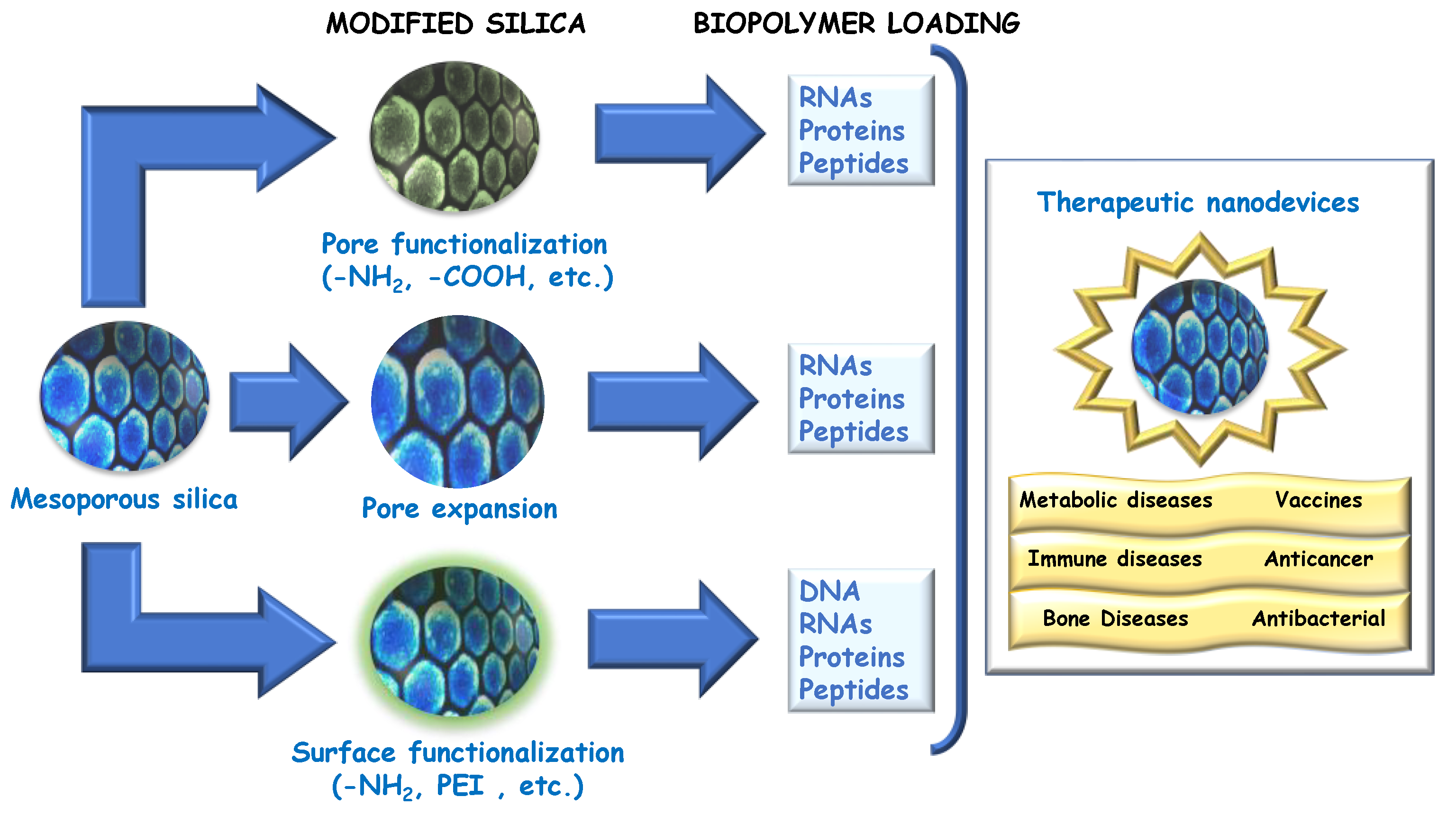
:1. Introduction
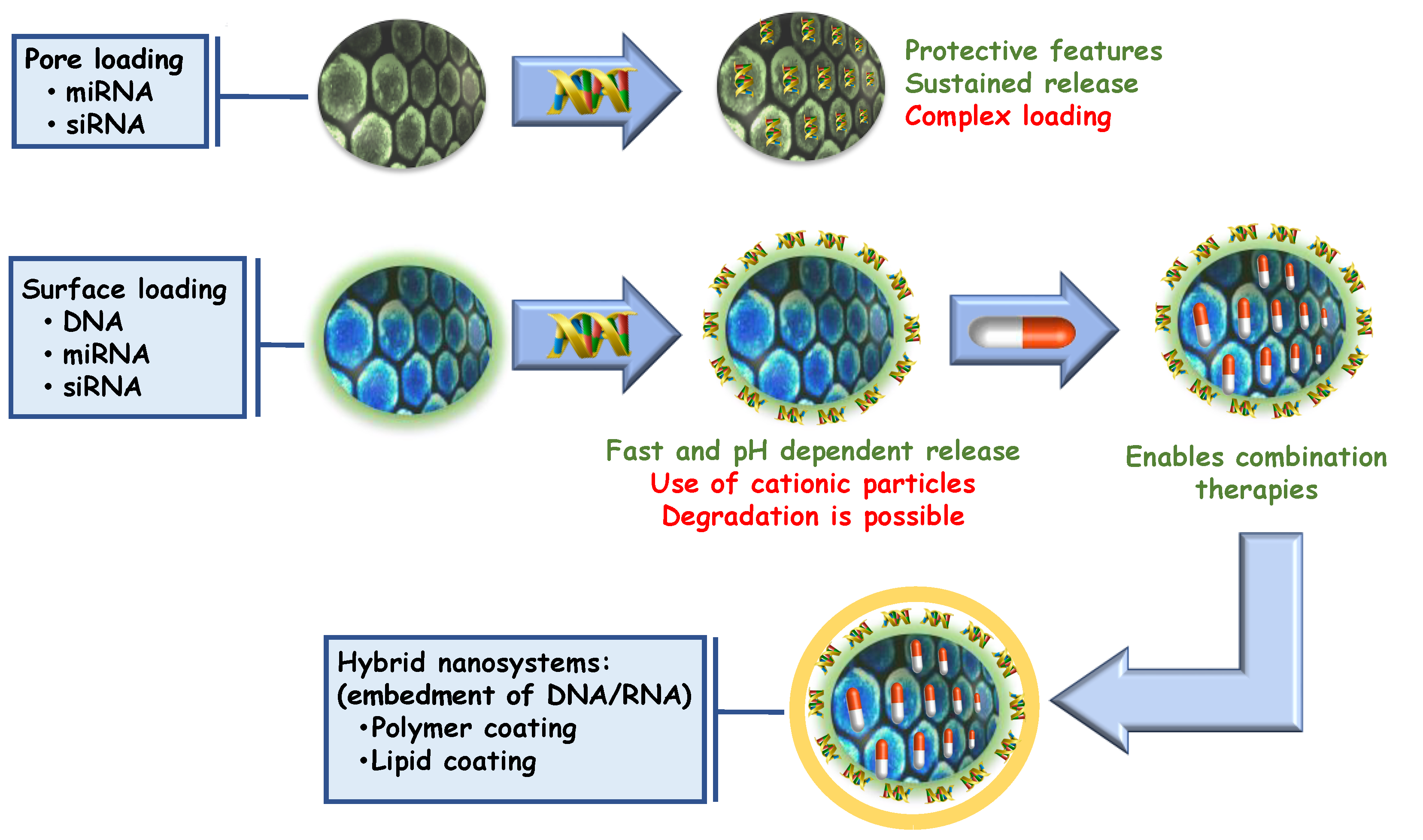
2. Development of Silica-Based Nanomedicines Using Therapeutic Nucleotides

| Nanosystem | Assembly Strategy | Nucleotide | Secondary Therapeutic | Therapy | Release Stimulus | Action Mechanism | Biological Evaluation | Ref. |
|---|---|---|---|---|---|---|---|---|
| Strategies for carrying oligonucleotides and plasmids with silica nanosystems | ||||||||
| LP-MSNs | Electrostatic deposition (PEI) | Plasmid (eGFP) | None | None (GFP transfection) | pH-driven detachment | Transfection | In vitro: HEK-293 | [50] |
| US-MSNs | PEI grafting (glutaraldehyde) | siRNA (siGADPH) | None | None (Knockdown quantification) | pH-driven detachment | Gene silencing | In vitro: HeLa, HEK-293 | [51] |
| Asymmetric MSNs (Phosphonate coated) | Electrostatic deposition (PEI) | Plasmid (eGFP) | None | None (GFP transfection) | pH-driven detachment | Transfection | In vitro: HEK-293T | [52] |
| MSNs (MCM-41) | Chemical grafting (PCL or CS) | siRNAs (α-tubulin, laminB1) | None | None (cytoskeleton reduction) | pH-driven detachment | Gene silencing | In vitro: HeLa, MCF-7 | [54] |
| LP-MSNs | Pore loading (Cystamine-PEI coated) | siRNA (anti-GFP) | None | None (Green florescence knockdown) | Redox (GSH) driven cleavage | Gene silencing | In vitro: MDA-MB-231 | [55] |
| MSN@ lipid Microbubbles | Pore loading | Plasmid (eGFP) | None | None (GFP transfection) | Ultrasound | Transfection | In vitro: SKOV3, HEK-293T In vivo: Mice | [57] |
| Strategies for gene therapy employing silica nanoparticles as non-viral vectors. | ||||||||
| MSNs | Pore loading | siRNA (anti-miR-155) | None | AS1411-targeted oncogene silencing | pH-sensitive PDA coating. | Adjuvant gene silencing for 5-FU chemotherapy | In vitro: SW480, HT-29, SW620, Lovo, Caco-2, NCM460 In vivo: Mice | [59] |
| MSNs | Electrostatic deposition (PEI) | siRNA (anti-HER2) | None | Transtuzumab-targeted oncogene silencing | pH-driven siRNA detachment | Gene silencing | In vitro: BT474 | [60] |
| MSNs | Pore loading | siRNA (anti-MDR1) | None | TAT-targeted anticancer siRNA delivery | Detachment of chitosan protecting layer | Gene silencing | In vitro: HeLa, EPG85.257 | [61] |
| CC-MSNs | Electrostatic deposition (PEI) | miRNA (rno-miRNA-26a-5p) | None | Osteogenic therapy | None | Gene expression | In vitro: rBMSC | [62] |
| Dual MSNs (Amino modified) | Pore loading | Plasmids (GLP-1AR and FGF-21) | None | Antidiabetic therapy | None | Transfection | In vitro: Hepa1-6 In vivo: Mice | [63] |
| Rambutan-like MSNs | Electrostatic deposition (PEI) | Plasmid (OVA) | None | Vaccine | pH-driven detachment | Transfection of an immune stimulator | In vitro: HEK-293 In vivo: Mice | [64] |
| Porous silica microrods | Electrostatic deposition (PEI) | Plasmid (OVA) | None | Vaccine | pH-driven detachment | Transfection of an immune stimulator | In vivo: Mice. | [65] |
| Ca-dopped Si NPs (amino modified) | Not determined | Plasmid (PRPF31-GFP) | None | Blindness treatment | pH-driven detachment | Transfection | In vivo: Mice | [66] |
| Dendritic MSNs (amino modified) | Not determined | Antisense plasmid (ASvicR) | None | Antibiofilm (anticaries) | pH-driven detachment | Transfection | In vitro: S. mutans UA159 | [67] |
| Strategies for combined gene and chemotherapy: siRNA delivery | ||||||||
| MSNs (amino modified) | Electrostatic deposition | siRNA (siVEGF) | Sorafenib | LA-targeted siRNA-drug combination | pH-driven detachment | Gene silencing plus kinase inhibition | In vitro: Huh7, HepG2, HeLa and A549 | [68] |
| MSNs | Electrostatic deposition (PEI) | siRNA (Survivin) | Docetaxel, etoposide and/or carfilzomib | siRNA-drug combination | pH-driven detachment | Gene silencing plus proteasome inhibition | In vitro: HEK-293, A549 | [69] |
| Disulfide bridged MSNs | Electrostatic deposition (HA-PEI) | siRNA (Bcl-2) | Doxorubicin | siRNA-drug combination | pH-driven detachment, GSH-driven SiO2 degradation | Gene silencing plus chemotherapeutic | In vitro: MCF7 | [70] |
| Ca2+ doped LP-MSNs | Pore loading (Ca2+) | siRNA (Bcl-2) | Chloroquine | siRNA-drug combination | Pore release | Gene silencing plus chemotherapeutic | In vitro: SKOV3 | [71] |
| Strategies for combined gene and chemotherapy: plasmid delivery | ||||||||
| UCNP@MSNs (amino modified) | Electrostatic deposition (H2A) | Plasmid (p53) | Bortezomib | H2A-targeted, combined genic and chemotherapy | pH-driven detachment | Transfection plus chemotherapeutic | In vitro: NCI-H1299 and HeLa | [72] |
| MSNs | Electrostatic deposition onto pH-labile polymer coating | Plasmid (p53) | 5-Fluoruracil | Combined genic and chemotherapy | pH-driven detachment | Transfection plus chemotherapeutic | In vitro: MCF-7 | [73] |
| MSNs | Electrostatic deposition (PEI) | Plasmid (HNF4α) | Cisplatin | Combined genic and chemotherapy | pH-driven detachment | Transfection plus chemotherapeutic | In vitro: Huh7 In vivo: Mice | [74] |
| Rod-shaped MSNs | Not Specified | Plasmid (Survivin) | Camptothecin | AS1411-targeted, Combined genic and chemotherapy | Not Specified | Transfection plus chemotherapeutic | In vitro: C26, CHO In vivo: Mice | [75] |
| Strategies for combined gene and chemotherapy: miRNA delivery | ||||||||
| MSNs | Lipid coating (erythrocytes’ membranes) | miRNA (miR137) | Indocyanine green (PDT) | RGD-targeted photothermal- miRNA | Detachment of protecting lipid layer | Photothermal and gene expression therapy. | In vitro: U87, RAW264.7 In vivo: Mice | [76] |
| MSNs | Pore loading | miRNA (hsa-miR-200c) siRNA (anti-Plk1) | Indocyanine green (PDT) | RGD-targeted photothermal- miRNA-siRNA | Detachment of protecting lipid layer | Photodynamic, gene silencing and expression | In vitro: MDA-MB-231 In vivo: Mice | [77] |
| MSNs | Pore loading | Dual miRNA (miR-34a and anti-miR-10b) | None | HA-targeted dual miRNA-combination | Detachment of protecting polymer layer (HA-PEG-PLGA) | Dual gene modulation | In vitro: MDA-MB-231, MDA-MB-468, HEK-293T, 4T1 In vivo: Chicken embryo In vivo: Mice | [78] |
| Strategies employing silica nanoparticles for gene therapy in bone diseases | ||||||||
| MSNs | Electrostatic deposition (PEI-PLL) | Plasmid (BMP-2) | Dexamethasone | Targeted and combined anti- osteoporotic treatment | pH-driven detachment | RGD-targeted osteoporosis gene silencing plus osteogenic stimulation | In vitro: HEK293T, BMSCs and RAW 264.7 | [79] |
| MSNs | Pore loading | Anti-miRNA (miR26) | None | Targeted anti-osteoporotic treatment | pH-driven detachment | KALA-targeted osteogenic stimulation | In vitro: rBMSCs | [80] |
| LP-MSNs | Pore loading | Anti-miRNA (miR26) | None | Targeted anti-osteoporotic treatment | Pore release | Osteogenic stimulation | In vitro: rBMSCs | [62] |
| MSNs | Electrostatic deposition (PEI) | siRNA (SOST) | Osteostatin | Combined anti- osteoporotic treatment | pH-driven detachment | Osteoporosis gene silencing plus osteogenic stimulation | In vitro: MEF In vivo: Mice | [81] |
| MSNs | Electrostatic deposition (PEI) | siRNA (SOST) | Osteostatin | Combined anti- osteoporotic treatment | pH-driven detachment | Alendronate-targeted Osteoporosis gene silencing plus osteogenic stimulation | In vitro: MC3T3-E1 In vivo: Mice | [82] |
| Other therapies | ||||||||
| MSNs | Electrostatic deposition | Plasmid (RhoG) | Curcumin | TAT-targeted neurite growth | pH-driven detachment | Neurite growth induction plus antioxidative effect | In vitro: N2a cells | [83] |
| MSNs | Electrostatic deposition | Anti-miRNA (miR33) | None | Metabolic lipid disorder treatment | pH-driven detachment | miRNA scavenging | In vitro: L02, LX02, RAW264.7 In vivo: Mice | [84] |
| LP-MSNs | Electrostatic driven loading | Plasmid (antisense vicR) | None | Biofilm disruption | pH-driven detachment | Reduction of extracellular polysaccharides | In vitro: Streptococcus Mutans | [67] |
2.1. Gene Therapy and Gene Silencing
2.2. Combined Anticancer Therapies
2.3. Gene-Based Therapies against Bone Diseases
2.4. Other Gene-Based Therapies Employing Silica Particles as Nucleotide Carriers
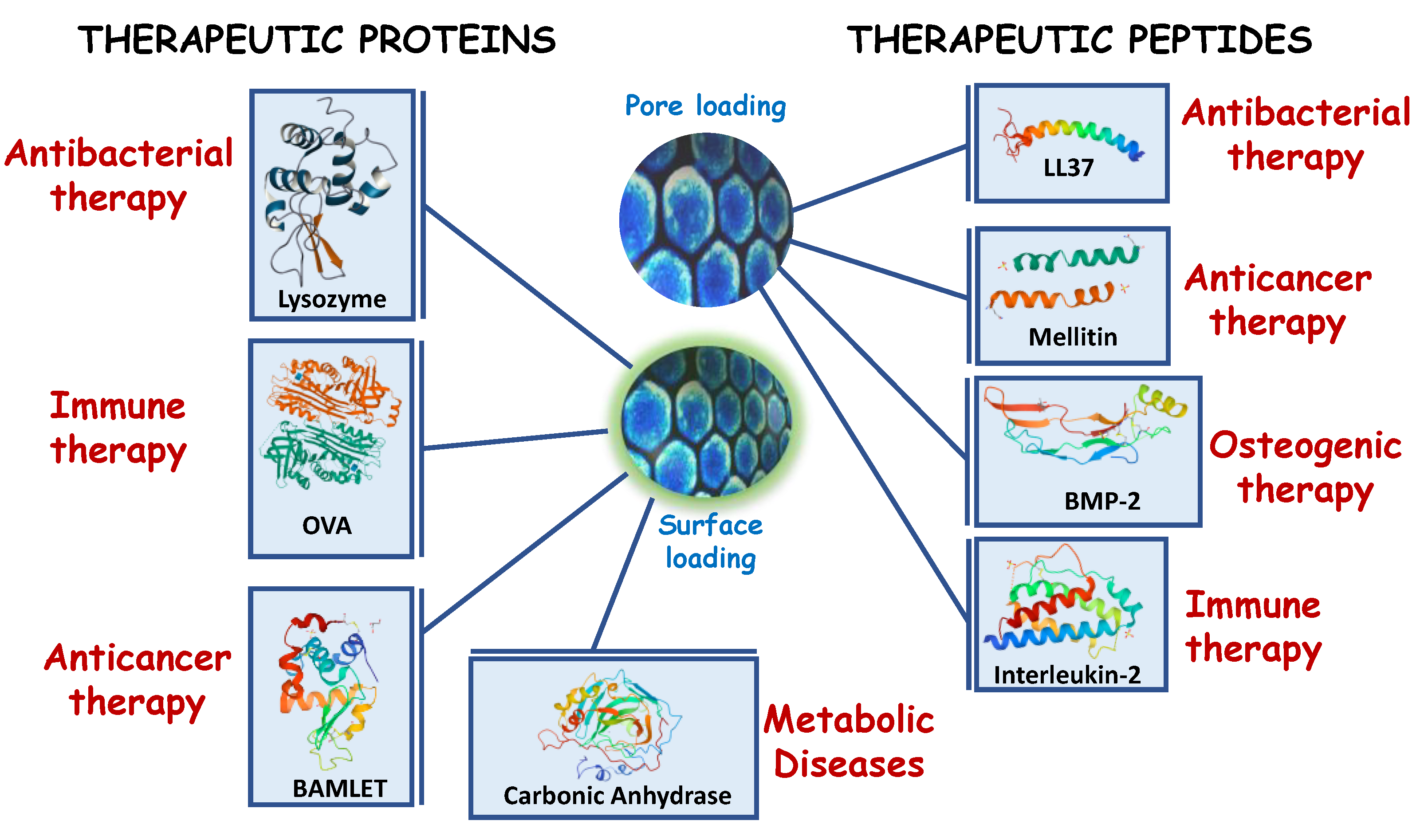
3. Protein- and Peptide-Based Therapeutics Employing Silica-Based Nanosystems
3.1. Anticancer Therapies
| Nanosystem | Assembly Strategy | Protein or Peptide | Secondary Therapeutic | Biomolecule Release Stimulus | Action Mechanism | Biological Evaluation | Ref. |
|---|---|---|---|---|---|---|---|
| Delivery of peptides and proteins in anticancer strategies | |||||||
| MSNs | Pore loading | Cytochrome C | None | Pore release | Cell apoptosis promoter | In vitro: HeLa | [111,112] |
| MSNs | Surface adsorption | None | Electrostatic detachment | None | [113] | ||
| MSNs | Surface grafting | None | None | In vitro: HeLa | [114] | ||
| MSNs | Surface grafting | Concanavalin A | None | None | Targeting plus upregulation of metalloproteinases | In vitro: MC3T3-E1, HOS. | [116] |
| MSNs | Surface grafting | BAMLET | Docetaxel | None | Cell apoptosis promoter plus chemotheraputic | In vitro: COS7, U87 MG In vivo: Zebrafish In vivo: Balb/c Mice | [117] |
| MSNs | Surface grafting | K8-Citraconate K8(RGD)2 | Doxorubicin | None | Membrane disruption plus chemotherapy | In vitro: COS7, U87 MG In vivo: Mice | [118] |
| MSNs | Surface grafting | TPP-K-(KLAKLAK)2- | Topotecan | None | Mitochondrial membrane disruption plus chemotherapy | In vitro: KB | [119] |
| MSNs | Surface grafting | C-GRK2R2QR3P2Q-RGDS C-GKGG-D(KLAKLAK)2 | Doxorubicin | None | Membrane disruption plus chemotherapy | In vitro: HeLa, COS7 | [120] |
| MSNs | Surface grafting | (RGDWWW)2KC | Doxorubicin | None | DNA-intercalation plus chemotherapy | In vitro: COS7, U87 MG | [121] |
| MSNs | Surface grafting | (KLAKLAK)2 | Doxorubicin | None | Membrane disruption plus chemotherapy | In vitro: HeLa | [122] |
| MSNs | Surface grafting and pore loading | ε-poly-L-lysine (surface) | C9h (pore) | Enzymatic degradation (Pore release) | Membrane disruption plus propapoptotic induction | In vitro: HeLa | [123] |
| MSNs | Pore loading | RDG-Hylin a1 | None | Pore release | Targeted cytolytic peptide | In vitro: HeLa Hep2 In vivo: Mice | [124] |
| HMSNs | Cavity loading | Pepstatin A | None | Release from cavity | Inhibition of Aspartyl protease | In vitro: MCF-7 | [125] |
| MSNs (SBA-16) | Pore loading | Alamandine | None | Pore release | Unknown | In vitro: 4T1, A549, HEK-293 | [126] |
| MSNs | Pore loading | Fabatin | None | Pore release | Mitochondrial disfunction | In vitro: MDA-MB-23, MCF-10A In vivo: Mice | [127] |
| SPION@MSNs | Pore loading | Mellitin | None | Thermosensitive–hydrolytic cleavage | Apoptosis induction and suppression of angiogenesis (VEGF) | In vitro: PANC-1 In vivo: Mice | [128] |
| Antibacterial therapies | |||||||
| MSNs | Adsorption | Lysozyme | None | Electrostatic detachment | Bacterial wall hydrolysis | In vitro: E. coli In vitro: HEK-293, LO2 In vivo: Mice | [129] |
| MSNs | Pore loading | None | Pore release | In vitro: E. coli | [130] | ||
| HMSNs | Surface adsorption | None | Electrostatic detachment | In vitro: E. coli In vivo: Mice | [131] | ||
| HMSNs | Cavity loading | None | Cavity release | In vitro: E. coli | [132] | ||
| MSNs | Surface grafting | Concanavalin A | Levofloxacin | None | Glycopeptide targeting | In vitro: E. coli | [133] |
| MSNs | Pore loading | Bactofencin A | None | Pore release | Defensin-like peptide | In vitro: S. aureus In vitro: HEK-293 | [134] |
| MSNs | Pore loading | β-defensin-2 | None | Pore release | Defensin-like peptide | In vitro: Clavibacter michiganensis | [135] |
| Solid SiO2 vs MSNs | Surface adsorption vs. pore loading | LL37 | None | Surface adsorption vs. pore release | Transmembrane pore formation (α-helical shape) | In vitro: E. coli | [136] |
| RSNs | Surface deposition | None | Release from rough surface | Transmembrane pore formation (α-helical shape) | In vitro: E. coli | [137] | |
| MSNs | Pore loading | NZX | None | Pore release | Antituberculotic peptide | In vitro: M. tuberculosis In vivo: Mice | [138] |
| MSNs | Surface grafting | Ovotransferrin | Gentamicin | None | Membrane lysis | In vitro: E. coli In vivo: Mice | [139] |
| MSNs (phosphonate vs. raw MCM-41) | Pore loading | Pexiganan, Indolicidin, or [I5,R8] Mastoparan (Electrostatically located at the surface) | trans-chalcone, curcumin, quercetin or berberine chloride | Electrostatic detachment | Sortase A inhibition (small molecules) plus antibiotic peptide. Pexiganan (cationic), Indolicidin (filamentator), Mastoparan (α-helix pore formation) | In vitro: S. aureus, S. aureus (Methilin resistant), E. coli, P. aeruginosa | [140] |
| Osteogenic therapies | |||||||
| MSNs | Pore loading | bFGF | None | Pore release | Growth factor (fibroblast) | In vitro: HUVEC | [141] |
| MSNs | Surface adsorption | BMP-2 | None | Electrostatic detachment | Bone morphogenetic protein | In vitro: bMSCs In vivo: Mice | [142] |
| MSN@SPION | Pore loading | Pore release | In vitro: bMSCs | [143] | |||
| MSNs | Pore loading | Osteostatin | None | Pore release | Hormone-related peptide | In vitro: MC3T3-E1 In vivo: Rabbit | [144,145,146] |
| MSNs MS-HANs | Pore loading | OGP | None | Pore release | Osteogenic growth peptide (Unknown mechanism) | In vitro: Mesenchymal stem cells | [147] |
| MSNs | Surface adsorption | BMP-2 derived peptide | None | Pore release | Bone morphogenetic protein derived peptide | In vitro: BMSCs In vivo: Rat | [148] |
| MCaSiNs | Pore loading | GL13K | Sr+2 doped matrix | Pore release (peptide) | Antibiotic peptide Osteoclastogenetic promoter | In vitro: HBMSCs In vitro: S. aureus | [149] |
| Immunotherapy | |||||||
| HMSNs | Cavity loading | IgG | None | Cavity release | Proof of concept | In vitro: HeLa | [150] |
| RSNs | Interparticle loading | Cyt c, IgG, Anti-pAkt | None | Cavity release | Proof of concept | None | [151] |
| HMSNs | Pore loading | OVA | None | Pore release | Xenoprotein induced immunostimulation | In vitro: NIH3T3 In vivo: Mice | [152,153] |
| DMOHS | Pore loading | None | Pore release | Xenoprotein induced immunostimulation | In vivo: Mice | [154] | |
| MSNs | Pore loading | CpG | Pore release | Xenoprotein induced immunostimulation plus a Toll-like Receptor agonist (CpG) | In vitro: RAW264.7 In vivo: Mice | [155] | |
| HMSNs | Cavity-pore loading | IL2 | Retinoic acid Doxorubicin | Lipid layer detachment. Multiple release | Immunostimulant protein (IL2) Chemotherapeutic (DOX) Apoptotic promoter (RA) | In vitro: L929 In vivo: Mice | [156] |
| MSNs | Pore loading | IL13 | None | Pore release | Immunostimulant protein (IL13) | In vitro: BMDMs In vivo: Mice | [157] |
| HMSNs | Surface adsorption | ORF2 | None | Electrostatic detachment | Anti circovirus vaccine | In vitro: PK15 In vivo: Mice | [158] |
| MSNs | Surface adsorption | SWAP | None | Electrostatic detachment | Anti-parasite vaccine | In vivo: Mice | [159] |
| MSNs | Surface adsorption | HSP70 | None | Electrostatic detachment | Anti-mycoplasma vaccine | In vivo: Mice | [160] |
| MSNs | Surface adsorption | EspA | None | Electrostatic detachment | Anti E. coli vaccine | In vivo: Mice | [161] |
| MSNs | Surface adsorption | rPb27 | None | Electrostatic detachment | Anti-fungi vaccine | In vitro: HEK-293 In vivo: Mice | [162] |
| HMSNs | Cavity loading | TRP2 (cavity) | HGP100 (pores) | Lipid layer detachment. Dual release | Dual antitumor immunostimulants | In vitro: BMDCs | [163] |
| MSNs | Surface | Hexahistidine | Chlorogenic acid | None | Ni scavenging (histidine) Antiinflamatory (Chlorogenic acid) | In vitro: BJ | [164] |
| Enzymes | |||||||
| MSNs | In-pore grafting | CA or HPR | None | None | Proof of concept | None | [165] |
| MSNs | In-pore grafting | CA | None | None | Proof of concept | In vitro: HeLa | [166] |
| MSNs | Pore loading | β-Galactosidase | None | None | Treatment of Morquio B syndrome | In vitro: N2a | [167] |
| MSNs | Surface grafting | SOD | None | None | Antioxidant effect (reduction of Reactive Oxygen Species) | In vitro: HeLa | [168] |
| MSNs | Surface grafting | SOD or GPx | None | None | Antioxidant effect (reduction of Reactive Oxygen Species) | In vitro: HeLa | [169] |
| MSNs | Surface grafting | Proteasomes | None | None | Anti Tau-protein aggregation | In vitro: HEK-293, HeLa | [170] |
| UC@MSNs | Surface adsorption | RNAase | Cisplatin prodrug | Electrostatic detachment | Combined anti-protein and chemotherapy | In vitro: HepG2, L929 In vivo: Mice | [171] |
3.2. Antibacterial Therapies
3.3. Osteogenic Therapies
3.4. Immunostimulant Proteins and Peptides
3.5. Enzymatic Therapy
4. Conclusions
Funding
Institutional Review Board Statement
Informed Consent Statement
Data Availability Statement
Acknowledgments
Conflicts of Interest
References
- Fus-Kujawa, A.; Prus, P.; Bajdak-Rusinek, K.; Teper, P.; Gawron, K.; Kowalczuk, A.; Sieron, A.L. An Overview of Methods and Tools for Transfection of Eukaryotic Cells in Vitro. Front. Bioeng. Biotechnol. 2021, 9, 701031. [Google Scholar] [CrossRef] [PubMed]
- Kulkarni, J.A.; Witzigmann, D.; Thomson, S.B.; Chen, S.; Leavitt, B.R.; Cullis, P.R.; van der Meel, R. The Current Landscape of Nucleic Acid Therapeutics. Nat. Nanotechnol. 2021, 16, 630–643. [Google Scholar] [CrossRef] [PubMed]
- Feng, R.; Patil, S.; Zhao, X.; Miao, Z.; Qian, A. RNA Therapeutics—Research and Clinical Advancements. Front. Mol. Biosci. 2021, 8, 710738. [Google Scholar] [CrossRef] [PubMed]
- Berraondo, P.; Sanmamed, M.F.; Ochoa, M.C.; Etxeberria, I.; Aznar, M.A.; Pérez-Gracia, J.L.; Rodríguez-Ruiz, M.E.; Ponz-Sarvise, M.; Castañón, E.; Melero, I. Cytokines in Clinical Cancer Immunotherapy. Br. J. Cancer 2019, 120, 6–15. [Google Scholar] [CrossRef] [PubMed] [Green Version]
- Cid, R.; Bolívar, J. Platforms for Production of Protein-Based Vaccines: From Classical to Next-Generation Strategies. Biomolecules 2021, 11, 1072. [Google Scholar] [CrossRef] [PubMed]
- Karpiński, T.; Adamczak, A. Anticancer Activity of Bacterial Proteins and Peptides. Pharmaceutics 2018, 10, 54. [Google Scholar] [CrossRef] [PubMed] [Green Version]
- Koo, H.B.; Seo, J. Antimicrobial Peptides under Clinical Investigation. Pept. Sci. 2019, 111, e24122. [Google Scholar] [CrossRef]
- Dijksteel, G.S.; Ulrich, M.M.W.; Middelkoop, E.; Boekema, B.K.H.L. Review: Lessons Learned from Clinical Trials Using Antimicrobial Peptides (AMPs). Front. Microbiol. 2021, 12, 616979. [Google Scholar] [CrossRef]
- Tian, Y.; van Tirrell, M.; LaBelle, J.L. Harnessing the Therapeutic Potential of Biomacromolecules through Intracellular Delivery of Nucleic Acids, Peptides, and Proteins. Adv. Healthc. Mater. 2022, 11, 2102600. [Google Scholar] [CrossRef]
- Morais, P.; Adachi, H.; Yu, Y.-T. The Critical Contribution of Pseudouridine to MRNA COVID-19 Vaccines. Front. Cell Dev. Biol. 2021, 9, 78942. [Google Scholar] [CrossRef]
- Xia, X. Detailed Dissection and Critical Evaluation of the Pfizer/BioNTech and Moderna MRNA Vaccines. Vaccines 2021, 9, 734. [Google Scholar] [CrossRef] [PubMed]
- Shi, Y.; van der Meel, R.; Chen, X.; Lammers, T. The EPR Effect and beyond: Strategies to Improve Tumor Targeting and Cancer Nanomedicine Treatment Efficacy. Theranostics 2020, 10, 7921–7924. [Google Scholar] [CrossRef]
- Zolnik, B.S.; González-Fernández, A.; Sadrieh, N.; Dobrovolskaia, M.A. Nanoparticles and the Immune System. Endocrinology 2010, 151, 458–465. [Google Scholar] [CrossRef] [PubMed]
- Amoozgar, Z.; Yeo, Y. Recent Advances in Stealth Coating of Nanoparticle Drug Delivery Systems. WIREs Nanomed. Nanobiotechnol. 2012, 4, 219–233. [Google Scholar] [CrossRef] [PubMed] [Green Version]
- Fam, S.Y.; Chee, C.F.; Yong, C.Y.; Ho, K.L.; Mariatulqabtiah, A.R.; Tan, W.S. Stealth Coating of Nanoparticles in Drug-Delivery Systems. Nanomaterials 2020, 10, 787. [Google Scholar] [CrossRef] [Green Version]
- Mazayen, Z.M.; Ghoneim, A.M.; Elbatanony, R.S.; Basalious, E.B.; Bendas, E.R. Pharmaceutical Nanotechnology: From the Bench to the Market. Futur. J. Pharm. Sci. 2022, 8, 12. [Google Scholar] [CrossRef]
- Sargazi, S.; Laraib, U.; Barani, M.; Rahdar, A.; Fatima, I.; Bilal, M.; Pandey, S.; Sharma, R.K.; Kyzas, G.Z. Recent Trends in Mesoporous Silica Nanoparticles of Rode-like Morphology for Cancer Theranostics: A Review. J. Mol. Struct. 2022, 1261, 132922. [Google Scholar] [CrossRef]
- Crommelin, D.J.A.; van Hoogevest, P.; Storm, G. The Role of Liposomes in Clinical Nanomedicine Development. What Now? Now What? J. Control. Release 2020, 318, 256–263. [Google Scholar] [CrossRef]
- Anselmo, A.C.; Mitragotri, S. Nanoparticles in the Clinic: An Update. Bioeng. Transl. Med. 2019, 4, e10143. [Google Scholar] [CrossRef] [Green Version]
- Anselmo, A.C.; Mitragotri, S. Nanoparticles in the Clinic: An Update Post COVID-19 Vaccines. Bioeng. Transl. Med. 2021, 6, e10246. [Google Scholar] [CrossRef]
- Hussein, H.A.; Nazir, M.S.; Azra, N.; Qamar, Z.; Seeni, A.; Tengku Din, T.A.D.A.-A.; Abdullah, M.A. Novel Drug and Gene Delivery System and Imaging Agent Based on Marine Diatom Biosilica Nanoparticles. Mar. Drugs 2022, 20, 480. [Google Scholar] [CrossRef] [PubMed]
- Farjadian, F.; Roointan, A.; Mohammadi-Samani, S.; Hosseini, M. Mesoporous Silica Nanoparticles: Synthesis, Pharmaceutical Applications, Biodistribution, and Biosafety Assessment. Chem. Eng. J. 2019, 359, 684–705. [Google Scholar] [CrossRef]
- Hosseinpour, S.; Walsh, L.J.; Xu, C. Biomedical Application of Mesoporous Silica Nanoparticles as Delivery Systems: A Biological Safety Perspective. J. Mater. Chem. B 2020, 8, 9863–9876. [Google Scholar] [CrossRef]
- Sun, L.; Sogo, Y.; Wang, X.; Ito, A. Biosafety of Mesoporous Silica Nanoparticles: A Combined Experimental and Literature Study. J. Mater. Sci. Mater. Med. 2021, 32, 102. [Google Scholar] [CrossRef] [PubMed]
- Mitchell, M.J.; Billingsley, M.M.; Haley, R.M.; Wechsler, M.E.; Peppas, N.A.; Langer, R. Engineering Precision Nanoparticles for Drug Delivery. Nat. Rev. Drug Discov. 2021, 20, 101–124. [Google Scholar] [CrossRef]
- Lee, N.K.; Kim, S.-N.; Park, C.G. Immune Cell Targeting Nanoparticles: A Review. Biomater. Res. 2021, 25, 44. [Google Scholar] [CrossRef]
- Rosenblum, D.; Joshi, N.; Tao, W.; Karp, J.M.; Peer, D. Progress and Challenges towards Targeted Delivery of Cancer Therapeutics. Nat. Commun. 2018, 9, 1410. [Google Scholar] [CrossRef] [Green Version]
- Moodley, T.; Singh, M. Current Stimuli-Responsive Mesoporous Silica Nanoparticles for Cancer Therapy. Pharmaceutics 2021, 13, 71. [Google Scholar] [CrossRef]
- Castillo, R.R.; Lozano, D.; González, B.; Manzano, M.; Izquierdo-Barba, I.; Vallet-Regí, M. Advances in Mesoporous Silica Nanoparticles for Targeted Stimuli-Responsive Drug Delivery: An Update. Expert Opin. Drug Deliv. 2019, 16, 415–439. [Google Scholar] [CrossRef]
- Shen, H.; Huang, X.; Min, J.; Le, S.; Wang, Q.; Wang, X.; Dogan, A.A.; Liu, X.; Zhang, P.; Draz, M.S.; et al. Nanoparticle Delivery Systems for DNA/RNA and Their Potential Applications in Nanomedicine. Curr. Top. Med. Chem. 2019, 19, 2507–2523. [Google Scholar] [CrossRef]
- Piperno, A.; Sciortino, M.T.; Giusto, E.; Montesi, M.; Panseri, S.; Scala, A. Recent Advances and Challenges in Gene Delivery Mediated by Polyester-Based Nanoparticles. Int. J. Nanomed. 2021, 16, 5981–6002. [Google Scholar] [CrossRef] [PubMed]
- Qin, S.; Tang, X.; Chen, Y.; Chen, K.; Fan, N.; Xiao, W.; Zheng, Q.; Li, G.; Teng, Y.; Wu, M.; et al. MRNA-Based Therapeutics: Powerful and Versatile Tools to Combat Diseases. Signal. Transduct. Target Ther. 2022, 7, 166. [Google Scholar] [CrossRef] [PubMed]
- Janowski, M.; Andrzejewska, A. The Legacy of MRNA Engineering: A Lineup of Pioneers for the Nobel Prize. Mol. Ther. Nucleic Acids 2022, 29, 272–284. [Google Scholar] [CrossRef] [PubMed]
- Dong, Y.; Siegwart, D.J.; Anderson, D.G. Strategies, Design, and Chemistry in SiRNA Delivery Systems. Adv. Drug Deliv. Rev. 2019, 144, 133–147. [Google Scholar] [CrossRef] [PubMed]
- Hattab, D.; Gazzali, A.M.; Bakhtiar, A. Clinical Advances of SiRNA-Based Nanotherapeutics for Cancer Treatment. Pharmaceutics 2021, 13, 1009. [Google Scholar] [CrossRef]
- Duan, L.; Ouyang, K.; Xu, X.; Xu, L.; Wen, C.; Zhou, X.; Qin, Z.; Xu, Z.; Sun, W.; Liang, Y. Nanoparticle Delivery of CRISPR/Cas9 for Genome Editing. Front. Genet. 2021, 12, 673286. [Google Scholar] [CrossRef]
- Chen, F.; Alphonse, M.; Liu, Q. Strategies for Nonviral Nanoparticle-based Delivery of CRISPR/Cas9 Therapeutics. WIREs Nanomed. Nanobiotechnol. 2020, 12, e1609. [Google Scholar] [CrossRef]
- Radu, D.R.; Lai, C.-Y.; Jeftinija, K.; Rowe, E.W.; Jeftinija, S.; Lin, V.S.-Y. A Polyamidoamine Dendrimer-Capped Mesoporous Silica Nanosphere-Based Gene Transfection Reagent. J. Am. Chem. Soc. 2004, 126, 13216–13217. [Google Scholar] [CrossRef]
- Castillo, R.R.; Baeza, A.; Vallet-Regí, M. Recent Applications of the Combination of Mesoporous Silica Nanoparticles with Nucleic Acids: Development of Bioresponsive Devices, Carriers and Sensors. Biomater. Sci. 2017, 5, 353–377. [Google Scholar] [CrossRef]
- Kim, T.; Nam, K.; Kim, Y.M.; Yang, K.; Roh, Y.H. DNA-Assisted Smart Nanocarriers: Progress, Challenges, and Opportunities. ACS Nano 2021, 15, 1942–1951. [Google Scholar] [CrossRef]
- Liu, M.; Wang, L.; Lo, Y.; Shiu, S.C.-C.; Kinghorn, A.B.; Tanner, J.A. Aptamer-Enabled Nanomaterials for Therapeutics, Drug Targeting and Imaging. Cells 2022, 11, 159. [Google Scholar] [CrossRef] [PubMed]
- Abu-Dief, A.M.; Alsehli, M.; Al-Enizi, A.; Nafady, A. Recent Advances in Mesoporous Silica Nanoparticles for Targeted Drug Delivery Applications. Curr. Drug Deliv. 2022, 19, 436–450. [Google Scholar] [CrossRef] [PubMed]
- Colilla, M.; Vallet-Regí, M. Chapter 13. Chemoresponsive Mesoporous Silica Nanoparticles for Targeted Drug Delivery in Cancer Therapy. In Chemoresponsive Materials: Smart Materials for Chemical and Biological Stimulation, 2nd ed.; Royal Society of Chemistry: London, UK, 2022; pp. 451–498. [Google Scholar]
- Barui, S.; Cauda, V. Multimodal Decorations of Mesoporous Silica Nanoparticles for Improved Cancer Therapy. Pharmaceutics 2020, 12, 527. [Google Scholar] [CrossRef]
- Yoo, H.; Jo, H.; Oh, S.S. Detection and beyond: Challenges and Advances in Aptamer-Based Biosensors. Mater. Adv. 2020, 1, 2663–2687. [Google Scholar] [CrossRef]
- Aznar, E.; Oroval, M.; Pascual, L.; Murguía, J.R.; Martínez-Máñez, R.; Sancenón, F. Gated Materials for On-Command Release of Guest Molecules. Chem. Rev. 2016, 116, 561–718. [Google Scholar] [CrossRef] [PubMed]
- Devika, V.; Sreelekshmi, P.J.; Rajeev, N.; Lakshmi, A.S.; Chandran, A.; Goutami, G.B.; Sadanandan, S. Recent Advances in Peptides-Based Stimuli-Responsive Materials for Biomedical and Therapeutic Applications: A Review. Mol. Pharm. 2022, 19, 1999–2021. [Google Scholar] [CrossRef]
- Castillo, R.R.; Lozano, D.; Vallet-Regí, M. Mesoporous Silica Nanoparticles as Carriers for Therapeutic Biomolecules. Pharmaceutics 2020, 12, 432. [Google Scholar] [CrossRef]
- Höbel, S.; Aigner, A. Polyethylenimines for SiRNA and MiRNA Delivery in Vivo. WIREs Nanomed. Nanobiotechnol. 2013, 5, 484–501. [Google Scholar] [CrossRef]
- Wang, Y.; Song, H.; Yu, M.; Xu, C.; Liu, Y.; Tang, J.; Yang, Y.; Yu, C. Room Temperature Synthesis of Dendritic Mesoporous Silica Nanoparticles with Small Sizes and Enhanced MRNA Delivery Performance. J. Mater. Chem. B 2018, 6, 4089–4095. [Google Scholar] [CrossRef]
- Chang, Q.; Liu, C.; Xie, Z.; Shu, Q.; Xie, Y.; Su, Q.; Deng, X. Polyethylenimine Functionalized Ultrasmall Mesoporous Silica Nanoparticles for SiRNA Delivery. ChemNanoMat. 2022, 8, e202100453. [Google Scholar] [CrossRef]
- Lin, X.; Wu, W.; Fu, J.; Yang, Y.; Guo, B.; Yu, C.; Song, H. Asymmetric Silica Nanoparticles with Tailored Spiky Coverage Derived from Silica-Polymer Cooperative Assembly for Enhanced Hemocompatibility and Gene Delivery. ACS Appl. Mater. Interfaces 2021, 13, 50695–50704. [Google Scholar] [CrossRef] [PubMed]
- Taranejoo, S.; Liu, J.; Verma, P.; Hourigan, K. A Review of the Developments of Characteristics of PEI Derivatives for Gene Delivery Applications. J. Appl. Polym. Sci. 2015, 132, 42096. [Google Scholar] [CrossRef]
- Nhavene, E.; Andrade, G.; Faria, J.; Gomes, D.; Sousa, E. Biodegradable Polymers Grafted onto Multifunctional Mesoporous Silica Nanoparticles for Gene Delivery. ChemEngineering 2018, 2, 24. [Google Scholar] [CrossRef] [Green Version]
- Slita, A.; Egorova, A.; Casals, E.; Kiselev, A.; Rosenholm, J.M. Characterization of Modified Mesoporous Silica Nanoparticles as Vectors for SiRNA Delivery. Asian J. Pharm. Sci. 2018, 13, 592–599. [Google Scholar] [CrossRef] [PubMed]
- Khan, M.A.; Kiser, M.R.; Moradipour, M.; Nadeau, E.A.; Ghanim, R.W.; Webb, B.A.; Rankin, S.E.; Knutson, B.L. Effect of Confinement in Nanopores on RNA Interactions with Functionalized Mesoporous Silica Nanoparticles. J. Phys. Chem. B 2020, 124, 8549–8561. [Google Scholar] [CrossRef] [PubMed]
- Du, M.; Chen, Y.; Tu, J.; Liufu, C.; Yu, J.; Yuan, Z.; Gong, X.; Chen, Z. Ultrasound Responsive Magnetic Mesoporous Silica Nanoparticle-Loaded Microbubbles for Efficient Gene Delivery. ACS Biomater. Sci. Eng. 2020, 6, 2904–2912. [Google Scholar] [CrossRef] [PubMed]
- Li, X.; Chen, Y.; Wang, M.; Ma, Y.; Xia, W.; Gu, H. A Mesoporous Silica Nanoparticle–PEI–Fusogenic Peptide System for SiRNA Delivery in Cancer Therapy. Biomaterials 2013, 34, 1391–1401. [Google Scholar] [CrossRef]
- Li, Y.; Duo, Y.; Zhai, P.; He, L.; Zhong, K.; Zhang, Y.; Huang, K.; Luo, J.; Zhang, H.; Yu, X. Dual Targeting Delivery of MiR-328 by Functionalized Mesoporous Silica Nanoparticles for Colorectal Cancer Therapy. Nanomedicine 2018, 13, 1753–1772. [Google Scholar] [CrossRef]
- Ngamcherdtrakul, W.; Sangvanich, T.; Reda, M.; Gu, S.; Bejan, D.; Yantasee, W. Lyophilization and Stability of Antibody-Conjugated Mesoporous Silica Nanoparticle with Cationic Polymer and PEG for SiRNA Delivery. Int. J. Nanomed. 2018, 13, 4015–4027. [Google Scholar] [CrossRef] [Green Version]
- Heidari, R.; Khosravian, P.; Mirzaei, S.A.; Elahian, F. SiRNA Delivery Using Intelligent Chitosan-Capped Mesoporous Silica Nanoparticles for Overcoming Multidrug Resistance in Malignant Carcinoma Cells. Sci. Rep. 2021, 11, 20531. [Google Scholar] [CrossRef]
- Hosseinpour, S.; Cao, Y.; Liu, J.; Xu, C.; Walsh, L.J. Efficient Transfection and Long-Term Stability of Rno-MiRNA-26a-5p for Osteogenic Differentiation by Large Pore Sized Mesoporous Silica Nanoparticles. J. Mater. Chem. B 2021, 9, 2275–2284. [Google Scholar] [CrossRef] [PubMed]
- Geng, S.; Qin, L.; He, Y.; Li, X.; Yang, M.; Li, L.; Liu, D.; Li, Y.; Niu, D.; Yang, G. Effective and Safe Delivery of GLP-1AR and FGF-21 Plasmids Using Amino-Functionalized Dual-Mesoporous Silica Nanoparticles in Vitro and in Vivo. Biomaterials 2021, 271, 120763. [Google Scholar] [CrossRef] [PubMed]
- Song, H.; Yang, Y.; Tang, J.; Gu, Z.; Wang, Y.; Zhang, M.; Yu, C. DNA Vaccine Mediated by Rambutan-Like Mesoporous Silica Nanoparticles. Adv. Ther. 2020, 3, 1900154. [Google Scholar] [CrossRef]
- Nguyen, T.L.; Yin, Y.; Choi, Y.; Jeong, J.H.; Kim, J. Enhanced Cancer DNA Vaccine via Direct Transfection to Host Dendritic Cells Recruited in Injectable Scaffolds. ACS Nano 2020, 14, 11623–11636. [Google Scholar] [CrossRef]
- Valdés-Sánchez, L.; Borrego-González, S.; Montero-Sánchez, A.; Massalini, S.; de la Cerda, B.; Díaz-Cuenca, A.; Díaz-Corrales, F.J. Mesoporous Silica-Based Nanoparticles as Non-Viral Gene Delivery Platform for Treating Retinitis Pigmentosa. J. Clin. Med. 2022, 11, 2170. [Google Scholar] [CrossRef]
- Tian, Y.; Zhang, Y.; Zhang, M.; Chen, X.; Lei, L.; Hu, T. Antisense vicR-Loaded Dendritic Mesoporous Silica Nanoparticles Regulate the Biofilm Organization and Cariogenicity of Streptococcus mutans. Int. J. Nanomed. 2022, 17, 1255–1272. [Google Scholar] [CrossRef]
- Zheng, G.; Zhao, R.; Xu, A.; Shen, Z.; Chen, X.; Shao, J. Co-Delivery of Sorafenib and SiVEGF Based on Mesoporous Silica Nanoparticles for ASGPR Mediated Targeted HCC Therapy. Eur. J. Pharm. Sci. 2018, 111, 492–502. [Google Scholar] [CrossRef]
- Dilnawaz, F.; Sahoo, S.K. Augmented Anticancer Efficacy by Si-RNA Complexed Drug-Loaded Mesoporous Silica Nanoparticles in Lung Cancer Therapy. ACS Appl. Nano Mater. 2018, 1, 730–740. [Google Scholar] [CrossRef]
- Zhang, B.; Liu, Q.; Liu, M.; Shi, P.; Zhu, L.; Zhang, L.; Li, R. Biodegradable Hybrid Mesoporous Silica Nanoparticles for Gene/Chemo-Synergetic Therapy of Breast Cancer. J. Biomater. Appl. 2019, 33, 1382–1393. [Google Scholar] [CrossRef]
- Choi, E.; Lim, D.-K.; Kim, S. Calcium-Doped Mesoporous Silica Nanoparticles as a Lysosomolytic Nanocarrier for Amine-Free Loading and Cytosolic Delivery of SiRNA. J. Ind. Eng. Chem. 2020, 81, 71–80. [Google Scholar] [CrossRef]
- Rong, J.; Li, P.; Ge, Y.; Chen, H.; Wu, J.; Zhang, R.; Lao, J.; Lou, D.; Zhang, Y. Histone H2A-Peptide-Hybrided Upconversion Mesoporous Silica Nanoparticles for Bortezomib/P53 Delivery and Apoptosis Induction. Colloids Surf. B Biointerfaces 2020, 186, 110674. [Google Scholar] [CrossRef] [PubMed]
- Zhou, S.; Ding, C.; Wang, C.; Fu, J. UV-Light Cross-Linked and PH de-Cross-Linked Coumarin-Decorated Cationic Copolymer Grafted Mesoporous Silica Nanoparticles for Drug and Gene Co-Delivery in Vitro. Mater. Sci. Eng. C 2020, 108, 110469. [Google Scholar] [CrossRef] [PubMed]
- Tsai, P.H.; Wang, M.L.; Chang, J.H.; Yarmishyn, A.A.; Nhi Nguyen, P.N.; Chen, W.; Chien, Y.; Huo, T.I.; Mou, C.Y.; Chiou, S.H. Dual Delivery of HNF4α and Cisplatin by Mesoporous Silica Nanoparticles Inhibits Cancer Pluripotency and Tumorigenicity in Hepatoma-Derived CD133-Expressing Stem Cells. ACS Appl. Mater. Interfaces 2019, 11, 19808–19818. [Google Scholar] [CrossRef] [PubMed]
- Babaei, M.; Abnous, K.; Taghdisi, S.M.; Taghavi, S.; Saljooghi, A.S.; Ramezani, M.; Alibolandi, M. Targeted Rod-Shaped Mesoporous Silica Nanoparticles for the Co-Delivery of Camptothecin and Survivin ShRNA into Colon Adenocarcinoma in Vitro and in Vivo. Eur. J. Pharm. Biopharm. 2020, 156, 84–96. [Google Scholar] [CrossRef] [PubMed]
- Li, M.; Cui, X.; Wei, F.; Li, C.; Han, X. RGD Peptide Modified Erythrocyte Membrane/Porous Nanoparticles Loading Mir-137 for NIR-Stimulated Theranostics of Glioblastomas. Nanomaterials 2022, 12, 1464. [Google Scholar] [CrossRef]
- Wang, Y.; Xie, Y.; Kilchrist, K.V.; Li, J.; Duvall, C.L.; Oupický, D. Endosomolytic and Tumor-Penetrating Mesoporous Silica Nanoparticles for SiRNA/MiRNA Combination Cancer Therapy. ACS Appl. Mater. Interfaces 2020, 12, 4308–4322. [Google Scholar] [CrossRef]
- Ahir, M.; Upadhyay, P.; Ghosh, A.; Sarker, S.; Bhattacharya, S.; Gupta, P.; Ghosh, S.; Chattopadhyay, S.; Adhikary, A. Delivery of Dual MiRNA through CD44-Targeted Mesoporous Silica Nanoparticles for Enhanced and Effective Triple-Negative Breast Cancer Therapy. Biomater. Sci. 2020, 8, 2939–2954. [Google Scholar] [CrossRef]
- Zhou, X.; Zhang, Q.; Chen, L.; Nie, W.; Wang, W.; Wang, H.; Mo, X.; He, C. Versatile Nanocarrier Based on Functionalized Mesoporous Silica Nanoparticles to Codeliver Osteogenic Gene and Drug for Enhanced Osteodifferentiation. ACS Biomater. Sci. Eng. 2019, 5, 710–723. [Google Scholar] [CrossRef]
- Yan, J.; Lu, X.; Zhu, X.; Hu, X.; Wang, L.; Qian, J.; Zhang, F.; Liu, M. Effects of MiR-26a on Osteogenic Differentiation of Bone Marrow Mesenchymal Stem Cells by a Mesoporous Silica Nanoparticle–PEI–Peptide System. Int. J. Nanomed. 2020, 15, 497–511. [Google Scholar] [CrossRef] [Green Version]
- Mora-Raimundo, P.; Lozano, D.; Manzano, M.; Vallet-Regí, M. Nanoparticles to Knockdown Osteoporosis-Related Gene and Promote Osteogenic Marker Expression for Osteoporosis Treatment. ACS Nano 2019, 13, 5451–5464. [Google Scholar] [CrossRef]
- Mora-Raimundo, P.; Lozano, D.; Benito, M.; Mulero, F.; Manzano, M.; Vallet-Regí, M. Osteoporosis Remission and New Bone Formation with Mesoporous Silica Nanoparticles. Adv. Sci. 2021, 8, 2101107. [Google Scholar] [CrossRef] [PubMed]
- Cheng, C.-S.; Liu, T.-P.; Chien, F.-C.; Mou, C.-Y.; Wu, S.-H.; Chen, Y.-P. Codelivery of Plasmid and Curcumin with Mesoporous Silica Nanoparticles for Promoting Neurite Outgrowth. ACS Appl. Mater. Interfaces 2019, 11, 15322–15331. [Google Scholar] [CrossRef] [PubMed]
- Tao, Y.; Xu, S.; Wang, J.; Xu, L.; Zhang, C.; Chen, K.; Lian, Z.; Zhou, J.; Xie, H.; Zheng, S.; et al. Delivery of MicroRNA-33 Antagomirs by Mesoporous Silica Nanoparticles to Ameliorate Lipid Metabolic Disorders. Front. Pharmacol. 2020, 11, 921. [Google Scholar] [CrossRef]
- Li, N.; Niu, D.; Jiang, Y.; Xu, C.; Pan, S.; He, J.; Chen, J.; Zhang, L.; Li, Y. Morphology Evolution and Spatially Selective Functionalization of Hierarchically Porous Silica Nanospheres for Improved Multidrug Delivery. Chem. Mater. 2017, 29, 10377–10385. [Google Scholar] [CrossRef]
- Castillo, R.R.; Vallet-Regí, M. Emerging Strategies in Anticancer Combination Therapy Employing Silica-Based Nanosystems. Biotechnol. J. 2021, 16, 1900438. [Google Scholar] [CrossRef]
- Kumar, K.; Rani, V.; Mishra, M.; Chawla, R. New Paradigm in Combination Therapy of SiRNA with Chemotherapeutic Drugs for Effective Cancer Therapy. Curr. Res. Pharmacol. Drug Discov. 2022, 3, 100103. [Google Scholar] [CrossRef]
- Castillo, R.R.; Colilla, M.; Vallet-Regí, M. Advances in Mesoporous Silica-Based Nanocarriers for Co-Delivery and Combination Therapy against Cancer. Expert Opin. Drug Deliv. 2017, 14, 229–243. [Google Scholar] [CrossRef]
- Paskeh, M.D.A.; Saebfar, H.; Mahabady, M.K.; Orouei, S.; Hushmandi, K.; Entezari, M.; Hashemi, M.; Aref, A.R.; Hamblin, M.R.; Ang, H.L.; et al. Overcoming Doxorubicin Resistance in Cancer: SiRNA-Loaded Nanoarchitectures for Cancer Gene Therapy. Life Sci. 2022, 298, 120463. [Google Scholar] [CrossRef]
- Mirzaei, S.; Gholami, M.H.; Hashemi, F.; Zabolian, A.; Hushmandi, K.; Rahmanian, V.; Entezari, M.; Girish, Y.R.; Sharath Kumar, K.S.; Aref, A.R.; et al. Employing SiRNA Tool and Its Delivery Platforms in Suppressing Cisplatin Resistance: Approaching to a New Era of Cancer Chemotherapy. Life Sci. 2021, 277, 119430. [Google Scholar] [CrossRef]
- Paris, J.L.; Vallet-Regí, M. Mesoporous Silica Nanoparticles for Co-Delivery of Drugs and Nucleic Acids in Oncology: A Review. Pharmaceutics 2020, 12, 526. [Google Scholar] [CrossRef]
- Mosammaparast, N.; Jackson, K.R.; Guo, Y.; Brame, C.J.; Shabanowitz, J.; Hunt, D.F.; Pemberton, L.F. Nuclear Import of Histone H2a and H2b Is Mediated by a Network of Karyopherins. J. Cell Biol. 2001, 153, 251–262. [Google Scholar] [CrossRef] [PubMed]
- Shi, X.; Singh, S.; Lin, E.; Li, H. Chimeric RNAs in Cancer. Adv. Clin. Chem. 2021, 100, 1–35. [Google Scholar] [PubMed]
- Živojević, K.; Mladenović, M.; Djisalov, M.; Mundzic, M.; Ruiz-Hernandez, E.; Gadjanski, I.; Knežević, N. Advanced Mesoporous Silica Nanocarriers in Cancer Theranostics and Gene Editing Applications. J. Control. Release 2021, 337, 193–211. [Google Scholar] [CrossRef] [PubMed]
- Liu, X.; Wu, F.; Ji, Y.; Yin, L. Recent Advances in Anti-Cancer Protein/Peptide Delivery. Bioconjug. Chem. 2019, 30, 305–324. [Google Scholar] [CrossRef] [PubMed]
- Yau, A.; Lee, J.; Chen, Y. Nanomaterials for Protein Delivery in Anticancer Applications. Pharmaceutics 2021, 13, 155. [Google Scholar] [CrossRef] [PubMed]
- Liu, H.-J.; Xu, P. Smart Mesoporous Silica Nanoparticles for Protein Delivery. Nanomaterials 2019, 9, 511. [Google Scholar] [CrossRef] [PubMed] [Green Version]
- Xu, C.; Lei, C.; Yu, C. Mesoporous Silica Nanoparticles for Protein Protection and Delivery. Front. Chem. 2019, 7, 290. [Google Scholar] [CrossRef] [Green Version]
- Liu, B.; Ejaz, W.; Gong, S.; Kurbanov, M.; Canakci, M.; Anson, F.; Thayumanavan, S. Engineered Interactions with Mesoporous Silica Facilitate Intracellular Delivery of Proteins and Gene Editing. Nano Lett. 2020, 20, 4014–4021. [Google Scholar] [CrossRef]
- Lau, J.L.; Dunn, M.K. Therapeutic Peptides: Historical Perspectives, Current Development Trends, and Future Directions. Bioorg. Med. Chem. 2018, 26, 2700–2707. [Google Scholar] [CrossRef]
- Marqus, S.; Pirogova, E.; Piva, T.J. Evaluation of the Use of Therapeutic Peptides for Cancer Treatment. J. Biomed. Sci. 2017, 24, 1–15. [Google Scholar] [CrossRef]
- Kurrikoff, K.; Aphkhazava, D.; Langel, Ü. The Future of Peptides in Cancer Treatment. Curr. Opin. Pharmacol. 2019, 47, 27–32. [Google Scholar] [CrossRef] [PubMed]
- Wang, Y.; Gao, Y.N.; Bai, R.; Chen, H.Y.; Wu, Y.Y.; Shang, J.J.; Bao, D.P. Identification of a Novel Anti-Cancer Protein, FIP-Bbo, from Botryobasidium botryosum and Protein Structure Analysis Using Molecular Dynamic Simulation. Sci. Rep. 2019, 9, 5818. [Google Scholar] [CrossRef] [PubMed] [Green Version]
- Gaspar, D.; Salomé Veiga, A.; Castanho, M.A.R.B. From Antimicrobial to Anticancer Peptides. A Review. Front. Microbiol. 2013, 4, 294. [Google Scholar] [CrossRef] [PubMed] [Green Version]
- Mahlapuu, M.; Håkansson, J.; Ringstad, L.; Björn, C. Antimicrobial Peptides: An Emerging Category of Therapeutic Agents. Front. Cell Infect. Microbiol. 2016, 6, 194. [Google Scholar] [CrossRef] [PubMed] [Green Version]
- Khan, M.M.; Filipczak, N.; Torchilin, V.P. Cell Penetrating Peptides: A Versatile Vector for Co-Delivery of Drug and Genes in Cancer. J. Control. Release 2021, 330, 1220–1228. [Google Scholar] [CrossRef]
- Kalmouni, M.; Al-Hosani, S.; Magzoub, M. Cancer Targeting Peptides. Cell. Mol. Life Sci. 2019, 76, 2171–2183. [Google Scholar] [CrossRef]
- Robinson, J.A. Folded Synthetic Peptides and Other Molecules Targeting Outer Membrane Protein Complexes in Gram-Negative Bacteria. Front. Chem. 2019, 7, 45. [Google Scholar] [CrossRef] [Green Version]
- Cheng, Z.; Li, Y.; Zhao, D.; Zhao, W.; Wu, M.; Zhang, W.; Cui, Y.; Zhang, P.; Zhang, Z. Nanocarriers for Intracellular Co-Delivery of Proteins and Small-Molecule Drugs for Cancer Therapy. Front. Bioeng. Biotechnol. 2022, 10, 994655. [Google Scholar] [CrossRef]
- Xu, J.; Li, G.; Zhang, Y.; Kang, Y.; Xu, S.; Liu, H. A Dual-Responsive Drug Delivery System Based on Mesoporous Silica Nanoparticles Covered with Zipper-Type Peptide for Intracellular Transport/Release. Colloids Surf. A Physicochem. Eng. Asp. 2021, 631, 127672. [Google Scholar] [CrossRef]
- Slowing, I.I.; Trewyn, B.G.; Lin, V.S.-Y. Mesoporous Silica Nanoparticles for Intracellular Delivery of Membrane-Impermeable Proteins. J. Am. Chem. Soc. 2007, 129, 8845–8849. [Google Scholar] [CrossRef]
- Méndez, J.; Morales Cruz, M.; Delgado, Y.; Figueroa, C.M.; Orellano, E.A.; Morales, M.; Monteagudo, A.; Griebenow, K. Delivery of Chemically Glycosylated Cytochrome c Immobilized in Mesoporous Silica Nanoparticles Induces Apoptosis in HeLa Cancer Cells. Mol. Pharm. 2014, 11, 102–111. [Google Scholar] [CrossRef] [PubMed] [Green Version]
- Shang, W.; Nuffer, J.H.; Muñiz-Papandrea, V.A.; Colón, W.; Siegel, R.W.; Dordick, J.S. Cytochrome c on Silica Nanoparticles: Influence of Nanoparticle Size on Protein Structure, Stability, and Activity. Small 2009, 5, 470–476. [Google Scholar] [CrossRef] [PubMed]
- Huang, W.-Y.; Davies, G.-L.; Davis, J.J. Engineering Cytochrome-Modified Silica Nanoparticles to Induce Programmed Cell Death. Chem. Eur. J. 2013, 19, 17891–17898. [Google Scholar] [CrossRef]
- Du, A.W.; Stenzel, M.H. Drug Carriers for the Delivery of Therapeutic Peptides. Biomacromolecules 2014, 15, 1097–1114. [Google Scholar] [CrossRef] [PubMed]
- Martínez-Carmona, M.; Lozano, D.; Colilla, M.; Vallet-Regí, M. Lectin-Conjugated PH-Responsive Mesoporous Silica Nanoparticles for Targeted Bone Cancer Treatment. Acta Biomater. 2018, 65, 393–404. [Google Scholar] [CrossRef]
- Pei, W.; Cai, L.; Gong, X.; Zhang, L.; Zhang, J.; Zhu, P.; Jiang, H.; Wang, C.; Wang, S.; Chen, J. Drug-Loaded Oleic-Acid Grafted Mesoporous Silica Nanoparticles Conjugated with α-Lactalbumin Resembling BAMLET-like Anticancer Agent with Improved Biocompatibility and Therapeutic Efficacy. Mater. Today Bio 2022, 15, 100272. [Google Scholar] [CrossRef] [PubMed]
- Luo, G.-F.; Chen, W.-H.; Liu, Y.; Zhang, J.; Cheng, S.-X.; Zhuo, R.-X.; Zhang, X.-Z. Charge-Reversal Plug Gate Nanovalves on Peptide-Functionalized Mesoporous Silica Nanoparticles for Targeted Drug Delivery. J. Mater. Chem. B 2013, 1, 5723–5732. [Google Scholar] [CrossRef]
- Luo, G.F.; Chen, W.H.; Liu, Y.; Lei, Q.; Zhuo, R.X.; Zhang, X.Z. Multifunctional Enveloped Mesoporous Silica Nanoparticles for Subcellular Co-Delivery of Drug and Therapeutic Peptide. Sci. Rep. 2014, 4, 6064. [Google Scholar] [CrossRef] [Green Version]
- Cheng, Y.J.; Zeng, X.; Cheng, D.B.; Xu, X.D.; Zhang, X.Z.; Zhuo, R.X.; He, F. Functional Mesoporous Silica Nanoparticles (MSNs) for Highly Controllable Drug Release and Synergistic Therapy. Colloids Surf. B Biointerfaces 2016, 145, 217–225. [Google Scholar] [CrossRef]
- Xiao, D.; Hu, J.-J.; Zhu, J.-Y.; Wang, S.-B.; Zhuo, R.-X.; Zhang, X.-Z. A Redox-Responsive Mesoporous Silica Nanoparticle with a Therapeutic Peptide Shell for Tumor Targeting Synergistic Therapy. Nanoscale 2016, 8, 16702–16709. [Google Scholar] [CrossRef]
- Zhang, J.; Shen, B.; Chen, L.; Chen, L.; Meng, Y.; Feng, J. A Dual-Sensitive Mesoporous Silica Nanoparticle Based Drug Carrier for Cancer Synergetic Therapy. Colloids Surf. B Biointerfaces 2019, 175, 65–72. [Google Scholar] [CrossRef] [PubMed]
- de la Torre, C.; Domínguez-Berrocal, L.; Murguía, J.R.; Marcos, M.D.; Martínez-Máñez, R.; Bravo, J.; Sancenón, F. ϵ-Polylysine-Capped Mesoporous Silica Nanoparticles as Carrier of the C9h Peptide to Induce Apoptosis in Cancer Cells. Chem. Eur. J. 2018, 24, 1890–1897. [Google Scholar] [CrossRef] [PubMed]
- Cao, J.; Zhang, Y.; Shan, Y.; Wang, J.; Liu, F.; Liu, H.; Xing, G.; Lei, J.; Zhou, J. A PH-Dependent Antibacterial Peptide Release Nano-System Blocks Tumor Growth in Vivo without Toxicity. Sci. Rep. 2017, 7, 11242. [Google Scholar] [CrossRef] [PubMed] [Green Version]
- Rahmani, S.; Budimir, J.; Sejalon, M.; Daurat, M.; Aggad, D.; Vivès, E.; Raehm, L.; Garcia, M.; Lichon, L.; Gary-Bobo, M.; et al. Large Pore Mesoporous Silica and Organosilica Nanoparticles for Pepstatin a Delivery in Breast Cancer Cells. Molecules 2019, 24, 332. [Google Scholar] [CrossRef] [PubMed] [Green Version]
- Leão, N.M.; Silva Borges, B.R.; Verano-Braga, T.; Morais, J.P.; Souza Santos, R.A.; Ferreira Soares, D.C. Mesoporous Silica Nanoparticles Loaded with Alamandine as a Potential New Therapy against Cancer. J. Drug Deliv. Sci. Technol. 2021, 61, 102216. [Google Scholar] [CrossRef]
- Ramya, S.; Paulpandi, M.; Kavithaa, K.; Saranya, T.; Winster, H.; Balachandar, V.; Narayanasamy, A. Fabatin-Loaded Silica Nanoparticle-Induced Apoptosis via Mitochondrial Dysfunction: Targeting the PI3K/AKT Molecular Pathway as a Therapeutic Implication against Triple Negative Breast Cancer. New J. Chem. 2021, 45, 17847–17861. [Google Scholar] [CrossRef]
- Lin, F.-C.; Yu, Q.; Zink, J.I. Self-Contained Nanocapsules Carrying Anticancer Peptides for Magnetically Activated and Enzyme-Cleaved Drug Delivery. ACS Appl. Nano Mater. 2021, 4, 10771–10783. [Google Scholar] [CrossRef]
- Li, L.; Wang, H. Enzyme-Coated Mesoporous Silica Nanoparticles as Efficient Antibacterial Agents in Vivo. Adv. Healthc. Mater. 2013, 2, 1351–1360. [Google Scholar] [CrossRef]
- Wang, Y.; Nor, Y.A.; Song, H.; Yang, Y.; Xu, C.; Yu, M.; Yu, C. Small-Sized and Large-Pore Dendritic Mesoporous Silica Nanoparticles Enhance Antimicrobial Enzyme Delivery. J. Mater. Chem. B 2016, 4, 2646–2653. [Google Scholar] [CrossRef]
- Song, H.; Ahmad Nor, Y.; Yu, M.; Yang, Y.; Zhang, J.; Zhang, H.; Xu, C.; Mitter, N.; Yu, C. Silica Nanopollens Enhance Adhesion for Long-Term Bacterial Inhibition. J. Am. Chem. Soc. 2016, 138, 6455–6462. [Google Scholar] [CrossRef]
- Xu, C.; He, Y.; Li, Z.; Ahmad Nor, Y.; Ye, Q. Nanoengineered Hollow Mesoporous Silica Nanoparticles for the Delivery of Antimicrobial Proteins into Biofilms. J. Mater. Chem. B 2018, 6, 1899–1902. [Google Scholar] [CrossRef] [PubMed]
- Martínez-Carmona, M.; Izquierdo-Barba, I.; Colilla, M.; Vallet-Regí, M. Concanavalin A-Targeted Mesoporous Silica Nanoparticles for Infection Treatment. Acta Biomater. 2019, 96, 547–556. [Google Scholar] [CrossRef] [PubMed]
- Durack, E.; Mallen, S.; O’Connor, P.M.; Rea, M.C.; Ross, R.P.; Hill, C.; Hudson, S. Protecting Bactofencin A to Enable Its Antimicrobial Activity Using Mesoporous Matrices. Int. J. Pharm. 2019, 558, 9–17. [Google Scholar] [CrossRef] [PubMed] [Green Version]
- Marcelino-Pérez, G.; Ruiz-Medrano, R.; Gallardo-Hernández, S.; Xoconostle-Cázares, B. Adsorption of Recombinant Human β-Defensin 2 and Two Mutants on Mesoporous Silica Nanoparticles and Its Effect against Clavibacter michiganensis Subsp. michiganensis. Nanomaterials 2021, 11, 2144. [Google Scholar] [CrossRef]
- Braun, K.; Pochert, A.; Lindén, M.; Davoudi, M.; Schmidtchen, A.; Nordström, R.; Malmsten, M. Membrane Interactions of Mesoporous Silica Nanoparticles as Carriers of Antimicrobial Peptides. J. Colloid Interface Sci. 2016, 475, 161–170. [Google Scholar] [CrossRef]
- Häffner, S.M.; Parra-Ortiz, E.; Browning, K.L.; Jørgensen, E.; Skoda, M.W.A.; Montis, C.; Li, X.; Berti, D.; Zhao, D.; Malmsten, M. Membrane Interactions of Virus-like Mesoporous Silica Nanoparticles. ACS Nano 2021, 15, 6787–6800. [Google Scholar] [CrossRef]
- Tenland, E.; Pochert, A.; Krishnan, N.; Rao, K.U.; Kalsum, S.; Braun, K.; Glegola-Madejska, I.; Lerm, M.; Robertson, B.D.; Lindén, M.; et al. Effective Delivery of the Anti-Mycobacterial Peptide NZX in Mesoporous Silica Nanoparticles. PLoS ONE 2019, 14, e0212858. [Google Scholar] [CrossRef] [Green Version]
- Ma, B.; Chen, Y.; Hu, G.; Zeng, Q.; Lv, X.; Oh, D.H.; Fu, X.; Jin, Y. Ovotransferrin Antibacterial Peptide Coupling Mesoporous Silica Nanoparticle as an Effective Antibiotic Delivery System for Treating Bacterial Infection In Vivo. ACS Biomater. Sci. Eng. 2022, 8, 109–118. [Google Scholar] [CrossRef]
- Alharthi, S.; Ziora, Z.M.; Janjua, T.; Popat, A.; Moyle, P.M. Formulation and Biological Evaluation of Mesoporous Silica Nanoparticles Loaded with Combinations of Sortase A Inhibitors and Antimicrobial Peptides. Pharmaceutics 2022, 14, 986. [Google Scholar] [CrossRef]
- Zhang, J.; Postovit, L.M.; Wang, D.; Gardiner, R.B.; Harris, R.; Abdul, M.M.; Thomas, A.A. In Situ Loading of Basic Fibroblast Growth Factor within Porous Silica Nanoparticles for a Prolonged Release. Nanoscale Res. Lett. 2009, 4, 1297–1302. [Google Scholar] [CrossRef]
- Gan, Q.; Zhu, J.; Yuan, Y.; Liu, H.; Qian, J.; Li, Y.; Liu, C. A Dual-Delivery System of PH-Responsive Chitosan-Functionalized Mesoporous Silica Nanoparticles Bearing BMP-2 and Dexamethasone for Enhanced Bone Regeneration. J. Mater. Chem. B 2015, 3, 2056–2066. [Google Scholar] [CrossRef] [PubMed]
- Gan, Q.; Zhu, J.; Yuan, Y.; Liu, C. PH-Responsive Fe3O4 Nanopartilces-Capped Mesoporous Silica Supports for Protein Delivery. J. Nanosci. Nanotechnol. 2016, 16, 5470–5479. [Google Scholar] [CrossRef] [PubMed]
- Lozano, D.; Manzano, M.; Doadrio, J.C.; Salinas, A.J.; Vallet-Regí, M.; Gómez-Barrena, E.; Esbrit, P. Osteostatin-Loaded Bioceramics Stimulate Osteoblastic Growth and Differentiation. Acta Biomater. 2010, 6, 797–803. [Google Scholar] [CrossRef] [PubMed]
- Trejo, C.G.; Lozano, D.; Manzano, M.; Doadrio, J.C.; Salinas, A.J.; Dapía, S.; Gómez-Barrena, E.; Vallet-Regí, M.; García-Honduvilla, N.; Buján, J.; et al. The Osteoinductive Properties of Mesoporous Silicate Coated with Osteostatin in a Rabbit Femur Cavity Defect Model. Biomaterials 2010, 31, 8564–8573. [Google Scholar] [CrossRef]
- Lozano, D.; Trejo, C.G.; Gómez-Barrena, E.; Manzano, M.; Doadrio, J.C.; Salinas, A.J.; Vallet-Regí, M.; García-Honduvilla, N.; Esbrit, P.; Buján, J. Osteostatin-Loaded onto Mesoporous Ceramics Improves the Early Phase of Bone Regeneration in a Rabbit Osteopenia Model. Acta Biomater. 2012, 8, 2317–2323. [Google Scholar] [CrossRef]
- Mendes, L.S.; Saska, S.; Martines, M.A.U.; Marchetto, R. Nanostructured Materials Based on Mesoporous Silica and Mesoporous Silica/Apatite as Osteogenic Growth Peptide Carriers. Mater. Sci. Eng. C 2013, 33, 4427–4434. [Google Scholar] [CrossRef]
- Zhou, X.; Feng, W.; Qiu, K.; Chen, L.; Wang, W.; Nie, W.; Mo, X.; He, C. BMP-2 Derived Peptide and Dexamethasone Incorporated Mesoporous Silica Nanoparticles for Enhanced Osteogenic Differentiation of Bone Mesenchymal Stem Cells. ACS Appl. Mater. Interfaces 2015, 7, 15777–15789. [Google Scholar] [CrossRef]
- Mutreja, I.; Kumar, D.; Hogan, K.; Campbell, E.; Mansky, K.; Aparicio, C. Strontium- and Peptide-Modified Silicate Nanostructures for Dual Osteogenic and Antimicrobial Activity. Biomater. Adv. 2022, 135, 212735. [Google Scholar] [CrossRef]
- Lim, J.-S.; Lee, K.; Choi, J.-N.; Hwang, Y.-K.; Yun, M.-Y.; Kim, H.-J.; Won, Y.S.; Kim, S.-J.; Kwon, H.; Huh, S. Intracellular Protein Delivery by Hollow Mesoporous Silica Capsules with a Large Surface Hole. Nanotechnology 2012, 23, 085101. [Google Scholar] [CrossRef]
- Niu, Y.; Yu, M.; Zhang, J.; Yang, Y.; Xu, C.; Yeh, M.; Taran, E.; Hou, J.J.C.; Gray, P.P.; Yu, C. Synthesis of Silica Nanoparticles with Controllable Surface Roughness for Therapeutic Protein Delivery. J. Mater. Chem. B 2015, 3, 8477–8485. [Google Scholar] [CrossRef]
- Wang, X.; Li, X.; Ito, A.; Yoshiyuki, K.; Sogo, Y.; Watanabe, Y.; Yamazaki, A.; Ohno, T.; Tsuji, N.M. Silica Nanospheres: Hollow Structure Improved Anti-Cancer Immunity of Mesoporous Silica Nanospheres In Vivo. Small 2016, 12, 3510–3515. [Google Scholar] [CrossRef] [PubMed]
- Wang, X.; Li, X.; Yoshiyuki, K.; Watanabe, Y.; Sogo, Y.; Ohno, T.; Tsuji, N.M.; Ito, A. Comprehensive Mechanism Analysis of Mesoporous-Silica-Nanoparticle-Induced Cancer Immunotherapy. Adv. Healthc. Mater. 2016, 5, 1169–1176. [Google Scholar] [CrossRef] [PubMed]
- Yang, Y.; Lu, Y.; Abbaraju, P.L.; Zhang, J.; Zhang, M.; Xiang, G.; Yu, C. Multi-Shelled Dendritic Mesoporous Organosilica Hollow Spheres: Roles of Composition and Architecture in Cancer Immunotherapy. Angew. Chem. Int. Ed. 2017, 56, 8446–8450. [Google Scholar] [CrossRef] [PubMed] [Green Version]
- Cha, B.G.; Jeong, J.H.; Kim, J. Extra-Large Pore Mesoporous Silica Nanoparticles Enabling Co-Delivery of High Amounts of Protein Antigen and Toll-like Receptor 9 Agonist for Enhanced Cancer Vaccine Efficacy. ACS Cent. Sci. 2018, 4, 484–492. [Google Scholar] [CrossRef] [PubMed] [Green Version]
- Kong, M.; Tang, J.; Qiao, Q.; Wu, T.; Qi, Y.; Tan, S.; Gao, X.; Zhang, Z. Biodegradable Hollow Mesoporous Silica Nanoparticles for Regulating Tumor Microenvironment and Enhancing Antitumor Efficiency. Theranostics 2017, 7, 3276–3292. [Google Scholar] [CrossRef]
- Park, J.; Choi, S.W.; Cha, B.G.; Kim, J.; Kang, S.J. Alternative Activation of Macrophages through Interleukin-13-Loaded Extra-Large-Pore Mesoporous Silica Nanoparticles Suppresses Experimental Autoimmune Encephalomyelitis. ACS Biomater. Sci. Eng. 2021, 7, 4446–4453. [Google Scholar] [CrossRef]
- Guo, H.C.; Feng, X.M.; Sun, S.Q.; Wei, Y.Q.; Sun, D.H.; Liu, X.T.; Liu, Z.X.; Luo, J.X.; Yin, H. Immunization of Mice by Hollow Mesoporous Silica Nanoparticles as Carriers of Porcine Circovirus Type 2 ORF2 Protein. Virol. J. 2012, 9, 108. [Google Scholar] [CrossRef] [Green Version]
- De Padua Oliveira, D.C.; de Barros, A.L.B.; Belardi, R.M.; de Goes, A.M.; de Oliveira Souza, B.K.; Soares, D.C.F. Mesoporous Silica Nanoparticles as a Potential Vaccine Adjuvant against Schistosoma Mansoni. J. Drug Deliv. Sci. Technol. 2016, 35, 234–240. [Google Scholar] [CrossRef]
- Virginio, V.G.; Bandeira, N.C.; dos Anjos Leal, F.M.; Lancellotti, M.; Zaha, A.; Ferreira, H.B. Assessment of the Adjuvant Activity of Mesoporous Silica Nanoparticles in Recombinant Mycoplasma hyopneumoniae Antigen Vaccines. Heliyon 2017, 3, e00225. [Google Scholar] [CrossRef] [Green Version]
- Hajizade, A.; Salmanian, A.H.; Amani, J.; Ebrahimi, F.; Arpanaei, A. EspA-Loaded Mesoporous Silica Nanoparticles Can Efficiently Protect Animal Model against Enterohaemorrhagic E. coli O157:H7. Artif. Cells Nanomed. Biotechnol. 2018, 46, S1067–S1075. [Google Scholar] [CrossRef]
- Ferreira Soares, D.C.; Soares, L.M.; Miranda de Goes, A.; Melo, E.M.; Branco de Barros, A.L.; Alves Santos Bicalho, T.C.; Leao, N.M.; Tebaldi, M.L. Mesoporous SBA-16 Silica Nanoparticles as a Potential Vaccine Adjuvant against Paracoccidioides Brasiliensis. Microporous Mesoporous Mater. 2020, 291, 109676. [Google Scholar] [CrossRef]
- Xie, J.; Yang, C.; Liu, Q.; Li, J.; Liang, R.; Shen, C.; Zhang, Y.; Wang, K.; Liu, L.; Shezad, K.; et al. Encapsulation of Hydrophilic and Hydrophobic Peptides into Hollow Mesoporous Silica Nanoparticles for Enhancement of Antitumor Immune Response. Small 2017, 13, 1701741. [Google Scholar] [CrossRef] [PubMed]
- Wang, T.; Yin, L.; Ma, Z.; Zhang, Y. Chlorogenic Acid-Loaded Mesoporous Silica Nanoparticles Modified with Hexa-Histidine Peptides Reduce Skin Allergies by Capturing Nickel. Molecules 2022, 27, 1430. [Google Scholar] [CrossRef] [PubMed]
- Gößl, D.; Singer, H.; Chiu, H.Y.; Schmidt, A.; Lichtnecker, M.; Engelke, H.; Bein, T. Highly Active Enzymes Immobilized in Large Pore Colloidal Mesoporous Silica Nanoparticles. New J. Chem. 2019, 43, 1671–1680. [Google Scholar] [CrossRef]
- Méndez, J.; Monteagudo, A.; Griebenow, K. Stimulus-Responsive Controlled Release System by Covalent Immobilization of an Enzyme into Mesoporous Silica Nanoparticles. Bioconjug. Chem. 2012, 23, 698–704. [Google Scholar] [CrossRef] [Green Version]
- Xu, C.; Yu, M.; Noonan, O.; Zhang, J.; Song, H.; Zhang, H.; Lei, C.; Niu, Y.; Huang, X.; Yang, Y.; et al. Core-Cone Structured Monodispersed Mesoporous Silica Nanoparticles with Ultra-Large Cavity for Protein Delivery. Small 2015, 11, 5949–5955. [Google Scholar] [CrossRef] [PubMed]
- Chen, Y.P.; Chen, C.T.; Hung, Y.; Chou, C.M.; Liu, T.P.; Liang, M.R.; Chen, C.T.; Mou, C.Y. A New Strategy for Intracellular Delivery of Enzyme Using Mesoporous Silica Nanoparticles: Superoxide Dismutase. J. Am. Chem. Soc. 2013, 135, 1516–1523. [Google Scholar] [CrossRef]
- Lin, Y.H.; Chen, Y.P.; Liu, T.P.; Chien, F.C.; Chou, C.M.; Chen, C.T.; Mou, C.Y. Approach to Deliver Two Antioxidant Enzymes with Mesoporous Silica Nanoparticles into Cells. ACS Appl. Mater. Interfaces 2016, 8, 17944–17954. [Google Scholar] [CrossRef]
- Han, D.H.; Na, H.-K.; Choi, W.H.; Lee, J.H.; Kim, Y.K.; Won, C.; Lee, S.-H.; Kim, K.P.; Kuret, J.; Min, D.-H.; et al. Direct Cellular Delivery of Human Proteasomes to Delay Tau Aggregation. Nat. Commun. 2014, 5, 5633. [Google Scholar] [CrossRef] [Green Version]
- Teng, B.; Ding, B.; Shao, S.; Wang, Z.; Tong, W.; Wang, S.; Cheng, Z.; Lin, J.; Ma, P. Intracellular RNA and Nuclear DNA-Dual-Targeted Tumor Therapy via Upconversion Nanoplatforms with UCL/MR Dual-Mode Bioimaging. Chem. Eng. J. 2021, 405, 126606. [Google Scholar] [CrossRef]
- Xie, M.; Liu, D.; Yang, Y. Anti-Cancer Peptides: Classification, Mechanism of Action, Reconstruction and Modification. Open Biol. 2020, 10, 200004. [Google Scholar] [CrossRef] [PubMed]
- Mader, J.S.; Hoskin, D.W. Cationic Antimicrobial Peptides as Novel Cytotoxic Agents for Cancer Treatment. Expert Opin. Investig. Drugs 2006, 15, 933–946. [Google Scholar] [CrossRef] [PubMed]
- Torchilin, V.P. Tat Peptide-Mediated Intracellular Delivery of Pharmaceutical Nanocarriers. Adv. Drug Deliv. Rev. 2008, 60, 548–558. [Google Scholar] [CrossRef] [PubMed]
- Boohaker, J.R.; Lee, W.M.; Vishnubhotla, P.; M. Perez, J.L.; R. Khaled, A. The Use of Therapeutic Peptides to Target and to Kill Cancer Cells. Curr. Med. Chem. 2012, 19, 3794–3804. [Google Scholar] [CrossRef] [PubMed]
- Khan, M.A.; Ghanim, R.W.; Kiser, M.R.; Moradipour, M.; Rogers, D.T.; Littleton, J.M.; Bradley, L.H.; Lynn, B.C.; Rankin, S.E.; Knutson, B.L. Strategy for Conjugating Oligopeptides to Mesoporous Silica Nanoparticles Using Diazirine-Based Heterobifunctional Linkers. Nanomaterials 2022, 12, 608. [Google Scholar] [CrossRef]
- Klabenkova, K.; Fokina, A.; Stetsenko, D. Chemistry of Peptide-Oligonucleotide Conjugates: A Review. Molecules 2021, 26, 5420. [Google Scholar] [CrossRef]
- Castillo, R.R.; Vallet-Regí, M. Recent Advances Toward the Use of Mesoporous Silica Nanoparticles for the Treatment of Bacterial Infections. Int. J. Nanomed. 2021, 16, 4409–4430. [Google Scholar] [CrossRef]
- Nordström, R.; Malmsten, M. Delivery Systems for Antimicrobial Peptides. Adv. Colloid Interface Sci. 2017, 242, 17–34. [Google Scholar] [CrossRef]
- Wang, C.; Hong, T.; Cui, P.; Wang, J.; Xia, J. Antimicrobial Peptides towards Clinical Application: Delivery and Formulation. Adv. Drug Deliv. Rev. 2021, 175, 113818. [Google Scholar] [CrossRef]
- Braun, K.; Pochert, A.; Gerber, M.; Raber, H.F.; Lindén, M. Influence of Mesopore Size and Peptide Aggregation on the Adsorption and Release of a Model Antimicrobial Peptide onto/from Mesoporous Silica Nanoparticles in Vitro. Mol. Syst. Des. Eng. 2017, 2, 393–400. [Google Scholar] [CrossRef]
- Tenland, E.; Krishnan, N.; Rönnholm, A.; Kalsum, S.; Puthia, M.; Mörgelin, M.; Davoudi, M.; Otrocka, M.; Alaridah, N.; Glegola-Madejska, I.; et al. A Novel Derivative of the Fungal Antimicrobial Peptide Plectasin Is Active against Mycobacterium tuberculosis. Tuberculosis 2018, 113, 231–238. [Google Scholar] [CrossRef] [PubMed]
- Zrelovs, N.; Kurbatska, V.; Rudevica, Z.; Leonchiks, A.; Fridmanis, D. Sorting out the Superbugs: Potential of Sortase A Inhibitors among Other Antimicrobial Strategies to Tackle the Problem of Antibiotic Resistance. Antibiotics 2021, 10, 164. [Google Scholar] [CrossRef]
- Zhang, W.; Guan, X.; Qiu, X.; Gao, T.; Yu, W.; Zhang, M.; Song, L.; Liu, D.; Dong, J.; Jiang, Z.; et al. Bioactive Composite Janus Nanofibrous Membranes Loading Ciprofloxacin and Astaxanthin for Enhanced Healing of Full-Thickness Skin Defect Wounds. Appl. Surf. Sci. 2023, 610, 155290. [Google Scholar] [CrossRef]
- Han, M.-C.; Cai, S.-Z.; Wang, J.; He, H.-W. Single-Side Superhydrophobicity in Si3N4-Doped and SiO2-Treated Polypropylene Nonwoven Webs with Antibacterial Activity. Polymers 2022, 14, 2952. [Google Scholar] [CrossRef] [PubMed]
- Hosseinpour, S.; Walsh, L.J.; Xu, C. Modulating Osteoimmune Responses by Mesoporous Silica Nanoparticles. ACS Biomater. Sci. Eng. 2022, 8, 4110–4122. [Google Scholar] [CrossRef] [PubMed]
- Burdușel, A.-C.; Gherasim, O.; Andronescu, E.; Grumezescu, A.M.; Ficai, A. Inorganic Nanoparticles in Bone Healing Applications. Pharmaceutics 2022, 14, 770. [Google Scholar] [CrossRef]
- Heras, C.; Sanchez-Salcedo, S.; Lozano, D.; Peña, J.; Esbrit, P.; Vallet-Regi, M.; Salinas, A.J. Osteostatin Potentiates the Bioactivity of Mesoporous Glass Scaffolds Containing Zn2+ Ions in Human Mesenchymal Stem Cells. Acta Biomater. 2019, 89, 359–371. [Google Scholar] [CrossRef]
- Pérez, R.; Sanchez-Salcedo, S.; Lozano, D.; Heras, C.; Esbrit, P.; Vallet-Regí, M.; Salinas, A.J. Osteogenic Effect of ZnO-Mesoporous Glasses Loaded with Osteostatin. Nanomaterials 2018, 8, 592. [Google Scholar] [CrossRef] [Green Version]
- Jiménez-Holguín, J.; Sánchez-Salcedo, S.; Vallet-Regí, M.; Salinas, A.J. Development and Evaluation of Copper-Containing Mesoporous Bioactive Glasses for Bone Defects Therapy. Microporous Mesoporous Mater. 2020, 308, 110454. [Google Scholar] [CrossRef]
- Jiménez-Holguín, J.; Sánchez-Salcedo, S.; Cicuéndez, M.; Vallet-Regí, M.; Salinas, A.J. Cu-Doped Hollow Bioactive Glass Nanoparticles for Bone Infection Treatment. Pharmaceutics 2022, 14, 845. [Google Scholar] [CrossRef]
- Viswanath, D.I.; Liu, H.-C.; Huston, D.P.; Chua, C.Y.X.; Grattoni, A. Emerging Biomaterial-Based Strategies for Personalized Therapeutic in Situ Cancer Vaccines. Biomaterials 2022, 280, 121297. [Google Scholar] [CrossRef] [PubMed]
- Wang, X.; Li, X.; Ito, A.; Sogo, Y.; Ohno, T. Particle-Size-Dependent Toxicity and Immunogenic Activity of Mesoporous Silica-Based Adjuvants for Tumor Immunotherapy. Acta Biomater. 2013, 9, 7480–7489. [Google Scholar] [CrossRef]
- Li, X.; Wang, X.; Sogo, Y.; Ohno, T.; Onuma, K.; Ito, A. Mesoporous Silica-Calcium Phosphate-Tuberculin Purified Protein Derivative Composites as an Effective Adjuvant for Cancer Immunotherapy. Adv. Healthc. Mater. 2013, 2, 863–871. [Google Scholar] [CrossRef] [PubMed]
- Wang, X.; Li, X.; Ito, A.; Watanabe, Y.; Sogo, Y.; Tsuji, N.M.; Ohno, T. Stimulation of In Vivo Antitumor Immunity with Hollow Mesoporous Silica Nanospheres. Angew. Chem. Int. Ed. 2016, 55, 1899–1903. [Google Scholar] [CrossRef] [PubMed]
- Kim, M.-G.; Park, J.Y.; Shon, Y.; Kim, G.; Shim, G.; Oh, Y.-K. Nanotechnology and Vaccine Development. Asian J. Pharm. Sci. 2014, 9, 227–235. [Google Scholar] [CrossRef] [Green Version]
- Pati, R.; Shevtsov, M.; Sonawane, A. Nanoparticle Vaccines Against Infectious Diseases. Front. Immunol. 2018, 9, 2224. [Google Scholar] [CrossRef] [PubMed] [Green Version]
- Lei, K.; Tang, L. T Cell Force-Responsive Delivery of Anticancer Drugs Using Mesoporous Silica Microparticles. Mater. Horiz. 2020, 7, 3196–3200. [Google Scholar] [CrossRef]
- Saewan, N. Effect of Coffee Berry Extract on Anti-Aging for Skin and Hair—In Vitro Approach. Cosmetics 2022, 9, 66. [Google Scholar] [CrossRef]
- Villegas, M.R.; Baeza, A.; Vallet-Regí, M. Nanotechnological Strategies for Protein Delivery. Molecules 2018, 23, 1008. [Google Scholar] [CrossRef] [Green Version]
- Villegas, M.R.; Baeza, A.; Usategui, A.; Ortiz-Romero, P.L.; Pablos, J.L.; Vallet-Regí, M. Collagenase Nanocapsules: An Approach to Fibrosis Treatment. Acta Biomater. 2018, 74, 430–438. [Google Scholar] [CrossRef]
- Zhao, M.; Liu, Y.; Hsieh, R.S.; Wang, N.; Tai, W.; Joo, K.-I.; Wang, P.; Gu, Z.; Tang, Y. Clickable Protein Nanocapsules for Targeted Delivery of Recombinant P53 Protein. J. Am. Chem. Soc. 2014, 136, 15319–15325. [Google Scholar] [CrossRef] [PubMed]
- Li, J.; Zhao, J.; Tan, T.; Liu, M.; Zeng, Z.; Zeng, Y.; Zhang, L.; Fu, C.; Chen, D.; Xie, T. Nanoparticle Drug Delivery System for Glioma and Its Efficacy Improvement Strategies: A Comprehensive Review. Int. J. Nanomed. 2020, 15, 2563–2582. [Google Scholar] [CrossRef] [PubMed]


Disclaimer/Publisher’s Note: The statements, opinions and data contained in all publications are solely those of the individual author(s) and contributor(s) and not of MDPI and/or the editor(s). MDPI and/or the editor(s) disclaim responsibility for any injury to people or property resulting from any ideas, methods, instructions or products referred to in the content. |
© 2023 by the authors. Licensee MDPI, Basel, Switzerland. This article is an open access article distributed under the terms and conditions of the Creative Commons Attribution (CC BY) license (https://creativecommons.org/licenses/by/4.0/).
Share and Cite
Gándara, Z.; Rubio, N.; Castillo, R.R. Delivery of Therapeutic Biopolymers Employing Silica-Based Nanosystems. Pharmaceutics 2023, 15, 351. https://doi.org/10.3390/pharmaceutics15020351
Gándara Z, Rubio N, Castillo RR. Delivery of Therapeutic Biopolymers Employing Silica-Based Nanosystems. Pharmaceutics. 2023; 15(2):351. https://doi.org/10.3390/pharmaceutics15020351
Chicago/Turabian StyleGándara, Zoila, Noelia Rubio, and Rafael R. Castillo. 2023. "Delivery of Therapeutic Biopolymers Employing Silica-Based Nanosystems" Pharmaceutics 15, no. 2: 351. https://doi.org/10.3390/pharmaceutics15020351