Review

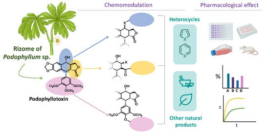
Podophyllotoxin: Recent Advances in the Development of Hybridization Strategies to Enhance Its Antitumoral Profile

by
Carolina Miranda-Vera
1,
Ángela Patricia Hernández
1,
Pilar García-García
1,
David Díez
2,
Pablo Anselmo García
1 and
María Ángeles Castro
1,*
1
Laboratorio de Química Farmacéutica, Departamento de Ciencias Farmacéuticas, CIETUS, IBSAL, Facultad de Farmacia, Campus Miguel de Unamuno, Universidad de Salamanca, 37007 Salamanca, Spain
2
Departamento de Química Orgánica, Facultad de Ciencias Químicas, Universidad de Salamanca, 37008 Salamanca, Spain
*
Author to whom correspondence should be addressed.
Pharmaceutics 2023, 15(12), 2728; https://doi.org/10.3390/pharmaceutics15122728
Submission received: 2 October 2023 / Revised: 22 November 2023 / Accepted: 28 November 2023 / Published: 4 December 2023
(This article belongs to the Special Issue Recent Advances in Drug Targeting for Cancer Treatment)

Abstract

:
Podophyllotoxin is a naturally occurring cyclolignan isolated from rhizomes of Podophyllum sp. In the clinic, it is used mainly as an antiviral; however, its antitumor activity is even more interesting. While podophyllotoxin possesses severe side effects that limit its development as an anticancer agent, nevertheless, it has become a good lead compound for the synthesis of derivatives with fewer side effects and better selectivity. Several examples, such as etoposide, highlight the potential of this natural product for chemomodulation in the search for new antitumor agents. This review focuses on the recent chemical modifications (2017–mid-2023) of the podophyllotoxin skeleton performed mainly at the C-ring (but also at the lactone D-ring and at the trimethoxyphenyl E-ring) together with their biological properties. Special emphasis is placed on hybrids or conjugates with other natural products (either primary or secondary metabolites) and other molecules (heterocycles, benzoheterocycles, synthetic drugs, and other moieties) that contribute to improved podophyllotoxin bioactivity. In fact, hybridization has been a good strategy to design podophyllotoxin derivatives with enhanced bioactivity. The way in which the two components are joined (directly or through spacers) was also considered for the organization of this review. This comprehensive perspective is presented with the aim of guiding the medicinal chemistry community in the design of new podophyllotoxin-based drugs with improved anticancer properties.

Graphical Abstract

1. Introduction

Cancer has become a major public health problem [1]. According to the WHO [2], following ischemic heart disease, this malignancy is the second leading cause of death, accounting for nearly 10 million deaths in 2020. There are many available treatments for this disease, such as surgery, radiation therapy, chemotherapy, and so on. Chemotherapy is the most common method used for treatment despite severe side effects and a high percentage of treatment failures due to drug resistance. Development of new drugs could overcome such limitations [3,4]. In this sense, natural products play an important role in the discovery and development of chemotherapeutic agents [5].
Natural products have been invaluable as tools for deciphering the logic of biosynthesis and as platforms for developing front-line drugs [6]. They offer huge structural diversity and, in some cases, great biological potency. Additionally, their intricate molecular frameworks offer a range of uncharted molecular structures for the discovery of chemical probes and drugs [7,8]. In fact, among the new chemical entities approved by the FDA throughout the last four decades, 48.5% of them were natural products, semisynthetic natural products analogues, or synthetic products based on natural products’ pharmacophores [9]. In the anticancer area, plant-derived agents such as vincristine, paclitaxel (Taxol®), docetaxel, and topotecan are among the most effective cancer chemotherapeutics currently available [10].
Among the natural products, lignans stand out for their interesting properties [11]. Podophyllotoxin (1) (Figure 1) is one of the most representative compounds of this family. It is isolated from the rhizomes of species of the genus Podophyllum (Berberidaceae) [12], and it has been used since ancient times as a laxative and as a remedy for various medical complications, such as gonorrhea, tuberculosis, menstrual disorders, psoriasis, dropsy, cough, syphilis, and venereal warts [13]. Several different pharmaceutical properties have also been described for podophyllotoxin derivatives, such as myotoxic, neurotoxic, insecticidal, antimicrobial, anti-inflammatory, antispasmodic, hypolipidemic, immunosuppressive, analgesic, and cathartic activities [12]. Furthermore, podophyllotoxin has other uses in dermatology: it is a useful agent in psoriasis vulgaris and has also proved to be effective in the treatment of rheumatoid arthritis [14]. The antiviral activity of podophyllotoxin was also investigated, and it was found that podophyllotoxin was the most active component in inhibiting the replication of measles and the herpes simplex type I virus. In fact, this compound is indicated in several pharmacopeias as an antiviral agent in the treatment of condyloma acuminatum caused by the human papilloma virus [15].
The most notorious activity of podophyllotoxin is its antitumor activity [14,15]. However, its severe side effects, such as bone marrow suppression, hair loss, neurotoxicity, and gastrointestinal toxicity [11], have limited its development as anticancer agent. Nevertheless, podophyllotoxin has become a good lead compound for the development of new derivatives with fewer side effects and better selectivity. Nowadays, some semisynthetic derivatives, such as etoposide (2), teniposide (3), or etopophos (4) (Figure 1), are in clinical use to treat a variety of cancers, including small cell lung cancer, germ-line malignancies, leukemias, lymphomas, and sarcomas such as Kaposi’s sarcoma [14,16,17,18]. These semisynthetic derivatives differ from podophyllotoxin in the antitumor mechanism of action. While the antitumor activity of podophyllotoxin is based on the inhibition of tubulin polymerization into microtubules, causing cell cycle arrest in the metaphase [15], etoposide and analogs block the activity of the topoisomerase II enzyme (Topo II), stabilizing the Topo II-DNA cleavage complex and preventing relegation of the DNA double strand during replication [19,20].
Despite the importance of these podophyllotoxin derivatives in chemotherapy, their clinical use is also associated with some secondary effects related to drug resistance, cytotoxicity toward normal cells, and poor bioavailability [16,17]. Thus, the search for less toxic podophyllotoxin derivatives is an active area of research to overcome these side effects associated with current cancer treatments.
In recent years, novel agents have been synthesized in an attempt to overcome podophyllotoxin’s and etoposide’s limitations, such as GL331 (5), TOP53 (6), azatoxin (7), F14512 (8), NK611 (9), and tafluposide (10), among others (Figure 2). GL331 has proven to be effective against colorectal cancer; whereas TOP53 is effective against metastatic lung cancer and has shown higher antitumor activity than etoposide; F14512, on its part, exploits the polyamine transport system to target tumor cells. NK611 has also proven to have better antineoplastic activity than etoposide in various in vitro and in vivo investigations, and tafluposide exhibits a potent cytotoxic action thanks to its ability to inhibit both DNA topoisomerases I and II [16].
The important biological properties described for podophyllotoxin since its isolation in the mid-1950s justify that this compound and its derivatives have been and continue to be the objective of extensive chemical modifications in order to obtain better and safer anticancer drugs. For all this, recent reviews have focused on different aspects of history [12], synthesis or semisynthesis [21], and biological properties [11,21,22,23,24] of podophyllotoxin analogs.
In a similar fashion to other research groups, our group has been working for years on the chemomodulation of podophyllotoxin, and we have obtained good results regarding selectivity and immunosuppressive activity [25,26,27]. Now, as a new contribution to the field, this review focuses on the recent chemical modifications performed on the cyclolignan skeleton of podophyllotoxin, with special emphasis on hybrids or conjugates of podophyllotoxin with other natural products or different moieties that contribute to improve its bioactivity.
Hybridization, or molecular conjugation, is a drug discovery approach based on rational design that combines two or more molecules of natural or synthetic origin, giving rise to “hybrid systems” or “conjugates” [28]. These new drugs are synthesized in order to improve the pharmacological profile of the original molecules by modulating pharmacokinetics, transporting across membranes, or avoiding metabolic degradation. It can also be used to achieve synergistic effects between the two hybrid components or even to overcome acquired drug resistance to the components separately [29,30].
The concept of hybridization is therefore a broad and versatile concept for drug design. It is worth noting that there are different approaches from a structural point of view for the synthesis of the new molecular hybrids. The most commonly used strategies for the synthesis of new molecular hybrids are shown in Figure 3. The keys to these approaches involve the chemical bonding of the two components (molecular approach) or the role that each conjugate fragment plays in the final activity of the hybrid (pharmacological approach) [31].
Designing hybrid molecules based on natural products has become an effective strategy for obtaining new antitumoral agents with lower toxicity than traditional single-target-molecule co-administration methods, and it can also improve dose compliance and reduce drug interactions [32]. Thus, some hybrids of podophyllotoxin and other natural compounds have been synthesized to combine the great properties of the two chemical entities that form the hybrid derivate.
This review covers the literature published during the last six years (2017–mid-2023) considering the chemical modifications performed mainly at the C-ring (but also modifications at the lactone ring and at the trimethoxyphenyl pendant ring) together with the biological properties described for them. Through this review, we analyzed the published data with the purpose of guiding the medicinal chemistry community in the design of novel podophyllotoxin-based drugs with improved anticancer properties.
Most of the literature dealing with podophyllotoxin-related compounds used a numbering system derived from that for the naphthalene that forms the skeleton rings B and C. However, to be consistent with our previous works, in this review we follow the IUPAC recommendations for the lignan family [33], which was based on the two phenylpropanoid subunits that biogenetically formed the natural product. Thus, the phenylpropanoid unit included in the tetracyclic moiety is numbered from 1 to 9. The other phenylpropanoid unit is numbered from 1′ to 9′ as shown in Figure 4.

2. C-Ring Modifications of the Podophyllotoxin Skeleton

C-ring modifications of the podophyllotoxin skeleton are quite important. Structure–activity relationship (SAR) studies have demonstrated that some substitutions at C7 can be tolerable. However, aromatization of the C-ring, which leads to chirality loss, affords derivatives that are either less potent or completely inactive [14,16]. On another note, the antimitotic activity of podophyllotoxin is actually influenced by the C7 position. The free hydroxyl group at C7 contributes to the cytotoxicity, and its loss does not elicit significant variations in the antitumor activity since deoxypodophyllotoxin also binds to tubulin [14]. Epimerization at the C7 position gives rise to epipodophyllotoxin, which is less potent against the depolymerization of tubulin but increases the inhibitory activity against DNA-Topo II [14]. In addition, glycosylation at C7β can suppress tubulin polymerization inhibition activity. The introduction of bulky groups at C7β can enhance the antitumor activity of podophyllotoxin caused by inhibition of DNA-Topo II [34]. Thus, in recent years, several different substituents have been introduced at that position either in α- or β-disposition; these are presented below. They are grouped depending on the structural nature of the substituent introduced as natural-product derivatives, heterocyclic systems, and other compounds.

2.1. Hybrids of Podophyllotoxin with Different Natural Products

Among the natural products that have been recently hybridized with podophyllotoxin, secondary metabolites such as flavonoids, coumarins, pterostilbenes, or alkaloids have been considered along with primary metabolites such as retinoic acid, bile acids, sugars, or vitamins. Ester or amide bonds have been used to combine both fragments starting either from podophyllotoxin or its analogues epipodophyllotoxin and 7β-aminopodophyllotoxin.

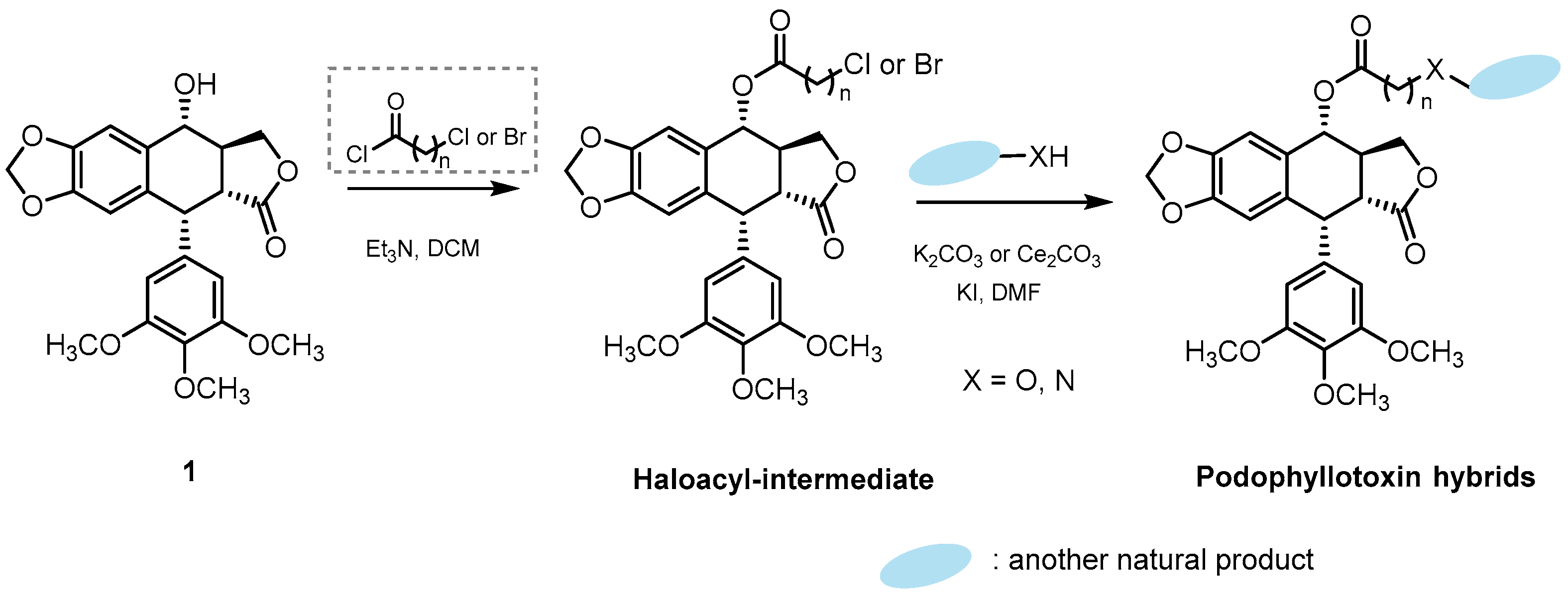
2.1.1. Podophyllotoxin Hybrids Synthesized through a Haloacyl Intermediate

Some hybrids of podophyllotoxin with formononetin, pterostilbenes, coumarins, and indarubicin are shown in Figure 5. They were synthesized from podophyllotoxin itself in two steps. First, a haloacyl intermediate was obtained via reaction with a chloro(bromo)acyl chloride followed by a substitution reaction of the halogen atom with a nucleophilic oxygen or nitrogen atom of the other natural compound under basic conditions, as stated in Scheme 1.
Following the above procedure, a series of podophyllotoxin–formononetin derivatives (11) were synthesized by Yang et al. [35]. Formononetin is an O-methylated isoflavone widely present in legumes, many species of clovers, and the traditional Chinese herb Astragalus membranaceus and whose structure mimics the activity of endogenous estrogens. It has a wide range of pharmacological activities, such as antiproliferative activity, growth inhibitory activity, vasorelaxant action, a neuroprotective or cardioprotective effect, and mammary gland proliferative activity, as well as antiapoptotic, antioxidant, anti-inflammatory, and antimicrobial activities. In addition, the anticancer properties of formononetin have been well documented against several types of cancer, such as breast, colon, glioma, osteosarcoma, and lung cancer, among others [36,37,38]. The antiproliferative activity of the new podophyllotoxin–formononetin hybrids was evaluated against several tumor cell lines, showing IC50 values at micromolar and nanomolar levels. Compound 11a (Figure 5) was the most promising compound, showing more sensitivity against the human lung carcinoma (A549) cell line than parental compound podophyllotoxin, with an IC50 value of 0.8 µM, which was 2.5-fold more potent than podophyllotoxin itself (IC50 = 1.9 µM). This derivative could also induce apoptosis in A549 cells through caspase-8 suppression and induce depolymerization of the microtubule network in these cells [35].
Pterostilbene is a natural demethylated analogue of resveratrol originally isolated from the heartwood of sandalwood with potential health benefits in inflammatory dermatoses, photoprotection, antioxidant activity, insulin sensitivity, blood glycemia and lipid levels, cardiovascular diseases, aging, memory, and cognition; it also exerts effects in cancer prevention and therapy in a wide range of tumors [39,40,41]. Pterostilbene was used by Zhang’s group to synthesize novel podophyllotoxin–pterostilbene hybrids (12) [42]. Conjugate 12a (Figure 5) showed a better anticancer effect against uveal melanoma cells (MuM-2B cells) than pterostilbene (IC50 = 0.08 µM and 40 µM, respectively) but less cytotoxicity against human umbilical vein endothelial cells (HUVEC cells) than podophyllotoxin (IC50 = 0.36 µM and 0.01 µM, respectively), which indicates that this hybrid had less toxicity than the parent compound. Compound 12a also had effects on the cell cycle since it arrested the MUM-2B cell cycle in the S cell cycle phase and promoted apoptosis in these cells. Furthermore, conjugate 12a showed great potential in inhibiting migration and metastasis of MuM-2B cells by increasing E-cadherin levels and downregulating the levels of vascular endothelial growth factor receptor 2 (VEGFR-2) and matrix metalloproteinase-2 (MMP-2) in these cells. Reductions in the expression levels of Topo IIα and Topo IIβ enzymes were also observed [42].
Two podophyllotoxin–coumarin conjugates (13) were synthesized by Bai et al. [43]. Coumarins are heterocyclic moieties widely distributed in nature, and they have exhibited multiple pharmacological properties, such as antioxidant, antibacterial, antimicrobial, antiviral, hepatoprotective, and anti-inflammatory effects, apart from being chemopreventive and anticancer agents in vitro [44]. Of the two synthesized hybrids (Figure 5), compound 13b showed significant cytotoxic activity against human oral squamous carcinoma HSC-2, SCC-9, and A-253 cells with IC50 values of 0.22, 0.23, and 0.25 µM, respectively. Additionally, this compound showed less toxicity than podophyllotoxin against human renal tubular epithelial HK-2 cells (IC50 values of 0.62 and 0.04 µM, respectively). It was able to induce depolymerization of the microtubule network, which caused cell cycle arrest in the G2 phase, and it could also induce apoptosis through decreasing the mitochondrial membrane potential in HSC-2 cells [43].
New podophyllotoxin–indirubin conjugates (14) were synthesized by Wang and colleagues [45] using the procedure described in Scheme 1. Indirubin, a bisindole alkaloid present in various plants, marine mollusks, and bacteria and detected in human urine, possesses several pharmacological activities like antipsoriatic, anti-Alzheimer’s, antiautoimmune, neuroprotective, anti-inflammatory, and anti-influenza activity. It also has antiproliferative activity, since it has been used for the treatment of chronic myelocytic leukemia by physicians in China for ages [46,47,48]. Among all the podophyllotoxin–indirubin derivatives synthesized (Figure 5), compound 14a, with an acetyl group as linker, displayed the most potent antiproliferative effect against myeloblastic leukemia K562 cells and myeloblastic leukemia’s vincristine resistant cells (K562/VCR cells) with IC50 values of 0.034 and 0.076 µM, respectively. Other derivatives with extended-length linkers were found to have lower activity than 14a, indicating that the linker length was critical to their antiproliferative effects. Compound 14a could also induce cell apoptosis through the mitochondrial pathway, block the cell cycle in the G2 phase in K562/VCR cells, and inhibit tubulin polymerization in a similar way to podophyllotoxin [45].

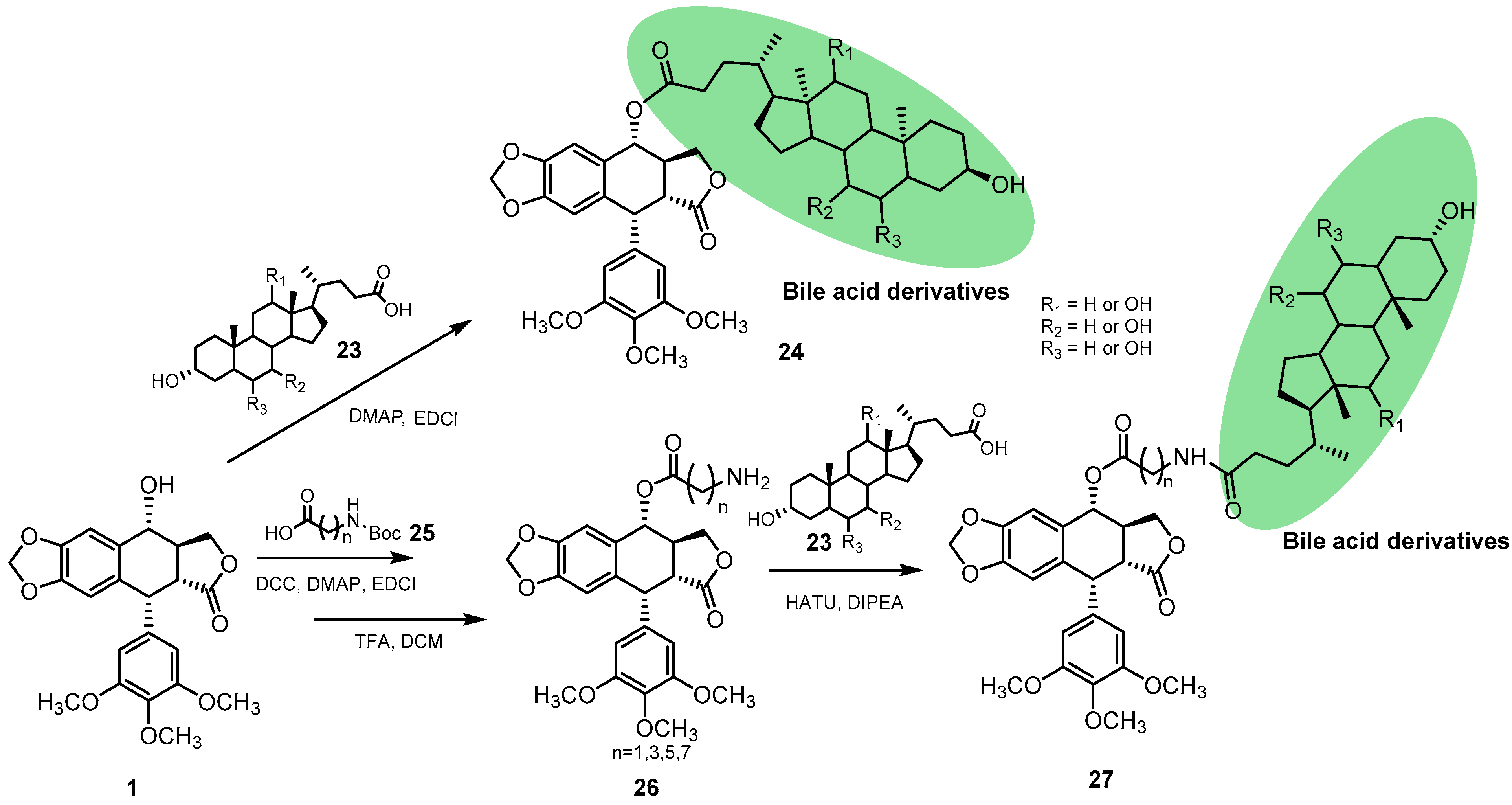
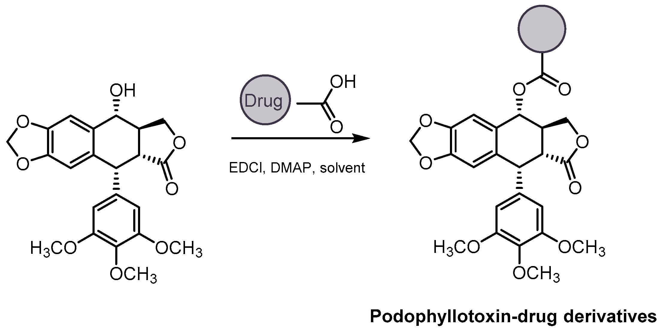
2.1.2. Podophyllotoxin Hybrids Synthesized via Direct Coupling with Natural-Product Derivatives Bearing a Carboxylic Group

An ester bond can be formed via the reaction of podophyllotoxin with a carboxylic acid in the presence of different coupling reagents, such as 1-(3-dimethylaminopropyl)-3-ethylcarbodiimide hydrochloride (EDCl), N,N′-dicyclohexylcarbodiimide (DCC), or O-(7-azabenzotriazol-1-yl)-N,N,N′,N′-tetramethyluronium hexafluorophosphate (HATU), as outlined in Scheme 2. Additionally, similar coupling reagents have been used to join podophyllotoxin to other natural products through an amide bond using 7β-aminopodophyllotoxin as the starting compound. The carboxylic acid can be an inherent part of the other natural product, as in the retinoic acid hybrid (15), or be included as an additional substituent as in the case of carboline derivatives (21). These conjugates are depicted in Figure 6.
All-trans retinoic acid (ATRA), a metabolite of vitamin A, has great anticancer properties. It could inhibit cell proliferation and induce apoptosis markers in non-small-cell lung carcinoma in addition to being of use in the treatment of various tumoral diseases like Kaposi’s sarcoma or ovarian carcinoma [49,50]. Following the previous scheme and taking into account the great anticancer properties of all-trans retinoic acid, Zhang’s group synthesized a new podophyllotoxin–ATRA derivative (15, Figure 6) [51]. This compound showed significant antiproliferative activity against the human gastric cell lines MKN-45 and BGC-823 with IC50 values of 0.42 and 0.20 µM, respectively. It could also block the MKN-45 cell cycle at the G1 phase and the BGC-823 cell cycle at the G2 phase by decreasing the levels of CDK1, CDK2, cyclin A, and cyclin B1. Apoptosis was also altered in these cells when they were treated with this compound, which produces an increment in the number of apoptotic cells in both cell lines by enhancing the levels of cleaved caspase-3, -8, and -9 [51].
Some podophyllotoxin-linked β-carboline congeners (21aq) that act as DNA-Topo II inhibitors were synthesized by Sathish and colleagues [52]. β-Carbolinic alkaloids are widely distributed in nature, being found in several families of plants, but they have also been found in cigarette smoke, overcooked foods, and wine. β-Carbolines have a wide spectrum of action, especially on the muscular, cardiovascular, and central nervous systems, and some reports indicate that β-carbolines have effective antioxidant properties [53]. Additionally, small molecules with a β-carboline scaffold possessed a tricyclic planar structure with suitable features for DNA interaction and enzyme inhibition activities like Topo II inhibition [54]. In order to obtain the podophyllotoxin-linked β-carboline derivatives, it was necessary to first synthesize β-carboline-3-carboxilic acids (20) from L-tryptophan (16) that was transformed into the corresponding methyl ester (17). Next, Pictet–Spengler condensation of compound 17 with different substituted benzaldehydes afforded methyl β-carboline-3-carboxilates (18, 19). Finally, compound 19 was hydrolyzed to the free carboxylic acids (20), as described in Scheme 3.
The final synthesis of the new β-carboline–podophyllotoxin derivatives was carried out following the procedure described above in Scheme 2 starting from 7β-aminopodophyllotoxin. Among all the derivatives synthesized (21aq), compounds 21i and 21j (Figure 6) showed promising cytotoxic activity with IC50 values of 1.1 µM against prostate cancer DU 145 cells. Cell cycle analysis and DNA-Topo II inhibition assays were carried out with these compounds. They showed that these conjugates blocked the DU 145 cell cycle at the S and G2/M phases and also could interfere with the catalytic activity of DNA-Topo II, acting as catalytic inhibitors [52].
Sometimes the carboxylic acid group is introduced through a spacer attached first to podophyllotoxin or to the other natural product, as shown for aurone and bile acid derivatives (Figure 7 and Scheme 4).
Aurones are secondary metabolites of plants that belong to the family of flavonoids. They have been used since ancient ages as natural dyes. In recent years, aurones have gained attention because of their biological activities, including anti-inflammatory, antibacterial, antifungal, antimalarial, antileishmanial and anti-Alzheimer’s activities. However, the most important property of these metabolites is their anticancer activity, showing low toxicity and a broad spectrum of anticancer mechanisms [55,56]. In order to synthesize 4β-acetamidobenzofuranone–podophyllotoxin hybrids (22Iad and 22IIab) (Figure 7), Paidakula and colleagues [57] first synthesized (Z)-2-benzylidenebenzofuran-3(2H)-one acid derivatives that were coupled with 7β-aminopodophyllotoxin, as indicated in Scheme 2. Among all the derivatives synthesized, compound 22Id showed the most promising antiproliferative activity against the MCF7 and MDA-MB-231 human breast cell lines, A549 human lung cell line, and DU 145 human prostate cell line (IC50 values = 0.13, 0.45, 0.10 and 0.97 µM, respectively) [57].
Similar approaches were followed to obtain hybrids of podophyllotoxin with bile acids or ligustrazine derivatives through different amino acid linkers. The amino acids that acted as linkers were first attached to podophyllotoxin through an ester bond and then to the other natural fragment through an amide bond.
Bile acids are a group of steroid-based molecules biosynthesized from cholesterol in the liver and in the gall bladder. Not only are they involved in glucose, lipid, and energy homeostasis, but these compounds also function as signaling molecules to regulate metabolism, the inflammatory response, and gut microbiota. In addition, bile acids are known to regulate cell growth and proliferation. Dysregulation of bile acid homeostasis and signaling can have an impact on liver regeneration and tumorigenesis [58,59]. Furthermore, they can act as drug carriers and enhance the stability and biocompatibility of drugs in the body without causing side effects [60]. Owing to the great properties of bile acids and based on the results obtained in a previous report [61], De-Sheng’s group synthesized new podophyllotoxin-linked bile acid derivatives (24 and 27, Scheme 4) attached directly or through ω-amino acid linkers, respectively [62]. For the latter, they first synthesized the acylated-N-Boc-protected derivatives of podophyllotoxin using N-Boc-aminocarboxylic acid carbon chains of different lengths and the corresponding coupling reagents. Deprotection of the amino group led to derivatives (26), and subsequent reaction with bile acids (23) led to the corresponding hybrids (27), as shown in Scheme 4. They used six different bile acids: cholic, deoxycholic, chenodeoxycholic, ursodeoxycholic, hyodeoxycholic and lithocholic. The antitumor activity of the 30 new hybrids was evaluated against some human cancer cell lines. The hybrid with hyodeoxycholic acid and 6-aminocaproic acid as the linker (n = 5, R1 = R3 = H, R2 = α-OH) presented the most potent activity against the HepG2 cell line (IC50 = 0.18 µM) and also showed great selectivity against this tumor cell line. This compound could also induce early apoptosis and arrest the HepG2 cell cycle in the S phase [62].
In a similar fashion, Wu and colleagues synthesized several podophyllotoxin–α-amino acid–ligustrazine derivatives (30, Scheme 5) [63]. Ligustrazine, or tetramethylpyrazine, is an alkaloid monomer isolated and extracted from the Chinese herbal medicine Lisgusticum wallichii that has various pharmacological activities, such as anticardiovascular, antiplatelet, ischemic stroke, anti-Alzheimer’s, neuroprotective, and anticancer effects. It has also been reported to suppress caspase-3 activation, increase Bcl-2 expression, and diminish Bax expression in some brain tissues [64,65,66]. To join ligustrazine to podophyllotoxin, one of the methyl groups was first oxidized to the corresponding acid and then attached to the podophyllotoxin skeleton using different L-α-amino acids as linkers. For that, the N-Boc-protected amino acids were first attached to podophyllotoxin through an ester bond to obtain 28. The ligustrazine moiety was then joined to the deprotected amino acids derivatives (29) via an amide function, affording hybrids (30) (Scheme 5). The antiproliferative activity of the new derivatives was tested against three tumor cell lines. Among all the tested compounds, the deprotected podophyllotoxin–amino acid derivatives (29) were more potent than the corresponding protected ones (28), and the podophyllotoxin–amino acid–ligustrazine derivatives (30) showed better selectivity against tumor cell lines. Interesting results were obtained with the intermediate 28b, which has a N-Boc-N-methyl glycine. Such derivative showed a similar antiproliferative activity to the N-Boc-deprotected compound (29b) toward the A549 tumor cell line, but it also showed less cytotoxicity toward L-02 normal cell lines. Compound 28b could also induce apoptosis and cell cycle arrest in the S phase in the A549 cell line [63].

2.1.3. Podophyllotoxin Hybrids with Other Natural Products Attached through a Triazole Ring

Although later in this review, several podophyllotoxin derivatives bearing a triazole ring in C7 position are considered separately, we have included some hybrids in this section that used such a heterocycle as the linker that joined podophyllotoxin and other natural compounds. They were obtained via click chemistry-type reactions, specifically the Huisgen 1,3-dipolar cycloaddition between a propargyl intermediate of a natural compound and an azido intermediate of podophyllotoxin (Scheme 6). Thus, the heterocycle was built at the same time as the hybrid was formed. Briefly, the propargyl rest was joined to the non-lignan natural compound via a reaction with propargyl bromide, and the azido intermediate (31) was synthesized using sodium azide (NaN3) in the presence of trifluoroacetic acid (TFA). Then, the coupling reaction between the two intermediates was carried out following the Huisgen 1,3-dipolar cycloaddition conditions, as shown in Scheme 6. Further information on the triazole scaffold and Huisgen 1,3-dipolar cycloaddition conditions can be found below in the section on podophyllotoxin–heterocycle hybrids (Section 2.2.1).
Several podophyllotoxin hybrids, such as podophyllotoxin–epigallocatechin-3-gallate (EGCG) conjugates (32), podophyllotoxin–camptothecin conjugates (33), a podophyllotoxin–curcumin hybrid (34), or bispodophyllotoxins (3537), have been synthesized through this strategy; these are shown in Figure 8.
Podophyllotoxin–EGCG derivatives (32-I and 32-II, Figure 8) were synthesized by Zi and colleagues using the procedure shown in Scheme 6 [67,68]. EGCG is the dominant active polyphenol of green tea (Camellia sinensis). It possesses antioxidant, antitumor, and anti-inflammatory features [69]. Several studies have demonstrated that the anticancer actions of this catechin include antiproliferative, pro-apoptotic, antiangiogenic, and anti-invasive functions and multiple antitumor mechanisms, such as alteration in the tumor cell cycle and production of epigenetic changes in gene expression [69,70]. The novel podophyllotoxin–EGCG derivatives exhibited good antiproliferative activity against all cancer cell lines tested. Among all the methylated EGCG–podophyllotoxin hybrids, compound 32-Ia (R1 = R2 = CH3) showed the most potent cytotoxicity against A549 cells with an IC50 value of 10 µM. Among the non-methylated EGCG–podophyllotoxin conjugates, compound 32-IIb (R1 = R2 = H) showed the most potent antiproliferative activity against the A549 cell line, even better than the methylated conjugate 32-IIa, with an IC50 value of 2.2 µM. Docking studies of these EGCG methylated and non-methylated conjugates suggested that such compounds could have a dual mechanism of action: inhibition of tubulin polymerization via binding at the colchicine binding site and inhibition of Topo II [67,68].
Camptothecin is a plant alkaloid present in wood, bark, and fruits of the Asian tree Camptotheca acuminata. It was discovered in 1966 by Wall and Wani in a systematic screening of natural products for anticancer drugs, and then in 1985 it was identified as a specific inhibitor of the enzyme DNA-topoisomerase I (Topo I) [71]. This drug reversibly induces single-strand breaks, thereby affecting a cell’s capacity to replicate. Camptothecin also stabilizes the so-called cleavable complex between Topo I and DNA [72]. As is known, some derivatives of podophyllotoxin, like etoposide, are DNA-Topo II inhibitors. So, the main purpose of the synthesis of camptothecin–podophyllotoxin conjugates was the development of compounds that can act on both Topo I and Topo II enzymes. The conjugates synthesized by Li et al. [73] (33ab, Figure 8) were tested against a panel of five cancer cell lines: leukemia (HL-60), hepatoma (SMMC-7721), lung cancer (A549), breast cancer (MCF7), and colon cancer (SW-480). It was found that both derivatives showed weak activity against the cancer cell lines tested, and only compound 33b displayed significant anticancer activity against the HL-60 cell line with an IC50 value of 18 µM [73], suggesting that 4′-O-demethylation is necessary for the activity of these hybrid compounds.
On their part, Duan and colleagues synthesized two novel tetrahydrocurcumin-podophyllotoxin hybrids (34ab, Figure 8) following the same protocol described in Scheme 6 [74]. Tetrahydrocurcumin is one of the major metabolites of curcumine, the main compound of the rhizomes of Curcuma longa (turmeric). However, it also exists naturally in the roots of Curcuma zeoaria, Zinger mioga, and Zinger officinale. Regarding its pharmacological properties, it has been recognized as anticancer, antiaging, and an antioxidant, and it can also prevent neurodegenerative disorders [75,76,77]. The new tetrahydrocurcumin–podophyllotoxin derivatives were tested against some human cancer cell lines, with both (34ab) showing promising anticancer activity against the HCT 116 and HeLa cell lines. Particularly, compound 34a, with IC50 values of 18 and 83 µM, respectively, was four times more potent against HCT 116 than against HeLa cells [74].
Dimeric podophyllotoxin derivatives synthesized by Zi et al. (35ab, 36af, and 37ah) [78] are shown in Figure 9. They include two triazole moieties joined through a previously dipropargyl functionalized glycerol or glucose scaffold that allowed the synthesis of the triazole moieties simultaneously. The derivatives were evaluated for their cytotoxicity against the same five human cell lines (HL-60, SMMC-7721, A549, MCF7, and SW-480). The IC50 values obtained revealed that most of these derivatives were nearly inactive (IC50 > 40 µM). However, compound 36c was active against all five cancer cell lines with IC50 values ranging from 0.43 to 3.5 µM. SAR studies suggested that the spacer between the two podophyllotoxin moieties can largely affect their activity [78].
Biotin, or vitamin H, is a water-soluble vitamin that belongs to the vitamin B complex and is an essential nutrient in the living organism, although it cannot be synthesized by higher organisms, in which a dietary intake is needed. Biotin is found in a large number of foods, including eggs, bread, green-leaf vegetables, or beef liver. In eukaryotic cells, biotin functions as a prosthetic group of enzymes, collectively known as biotin-dependent carboxylases, which catalyze key reactions in gluconeogenesis, fatty acid synthesis, and amino acid catabolism. Furthermore, recent evidence suggests that biotin also plays unique roles in cell signaling, the epigenetic regulation of genes, and chromatin structure [79,80]. Nonetheless, despite that biotin and other vitamins are necessary for the survival and division of normal cells, biotin requirement in tumor cells is higher than in normal cells due to their rapid growth and division. In addition, recent studies have shown that some cell lines express higher levels of biotin receptors than normal cells, such as L1210RF (leukemia), Ov2008 (ovarian), Colo–26 (colon), P815 (mastocytoma), M109 (lung), RENCA (renal), and 4T1 (breast) [81,82]. Due to the importance of biotin in cancer, several biotinylated podophyllotoxin derivatives (4245, Scheme 7) were synthesized by Zi and colleagues [83]. Three different series of derivatives were synthesized: ester-linked derived from epipodophyllotoxin (42ab) and from podophyllotoxin (43), amide-linked obtained from 4β-aminopodophyllotoxin (44ad), and triazole-linked derivatives (45ad). The latter were synthesized first by converting podophyllotoxin into the 7β-azide derivative 31 followed by reaction with propargyl alcohol to obtain the triazole derivative 40. Three series of compounds were synthesized via the reaction of 6-biotinylaminocaproic acid (41) with the corresponding podophyllotoxin derivates 3840 (Scheme 7).
The cytotoxicity of these conjugates was evaluated in several cancer cell lines. The results suggested that compounds 42 and 43, with an ester linkage, exhibited better antiproliferative activities with IC50 values ranging from micromolar to nanomolar. They also suggested that the 6-aminocaproic acid linking spacer had a variable influence on the cytotoxicity. Among these derivatives, compound 42a, with the biotin directly attached to podophyllotoxin, showed the most promising antiproliferative activity, even higher than podophyllotoxin in hepatoma and colon cancer cell lines (the SW480 and SMMC-7721 cell lines; IC50 0.23 and 0.56 µM for 42a; 4.1 and 9.4 µM for podophyllotoxin). Also, compound 42a could induce cell apoptosis in the lung cancer cell lines NCI-H1299 and H1975 through an increment in the expression levels of cleaved-caspase-3 and cleaved-PARP in a dose-dependent manner. It was also able to significantly suppress the growth of S180 tumor xenografts in mice [83].
Finally, we have included in this section several podophyllotoxin–polyamine hybrids despite their synthesis prior to the time period covered by this review [84,85] due to the fact that a study about their activity against different Topo II isoforms was published in 2018 in order to compare their activity with that of etoposide. The importance of etoposide in cancer treatment is well known. However, the drug is associated with very severe side effects, one of the most dramatic of which is the generation of treatment-related acute myelogenous leukemia [85]. To minimize off-target effects, polyamine–podophyllotoxin conjugates were evaluated by Oviatt and colleagues [86]. Among them, compound 8, called F14512 (Figure 2 and Figure 10), with a tetramine spermine side chain that was a candidate for phase I clinical trials [87], had the highest ability to induce double-stranded breaks in DNA and turned out to be a much stronger Topo II poison than etoposide. Moreover, in vitro studies of this compound against different Topo II isoforms revealed that it is more potent against the Topo IIβ isoform than to the α-isoform [86]. Additionally, this compound showed strong therapeutic efficacy against spontaneous non-Hodgkin lymphoma in a randomized double-blind trial in dogs that justified its evaluation in human clinical trials [88].

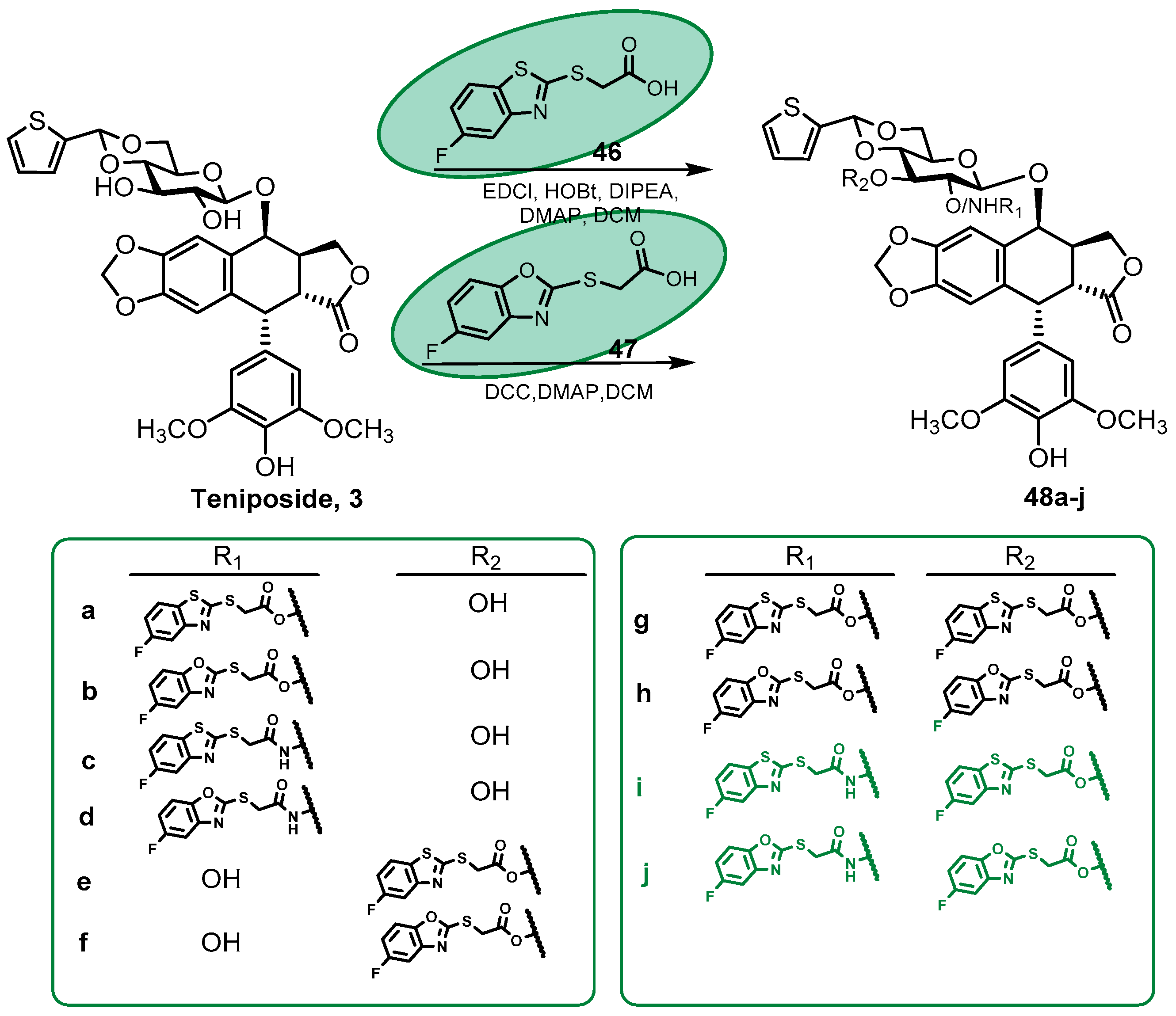
2.1.4. Podophyllotoxin Glycoconjugates

Among the primary metabolites, carbohydrates have been of interest in cancer therapy for over a century [89] since Otto Warburg observed differences in the 1920s between cancer cells and normal cells in glucose metabolism. Cancer cells tend to depend on glycolysis for acquiring energy even in aerobic conditions, and this fact was later named the “Warburg effect”. Therefore, cancer cells usually have a higher expression of glucose transporters, which the scientific community has used to facilitate the entry of carbohydrate-conjugated antitumor drugs into cells [90].
Glycoconjugates that act as anticancer drugs contain sugar moieties that allow the specific binding of the antitumor drugs with those specific glucose transporters. Glucose transporters transfer the drugs to the tumor cells, where they are selectively uptaken and thus the organ toxicity thus is minimized. This has become an attractive strategy in drug design in order to improve drug efficacy and pharmacokinetics and to reduce side effects [91,92].
The podophyllotoxin glycoconjugates are not new, since etoposide and teniposide, two of the semisynthetic derivatives of podophyllotoxin that are in clinical use, include a sugar moiety in their structure. The introduction of a β-glycosidic moiety at C7 position of podophyllotoxin together with the 4′-O-demethylation is related to the change in the mechanism of action of these compounds, as mentioned in the Introduction. Instead of being inhibitors of tubulin polymerization like their parent compound podophyllotoxin, they are potent DNA-Topo II inhibitors [7,15].
Teniposide is less frequently used for chemotherapy in comparison with etoposide, although it has shown higher in vitro DNA-Topo II inhibition than etoposide [16]. New derivatives of teniposide have been synthesized in recent years by Cheng and colleagues [93] by introducing different fluorobenzoheterocyclic derivatives into glucose positions 2 and 3 in teniposide using different carbodiimides as coupling reagents (Scheme 8). Among them, only compounds 48i and 48j presented a similar antitumor activity to teniposide against the HepG2, HeLa, A549, and MCF7 tumor cell lines, but they presented a reduction in the toxicity by around 10 times against HL-7702, H8, MRC-5, and HMEC human cells compared to teniposide. Cell cycle analysis and apoptosis studies revealed that these derivatives could block the cell cycle in the G2/M phase like teniposide, and they also could induce apoptosis in HepG2 cells, but in a weaker manner than etoposide. DNA-Topo II inhibitory activity was also measured, and the results indicated that compounds 48i and 48j inhibited this enzyme in a concentration-dependent manner [93].
In the last six years, other podophyllotoxin glycoconjugates have been synthesized by Zi and colleagues, such as the per-butyrylated and per-acetylated glycoside derivatives 49 and 51 (Scheme 9) [94,95]. The final glucoside conjugates were synthesized by coupling the tetra-acetylated residue of glucose (54) or the tri-butyrylated residue of xylose (56) with podophyllotoxin/demethylpodophyllotoxin. The corresponding deacylated analogues 50 and 52 with free glucose or xylose residues were also obtained. At this point, we would like to notice that during deprotection, isomerization of the lactone ring must have taken place under the strong basic conditions used. The authors claimed that the trans-lactone ring of the podophyllotoxin skeleton is maintained in compounds 50 and 52. However the chemical shift reported for C9′ in the 13C NMR spectra did not indicate that, since it appeared at over 175 ppm in all the compounds, which is characteristic of cis-lactones [96].
The per-acetylated glycoside was synthesized first by treating D-glucose (53) with acetic anhydride and sodium acetate. Then, 1,2,3,4,6-penta-O-acetyl-α/β-glucopyranoside was treated with ammonia to get the free hydroxy group at the anomeric position, obtaining 2,3,4,6-tetra-O-acetyl-α/β-D-glucopyranoside (54) in this manner. The tri-butyrylated residue of xylose (56) was synthesized in a similar manner through the treatment of D-xylose (55) with butyric anhydride and iodine followed by ammonia treatment (Scheme 10).
Novel glucoside and xyloside derivatives, per-acylated or not, were evaluated for their antiproliferative activity against the HL-60, SMMC-7721, A549, MCF7, and SW480 cancer cell lines. In both cases, the per-acylated derivatives were more cytotoxic than the derivates with the free sugar rests, as expected for cis-lactones; among them, the 4′-O-demethylated derivatives (compounds 49b and 51b) showed the highest cytotoxicity against the five cancer cell lines tested. Thus, the per-butyrylated xyloside (49b) showed the highest cytotoxicity for the HL-60 cell line with an IC50 value of 2.9 µM and for the A549 cell line with an IC50 value of 4.4 µM [95]. In the case of the corresponding per-acetylated glucoside (51b), it showed IC50 values ranging from 3.2 to 11 µM; the highest cytotoxicity and selectivity were against the SW480 colon cancer cell line (IC50 = 3.3 µM; Selectivity Index = 6.7) [94].
During the time covered by this review, other podophyllotoxin glycoconjugates that used 1,2,3-triazole as a linker between podophyllotoxin and the sugar residues have been synthesized following the conditions outlined at Scheme 6. The use of this strategy is exemplified by the 1,2,3-triazole-linked picropodophyllin glycoconjugates synthesized by Zi et al. [97] containing maltose, 1,6-β-D-diglucose, or several other disaccharides and trisaccharides, in which the sugar residues were also joined by triazole rings (57I-V, Scheme 11). Most of the compounds synthesized showed weak cytotoxicity with IC50 values > 40 µM against the cell lines tested, as expected for picropodophyllin derivatives. However, compound 57II with a 1,6-β-D-diglucose residue showed strong anticancer activity against all cancer cell lines tested (HL-60, SMMC-7721, A549, MCF7, and SW480) with IC50 values ranging from 0.67 to 7.4 µM [97].
The podophyllotoxin glycoconjugates 59al obtained by Nerella and colleagues [98] were synthesized through a click chemistry reaction between 4β-O-propargylated podophyllotoxin (59) and sugar azides, although in this case, silver (I)- N-heterocyclic carbenes were used as catalysts instead of copper salts (Scheme 12). Compound 59a, with a tetraacetylglucosyl residue, was the most promising of the series against the DU 145 prostate cancer cell line with IC50 values of 1.0 µM. Compound 59e, bearing a tetraacetylgalactosyl residue, was the most promising against the MCF7 cell line with an IC50 value of 1.3 µM. Docking studies of compounds 59a and 59e revealed that both derivatives showed a high binding affinity toward DNA-Topo II and that they were in good agreement with observed IC50 values [98].

2.2. Podophyllotoxin Derivatives with Heterocyclic Systems

Heterocyclic compounds, aromatic or not, are a significant source of pharmacologically active scaffolds, which can be prepared synthetically or isolated from natural sources [99]. An important feature of heterocycle bioactive compounds is the presence of constituent heteroatoms (mainly nitrogen, sulfur, and oxygen in many combinations) that directly affect the reactivity of the skeleton, the bioactivity of the compounds, and the interactions between biological targets and target modulators [100]. Therefore, their significance in drugs rests on the diversity and versatility of substitution patterns that these rings can undergo.
In this section, heterocyclic substituents at podophyllotoxin’s C7 position are considered and grouped according to the heterocyclic size: five- or six-membered rings and benzoheterocycles with one, two, or three heteroatoms. In some cases, the heterocycle is joined directly to the podophyllotoxin without any linker. In other cases, the linker is only one heteroatom, and in most of the cases, they are connected by linkers with different length chains through ester or amide bonds. Also, the heterocycle could be introduced as such or formed at the last reaction step.

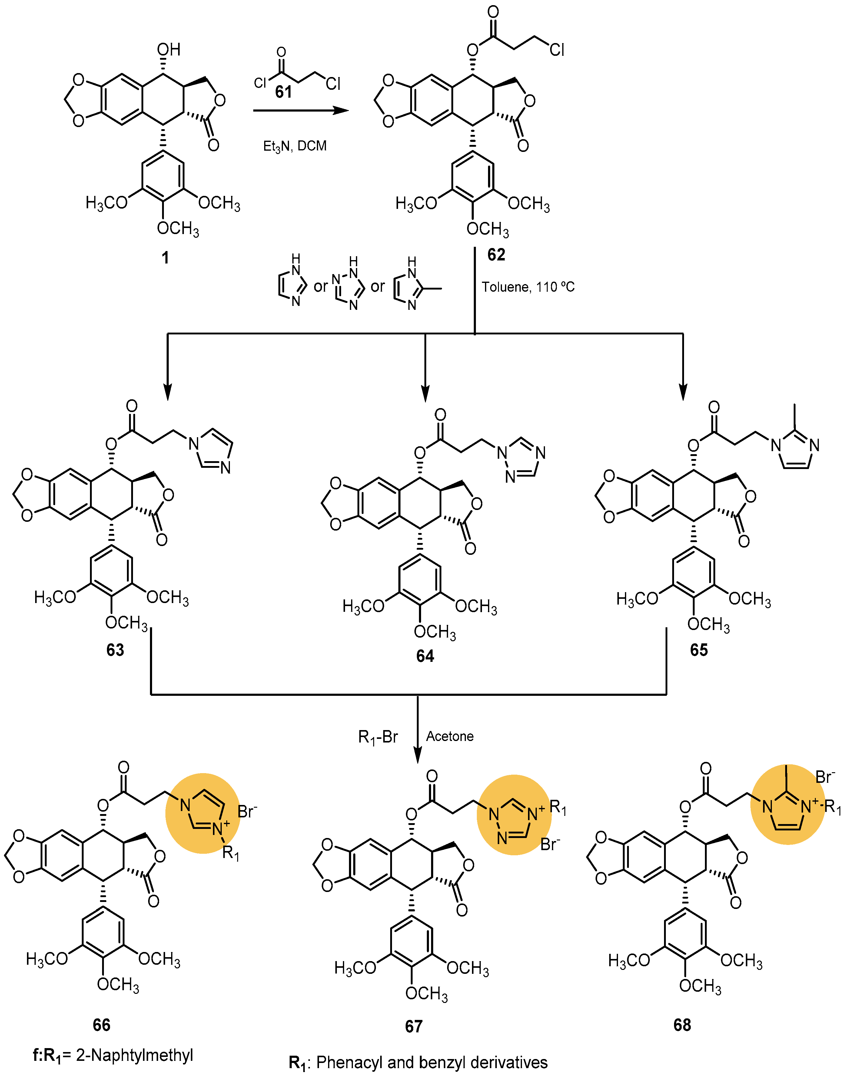
2.2.1. Five-Membered Heterocycles with Two Heteroatoms

The imidazole nucleus is commonly found in numerous endogenous biomolecules, including the neurotransmitter histamine and the amino acid histidine, among others. Apart from that, the imidazole unit is present in a great variety of medicinal agents and shows several pharmacological activities, such as antibacterial, antitumor, antitubercular, analgesic, anti-VIH, and antifungal [101,102,103].
Cao and his colleagues synthesized the N-acyl imidazole derivative of podophyllotoxin GMZ-1 (60, Figure 11) [104]. Compound 60 showed high antiproliferative activity against the K562 and K562/A02 cell lines with IC50 values of 0.08 and 0.12 µM, respectively. In addition, this compound was able to induce apoptosis in the K562/A02 cell line in a time- and concentration-dependent manner, which indicated its potentiality to overcome leukemia cell resistance to apoptosis induced by chemotherapy [104].
On their part, Yin and colleagues have synthesized new podophyllotoxin derivatives bearing nitrogen-containing heterocycles, such as imidazole, methylimidazole, or 1,2,4-triazole [105]. With the aim of synthesizing these derivatives, they first created a reaction of podophyllotoxin with 2-chloropropionyl chloride (61) following the procedure described previously in Scheme 1. Then, compound 62 reacted with imidazole, 1,2,4-triazole, and 2-methylimidazole to obtain compounds 6365, whose treatment with various bromides generated the subsequent podophyllotoxin imidazolium/ triazolium salts (compounds 6668), as shown in Scheme 13.
The cytotoxic activity of the new conjugates was evaluated against some human cancer cell lines, including HepG2, A549, MDA-MB-231, and HCT 116 cells. All the imidazole and 1,2,4-triazole derivatives 6668 showed an excellent cytotoxic activity against all cancer cell lines tested. Among them, compound 66f, bearing a 2-naphthylmethyl substituent at the third position of the imidazole ring, showed the most potent antitumor activity with IC50 values of 0.07, 0.29, 0.11, and 0.04 against the HepG2, A549, MDA-MB-231, and HCT 116 tumor cell lines, respectively. On another note, compound 66f could also induce apoptosis in the HCT 116 cell line and arrest the cell cycle of this tumor cell line in the G2/M phase [105].
The thiazole nucleus is a very important heterocycle in many biologically active compounds, and it is also a fascinating building block in medicinal chemistry, since a wide range of activities have been described for compounds containing thiazole rings, like antioxidant, anti-inflammatory, antimicrobial, antifungal, antiviral, anticonvulsant, neuroprotective, and antitumor activities [106,107].
Sang and colleagues synthesized 7β-thiazole derivatives of 4′-O-demethylepipodophyllotoxin (71ai) linked only by a nitrogen atom, as shown in Scheme 14 [108]. Firstly, the corresponding 2-(2-aminothiazol-4-yl)acetic esters or amides (70) were synthesized from ethyl 2-(2-aminothiazol-4-yl)acetate as previously described [109]. 4′-O-Demethyl-7β-iodoepipodophyllotoxin (69) on its part was synthesized by using methanesulfonic acid and sodium iodide and then coupled to the heterocycle in the presence of triethylamine. The cytotoxic activities of the nine obtained derivatives (71ai) were evaluated against four human cancer cell lines (A549, HepG2, HeLa, and LOVO).
The results showed that the new compounds had greater or equal antitumor activity against these cancer cell lines, and 2-aminothiazol-4-yl esters 71g and 71h appeared to be more potent than the other derivatives synthesized. Their effect on DNA-Topo II and double-strand breaks was also studied, showing that compound 71h (R = OCH2CH3 and n = 0) decreased the expression of both Topo IIα and Topo IIβ in the A-569 cell line in a dose-dependent manner. Furthermore, it induced supercoiled pBR322 DNA relaxation to form linear DNA [108].
Another heterocycle with two heteroatoms that has been attached to podophyllotoxin is isoxazoline. The isoxazoline scaffold is found in several compounds exerting a wide range of biological activities and pharmacological properties, occupying a prominent position in psychotropic drugs, but it also serves as a valuable precursor for the construction of diverse molecules, including natural products [110,111].
Yang and colleagues synthesized novel isoxazoline-containing podophyllotoxin/2′,6′-dihalogenopodophyllotoxin derivatives (78 IIVae, Scheme 15) [112] with improved insecticidal/acaricidal activities compared to other previously synthesized dihalogenopodophyllotoxin derivates [113,114]. They were synthesized using 1,3-dipolar cycloadditions between dihalogenopodophyllotoxin acrylates (77) and nitride oxide dipoles (74) obtained through the corresponding oximes (73), as shown in Scheme 15.
Among all the derivatives, compounds 78 IIc and 78 IIIc, bearing 4-chlorophenyl as isoxazoline substituent and chlorine atoms in the C6′ or C6′ and C2′ positions of podophyllotoxin, respectively, exhibited good insecticidal and acaricidal activities that were even better than the toosendanin used as a reference but not a higher cytotoxicity. This fact demonstrates that the introduction of the halogen atom/atoms at the C2′, C6′ position of podophyllotoxin and the introduction of chlorine atom at the C4 position on the phenyl substituent of the isoxazoline fragment were very important for their insecticidal and acaricidal activities [112].
Zefirov and colleagues also synthesized several podophyllotoxin and epipodophyllotoxin derivatives bearing substituted isoxazole fragments during the time covered by this review [115]. They first synthesized the isoxazole unit from ethyl-2,4-dioxopentanoate (79) following a known procedure [116,117,118], as shown in Scheme 16. Then, bromination, iodination, and nitration at position 4 of the isoxazole ring was carried out following several procedures previously described [119,120,121]. The final derivatives (81Iad and 81IIad) were obtained by coupling isoxazolecarboxylic acids 80ad with podophyllotoxin and epipodophyllotoxin using coupling reagents.
A primary screening of the cytotoxicity of these compounds was conducted using a test based on determining the survival of co-cultivated modified cell lines of tumor and non-tumor etiology (A549, EGFP, and VA13_katushka). The results showed that cell treatment with such compounds at a concentration of 100 nmol/L for 72 h caused a decrease in the amount of both the tumor and non-tumor cell lines by 40–60% with IC50 values in the range of nanomolar. However, the authors could not establish whether the introduction of an halogen atom in the fourth position of the isoxazole ring could be a key feature in the cytotoxicity of the compounds [115].

2.2.2. Five-Membered Heterocycles with Three Heteroatoms

In this section, several triazole, oxadiazole, and thiadiazole heterocycles are considered.
Under the generic name of triazole, two heterocycles are possible: 1,2,3-triazole and 1,2,4-triazole. The most frequent is 1,2,3-triazole, which is a fundamental building block in different bioactive compounds because it is remarkably stable toward hydrolysis, oxidative/reductive conditions, and enzymatic degradation [122,123].
1,4-Substituted-1,2,3-triazoles have multiple roles in bioactive molecules: as a basic pharmacophore element, they participate in the formation of hydrogen bonding or hydrophobic interactions; as a molecular scaffold, they regulate other pharmacophore elements to maintain an active conformation; as a linker group, they link conjugates, molecules, or probes [124]. Triazoles can also show bioisosteric effects on peptide linkage, the aromatic ring, double bonds, and the ubiquitous bioheterocycle imidazole ring. Consequently, these central structural motifs of biological interest have manifested therapeutical potential in different domains: antitumor, anti-inflammatory, analgesic, anti-HIV, anticandidal, antiparasitic, etc.; they have also been used as various enzyme inhibitors, such as histone deacetylase, alkaline phosphatase, etc. [122].
Due to the importance of the triazole skeleton in medicinal chemistry, in the period covered by this review, several podophyllotoxin hybrids in which the triazole ring acts as a linker have been synthesized through the click chemistry strategy. A few examples of podophyllotoxin conjugates with other natural products obtained with this strategy were described in the previous Section 2.1.3. The concept of click chemistry, first introduced by Sharpless, was used to describe a synthetic strategy to join organic and bioorganic molecules. Click chemistry refers to the set of highly efficient, reliable, and stereoselective reactions that can be used to develop promising structures using facile reaction conditions and easily available starting materials [125]. Among all the type of click reactions, Huisgen 1,3-dipolar cycloaddition of alkynes and azides yielding triazoles is the most representative [126]. In the case of the conjugates previously described in this review, the triazole ring was placed directly at podophyllotoxin’s C7 position, acting as the linker between the two natural-product moieties. In this section, the compounds shown below present the triazole ring separated from the podophyllotoxin skeleton by ester or amide bonds. They were synthesized by coupling an azido derivative of podophyllotoxin with an alkyne derivative of an aliphatic or aromatic compound using Cu (I) in its salt form (Cu2SO4·5H2O) as a catalyst, as indicated in Scheme 17.
The 7α-triazolacetyl–podophyllotoxin derivatives (82aw, Figure 12) synthesized by Hou and colleagues [127] from podophyllotoxin following the method described in Scheme 17 exhibited low nanomolar IC50 values against the three cancer cell lines tested (A549, MCF7, and prostate cancer PC-3). Compound 82u (Figure 12) was the most representative of the phenyl-triazolo derivatives of the series, presenting the greatest antiproliferative activity against all the cell lines (IC50 values = 30.1 µM for A549 cells, 21.1 µM for PC-3 cells, and 25.2 µM for MCF7 cells). It could also induce apoptosis in PC-3 cells in a dose-dependent manner through an increment in the level of cellular ROS and blocking the cell cycle in PC-3 cells at the G2/M phase [127].
Vishnuvardhan et al. synthesized novel 7β-[(4-substituted)-1,2,3-triazol-1-yl]acylaminopodophyllotoxin derivatives (83al, Figure 12) from 7β-aminopodophyllotoxin [128]. Compounds 83c and 83f, bearing substituted phenyl derivatives, were able to increase their cytotoxicity against HeLa cells compared to podophyllotoxin with IC50 values of 0.9 and 0.07 µM, respectively. These compounds could also induce apoptosis in these cells and arrest HeLa cells cell cycle in the G2/M phase. Furthermore, compound 83f could increase its cytotoxic activity against MCF7 and HT-29 compared to podophyllotoxin with IC50 values of 0.9 and 0.1 µM, respectively [128].
Several 7β-triazolamides of podophyllotoxin (85at, Scheme 18) with the ability to cause DNA-Topo IIα inhibition were synthesized by Reddy and colleagues [129]. In this case, the triazole ring was built before coupling it with podophyllotoxin. First, a substituted triazolic acid (84) was synthesized that subsequently was coupled with 7β-aminopodophyllotoxin (38), as shown in Scheme 18. Among the 20 derivatives synthesized, compounds 85b, with a 4-methoxyphenyl substituent, and 85g and 85i, with halogen-phenyl substituents, exhibited significant cytotoxicity with IC50 values of less than 1 µM against all the cell lines tested (HeLa, MCF7, DU 145, HepG2, and HT-29). Moreover, compound 85g, with a 4-chlorophenyl substituent, was the most promising compound in the series with IC50 values in the range of 0.70–4.11 µM. A DNA-Topo II inhibition assay confirmed that compounds 85b, 85g, and 85i inhibited the activity of Topo II [129].
Apart from the imidazole derivatives of podophyllotoxin described previously (Scheme 13), Yin and colleagues also synthesized 1,2,3-triazole derivatives of podophyllotoxin using a click chemistry protocol (Scheme 19). For this, they created a reaction of podophyllotoxin with thionyl chloride, obtaining 7-chlorinated podophyllotoxin (86). Then, a nucleophilic substitution was carried out with sodium azide to obtain compound 87, and finally, the Huisgen 1,3-dipolar cycloaddition was performed using various terminal alkynes. All the 1,2,3-triazole derivatives (88ae) except compound 88e, bearing a naphthalene ring at the fourth position of the triazole ring, showed IC50 values in the range of 0.04–9.64 µM against the human tumor cell lines tested (the HepG2, A549, MDA-MB-231, and HCT 116 cell lines) [106].
As far as we know, only one example with 1,2,4-triazole heterocycle joined directly to podophyllotoxin was found during the period covered by this review. In this case, the triazole was attached to podophyllotoxin through a sulfur atom. Zhao’s group previously synthesized the antitumor agent 7β-(1,2,4-triazol-3-ylthio)-7-deoxypodophyllotoxin (90) by reacting podophyllotoxin with 1,2,4-triazole-3-thiol (89), as shown in Scheme 20 [130]. In 2017, they published the results obtained in the cytotoxic studies of this derivative against the HeLa, BGC-823, and A549 tumor cell lines [131]. Compound 90 showed IC50 values (IC50 = 0.17, 0.34, and 0.79 µM, respectively) lower than those of colchicine and podophyllotoxin (IC50 = 33.10, 21.75, and 35.08 µM for colchicine and 8.12, 15.41, and 13.64 µM for podophyllotoxin for the HeLa, BGC-823, and A549 tumor cell lines, respectively). Also, they were able to crystalize its tubulin complex, and the interactions at the α,β-tubulin interface were studied [131].
On another note, among the five-membered heterocycles with three different heteroatoms, several examples of podophyllotoxin derivatives bearing a 1,3,4-oxadiazole ring were found. This five-membered heterocycle has gained a lot of attention in pharmaceutical sciences in recent years since it is a commonly used pharmacophore for drug design due to its metabolic profile and ability to engage hydrogen bonding with receptor sites [132]. Also, this heterocycle ring is a very good bioisostere of amides and esters. 1,3,4-Oxadiazole-based compounds have shown interesting pharmacological properties, including antitumor, tyrosinase inhibitor, antibacterial, antifungal, antitubercular, antimalarial, muscle relaxant, anticonvulsant, antihypoglycemic, antioxidant, insecticidal, and anti-VIH activity [133,134].
The synthetic strategies to introduce this heterocycle on the podophyllotoxin skeleton are similar to those described before for the synthesis of other heterocycles. Researchers either built the ring at the last step of the synthetic procedure or coupled the cyclolignan with the previously formed heterocycle. Thus, Ren and colleagues, during the period covered by this review, prepared several novel podophyllotoxin derivatives with a 1,3,4-oxadiazole heterocycle (Scheme 21) by first converting podophyllotoxin into 7β-aminopodophyllotoxin (38). The amino group was then transformed into the corresponding isothiocyanate (92) through a dithiocarbamate intermediate (91). Finally, reaction with several hydrazides followed by treatment with TsCl yielded the final oxadiazole derivatives (93ai), as shown in Scheme 21 [135].
The most interesting derivative was 93b, with a methyl substituent, which caused a strong inhibition of the survival of HepG2 tumor cell lines in a dose- and time-dependent manner when they were treated with this compound. In addition, compound 93b could arrest the cell cycle of HepG2 cells in the S phase in a time-dependent manner through the suppression of the expression of cyclin D1 and a decrease in the expression of CDK2 and CDK4 in a dose-dependent manner. Regarding apoptosis, this podophyllotoxin derivative could produce significant apoptosis in HepG2 cells through the activation of the mitochondria-based intrinsic apoptosis pathway [136].
Wang and colleagues and Han et al. both obtained 1,3,4-oxadiazole and 1,3,4-thiadiazole derivatives of podophyllotoxin using an alternative path of synthesizing the heterocycle before it joining to podophyllotoxin [137,138]. First, they synthesized 1,3,4-oxa(thia)diazole-acid intermediates (97) from benzoate derivatives, as shown in Scheme 22.
The final derivatives 98 and 99 were synthesized via a reaction with podophyllotoxin using coupling reagents (Scheme 22).
Among all the 1,3,4-oxadiazole derivatives synthesized by Wang and colleagues (Figure 13) [137], compound 98c, with a 4-methoxyphenyl substituent, exhibited the most potent antiproliferative activity against the four cancer cell lines, but it specifically had better antiproliferative activity against the MCF7 cell line with an IC50 value of 2.54 µM. Further studies were carried out with this compound, and the results showed that it could induce cell cycle arrest in the G2/M phase in a dose-dependent manner in MCF7 cells via an increment in the expression of the proteins cyclin A2 and CDK2. Compound 98c could lead to tubulin depolymerization in MCF7 cells [137].
At the same time, Han and colleagues synthesized both 1,3,4-oxadiazole and 1,3,4-thiadiazole derivatives of podophyllotoxin and compounds 99m and 99i, belonging to the 1,3,4-oxadiazole series and bearing an halogen atom; these were the most notorious derivatives (Figure 13) [138]. Compound 99m showed better anticancer activity than podophyllotoxin against MDA-MB-231 cells (IC50 = 7.28 µM), while compound 99i displayed better antiproliferative against MCF7 cells (IC50 = 2.46 µM). The antiproliferative activity of Compound 99i was further investigated in other breast cancer cell lines such as the BT474, SK-BR-3, and MDA-MB-453 cell lines, exhibiting excellent antiproliferative activity in all the breast cancer cell lines tested (IC50 values = 5.23, 4.48, and 8.23 µM, respectively). In addition, compound 99i could arrest the MCF7 cell cycle in the G2/M phase by increasing the protein levels of cyclin B1 and CDK1 and also could induce apoptosis by increasing the levels of proapoptotic proteins [138].

2.2.3. Six-Membered Heterocycles

During the period included in this review, piperazine and pyridine were the six-membered heterocycles used by researchers to synthesize new podophyllotoxin derivatives.
Pyridine-based ring systems are one of the most abundant heterocycles in nature and one of the most widely used in the field of drug design. In biological systems, pyridine-based systems take part in redox reactions where NAD reduces its pyridine into dihydropyridine, rendering NADH; in plants, they are mostly found in alkaloids [139]. In addition, pyridine derivatives are known for their antimicrobial, antiviral, antioxidant, anti-inflammatory, antidiabetic, antimalarial, and antipsychotic activities [140].
Wang’s group, on its part, has synthesized new derivatives of podophyllotoxin with 2-aminopyridines [141]. They were synthesized by reacting podophyllotoxin or 4′-O-demethylepipodophyllotoxin with HBr, obtaining in this manner a 7β-bromide intermediate that experimented with nucleophilic substitution via 2-aminopyridines to yield the desired podophyllotoxin derivatives (Scheme 23).
The cytotoxic activity of these pyridine derivatives was evaluated against several tumoral cell lines. Among all the derivatives, those containing a fluorine atom (compounds 101c and 101d) were, in general, the most cytotoxic of the series with an IC50 under the micromolar level (IC50 values = 0.20, 2.18, 4.49, 9.04, and 26.23 µM for 101c and 0.06, 0.96, 1.42, 6.65, and 19.63 µM for 101d against the HeLa, BGC-823, MCF7, A549, and Huh cell lines, respectively). Furthermore, cell cycle arrest studies and apoptotic studies were carried out to elucidate the mechanisms of the new derivatives. The cell cycle studies showed that the treatment of HeLa cells with 101c and 101d increased the percentage of cells at the G2/M phase, but interestingly, the change in the fluorine with a methyl group in 101a did not induce cell cycle arrest significantly at that phase of the cell cycle. These results showed that fluorine derivatives exhibited the most potent cytotoxic activity against HeLa cells compared to the other derivatives. Regarding the apoptotic studies, 101c and 101d induced apoptosis in a concentration-dependent manner through an increase in p53 protein levels [141].
Considering the great importance of derivative 101c (R1 = CH3; R2 = F), further studies were carried out with this compound in order to investigate its biological effects on the microtubule and in cell cycle- and apoptosis-related proteins [142]. These studies revealed that when cells were treated with different concentrations of 101c, they displayed a disorder in the microtubule distribution and that the microtubule network completely disappeared, with all cells becoming roundish, showing evident apoptosis. Furthermore, it inhibited the phosphorylation of MDM2, an ubiquitin ligase of p53 [142].
Compounds 107 and 108 are novel dithiocarbamates derivatives obtained by Li et al. bearing one and two pyridine rings, respectively (Scheme 24B) [143]. Both were prepared by reacting 4′-O-demethylepipodophyllotoxin with the corresponding dithiocarbamates (104 and 106) that were synthesized from (pyridin-2-yl)methanal or di(pyridin-2-yl)methanone, as shown in Scheme 24A [144].
Compound 107 displayed significant inhibitory activity against HepG2 cells and HCCLM3 cells with an IC50 of 3.89 and 11.01 µM, and it also inhibited migration and invasion ability of HCCLM3 by reducing the expression levels of metalloproteinase 2 and 9 (MMP-2 and MMP-9). Furthermore, treatment of HepG2 cells with this compound induced the apoptotic ratio of these cells caused by the upregulation of the expression levels of cleaved-caspase 3 and Bax protein and the downregulation of the expression levels of Bcl-2 protein [145].
Compound 108, on its part, exhibited significant antiproliferative activity against three hepatocellular carcinoma cell lines (the HepG2, Bel-7402, and HCCLM3 cell lines) with IC50 values < 3 µM. Additionally, 108 inhibited cell migration and invasion in the HCCLM3 cell line by reducing both MMP-2 and MMP-9 in a similar manner to 107. Furthermore, 108 could trigger the upregulation of the p53 protein and induce cellular ROS production [143].
In recent years, several piperazine–podophyllotoxin derivatives have been synthesized due to the notorious importance that the piperazine scaffold has gained lately in drug design. Piperazine is one of the most versatile six-member-containing heterocycles. The N-4 nitrogen of piperazine can be used as a basic amine, while hydrogen bond acceptors and hydrophobic groups can be easily introduced at the N-1 of nitrogen without the addition of a stereocenter. Piperazine is present in several natural bioactive or synthetic derivatives, and it is one of the major components of the plethora of clinically useful compounds like antiemetics, antihelminthics, antiallergic anxiolytics, antipsychotics, antidepressants, and so on [146,147,148,149]. As with other heterocycles, the piperazine ring can be attached directly to the C7 position or through a spacer.
Piperazine-acetyl–podophyllotoxin ester derivatives (110ap, Scheme 25) were synthesized by Xue et al. by joining the acid intermediates of piperazine 109 with podophyllotoxin in the presence of coupling agents [150]. Most of the obtained derivatives showed antitumor activity against the cell lines tested. Compound 110e, bearing a 3,4-dichlorophenyl substituent, displayed the most potent inhibitory activity against the MCF7 cell line with an IC50 value of 2.78 µM. Cell cycle studies of this compound showed that treatment of MCF7 cells with 110e led to G2/M phase arrest in a dose-dependent manner due to the phenotypic changes in the skeleton network of tubulin that triggered tubulin depolymerization and an increment in the expression levels of the protein CDK1 [150].
Zhang and colleagues and Wu et al. reported new piperazine–podophyllotoxin derivatives in which the piperazine scaffold was introduced directly at podophyllotoxin’s C7β position [151,152]. First, they synthesized a 7α/β-iodo derivative of podophyllotoxin and demethylepipodophyllotoxin that was subsequently treated with the piperazine intermediates to afford 7α or β-piperazine podophyllotoxin derivatives, as shown in Scheme 26. Additionally, the substituent at N-4 of the piperazinyl ring can also be introduced before or after the heterocycle is coupled with podophyllotoxin.
After obtaining 7-piperazinyl-podophyllotoxin (111), Zhang and colleagues synthesized the final products (113I and 113II) via condensation of 111 with cinnamic acid derivatives (112) (Scheme 27) [151]. The antiproliferative activity of the 12 synthesized derivatives was screened against some human cancer cell lines, with most of the compounds showing a remarkable increment in the cytotoxicity against the tumor cell lines. However, compounds with a 7α configuration (113I) exhibited better cytotoxicity against the tumor cell lines than compounds with a 7β configuration (113II). Compound 113Ie, with an α configuration and no substituent in the phenyl ring of cinnamic acid, showed the most promising antiproliferative activity against the HeLa cell line with an IC50 value of 0.14 µM. In addition, this compound was able to inhibit colony formation in all the cancer cell lines tested, especially in MCF7 cells, in which compound 113Ie could also reduce the mitochondria membrane potential, induce apoptosis by reducing the Bcl-2/Bax ratio, and induce depolymerization of the microtubule cytoskeleton [151].

2.2.4. Benzoheterocycle Derivatives

Indole is one of the most biologically active, abundantly distributed heterocycles in nature; it is found in natural products such as alkaloids, plant and animal hormones, etc. Biological evaluations and mechanism of action studies of anticancer indoles revealed that these compounds target diverse pathways in cancer cells. In particular, a great number of small molecules containing an indole scaffold have been described as tubulin polymerization inhibitors with the potential of interacting with colchicine’s binding site. Other important targets for anticancer indoles are histone deacetylases, sirtuins, PIM kinases, or DNA topoisomerases [153].
Some podophyllotoxin–heterocyclic/benzoheterocyclic conjugates have been synthesized by coupling the C7 hydroxyl group from podophyllotoxin with the corresponding heterocyclic acid using coupling reagents, as shown in Scheme 28.
Han and colleagues synthesized several new podophyllotoxin–benzoheterocyclic derivatives (114aj, Figure 14) from podophyllotoxin and the corresponding benzoheterocyclic acid following the procedure described above (Scheme 28) [154]. Among all these derivatives, compound 114d, with an indole moiety, showed the greatest cytotoxicity with IC50 values of 1.93, 2.20, 5.94, and 10.10 µM against the HepG2, HeLa, A549, and MCF7 tumor cell lines, respectively. In addition, compound 114d blocked the HepG2 cell cycle in G2/M caused by the upregulation of CDK1 protein expression and the downregulation of the expression levels of cyclins A, B, and D1. Additionally, compound 114d induced apoptosis in HepG2 cells through the downregulation of the expression of Bcl-2 and the upregulation of the protein levels of Bad and Bax. Alteration in the tubulin cytoskeleton was also reported when HepG2 cells were treated with 100d at 4 µM [154].
In a similar fashion, Zhang et al. synthesized some new indole esters of podophyllotoxin (115aj, Figure 14) that might be accommodated deeply into the colchicine binding site of β-tubulin and simultaneously occupy the hydrophobic cavity of α-tubulin [155]. The 10 derivatives obtained were tested for their cytotoxicity against drug-sensitive and drug-resistant human leukemia cells (the K562 and K562/VCR cell lines). Compounds 115b, 115h, and 115i showed better antiproliferative activities against K562 cells and K562/VCR cells with IC50 values of 0.04, 0.08, and 0.1 µM and 0.20, 0.23, and 0.22 µM, respectively. Cell cycle analysis of 115i revealed that this compound could cause arrest of the cell cycle of K562/VCR cells in the G2 phase, and apoptotic analysis suggested that 115i exerted its antiproliferative activity through the induction of cell apoptosis [155].
Finally, Zhao and colleagues, on their part, synthesized 7β-aminopodophyllotoxin derivatives (116, Scheme 29) in which the benzoheterocycle was attached to the cyclolignan skeleton through a nitrogen atom by reacting 7β-iodopodophyllotoxin (69) with the corresponding amino-substituted indole, indazole, and quinolone derivatives (Scheme 29) [156]. The 12 novel derivatives synthesized were assessed against four human tumor cell lines: HepG2, HeLa, A549, and MCF7; the results showed that most of the new congeners exhibited higher cytotoxic activities against the tumor cell lines than the parental compounds. In addition, compounds 116c and 116f, with 1H-indol-6-yl and 1H-indazol-5-yl substituents, reached nanomolar concentration levels (IC50 values of 0.1, 0.08, 0.08, and 0.07 µM for compound 116c and 0.3, 0.2, 0.2, and 0.2 µM for compound 116f). These compounds could also arrest the cell cycle of MCF7, HeLa, and A549 in the G2/M phase and increase the rate of apoptosis in a dose- and time-dependent manner in the MCF7 cell line [156].
Zhang et al. synthesized 18 new benzo-heterocyclic derivatives of podophyllotoxin from 4′-O-demethylepipodophyllotoxin and SH-containing benzo-five-membered heterocycles (118ar, Scheme 30) [157]. The antiproliferative activity of the new derivatives obtained was evaluated against the HepG2, HeLa, A549, and MCF7 cancer cell lines. All the compounds showed promising activity against the tumor cell lines tested. Among them, fluorobenzoxazole and fluorobenzothiazol derivatives 118b and 118n showed the best antiproliferative activity (IC50 values of 1.5, 1.5, 2.3, and 1.4 µM for compound 118b and 1.4, 2.3, 3.4, and 1.2 µM for compound 118n against HepG2, HeLa, A549, and MCF7 tumor cells, respectively) with low toxicity (millimolar level) on normal cells. Further studies of cell cycle arrest and apoptosis showed that they could block the cell cycle in HepG2 cells at the G2/M phase and improve their ability to induce apoptosis because they could inhibit microtubule polymerization and induce DNA double-strand breaks [157].

2.3. Hybrids of Podophyllotoxin with Other Moieties

Podophyllotoxin was also coupled to commercial drugs bearing a carboxylic acid moiety, such as non-steroidal anti-inflammatory drugs (NSAIDs) and the antitumoral drug lonidamine, by Zang’s group [158,159]. The hybrids were synthesized following the same procedure of coupling podophyllotoxin with the corresponding acids (aspirin, indomethacin, and ibuprofen as the NSAIDs and lonidamine), as shown in Scheme 31.
Podophyllotoxin–NSAID conjugates (119121, Figure 15) were synthesized with the purpose of overcoming cell multidrug resistance (MDR) [159]. The conjugates were tested against drug-sensitive and drug-resistant hepatocellular carcinoma cells (Bel-7402 and Bel-7402/5-FU), with all of them showing potent inhibitory effects on the proliferation of both types of cells. Notably, a podophyllotoxin–aspirin conjugate (compound 119) showed the best antiproliferative activity against Bel-7402 and Bel-7402/5-FU (IC50 = 0.19 and 0.06 µM, respectively), and it also possessed selective cytotoxicity against the drug-resistant Bel-7402/5-FU cells with a resistance factor of 0.32, which suggested that the podophyllotoxin–aspirin hybrid had better potential than other podophyllotoxin derivatives to overcome drug resistance in Bel-7402/5-FU cells. Additionally, several studies of P-gp and MRP protein expression levels carried out via Western blotting indicated that the podophyllotoxin–aspirin conjugate significantly reduced the expression level of multidrug-resistance-associated protein 1 (MRP1) and slightly decreased the level of P-gp [159].
Lonidamine (LND, 1-(2,4-dichlorobenzyl)-1H-indazole-3-carboxylic acid), an antispermatogenic and antineoplastic agent, is an inhibitor of the glycolytic enzyme hexokinase II, which is upregulated in several tumors. It has low anticancer activity when used alone but presents selectivity to various tumors, so it is commonly used as a sensitizer to chemotherapeutic agents and physical therapy. Additionally, new targets for this compound have been found recently, such as some components of the mitochondrial electron chain and some mitochondrial transporters [160,161,162].
The antiproliferative activity of the podophyllotoxin–lonidamine hybrid (compound 122, Figure 15) [158] was evaluated against the K562, K562/VCR, and K562/ADR cell lines, displaying stronger cytotoxicity against such cell lines when compared to etoposide, with IC50 values of 0.423, 0.578, and 0.560 µM, respectively. In addition, cell cycle and apoptosis analyses showed that compound 122 could arrest the K562/VCR cell cycle in the G2 phase and that the compound could increase the rate of K562/VCR apoptotic cells caused by the disruption in the mitochondrial function [158].
With the aim of overcoming multidrug resistance, new nitric oxide-donating podophyllotoxin derivatives (123ab) were obtained by Zhang and colleagues (Scheme 32) [163]. They were synthesized by coupling podophyllotoxin with chloroacyl chlorides, as stated for other hybrids, and then the acyl-intermediate was directly treated with silver nitrate (AgNO3) to produce the final compounds (123ab). Both compounds 123a and 123b showed potent antiproliferative activity against three leukemic cell lines (K562, K562/VCR, and K562/ADR) with nanomolar IC50 values in the range of 0.007–0.08 µM in all cell lines tested, and they could also arrest the K562/ADR cell cycle in the G2 phase. Due to the presence of the -ONO2 moiety in the derivatives, the intracellular NO levels were investigated, and the results suggested that compound 123a could induce an increment in the intracellular NO level in K562/ADR cells. Additionally, the impact of compound 123a on the expression levels of MDR-related proteins in K562/ADR cells was determined via Western blotting, and it was found that derivative 123a could significantly reduce P-gp levels. However, this compound displayed no significant effect on MRP1 expression [163].
A nitrosyl derivative was also created by Yang and colleagues [164]. 4β-[(2″,2″,6″,6″-Tetramethyl-1″-oxyl-piperidin-4″-yl)amino]-4′-O-demethylepipodophyllotoxin (124, Figure 16) is a new podophyllotoxin derivative spin-labeled at C7 position and whose synthesis was described in a previous report [165], but in the time covered by this review, new antiproliferative studies have been performed. Those studies revealed that 124 inhibited proliferation of different human gastric cancer cells at different differentiation degrees. For example, within gastric cancer cells at different degrees of proliferation, compound 124 was more potent against MKN28 cells from well-differentiated adenocarcinoma than HGC-27 cells from undifferentiated adenocarcinoma. Additionally, the compound decreased the expression of HSP90 in the BGC-823 and HGC-27 cell lines in a concentration-dependent manner and could induce apoptosis in these cells after 48 h of treatment [164].
Several prodrugs of podophyllotoxin that can be activated by NQO1 oxidoreductase were synthesized by Qu and colleagues (Scheme 33 and Scheme 34) [166]. They were constituted using podophyllotoxin as the active drug, a self-immolative linker, and a benzoquinone as a NQO1-responsive trigger group. NQO1 is an oxidoreductase enzyme that catalyzes the direct two-electron reduction for various quinones, and it is upregulated in some human cancer tissues, such as non-small-cell lung carcinoma or pancreatic carcinoma [167,168]. Compound 128 was synthesized following Scheme 33. First, trimethylhydroquinone (125) reacted with 3,3-dimethylacrylic acid (3,3-DMAC), and the product (126) was then subjected to N-bromosuccinimide treatment to give quinone acid (127), which was then coupled to podophyllotoxin.
Other similar prodrugs with longer self-immolative linkers, including an aromatic ring, were also synthesized. As shown in Scheme 34, connection to podophyllotoxin was achieved by means of a carbonate group (130). The synthesized prodrugs were tested against NQO1-overexpressing A549 and HepG2, hypoxia-induced A549 and HepG2, and Taxol-resistant A549 human cancer cell lines. Compound 131II (R = CH3) displayed the most potent activity and selectivity against NQO1-overexpressing A549 and HepG2 cells (IC50 = 0.1 and 0.08 µM, respectively) and had a significant inhibitory effect on A549/T cells, HepG2/Hyp, and A549/Hyp cell lines with low toxicity to normal cells. The in vitro podophyllotoxin release profile of compound 131II was investigated through an HPLC assay, showing that the release of the prodrug followed a time-dependent pattern [166].
Sun and colleagues synthesized 18 podophyllotoxin-β-aroylacrylic-related acids (135am, Scheme 35) [169]. β-Aroylacrylic acids can be considered a structural combination of chalcones and cinnamic acids, two naturally occurring aromatic compounds that exert a broad spectrum of pharmacological activities that are mostly dependent on the position of the substituent groups [170,171]. β-Aroylacrylic acid derivatives (134) were synthesized from several acid anhydrides and substituted benzenes. Once the acid derivatives were obtained, they were coupled to podophyllotoxin using coupling reagents (Scheme 35). The new derivatives exhibited significant toxicity against the three cancer cell lines tested and were more potent than etoposide. Compound 135l, the one bearing 3-bromo aroylacrylic acid, showed the most potent antiproliferative activity in the series against the HGC-27 human gastric cell line with an IC50 value of 0.89 µM, and it also showed lower cytotoxic activity against the normal cell lines. In addition, this compound could arrest the cell cycle of HGC-27 cells in the G2/M phase caused by the upregulation of CDK1 protein expression and the downregulation of cyclin B1 and CDC25C. Apoptosis was also induced by compound 135l in HGC-27 cells in a time-dependent manner through the mitochondrial pathway [169].
In recent years, it was discovered that ferrocenyl derivatives and their analogues are able to show novel and unexpected properties. For example, it has been demonstrated that a ferrocenyl analogue of paclitaxel is more potent in terms of its antiproliferative activity than the parental compound [172].
Hence, some podophyllotoxin–ferrocenyl conjugates were synthesized by Wieczorek and colleagues (Scheme 36) [173]. Three types of conjugates were synthesized using podophyllotoxin and ϖ-ferrocenylalkanoic acids (136) and ϖ-alkynoylferrocenes (137) as starting materials to create compounds linked by ester or amide bonds and by a 1,2,3-triazole ring, respectively. Ester–ferrocenyl derivatives (138) were synthesized via the O-acylation of podophyllotoxin with ϖ-ferrocenylalkanoic acids (136) following a previously described procedure [174]. On their part, the aminoferrocenyl derivatives (140) were synthesized by coupling 7β-aminopodophyllotoxin (38) and the same ϖ-ferrocene carboxylic acids (136). To synthesize the triazolo–ferrocenyl conjugates (139), ϖ-alkynoylferrocenes (137) were first prepared via acylation of ferrocene with ϖ-alkynoic acids, and then they were coupled with 7β-azidopodophyllotoxin (31). The antiproliferative activity of these derivatives was tested against some human cancer cell lines. The results showed that the ester conjugates with a ferrocenyl moiety (138) were more potent than the 1,2,3-triazolo conjugates (139). Amide podophyllotoxin–ferrocenyl conjugates (140) were the least cytotoxic. The most active compounds of the series were 138c and 138d with IC50 values between 0.11 and 0.68 µM against the colorectal adenocarcinoma Colo-205, HCT 116, and SW620 cell lines and the A549, HepG2, and MCF7 cell lines [173].
With the purpose of improving podophyllotoxin’s interactions with DNA Topo IIα and overcoming its problems related to toxicity, Wei and colleagues synthesized several 7β-acetylamino-substituted podophyllotoxin derivatives (142at, Scheme 37) [175] via a reaction of podophyllotoxin or 4′-O-demethylepipodophyllotoxin with chloroacetonitrile (ClCH2CN) instead of the recurrent 2-chloroacetyl chloride (Scheme 37). Thus, 7β-chloroamido podophyllotoxin/demethylpodophyllotoxin intermediates (141) were formed directly. After that, such intermediates reacted with substituted amines to afford the final new derivatives (142at). The antiproliferative activity of the derivatives was tested against some human cancer cell lines. Compound 142g, with a disubstituted n-propyl amine group, showed improved and selective toxicity against the HeLa and human bladder carcinoma (T24) cell lines with IC50 values of 2.34 and 2.98 µM, respectively. This compound could also induce apoptosis and cell cycle arrest in the G2/M phase in HeLa cells [175].
Several new ester derivatives of podophyllotoxin were obtained (143at, Scheme 38) by Zhao et al. [176] through the combination of podophyllotoxin and N-Boc-amino acids and other moieties. The antiproliferative activity of the novel derivatives was evaluated against five human cancer cell lines (PC-3M, HEMEC, A549, MCF7, and HepG2 cells). Compound 143e, with a Boc-glycine rest, exhibited the strongest antiproliferative activity against the PC-3 cell line with IC50 value of 1.28 µM. This compound could also induce apoptosis in this tumor cell line in a concentration-dependent manner [176].
Considering the importance of the sulfamate moiety in drug design, Bader and colleagues synthesized new podophyllotoxin–sulfamate derivatives (144af, Scheme 39) [177]. The novel derivatives were synthesized from podophyllotoxin, reacting first with chlorosulfonic acid and then with ammonia or aryl/heteroarylamines. Compound 144b of the series, bearing a 2-pyridylmethyl substituent, showed great antiproliferative properties against tumor cell lines such as the MCF7, A-2780, and HT-29 cell lines with IC50 values in the range of 0.15–0.22 µM, and it could also increase the apoptotic rate in MCF7 cells in a dose-dependent manner [177].
Yang et al., on their part, synthesized novel N-sulfonyl amidine derivatives of podophyllotoxin through a cooper-catalyzed sulfonyl azide–alkyne cycloaddition/ring cleavage (CuAAC/ring-opening reaction) [178]. This reaction was carried out in a one-pot fashion with 7β-aminopodophyllotoxin (38) or 4′-O-demethyl-7β-aminopodophyllotoxin, terminal alkynes, sulfonyl azides, CuI, and Et3N to afford the new derivatives (145 Iav; 145 IIaf, Scheme 40). The antiproliferative activity of the new analogs was tested against the A549 cell line, with many of them showing moderate to good anticancer activity. Compounds 145Ir, 145Iv, and 145IIc, which possessed -CF3, 10-camphor, and -CF3 groups, respectively, showed the most promising antiproliferative activity, even more potent than the parent compound etoposide (IC50 values: 2.44, 5.21, 1.65, and 12 µM, respectively) [178].
Another interesting functional group used to join podophyllotoxin to other moieties is carbamate, which also has great properties in medicinal chemistry. The emerging role of carbamates in drug design is related to their chemical and metabolic stability, their capability to increase permeability across cellular membranes, and their ability to improve the biological activity of active pharmacophores of structurally different natural or synthetic compounds [179,180].
Hence, Xu and colleagues synthesized a new series of aminosulfonyl carbamates (147al, Scheme 41) [181]. The derivatives were prepared by reacting podophyllotoxin with chlorosulfonyl isocyanate (CSI) to give the intermediate 146, which subsequently reacted with aliphatic or heterocyclic amines to obtain N-(aminosulfonyl)-4-podophyllotoxin carbamates (147ai). The cytotoxic activity of the new derivatives was evaluated, and compound 147e, with a morpholine residue, showed the most promising antiproliferative activities with IC50 values of 0.5, 1.5, 16.5, and 0.7 µM for HeLa, A549, HCT-8, and HepG2 cells, respectively. Furthermore, cell cycle studies suggested that compound 147e could inhibit the progression of the cell cycle in HeLa cells in the G2/M phase, and nuclear condensation studies showed that compound 147e produced nuclear fragmentation, which meant that such compound could also induce apoptosis in HeLa cells [181].
Novel aliphatic, five-membered heterocyclic and benzoheterocyclic amino derivatives of podophyllotoxin were synthesized by Tian’s group [178]. Some of these derivatives could also be included in previous sections of this review; however, we decided to collect them together at this point because they were prepared through an imine intermediate formed between 7β-aminopodophyllotoxin (38) and several aldehydes [182]. The imine intermediate of podophyllotoxin (148) was reduced to the corresponding amine to yield the final compounds (149ak, Scheme 42). Some of the new derivatives showed better antiproliferative activities against the HeLa cell line than etoposide. However, the nature of the R1 substituent influenced the cytotoxic activity. Compounds with an aliphatic amine showed better cytotoxicity than derivatives with heterocyclic moieties. Also, benzoheterocyclic compounds showed lower activity than those with five-membered heterocycles [182].
Finally, several phenyl derivatives of podophyllotoxin were synthesized during the time covered in this review through multiple synthetic strategies.
Cui Hu et al. synthesized new podophyllotoxin–phenoxyacetyl derivatives (151al, Scheme 43) by binding the corresponding phenoxyacetic acid derivatives (150al) [183]. Among the novel derivatives, compounds 151d and 151i exhibited the best antiproliferative effect against the HepG2 cell line (IC50 values of 5.21 and 4.92 µM, respectively), and compound 151d, with a 2,5-dimehtylphenyl group, exhibited the best antiproliferative activity against the HeLa cell lines (1.64 µM), even better than podophyllotoxin. Furthermore, compound 151d could block the HeLa cell cycle at the G2/M phase, which was caused by the suppression of Cdc25 protein levels [183].
On their part, Xi et al. synthesized new podophyllotoxin derivatives targeting DNA-Topo II (154aj and 155ao), as shown in Scheme 44 [184]. Briefly, after podophyllotoxin 4′-O-demethylation, displacement of the C7 hydroxyl group gave compound 153, which was subsequently reduced via a Pd-C catalytic hydrogenation. Derivatives 154aj were then obtained through a condensation reaction between 153 and the corresponding acids. In the case of derivatives 155ao, the synthetic route was similar, but intermediate 7-azidoepipodophyllotoxin (31) was used instead. Among all the synthesized derivatives, compound 155i with a 2-(N-benzyl-N-methylamino)ethyl group was the most effective against the human cancer cell lines tested with IC50 values between 4.1 and 10 µM and showing high potency against the HepG2 cell line with an IC50 value of 2.18 µM [184].
Related to the above derivatives, compound 156 was synthesized by Wang and colleagues (Figure 17); it had dimethylaminoacetamido as the phenyl substituent and was methylated at C4′. It was designed as an oral Topo II inhibitor and was evaluated in a breast cancer cell model in vitro and in vivo [185]. This compound showed great antiproliferative properties against the MCF7 and MDA-MB1 breast cancer cell lines, and it decreased the colony number of these cells in a dose-dependent manner. A kDNA-decatenation assay of compound 156 showed that it significantly inhibited the activity of DNA-Topo II in a concentration-dependent manner. At the same time, compound 156 also improved intracellular ROS levels and expression of the γ-H2AX protein, a DNA damage marker, whereas it arrested the MCF7 cell cycle in the S phase and increased the rate of apoptosis through activation of the mitochondrial pathway [185].

3. D-Ring Modifications of the Podophyllotoxin Skeleton

The trans-γ-lactone ring, or D-ring (Figure 3), of podophyllotoxin was initially considered essential for the cytotoxicity of cyclolignans and for the inhibition of tubulin polymerization [26]; however, it was found later that this ring was dispensable for the tubulin-binding activity of podophyllotoxin, although the Topo II inhibition ability can be influenced in some derivatives lacking this ring [16,186].
Our research group has been interested for many years in the design of podophyllotoxin derivatives lacking the lactone ring. In this regard, several podophyllic aldehyde derivatives hybridized with other bioactive compounds have been described, such as lignopurines [25] and more recently lignoquinones [187] and biscyclolignans [188].
Quinone and naphthoquinone derivatives are widely distributed in nature, and several compounds of this type have therapeutic properties. The in vitro cytotoxicity of naphthoquinones has been extensively studied [189,190]. Naphthoquinones undergo both one- and two-electron reduction, forming semiquinones or hydroquinones that can participate in different redox reactions. Terpenylquinones, on their part, are mixed biogenetic secondary metabolites widespread in nature that have interesting pharmacological activities, such as antifungal, antiviral, antileishmanial, or antineoplastic, among others [191]. Our research group also has a great deal of experience in the semisynthesis of this type of compound from natural terpenoids [192,193].
Lignohydroquinones are a family of molecular hybrids formed by the junction of the podophyllotoxin derivative, the podophyllic aldehyde, and several diterpenylnapthohydroquinones (DNHQs) and monoterpenylhydroquinones (MNHQs) through aliphatic or aromatic linkers [187,194] (Scheme 45). The first class of compounds were synthesized by attaching the linker to the DNHQ (160) through the carboxylic acid presented in myrceocommunic acid (157) [188]. In the case of lignohydroquinones derived from MNHQs, oxidation of the β-myrcene rest double bond to carboxylic acid was first accomplished before linker attachment [194]. The cyclolignan fragment, on its part, was obtained from podophyllotoxin via opening of the trans-γ-lactone ring and subsequent protection of the two hydroxyl groups formed as an acetonide (159) before joining the hydroquinone rest through an ester bond. The last steps comprised deprotection of the acetonide and subsequent oxidation of the free hydroxyl groups under Swern conditions to obtain the α,β-unsaturated aldehyde, which is the main structural feature of podophyllic aldehyde. The antiproliferative activity of the new hybrids (162, 163) was tested against three tumor cell lines. Among them, the DNHQ hybrid with the aromatic linker (162) showed the best antiproliferative activity against the MG-63 osteosarcoma cell line with an IC50 value of 0.15 µM. The results indicate a degree of selectivity of this compound toward the MG-63 cancer cell line. Compound 162 also had the ability to induce cellular apoptosis and cell cycle arrest in the G2/M phase in the MG-63 cell line, showing that the hybrid retained the tubulin effect associated with cyclolignans [187]. The NHQ-derived hybrid with the aromatic linker (163) was also found to be the most potent in its series. In this case, selectivity was found in the HT-29 line, where the hybrid reached the nanomolar IC50 range.
The 7β,9-biscyclolignan 166 (Scheme 46) that was also synthesized by Castro’s group [188] was formed by two cyclolignan fragments joined through imine/amine-type bonds. One of the fragments was derived from 4′-O-demethylpodophyllotoxin, and the other was derived from podophyllic aldehyde. This hybrid was designed with the purpose to combine in a single structure the requirements to directly target both tubulin and Topo II. On one hand, the podophyllic aldehyde derivative (164) was synthesized via a procedure described previously by Castro et al. [26]. On the other hand, the fragment derived from 4′-O-demethylpodophyllotoxin (165) was synthesized in a one-pot reaction in which the 4′-O-demethylation and the 7-epimerization was carried out, and p-phenylenediamine acted as a linker to join both cyclolignans, as shown in Scheme 46.
Cytotoxicity of this derivative (166) was evaluated at different concentrations against three human cell lines (MG-63, HT-29, and MCF7), and it exhibited remarkable activity against an osteosarcoma cell line (MG-63), even more than its precursors. Cell cycle and apoptosis studies showed that this biscyclolignan could induce apoptosis and cell cycle arrest at the G2/M phase in the MG-63 cell line. Finally, docking studies revealed that the new hybrid had the ability to interact with both tubulin and Topo II with good binding energies [188].
Other cyclolignans lacking the lactone ring described in the literature during the time covered by this review include hydrazide derivatives (167aj, Scheme 47) synthesized by Nerella and colleagues from podophyllotoxin and the corresponding anhydrous hydrazines [195]. Hydrazides have wide applications as chemical preservers for plants for manufacturing polymers and glues. In pharmacology, this chemical scaffold possesses a wide spectrum of bioactivity, including antibacterial, antitubercular, antifungal, anticancer, anti-inflammatory, anticonvulsant, antidepressant, antiviral, and antiprotozoal properties [196,197]. The antiproliferative activity of several podophyllotoxin hydrazide derivatives was evaluated. Compounds 167c and 167f, having ethyl and cyclohexyl substituents on the hydrazine, exhibited significant anticancer activity, almost similar to that of the standard drug etoposide, against all the cell lines tested. SAR studies revealed that compounds 167c and 167f showed the most promising apoptotic activity, while compounds with a benzyl moiety in hydrazine (compounds 167h, 167i, and 167j) showed moderate activity [195].
Ganaie and colleagues synthesized C9′ triazolyl analogs of podophyllotoxin [198] while considering the importance of the triazole heterocycle in medicinal chemistry, as was mentioned previously in this article. The new derivatives were synthesized in a manner similar to that used by our own group some years before for several isooxazoline cyclolignans [199]. The triazole-containing podophyllotoxin derivatives (171ak) were subsequently synthesized via Huisgen 1,3-dipolar cycloaddition with aromatic azides (Scheme 48). Cytotoxicity of the new derivatives was tested against some human tumor cell lines. The results showed that compounds 171c, 171e, 171f, and 171i displayed better anticancer activity than the parent compound podophyllotoxin. The para-substituted derivatives 171e and 171f were the most promising derivatives of the series with IC50 values of 19 µM against the PC-3 prostatic tumor cell line for compound 171f and 20µM against the A549 cell line for compound 171e [198].

4. E-Ring Modifications of the Podophyllotoxin Skeleton

It has been observed that the maintenance of the free rotation of the E-ring (Figure 3) is necessary for antitumor activity. Additionally, a free 4′-hydroxy group is crucial in the DNA Topo II inhibitory activity of cyclolignans, and demethylation at this position seems to be necessary for DNA breakage activity to occur [14,16].
Due to the importance of the E-ring in the inhibition of DNA Topo II, E-ring modifications have only actually focused on enhancing E-ring involvement in the cytotoxic mechanism of the compound [12]. Frequently, modifications at this point complemented other changes, as occurred with some nitrophenylpiperazine derivatives prepared by Wu et al. [152] and mentioned previously in this article.
Wu and colleagues incorporated O-nitrophenoxyacetyl groups at the C4′ position with different substituents [152] (Scheme 49). Derivative 172 was synthesized through the procedure described previously for the introduction of a piperazine scaffold at C7 (Scheme 28) and then, final derivatives (174) were obtained (Scheme 49). Most of the newly synthesized derivatives had antiproliferative activity against the tumor cell lines tested (the HeLa, A549, and HepG2 cell lines), but they showed the best potency in A549 and HeLa cells and the lowest potency in HepG2 cells. Compounds 174b and 174d, with 4-methyl and 4-methoxy groups as the R1, were the compounds that showed the best IC50 values of the series (0.77, 0.83, and 1.19 µM and 0.98, 0.91, and 1.58 µM against the A549, HeLa, and HepG2 cell lines, respectively). These compounds arrested the cell cycle in the G2/M phase in a dose-dependent manner and induced apoptosis in the HeLa cell line [152].
Compound 175 (Figure 18) was synthesized by Xiang and colleagues [200]. It combined deoxypodophyllotoxin and 5-FU, an antimetabolite widely used in the treatment of a wide range of cancers [201]. Compound 175 exhibited increased cytotoxicity in cancer cells and decreased toxicity in non-cancerous cells compared with etoposide. It also inhibited A549 cell migration via downregulation of MMP-9 expression and upregulation of TIMP-1 expression. It was synthesized using a procedure reported previously [202], but in the time covered by this review, further cytotoxic assays were made [200]. Interestingly, it was found recently that treatment with 175 resulted in a dramatic dose- and time-dependent inhibition of endothelial cell proliferation in low concentrations (20, 50, and 100 µM) compared to etoposide (30 µM) in normal human cells (WI-38). Also, compound 175 inhibited the wound-healing migration and the angiogenesis of umbilical vein endothelial cells (HUVECs) in vivo and in vitro in a dose-dependent manner [200].
Kwon et al., on their part, synthesized compound 178, a new β-apopicropodophyllotoxin derivative [203]. β-Apopicropodophyllotoxin (176) was obtained from podophyllotoxin using p-toluensulfonyl chloride and cyclopentylamine. Then, it was 4′-O-demethylated using methanesulfonic acid (177), and finally, reaction with benzyl bromide yielded compound 178 (Scheme 50). The cytotoxic activity of this compound was evaluated against the HTC 116 and DLD-1 colorectal cancer cell lines and the CCD-18CO human colon fibroblast cell line, showing less cytotoxicity against the CCD-18CO cell line and similar values of cytotoxicity against the colorectal cancer cell lines than podophyllotoxin and apopicropodophyllotoxin. This compound could induce DNA damage based on the increase in γH2AX levels, and it also showed radiosensitizer effects due to the increase in ROS levels in the HTC 116 and DLD-1 cells when treated with compound 178 in combination with radiation [203].

5. Conclusions and Future Perspectives

The present review shows that there is an ongoing search for new chemical entities containing podophyllotoxin in their structure.
Concerning the hybridization strategy, it should be noted that there is a wide variety of structures that have been conjugated with this natural product, including other natural products and different compounds of synthetic origin. Cytotoxic activity enhancement has been mainly pursued, but it is not the only strategy. In addition to scaffolds with antitumor activity, examples have been found of other molecules with activities that may synergize with cytotoxicity, such as redox ability or anti-inflammatory activities.
Regarding the chemical coupling of other scaffolds to podophyllotoxin, numerous examples were found. Most of the results presented in this review showed modifications of the C-ring at the C7 position of podophyllotoxin, mimicking the requirements and the mechanism of action of the drug etoposide. Etoposide is the reference molecule for the introduction of bulky substituents at such position; however, the binding strategies are not limited to that found in etoposide. The hydroxyl group at C7 has allowed linkage via oxygenated functional groups and also with other heteroatoms (N or S) at that position. Chemical strategies frequently used to introduce the second component of the hybrids include click chemistry, esterification, or substitution reactions, among others. The presence of heterocycles in the hybrid molecules is interesting and noteworthy, which motivated the organization of a single section within this review dedicated to systematizing the chemistry associated with these structures. Particularly, the use of 1,2,3-triazole systems obtained from 7β-azidopodophyllotoxin via click chemistry methodology is one of the most remarkable approaches. This heterocycle often serves as a bridge or linker for binding other types of molecules.
Other positions, such as D- and E-rings, for the formation of hybrids or conjugates have been less explored in this period. It might be interesting to explore these modifications further to chemomodulate podophyllotoxin by exploring its antimitotic profile, unlike the substitution at C7β, which leads to derivatives focused on Topo II inhibition.
According to the results presented in this review, it can be stated that podophyllotoxin is far from being just another compound of natural origin. It is nowadays a versatile and modulable lead compound in the search for new antitumor agents. Considering its properties and the possibilities of chemomodulation, it will be possible to continue searching for antitumor agents with better profiles and properties from this natural product.
Apart from the molecular hybridization described throughout this review, several other approaches recently have been gaining attention, such as bioconjugation or nanoconjugation. Nonetheless, such strategies can also be considered hybridization methods by which the natural product biopharmaceutical properties can be improved when increasing effectiveness of the natural product itself or promoting its targeted distribution and delivery [204]. These strategies well deserve to be the focus of a different review.
Moreover, podophyllotoxin is not only limited to its cytotoxic activity, although it seems to be the most emphasized activity in its chemomodulation. Its antiviral activity has long been well known [205,206]. In recent literature, examples of antiviral activity of podophyllotoxin derivatives different from the clinical indication against warts can be found [207]. In this regard, many authors have proposed podophyllotoxin [208] or etoposide [209] as useful drugs for the treatment of SARS-CoV-2 infection. In these cases, both compounds are used to ameliorate the symptoms associated with the inflammatory disease that accompanies the pathology. This is an example of a drug-repurposing approach. Furthermore, the immunomodulatory effect of these types of compounds [210,211] could be used to enhance the antitumor activity of other drugs or even be included as co-adjuvants in immunotherapy. Another perspective on the use of podophyllotoxin is in combination with other types of antitumor therapies. Thus, the radiodensity properties of podophyllotoxin and derivates have been described both in vitro and in vivo in studies of colon cancer and head and neck squamous carcinoma [203,212]. As described in this review, the combination in a single molecule with other compounds that can promote this activity could enhance said bioactivity and contribute to the repositioning of podophyllotoxin in combination with other therapeutic approaches.

Author Contributions

C.M.-V., Á.P.H. and M.Á.C., conceptualization and writing—original draft; C.M.-V., methodology and data curation; M.Á.C., P.A.G. and P.G.-G., supervision; D.D., P.G.-G. and M.Á.C., funding acquisition; Á.P.H., P.G.-G., D.D., P.A.G. and M.Á.C., writing—review and editing. All authors have read and agreed to the published version of the manuscript.

Funding

Financial support came from Project PID2020-118303GB-I00 (funded by MCIN/AEI/10.13039/501100011033) and Project SA076P20 (funded by the Junta de Castilla y León and co-funded by the European Social Fund). C.M.-V. acknowledges Junta de Castilla y León for the predoctoral fellowship co-funded by the European Social Fund (Orden EDU/842/2022).

Conflicts of Interest

The authors declare no conflict of interest.

References

  1. Siegel, R.L.; Miller, K.D.; Fuchs, H.E.; Jemal, A. Cancer Statistics. Cancer J. Clin. 2021, 71, 7–33. [Google Scholar] [CrossRef]
  2. WHO. Cancer. Available online: https://www.who.int/news-room/fact-sheets/detail/cancer (accessed on 29 September 2023).
  3. Mansoori, B.; Mohammadi, A.; Davudian, S.; Shirjang, S.; Baradaran, B. The Different Mechanisms of Cancer Drug Resistance: A Brief Review. Adv. Pharm. Bull. 2017, 7, 339–348. [Google Scholar] [CrossRef] [PubMed]
  4. Panda, M.; Biswal, B.K. Cell signaling and cancer: A mechanistic insight into drug resistance. Mol. Biol. Rep. 2019, 46, 5645–5659. [Google Scholar] [CrossRef] [PubMed]
  5. Paterson, I.; Anderson, E.A. The renaissance of natural products as drug candidates. Science 2005, 310, 451–453. [Google Scholar] [CrossRef] [PubMed]
  6. Clardy, J.; Walsh, C. Lessons from natural molecules. Nature 2004, 432, 829–837. [Google Scholar] [CrossRef] [PubMed]
  7. Gordaliza, M. Natural products as leads to anticancer drugs. Clin. Transl. Oncol. 2007, 9, 767–776. [Google Scholar] [CrossRef] [PubMed]
  8. Rodrigues, T.; Reker, D.; Schneider, P.; Schneider, G. Counting on natural products for drug design. Nat. Chem. 2016, 8, 531–541. [Google Scholar] [CrossRef] [PubMed]
  9. Newman, D.J.; Cragg, G.M. Natural Products as Sources of New Drugs over the Nearly Four Decades from 01/1981 to 09/2019. J. Nat. Prod. 2020, 83, 770–803. [Google Scholar] [CrossRef]
  10. Cragg, G.M.; Pezzuto, J.M. Natural Products as a Vital Source for the Discovery of Cancer Chemotherapeutic and Chemopreventive Agents. Med. Princ. Pract. 2016, 25, 41–59. [Google Scholar] [CrossRef]
  11. Zhao, W.; Cong, Y.; Li, H.M.; Li, S.; Shen, Y.; Qi, Q.; Zhang, Y.; Li, Y.Z.; Tang, Y.J. Challenges and potential for improving the druggability of podophyllotoxin-derived drugs in cancer chemotherapy. Nat. Prod. Rep. 2021, 38, 470–488. [Google Scholar] [CrossRef]
  12. Shah, Z.; Gohar, U.F.; Jamshed, I.; Mushtaq, A.; Mukhtar, H.; Zia-UI-Haq, M.; Ionut Toma, S.; Manea, R.; Moga, M.; Popovici, B. Podophyllotoxin: History, Recent Advances and Future Prospects. Biomolecules 2021, 11, 603. [Google Scholar] [CrossRef] [PubMed]
  13. Bohlin, L.; Rosén, B. Podophyllotoxin derivatives: Drug discovery and development. Drug Discov. Today 1996, 1, 343–351. [Google Scholar] [CrossRef]
  14. Gordaliza, M.; Castro, M.; Miguel del Corral, J.; San Feliciano, A. Antitumor Properties of Podophyllotoxin and Related Compounds. Curr. Pharm. Des. 2000, 6, 1811–1839. [Google Scholar] [CrossRef] [PubMed]
  15. Gordaliza, M.; García, P.A.; Miguel del Corral, J.M.; Castro, M.A.; Gómez-Zurita, M.A. Podophyllotoxin: Distribution, sources, applications and new cytotoxic derivatives. Toxicon 2004, 44, 441–459. [Google Scholar] [CrossRef] [PubMed]
  16. Guerram, M.; Jiang, Z.Z.; Zhang, L.Y. Podophyllotoxin, a medicinal agent of plant origin: Past, present and future. Chin. J. Nat. Med. 2012, 10, 161–169. [Google Scholar] [CrossRef]
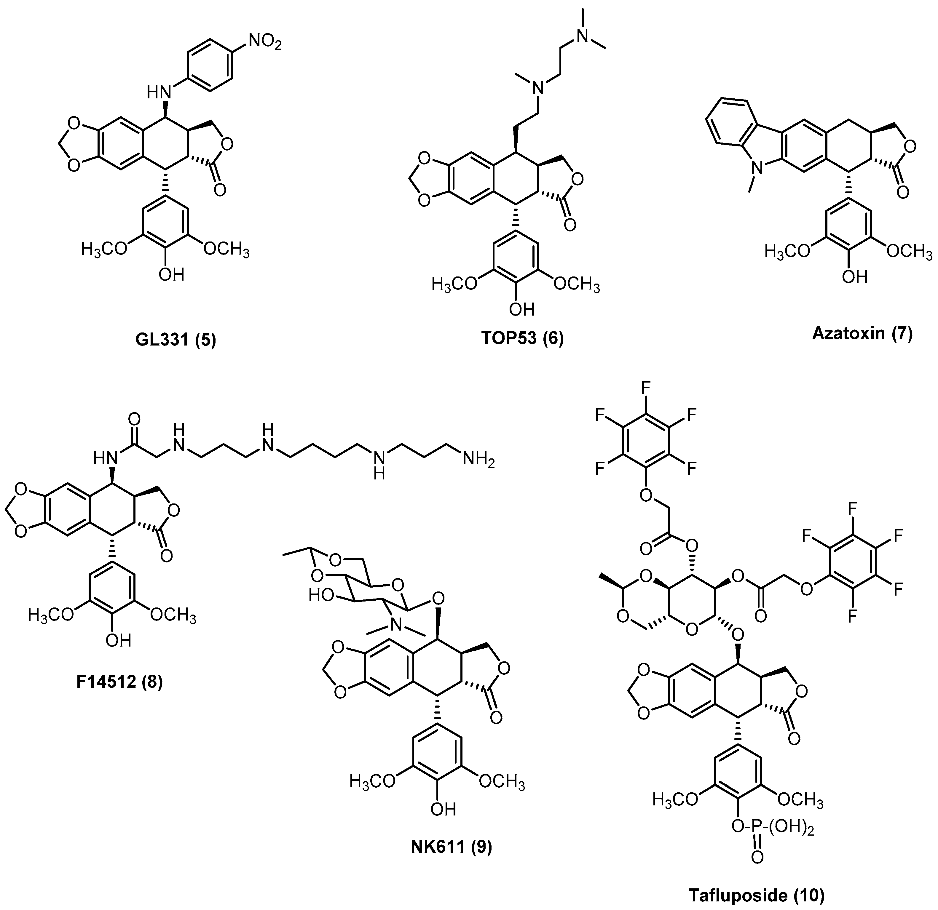
  17. Baldwin, E.L.; Osheroff, N. Etoposide, topoisomerase II and cancer. Curr. Med. Chem. Anti-Cancer Agents 2005, 5, 363–372. [Google Scholar] [CrossRef] [PubMed]
  18. Van Maanen, J.M.S.; Retèl, J.; De Vries, J.; Pinedo, H.M. Mechanism of Action of Antitumor Drug Etoposide: A Review. JNCI J. Natl. Cancer Inst. 1988, 80, 1526–1533. [Google Scholar] [CrossRef]
  19. Pitts, S.L.; Jablonksy, M.J.; Duca, M.; Dauzonne, D.; Monneret, C.; Arimondo, P.B.; Anklin, C.; Graves, D.E.; Osheroff, N. Contributions of the D-Ring to the Activity of Etoposide Against Human Topoisomerase IIα: Potential Interactions with DNA in the Ternary Enzyme-Drug-DNA Complex. Biochemistry 2011, 50, 5058. [Google Scholar] [CrossRef]
  20. Bromberg, K.D.; Burgin, A.B.; Osheroff, N. A Two-drug Model for Etoposide Action against Human Topoisomerase IIα. J. Biol. Chem. 2003, 278, 7406–7412. [Google Scholar] [CrossRef]
  21. Yu, X.; Che, Z.; Xu, H. Recent Advances in the Chemistry and Biology of Podophyllotoxins. Chem. A Eur. J. 2017, 23, 4467–4526. [Google Scholar] [CrossRef]
  22. Fan, H.; Zhu, Z.; Xian, H.; Wang, H.; Chen, B. Insight Into the Molecular Mechanism of Podophyllotoxin Derivatives as Anticancer Drugs. Front. Cell Dev. Biol. 2021, 9, 709075. [Google Scholar] [CrossRef] [PubMed]
  23. Xiao, J.; Gao, M.; Sun, Z.; Diao, Q.; Wang, P.; Gao, F. Recent advances of podophyllotoxin/epipodophyllotoxin hybrids in anticancer activity, mode of action, and structure-activity relationship: An update (2010–2020). Eur. J. Med. Chem. 2020, 208, 112830. [Google Scholar] [CrossRef]
  24. Zhang, X.; Rakesh, K.P.; Shantharam, C.S.; Manukumar, H.M.; Asiri, A.M.; Marwani, H.M.; Qin, H.L. Podophyllotoxin derivatives as an excellent anticancer aspirant for future chemotherapy: A key current imminent needs. Bioorg. Med. Chem. 2018, 26, 340–355. [Google Scholar] [CrossRef] [PubMed]
  25. Castro, M.Á.; Miguel del Corral, J.M.; García, P.A.; Victoria Rojo, M.; Bento, A.C.; Mollinedo, F.; Francesch, A.M.; San Feliciano, A. Lignopurines: A new family of hybrids between cyclolignans and purines. Synthesis and biological evaluation. Eur. J. Med. Chem. 2012, 58, 377–389. [Google Scholar] [CrossRef] [PubMed]
  26. Castro, M.Á.; Miguel del Corral, J.M.; García, P.A.; Rojo, M.V.; De La Iglesia-Vicente, J.; Mollinedo, F.; Cuevas, C.; San Feliciano, A. Synthesis and biological evaluation of new podophyllic aldehyde derivatives with cytotoxic and apoptosis-inducing activities. J. Med. Chem. 2010, 53, 983–993. [Google Scholar] [CrossRef] [PubMed]
  27. Castro, M.A.; Miguel del Corral, J.M.; Gordaliza, M.; García, P.A.; Gómez-Zurita, M.A.; García-Grávalos, M.D.; De La Iglesia-Vicente, J.; Gajate, C.; An, F.; Mollinedo, F.; et al. Synthesis and Biological Evaluation of New Selective Cytotoxic Cyclolignans Derived from Podophyllotoxin. J. Med. Chem. 2004, 47, 1214–1222. [Google Scholar] [CrossRef]
  28. Sampath Kumar, H.M.; Herrmann, L.; Tsogoeva, S.B. Structural hybridization as a facile approach to new drug candidates. Bioorg. Med. Chem. Lett. 2020, 30, 127514. [Google Scholar] [CrossRef]
  29. Bansal, Y.; Silakari, O. Multifunctional compounds: Smart molecules for multifactorial diseases. Eur. J. Med. Chem. 2014, 76, 31–42. [Google Scholar] [CrossRef]
  30. Bérubé, G. An overview of molecular hybrids in drug discovery. Expert Opin. Drug Discov. 2016, 11, 281–305. [Google Scholar] [CrossRef]
  31. Fortin, S.; Bérubé, G. Advances in the development of hybrid anticancer drugs. Expert Opin. Drug Discov. 2013, 8, 1029–1047. [Google Scholar] [CrossRef]
  32. Choudhary, S.; Singh, P.K.; Verma, H.; Singh, H.; Silakari, O. Success stories of natural product-based hybrid molecules for multi-factorial diseases. Eur. J. Med. Chem. 2018, 151, 62–97. [Google Scholar] [CrossRef]
  33. Moss, G.P.; Mary, Q.; Road, M.E. Nomenclature of lignans and neolignans. Pure Appl. Chem 2000, 72, 1493–1523. [Google Scholar] [CrossRef]
  34. Kumar, A.; Kumar, V.; Alegria, A.E.; Malhotra, S. V Synthetic and Application Perspectives of Azapodophyllotoxins: Alternative Scaffolds of Podophyllotoxin. Curr. Med. Chem. 2011, 18, 3853–3870. [Google Scholar] [CrossRef]
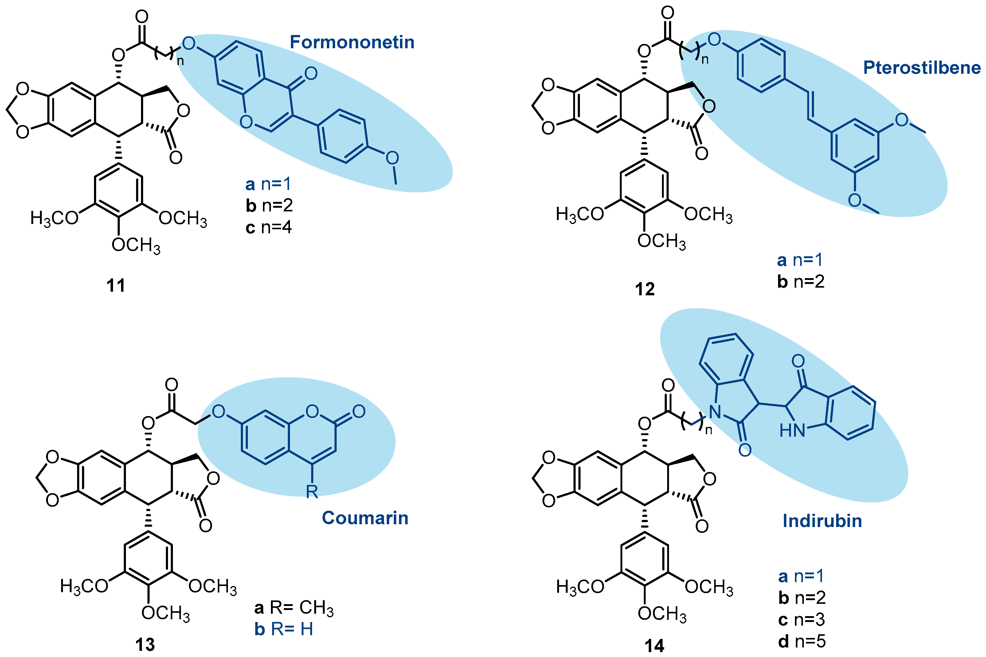
  35. Yang, C.; Xie, Q.; Zeng, X.; Tao, N.; Xu, Y.; Chen, Y.; Wang, J. Novel hybrids of podophyllotoxin and formononetin inhibit the growth, migration and invasion of lung cancer cells. Bioorg. Chem. 2019, 85, 445–454. [Google Scholar] [CrossRef]
  36. Tay, K.-C.; Tan, L.T.-H.; Chan, C.K.; Hong, S.L.; Chan, K.-G.; Yap, W.H.; Pusparajah, P.; Lee, L.-H.; Goh, B.-H. Formononetin: A Review of Its Anticancer Potentials and Mechanisms. Front. Pharmacol. 2019, 10, 820. [Google Scholar] [CrossRef]
  37. Jiang, D.; Rasul, A.; Batool, R.; Sarfraz, I.; Hussain, G.; Tahir, M.M.; Qin, T.; Selamoglu, Z.; Ali, M.; Li, J.; et al. Potential Anticancer Properties and Mechanisms of Action of Formononetin. BioMed Res. Int. 2019, 2019, 5854315. [Google Scholar] [CrossRef]
  38. Ong, S.K.L.; Shanmugam, M.K.; Fan, L.; Fraser, S.E.; Arfuso, F.; Ahn, K.S.; Sethi, G.; Bishayee, A. Focus on Formononetin: Anticancer Potential and Molecular Targets. Cancers 2019, 11, 611. [Google Scholar] [CrossRef]
  39. Estrela, J.M.; Ortega, A.; Mena, S.; Rodriguez, M.L.; Asensi, M. Pterostilbene: Biomedical applications. Crit. Rev. Clin. Lab. Sci. 2013, 50, 65–78. [Google Scholar] [CrossRef]
  40. Kim, H.; Seo, K.H.; Yokoyama, W. Chemistry of Pterostilbene and Its Metabolic Effects. J. Agric. Food Chem. 2020, 68, 12836–12841. [Google Scholar] [CrossRef]
  41. Obrador, E.; Salvador-Palmer, R.; Jihad-Jebbar, A.; López-Blanch, R.; Dellinger, T.H.; Dellinger, R.W.; Estrela, J.M. Pterostilbene in Cancer Therapy. Antioxidants 2021, 10, 492. [Google Scholar] [CrossRef]
  42. Zhang, L.; Wang, J.; Liu, L.; Zheng, C.; Wang, Y. Podophyllotoxin-pterostilbene fused conjugates as potential multifunctional antineoplastic agents against human uveal melanoma cells. RSC Adv. 2017, 7, 10601–10608. [Google Scholar] [CrossRef]
  43. Bai, G.; Zhao, D.; Ran, X.; Zhang, L.; Zhao, D. Novel Hybrids of Podophyllotoxin and Coumarin Inhibit the Growth and Migration of Human Oral Squamous Carcinoma Cells. Front. Chem. 2021, 8, 626075. [Google Scholar] [CrossRef]
  44. Gkionis, L.; Kavetsou, E.; Kalospyros, A.; Manousakis, D.; Garzon Sanz, M.; Butterworth, S.; Detsi, A.; Tirella, A. Investigation of the cytotoxicity of bioinspired coumarin analogues towards human breast cancer cells. Mol. Divers. 2021, 25, 307–321. [Google Scholar] [CrossRef] [PubMed]
  45. Wang, J.; Long, L.; Chen, Y.; Xu, Y.; Zhang, L. Design, synthesis and antineoplastic activity of novel hybrids of podophyllotoxin and indirubin against human leukaemia cancer cells as multifunctional anti-MDR agents. Bioorg. Med. Chem. Lett. 2018, 28, 1817–1824. [Google Scholar] [CrossRef] [PubMed]
  46. Eisenbrand, G.; Hippe, F.; Jakobs, S.; Muehlbeyer, S. Molecular mechanisms of indirubin and its derivatives: Novel anticancer molecules with their origin in traditional Chinese phytomedicine. J. Cancer Res. Clin. Oncol. 2004, 130, 627–635. [Google Scholar] [CrossRef] [PubMed]
  47. Wang, H.; Wang, Z.; Wei, C.; Wang, J.; Xu, Y.; Bai, G.; Yao, Q.; Zhang, L.; Chen, Y. Anticancer potential of indirubins in medicinal chemistry: Biological activity, structural modification, and structure-activity relationship. Eur. J. Med. Chem. 2021, 223, 113652. [Google Scholar] [CrossRef] [PubMed]
  48. Blažević, T.; Heiss, E.H.; Atanasov, A.G.; Breuss, J.M.; Dirsch, V.M.; Uhrin, P. Indirubin and Indirubin Derivatives for Counteracting Proliferative Diseases. Evid.-Based Complement. Altern. Med. 2015, 2015, 654098. [Google Scholar] [CrossRef]
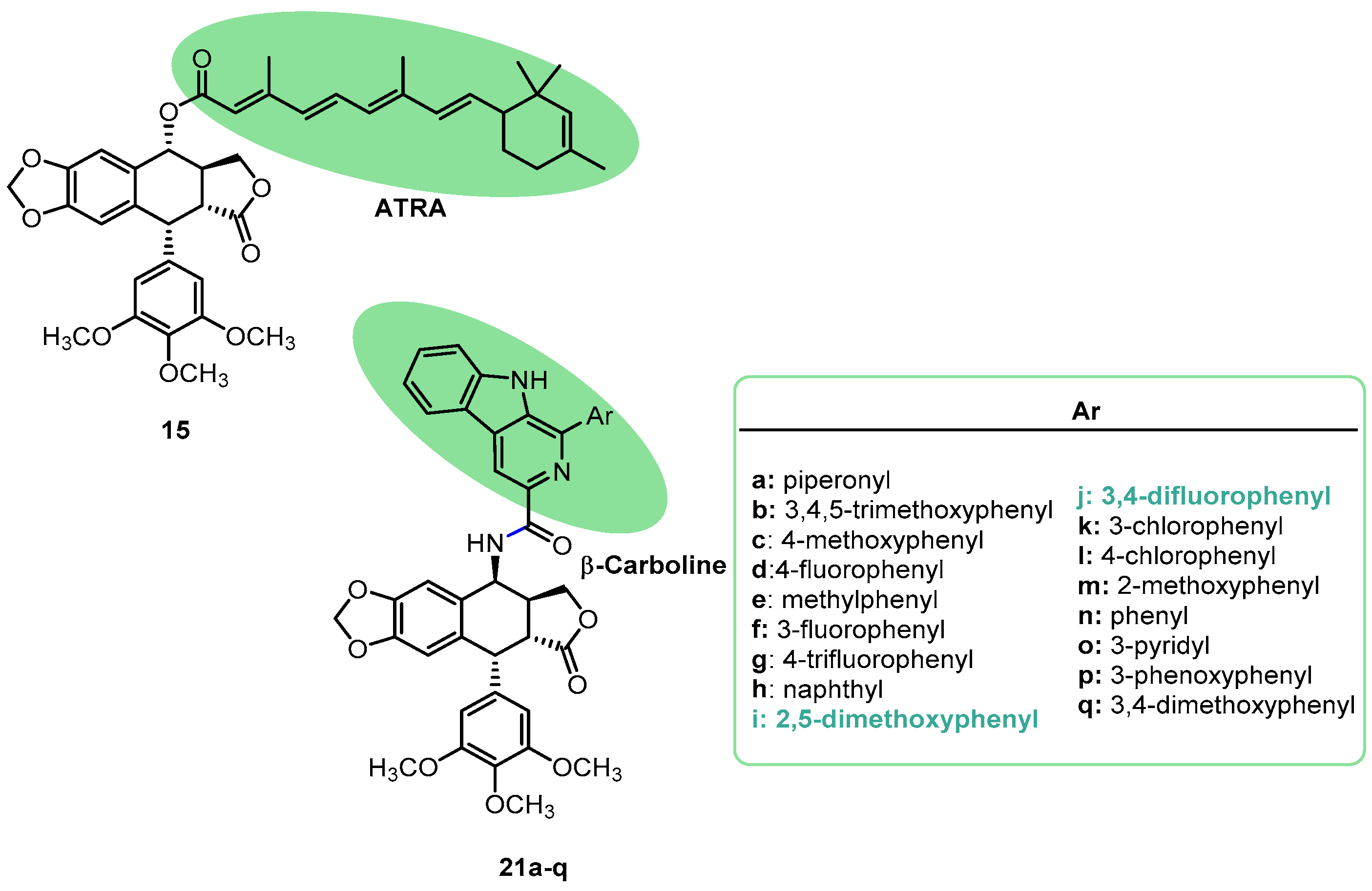
  49. Siddikuzzaman; Guruvayoorappan, C.; Berlin Grace, V.M. All trans retinoic acid and cancer. Immunopharmacol. Immunotoxicol. 2011, 33, 241–249. [Google Scholar] [CrossRef]
  50. Ni, X.; Hu, G.; Cai, X. The success and the challenge of all-trans retinoic acid in the treatment of cancer. Crit. Rev. Food Sci. Nutr. 2019, 59, S71–S80. [Google Scholar] [CrossRef]
  51. Zhang, L.; Wang, J.; Liu, L.; Zheng, C.; Wang, Y. Synthesis and Antiproliferative Activity of Novel All-Trans-Retinoic Acid-Podophyllotoxin Conjugate towards Human Gastric Cancer Cells. Molecules 2017, 22, 628. [Google Scholar] [CrossRef]
  52. Sathish, M.; Kavitha, B.; Nayak, V.L.; Tangella, Y.; Ajitha, A.; Nekkanti, S.; Alari, A.; Shankaraiah, N. Synthesis of podophyllotoxin linked b-carboline congeners as potential anticancer agents and DNA topoisomerase II inhibitors. Eur. J. Med. Chem. 2018, 144, 557–571. [Google Scholar] [CrossRef]
  53. Moura, D.J.; Richter, M.F.; Boeira, J.M.; Pêgas Henriques, J.A.; Saffi, J. Antioxidant properties of β-carboline alkaloids are related to their antimutagenic and antigenotoxic activities. Mutagenesis 2007, 22, 293–302. [Google Scholar] [CrossRef]
  54. Osorio, E.J.; Robledo, S.M.; Bastida, J. Alkaloids with antiprotozoal activity. Alkaloids Chem. Biol. 2008, 66, 113–190. [Google Scholar]
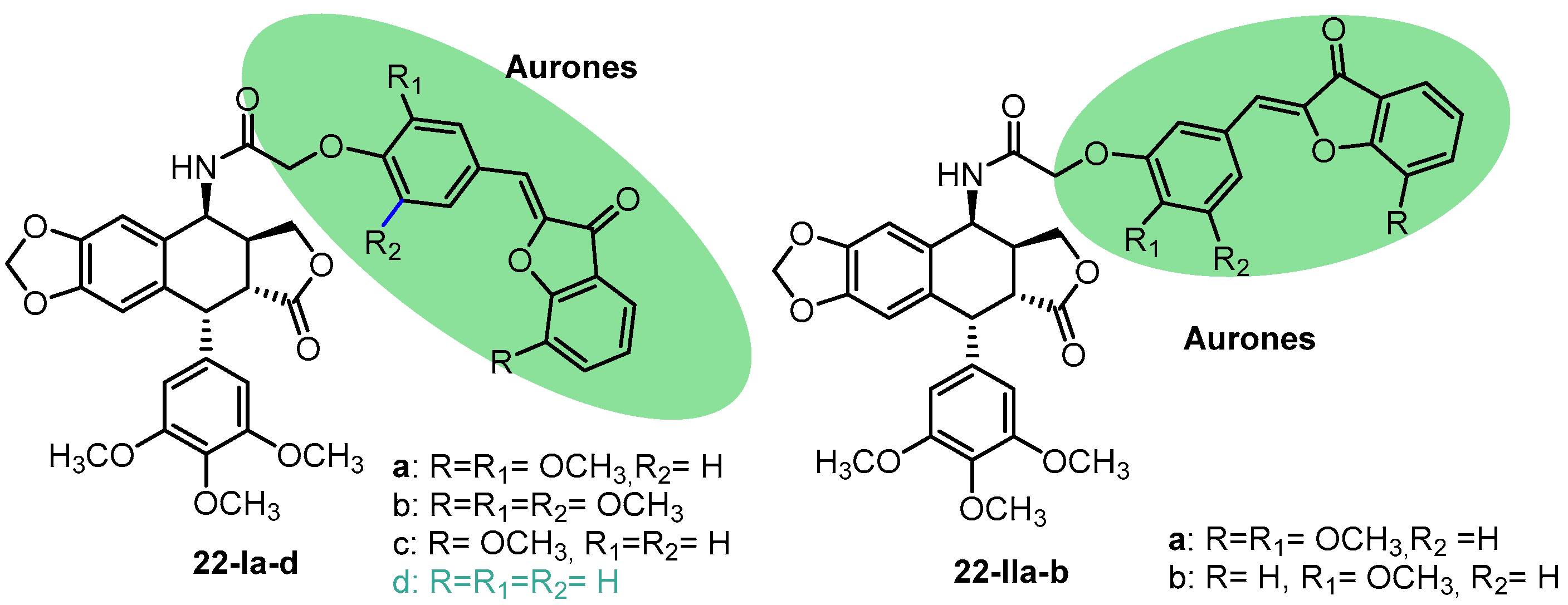
  55. Alsayari, A.; Muhsinah, A.B.; Hassan, M.Z.; Ahsan, M.J.; Alshehri, J.A.; Begum, N. Aurone: A biologically attractive scaffold as anticancer agent. Eur. J. Med. Chem. 2019, 166, 417–431. [Google Scholar] [CrossRef] [PubMed]
  56. Mazziotti, I.; Petrarolo, G.; La Motta, C. Aurones: A Golden Resource for Active Compounds. Molecules 2021, 27, 2. [Google Scholar] [CrossRef] [PubMed]
  57. Paidakula, S.; Nerella, S.; Vadde, R.; Kamal, A. Design and synthesis of 4β-Acetamidobenzofuranone-podophyllotoxin hybrids and their anti-cancer evaluation. Bioorg. Med. Chem. Lett. 2019, 29, 2153–2156. [Google Scholar] [CrossRef] [PubMed]
  58. Wu, L.; Feng, J.; Li, J.; Yu, Q.; Ji, J.; Wu, J.; Dai, W.; Guo, C. The gut microbiome-bile acid axis in hepatocarcinogenesis. Biomed. Pharmacother. 2021, 133, 111036. [Google Scholar] [CrossRef] [PubMed]
  59. Li, T.; Apte, U. Bile Acid Metabolism and Signaling in Cholestasis, Inflammation, and Cancer. Adv. Pharmacol. 2015, 74, 263–302. [Google Scholar] [PubMed]
  60. Park, J.; Choi, J.U.; Kim, K.; Byun, Y. Bile acid transporter mediated endocytosis of oral bile acid conjugated nanocomplex. Biomaterials 2017, 147, 145–154. [Google Scholar] [CrossRef]
  61. Gong, Y.; Zhang, X.; Zhang, Y.; Chu, F.; Li, G.; Zhang, H.; Xu, B.; Zhang, H.; Li, B.; Wang, P.; et al. Bile acids, carriers of hepatoma-targeted drugs? Pharmazie 2016, 71, 139–145. [Google Scholar]
  62. Cai, D.S.; Lou, S.Y.; Huo, S.; Yang, Y.Q.; Gao, F.; Pi, W.M.; Chen, K.D.; Wang, C.; Yang, X.Y.; Jiao, J.Y.; et al. Synthesis and biological activity evaluation of podophyllotoxin-linked bile acid derivatives as potential anti-liver cancer agents. Bioorg. Chem. 2022, 128, 106066. [Google Scholar] [CrossRef] [PubMed]
  63. Wu, G.; Xu, B.; Yang, Y.; Zhang, X.; Fang, K.; Ma, T.; Wang, H.; Xue, N.; Chen, M.; Guo, W.; et al. Synthesis and biological evaluation of podophyllotoxin derivatives as selective antitumor agents. Eur. J. Med. Chem. 2018, 155, 183–196. [Google Scholar] [CrossRef] [PubMed]
  64. Gao, H.; Liu, P.; Li, P.; Huang, Z.; Yu, F.; Lei, T.; Chen, Y.; Cheng, Y.; Mu, Q.; Huang, H. Ligustrazine monomer against cerebral ischemia/reperfusion injury. Neural Regen. Res. 2015, 10, 832. [Google Scholar] [PubMed]
  65. Zou, J.; Gao, P.; Hao, X.; Xu, H.; Zhan, P.; Liu, X. Recent progress in the structural modification and phamacological activities of ligustrazine derivatives. Eur. J. Med. Chem. 2018, 147, 150–162. [Google Scholar] [CrossRef]
  66. Shao, H.; He, X.; Zhang, L.; Du, S.; Yi, X.; Cui, X.; Liu, X.; Huang, S.; Tong, R. Efficacy of Ligustrazine injection as adjuntive therapy in treating acute cerebral infarction: A systematic review and meta-analysis. Front. Pharmacol. 2021, 12, 761722. [Google Scholar] [CrossRef]
  67. Zi, C.; Yang, L.; Hu, Y.; Zhang, P.; Tang, H.; Zhang, B.; Shen, J.; Kong, Q.; Wang, Y.; Wang, X.; et al. Synthesis, antitumor activity, and molecular docking of (−)-epigallocatechin-3-gallate-4β-triazolopodophyllotoxin conjugates. J. Asian Nat. Prod. Res. 2021, 23, 772–780. [Google Scholar] [CrossRef]
  68. Zi, C.; Wang, Z.; Shi, J.; Shi, B.; Zhang, N.; Wu, Y.; Xie, Y.; Zhou, L.; Xiao, C.; Wang, X.; et al. Synthesis, cytotoxicity, and molecular docking of methylated (–)-epigallocatechin-3-gallate- 4 β -triazolopodophyllotoxin derivatives as novel antitumor agents. J. Chem. Res. 2021, 1, 4–10. [Google Scholar] [CrossRef]
  69. Bimonte, S.; Cascella, M. The Potential Roles of Epigallocatechin-3-Gallate in the Treatment of Ovarian Cancer: Current State of Knowledge. Drug Des. Dev. Ther. 2020, 14, 4245–4250. [Google Scholar] [CrossRef]
  70. Negri, A.; Naponelli, V.; Rizzi, F.; Bettuzzi, S. Molecular Targets of Epigallocatechin—Gallate (EGCG): A Special Focus on Signal Transduction and Cancer. Nutrients 2018, 10, 1936. [Google Scholar] [CrossRef]
  71. Pizzolato, J.F.; Saltz, L.B. The camptothecins. Lancet 2003, 361, 2235–2242. [Google Scholar] [CrossRef]
  72. Hsiang, Y.H.; Hertzberg, R.; Hecht, S.; Liu, L.F. Camptothecin induces protein-linked DNA breaks via mammalian DNA topoisomerase I. J. Biol. Chem. 1985, 260, 14873–14878. [Google Scholar] [CrossRef] [PubMed]
  73. Zi, C.; Yang, L.; Dong, F.; Kong, Q.; Ding, Z. Synthesis and antitumor activity of camptothecin-4β-triazolopodophyllotoxin. Nat. Prod. Res. 2019, 34, 2301–2309. [Google Scholar] [CrossRef] [PubMed]
  74. Duan, M.; Mahal, A.; Mohammed, B.; Zhu, Y.; Tao, H.; Mai, S.; Al-Haideri, M.; Zhu, Q. Synthesis and antitumor activity of new tetrahydrocurcumin derivatives via click reaction. Nat. Prod. Res. 2022, 36, 5268–5276. [Google Scholar] [CrossRef] [PubMed]
  75. Wu, J.C.; Tsai, M.L.; Lai, C.S.; Wang, Y.J.; Ho, C.T.; Pan, M.H. Chemopreventative effects of tetrahydrocurcumin on human diseases. Food Funct. 2014, 5, 12–17. [Google Scholar] [CrossRef] [PubMed]
  76. Lai, C.S.; Ho, C.T.; Pan, M.H. The cancer chemopreventive and therapeutic potential of tetrahydrocurcumin. Biomolecules 2020, 10, 831. [Google Scholar] [CrossRef] [PubMed]
  77. Gupta, S.C.; Patchva, S.; Aggarwal, B.B. Therapeutic roles of curcumin: Lessons learned from clinical trials. Am. Assoc. Pharm. Sci. J. 2013, 15, 195–218. [Google Scholar] [CrossRef] [PubMed]
  78. Xu, F.; Dong, F.; Yang, D.; Li, Y.; Ding, Z.; Zhou, J.; Jiang, Z.; Hu, J. Synthesis and anticancer activity of dimeric podophyllotoxin derivatives. Drug Des. Dev. Ther. 2018, 12, 3393–3406. [Google Scholar]
  79. Zempleni, J.; Wijeratne, S.S.K.; Hassan, Y.I. Biotin. Biofactors 2009, 35, 36–46. [Google Scholar] [CrossRef]
  80. León-Del-Río, A. Biotin in metabolism, gene expression, and human disease. J. Inherit. Metab. Dis. 2019, 42, 647–654. [Google Scholar] [CrossRef]
  81. Russell-Jones, G.; McTavish, K.; McEwan, J.; Rice, J.; Nowotnik, D. Vitamin-mediated targeting as a potential mechanism to increase drug uptake by tumours. J. Inorg. Biochem. 2004, 98, 1625–1633. [Google Scholar] [CrossRef]
  82. Chen, S.; Zhao, X.; Chen, J.; Chen, J.; Kuznetsova, L.; Wong, S.S.; Ojima, I. Mechanism-Based Tumor-Targeting Drug Delivery System. Validation of Efficient Vitamin Receptor-Mediated Endocytosis and Drug Release. Bioconjug. Chem. 2010, 21, 979–987. [Google Scholar] [CrossRef]
  83. Jiang, Z.; Yuan, S.; Hu, J. Design, Synthesis, and Biological Evaluation of Novel Biotinylated Podophyllotoxin Derivatives as Potential Antitumor Agents. Front. Chem. 2019, 7, 434. [Google Scholar]
  84. Gentry, A.C.; Pitts, S.L.; Jablonsky, M.J.; Bailly, C.; Graves, D.E.; Osheroff, N. Interactions between the Etoposide Derivative F14512 and Human Type II Topoisomerases: Implications for the C4 Spermine Moiety in Promoting Enzyme-mediated DNA Cleavage. Biochemistry 2011, 50, 3240–3249. [Google Scholar] [CrossRef] [PubMed]
  85. Pendleton, M.; Lindsey, R.H.; Felix, C.A.; Grimwade, D.; Osheroff, N. Topoisomerase II and leukemia. Ann. N. Y. Acad. Sci. 2014, 1310, 98. [Google Scholar] [CrossRef]
  86. Oviatt, A.A.; Kuriappan, J.A.; Minniti, E.; Vann, K.R.; Minarini, A.; De Vivo, M.; Osheroff, N.; Discovery, D.; Morego, V. Polyamine-containing etoposide derivatives as poisons of human type II topoisomerases: Differential effects on topoisomerase IIa and IIb. Bioorg. Med. Chem. Lett. 2018, 28, 2961–2968. [Google Scholar] [CrossRef] [PubMed]
  87. Tierny, D.; Serres, F.; Segaoula, Z.; Bemelmans, I.; Bouchaert, E.; Pétain, A.; Brel, V.; Couffin, S.; Marchal, T.; Nguyen, L.; et al. Phase i Clinical Pharmacology Study of F14512, a New Polyamine-Vectorized Anticancer Drug, in Naturally Occurring Canine Lymphoma. Clin. Cancer Res. 2015, 21, 5314–5323. [Google Scholar] [CrossRef]
  88. Boyé, P.; Floch, F.; Serres, F.; Segaoula, Z.; Hordeaux, J.; Pascal, Q.; Coste, V.; Courapied, S.; Bouchaert, E.; Rybicka, A.; et al. Randomized, double-blind trial of F14512, a polyaminevectorized anticancer drug, compared with etoposide phosphate, in dogs with naturally occurring lymphoma. Oncotarget 2020, 11, 671–686. [Google Scholar] [CrossRef] [PubMed]
  89. Fu, J.; Yang, J.; Seeberger, P.H.; Yin, J. Glycoconjugates for glucose transporter-mediated cancer-specific targeting and treatment. Carbohydr. Res. 2020, 498, 108195. [Google Scholar] [CrossRef]
  90. Heiden, M.G.V.; Cantley, L.C.; Thompson, C.B. Understanding the warburg effect: The metabolic requirements of cell proliferation. Science 2009, 324, 1029–1033. [Google Scholar] [CrossRef]
  91. Calvaresi, E.C.; Hergenrother, P.J. Glucose conjugation for the specific targeting and treatment of cancer. Chem. Sci. 2013, 4, 2319–2333. [Google Scholar] [CrossRef]
  92. Arafa, H.M.M. Possible contribution of β-glucosidase and caspases in the cytotoxicity of glufosfamide in colon cancer cells. Eur. J. Pharmacol. 2009, 616, 58–63. [Google Scholar] [CrossRef]
  93. Cheng, J.; Zhao, W.; Yao, H.; Shen, Y.; Zhang, Y.; Li, Y.; Wongprasert, K.; Tang, Y. Discovery of 4,6-O-Thenylidene-β-D-glucopyranoside-(2″-acetamido,3″-acetyl-di-S-5-fluorobenzothizole/5-fluorobenzoxazole)-4′-demethylepipodophyllotoxin as Potential Less Toxic Antitumor Candidate Drugs by Reducing DNA Damage and Less Inhibition of. J. Med. Chem. 2020, 63, 2877–2893. [Google Scholar] [CrossRef]
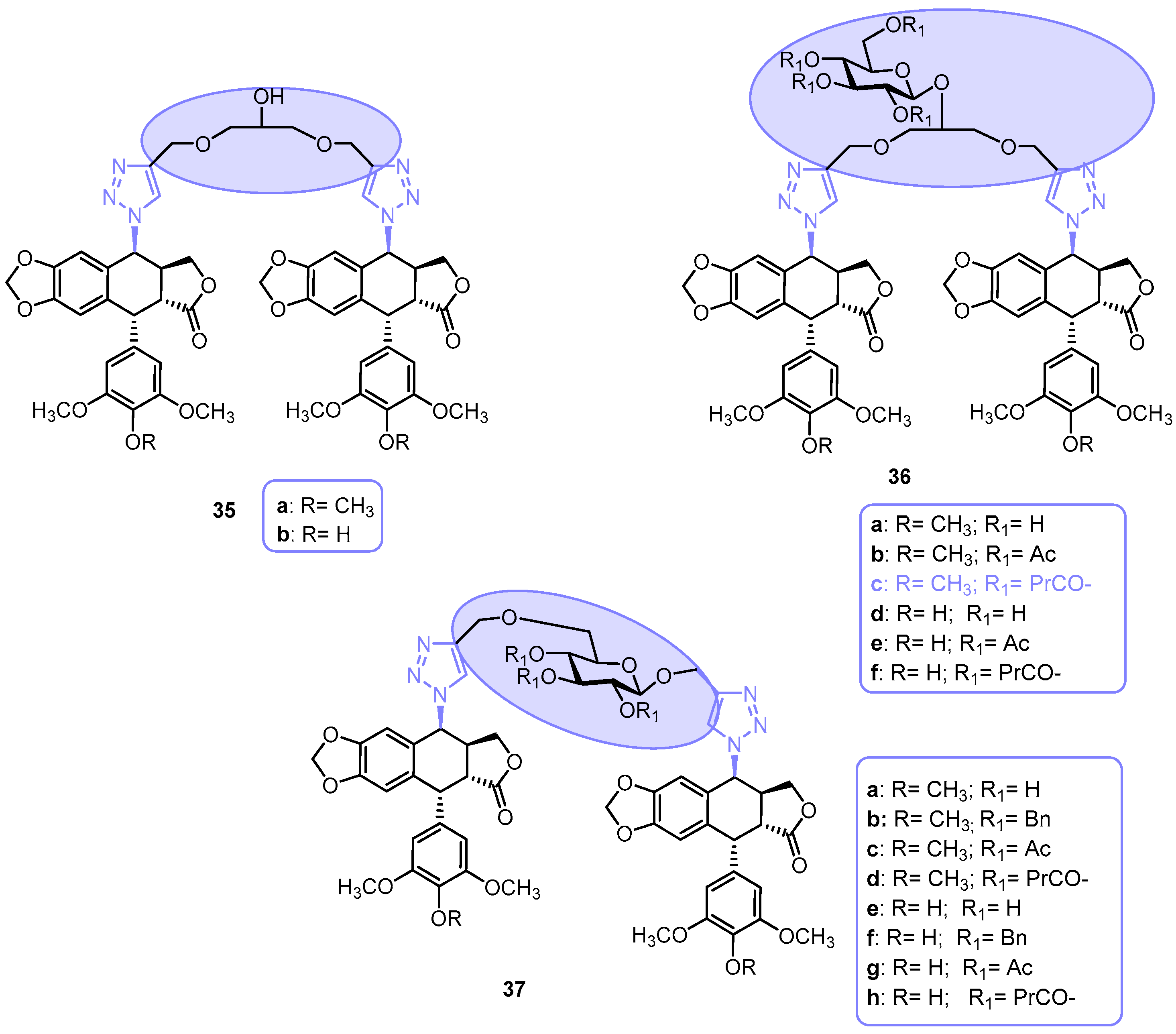
  94. Zi, C.T.; Yang, L.; Kong, Q.H.; Li, H.M.; Yang, X.Z.; Ding, Z.T.; Jiang, Z.H.; Hu, J.M.; Zhou, J. Glucoside Derivatives of Podophyllotoxin: Synthesis, Physicochemical Properties, and Cytotoxicity. Drug Des. Dev. Ther. 2019, 13, 3683–3692. [Google Scholar] [CrossRef] [PubMed]
  95. Zi, C.; Yang, L.; Zhang, B.; Li, Y.; Ding, Z.; Jiang, Z.; Hu, J.; Zhou, J. Synthesis and Cytotoxicities of Novel Podophyllotoxin Xyloside Derivatives. Nat. Prod. Commun. 2019, 14, 1934578X19860668. [Google Scholar] [CrossRef]
  96. San Feliciano, A.; Miguel Del Corral, J.M.; Gordaliza, M.; Castro, M.A. 13C NMR data for several cyclolignans. Magn. Reson. Chem. 1993, 31, 868–875. [Google Scholar] [CrossRef]
  97. Zi, C.; Yang, L.; Gao, W.; Li, Y.; Zhou, J.; Ding, Z. Click Glycosylation for the Synthesis of 1,2,3- Triazole-Linked Picropodophyllotoxin Glycoconjugates and Their Anticancer Activity. Med. Chem. Drug Discov. 2017, 2, 5038–5044. [Google Scholar] [CrossRef]
  98. Nerella, S.; Kankala, S.; Gavaji, B. Synthesis of podophyllotoxin-glycosyl triazoles via click protocol mediated by silver (I)-N -heterocyclic carbenes and their anticancer evaluation as topoisomerase-II inhibitors. Nat. Prod. Res. 2021, 35, 9–16. [Google Scholar] [CrossRef]
  99. Ali, I.; Lone, M.; Al-Othman, Z.; Al-Warthan, A.; Sanagi, M. Heterocyclic Scaffolds: Centrality in Anticancer Drug Development. Curr. Drug Targets 2015, 16, 711–734. [Google Scholar] [CrossRef]
  100. Bozorov, K.; Zhao, J.; Aisa, H.A. 1,2,3-Triazole-containing hybrids as leads in medicinal chemistry: A recent overview. Bioorg. Med. Chem. 2019, 27, 3511–3531. [Google Scholar] [CrossRef]
  101. Beltran-Hortelano, I.; Alcolea, V.; Font, M.; Pérez-Silanes, S. The role of imidazole and benzimidazole heterocycles in Chagas disease: A review. Eur. J. Med. Chem. 2020, 206, 112692. [Google Scholar] [CrossRef]
  102. Verma, A.; Joshi, S.; Singh, D. Imidazole: Having versatile biological activities. J. Chem. 2013, 2013, 329412. [Google Scholar] [CrossRef]
  103. Shalini, K.; Sharma, P.K.; Kumar, N. Imidazole and its biological activities: A review. Der Chem. Sin. 2010, 1, 36–47. [Google Scholar]
  104. Cao, B.O.; Yang, S.; Li, W.; Chen, H.; Chen, Y.; Liu, Y.; Liu, B.I.N. GMZ-1 is a podophyllotoxin derivative that suppresses growth and induces apoptosis in adriamycin-resistant K562 / A02 cells through modulation of MDR1 expression. Mol. Med. Rep. 2018, 17, 474–478. [Google Scholar] [CrossRef]
  105. Yin, M.; Fang, Y.; Sun, X.; Xue, M.; Zhang, C.; Zhu, Z.; Meng, Y.; Kong, L.; Myint, Y.Y.; Li, Y.; et al. Synthesis and anticancer activity of podophyllotoxin derivatives with nitrogen-containing heterocycles. Front. Chem. 2023, 11, 1191498. [Google Scholar] [CrossRef] [PubMed]
  106. Ayati, A.; Emami, S.; Asadipour, A.; Shafiee, A.; Foroumadi, A. Recent applications of 1,3-thiazole core structure in the identification of new lead compounds and drug discovery. Eur. J. Med. Chem. 2015, 97, 699–718. [Google Scholar] [CrossRef] [PubMed]
  107. Ayati, A.; Emami, S.; Moghimi, S.; Foroumadi, A. Thiazole in the targeted anticancer drug discovery. Future Med. Chem. 2019, 11, 1929–1952. [Google Scholar] [CrossRef] [PubMed]
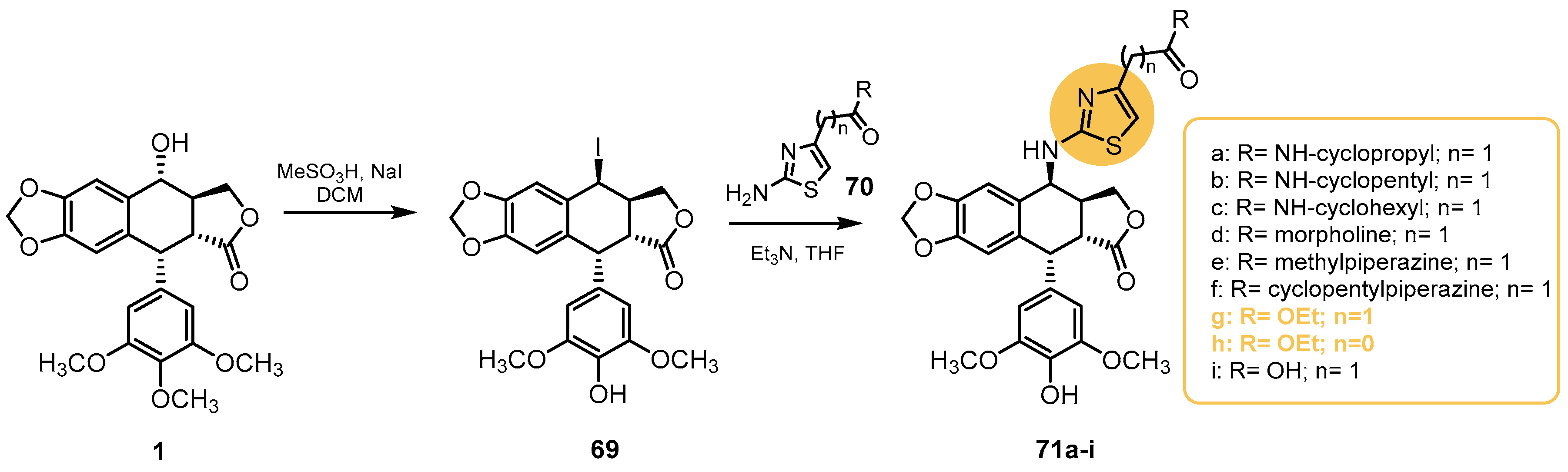
  108. Sang, C.; Tian, H.; Chen, Y.; Liu, J.; Chen, S.; Hui, L. Synthesis and biological evaluation of 4b-(thiazol-2-yl)amino-4′-O demethyl-4-deoxypodophyllotoxins as topoisomerase-II inhibitors. Bioorg. Med. Chem. Lett. 2018, 28, 71–76. [Google Scholar] [CrossRef] [PubMed]
  109. Vágó, I.; Kalaus, G.; Greiner, I.; Kajtár-Peredy, M.; Brlik, J.; Szabó, L.; Szántay, C. Synthesis of vinca alkaloids and related compounds. Heterocycles 2001, 55, 873–880. [Google Scholar]
  110. Pairas, G.N.; Perperopoulou, F.; Tsoungas, P.G.; Varvounis, G. The Isoxazole Ring and Its N-Oxide: A Privileged Core Structure in Neuropsychiatric Therapeutics. ChemMedChem 2017, 12, 408–419. [Google Scholar] [CrossRef]
  111. Ji Ram, V.; Sethi, A.; Nath, M.; Pratap, R. Five-Membered Heterocycles. In The Chemistry of Heterocycles; Elsevier Inc.: Amsterdam, The Netherlands, 2019; pp. 149–478. ISBN 9780081010334. [Google Scholar]
  112. Yang, R.; Zhang, Y.; Xu, H. Synthesis of novel isoxazoline-containing podophyllotoxin/2′(2′6′)-(di) halogenopodophyllotoxin derivatives and their insecticidal/acaricidal activities. Bioorg. Med. Chem. Lett. 2018, 28, 1410–1416. [Google Scholar] [CrossRef]
  113. Fan, L.; Guo, Y.; Zhi, X.; Yu, X.; Xu, H. Stereoselective synthesis of 2α-chloropicropodophyllotoxins and insecticidal activity of their esters against oriental armyworm, mythimna separata walker. J. Agric. Food Chem. 2014, 62, 3726–3733. [Google Scholar] [CrossRef]
  114. Che, Z.; Yu, X.; Fan, L.; Xu, H. Insight into dihalogenation of E-ring of podophyllotoxins, and their acyloxyation derivatives at the C4 position as insecticidal agents. Bioorg. Med. Chem. Lett. 2013, 23, 5592–5598. [Google Scholar] [CrossRef]
  115. Zefirov, N.A.; Nurieva, E.V.; Mamaeva, A.V.; Vasilenko, D.A.; Sadovnikov, K.S.; Averina, E.B.; Kolchanova, A.Y.; Milaeva, E.R.; Zefirova, O.N. New podophyllotoxin and epipodophyllotoxin derivatives with substituted isoxazole fragments. Russ. Chem. Bull. 2023, 72, 1029–1035. [Google Scholar] [CrossRef]
  116. Huang, D.; Huang, M.; Liu, W.; Liu, A.; Liu, X.; Chen, X.; Pei, H.; Sun, J.; Yin, D.; Wang, X. Design, synthesis and biological evaluation of 1H-pyrazole-5-carboxamide derivatives as potential fungicidal and insecticidal agents. Chem. Pap. 2017, 71, 2053–2061. [Google Scholar] [CrossRef]
  117. Baraldi, P.G.; Simoni, D.; Moroder, F.; Manfredini, S.; Mucchi, L.; Dalla Vecchia, F.; Orsolini, P. Synthesis of 2-(5′-substituted isoxazol-3′-yl)-4-oxo-3-thiazolidinylalkanoic acids. J. Heterocycl. Chem. 1982, 19, 557–560. [Google Scholar] [CrossRef]
  118. Jackson, P.L.; Hanson, C.D.; Farrell, A.K.; Butcher, R.J.; Stables, J.P.; Eddington, N.D.; Scott, K.R. Enaminones An explanation of anticonvulsant activity and toxicity per Linus Pauling’s clathrate hypothesis. Eur. J. Med. Chem. 2012, 51, 42–51. [Google Scholar] [CrossRef] [PubMed]
  119. Duan, J.; Zhang, L.H.; Dolbier, W.R. A convenient new method for the bromination of deactivated aromatic compounds. Synlett 1999, 1999, 1245–1246. [Google Scholar] [CrossRef]
  120. Datta, R.L.; Chatterjbe, N.R. Halogenation. XVIII. Direct ionization by means of iodine and nitric acid. J. Am. Chem. Soc. 1919, 41, 292–295. [Google Scholar] [CrossRef]
  121. Trukhacheva, L.A.; Levina, V.I.; Grigor’ev, N.B.; Arzamastsev, A.P.; Dalinger, I.L.; Vatsadze, I.A.; Popova, G.P.; Shevelev, S.A.; Granik, V.G. Kinetics of hydrolysis of five-membered C-nitroheterocycles: Pyrazole, imidazole, 1,2,4-triazole, and isoxazole derivatives. Russ. Chem. Bull. 2005, 54, 2813–2819. [Google Scholar] [CrossRef]
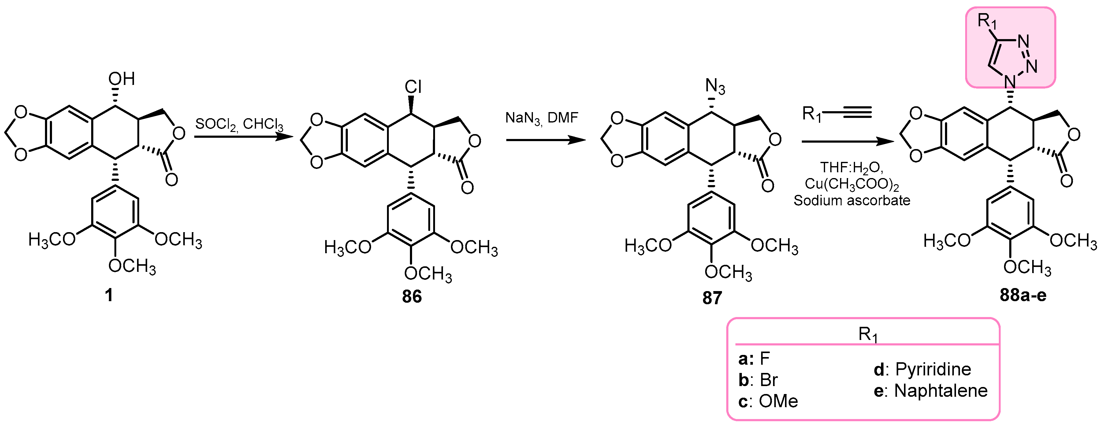
  122. Dheer, D.; Singh, V.; Shankar, R. Medicinal attributes of 1,2,3-triazoles: Current developments. Bioorg. Chem. 2017, 71, 30–54. [Google Scholar] [CrossRef] [PubMed]
  123. Bonandi, E.; Christodoulou, M.S.; Fumagalli, G.; Perdicchia, D.; Rastelli, G.; Passarella, D. The 1,2,3-triazole ring as a bioisostere in medicinal chemistry. Drug Discov. Today 2017, 22, 1572–1581. [Google Scholar] [CrossRef]
  124. Jiang, X.; Hao, X.; Jing, L.; Wu, G.; Kang, D.; Liu, X.; Zhan, P. Recent applications of click chemistry in drug discovery. Expert Opin. Drug Discov. 2019, 14, 779–789. [Google Scholar] [CrossRef]
  125. Kaur, J.; Saxena, M.; Rishi, N. An Overview of Recent Advances in Biomedical Applications of Click Chemistry. Bioconjug. Chem. 2021, 32, 1455–1471. [Google Scholar] [CrossRef]
  126. Kolb, H.C.; Sharpless, K.B. The growing impact of click chemistry on drug discovery. Drug Discov. Today 2003, 8, 1128–1137. [Google Scholar] [CrossRef]
  127. Hou, W.; Zhang, G.; Luo, Z.; Su, L.; Xu, H. Click chemistry-based synthesis and cytotoxic activity evaluation of 4α-triazole acetate podophyllotoxin derivatives. Chem. Biol. Drug Des. 2019, 93, 473–483. [Google Scholar] [CrossRef]
  128. Vishnuvardhan, M.V.P.S.; Chandrasekhar, K.; Nayak, V.L.; Sayeed, I.B.; Alarifi, A.; Kamal, A. Click chemistry-assisted synthesis of triazolo linked podophyllotoxin conjugates as tubulin. Med. Chem. Commun. 2017, 8, 1817–1823. [Google Scholar] [CrossRef]
  129. Reddy, V.G.; Bonam, S.R.; Reddy, T.S.; Akunuri, R.; Naidu, V.G.M.; Nayak, V.L.; Bhargava, S.K.; Kumar, H.M.S.; Srihari, P.; Kamal, A. 4b-amidotriazole linked podophyllotoxin congeners: DNA topoisomerase-IIa inhibition and potential anticancer agents for prostate cancer. Eur. J. Med. Chem. 2018, 20, 595–611. [Google Scholar] [CrossRef]
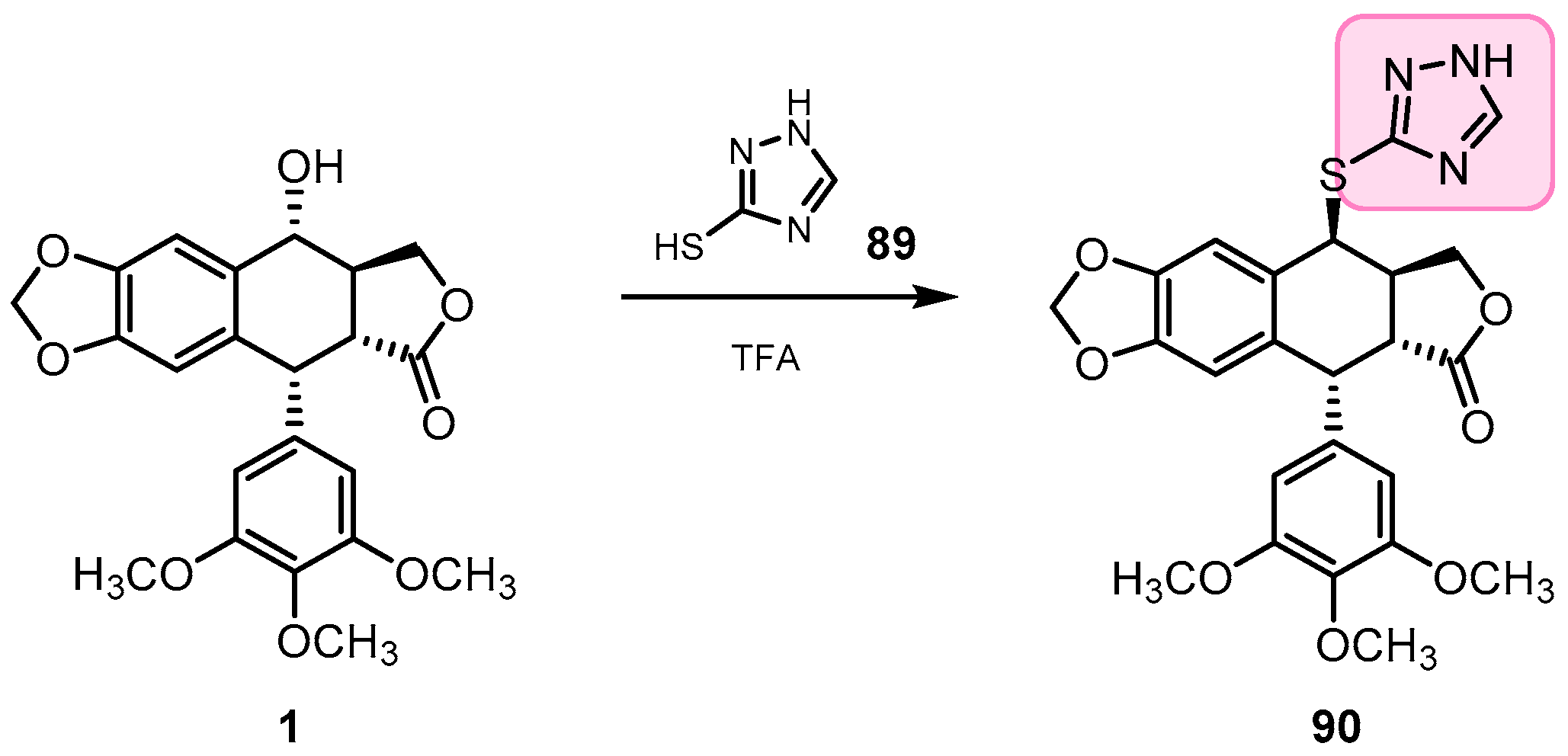
  130. Li, J.L.; Zhao, W.; Zhou, C.; Zhang, Y.X.; Li, H.M.; Tang, Y.L.; Liang, X.H.; Chen, T.; Tang, Y.J. Comparison of carbon-sulfur and carbon-amine bond in therapeutic drug: 4β-S-aromatic heterocyclic podophyllum derivatives display antitumor activity. Nat. Sci. Rep. 2015, 5, 14814. [Google Scholar] [CrossRef]
  131. Zhao, W.; Zhou, C.; Guan, Z.; Yin, P.; Chen, F.; Tang, Y. Structural insights into the inhibition of tubulin by the antitumor agent 4b-(1,2,4-triazol-3-ythio)-4-deoxypodophyllotoxin. ACS Chem. Biol. 2017, 12, 745–752. [Google Scholar] [CrossRef]
  132. Chawla, G.; Naaz, B.; Siddiqui, A. Exploring 1,3,4-Oxadiazole Scaffold for Anti-inflammatory and Analgesic Activities: A Review of Literature from 2005–2016. Mini-Rev. Med. Chem. 2018, 18, 216–233. [Google Scholar] [CrossRef]
  133. Bajaj, S.; Asati, V.; Singh, J.; Roy, P.P. 1,3,4-Oxadiazoles: An emerging scaffold to target growth factors, enzymes and kinases as anticancer agents. Eur. J. Med. Chem. 2015, 97, 124–141. [Google Scholar] [CrossRef] [PubMed]
  134. Vaidya, A.; Pathak, D.; Shah, K. 1,3,4-oxadiazole and its derivatives: A review on recent progress in anticancer activities. Chem. Biol. Drug Des. 2021, 97, 572–591. [Google Scholar] [CrossRef] [PubMed]
  135. Ren, J.; Wu, L.; Xin, W.Q.; Chen, X.; Hu, K. Synthesis and biological evaluation of novel 4β-(1,3,4-oxadiazole-2-amino)-podophyllotoxin derivatives. Bioorg. Med. Chem. Lett. 2012, 22, 4778–4782. [Google Scholar] [CrossRef] [PubMed]
  136. Ren, J.; Liu, Y.; Li, L.; Zhao, Y.; Li, Z.; Wu, C.; Chen, L.; Hu, K. OAMDP, a novel podophyllotoxin derivative, induces apoptosis, cell cycle arrest and autophagy in hepatoma HepG2 cells. Cell Biollogy Int. 2018, 42, 194–204. [Google Scholar] [CrossRef] [PubMed]
  137. Wang, Z.; Sun, W.; Wang, X.; Zhang, Y.; Qiu, H.; Qi, J.; Pang, Y.; Lu, G.; Wang, X.; Yu, F.; et al. Design, Synthesis, Biological evaluation and 3D−QSAR Analysis of podophyllotoxin dioxazole combination as tubulin targeting anticancer agents. Chem. Biol. Drug Des. 2017, 90, 236–243. [Google Scholar] [CrossRef] [PubMed]
  138. Han, H.; Xu, X.; Ma, Y.; Luo, Y.; Wang, Z.; Yang, M. Discovering Podophyllotoxin Derivatives as Potential Anti-Tubulin Agents: Design, Synthesis and Biological Evaluation. Biol. Chem. Chem. Biol. 2020, 5, 10526–10536. [Google Scholar] [CrossRef]
  139. Ling, Y.; Hao, Z.Y.; Liang, D.; Zhang, C.L.; Liu, Y.F.; Wang, Y. The expanding role of pyridine and dihydropyridine scaffolds in drug design. Drug Des. Dev. Ther. 2021, 15, 4289–4338. [Google Scholar] [CrossRef]
  140. Tahir, T.; Ashfaq, M.; Saleem, M.; Rafiq, M.; Shahzad, M.I.; Kotwica-Mojzych, K.; Mojzych, M. Pyridine Scaffolds, Phenols and Derivatives of Azo Moiety: Current Therapeutic Perspectives. Molecules 2021, 26, 4872. [Google Scholar] [CrossRef]
  141. Wang, H. Podophyllum derivatives containing fluorine atom in the 3-position of 2-aminopyridine improved the antitumor activity by inducing P53-dependent apoptosis. Med. Chem. Res. 2017, 26, 1279–1290. [Google Scholar] [CrossRef]
  142. Wang, H.; Feng, J.; Zhou, T.; Wei, L.; Zhou, J. International Journal of Biochemistry and Cell Biology P-3F, a microtubule polymerization inhibitor enhances P53 stability through the change in localization of RPS27a. Int. J. Biochem. Cell Biol. 2017, 92, 53–62. [Google Scholar] [CrossRef]
  143. Li, Y.; Wang, T.; Sun, Y.; Huang, T.; Li, C.; Fu, Y.; Li, Y.; Li, C. p53-Mediated PI3K/AKT/mTOR Pathway Played a Role in Ptox Dpt -Induced EMT Inhibition in Liver Cancer Cell Lines. Oxidative Med. Cell. Longev. 2019, 2019, 2531493. [Google Scholar]
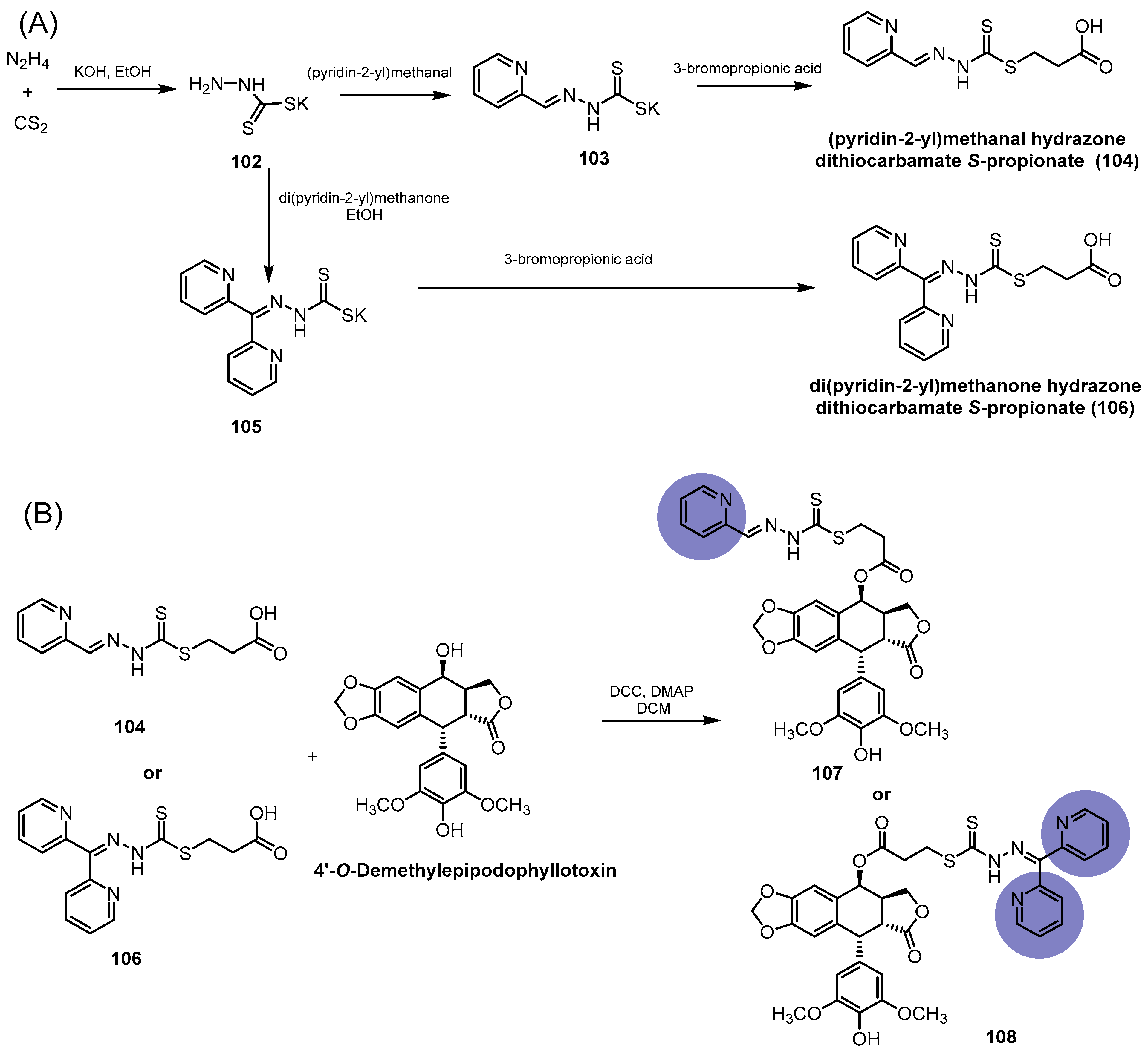
  144. Wang, T.; Fu, Y.; Huang, T.; Liu, Y.; Wu, M.; Yuan, Y.; Li, S.; Li, C. Copper Ion Attenuated the Antiproliferative Activity of Di-2-pyridylhydrazone Dithiocarbamate Derivative; However, There Was a Lack of Correlation between ROS Generation and Antiproliferative Activity. Molecules 2016, 21, 1088. [Google Scholar] [CrossRef]
  145. Li, Y.; Huang, T.; Fu, Y.; Wang, T.; Zhao, T.; Guo, S.; Sun, Y.; Yang, Y.; Id, C.L. Antitumor activity of a novel dual functional podophyllotoxin derivative involved PI3K/AKT/mTOR pathway. PLoS ONE 2019, 14, e0215886. [Google Scholar] [CrossRef]
  146. Jain, A.; Chaudhary, J.; Khaira, H.; Chopra, B.; Dhingra, A. Piperazine: A Promising Scaffold with Analgesic and Anti-inflammatory Potential. Drug Res. 2021, 71, 62–72. [Google Scholar] [CrossRef]
  147. Zhang, R.H.; Guo, H.Y.; Deng, H.; Li, J.; Quan, Z.S. Piperazine skeleton in the structural modification of natural products: A review. J. Enzyme Inhib. Med. Chem. 2021, 36, 1165–1197. [Google Scholar] [CrossRef]
  148. Singh, K.; Siddiqui, H.H.; Shakya, P.; Bagga, P.; Kumar, A.; Khalid, M.; Arif, M.; Alok, S. Piperazine-A Biologically Active Scaffold. Int. J. Pharceutical Sci. Res. 2015, 6, 4145–4158. [Google Scholar]
  149. Markandewar, R.A.; Baseer, M.A. Exploring Pharmacological Significance of Piperazine Scaffold. World J. Pharm. Res. 2016, 5, 1409–1420. [Google Scholar]
  150. Sun, W.; Ji, Y.; Wan, Y.; Han, H.; Lin, H.; Qi, J.; Wang, X.; Yang, Y. Design and synthesis of piperazine acetate podophyllotoxin ester derivatives targeting tubulin depolymerization as new anticancer agents. Bioorg. Med. Chem. Lett. 2017, 27, 4066–4074. [Google Scholar] [CrossRef]
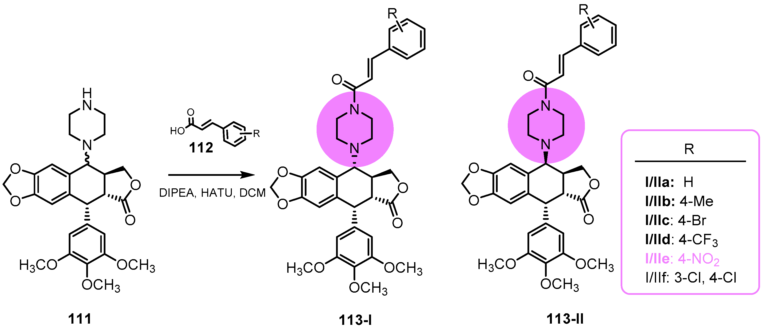
  151. Zhang, X.; Yang, T.; Jin, X.; Lin, K.; Dai, X.; Gao, T.; Huang, G.; Fan, M.; Ma, L.; Liu, Z.; et al. Synthesis and biological evaluation of cytotoxic activity of novel podophyllotoxin derivatives incorporating piperazinyl-cinnamic amide moieties. Bioorg. Chem. 2022, 123, 105761. [Google Scholar] [CrossRef] [PubMed]
  152. Wu, Z.R.; Deng, W.; He, D. Synthesis and biological evaluation of prodrugs for nitroreductase based 4-β-amino-4′-Demethylepipodophyllotoxin as potential anticancer agents. Med. Chem. Res. 2022, 31, 1099–1108. [Google Scholar] [CrossRef]
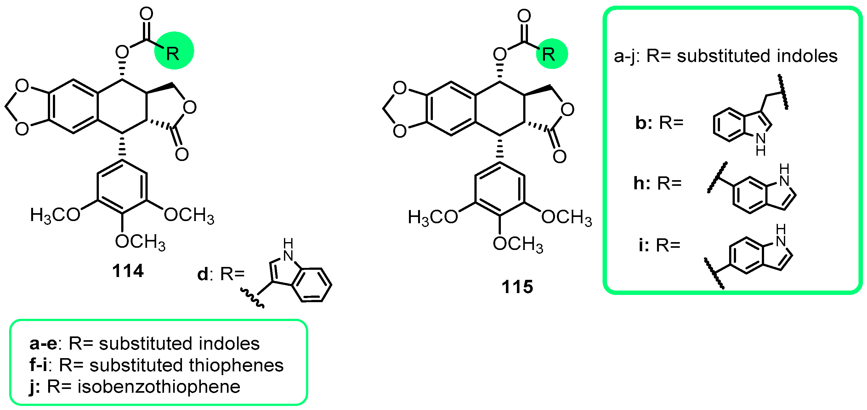
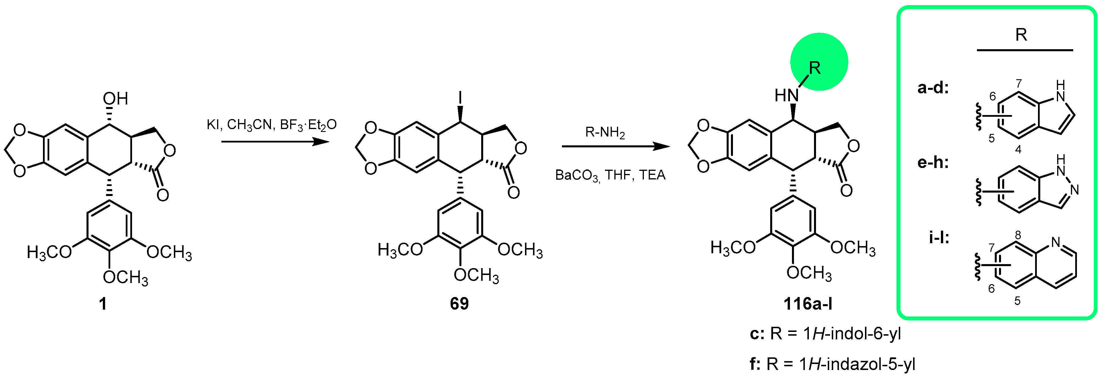
  153. Dadashpour, S.; Emami, S. Indole in the target-based design of anticancer agents: A versatile scaffold with diverse mechanisms. Eur. J. Med. Chem. 2018, 150, 9–29. [Google Scholar] [CrossRef] [PubMed]
  154. Han, H.; Lin, H.; He, D.; Ren, Y.; Sun, W.; Liang, L. Novel Podophyllotoxin Derivatives as Potential Tubulin Inhibitors: Design, Synthesis, and Antiproliferative Activity Evaluation. Chem. Biodivers. 2018, 15, e1800289. [Google Scholar] [CrossRef] [PubMed]
  155. Zhang, L.; Zeng, X.; Ren, X.; Tao, N.; Yang, C.; Xu, Y.; Chen, Y.; Wang, J. Design, synthesis, and biological evaluation of indole carboxylic acid esters of podophyllotoxin as antiproliferative agents. Med. Chem. Res. 2019, 28, 81–94. [Google Scholar] [CrossRef]
  156. Zhao, W.; He, L.; Xiang, T.; Tang, Y. Discover 4b-NH-(6-aminoindole)-4-desoxy-podophyllotoxin with nanomolar-potency antitumor activity by improving the tubulin binding affinity on the basis of a potential binding site nearby colchicine domain. Eur. J. Med. Chem. 2019, 170, 73–86. [Google Scholar] [CrossRef] [PubMed]
  157. Zhang, Q.; Zhao, W.; Tang, Y. Discover the leading compound of 4b-S-(5-fluorobenzoxazole)-4deoxy-4′-demethylepipodophyllotoxin with millimolar potency toxicity by modifiyng the molecule structure of 4′-demethylepipodophyllotoxin. Eur. J. Med. Chem. 2018, 158, 951–964. [Google Scholar] [CrossRef]
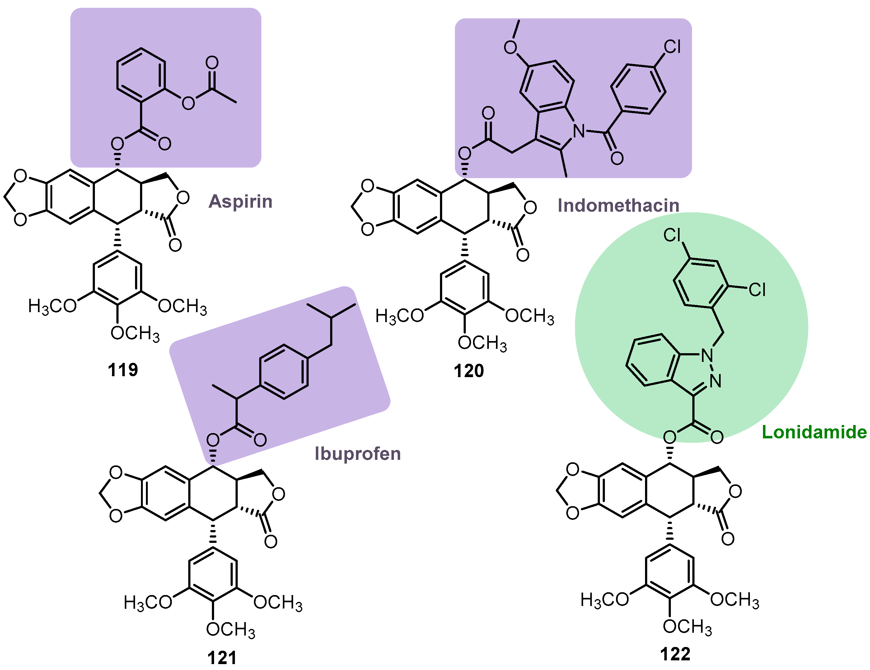
  158. Zang, L.; Zheng, J.; Rong, Y.; Chengli, Y.; Long, L.; Xu, Y.; Chen, Y.; Wang, J.; Yao, Q. Synthesis, antitumor evaluation and molecular docking study of a novel podophyllotoxin-Ionidamine hybrid. Med. Chem. Res. 2018, 27, 2231–2238. [Google Scholar] [CrossRef]
  159. Zhang, L.; Liu, L.; Zheng, C.; Wang, Y.; Nie, X.; Shi, D. Synthesis and biological evaluation of novel podophyllotoxin-NSAIDs conjugates as multifunctional anti-MDR agents against resistant human hepatocellular carcinoma Bel-7402/5-FU cells. Eur. J. Med. Chem. 2017, 131, 81–91. [Google Scholar] [CrossRef] [PubMed]
  160. Huang, Y.; Sun, G.; Sun, X.; Li, F.; Zhao, L.; Zhong, R.; Peng, Y. The potential of lonidamine in combination with chemotherapy and physical therapy in cancer treatment. Cancers 2020, 12, 3332. [Google Scholar] [CrossRef]
  161. Bhutia, Y.D.; Babu, E.; Ganapathy, V. Re-programming tumour cell metabolism to treat cancer: No lone target for lonidamine. Biochem. J. 2016, 473, 1503–1506. [Google Scholar] [CrossRef]
  162. Nath, K.; Guo, L.; Nancolas, B.; Nelson, D.S.; Shestov, A.A.; Lee, S.-C.; Roman, J.; Zhou, R.; Leeper, D.B.; Halestrap, A.P.; et al. Mechanism of antineoplastic activity of lonidamide. Biochim. Biophys. Acta 2016, 1866, 151–162. [Google Scholar]
  163. Zhang, L.; Rong, Y.; Zheng, J.; Yang, C.; Chen, Y.; Wang, J.; Wei, G. Desing, synthesis and biological evaluation of novel nitric oxide-donating podophyllotoxin derivatives as potential antiproliferative agents against multi-drug resistant leukemia cells. RSC Adv. 2018, 8, 34266–34274. [Google Scholar] [CrossRef]
  164. Yang, Y.; Qi, S.; Shi, R.; Yao, J.; Wang, L.; Yuan, H. Induction of apoptotic DNA fragmentation mediated by mitochondrial pathway with caspase-3-dependent BID cleavage in human gastric cancer cells by a new nitroxyl spin-labeled derivative of podophyllotoxin. Biomed. Pharmacother. 2017, 90, 131–138. [Google Scholar] [CrossRef]
  165. Tian, X.; Wang, Y.; Yang, M.; Chen, Y. Synthesis and antitumor activity of spin labeled derivatives of podophyllotoxin. Life Sci. 1996, 60, 511–517. [Google Scholar] [CrossRef]
  166. Qu, Y.; Zhang, C.; Ma, X.; Gao, Y.; Liu, J.; Wu, L. Synthesis and biological evaluation of NQO1-activated prodrugs of podophyllotoxin as antitumor agents. Bioorg. Med. Chem. 2020, 28, 115821. [Google Scholar] [CrossRef]
  167. Belinsky, M.; Jaiswal, A.K. NAD(P)H:Quinone oxidoreductase I (DT-diaphorase) expression in normal and tumor tissues. Cancer Metastasis Rev. 1993, 12, 103–117. [Google Scholar] [CrossRef] [PubMed]
  168. Siegel, D.; Gustafson, D.L.; Dehn, D.L.; Han, J.Y.; Boonchoong, P.; Berliner, L.J.; Ross, D. NAD(P)H:quinone oxidoreductase 1: Role as a superoxide scavenger. Mol. Pharmacol. 2004, 65, 1238–1247. [Google Scholar] [CrossRef] [PubMed]
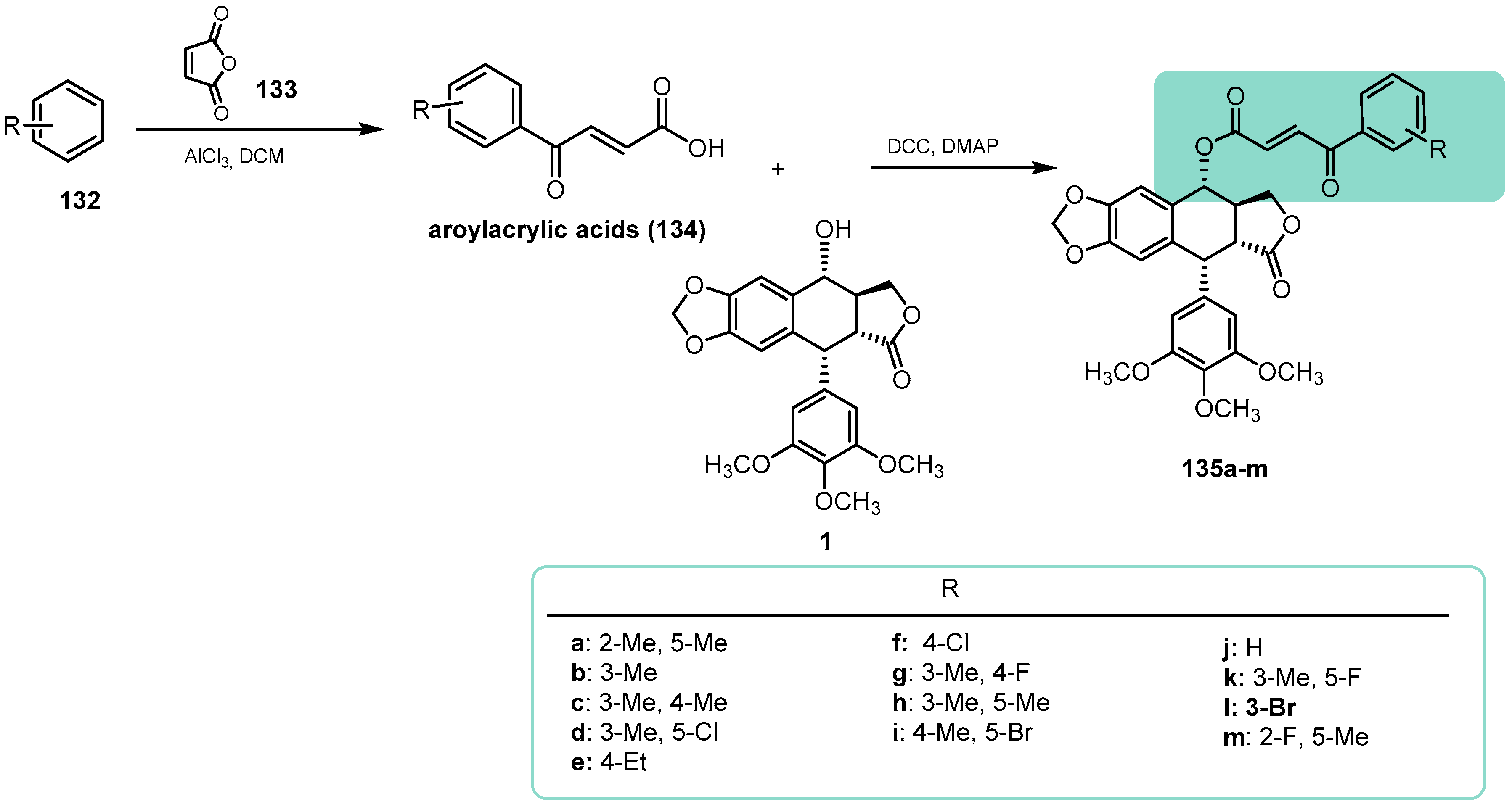
  169. Sun, W.; Sun, F.; Meng, J.; Cao, X.; Zhao, S.; Wang, C.; Li, L.; Pei, J. Design, semi-synthesis and bioactivity evaluation of novel podophyllotoxin derivatives as potent anti-tumor agents. Bioorg. Chem. 2022, 126, 105906. [Google Scholar] [CrossRef] [PubMed]
  170. Salehi, B.; Quispe, C.; Chamkhi, I.; El Omari, N.; Balahbib, A.; Sharifi-Rad, J.; Bouyahya, A.; Akram, M.; Iqbal, M.; Docea, A.O.; et al. Pharmacological Properties of Chalcones: A Review of Preclinical Including Molecular Mechanisms and Clinical Evidence. Front. Pharmacol. 2021, 11, 592654. [Google Scholar] [CrossRef] [PubMed]
  171. Zhang, M.; Lu, X.; Zhang, H.-J.; Li, N.; Xiao, Y.; Zhu, H.-L.; Ye, Y.-H. Synthesis, structure, and biological assay of cinnamic amides as potential EGFR kinase inhibitors. Med. Chem. Res. 2013, 22, 986–994. [Google Scholar] [CrossRef]
  172. Plazuk, D.; Wieczorek, A.; Błauz, A.; Rychlik, B. Synthesis and biological activities of ferrocenyl derivatives of paclitaxel. Medchemcomm 2012, 3, 498–501. [Google Scholar] [CrossRef]
  173. Wieczorek, A.; Blauz, A.; Makal, A.; Rychlik, B.; Plazuk, D. Synthesis and evaluation of biological properties of ferrocenyl—Podophyllotoxin conjugates. Dalton Trans. 2017, 47, 10847–10858. [Google Scholar] [CrossRef] [PubMed]
  174. Wieczorek, A.; Błauż, A.; Żal, A.; Arabshahi, H.J.; Reynisson, J.; Hartinger, C.G.; Rychlik, B.; Plażuk, D. Ferrocenyl Paclitaxel and Docetaxel Derivatives: Impact of an Organometallic Moiety on the Mode of Action of Taxanes. Chem. A Eur. J. 2016, 22, 11413–11421. [Google Scholar] [CrossRef] [PubMed]
  175. Wei, J.; Chen, J.; Ju, P.; Ma, L.; Chen, L.; Ma, W.; Zheng, T. Synthesis and Biological Evaluation of 4 β -N-Acetylamino Substituted Podophyllotoxin Derivatives as Novel Anticancer Agents. Agents Front. Chem. 2019, 7, 253. [Google Scholar] [CrossRef] [PubMed]
  176. Zhao, Y.; Li, D.; Wei, M.; Du, R.; Yan, Z. The ester derivatives obtained by C-ring modification of podophyllotoxin induce apoptosis and inhibited proliferation in PC-3M cells via down-regulation of PI3K/Akt signaling pathway. Bioorg. Med. Chem. Lett. 2021, 46, 128174. [Google Scholar] [CrossRef]
  177. Bader, A.; Bkhaitan, M.M.; Abdalla, A.N.; Abdallah, Q.M.A.; Ali, H.I.; Sabbah, D.A.; Albadawi, G.; Abushaikha, G.M. Design and Synthesis of 4-O-Podophyllotoxin Sulfamate Derivatives as Potential Cytotoxic Agents. Evid.-Based Complement. Altern. Med. 2021, 2021, 6672807. [Google Scholar] [CrossRef]
  178. Yang, Z.; Zhou, Z.; Luo, X.; Luo, X.; Luo, H.; Luo, L.; Yang, W. Desing and Synthesis of Novel Podophyllotoxins Hybrids and the Effects of Different functional Groups on Cytotoxicity. Molecules 2022, 27, 220. [Google Scholar] [CrossRef]
  179. Ghosh, A.K.; Brindisi, M. Organic Carbamates in Drug Design and Medicinal Chemistry. J. Med. Chem. 2015, 58, 2895–2940. [Google Scholar] [CrossRef]
  180. Matošević, A.; Bosak, A. Carbamate group as structural motif in drugs: A review of carbamate derivatives used as therapeutic agents. Arh. Hig. Rada Toksikol. 2020, 71, 285–299. [Google Scholar] [CrossRef]
  181. Xu, X.; Guan, X.; Feng, S.; Ma, Y.; Chen, S.; Hui, L. Chemistry Letters One-pot synthesis and biological evaluation of N-(aminosulfonyl)-4-podophyllotoxin carbamates as potential anticancer agents. Bioorg. Med. Chem. Lett. 2017, 27, 2890–2894. [Google Scholar] [CrossRef]
  182. Tian, D.; Chen, H.; Luo, G.; Liang, C. Synthesis and cytotoxicity of heterocyclic amine derivatives of podophyllotoxin. Chem. Nat. Compd. 2020, 56, 858–862. [Google Scholar] [CrossRef]
  183. Hu, C.; Zhu, X.; Wang, G.; Wu, X.; Han, H.; Lu, G.; Qi, J.; Pang, Y.; Yang, R.; Wang, X.; et al. Design, synthesis and anti-cancer evaluation of novel podophyllotoxin derivatives as potent tubulin-targeting agents. Med. Chem. Res. 2018, 27, 351–365. [Google Scholar] [CrossRef]
  184. Xi, W.; Sun, H.; Bastow, K.F.; Xiao, Z.; Lee, K.H. Identification of Novel 4′-O-Demethyl-epipodophyllotoxin Derivatives as Antitumor Agents Targeting Topoisomerase II. Molecules 2022, 27, 5029. [Google Scholar] [CrossRef] [PubMed]
  185. Wang, Y.; Sun, H.; Xiao, Z.; Zhang, G.; Zhang, D.; Bao, X.; Li, F.; Wu, S. DNA damage and apoptosis induced by a potent orally podophyllotoxin derivative in breast cancer. Cell Commun. Signal. 2018, 16, 52. [Google Scholar] [CrossRef]
  186. Castro, M.A.; García, P.A.; Hernández, Á.P.; Díez, D. An overview on heterocyclic podophyllotoxin derivatives. Targets Heterocycl. Syst. 2015, 19, 28–61. [Google Scholar]
  187. Hernández, Á.P.; Díez, P.; García, P.A.; Miguel Del Corral, J.M.; Pérez-Andrés, M.; Díez, D.; San Feliciano, A.; Fuentes, M.; Castro, M.Á. New Hybrids Derived from Podophyllic Aldehyde and Diterpenylhydroquinones with Selectivity toward Osteosarcoma Cells. ACS Med. Chem. Lett. 2018, 9, 328–333. [Google Scholar] [CrossRef] [PubMed]
  188. Hernández, Á.P.; Díez, P.; García, P.A.; Pérez-Andrés, M.; Ortega, P.; Jambrina, P.G.; Díez, D.; Castro, M.Á.; Fuentes, M. A Novel Cytotoxic Conjugate Derived from the Natural Product Podophyllotoxin as a Direct-Target Protein Dual Inhibitor. Molecules 2020, 25, 4258. [Google Scholar] [CrossRef] [PubMed]
  189. Munday, R. Autoxidation of naphthohydroquinones: Effects on metals, chelating agents, and superoxide dismutase. Free Radic. Biol. Med. 1997, 22, 689–695. [Google Scholar] [CrossRef]
  190. Pedroza, D.A.; De Leon, F.; Valera-Ramírez, A.; Lema, C.; Aguilera, R.J.; Mito, S. The cytotoxic effec of 2-acylated-1,4-naphthohydroquinones on leukemia/lymphoma cells. Bioorg. Med. Chem. 2014, 22, 842–847. [Google Scholar] [CrossRef]
  191. García, P.A.; Hernández, Á.P.; San Feliciano, A.; Castro, M.A. Bioactive prenyl- and terpenyl-quinones/Hydroquinones of marine origin. Mar. Drugs 2018, 16, 292. [Google Scholar] [CrossRef]
  192. Hernández, Á.P.; Chamorro, P.; Rodríguez, M.L.; Miguel del Corral, J.M.; García, P.A.; Francesch, A.; San Feliciano, A.; Castro, M.Á. New antineoplastic naphthohydroquinones attached to labdane and rearranged diterpene skeletons. Molecules 2021, 26, 474. [Google Scholar] [CrossRef]
  193. Roa-Linares, V.C.; Miranda-Brand, Y.; Tangarife-Castaño, V.; Ochoa, R.; García, P.A.; Castro, M.Á.; Betancur-Galvis, L.; San Feliciano, A. Anti-herpetic, anti-dengue and antineoplastic activities of simple and heterocycle-fused derivatives of terpenyl-1,4-naphthoquinone and 1,4-anthraquinone. Molecules 2019, 24, 1279. [Google Scholar] [CrossRef]
  194. Hernández, Á.-P.; Díez, P.; García, P.A.; Pérez-Andrés, M.; Veselinova, A.; Jambrina, P.G.; San Feliciano, A.; Díez, D.; Fuentes, M.; Castro, M.Á. Improving Properties of Podophyllic Aldehyde-Derived Cyclolignans: Design, Synthesis and Evaluation of Novel Lignohydroquinones, Dual-Selective Hybrids against Colorectal Cancer Cells. Pharmaceutics 2023, 15, 886. [Google Scholar] [CrossRef] [PubMed]
  195. Nerella, S.; Kankala, S.; Paidakula, S.; Gavaji, B. Synthesis of D-ring modified acid hydrazide derivatives of podophyllotoxin and their anticancer studies as Tubulin inhibiting agents. Bioorg. Chem. 2020, 94, 103384. [Google Scholar] [CrossRef] [PubMed]
  196. Popiołek, Ł. Updated Information on Antimicrobial Activity of Hydrazide–Hydrazones. Int. J. Mol. Sci. 2021, 22, 9389. [Google Scholar] [CrossRef] [PubMed]
  197. Mali, S.N.; Thorat, B.R.; Gupta, D.R.; Pandey, A. Mini-Review of the Importance of Hydrazides and Their Derivatives—Synthesis and Biological Activity. Eng. Proc. 2021, 11, 21. [Google Scholar]
  198. Ganaie, B.; Banday, J.A.; Baht, B.A.; Ara, T. Synthesis and In Vitro Anticancer Activity of Triazolyl Analogs of Podophyllotoxin, a Natural Occurring Lignin. Russ. J. Org. Chem. 2021, 57, 2039–2047. [Google Scholar] [CrossRef]
  199. Miguel del Corral, J.M.; Gordaliza, M.; Castro, M.A.; López-Vázquez, M.L.; García-Grávalos, M.D.; Broughton, H.B.; San Feliciano, A. Bioactive isoxazoline and oxime derivatives from 7-ketolignans. Tetrahedron 1997, 53, 6555–6564. [Google Scholar] [CrossRef]
  200. Xiang, R.; Guan, X.; Hui, L.; Jin, Y.; Chen, S. Investigation of the anti-angiogenesis effects induced by deoxypodophyllotoxin-5-FU conjugate C069 against HUVE cells. Bioorg. Med. Chem. Lett. 2017, 27, 713–717. [Google Scholar] [CrossRef]
  201. Longley, D.B.; Harkin, D.P.; Johnston, P.G. 5-Fluorouracil: Mechanisms of action and clinical strategies. Nat. Rev. Cancer 2003, 3, 330–338. [Google Scholar] [CrossRef]
  202. Guan, X.W.; Xu, X.H.; Feng, S.L.; Tang, Z.B.; Chen, S.W.; Hui, L. Synthesis of hybrid 4-deoxypodophyllotoxin-5-fluorouracil compounds that inhibit cellular migration and induce cell cycle arrest. Bioorg. Med. Chem. Lett. 2016, 26, 1561–1566. [Google Scholar] [CrossRef]
  203. Kwon, J.; Lee, N.; Kang, A.; Ahn, I.; Choi, I.; Song, J.; Hwang, S.; Um, H.; Choi, J.; Kim, J.; et al. JNC-1043, a novel podophyllotoxin derivative, exerts anticancer drug and radiosensitizer effects in colorectal cancer cells. Molecules 2022, 27, 7008. [Google Scholar] [CrossRef] [PubMed]
  204. Shi, R.J.; Fan, H.Y.; Yu, X.H.; Tang, Y.L.; Jiang, J.; Liang, X.H. Advances of podophyllotoxin and its derivatives: Patterns and mechanisms. Biochem. Pharmacol. 2022, 200, 115039. [Google Scholar] [CrossRef] [PubMed]
  205. Charlton, J.L. Antiviral activity of lignans. J. Nat. Prod. 1998, 61, 1447–1451. [Google Scholar] [CrossRef] [PubMed]
  206. Sudo, K.; Konno, K.; Shigeta, S.; Yokota, T. Inhibitory effects of podophyllotoxin derivatives on herpes simplex virus replication. Antivir. Chem. Chemother. 1998, 9, 263–267. [Google Scholar] [CrossRef]
  207. Brand, Y.M.; Roa-Linares, V.; Santiago-Dugarte, C.; del Olmo, E.; López-Pérez, J.L.; Betancur-Galvis, L.; Gallego-Gómez, J.C.; San Feliciano, A. A new host-targeted antiviral cyclolignan (SAU-22.107) for Dengue Virus infection in cell cultures. Potential action mechanisms based on cell imaging. Virus Res. 2023, 323, 198995. [Google Scholar] [CrossRef]
  208. Hensel, A.; Bauer, R.; Heinrich, M.; Spiegler, V.; Kayser, O.; Hempel, G.; Kraft, K. Challenges at the Time of COVID-19: Opportunities and Innovations in Antivirals from Nature. Planta Med. 2020, 86, 659–664. [Google Scholar] [CrossRef]
  209. Miao, Y.; Fan, L.; Jian-Yong, L. Potential treatments for COVID-19 related cytokine storm-Beyond corticosteroids. Front. Immunol. 2020, 11, 1445. [Google Scholar] [CrossRef]
  210. Zhang, J.; Zhao, H.; Feng, Y.; Xu, X.; Yang, Y.; Zhang, P.; Lu, Z.; Zhang, T. Topoisomerase 2 inhibitor etoposide promotes interleukin-10 production in LPS-induced macrophages via upregulating transcription factor Maf and activating PI3K/Akt pathway. Int. Immunopharmacol. 2021, 101, 108264. [Google Scholar] [CrossRef]
  211. Johnson, T.S.; Terrell, C.E.; Millen, S.H.; Katz, J.D.; Hildeman, D.A.; Jordan, M.B. Etoposide Selectively Ablates Activated T Cells To Control the Immunoregulatory Disorder Hemophagocytic Lymphohistiocytosis. J. Immunol. 2014, 192, 84–91. [Google Scholar] [CrossRef]
  212. Resendez, A.; Tailor, D.; Graves, E.; Malhotra, S.V. Radiosensitization of Head and Neck Squamous Cell Carcinoma (HNSCC) by a Podophyllotoxin. ACS Med. Chem. Lett. 2019, 10, 1314–1321. [Google Scholar] [CrossRef]
Figure 1. Structures of podophyllotoxin and its semisynthetic derivatives.
Pharmaceutics 15 02728 g001
Figure 2. Novel derivatives of podophyllotoxin and etoposide.
Pharmaceutics 15 02728 g002
Figure 3. Chemical and pharmacological strategies for hybridization design.
Pharmaceutics 15 02728 g003
Figure 4. Numbering of the carbon atoms and ring nomenclature of podophyllotoxin.
Pharmaceutics 15 02728 g004
Scheme 1. General procedure for the synthesis of hybrids between podophyllotoxin and another natural product through a haloacyl intermediate.
Pharmaceutics 15 02728 sch001
Figure 5. Podophyllotoxin–natural-product hybrids 1114 obtained through a haloacyl intermediate.
Pharmaceutics 15 02728 g005
Scheme 2. General procedure for the synthesis of hybrids between podophyllotoxin and another natural product using coupling reagents.
Pharmaceutics 15 02728 sch002
Figure 6. Podophyllotoxin–all-trans retinoic acid hybrid and podophyllotoxin–β-carboline derivatives.
Pharmaceutics 15 02728 g006
Scheme 3. Synthesis of β-carboline-3-carboxilic acids from L-tryptophan.
Pharmaceutics 15 02728 sch003
Figure 7. Podophyllotoxin–aurone hybrids.
Pharmaceutics 15 02728 g007
Scheme 4. Synthesis of podophyllotoxin–bile acid hybrids.
Pharmaceutics 15 02728 sch004
Scheme 5. General synthesis of podophyllotoxin–amino acid–ligustrazine derivatives.
Pharmaceutics 15 02728 sch005
Scheme 6. General procedure for the synthesis of 1,4-disubstituted 1,2,3-triazolo derivatives of podophyllotoxin.
Pharmaceutics 15 02728 sch006
Figure 8. General structure of podophyllotoxin–epigallocatechin-3-gallate, podophyllotoxin–camptothecin, and podophyllotoxin–tetrahydrocurcumin conjugates.
Pharmaceutics 15 02728 g008
Figure 9. Structures of the dimeric podophyllotoxin derivatives.
Pharmaceutics 15 02728 g009
Scheme 7. General procedure for the synthesis of the biotinylated derivatives of podophyllotoxin.
Pharmaceutics 15 02728 sch007
Figure 10. F14512, the tetramine spermine derivative of podophyllotoxin evaluated by Oviatt and colleagues.
Pharmaceutics 15 02728 g010
Scheme 8. Synthesis of new teniposide derivatives by Cheng and colleagues.
Pharmaceutics 15 02728 sch008
Scheme 9. General procedure for the synthesis of the podophyllotoxin–glucoside derivatives. The configuration of the lactone in 50 and 52 has been corrected.
Pharmaceutics 15 02728 sch009
Scheme 10. Synthesis of acetylated and butyrylated glucoside residues.
Pharmaceutics 15 02728 sch010
Scheme 11. General synthesis of picropodophyllin glycoconjugates.
Pharmaceutics 15 02728 sch011
Scheme 12. General procedure for the synthesis of the triazolopodophyllotoxin glycoconjugates.
Pharmaceutics 15 02728 sch012
Figure 11. Compound 60, N-acyl imidazole derivative of podophyllotoxin.
Pharmaceutics 15 02728 g011
Scheme 13. General procedure for the synthesis of podophyllotoxin derivatives with nitrogen-containing heterocycles.
Pharmaceutics 15 02728 sch013
Scheme 14. Synthesis of 7β-thiazole derivatives of podophyllotoxin.
Pharmaceutics 15 02728 sch014
Scheme 15. Synthesis of isoxazoline–podophyllotoxin derivatives.
Pharmaceutics 15 02728 sch015
Scheme 16. Synthesis of podophyllotoxin and epipodophyllotoxin derivatives with substituted isoxazole fragments.
Pharmaceutics 15 02728 sch016
Scheme 17. General procedure for the synthesis of triazolo derivatives of podophyllotoxin.
Pharmaceutics 15 02728 sch017
Figure 12. General structure of 7α/β-(4-substitutedtriazol)acetyl–podophyllotoxin derivatives.
Pharmaceutics 15 02728 g012
Scheme 18. Synthesis of aminotriazol derivatives of podophyllotoxin.
Pharmaceutics 15 02728 sch018
Scheme 19. General procedure for the synthesis of new podophyllotoxin–triazole derivatives by Yin and colleagues.
Pharmaceutics 15 02728 sch019
Scheme 20. Synthesis of 7β-(1,2,4-triazol-3-ylthio)-7-deoxypodophyllotoxin by Zhao’s group.
Pharmaceutics 15 02728 sch020
Scheme 21. General procedure for the synthesis of 1,3,4-oxadiazole derivatives of podophyllotoxin by Ren et al.
Pharmaceutics 15 02728 sch021
Scheme 22. General procedure for the synthesis of podophyllotoxin 1,3,4-oxadiazole derivatives.
Pharmaceutics 15 02728 sch022
Figure 13. General structure of the podophyllotoxin-1,3,4-oxadiazole derivatives synthesized by Wang’s and Han’s groups.
Pharmaceutics 15 02728 g013
Scheme 23. General procedure for the synthesis of 2-aminopyridine derivatives of podophyllotoxin and 4′-O-demethylepipodophyllotoxin.
Pharmaceutics 15 02728 sch023
Scheme 24. Synthesis of podophyllotoxin-pyridine thiocarbamates conjugates. (A) General procedure for the synthesis of pyridine hydrazone thiocarbamates. (B) General procedure for the synthesis of the hybrids bearing pyridine hydrazone thiocarbamates.
Pharmaceutics 15 02728 sch024
Scheme 25. Synthesis of podophyllotoxin–piperazine acetate derivatives.
Pharmaceutics 15 02728 sch025
Scheme 26. General procedure for the synthesis of 7α/β-piperazine derivatives of podophyllotoxin.
Pharmaceutics 15 02728 sch026
Scheme 27. Synthesis of podophyllotoxin–piperazinyl cinnamic acid conjugates.
Pharmaceutics 15 02728 sch027
Scheme 28. General procedure for the synthesis of podophyllotoxin–benzoheterocyclic derivatives.
Pharmaceutics 15 02728 sch028
Figure 14. General structure of podophyllotoxin–benzoheterocyclic derivatives.
Pharmaceutics 15 02728 g014
Scheme 29. Synthetic procedure for 7β-aminopodophyllotoxin derivatives.
Pharmaceutics 15 02728 sch029
Scheme 30. Synthesis of 4′-O-demethylepipodophyllotoxin-SH containing benzo-five-membered heterocycles by Zhang group.
Pharmaceutics 15 02728 sch030
Scheme 31. General procedure for the synthesis of podophyllotoxin–drug derivatives.
Pharmaceutics 15 02728 sch031
Figure 15. Podophyllotoxin–commercial drug derivatives.
Pharmaceutics 15 02728 g015
Scheme 32. General synthesis of podophyllotoxin–nitric oxide-donating derivatives.
Pharmaceutics 15 02728 sch032
Figure 16. Structure of podophyllotoxin nitrosyl derivative 124.
Pharmaceutics 15 02728 g016
Scheme 33. General procedure for the synthesis of compound 128.
Pharmaceutics 15 02728 sch033
Scheme 34. General procedure for the synthesis of podophyllotoxin prodrugs 131I and II.
Pharmaceutics 15 02728 sch034
Scheme 35. General synthetic procedure for podophyllotoxin–aroylacrylic derivatives.
Pharmaceutics 15 02728 sch035
Scheme 36. Synthesis of podophyllotoxin–ferrocenyl derivatives.
Pharmaceutics 15 02728 sch036
Scheme 37. Synthesis of 7β-acetylamino-substituted podophyllotoxin derivatives.
Pharmaceutics 15 02728 sch037
Scheme 38. General procedure for the synthesis of podophyllotoxin–N-Boc amino acid derivatives.
Pharmaceutics 15 02728 sch038
Scheme 39. General procedure for the synthesis of new podophyllotoxin–sulfamate derivatives.
Pharmaceutics 15 02728 sch039
Scheme 40. Synthetic procedure for the podophyllotoxin–N-sulfonyl amidine derivatives.
Pharmaceutics 15 02728 sch040
Scheme 41. Synthetic route for the synthesis of podophyllotoxin–aminosulfonyl carbamate derivatives.
Pharmaceutics 15 02728 sch041
Scheme 42. General procedure for the synthesis of heterocyclic and benzoheterocyclic amino derivatives of podophyllotoxin.
Pharmaceutics 15 02728 sch042
Scheme 43. Synthesis of podophyllotoxin–phenoxyacetyl derivatives.
Pharmaceutics 15 02728 sch043
Scheme 44. Synthetic procedure for novel 4′-O-demethylepipodophyllotoxin derivatives obtained by Xi and colleagues.
Pharmaceutics 15 02728 sch044
Figure 17. Structure of compound 156.
Pharmaceutics 15 02728 g017
Scheme 45. Schematic route for the synthesis of the lignohydroquinones.
Pharmaceutics 15 02728 sch045
Scheme 46. General procedure for the synthesis of 7β-9-biscyclolignans.
Pharmaceutics 15 02728 sch046
Scheme 47. General procedure for the synthesis of podophyllotoxin–hydrazine derivatives.
Pharmaceutics 15 02728 sch047
Scheme 48. Synthetic procedure for C9′ triazolyl analogues of podophyllotoxin.
Pharmaceutics 15 02728 sch048
Scheme 49. Synthesis of 7β-(4-nitrobenzyl)piperazine-4′-O-nitrophenoxyacetyl derivatives of podophyllotoxin.
Pharmaceutics 15 02728 sch049
Figure 18. Structure of compound 175.
Pharmaceutics 15 02728 g018
Scheme 50. General procedure for the obtention of compound 178.
Pharmaceutics 15 02728 sch050
Disclaimer/Publisher’s Note: The statements, opinions and data contained in all publications are solely those of the individual author(s) and contributor(s) and not of MDPI and/or the editor(s). MDPI and/or the editor(s) disclaim responsibility for any injury to people or property resulting from any ideas, methods, instructions or products referred to in the content.

