
Strengthening the Key Features of Volumizing Fillers: Projection Capacity and Long-Term Persistence

Abstract
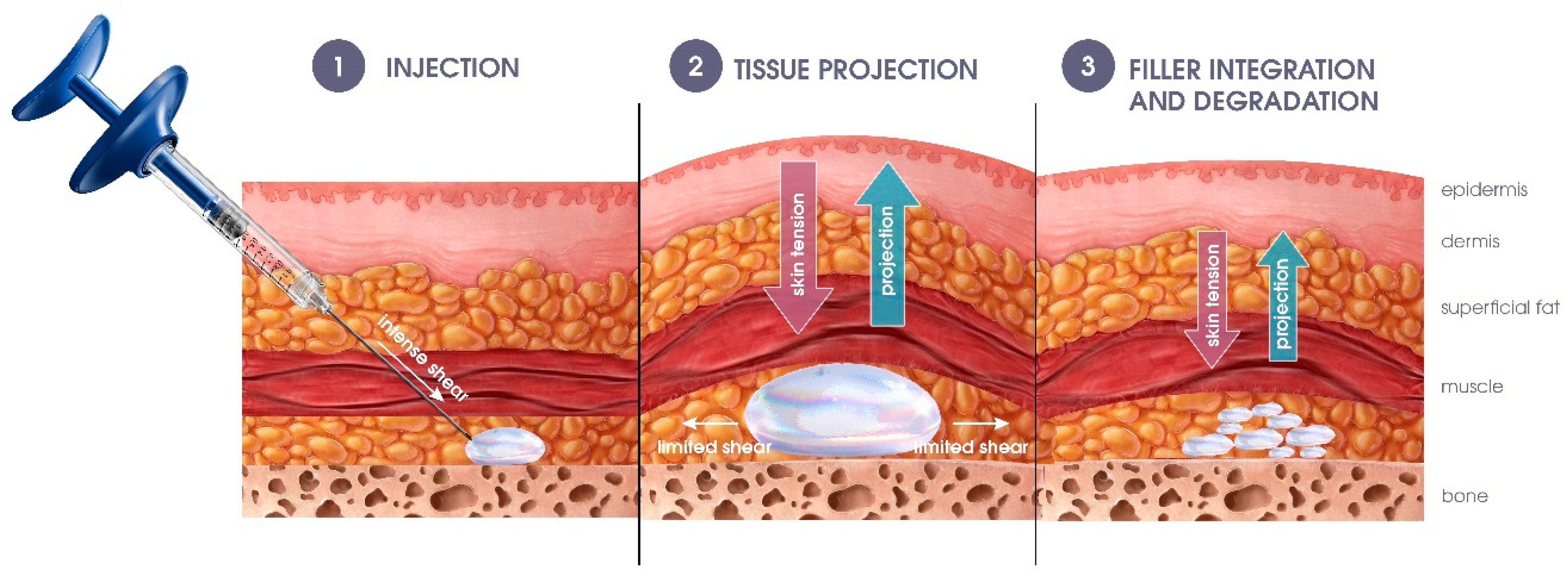
:1. Introduction
2. Materials and Methods
2.1. List of Investigated Fillers
2.2. Determination of the Dynamic Elastic Modulus in Compression Mode
2.3. Projection Index Measurement
2.4. Persistence of Volumizers Assessed by Enzymatic Degradation
2.5. Data Presentation
3. Results
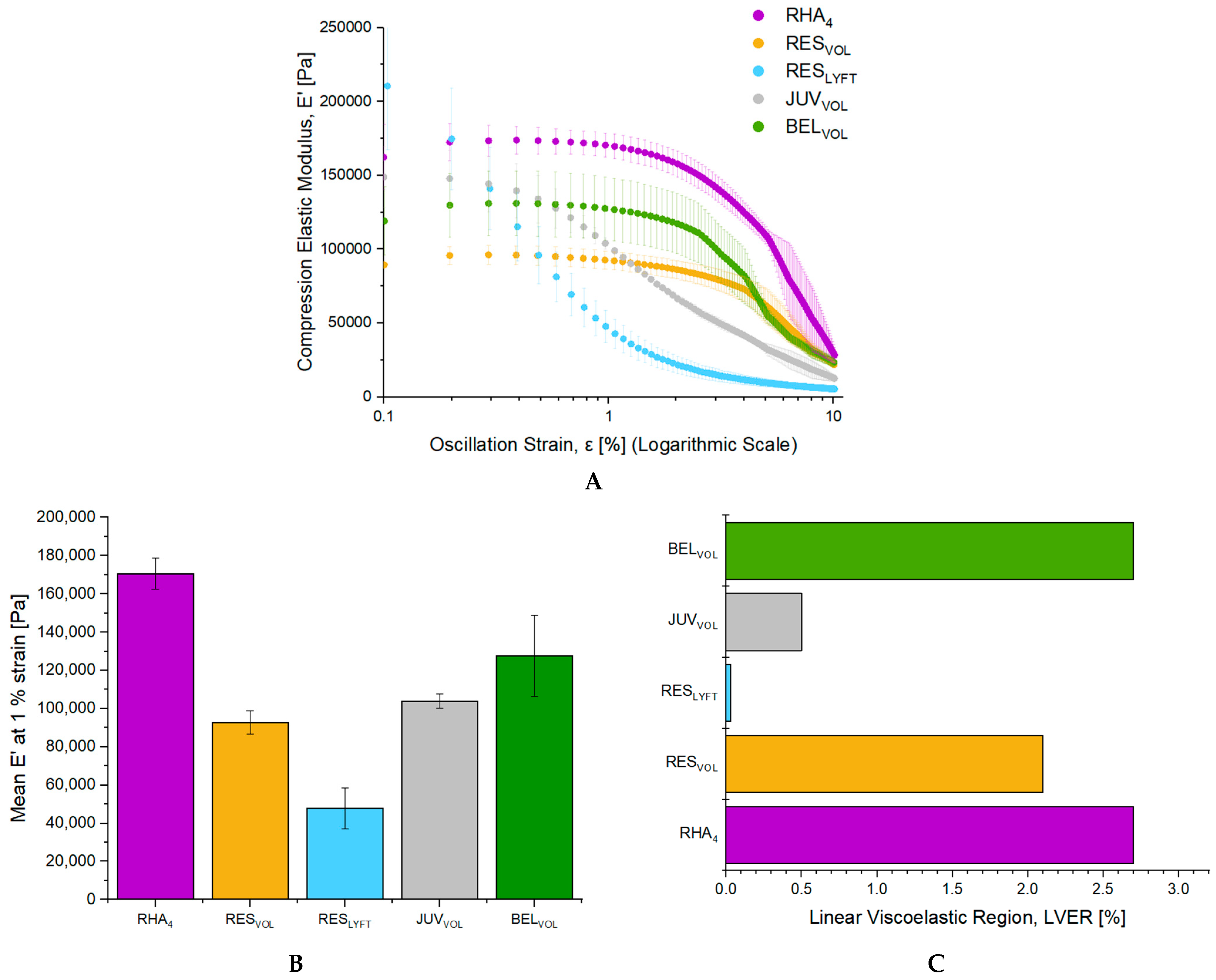
3.1. Viscoelasticity Characterization of Volumizers in Compression Mode
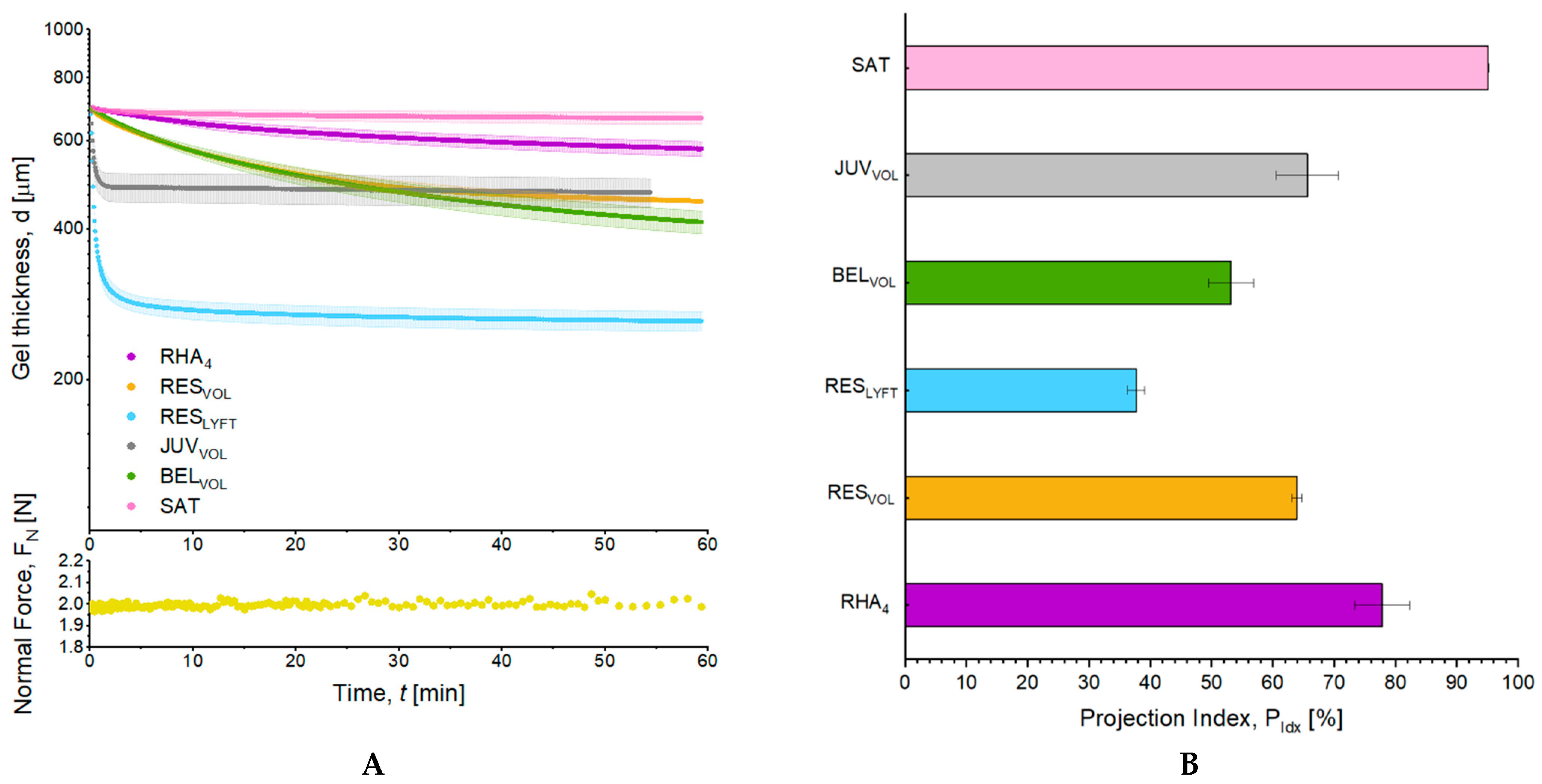
3.2. Volumizing Capacities and Projection Index Measurement
3.3. Resistance of Volumizers to Enzymatic Degradation
4. Discussion
5. Conclusions
6. Patents
Author Contributions
Funding
Institutional Review Board Statement
Informed Consent Statement
Data Availability Statement
Conflicts of Interest
References
- Sadick, N.; Sorhaindo, L. The utility of soft tissue fillers in clinical dermatology: Treatment of fine wrinkles and skin defects. Expert Rev. Med. Devices 2007, 4, 559–565. [Google Scholar] [CrossRef]
- Beasley, K.L.; Weiss, M.A.; Weiss, R.A. Hyaluronic acid fillers: A comprehensive review. Facial Plast. Surg. FPS 2009, 25, 86–94. [Google Scholar] [CrossRef]
- John, H.E.; Price, R.D. Perspectives in the selection of hyaluronic acid fillers for facial wrinkles and aging skin. Patient Prefer Adherence 2009, 3, 225–230. [Google Scholar]
- Guarise, C.; Barbera, C.; Pavan, M.; Panfilo, S.; Beninatto, R.; Galesso, D. HA-based dermal filler: Downstream process comparison, impurity quantitation by validated HPLC-MS analysis, and in vivo residence time study. J. Appl. Biomater. Funct. Mater. 2019, 17, 2280800019867075. [Google Scholar] [CrossRef]
- Faivre, J.; Pigweh, A.I.; Iehl, J.; Maffert, P.; Goekjian, P.; Bourdon, F. Crosslinking hyaluronic acid soft-tissue fillers: Current status and perspectives from an industrial point of view. Expert Rev. Med. Devices 2021, 18, 1175–1187. [Google Scholar] [CrossRef] [PubMed]
- Luo, Y.; Tan, J.; Zhou, Y.; Guo, Y.; Liao, X.; He, L.; Li, D.; Li, X.; Liu, Y. From crosslinking strategies to biomedical applications of hyaluronic acid-based hydrogels: A review. Int. J. Biol. Macromol. 2023, 231, 123308. [Google Scholar] [CrossRef] [PubMed]
- Sundaram, H.; Cassuto, D. Biophysical characteristics of hyaluronic acid soft-tissue fillers and their relevance to aesthetic applications. Plast. Reconstr. Surg. 2013, 132 (Suppl. S2), 5S–21S. [Google Scholar] [CrossRef]
- Gavard Molliard, S.; Albert, S.; Mondon, K. Key importance of compression properties in the biophysical characteristics of hyaluronic acid soft-tissue fillers. J. Mech. Behav. Biomed. Mater. 2016, 61, 290–298. [Google Scholar] [CrossRef] [PubMed]
- Gavard Molliard, S.; Bon Bétemps, J.; Hadjab, B.; Topchian, D.; Micheels, P.; Salomon, D. Key rheological properties of hyaluronic acid fillers: From tissue integration to product degradation. Plast. Aesthet Res. 2018, 5, 17. [Google Scholar] [CrossRef]
- Pierre, S.; Liew, S.; Bernardin, A. Basics of dermal filler rheology. Dermatol. Surg. 2015, 41 (Suppl. S1), S120–S126. [Google Scholar] [CrossRef] [PubMed]
- Trevidic, P.; Kaufman-Janette, J.; Weinkle, S.; Wu, R.; Dhillon, B.; Antunes, S.; Macé, E.; Maffert, P. Injection Guidelines for Treating Midface Volume Deficiency with Hyaluronic Acid Fillers: The ATP Approach (Anatomy, Techniques, Products). Aesthetic Surg. J. 2022, 42, 920–934. [Google Scholar] [CrossRef]
- Faivre, J.; Gallet, M.; Tremblais, E.; Trévidic, P.; Bourdon, F. Advanced Concepts in Rheology for the Evaluation of Hyaluronic Acid-Based Soft Tissue Fillers. Dermatol. Surg. 2021, 47, e159–e167. [Google Scholar] [CrossRef]
- Fagien, S.; Bertucci, V.; von Grote, E.; Mashburn, J.H. Rheologic and Physicochemical Properties Used to Differentiate Injectable Hyaluronic Acid Filler Products. Plast. Reconstr. Surg. 2019, 143, 707e–720e. [Google Scholar] [CrossRef] [PubMed]
- Farrell, K.; Joshi, J.; Kothapalli, C.R. Injectable uncrosslinked biomimetic hydrogels as candidate scaffolds for neural stem cell delivery. J. Biomed. Mater. Res. Part A 2017, 105, 790–805. [Google Scholar] [CrossRef] [PubMed]
- Fan, Z.; Cheng, P.; Zhang, P.; Gao, Y.; Zhao, Y.; Liu, M.; Gu, J.; Wang, Z.; Han, J. A novel multifunctional Salecan/κ-carrageenan composite hydrogel with anti-freezing properties: Advanced rheology, thermal analysis and model fitting. Int. J. Biol. Macromol. 2022, 208, 1–10. [Google Scholar] [CrossRef]
- Chaudhuri, O.; Gu, L.; Klumpers, D.; Darnell, M.; Bencherif, S.A.; Weaver, J.C.; Huebsch, N.; Lee, H.-P.; Lippens, E.; Duda, G.N.; et al. Hydrogels with tunable stress relaxation regulate stem cell fate and activity. Nat. Mater. 2016, 15, 326–334. [Google Scholar] [CrossRef]
- Brusini, R.; Iehl, J.; Clerc, E.; Gallet, M.; Bourdon, F.; Faivre, J. Comparative Preclinical Study of Lidocaine and Mepivacaine in Resilient Hyaluronic Acid Fillers. Pharmaceutics 2022, 14, 1553. [Google Scholar] [CrossRef]
- Flégeau, K.; Jing, J.; Brusini, R.; Gallet, M.; Moreno, C.; Walker, L.; Bourdon, F.; Faivre, J. Multidose Hyaluronidase Administration as an Optimal Procedure to Degrade Resilient Hyaluronic Acid Soft Tissue Fillers. Molecules 2023, 28, 1003. [Google Scholar] [CrossRef]
- Schelke, L.W.; Velthuis, P.; Kadouch, J.; Swift, A. Early ultrasound for diagnosis and treatment of vascular adverse events with hyaluronic acid fillers. J. Am. Acad. Dermatol. 2019, 88, 79–85. [Google Scholar] [CrossRef] [PubMed]
- Shumate, G.T.; Chopra, R.; Jones, D.; Messina, D.J.; Hee, C.K. In Vivo Degradation of Crosslinked Hyaluronic Acid Fillers by Exogenous Hyaluronidases. Dermatol. Surg. Off. Publ. Am. Soc. Dermatol. Surg. 2018, 44, 1075–1083. [Google Scholar] [CrossRef]
- Ryu, H.-J.; Kwak, S.-S.; Rhee, C.-H.; Yang, G.-H.; Yun, H.-Y.; Kang, W.-H. Model-Based Prediction to Evaluate Residence Time of Hyaluronic Acid Based Dermal Fillers. Pharmaceutics 2021, 13, 133. [Google Scholar] [CrossRef] [PubMed]
- Ranamukhaarachchi, S.A.; Lehnert, S.; Ranamukhaarachchi, S.L.; Sprenger, L.; Schneider, T.; Mansoor, I.; Rai, K.; Hafeli, U.O.; Stoeber, B. A micromechanical comparison of human and porcine skin before and after preservation by freezing for medical device development. Sci. Rep. 2016, 6, 32074. [Google Scholar] [CrossRef] [PubMed]
- La Gatta, A.; Salzillo, R.; Catalano, C.; D’Agostino, A.; Pirozzi, A.V.A.; De Rosa, M.; Schiraldi, C. Hyaluronan-based hydrogels as dermal fillers: The biophysical properties that translate into a “volumetric” effect. PLoS ONE 2019, 14, e0218287. [Google Scholar] [CrossRef]
- Patel, P.N.; Smith, C.K.; Patrick, C.W., Jr. Rheological and recovery properties of poly(ethylene glycol) diacrylate hydrogels and human adipose tissue. J. Biomed. Mater. Res. Part A 2005, 73, 313–319. [Google Scholar] [CrossRef]
- Sun, Z.; Gepner, B.D.; Lee, S.H.; Rigby, J.; Cottler, P.S.; Hallman, J.J.; Kerrigan, J.R. Multidirectional mechanical properties and constitutive modeling of human adipose tissue under dynamic loading. Acta Biomater. 2021, 129, 188–198. [Google Scholar] [CrossRef]
- Bertsch, P.; Andrée, L.; Besheli, N.; Leeuwenburgh, S. Colloidal hydrogels made of gelatin nanoparticles exhibit fast stress relaxation at strains relevant for cell activity. Acta Biomater. 2021, 138, 124–132. [Google Scholar] [CrossRef] [PubMed]
- Grolman, J.M.; Weinand, P.; Mooney, D.J. Extracellular matrix plasticity as a driver of cell spreading. Proc. Natl. Acad. Sci. USA 2020, 117, 25999–26007. [Google Scholar] [CrossRef] [PubMed]
- Carvalho, E.M.; Kumar, S. Lose the stress: Viscoelastic materials for cell engineering. Acta Biomater. 2023, 163, 146–157. [Google Scholar] [CrossRef]
- Wang, X.; Xi, H.; Wei, C. Connections between cohesion and properties that related to safety and effectiveness of the hyaluronic acid dermal fillers: A comparative study of the cohesive and non-cohesive gels. Ski. Res. Technol. 2023, 29, e13395. [Google Scholar] [CrossRef]
- Ferraz, R.M.; Sandkvist, U.; Lundgren, B. Degradation of Hylauronic Acid Fillers Using Hyaluronidase in an In Vivo Model. J. Drugs Dermatol. JDD 2018, 17, 548–553. [Google Scholar]
- Jons, C.K.; Grosskopf, A.K.; Baillet, J.; Yan, J.; Klich, J.H.; Saouaf, O.M.; Appel, E.A. Yield-Stress and Creep Control Depot Formation and Persistence of Injectable Hydrogels Following Subcutaneous Administration. Adv. Funct. Mater. 2022, 32, 2203402. [Google Scholar] [CrossRef]
- Wissing, T.B.; Bonito, V.; van Haaften, E.E.; van Doeselaar, M.; Brugmans, M.M.C.P.; Janssen, H.M.; Bouten, C.V.C.; Smits, A.I.P.M. Macrophage-Driven Biomaterial Degradation Depends on Scaffold Microarchitecture. Front. Bioeng. Biotechnol. 2019, 7, 87. [Google Scholar] [CrossRef] [PubMed]


| Filler | Manufacturer | Technology | HA Concentration (mg/mL) a | MOD (%) b | Shear Elastic Modulus G′ (Pa) c | Injection Forces (N) d | Batch References |
|---|---|---|---|---|---|---|---|
| Belotero® Volume (BELVOL) | Merz, Plan-les-Ouates, Switzerland | Cohesive Polydensified Matrix | 26 | 8 | 198 | 19.5 (TSK 30G × ½″) | 557104/1 |
| Restylane® Volume/Contour (RESVOL) | Galderma, Uppsala, Sweden | Optimal Balance Technology/XpresHAn | 20 | 7 | 172 | 10.3 (TSK 27G × ½″) | 17936-1 |
| Restylane® Lyft (RESLYFT) | Galderma, Uppsala, Sweden | NASHA | 20 | 1 | 855 | 21.7 (29G × ½″TW) | 15085-1 |
| Juvéderm® Voluma (JUVVOL) | Abbvie (Allergan), Pringy, France | Vycross | 20 | 6 | 312 | 8.1 (TSK 27G × ½″) | VB20A60547 |
| Teosyal® RHA 4 (RHA4) | Teoxane, Geneva, Switzerland | Preserved Network | 23 | 4 | 260 | 9.0 (TSK 27G × ½″) | TPUL-194921-A |
Disclaimer/Publisher’s Note: The statements, opinions and data contained in all publications are solely those of the individual author(s) and contributor(s) and not of MDPI and/or the editor(s). MDPI and/or the editor(s) disclaim responsibility for any injury to people or property resulting from any ideas, methods, instructions or products referred to in the content. |
© 2023 by the authors. Licensee MDPI, Basel, Switzerland. This article is an open access article distributed under the terms and conditions of the Creative Commons Attribution (CC BY) license (https://creativecommons.org/licenses/by/4.0/).
Share and Cite
Flégeau, K.; Jing, J.; Vantou, C.; Brusini, R.; Bourdon, F.; Faivre, J. Strengthening the Key Features of Volumizing Fillers: Projection Capacity and Long-Term Persistence. Pharmaceutics 2023, 15, 2585. https://doi.org/10.3390/pharmaceutics15112585
Flégeau K, Jing J, Vantou C, Brusini R, Bourdon F, Faivre J. Strengthening the Key Features of Volumizing Fillers: Projection Capacity and Long-Term Persistence. Pharmaceutics. 2023; 15(11):2585. https://doi.org/10.3390/pharmaceutics15112585
Chicago/Turabian StyleFlégeau, Killian, Jing Jing, Camille Vantou, Romain Brusini, François Bourdon, and Jimmy Faivre. 2023. "Strengthening the Key Features of Volumizing Fillers: Projection Capacity and Long-Term Persistence" Pharmaceutics 15, no. 11: 2585. https://doi.org/10.3390/pharmaceutics15112585