
Biopolymer- and Natural Fiber-Based Biomimetic Tissues to Realize Smart Cosmeceuticals and Nutraceuticals Using an Innovative Approach


 , , , , , and
Abstract
:1. Introduction
2. Materials and Methods
2.1. Materials
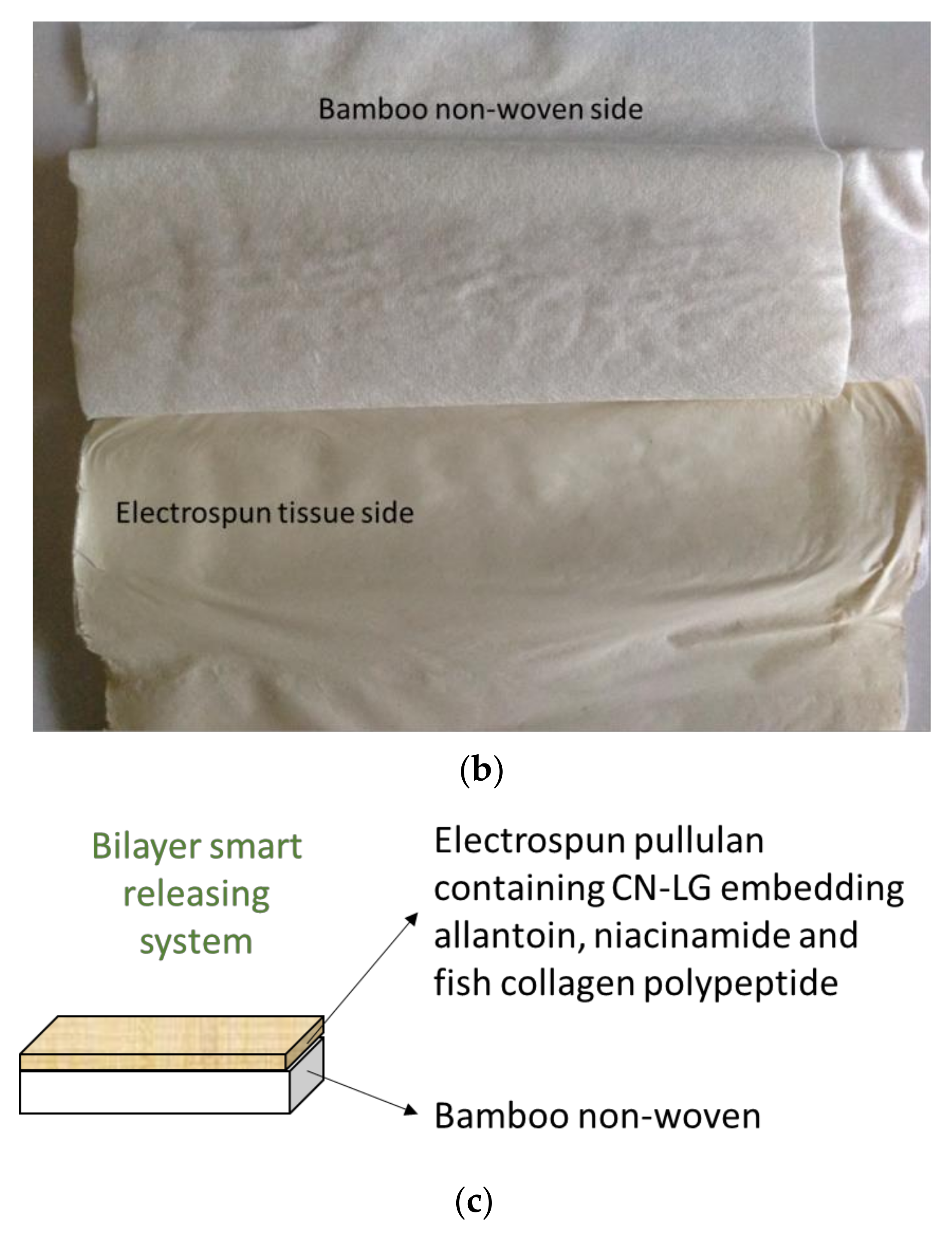
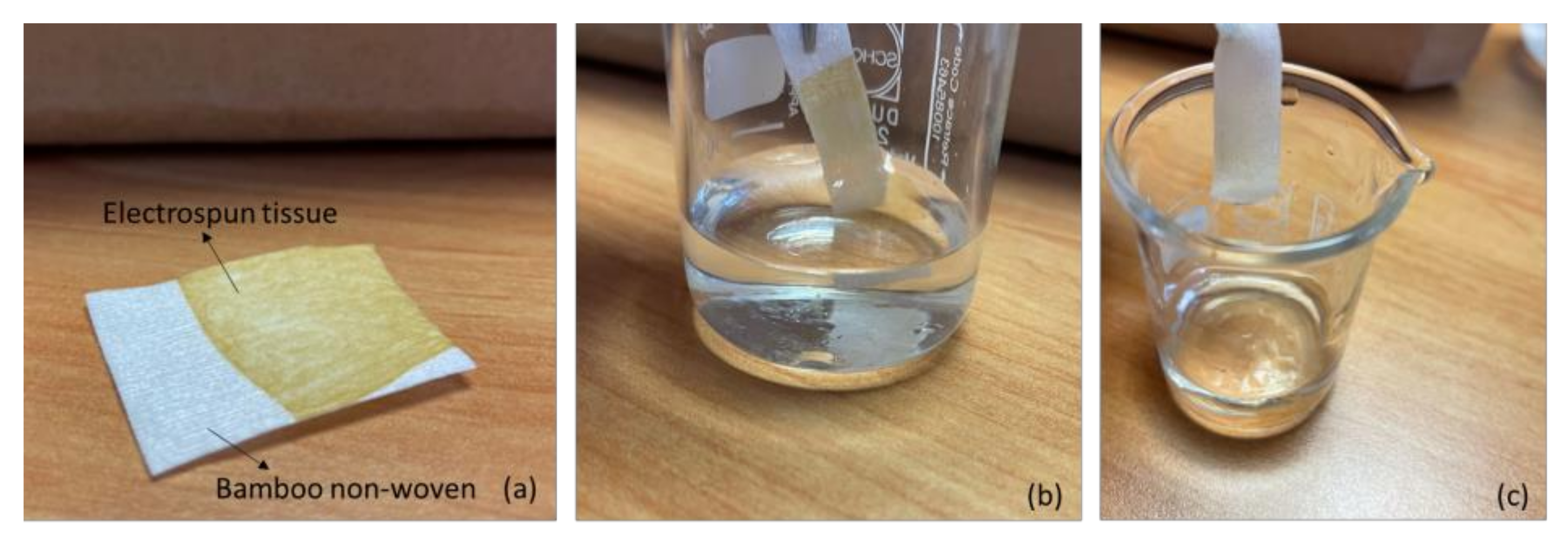
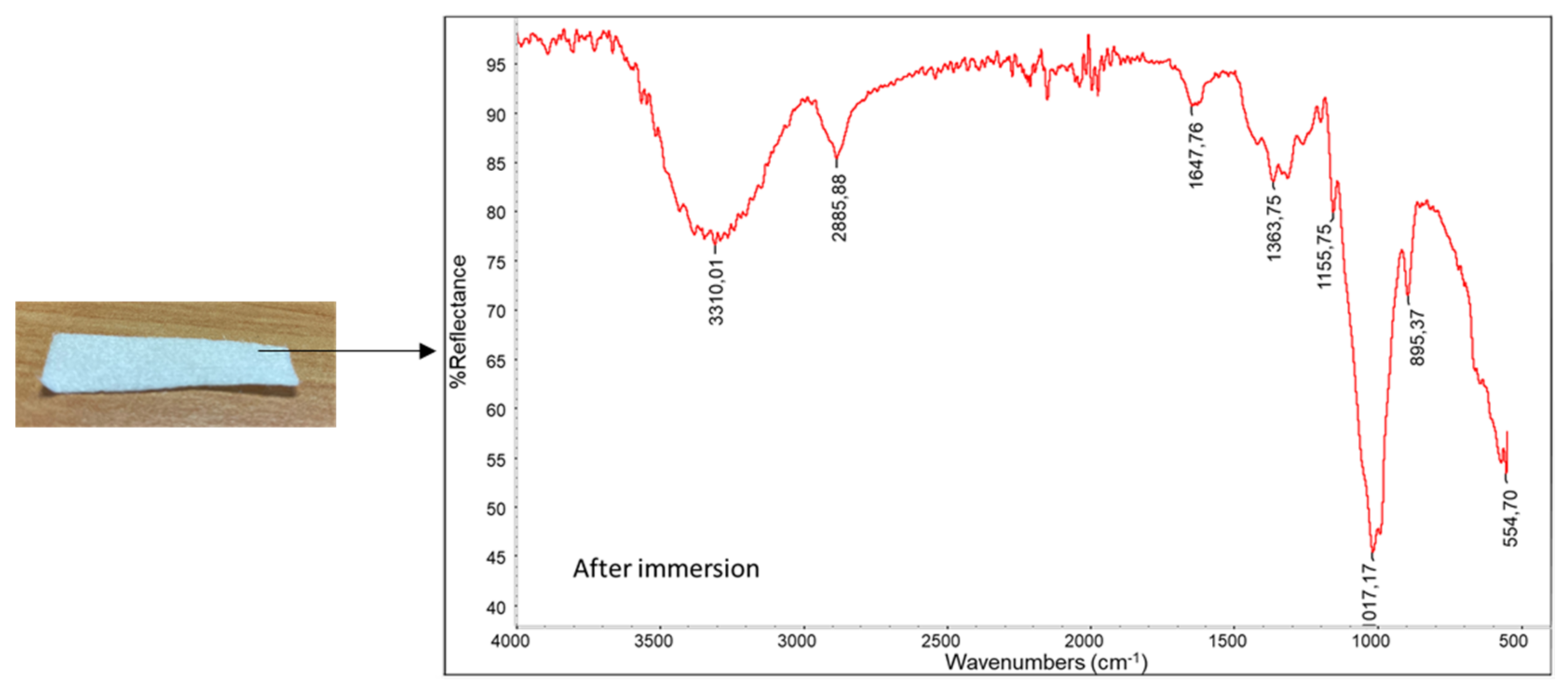
2.2. Methods
2.3. Cell Culture
2.3.1. Alamar Blue Assay
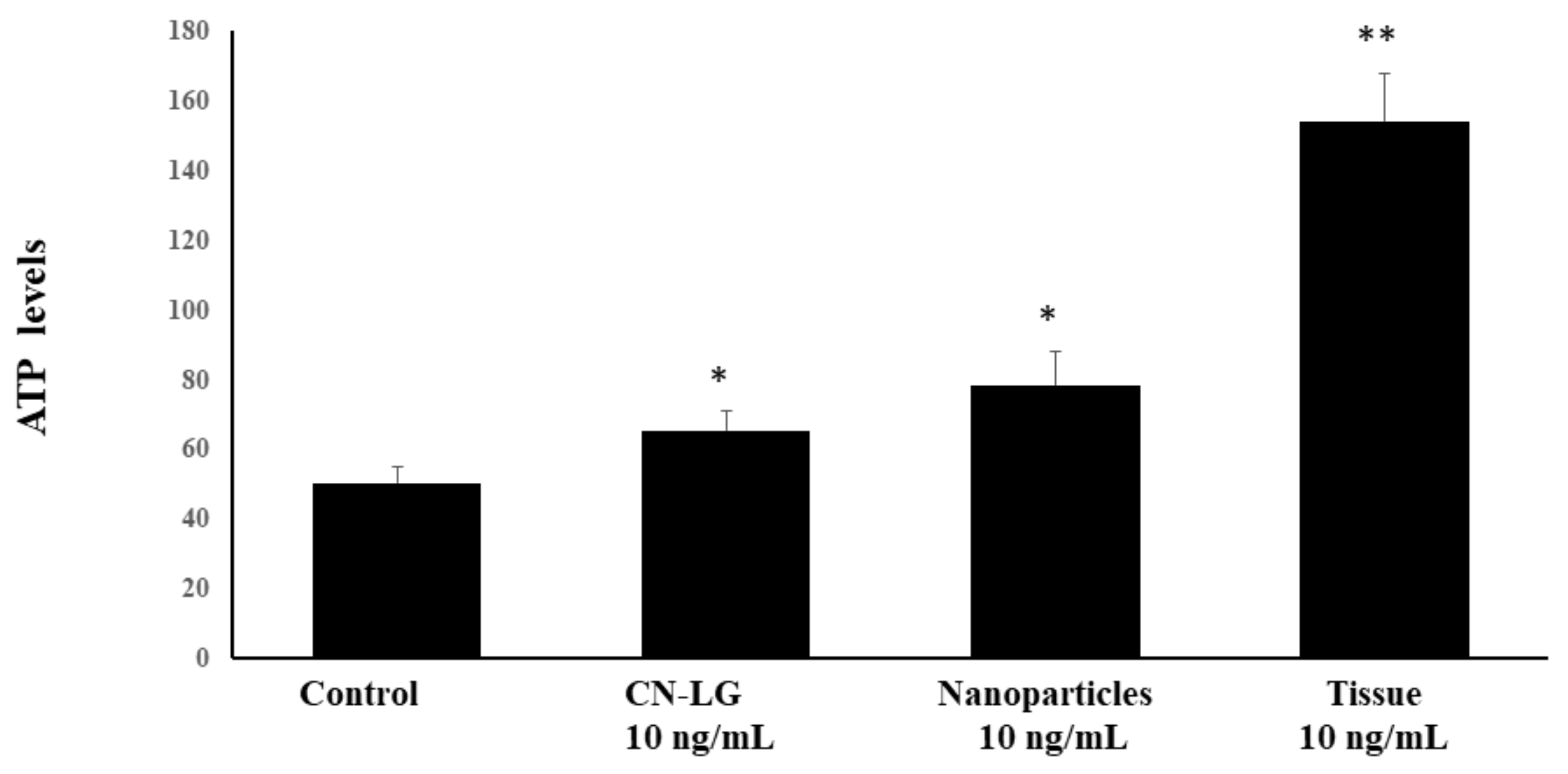
2.3.2. UV-Irradiated Keratinocytes and Intracellular ATP Level Determination
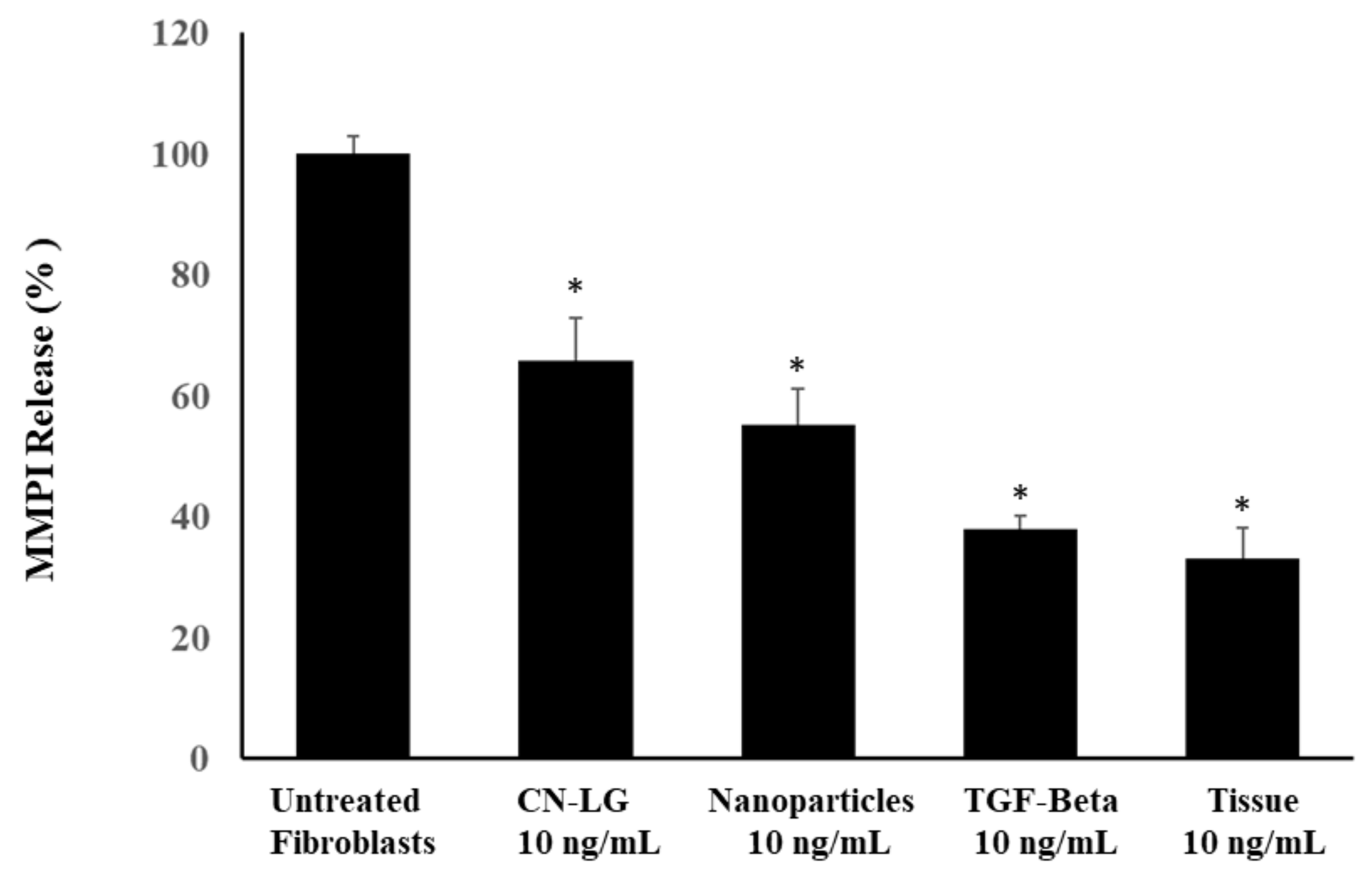
2.3.3. Metalloproteinase (MMPI) Recovery
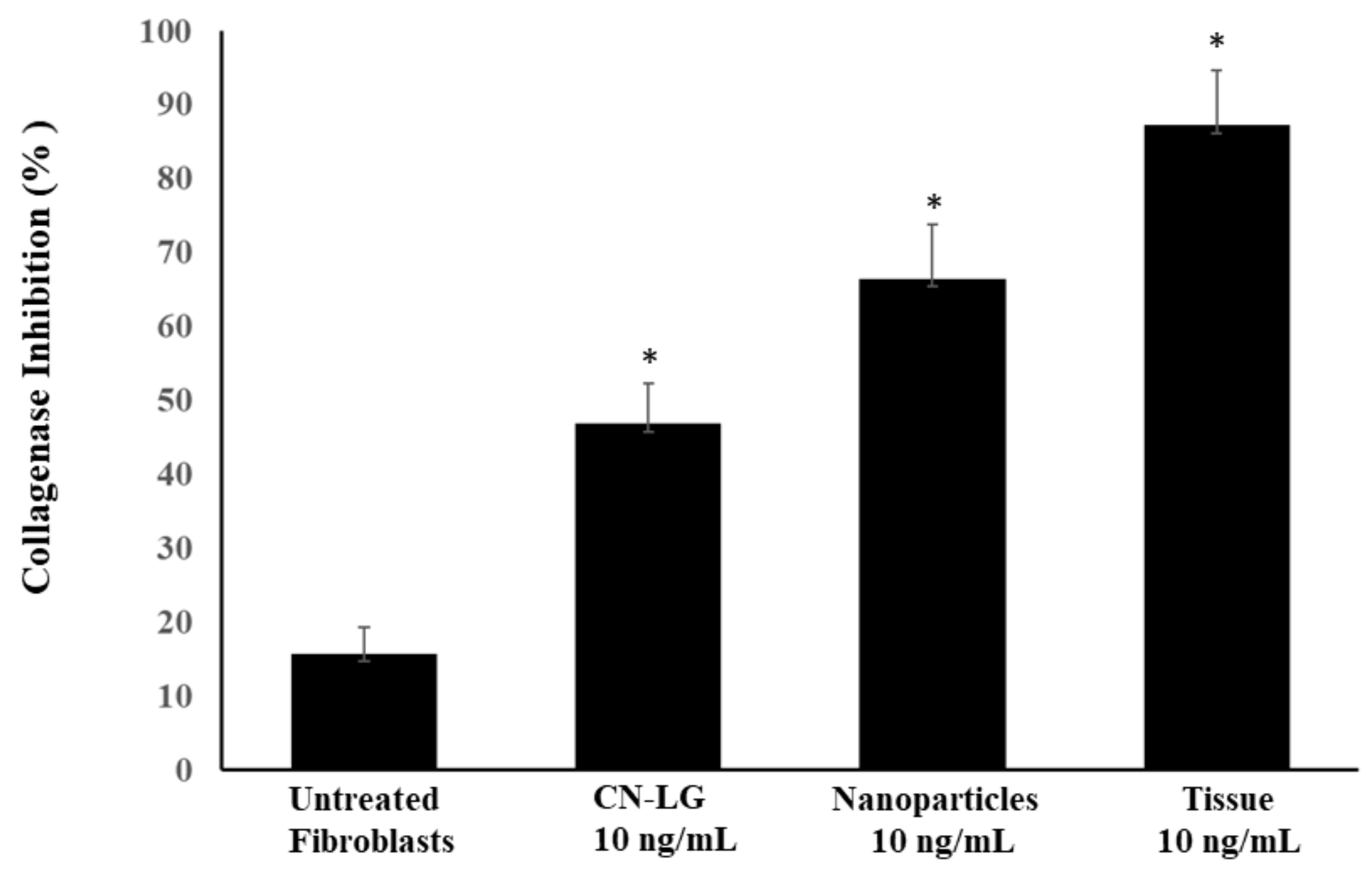
2.3.4. Collagen Synthesis
2.3.5. Statistical Analysis
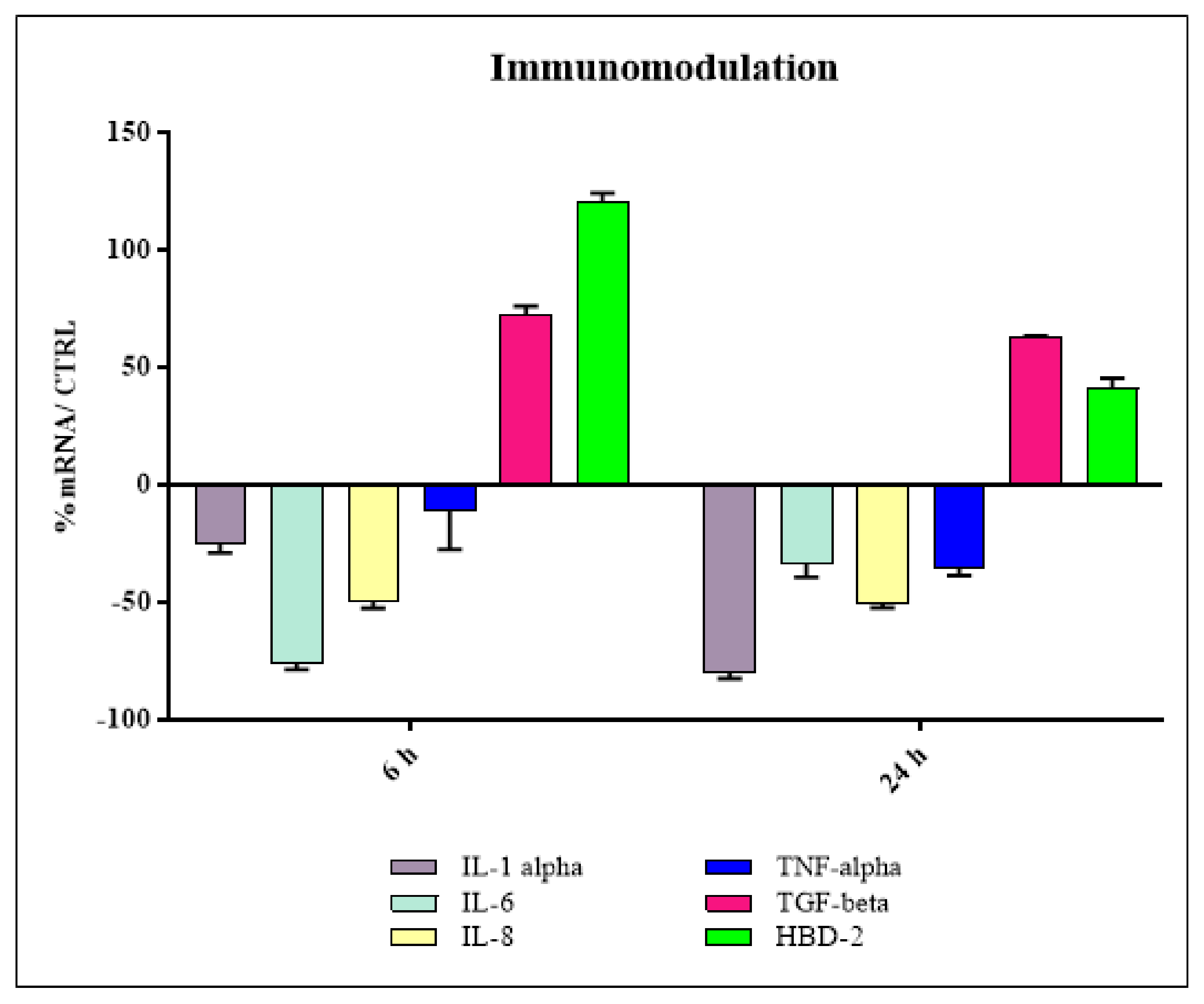
2.4. Immunomodulatory Tests
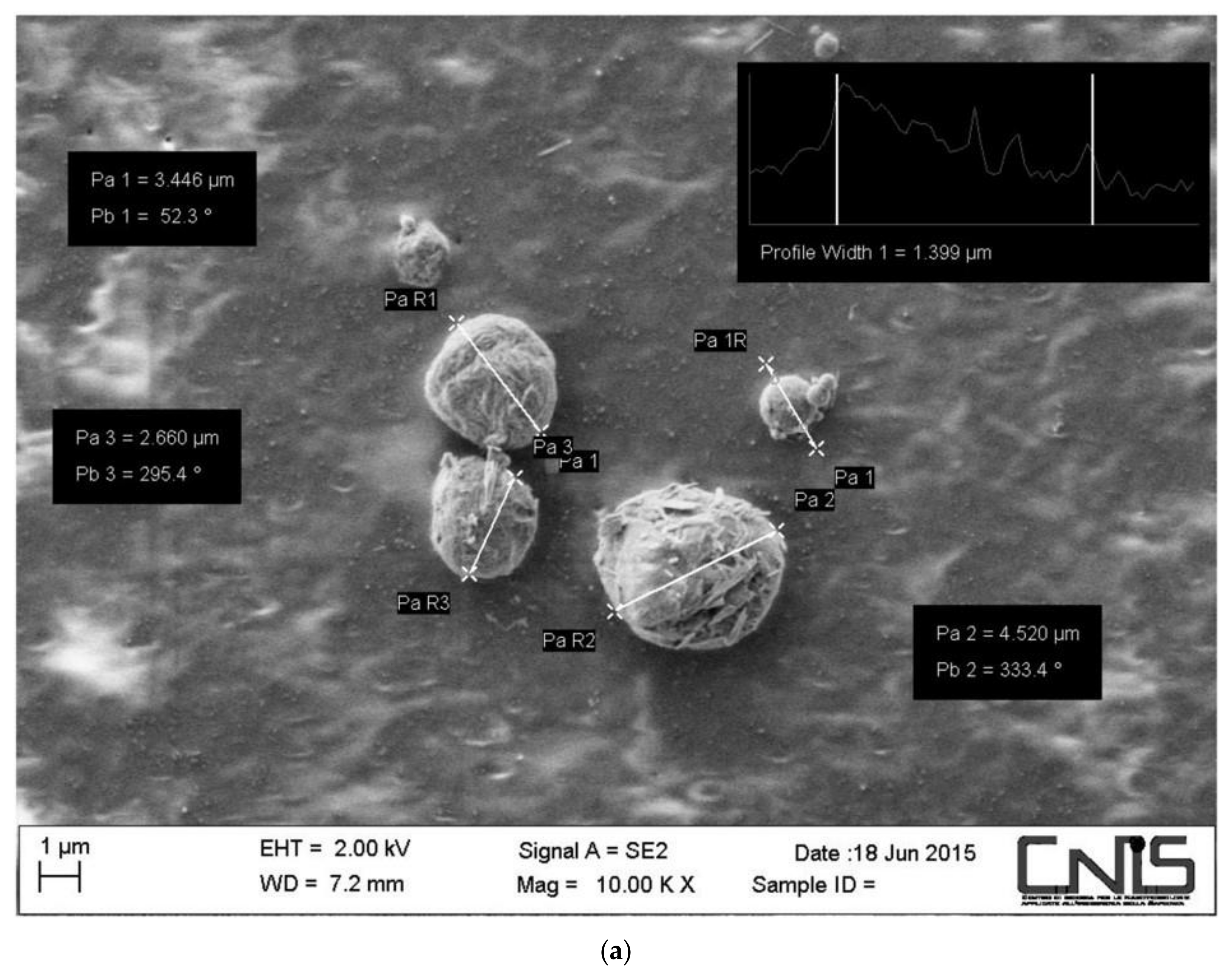
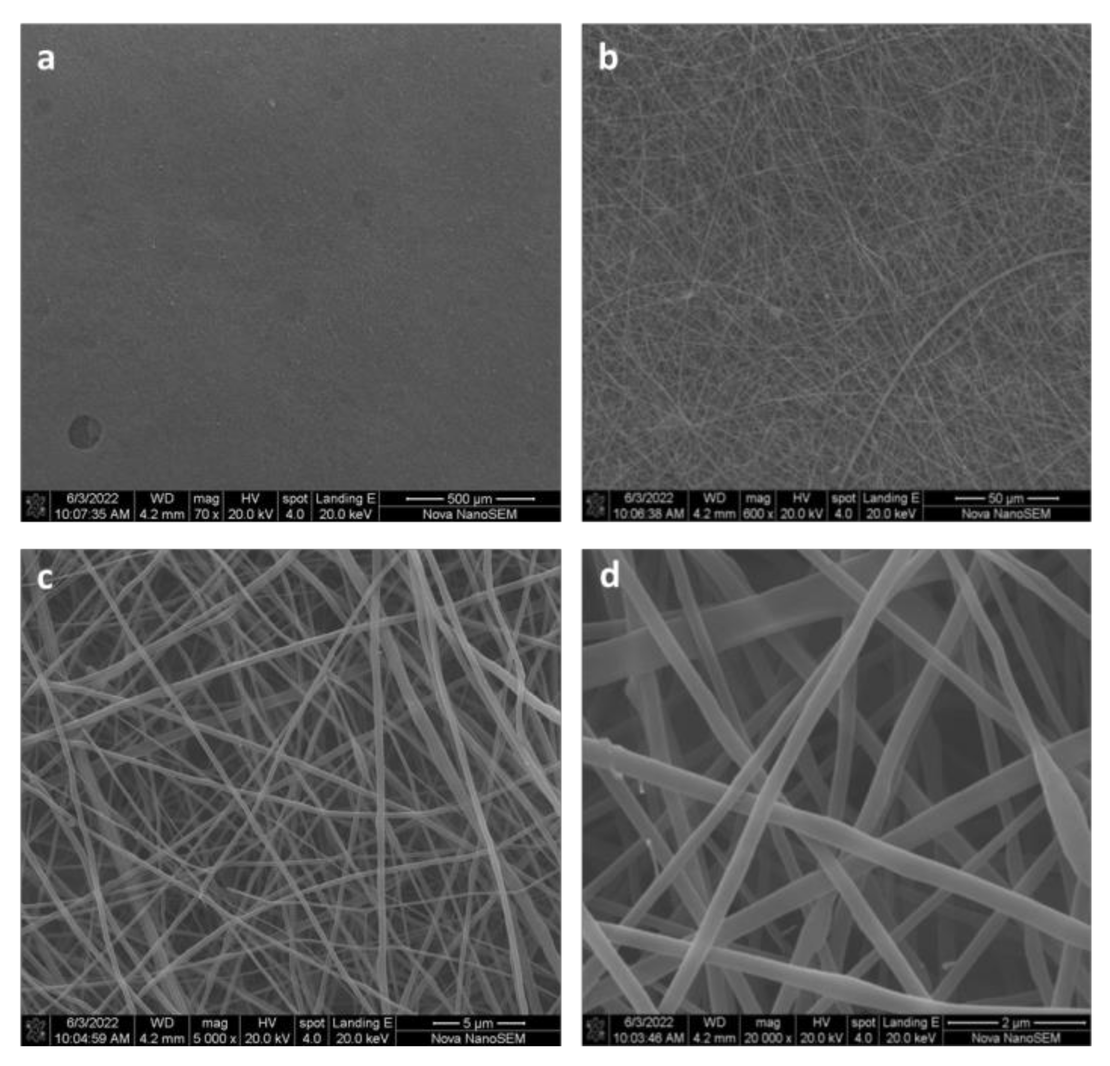
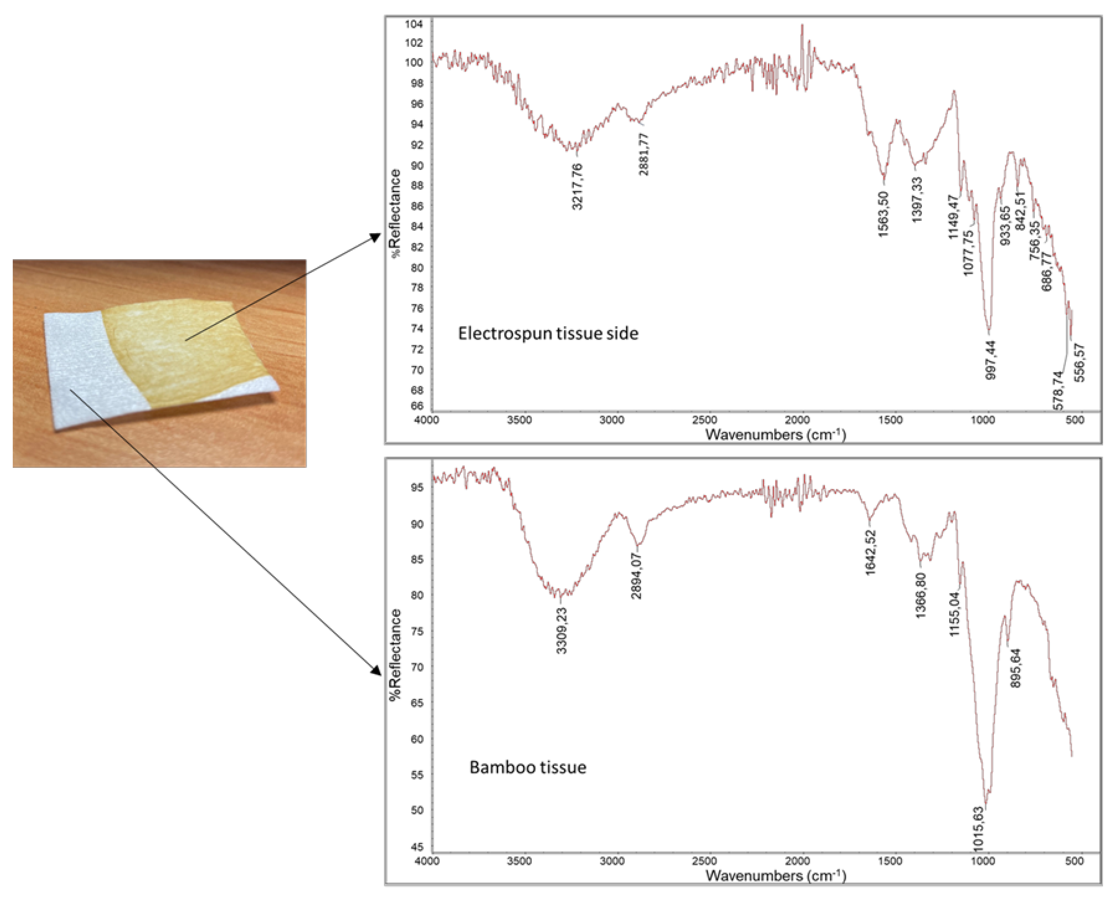
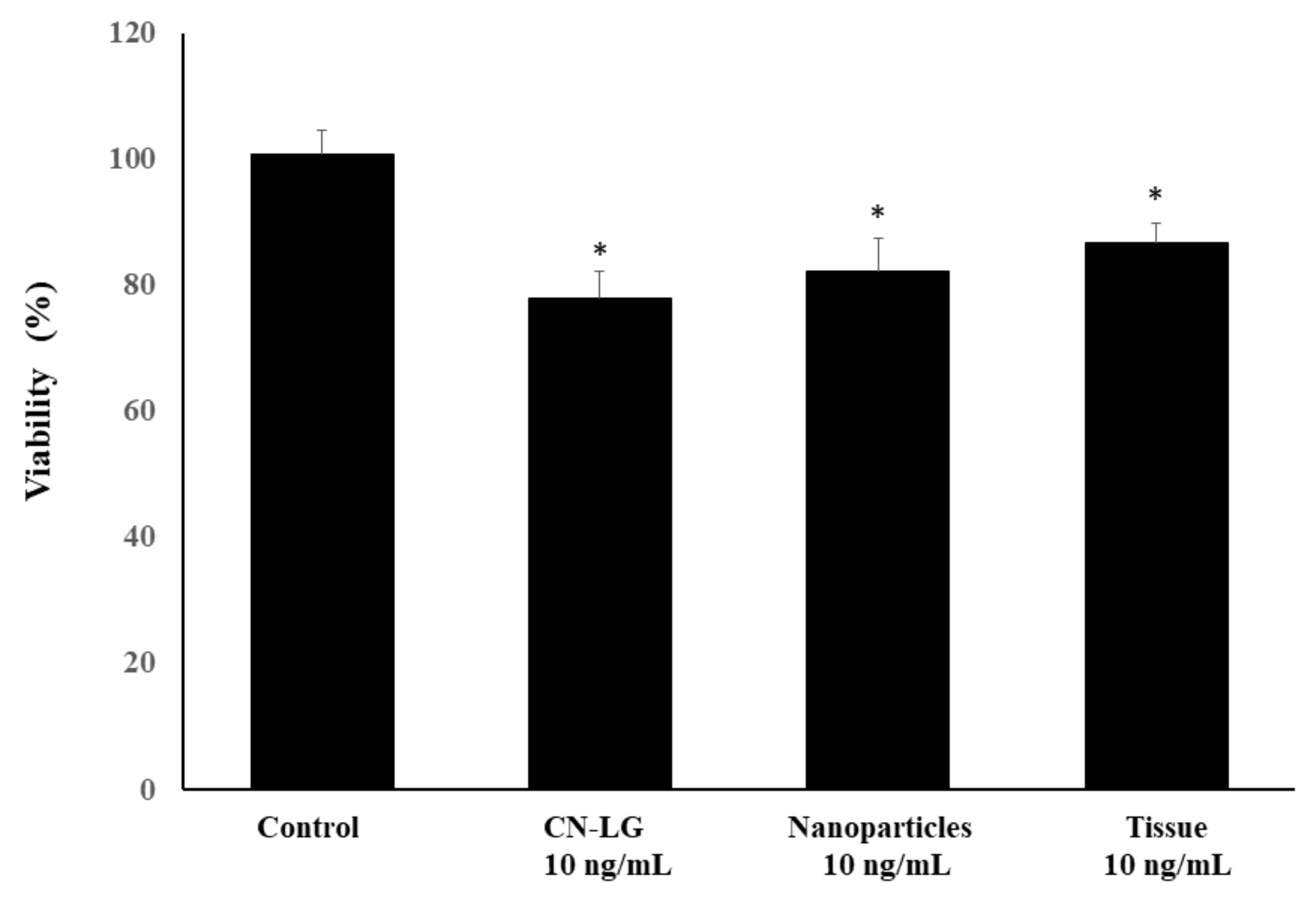
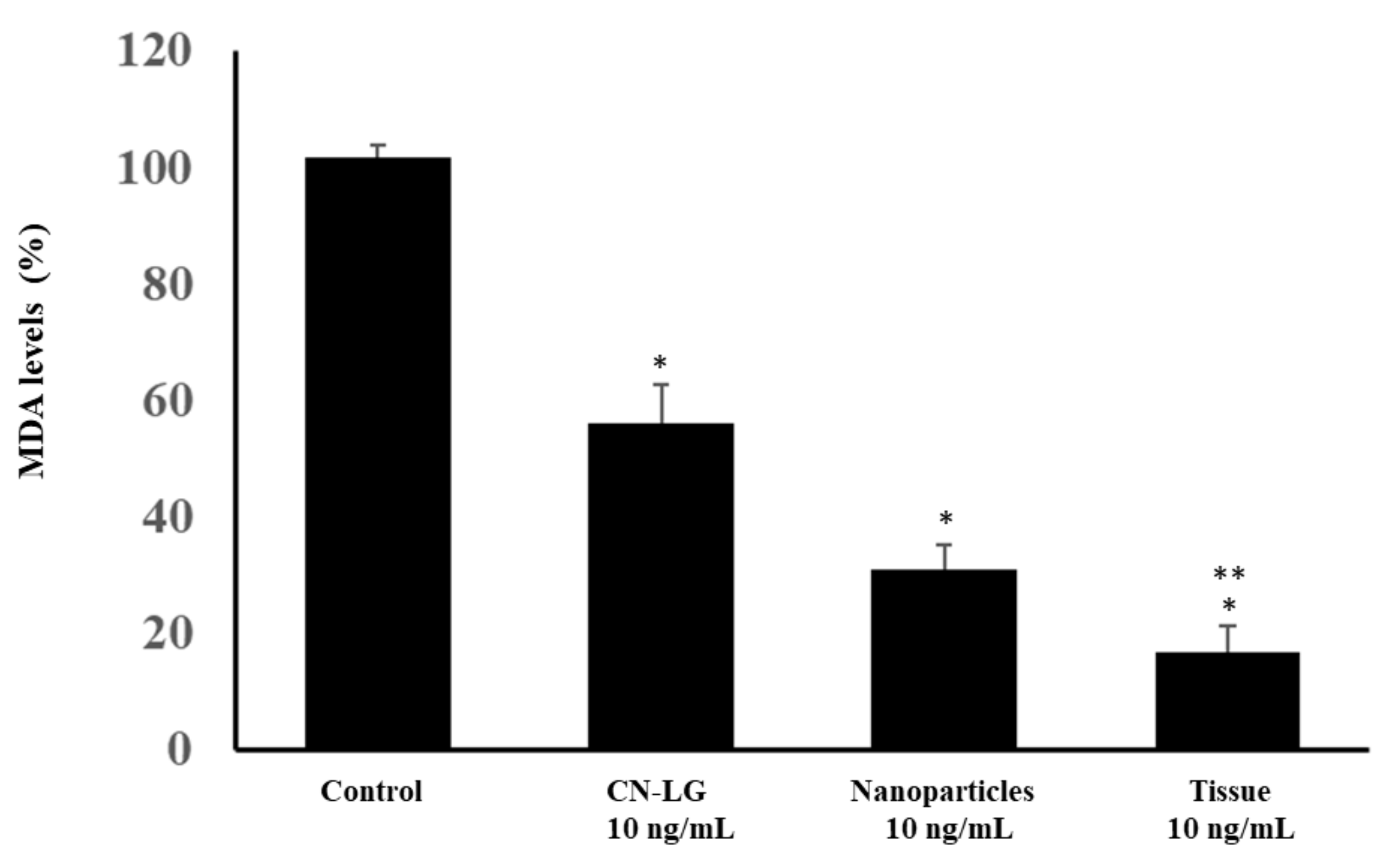
3. Results
4. Discussion
5. Conclusions
Author Contributions
Funding
Institutional Review Board Statement
Informed Consent Statement
Data Availability Statement
Conflicts of Interest
References
- John, D.A.; Babu, G.R. Lessons from the Aftermaths of Green Revolution on Food System and Health. Front. Sustain. Food Syst. Health 2021, 5, 644559. [Google Scholar] [CrossRef]
- Faria-Silva, C.; Ascenso, A.; Costa, A.M.; Marto, J.; Carvalheiro, M.; Ribeiro, H.M.; Simões, S. Feeding the skin: A new trend in food and cosmetics convergence. Trends Food Sci. Technol. 2020, 95, 21–32. [Google Scholar] [CrossRef]
- EMAF. From Linear to Circular Economy. A Global Learning Origramme. EllenMcArthurFoundation. 15 April 2020. Available online: www.ellenmacarthurfoundation.org (accessed on 25 July 2022).
- Cuc, S.; Tripa, S. Redesign and up cycling—A solution for the competitive es of small and medium-sized enterprises in the clothing Industry. Ind. Textila 2018, 69, 31–36. [Google Scholar] [CrossRef]
- FAO. The State of Food and Agriculture. Moving forward on Food Loss and Waste Reduction; Food and Agriculture Organization of United Nations: Rome, Italy, 2019. [Google Scholar]
- Bauer, F.; Nielsen, T.D.; Nilsson, L.J.; Palm, E.; Ericsson, K.; Fråne, A.; Cullen, J. Plastics and climate change—Breaking carbon lock-ins through three mitigation pathways. One Earth 2022, 5, 361–376. [Google Scholar] [CrossRef]
- Avionics, C.H.; Gorbi, S.; Regoli, F. Plastics and Microplastics in the Oceans:From Emerging Pollutants to Emerged Threat. Mar. Environ. Res. 2017, 128, 2–11. [Google Scholar]
- Hernandez, L.M.; Xu, E.G.; Larsson, H.C.E.; Tahara, R.; Mhisura, V.B.; Tufenkji, N. Plastic Teabags release billions of microplastics and Nanoparticles into tea. Environ. Sci. Technol. 2019, 53, 12300–12310. [Google Scholar] [CrossRef] [PubMed]
- Ragusa, A.; Svelato, A.; Santacroce, C.; Catalano, P.; Notarstefano, V.; Carnevali, O. First Evidence of Microplastics in Human Placenta. Environ. Int. 2021, 146, 106274. [Google Scholar] [CrossRef] [PubMed]
- Morganti, P.; Palombo, M.; Tischenko, G.; Putin, V.E.; Guarneri, F.; Cardillo, A. Chitin Hyaluronan Nanoparticles: A Multifunctional Carrier to Deliver Anti-Aging Active Ingrediens through the Skin. Cosmetics 2014, 1, 140–158. [Google Scholar] [CrossRef]
- Malevskaia, E.N.; Kirichuk, O.P.; Kuznetzov, S.I.; Dresvyanina, E.N.; Yudin, V.E.; Morganti, P. Hemocompatible chitin-chitosan composite fibers. Cosmetics 2020, 7, 28. [Google Scholar] [CrossRef]
- Morganti, P.; Morganti, G.; Memic, A.; Coltelli, M.B.; Chen, H.-D. The new renaissance of beauty and wellness through the green Economy. Latest Trends Text. Fash. Des. 2021, 4, 749–760. [Google Scholar]
- Panariello, L.; Vannozzi, A.; Morganti, P.; Coltelli, M.B.; Lazzeri, A. Biobased and eco compatible beauty films coated with chitin nanofibers, Nanolignin and vitamin E. Cosmetics 2021, 8, 27. [Google Scholar] [CrossRef]
- Wieckers, J.W. Skin Delivery: What it is and Why we Need it. In Skin Delivery Systems; Wiekers, J.W., Ed.; Allured Publishing Corporation: Carol Stream, IL, USA, 2008; pp. 1–21. [Google Scholar]
- Wieckers, J.W. The barrier function of the skin in relation to percutaneous absorption of drugs. Pharm. Weekbl. 1989, 11, 185–198. [Google Scholar] [CrossRef]
- Haun, J.B.; Hammerless, D.A. Quantifying nanoparticles adhesion mediate D by specific molecular Interactions. Langmuir 2008, 24, 8821–8832. [Google Scholar] [CrossRef]
- Desai, P.; Patlolla, R.R.; Sungh, M. Interaction of nanoparticles and cell-penetrating peptides with skin for transdermal delivery. Mol. Membr. Biol. 2010, 27, 247–259. [Google Scholar] [CrossRef] [PubMed]
- Iqbal, A.; Naqvi, S.A.R.; Sherazi, T.A.; Asif, M.; Shahzad, S.A. Thin films as an emerging platform for drug delivery. In Novel Platforms for Drug Delivery Applications; Elsevier: Amsterdam, The Netherlands, 2023. [Google Scholar] [CrossRef]
- Jacob, S.; Nair, A.B.; Boddu, S.H.S.; Gorain, B.; Sreeharsha, N.; Shah, J. An Updated Overview of the Emerging Role of Patch and Film-Based Buccal Delivery Systems. Pharmaceutics 2021, 13, 1206. [Google Scholar] [CrossRef] [PubMed]
- Luraghi, A.; Peri, F.; Moroni, L. Electrospinning for drug delivery applications: A review. J. Control. Release 2021, 334, 463–484. [Google Scholar] [CrossRef] [PubMed]
- Zulkifli, M.Z.A.; Nordin, D.; Shaari, N.; Kamarudin, S.K. Overview of Electrospinning for Tissue Engineering Applications. Polymers 2023, 15, 2418. [Google Scholar] [CrossRef]
- Anand, S.; Azimi, B.; Lucena, M.; Ricci, C.; Candito, M.; Zavagna, L.; Astolfi, L.; Coltelli, M.B.; Lazzeri, A.; Berrettini, S.; et al. Chitin nanofibrils modulate mechanical response in tympanic membrane replacements. Carbohydr. Polym. 2023, 310, 120732. [Google Scholar] [CrossRef]
- Shabunin, A.S.; Yudin, V.E.; Dobrovolskaya, I.P.; Zynovyen, E.V.; Zubov, F.V.; Ivan’kova, E.M.; Morganti, P. Composite wound dressing on chitin/chitosan nanofibers:processing and biomedical applications. Cosmetics 2019, 6, 16. [Google Scholar] [CrossRef]
- Morganti, P.; Morganti, G.; Palombo, M. Research & Innovation for sustainable products: Polysaccharides for a Smart Circular Economy at zero waste. Auctores Publ. 2021, 3, 1–37. [Google Scholar] [CrossRef]
- Sivan, M.; Madheswaran, D.; Valtera, J.; Kuzelova Kostakova, E.; Lukas, D. Alternating current electrospinning: The impacts of various high-voltage signal shapes and frequencies on the spinnability and productivity of polycaprolactone nanofibers. Mater. Des. 2022, 213, 110308. [Google Scholar] [CrossRef]
- Imadi, S.R.; Mahmood, I.; Kazi, A.G. Bamboo Fiber Processing, Properties, and Applications. In Biomass and Bioenergy; Hakeem, K., Jawaid, M., Rashid, U., Eds.; Springer: Cham, Switzerland, 2014. [Google Scholar] [CrossRef]
- Tsuzuki, T.; Kanwar, R.K.; Wang, X. The origin of the antibacterial property of bamboo. J. Text. Inst. 2012, 103, 844–849. [Google Scholar] [CrossRef]
- Teno, J.; Pardo-Figuerez, M.; Hummel, N.; Bonin, V.; Fusco, A.; Ricci, C.; Donnarumma, G.; Coltelli, M.-B.; Danti, S.; Lagaron, J.M. Preliminary Studies on an Innovative Bioactive Skin Soluble Beauty Mask Made by Combining Electrospinning and Dry Powder Impregnation. Cosmetics 2020, 7, 96. [Google Scholar] [CrossRef]
- Pandey, R.; Poljsak, B.; Godic, A.; Dahmane, R. Skin Photoaging and the role of antioxidants in its prevention. Dermatology 2013, 2013, 930164. [Google Scholar] [CrossRef]
- Guarneri, B. Young skin versus aged skin. In Biofunctional Textiles for an Aging Skin; Morganti, P., Ed.; Lambert Academic Publishing: Chisinau, Moldava, 2022; Volume 1, pp. 3–30. [Google Scholar]
- Coltelli, M.B.; Danti, S.; De Clerck, K.; Lazzeri, A.; Morganti, P. Pullulan for Advanced Sustainable Body- and Skin-Contact Applications. J. Funct. Biomater. 2020, 11, 20. [Google Scholar] [CrossRef] [PubMed]
- Morganti, P.; Del Ciotto, P.; Fabrizi, G.; Guarneri, F.; Cardillo, A.; Palombo, M.; Morganti, G. Safety and Tolerability of Chitin Nanofibril/Hyaluronic Acid Nanoparticles Entrapping Lutein. Note I. Nanoparticles characterization, bioavailability and biodegradability. SOFW J. 2013, 139, 12–23. [Google Scholar]
- Jacobson, E.L.; Giacomoni, P.U.; Roberts, M.J.; Wondrak, G.T.; Jacobson, M.K. Metabolic effects of solar radiation and enhancers of energy metabolism. In Sun Protection in Man; Giacomini, P.U., Ed.; Elsevier: Amsterdam, The Netherlands, 2001; pp. 677–690. [Google Scholar]
- Surjana, D.; Halliday, G.M.; Damian, D.L. Nicotinamide enhances repair of ultraviolet radiation-induced DNA damage in human keratinocytes and ex vivo skin. Carcinogenesis 2013, 34, 1144–1149. [Google Scholar] [CrossRef] [PubMed]
- Tan, C.Y.R.; Tan, C.L.; Chin, T.; Morenc, M.; Ho, C.Y.; Rovito, H.A. Nicotinamide prevents UVB- and oxidative stress in human primary keratinocytes. Investig. Dermatol. 2022, 142, 1670–1682. [Google Scholar] [CrossRef] [PubMed]
- Sanchez, A.; Blanco, M.; Correa, B.; Perez-Martin, R.I.; Sotelo, C.G. Effect of Collagen hydrolysates on type I collagen mRNA levels of humans dermal fibroblast culture. Mar. Drugs 2018, 16, 144. [Google Scholar] [CrossRef] [PubMed]
- Geahchan, S.; Bahariouei, P.; Rahman, A. Marine collagen: A promising biomedical for wound healing, skin anti/Aging, and bone regeneration. Mar. Drugs 2022, 20, 61. [Google Scholar] [CrossRef]
- Li, C.; Fu Dai, H.; Wang, Q.; Gao, R.; Zhang, Y. Recent progress in preventive effect ofcollagen peptides on photoaging akin and action mechanism. Food Sci. Hum. Wellness 2022, 11, 2018–2229. [Google Scholar] [CrossRef]
- Dinica, R.M.; Sanduc, C.; Botezatu, A.U.D.; Busuioc, A.C.; Balanescu, F.; Mihaila, M.D.I. Allantoin from valuable Romanian animal and plant sources with promising antiinflammatory activity as nutricosmetic ingredient. Sustainability 2021, 13, 170. [Google Scholar] [CrossRef]
- Song, H.; Zhang, L.; Luo, Y.; Zhang, S.; Li, B. Effects of collagen peptides on skin ageing and platelet release in chronological aged mice revealed by cytokine array analysis. J. Cell. Mol. Med. 2018, 22, 277–288. [Google Scholar] [CrossRef]
- Al-Atif, H. Collagen supplements for aging and wrinkles:A paradigm shift in the fields of Dermatology and Cosmetics. Dermatol. Pract. Concepy 2022, 12, 2022018. [Google Scholar] [CrossRef] [PubMed]
- Ursini, F.; Maiani, G.; Polito, A.; Coassin, M.; Ferro-Luzzi, A. TBA reactive material in human plasma and its relation to nutritional parameter. Nutr. Rep. Intern. 1989, 39, 1263–1274. [Google Scholar]
- Azimi, B.; Ricci, C.; Fusco, A.; Zavagna, L.; Linari, S.; Donnarumma, G.; Hadrich, A.; Cinelli, P.; Coltelli, M.-B.; Danti, S.; et al. Electrosprayed Shrimp and Mushroom Nanochitins on Cellulose Tissue for Skin Contact Application. Molecules 2021, 26, 4374. [Google Scholar] [CrossRef] [PubMed]
- Edwards, C.; O’Brien, W.D., Jr. Modified assay for the determination of hydroxyproline in a tissue hydrolizate. Clin. Chem. Acta 1980, 104, 119–1428. [Google Scholar] [CrossRef]
- Camargo, L.A.; Pereira, S.C.; Correa, A.C.; Farinas, C.S.; Marconcini, J.M.; Mattoso, L.H.C. Feasibility of Manufacturing Cellulose Nanocrystals from the Solid Residues of Second-Generation Ethanol Production from Sugarcane Bagasse. BioEnergy Res. 2016, 9, 894–906. [Google Scholar] [CrossRef]
- Sawangrat, C.; Thipchai, P.; Kaewapai, K.; Jantanasakulwong, K.; Suhr, J.; Wattanachai, P.; Rachtanapun, P. Surface Modification and Mechanical Properties Improvement of Bamboo Fibers Using Dielectric Barrier Discharge Plasma Treatment. Polymers 2023, 15, 1711. [Google Scholar] [CrossRef]
- Shingel, K. Determination of structural peculiarities of dexran, pullulan and γ-irradiated pullulan by Fourier-transform IR spectroscopy. Carbohydr. Res. 2002, 337, 1445–1451. [Google Scholar] [CrossRef] [PubMed]
- Tomasula, P.; De Sousa, A.M.M.; Liu, S.-C.; Tunick, M.H.; Liu, Z.; Liu, L.S. Electrospinning Pullulan Fibers from Salt Solutions. Polymers 2017, 9, 32. [Google Scholar]
- Panyamao, P.; Ruksiriwanich, W.; Sirisa-Ard, P.; Charumanee, S. Injectable Thermosensitive Chitosan/Pullulan-Based Hydrogels with Improved Mechanical Properties and Swelling Capacity. Polymers 2020, 12, 2514. [Google Scholar] [CrossRef]
- Bock, P.; Nouslainen, P.; Elder, T.; Blaukopf, M.; Amer, H.; Zirbs, R.; Potthast, A.; Gierlinger, N. Infrared and Raman spectra of lignin substructures: Dibenzodioxocin. J. Raman Spectrosc. 2020, 51, 422–431. [Google Scholar] [CrossRef]
- Sparavigna, A. Role of the extracellular matrix in skin aging and dedicated treatment -State of the art. Plast. Aesthet. Res. 2020, 7, 14. [Google Scholar] [CrossRef]
- Lee, H.; Hong, Y.; Kim, M. Structural and Functional Changes and Possible Molecular Mechanisms in Aged Skin. Int. J. Mol. Sci. 2021, 22, 12489. [Google Scholar] [CrossRef]
- Zorina, A.; Zorin, V.; Kudlay, D.; Kopnin, P. Molecular Mechanisms of Changes in Homeostasis of the Dermal Extracellular Matrix: Both Involutional and Mediated by Ultraviolet Radiation. Int. J. Mol. Sci. 2022, 23, 6655. [Google Scholar] [CrossRef]
- Glim, J.E.; Everts, V.; Niessen, F.B.; Ulrich, M.M.; Beelen, R.H.J. Extracellular Matrix Components of Oral Mucosa Differ from Skin and Resemble That of Foetal Skin. Arch. Oral Biol. 2014, 59, 1048–1055. [Google Scholar] [CrossRef] [PubMed]
- Boza, Y.; Yefi, R.; Rudolph, M.; Smith, P.; Oberyszyn, T.; Tober, K.; Rojas, I. Single Exposure of Human Oral Mucosa Fibroblasts to Ultraviolet B Radiation Reduces Proliferation and Induces COX-2 Expression and Activation. Rev. Clínica Periodoncia Implantol. Rehabil. Oral 2010, 3, 123–127. [Google Scholar] [CrossRef]
- Charoenchon, N.; Rhodes, L.E.; Nicolaou, A.; Williamson, G.; Watson, R.E.B.; Farrar, M.D. Ultraviolet radiation-induced degradation of dermal extracellular matrix and protection by green tea catechins: A randomized controlled trial. Clin. Exp. Dermatol. 2022, 47, 1314–1323. [Google Scholar] [CrossRef]
- Shin, J.-W.; Kwon, S.-H.; Choi, J.-Y.; Na, J.-I.; Huh, C.-H.; Choi, H.-R.; Park, K.-C. Molecular Mechanisms of Dermal Aging and Antiaging Approaches. Int. J. Mol. Sci. 2019, 20, 2126. [Google Scholar] [CrossRef]
- Kim, S.-H.; Turnbull, J.; Guimond, S. Extracellular Matrix and cell signaling: The dynamic cooperation of integrin, Proteoglycan and growth factor receptor. J. Endocrinol. 2011, 209, 139–151. [Google Scholar] [CrossRef]
- Elango, J.; Hou, C.; Bao, B.; Wang, S.; Mate’Sanchez de Val, J.E.; Wenhui, W. The Molecular Interaction of Collagen with Cell Receptors for Biological Function. Polymers 2022, 14, 876. [Google Scholar] [CrossRef] [PubMed]
- Batra, H.; Poeppelman, C.; Wallach, J. Sustainability Value in Chemicals: Market Tailwinds versus ESG Scores; Mckinsey Report, NY, USA. 2022. Available online: https://www.mckinsey.com/industries/chemicals/our-insights/sustainability-value-in-chemicals-market-tailwinds-versus-esg-scores (accessed on 10 October 2023).
- Park, J.; Halliday, G.M.; Suriana, D.; Damian, D.L. Nicotinamide prevents ultraviolet radiation-induced cellular energy loss. Photochem. Photobiol. 2010, 86, 942–948. [Google Scholar] [CrossRef] [PubMed]
- Svapirabu, G.; Yasemides, E.; Halliday, G.M.; Park, J.; Damian, D.L. Topical Nicotinamide modulates cellular energy metabolism and provides broad-spectrum protection against ultraviolet radiation-induced immunosuppression in Humans. Br. J. Dermatol. 2009, 161, 1357–1364. [Google Scholar] [CrossRef]
- Vaziri, H.; Dessain, S.K.; Ng, E.; Imai, S.; Frye, R.A.; Pandita, T.K. hSIR2 (SIRT1) functions as an NAD-dependent p53 deacetylase. Cell 2001, 107, 149–159. [Google Scholar] [CrossRef]
- Damian, D.L.; Patterson, C.R.S.; Stapelberg, M.; Park, J.; Barnetson, R.S.C.; Halliday, G.M. UV radiation-induced immunosuppression is greater in man and prevented by topical Nicotinamide. J. Investig. Dermatol. 2008, 128, 447–454. [Google Scholar] [CrossRef]
- Boo, Y.C. Mechanistic Basis and Clinical Evidence for the Applications of Nicotinamide (Niacinamide) to Control Skin Aging and Pigmentation. Antioxidants 2021, 10, 1315. [Google Scholar] [CrossRef] [PubMed]
- Sun, P.; Qie, S.; Pan, B. Nicotinamide Riboside will Play an Important Role in Anti-aging Therapy in Humans, Especially in the Face Skin Anti-aging Treatment. Aesthetic Plast. Surg. 2022, 46, 192–194. [Google Scholar] [CrossRef]
- Morganti, P.; Morganti, G.; Gagliardini, A.; Lohani, A. From Cosmetics to innovative cosmeceutical-non-woven tissues as new biodegradable carriers. Cosmetics 2021, 8, 65. [Google Scholar] [CrossRef]
- Hu, M.; Ling, Z.; Ren, X. Extracellular matrix dynamics: Tracking in biological systems and their implications. J. Biol. Eng. 2022, 16, 13. [Google Scholar] [CrossRef] [PubMed]
- Rosenboom, J.G.; Langer, R.; Traverso, G. Bioplastics for a circular economy. Nat. Rev. Mater. 2022, 7, 117–137. [Google Scholar] [CrossRef]
- Tenhunen-Lunkka, A.; Rommens, T.; Vanderreydt, I. Greenhouse Gas Emission Reduction Potential of European Union’s Circularity Related Targets for Plastics. Circ. Econ. Sust. 2023, 3, 475–510. [Google Scholar] [CrossRef]
- Venkatachalam, V.; Pohler, M.; Spierling, S.; Nickel, L.; Barner, L.; Endres, H.J. Design for Recycling Strategies Based on the Life Cycle Assessment and End of Life Options of Plastics in a Circular Economy. Macromol. Chem. Phys. 2022, 223, 2200046. [Google Scholar] [CrossRef]
- Antelava, A.; Damilos, S.; Hafeez, S.; Manos, G.; Al-Salem, S.M.; Sharma, B.K.; Kohli, K.; Constantinou, A. Plastic Solid Waste (PSW) in the Context of Life Cycle Assessment (LCA) and Sustainable Management. Environ. Manag. 2019, 64, 230–244. [Google Scholar] [CrossRef]
- Balwada, J.; Samaiya, S.; Mishra, R.P. Packaging Plastic Waste Management for a Circular Economy and Identifying a Better Waste Collection System Using Analytical Hierarchy Process (AHP). Procedia CIRP 2021, 98, 270–275. [Google Scholar] [CrossRef]
- Dube, M.; Dube, S. Towards Sustainable Color Cosmetics Packaging. Cosmetics 2023, 10, 139. [Google Scholar] [CrossRef]
- Jimenez, J. Hybrid Beauty and Cosmetics of the Future, In-Cosmetics Connect. 2021. Available online: www.connect.in-cosmetics.com/trends-industry/hybrid-beauty-and-the-cosmeticsof-the-future/ (accessed on 19 August 2022).
- Jimenez, J. Beautyverse: Trends for the Beauty Industry, In-Cosmetics Connect. 2022. Available online: www.connect.in-cosmetics.com/trends-en/beautyverse-trends-for-the-beautyindustry/ (accessed on 10 October 2023).
- Kim, T.-W.; Lee, C.-H.; Kim, D.-D.; Kim, D.-H.; Park, S.-Y.; Kim, H.-W. Skincare device product design based on factor analysis of Korean anthropometric data. Cosmetics 2022, 9, 42. [Google Scholar] [CrossRef]
- Global NewsWire. $144.2 Billion Global Beauty Devices Market Size, Share, Growth Research Report 2022–2028; Globe Newswire: Washington, DC, USA, 2022. [Google Scholar]


| CN-LG Mean Size (nm) | Nanoparticles Yield (%) | Polypeptide Content (%) | Nicotinamide Content (%) | Allantoin Content (%) | Entrapment Efficacy (%) |
|---|---|---|---|---|---|
| 185 ± 15 | 48 ± 8 | 18 ± 2 | 15 ± 3 | 14 ± 4 | 65 ± 6 |
Disclaimer/Publisher’s Note: The statements, opinions and data contained in all publications are solely those of the individual author(s) and contributor(s) and not of MDPI and/or the editor(s). MDPI and/or the editor(s) disclaim responsibility for any injury to people or property resulting from any ideas, methods, instructions or products referred to in the content. |
© 2023 by the authors. Licensee MDPI, Basel, Switzerland. This article is an open access article distributed under the terms and conditions of the Creative Commons Attribution (CC BY) license (https://creativecommons.org/licenses/by/4.0/).
Share and Cite
Morganti, P.; Coltelli, M.-B.; Gagliardini, A.; Lazzeri, A.; Morganti, G.; Simonetti, G.; Fritsch, T.; Calabrese, V.; Fusco, A.; Donnarumma, G. Biopolymer- and Natural Fiber-Based Biomimetic Tissues to Realize Smart Cosmeceuticals and Nutraceuticals Using an Innovative Approach. Pharmaceutics 2023, 15, 2525. https://doi.org/10.3390/pharmaceutics15112525
Morganti P, Coltelli M-B, Gagliardini A, Lazzeri A, Morganti G, Simonetti G, Fritsch T, Calabrese V, Fusco A, Donnarumma G. Biopolymer- and Natural Fiber-Based Biomimetic Tissues to Realize Smart Cosmeceuticals and Nutraceuticals Using an Innovative Approach. Pharmaceutics. 2023; 15(11):2525. https://doi.org/10.3390/pharmaceutics15112525
Chicago/Turabian StyleMorganti, Pierfrancesco, Maria-Beatrice Coltelli, Alessandro Gagliardini, Andrea Lazzeri, Gianluca Morganti, Giovanna Simonetti, Tilman Fritsch, Vittorio Calabrese, Alessandra Fusco, and Giovanna Donnarumma. 2023. "Biopolymer- and Natural Fiber-Based Biomimetic Tissues to Realize Smart Cosmeceuticals and Nutraceuticals Using an Innovative Approach" Pharmaceutics 15, no. 11: 2525. https://doi.org/10.3390/pharmaceutics15112525