
Influence of Green Synthesized Zinc Oxide Nanoparticles on Molecular Interaction and Comparative Binding of Azure Dye with Chymotrypsin: Novel Nano-Conjugate for Cancer Phototherapy

Abstract
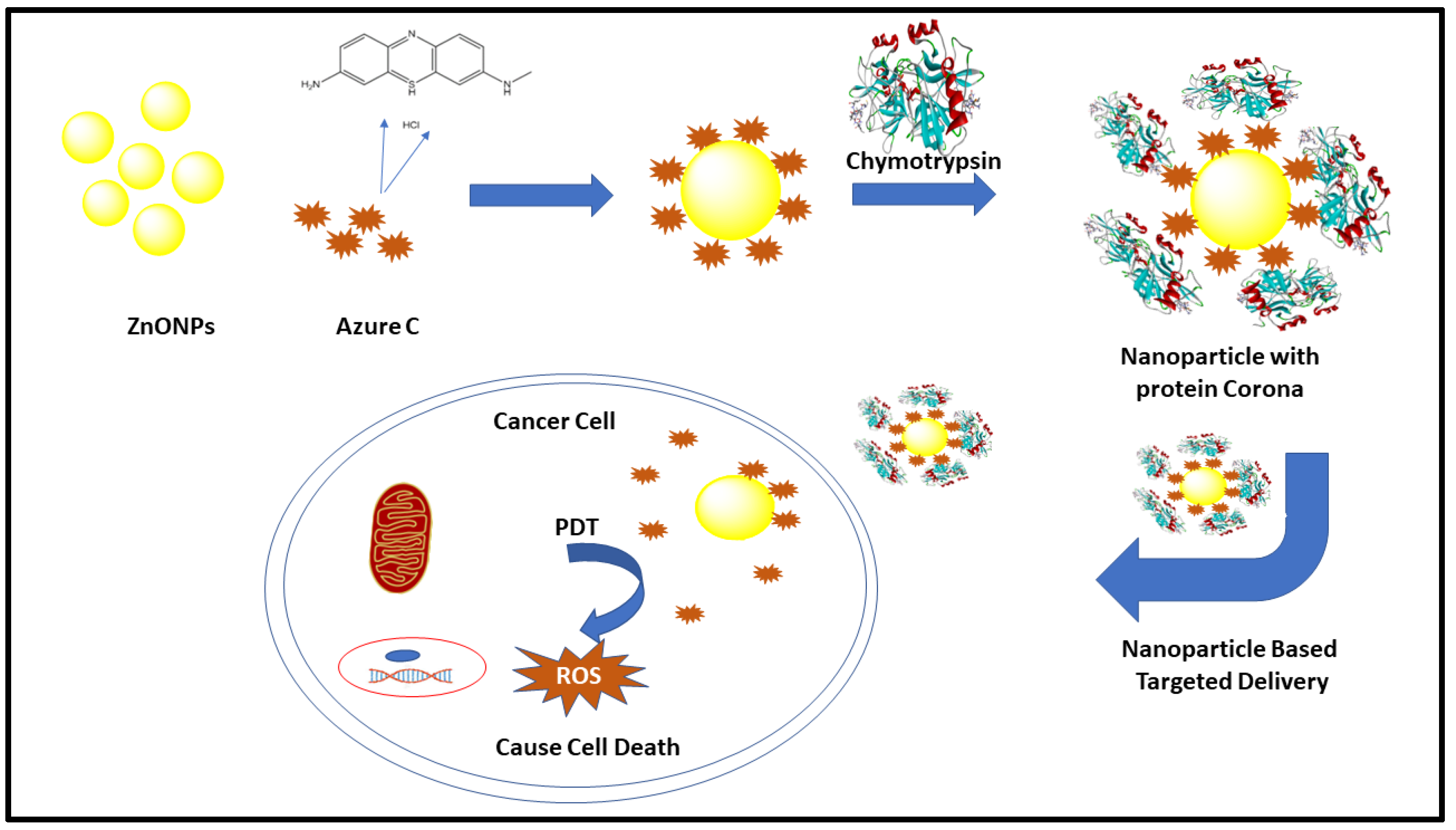
:1. Introduction
2. Material
3. Methods
3.1. Synthesis of Zinc Oxide Nanoparticles
3.2. UV-Visible Experiments
3.3. Fluorescence Experiments
3.4. Circular Dichroism Experiments
3.5. Molecular Docking Studies
3.6. In Vitro Cytotoxicity Assay
4. Results and Discussion
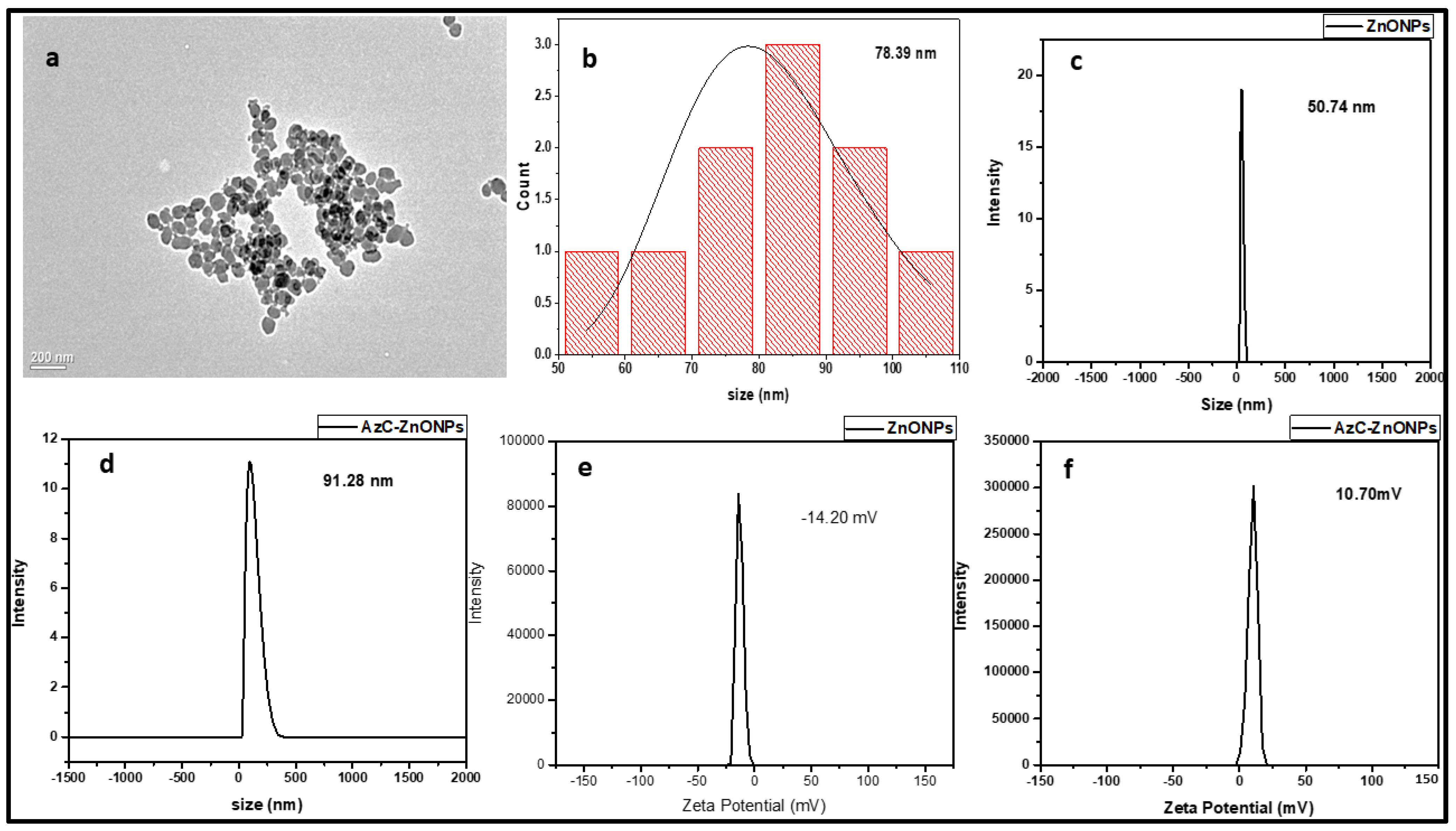
4.1. Characterization of AzC-ZnONPs
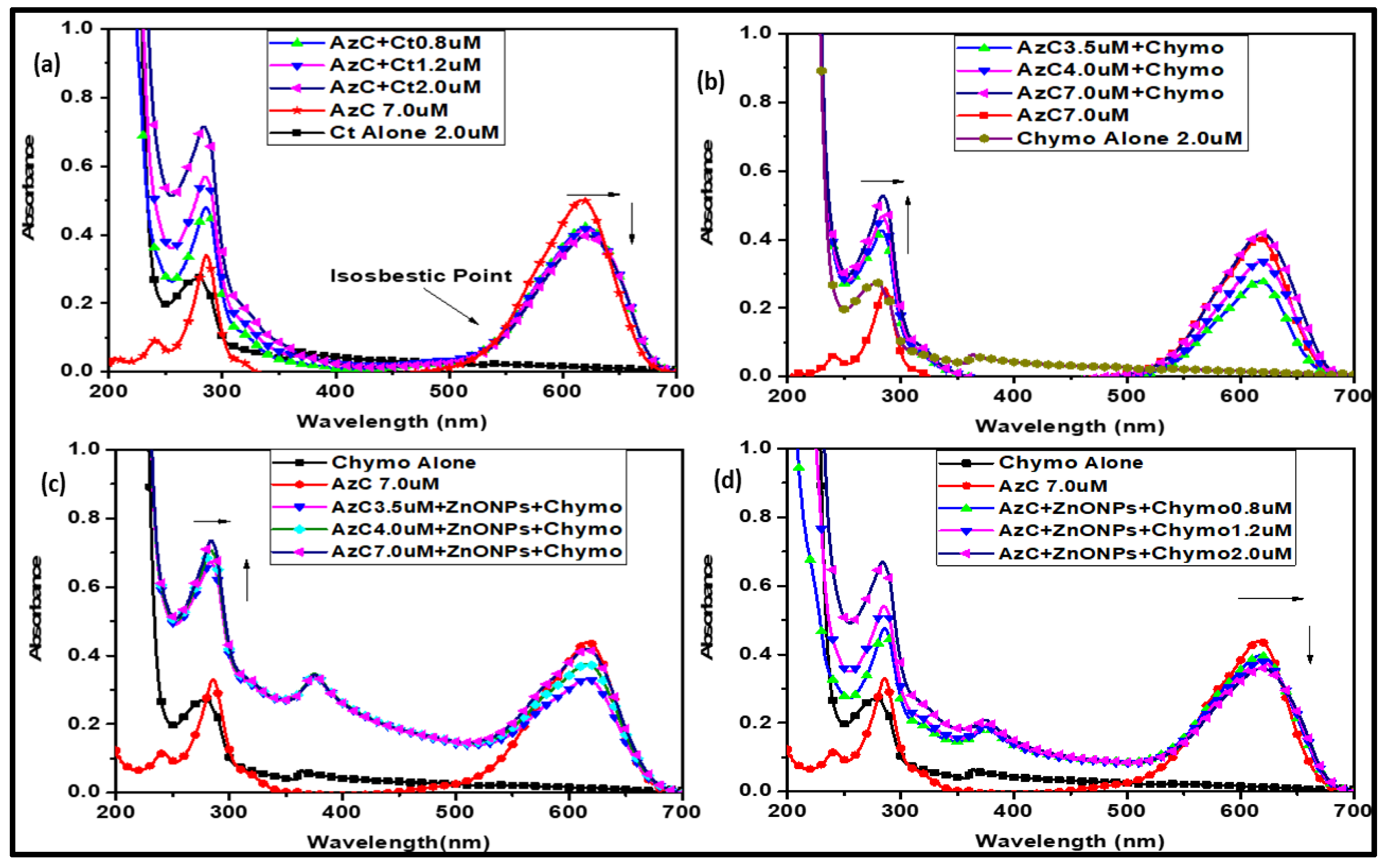
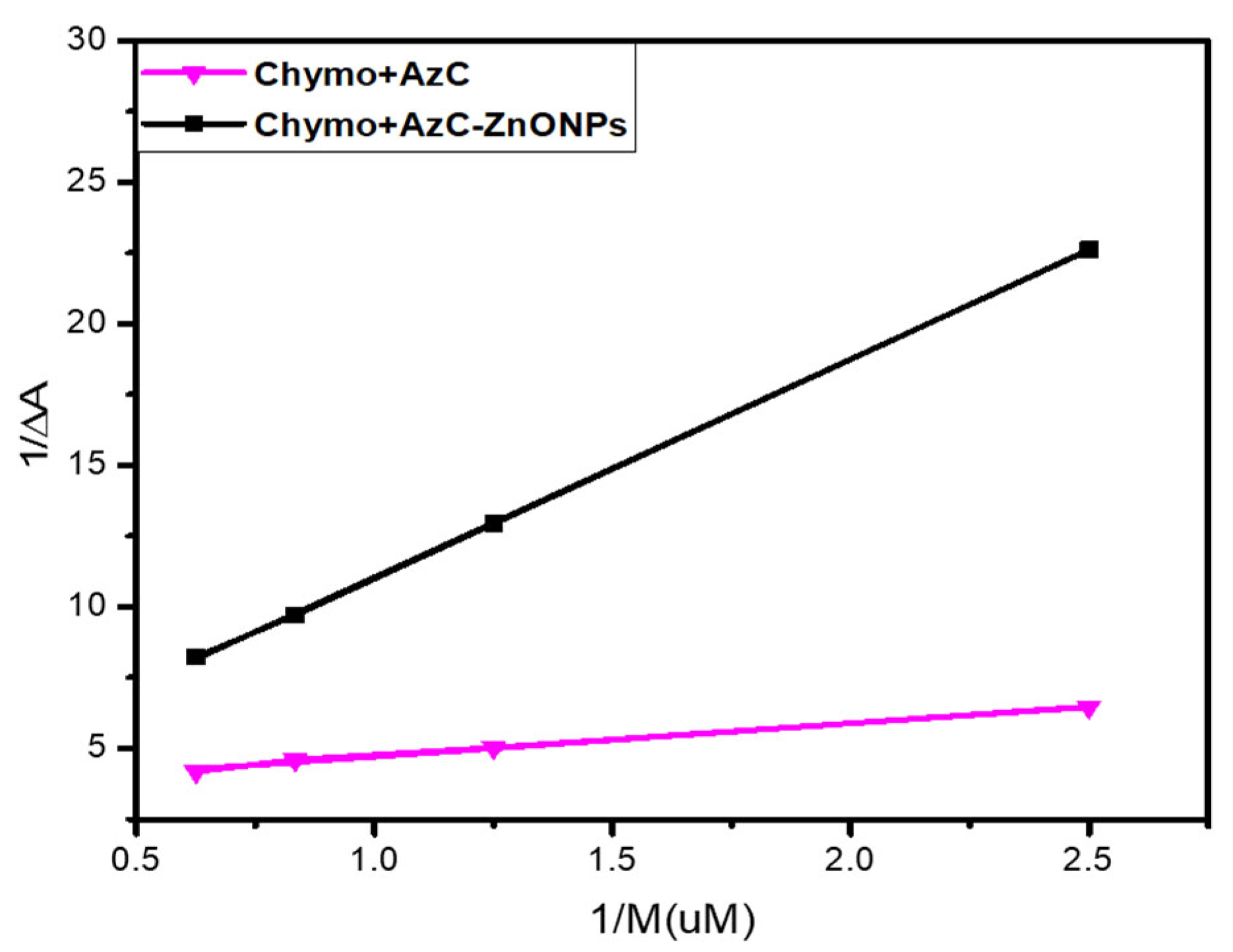
4.2. UV-Visible Spectroscopic Studies
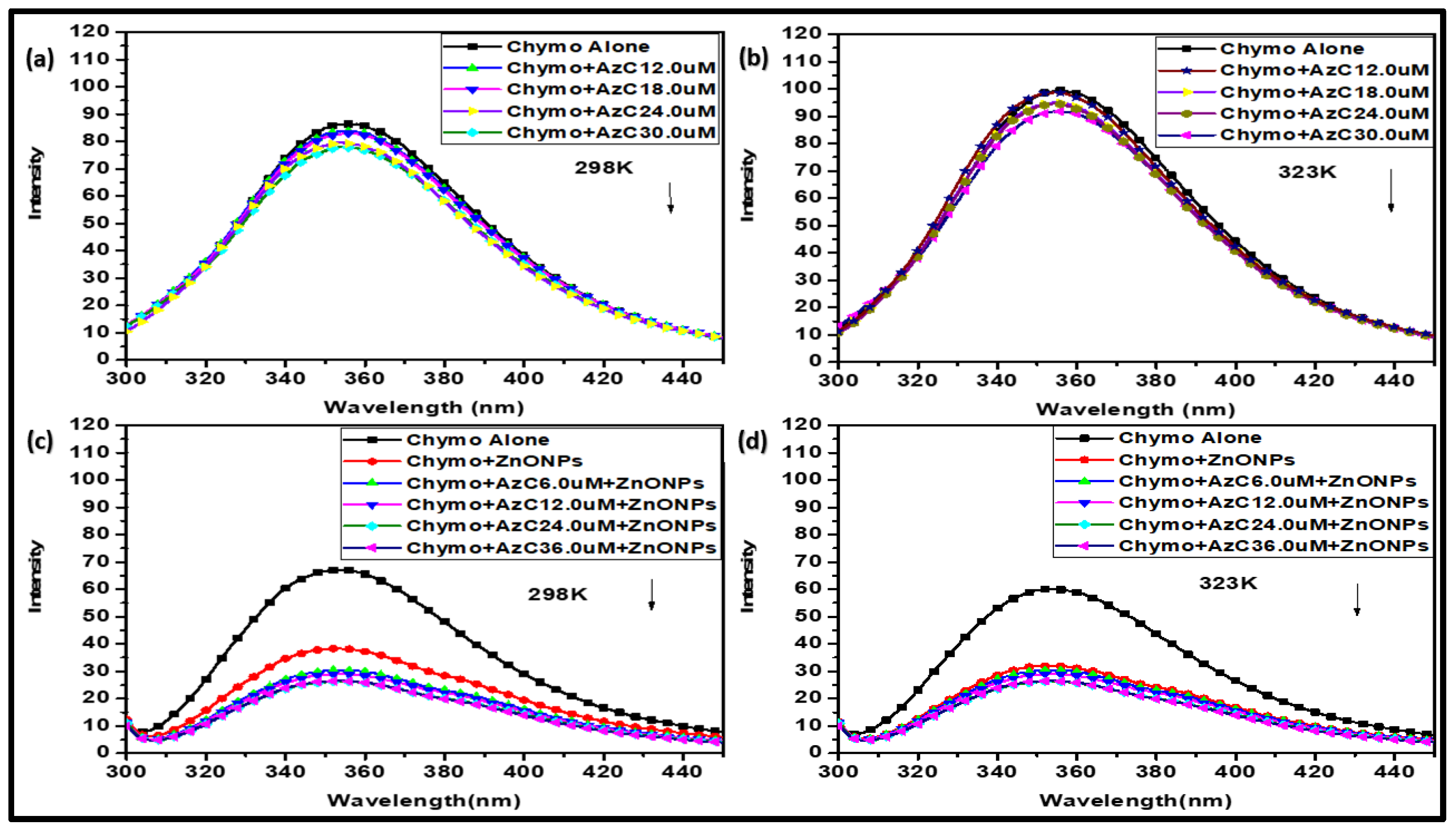
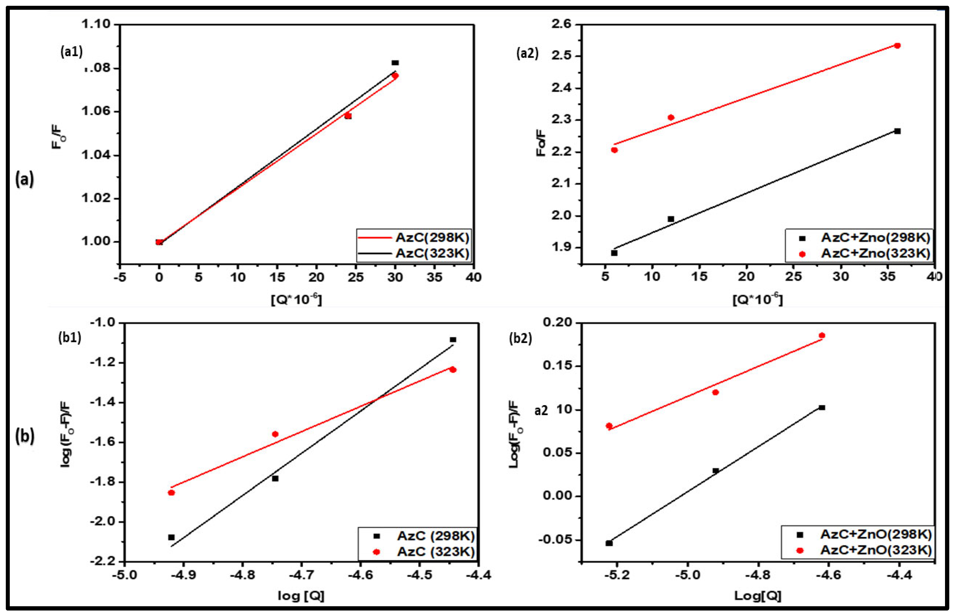
4.3. Fluorescence Study
4.4. Mode of Fluorescence Quenching
4.5. Thermodynamic Analysis of Binding
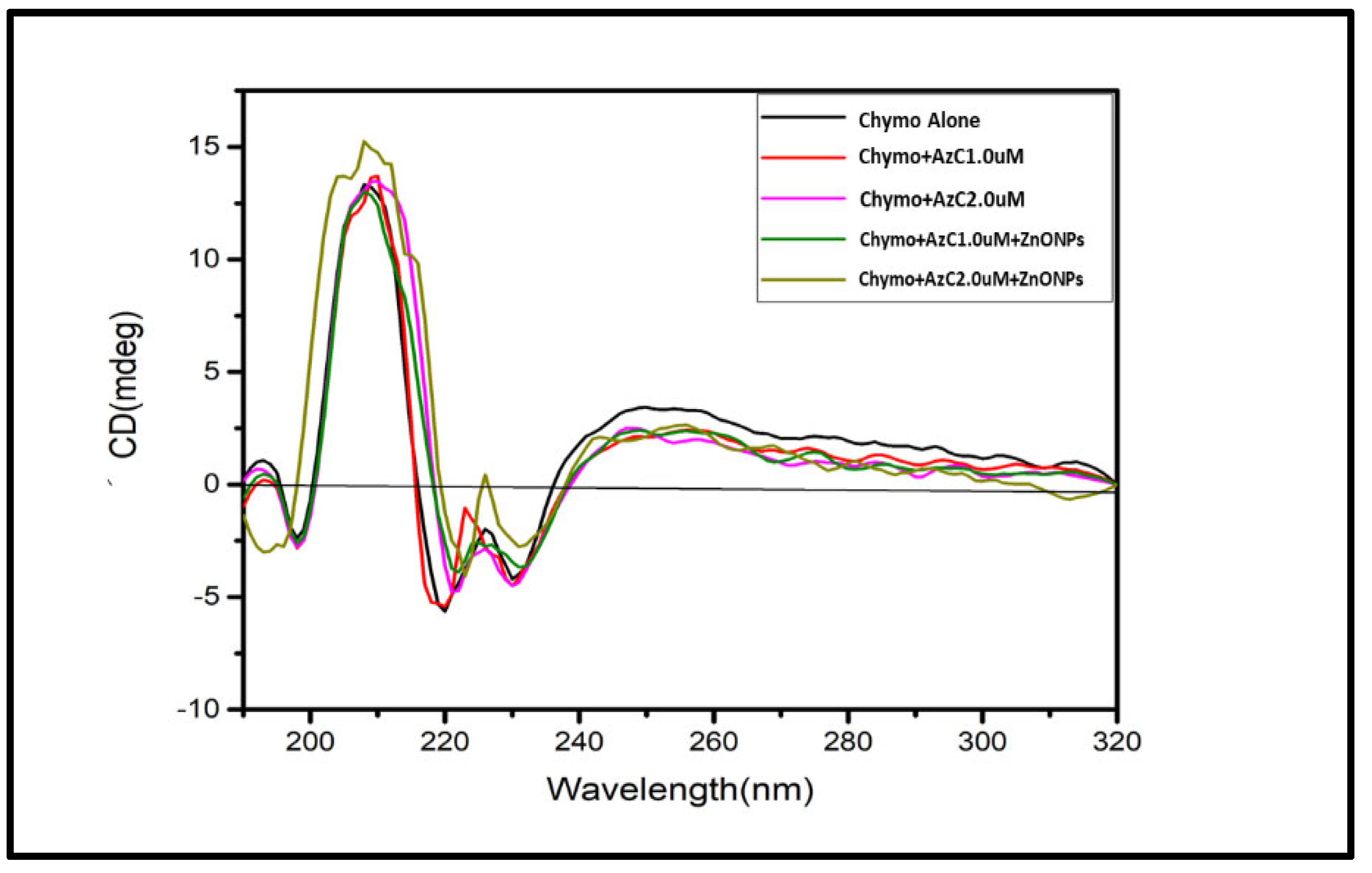
4.6. Circular Dichroism (CD) Study
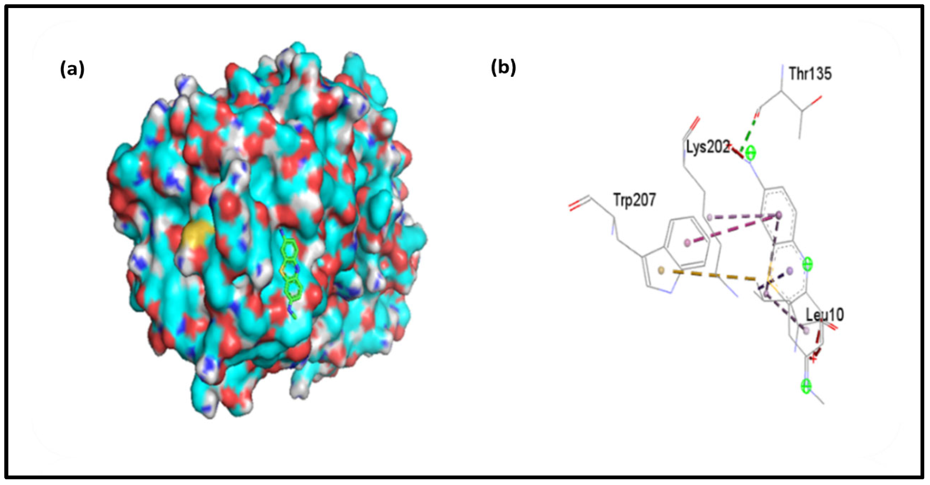
4.7. Molecular Docking
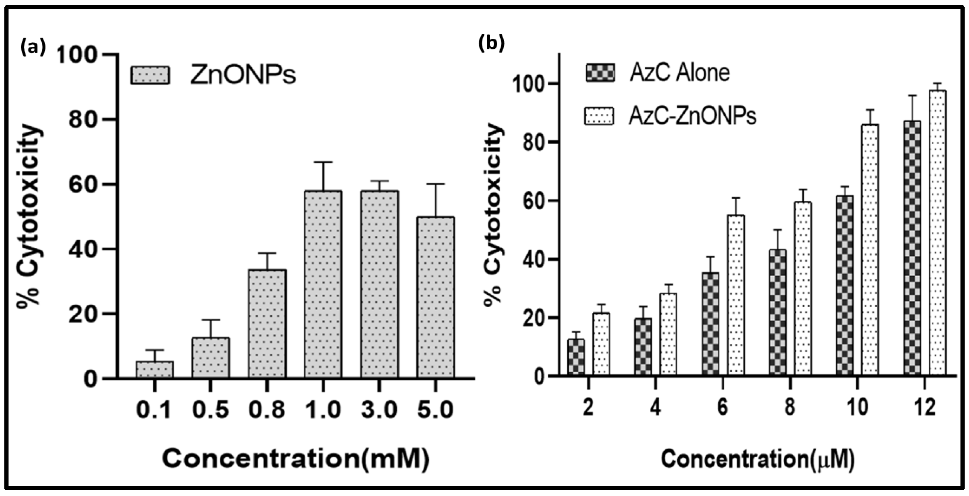
4.8. In Vitro Cytotoxicity
4.9. Biological Relevance
5. Conclusions
Author Contributions
Funding
Institutional Review Board Statement
Informed Consent Statement
Data Availability Statement
Acknowledgments
Conflicts of Interest
References
- Chhikara, B.S.; Parang, K. Global Cancer Statistics 2022: The trends projection analysis. Chem. Biol. Lett. 2022, 10, 451. [Google Scholar]
- Miller, K.D.; Nogueira, L.; Devasia, T.; Mariotto, A.B.; Yabroff, K.R.; Jemal, A.; Kramer, J.; Siegel, R.L. Cancer treatment and survivorship statistics, 2022. CA A Cancer J. Clin. 2022, 72, 409–436. [Google Scholar] [CrossRef] [PubMed]
- Sahu, A.; Choi, W.I.; Lee, J.H.; Tae, G. Graphene oxide mediated delivery of methylene blue for combined photodynamic and photothermal therapy. Biomaterials 2013, 34, 6239–6248. [Google Scholar] [CrossRef] [PubMed]
- Didamson, O.C.; Chandran, R.; Abrahamse, H. A Gold Nanoparticle Bioconjugate Delivery System for Active Targeted Photodynamic Therapy of Cancer and Cancer Stem Cells. Cancers 2022, 14, 4558. [Google Scholar] [CrossRef] [PubMed]
- Pothipor, C.; Bamrungsap, S.; Jakmunee, J.; Ounnunkad, K. A gold nanoparticle-dye/poly (3-aminobenzylamine)/two dimensional MoSe2/graphene oxide electrode towards label-free electrochemical biosensor for simultaneous dual-mode detection of cancer antigen 15-3 and microRNA-21. Colloids Surf. B Biointerfaces 2022, 210, 112260. [Google Scholar] [CrossRef]
- Saha, B.; Kumar, G.S. Binding interaction of phenothiazinium dyes with double stranded RNAs: Spectroscopic and calorimetric investigation. J. Photochem. Photobiol. B Biol. 2017, 167, 99–110. [Google Scholar] [CrossRef]
- Paul, P.; Mati, S.S.; Kumar, G.S. Insights on the interaction of phenothiazinium dyes methylene blue and new methylene blue with synthetic duplex RNAs through spectroscopy and modeling. J. Photochem. Photobiol. B Biol. 2020, 204, 111804. [Google Scholar] [CrossRef]
- Sonia Singh, A.; Shivangi Kukreti, R.; Kukreti, S.; Kaushik, M. Probing multifunctional azure B conjugated gold nanoparticles with serum protein binding properties for trimodal photothermal, photodynamic, and chemo therapy: Biophysical and photophysical investigations. Mater. Sci. Eng. C 2022, 134, 112678. [Google Scholar]
- Shang, W.; Nuffer, J.H.; Dordick, J.S.; Siegel, R.W. Unfolding of ribonuclease A on silica nanoparticle surfaces. Nano Lett. 2007, 7, 1991–1995. [Google Scholar] [CrossRef]
- Riley, M.B.; Strandquist, E.; Weitzel, C.S.; Driskell, J.D. Structure and activity of native and thiolated α-chymotrypsin adsorbed onto gold nanoparticles. Colloids Surf. B Biointerfaces 2022, 220, 112867. [Google Scholar] [CrossRef]
- Wang-Bennett, L.T.; Liebl, D.J.; Bennett, G.N. Targeted neuronal lesion induced by photosensitizing dyes. Brain Res. 1990, 534, 122–128. [Google Scholar] [CrossRef] [PubMed]
- Kamaly, N.; Farokhzad, O.C.; Corbo, C. Nanoparticle protein corona evolution: From biological impact to biomarker discovery. Nanoscale 2022, 14, 1606–1620. [Google Scholar] [CrossRef] [PubMed]
- Wang, X.; Zhong, X.; Li, J.; Liu, Z.; Cheng, L. Inorganic nanomaterials with rapid clearance for biomedical applications. Chem. Soc. Rev. 2021, 50, 8669–8742. [Google Scholar] [CrossRef] [PubMed]
- Kim, S.; Lee, S.Y.; Cho, H.J. Berberine and zinc oxide-based nanoparticles for the chemo-photothermal therapy of lung adenocarcinoma. Biochem. Biophys. Res. Commun. 2018, 501, 765–770. [Google Scholar] [CrossRef] [PubMed]
- Rasmussen, J.W.; Martinez, E.; Louka, P.; Wingett, D.G. Zinc oxide nanoparticles for selective destruction of tumor cells and potential for drug delivery applications. Expert Opin. Drug Deliv. 2010, 7, 1063–1077. [Google Scholar] [CrossRef] [Green Version]
- Singh, A.; Kaushik, M. Physicochemical investigations of zinc oxide nanoparticles synthesized from Azadirachta Indica (Neem) leaf extract and their interaction with Calf-Thymus DNA. Results Phys. 2019, 13, 102168. [Google Scholar] [CrossRef]
- Martínez-Cabanas, M.; López-García, M.; Rodríguez-Barro, P.; Vilariño, T.; Lodeiro, P.; Herrero, R.; Barriada, J.L.; Sastre de Vicente, M.E. Antioxidant Capacity Assessment of Plant Extracts for Green Synthesis of Nanoparticles. Nanomaterials 2021, 11, 1679. [Google Scholar] [CrossRef]
- Sonia Kukreti, S.; Kaushik, M. Gold Nanoclusters: An Ultrasmall Platform for Multifaceted Applications. Talanta 2021, 234, 122623. [Google Scholar] [CrossRef]
- Khdair, A.; Gerard, B.; Handa, H.; Mao, G.; Shekhar, M.P.V.; Panyam, J. Surfactant polymer nanoparticles enhance the effectiveness of anticancer photodynamic therapy. Mol. Pharm. 2008, 5, 795–807. [Google Scholar] [CrossRef]
- Bardhan, M.; Mandal, G.; Ganguly, T. Steady state, time resolved, and circular dichroism spectroscopic studies to reveal the nature of interactions of zinc oxide nanoparticles with transport protein bovine serum albumin and to monitor the possible protein conformational changes. J. Appl. Phys. 2009, 106, 034701. [Google Scholar] [CrossRef]
- Kathiravan, A.; Paramaguru, G.; Renganathan, R. Study on the binding of colloidal zinc oxide nanoparticles with bovine serum albumin. J. Mol. Struct. 2009, 934, 129–137. [Google Scholar] [CrossRef]
- Casals, E.; Pfaller, T.; Duschl, A.; Oostingh, G.J.; Puntes, V. Time Evolution of the Nanoparticle Protein Corona. ACS Nano 2010, 4, 3623–3632. [Google Scholar] [CrossRef] [PubMed]
- Kishen, A.; Upadya, M.; Tegos, G.P.; Hamblin, M.R. Efflux pump inhibitor potentiates antimicrobial photodynamic inactivation of Enterococcus faecalis biofilm. Photochem. Photobiol. 2010, 86, 1343–1349. [Google Scholar] [CrossRef] [PubMed] [Green Version]
- Tuite, E.M.; Kelly, J.M. New trends in photobiology: Photochemical interactions of methylene blue and analogues with DNA and other biological substrates. J. Photochem. Photobiol. B Biol. 1993, 21, 103–124. [Google Scholar] [CrossRef] [PubMed]
- Cedervall, T.; Lynch, I.; Lindman, S.; Berggård, T.; Thulin, E.; Nilsson, H.; Dawson, K.A.; Linse, S. Understanding the nanoparticle–protein corona using methods to quantify exchange rates and affinities of proteins for nanoparticles. Proc. Natl. Acad. Sci. USA 2007, 104, 2050–2055. [Google Scholar] [CrossRef] [Green Version]
- Deng, Z.J.; Mortimer, G.; Schiller, T.; Musumeci, A.; Martin, D.; Minchin, R.F. Differential plasma protein binding to metal oxide nanoparticles. Nanotechnology 2009, 20, 455101. [Google Scholar] [CrossRef]
- Reddy, P.M.; Umapathi, R.; Venkatesu, P. A green approach to offset the perturbation action of 1-butyl-3-methylimidazolium iodide on α-chymotrypsin. Phys. Chem. Chem. Phys. 2015, 17, 184–190. [Google Scholar] [CrossRef]
- Tian, J.; Wei, S.; Zhao, Y.; Liu, R.; Zhao, S. Studies on interaction between CdTe quantum dots and α-chymotrypsin by molecular spectroscopy. J. Chem. Sci. 2010, 122, 391–400. [Google Scholar] [CrossRef]
- Kumar, M.; Kaushik, M.; Chaudhary, S.; Kukreti, S. Spectroscopic studies of the binding interactions of phenothiazininum dyes (Thionine Acetate, Azure A and Azure B) with Calf-thymus DNA. J. Drug Metab. Toxicol. 2016, 7, 1–7. [Google Scholar]
- Das, S.; Islam, M.M.; Jana, G.C.; Patra, A.; Jha, P.K.; Hossain, M. Molecular binding of toxic phenothiazinium derivatives, azures to bovine serum albumin: A comparative spectroscopic, calorimetric, and in silico study. J. Mol. Recognit. 2017, 30, e2609. [Google Scholar] [CrossRef]
- Farhadian, S.; Shareghi, B.; Saboury, A.A.; Babaheydari, A.K.; Raisi, F. Molecular aspects of the interaction of spermidine and α-chymotrypsin. Int. J. Biol. Macromol. 2016, 92, 523–532. [Google Scholar] [CrossRef] [PubMed]
- Qi, Z.D.; Zhou, B.; Qi, X.; Chuan, S.; Liu, Y.; Dai, J. Interaction of rofecoxib with human serum albumin: Determination of binding constants and the binding site by spectroscopic methods. J. Photochem. Photobiol. A Chem. 2008, 193, 81–88. [Google Scholar] [CrossRef]
- Li, H.; Pu, J.; Wang, Y.; Liu, C.; Yu, J.; Li, T.; Wang, R. Comparative study of the binding of Trypsin with bifendate and analogs by spectrofluorimetry. Spectrochim. Acta Part A Mol. Biomol. Spectrosc. 2013, 115, 1–11. [Google Scholar] [CrossRef] [PubMed]
- Alam, P.; Chaturvedi, S.K.; Anwar, T.; Siddiqi, M.K.; Ajmal, M.R.; Badr, G.; Mahmoud, M.H.; Khan, R.H. Biophysical and molecular docking insight into the interaction of cytosine β-D arabinofuranoside with human serum albumin. J. Lumin. 2015, 164, 123–130. [Google Scholar] [CrossRef]
- Alam, P.; Siddiqi, K.; Chturvedi, S.K.; Khan, R.H. Protein aggregation: From background to inhibition strategies. Int. J. Biol. Macromol. 2017, 103, 208–219. [Google Scholar] [CrossRef]
- Farhadian, S.; Shareghi, B.; Saboury, A.A.; Evini, M. The influence of putrescine on the structure, enzyme activity and stability of α-chymotrypsin. RSC Adv. 2016, 6, 29264–29278. [Google Scholar] [CrossRef]
- Farhadian, S.; Shareghi, B.; Momeni, L.; Abou-Zied, O.K.; Sirotkin, V.A.; Tachiya, M.; Saboury, A.A. Insights into the molecular interaction between sucrose and α-chymotrypsin. Int. J. Biol. Macromol. 2018, 114, 950–960. [Google Scholar] [CrossRef]
- Kumar, A.; Rani, A.; Venkatesu, P. A comparative study of the effects of the Hofmeister series anions of the ionic salts and ionic liquids on the stability of α-chymotrypsin. New J. Chem. 2015, 39, 938–952. [Google Scholar] [CrossRef]
- Woody, R.W.; Dunker, A.K. Aromatic and cystine side-chain circular dichroism in proteins. In Circular Dichroism and the Conformational Analysis of Biomolecules; Springer: Boston, MA, USA, 1996; pp. 109–115. [Google Scholar]
- Fakhar-e-Alam, M.; Ali, S.M.; Ibupoto, Z.H.; Kimleang, K.; Atif, M.; Kashif, M.; Loong, F.K.; Hashim, U.; Willander, M. Sensitivity of A-549 human lung cancer cells to nanoporous zinc oxide conjugated with Photofrin. Lasers Med. Sci. 2012, 27, 607–614. [Google Scholar] [CrossRef]
- Nagi, J.S.; Skorenko, K.; Bernier, W.; Jones, W.E.; Doiron, A.L. Near infrared-activated dye-linked ZnO nanoparticles release reactive oxygen species for potential use in photodynamic therapy. Materials 2019, 13, 17. [Google Scholar] [CrossRef] [Green Version]
- Vilsinski, B.H.; Gonçalves, R.S.; Caetano, W.; de Souza, P.R.; de Oliveira, A.C.; Gomes, Y.S.; Gerola, A.P.; Martins, A.F.; Valente, A.J.; Muniz, E.C. Photodynamic Therapy: Use of Nanocarrier Systems to Improve Its Effectiveness. In Functional Properties of Advanced Engineering Materials and Biomolecules; Springer: Cham, Switzerland, 2021; pp. 289–316. [Google Scholar]
- Zolfaghari, P.S.; Packer, S.; Singer, M.; Nair, S.P.; Bennett, J.; Street, C.; Wilson, M. In vivo killing of Staphylococcus aureus using a light-activated antimicrobial agent. BMC Microbiol. 2009, 9, 27. [Google Scholar] [CrossRef] [PubMed] [Green Version]
- Papin, J.F.; Floyd, R.A.; Dittmer, D.P. Methylene blue photoinactivation abolishes West Nile virus infectivity in vivo. Antivir. Res. 2005, 68, 84–87. [Google Scholar] [CrossRef] [PubMed]
- Orth, K.; Beck, G.; Genze, F.; Rück, A. Methylene blue mediated photodynamic therapy in experimental colorectal tumors in mice. J. Photochem. Photobiol. B Biol. 2000, 57, 186–192. [Google Scholar] [CrossRef] [PubMed]
- De Rosa, F.S.; Bentley, M.V.L. Photodynamic therapy of skin cancers: Sensitizers, clinical studies and future directives. Pharm. Res. 2000, 17, 1447–1455. [Google Scholar] [CrossRef]
- Ohulchanskyy, T.Y.; Roy, I.; Goswami, L.N.; Chen, Y.; Bergey, E.J.; Pandey, R.K.; Oseroff, A.R.; Prasad, P.N. Organically modified silica nanoparticles with covalently incorporated photosensitizer for photodynamic therapy of cancer. Nano Lett. 2007, 7, 2835–2842. [Google Scholar] [CrossRef]
- Roy, I.; Ohulchanskyy, T.Y.; Pudavar, H.E.; Bergey, E.J.; Oseroff, A.R.; Morgan, J.; Dougherty, T.J.; Prasad, P.N. Ceramic-based nanoparticles entrapping water insoluble photosensitizing anticancer drugs: A novel drug-carrier system for photodynamic therapy. J. Am. Chem. Soc. 2003, 125, 7860–7865. [Google Scholar] [CrossRef]
- Zhang, P.; Steelant, W.; Kumar, M.; Scholfield, M. Versatile photosensitizers for photodynamic therapy at infrared excitation. J. Am. Chem. Soc. 2007, 129, 4526–4527. [Google Scholar] [CrossRef] [Green Version]
- Rajan, D.; Ilanchelian, M. Exploring the interaction of Azure dyes with t-RNA by hybrid spectroscopic and computational approaches and its applications toward human lung cancer cell line. Int. J. Biol. Macromol. 2018, 113, 1052–1061. [Google Scholar] [CrossRef]
- Avgustinovich, A.V.; Bakina, O.V.; Afanas’ ev, S.G.; Cheremisina, O.V.; Spirina, L.V.; Dobrodeev, A.Y.; Buldakov, M.; Choynzonov, E.L. Nanoparticles in Gastric Cancer Management. Curr. Pharm. Des. 2021, 27, 2436–2444. [Google Scholar] [CrossRef]


| S. No. | Complex | Binding Constant |
|---|---|---|
| 1, | Chymo+AzC | 5.72 × 10⁴ M−1 |
| 2, | Chymo+AzC-ZnONPs | 11.49 × 10⁵ M−1 |
| S.No | Complex (Temperature) | Ksv (mole−1) | Kq (Quenching Rate Constant) (M−1 s −1) | R2 |
|---|---|---|---|---|
| 1, | Chymo+AzC (298 K) | 2.6 × 10³ | 8.78 × 10¹¹ | 0.991 |
| 2, | Chymo+AzC (323 K) | 2.5 × 10³ | 8.44 × 10¹¹ | 0.998 |
| 3, | Chymo+AzC-ZnONPs (298 K) | 12.4 × 10³ | 4.18 × 10¹¹ | 0.985 |
| 4, | Chymo+AzC-ZnONPs (323 K) | 10.4 × 10³ | 3.51 × 10¹¹ | 0.993 |
| Complex | H (Kjmol−1) | G (Kjmol−1) | S (Kjmol−1 K−1) |
|---|---|---|---|
| Chymo+AzC (298 K) | −0.354 | −7.434 | −2.19 × 10⁻² |
| Chymo+AzC (323 K) | −0.354 | −5.823 | −1.83 × 10⁻² |
| Chymo+AzC-ZnONPs (298 K) | −281.666 | −47.489 | −78.58 × 10⁻² |
| Chymo+AzC-ZnONPs (323 K) | −281.666 | −22.364 | −80.27 × 10⁻² |
Disclaimer/Publisher’s Note: The statements, opinions and data contained in all publications are solely those of the individual author(s) and contributor(s) and not of MDPI and/or the editor(s). MDPI and/or the editor(s) disclaim responsibility for any injury to people or property resulting from any ideas, methods, instructions or products referred to in the content. |
© 2022 by the authors. Licensee MDPI, Basel, Switzerland. This article is an open access article distributed under the terms and conditions of the Creative Commons Attribution (CC BY) license (https://creativecommons.org/licenses/by/4.0/).
Share and Cite
Singh, A.; Kumar, P.; Sarkar, N.; Kaushik, M. Influence of Green Synthesized Zinc Oxide Nanoparticles on Molecular Interaction and Comparative Binding of Azure Dye with Chymotrypsin: Novel Nano-Conjugate for Cancer Phototherapy. Pharmaceutics 2023, 15, 74. https://doi.org/10.3390/pharmaceutics15010074
Singh A, Kumar P, Sarkar N, Kaushik M. Influence of Green Synthesized Zinc Oxide Nanoparticles on Molecular Interaction and Comparative Binding of Azure Dye with Chymotrypsin: Novel Nano-Conjugate for Cancer Phototherapy. Pharmaceutics. 2023; 15(1):74. https://doi.org/10.3390/pharmaceutics15010074
Chicago/Turabian StyleSingh, Amit, Pankaj Kumar, Niloy Sarkar, and Mahima Kaushik. 2023. "Influence of Green Synthesized Zinc Oxide Nanoparticles on Molecular Interaction and Comparative Binding of Azure Dye with Chymotrypsin: Novel Nano-Conjugate for Cancer Phototherapy" Pharmaceutics 15, no. 1: 74. https://doi.org/10.3390/pharmaceutics15010074