
Beyond the Interface: Improved Pulmonary Surfactant-Assisted Drug Delivery through Surface-Associated Structures
 , , , and
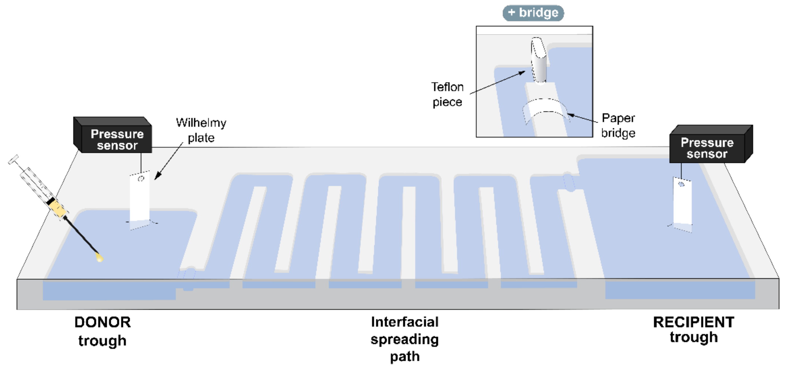
Abstract
:1. Introduction
2. Materials and Methods
2.1. Lipids
2.2. Budesonide
2.3. Pulmonary Surfactant
2.4. Vehiculization Surface Balance
2.5. Fluorescence Spectroscopy
2.6. Statistics
3. Results
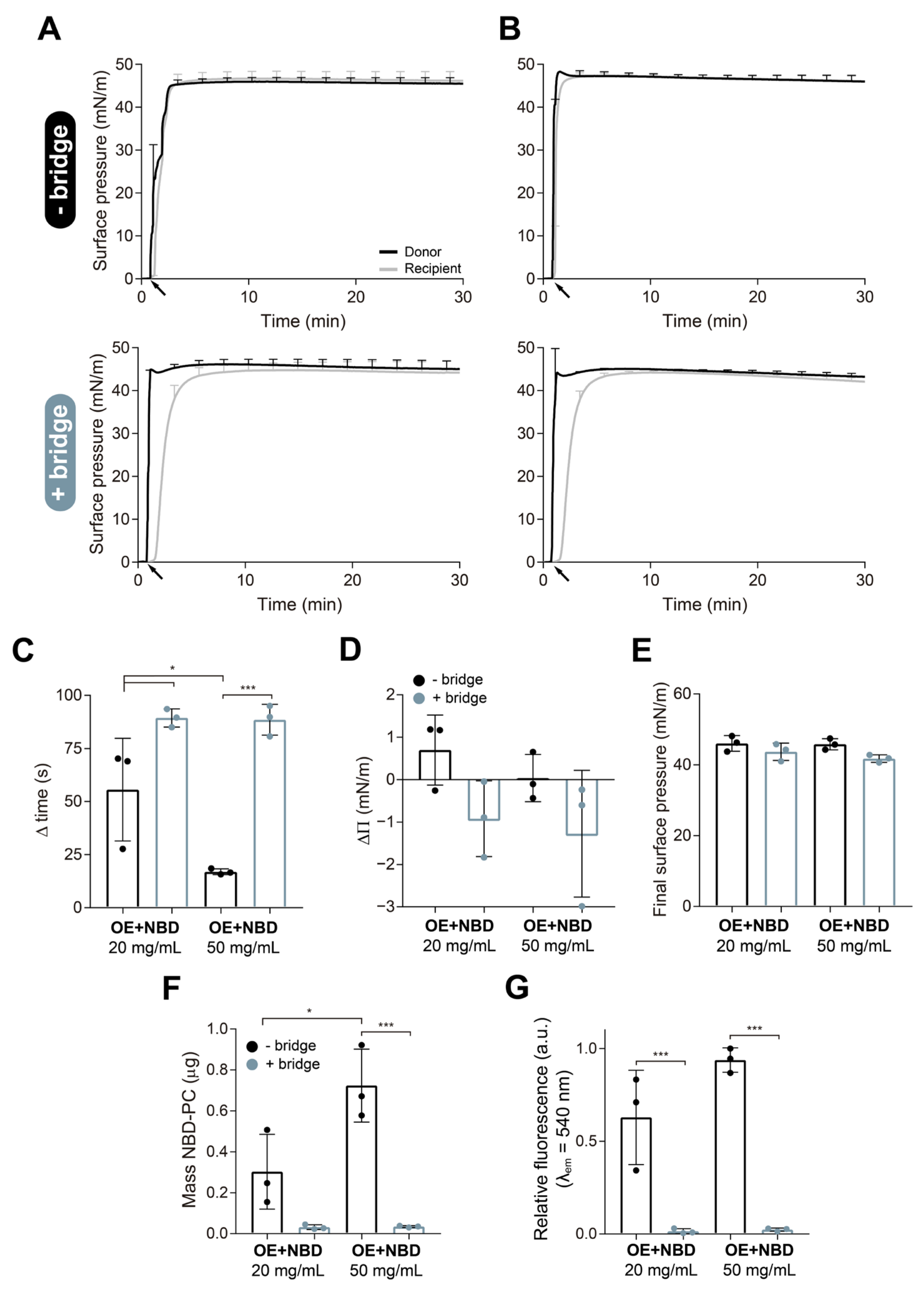
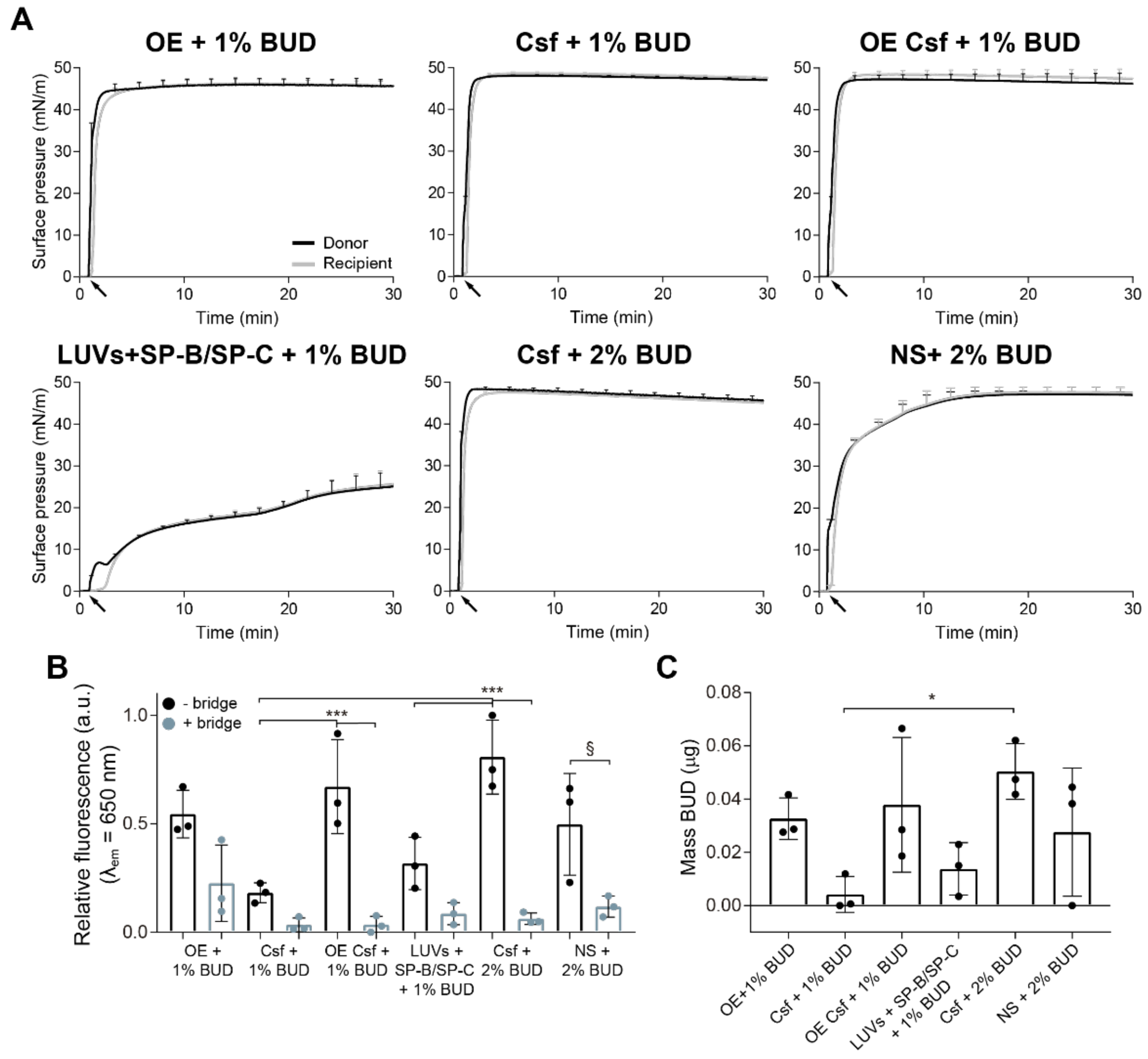
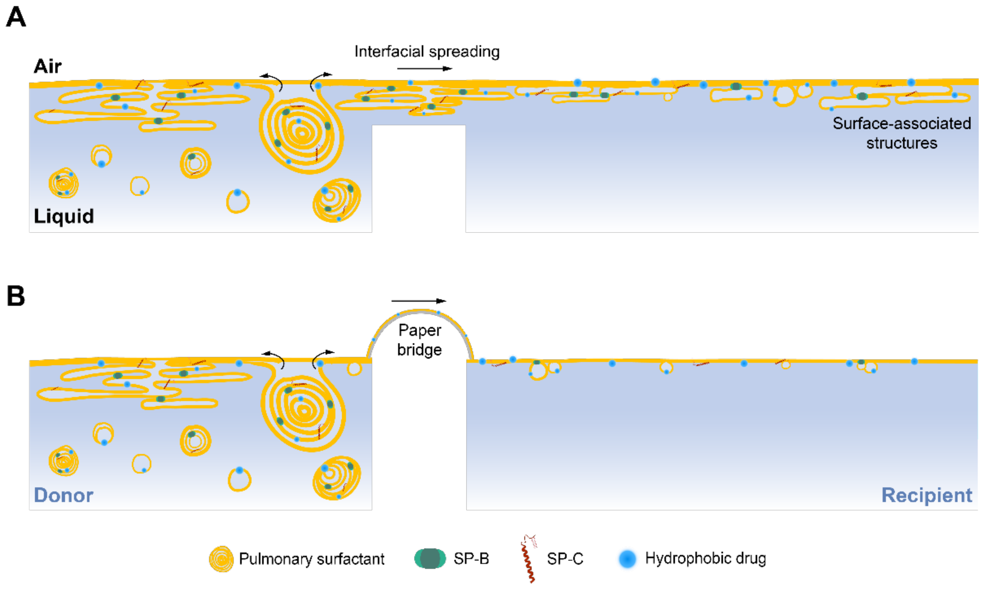
3.1. Contribution of Surface-Associated Structures to the Interfacial Delivery
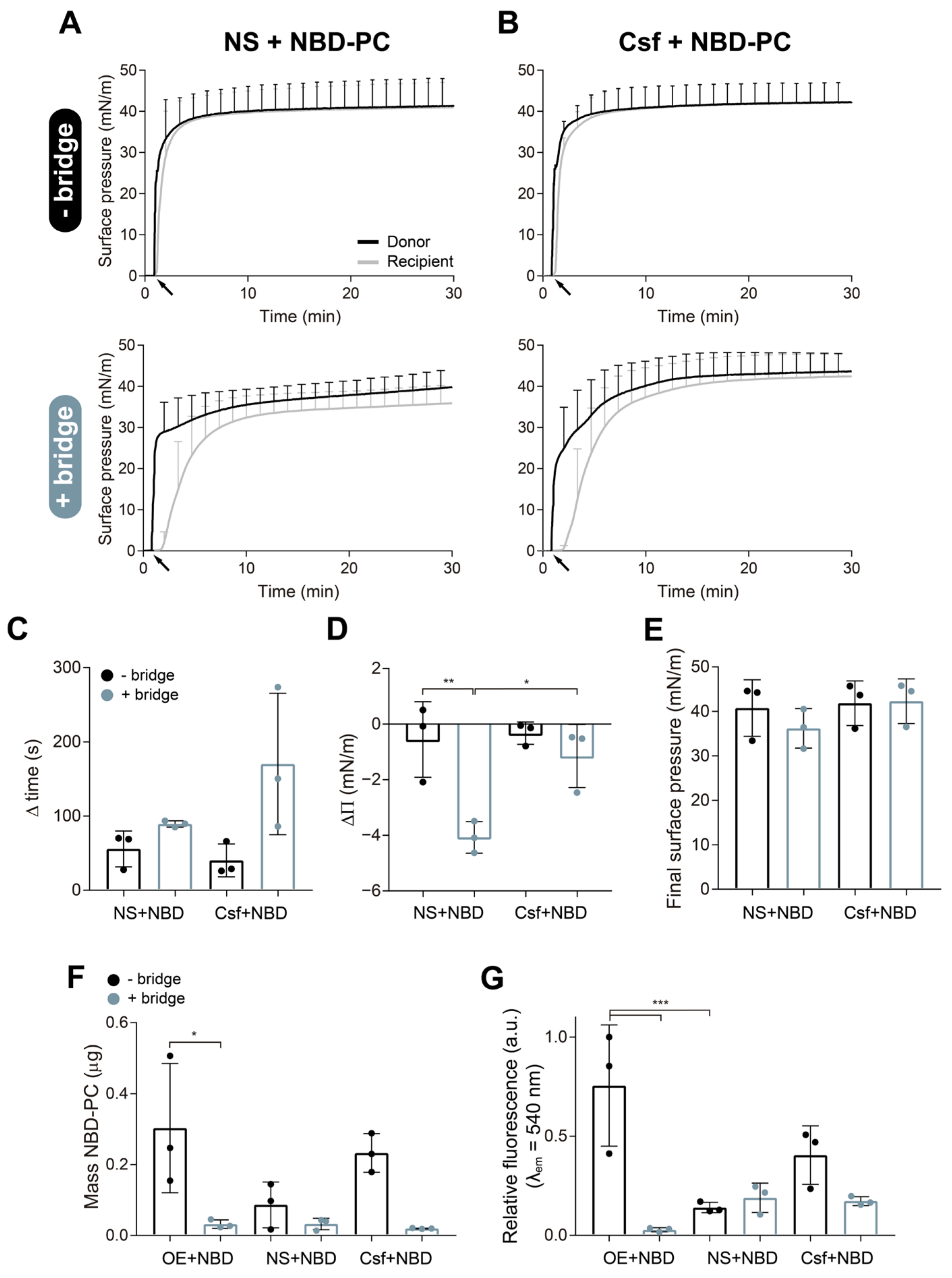
3.2. Transport of Budesonide over the Air–Liquid Interface by Different Materials
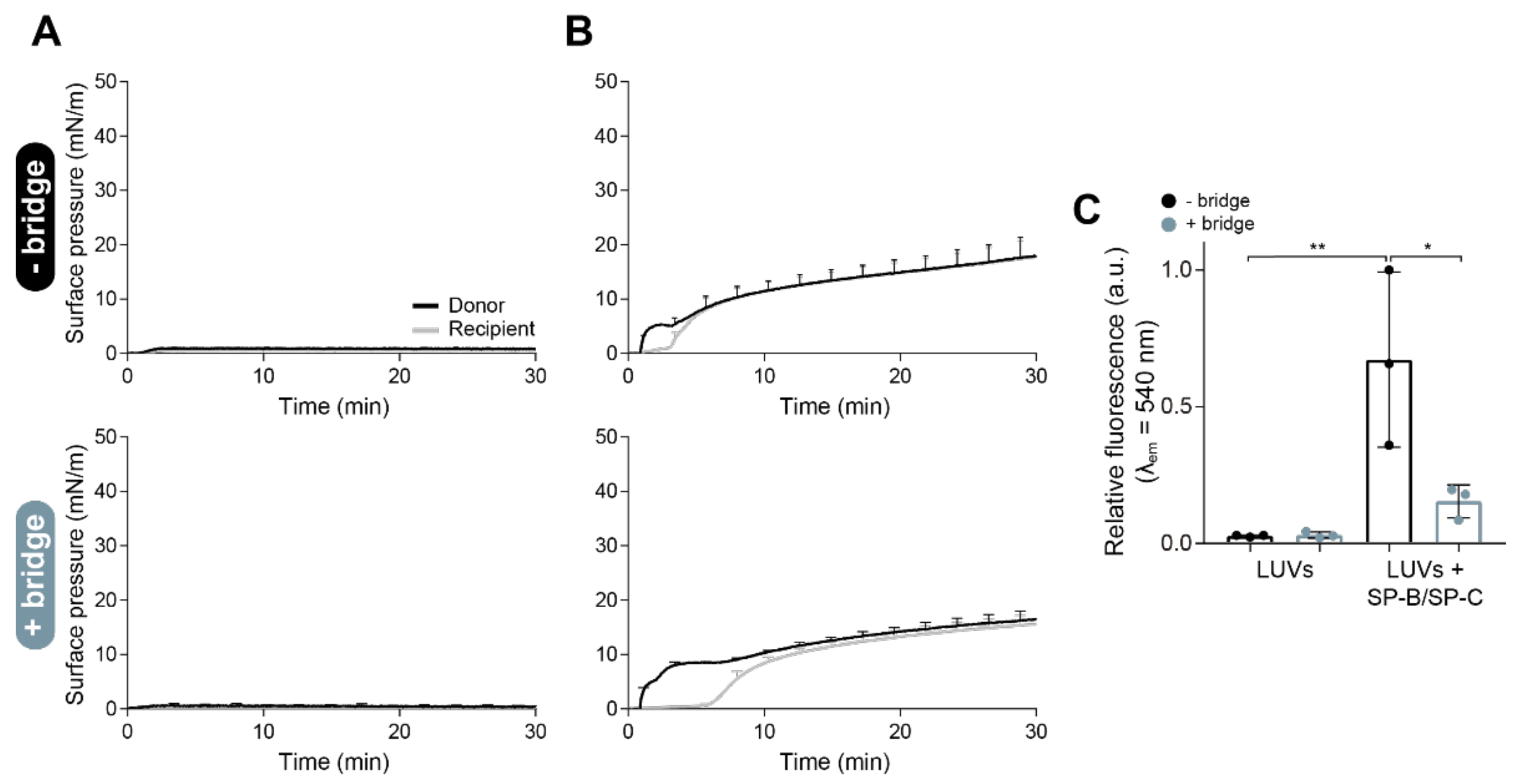
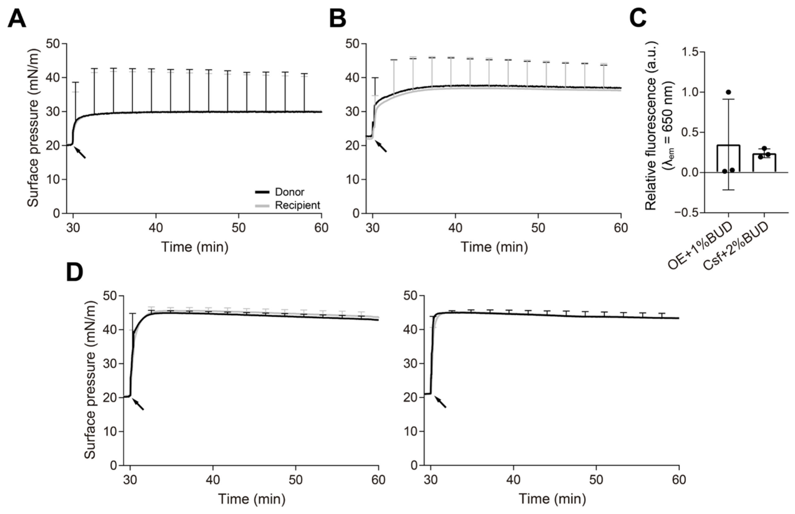
3.3. Spreading of PS over an Interface with a Pre-Existing Surfactant Monolayer
4. Discussion
Supplementary Materials
Author Contributions
Funding
Institutional Review Board Statement
Informed Consent Statement
Data Availability Statement
Acknowledgments
Conflicts of Interest
References
- Parra, E.; Pérez-Gil, J. Composition, structure and mechanical properties define performance of pulmonary surfactant membranes and films. Chem. Phys. Lipids 2015, 185, 153–175. [Google Scholar] [CrossRef]
- Schürch, S.; Qanbar, R.; Bachofen, H.; Possmayer, F. The surface-associated surfactant reservoir in the alveolar lining. Neonatology 1995, 67 (Suppl. S1), 61–76. [Google Scholar] [CrossRef]
- Xu, L.; Yang, Y.; Zuo, Y.Y. Atomic force microscopy imaging of adsorbed pulmonary surfactant films. Biophys. J. 2020, 119, 756–766. [Google Scholar] [CrossRef] [PubMed]
- Schürch, D.; Ospina, O.L.; Cruz, A.; Pérez-Gil, J. Combined and independent action of proteins SP-B and SP-C in the surface behavior and mechanical stability of pulmonary surfactant films. Biophys. J. 2010, 99, 3290–3299. [Google Scholar] [CrossRef] [PubMed] [Green Version]
- Serrano, A.G.; Pérez-Gil, J. Protein–lipid interactions and surface activity in the pulmonary surfactant system. Chem. Phys. Lipids 2006, 141, 105–118. [Google Scholar] [CrossRef] [PubMed]
- Goerke, J. Pulmonary surfactant: Functions and molecular composition. Biochim. Biophys. Acta Mol. Basis Dis. 1998, 1408, 79–89. [Google Scholar] [CrossRef] [Green Version]
- Lopez-Rodriguez, E.; Pérez-Gil, J. Structure-function relationships in pulmonary surfactant membranes: From biophysics to therapy. Biochim. Biophys. Acta Biomembr. 2014, 1838, 1568–1585. [Google Scholar] [CrossRef] [Green Version]
- Zuo, Y.Y.; Veldhuizen, R.A.; Neumann, A.W.; Petersen, N.O.; Possmayer, F. Current perspectives in pulmonary surfactant—Inhibition, enhancement and evaluation. Biochim. Biophys. Acta Biomembr. 2008, 1778, 1947–1977. [Google Scholar] [CrossRef] [Green Version]
- Perez-Gil, J.; Weaver, T.E. Pulmonary surfactant pathophysiology: Current models and open questions. Physiology 2010, 25, 132–141. [Google Scholar] [CrossRef] [PubMed] [Green Version]
- Baer, B.; McCaig, L.; Yamashita, C.; Veldhuizen, R. Exogenous surfactant as a pulmonary delivery vehicle for budesonide in vivo. Lung 2020, 198, 909–916. [Google Scholar] [CrossRef]
- Banaschewski, B.J.; Veldhuizen, E.J.; Keating, E.; Haagsman, H.P.; Zuo, Y.Y.; Yamashita, C.M.; Veldhuizen, R.A. Antimicrobial and biophysical properties of surfactant supplemented with an antimicrobial peptide for treatment of bacterial pneumonia. Antimicrob. Agents Chemother. 2015, 59, 3075–3083. [Google Scholar] [CrossRef] [Green Version]
- Hidalgo, A.; Garcia-Mouton, C.; Autilio, C.; Carravilla, P.; Orellana, G.; Islam, M.N.; Bhattacharya, J.; Bhattacharya, S.; Cruz, A.; Pérez-Gil, J. Pulmonary surfactant and drug delivery: Vehiculization, release and targeting of surfactant/tacrolimus formulations. J. Control. Release 2021, 329, 205–222. [Google Scholar] [CrossRef]
- Merckx, P.; Lammens, J.; Nuytten, G.; Bogaert, B.; Guagliardo, R.; Maes, T.; Vervaet, C.; De Beer, T.; De Smedt, S.C.; Raemdonck, K. Lyophilization and nebulization of pulmonary surfactant-coated nanogels for siRNA inhalation therapy. Eur. J. Pharm. Biopharm. 2020, 157, 191–199. [Google Scholar] [CrossRef] [PubMed]
- Hidalgo, A.; Cruz, A.; Pérez-Gil, J. Barrier or carrier? Pulmonary surfactant and drug delivery. Eur. J. Pharm. Biopharm. 2015, 95, 117–127. [Google Scholar] [CrossRef] [PubMed]
- Khanal, A.; Sharma, R.; Corcoran, T.E.; Garoff, S.; Przybycien, T.M.; Tilton, R.D. Surfactant driven post-deposition spreading of aerosols on complex aqueous subphases. 1: High deposition flux representative of aerosol delivery to large airways. J. Aerosol Med. Pulm. 2015, 28, 382–393. [Google Scholar] [CrossRef] [PubMed] [Green Version]
- Halpern, D.; Jensen, O.; Grotberg, J. A theoretical study of surfactant and liquid delivery into the lung. J. Appl. Physiol. 1998, 85, 333–352. [Google Scholar] [CrossRef] [Green Version]
- Stetten, A.Z.; Iasella, S.V.; Corcoran, T.E.; Garoff, S.; Przybycien, T.M.; Tilton, R.D. Surfactant-induced Marangoni transport of lipids and therapeutics within the lung. Curr. Opin. Colloid Interface Sci. 2018, 36, 58–69. [Google Scholar] [CrossRef]
- Espinosa, F.; Shapiro, A.; Fredberg, J.; Kamm, R. Spreading of exogenous surfactant in an airway. J. Appl. Physiol. 1993, 75, 2028–2039. [Google Scholar] [CrossRef] [PubMed]
- García-Mouton, C.; Hidalgo, A.; Arroyo, R.; Echaide, M.; Cruz, A.; Pérez-Gil, J. Pulmonary surfactant and drug delivery: An interface-assisted carrier to deliver surfactant protein SP-D into the airways. Front. Bioeng. Biotechnol. 2020, 8, 613276. [Google Scholar] [CrossRef]
- Hidalgo, A.; Salomone, F.; Fresno, N.; Orellana, G.; Cruz, A.; Perez-Gil, J. Efficient interfacially driven vehiculization of corticosteroids by pulmonary surfactant. Langmuir 2017, 33, 7929–7939. [Google Scholar] [CrossRef]
- Baer, B.; Veldhuizen, E.J.; Molchanova, N.; Jekhmane, S.; Weingarth, M.; Jenssen, H.; Lin, J.S.; Barron, A.E.; Yamashita, C.; Veldhuizen, R. Optimizing exogenous Surfactant as a pulmonary Delivery Vehicle for Chicken Cathelicidin-2. Sci. Rep. 2020, 10, 9392. [Google Scholar] [CrossRef]
- Baer, B.; Veldhuizen, E.J.; Possmayer, F.; Yamashita, C.; Veldhuizen, R. The wet bridge transfer system: A novel tool to assess exogenous surfactant as a vehicle for intrapulmonary drug delivery. Discov. Med. 2018, 26, 207–218. [Google Scholar] [PubMed]
- Yu, S.-H.; Possmayer, F. Lipid compositional analysis of pulmonary surfactant monolayers and monolayer-associated reservoirs. J. Lipid Res. 2003, 44, 621–629. [Google Scholar] [CrossRef] [PubMed] [Green Version]
- Yu, S.-H.; Possmayer, F. Dipalmitoylphosphatidylcholine and cholesterol in monolayers spread from adsorbed films of pulmonary surfactant. J. Lipid Res. 2001, 42, 1421–1429. [Google Scholar] [CrossRef]
- Venkataraman, R.; Kamaluddeen, M.; Hasan, S.U.; Robertson, H.L.; Lodha, A. Intratracheal administration of budesonide-surfactant in prevention of bronchopulmonary dysplasia in very low birth weight infants: A systematic review and meta-analysis. Pediatr. Pulmonol. 2017, 52, 968–975. [Google Scholar] [CrossRef] [PubMed]
- Halliday, H.L. Update on postnatal steroids. Neonatology 2017, 111, 415–422. [Google Scholar] [CrossRef]
- Taeusch, H.W.; De La Serna, J.B.; Perez-Gil, J.; Alonso, C.; Zasadzinski, J.A. Inactivation of pulmonary surfactant due to serum-inhibited adsorption and reversal by hydrophilic polymers: Experimental. Biophys. J. 2005, 89, 1769–1779. [Google Scholar] [CrossRef] [Green Version]
- Bligh, E.; Dyer, W. A rapid method of lipid extraction and purification. Can. J. Biochem. Physiol. 1959, 37, 911–917. [Google Scholar] [CrossRef] [PubMed]
- Rouser, G.; Siakotos, A.; Fleischer, S. Quantitative analysis of phospholipids by thin-layer chromatography and phosphorus analysis of spots. Lipids 1966, 1, 85–86. [Google Scholar] [CrossRef]
- Pérez-Gil, J.; Cruz, A.; Casals, C. Solubility of hydrophobic surfactant proteins in organic solvent/water mixtures. Structural studies on SP-B and SP-C in aqueous organic solvents and lipids. Biochim. Biophys. Acta Mol. Cell Biol. Lipids 1993, 1168, 261–270. [Google Scholar] [CrossRef]
- Bernhard, W.; Mottaghian, J.; Gebert, A.; Rau, G.A.; Von der Hardt, H.; Poets, C.F. Commercial versus native surfactants: Surface activity, molecular components, and the effect of calcium. Am. J. Respir. Crit. 2000, 162, 1524–1533. [Google Scholar] [CrossRef] [PubMed] [Green Version]
- Chen, C.-M.; Chang, C.-H.; Chao, C.-H.; Wang, M.-H.; Yeh, T.-F. Biophysical and chemical stability of surfactant/budesonide and the pulmonary distribution following intra-tracheal administration. Drug Deliv. 2019, 26, 604–611. [Google Scholar] [CrossRef] [PubMed] [Green Version]
- Zhang, H.; Wang, Y.E.; Neal, C.R.; Zuo, Y.Y. Differential effects of cholesterol and budesonide on biophysical properties of clinical surfactant. Pediatr. Res. 2012, 71, 316–323. [Google Scholar] [CrossRef] [PubMed]
- Wang, Y.E.; Zhang, H.; Fan, Q.; Neal, C.R.; Zuo, Y.Y. Biophysical interaction between corticosteroids and natural surfactant preparation: Implications for pulmonary drug delivery using surfactant as a carrier. Soft Matter 2012, 8, 504–511. [Google Scholar] [CrossRef] [Green Version]
- Hidalgo, A.; Cruz, A.; Pérez-Gil, J. Pulmonary surfactant and nanocarriers: Toxicity versus combined nanomedical applications. Biochim. Biophys. Acta Biomembr. 2017, 1859, 1740–1748. [Google Scholar] [CrossRef]
- Haitsma, J.J.; Lachmann, U.; Lachmann, B. Exogenous surfactant as a drug delivery agent. Adv. Drug Deliv. Rev. 2001, 47, 197–207. [Google Scholar] [CrossRef]
- Dani, C.; Corsini, I.; Burchielli, S.; Cangiamila, V.; Romagnoli, R.; Jayonta, B.; Longini, M.; Paternostro, F.; Buonocore, G. Natural surfactant combined with beclomethasone decreases lung inflammation in the preterm lamb. Respiration 2011, 82, 369–376. [Google Scholar] [CrossRef]
- Van’t Veen, A.; Mouton, J.W.; Gommers, D.; Lachmann, B. Pulmonary surfactant as vehicle for intratracheally instilled tobramycin in mice infected with Klebsiella pneumoniae. Br. J. Pharmacol. 1996, 119, 1145. [Google Scholar] [CrossRef] [Green Version]
- Kharasch, V.S.; Sweeney, T.D.; Fredberg, J.; Lehr, J.; Damokosh, A.I.; Avery, M.E.; Brain, J.D. Pulmonary surfactant as a vehicle for intratracheal delivery of technetium sulfur colloid and pentamidine in hamster lungs. Am. Rev. Respir. Dis. 1991, 144, 909–913. [Google Scholar] [CrossRef]
- Guagliardo, R.; Merckx, P.; Zamborlin, A.; De Backer, L.; Echaide, M.; Pérez-Gil, J.; De Smedt, S.C.; Raemdonck, K. Nanocarrier lipid composition modulates the impact of pulmonary surfactant protein B (SP-B) on cellular delivery of siRNA. Pharmaceutics 2019, 11, 431. [Google Scholar] [CrossRef]
- Grotberg, J.B.; Gaver, D.P., III. A synopsis of surfactant spreading research. J. Colloid Interface Sci. 1996, 178, 377–378. [Google Scholar] [CrossRef]
- Bachofen, H.; Gerber, U.; Gehr, P.; Amrein, M.; Schürch, S. Structures of pulmonary surfactant films adsorbed to an air–liquid interface in vitro. Biochim. Biophys. Acta Biomembr. 2005, 1720, 59–72. [Google Scholar] [CrossRef] [Green Version]
- Schenck, D.; Goettler, S.; Fiegel, J. Surfactant-induced spreading of nanoparticles is inhibited on mucus mimetic surfaces that model native lung conditions. Phys. Biol. 2019, 16, 065001. [Google Scholar] [CrossRef] [PubMed]
- Grotberg, J.; Halpern, D.; Jensen, O. Interaction of exogenous and endogenous surfactant: Spreading-rate effects. J. Appl. Physiol. 1995, 78, 750–756. [Google Scholar] [CrossRef]
- Bull, J.; Nelson, L.; Walsh, J., Jr.; Glucksberg, M.; Schürch, S.; Grotberg, J. Surfactant-spreading and surface-compression disturbance on a thin viscous film. J. Biomech. Eng. 1999, 121, 89–98. [Google Scholar] [CrossRef]
- Krüger, P.; Schalke, M.; Wang, Z.; Notter, R.H.; Dluhy, R.A.; Lösche, M. Effect of hydrophobic surfactant peptides SP-B and SP-C on binary phospholipid monolayers. I. Fluorescence and dark-field microscopy. Biophys. J. 1999, 77, 903–914. [Google Scholar] [CrossRef] [Green Version]
- Guagliardo, R.; Perez-Gil, J.; De Smedt, S.; Raemdonck, K. Pulmonary surfactant and drug delivery: Focusing on the role of surfactant proteins. J. Control. Release 2018, 291, 116–126. [Google Scholar] [CrossRef] [PubMed]
- Kern, J.C.; Dooney, D.; Zhang, R.; Liang, L.; Brandish, P.E.; Cheng, M.; Feng, G.; Beck, A.; Bresson, D.; Firdos, J. Novel phosphate modified cathepsin B linkers: Improving aqueous solubility and enhancing payload scope of ADCs. Bioconjug. Chem. 2016, 27, 2081–2088. [Google Scholar] [CrossRef] [PubMed]


Disclaimer/Publisher’s Note: The statements, opinions and data contained in all publications are solely those of the individual author(s) and contributor(s) and not of MDPI and/or the editor(s). MDPI and/or the editor(s) disclaim responsibility for any injury to people or property resulting from any ideas, methods, instructions or products referred to in the content. |
© 2023 by the authors. Licensee MDPI, Basel, Switzerland. This article is an open access article distributed under the terms and conditions of the Creative Commons Attribution (CC BY) license (https://creativecommons.org/licenses/by/4.0/).
Share and Cite
García-Mouton, C.; Echaide, M.; Serrano, L.A.; Orellana, G.; Salomone, F.; Ricci, F.; Pioselli, B.; Amidani, D.; Cruz, A.; Pérez-Gil, J. Beyond the Interface: Improved Pulmonary Surfactant-Assisted Drug Delivery through Surface-Associated Structures. Pharmaceutics 2023, 15, 256. https://doi.org/10.3390/pharmaceutics15010256
García-Mouton C, Echaide M, Serrano LA, Orellana G, Salomone F, Ricci F, Pioselli B, Amidani D, Cruz A, Pérez-Gil J. Beyond the Interface: Improved Pulmonary Surfactant-Assisted Drug Delivery through Surface-Associated Structures. Pharmaceutics. 2023; 15(1):256. https://doi.org/10.3390/pharmaceutics15010256
Chicago/Turabian StyleGarcía-Mouton, Cristina, Mercedes Echaide, Luis A. Serrano, Guillermo Orellana, Fabrizio Salomone, Francesca Ricci, Barbara Pioselli, Davide Amidani, Antonio Cruz, and Jesús Pérez-Gil. 2023. "Beyond the Interface: Improved Pulmonary Surfactant-Assisted Drug Delivery through Surface-Associated Structures" Pharmaceutics 15, no. 1: 256. https://doi.org/10.3390/pharmaceutics15010256