Article

Composite Nanoarchitectonics of Photoactivated Titania-Based Materials with Anticancer Properties

by
Nefeli Papadopoulou-Fermeli
1,†,
Nefeli Lagopati
1,2,3,†,
Natassa Pippa
4,
Elias Sakellis
5,
Nikos Boukos
5,
Vassilis G. Gorgoulis
3,6,7,8,9,10,
Maria Gazouli
2,11 and
Evangelia A. Pavlatou
1,*
1
Laboratory of General Chemistry, School of Chemical Engineering, National Technical University of Athens, Zografou Campus, 15789 Zografou, Greece
2
Laboratory of Biology, Department of Basic Medical Sciences, Medical School, National and Kapodistrian University of Athens, 11527 Athens, Greece
3
Laboratory of Histology-Embryology, Molecular Carcinogenesis Group, Department of Basic Medical Sciences, Medical School, National and Kapodistrian University of Athens, 11527 Athens, Greece
4
Section of Pharmaceutical Technology, Department of Pharmacy, School of Health Sciences, National and Kapodistrian University of Athens, 15771 Athens, Greece
5
Institute of Nanoscience and Nanotechnology, National Center for Scientific Research “Demokritos”, 15310 Agia Paraskevi, Greece
6
Biomedical Research Foundation, Academy of Athens, 11527 Athens, Greece
7
Clinical Molecular Pathology, Medical School, University of Dundee, Dundee DD1 9SY, UK
8
Molecular and Clinical Cancer Sciences, Manchester Cancer Research Centre, Manchester Academic Health Sciences Centre, University of Manchester, Manchester M20 4GJ, UK
9
Center for New Biotechnologies and Precision Medicine, Medical School, National and Kapodistrian University of Athens, 11527 Athens, Greece
10
Faculty of Health and Medical Sciences, University of Surrey, Guildford GU2 7YH, UK
11
School of Science and Technology, Hellenic Open University, 26335 Patra, Greece
*
Author to whom correspondence should be addressed.
These authors contributed equally to this work.
Pharmaceutics 2023, 15(1), 135; https://doi.org/10.3390/pharmaceutics15010135
Submission received: 28 November 2022 / Revised: 23 December 2022 / Accepted: 26 December 2022 / Published: 30 December 2022
(This article belongs to the Special Issue Functionalized Nanoparticles in Cancer Therapeutics)

Abstract

:
The synthesis of titania-based composite materials with anticancer potential under visible-light irradiation is the aim of this study. In specific, titanium dioxide (TiO2) nanoparticles (NPs) chemically modified with silver were embedded in a stimuli-responsive microgel (a crosslinked interpenetrating network (IP) network that was synthesized by poly (N-Isopropylacrylamide) and linear chains of polyacrylic acid sodium salt, forming composite particles. The ultimate goal of this research, and for our future plans, is to develop a drug-delivery system that uses optical fibers that could efficiently photoactivate NPs, targeting cancer cells. The produced Ag-TiO2 NPs, the microgel and the composite materials were characterized through X-ray diffraction (XRD), Fourier transform infrared spectroscopy (FT-IR), micro-Raman spectroscopy, ultraviolet-visible spectroscopy (UV-Vis), dynamic light scattering (DLS) and transmission electron microscopy (TEM). Our results indicated that Ag-TiO2 NPs were successfully embedded within the thermoresponsive microgel. Either Ag-TiO2 NPs or the composite materials exhibited high photocatalytic degradation efficiency on the pollutant rhodamine B and significant anticancer potential under visible-light irradiation.

1. Introduction

The bioactivity of photoinduced titanium dioxide (TiO2) has been studied in recent years in order to propose an alternative therapeutic approach, minimizing the side effects caused by conventional cancer treatments [1]. Pure TiO2 can induce cyto- and genotoxicity through programmed cell death, eradicating cancer cells thanks to its photocatalytic properties [2,3]. TiO2 is widely used in photocatalysis because it is considered the most suitable photocatalyst thanks to its high photocatalytic efficiency, physicochemical stability, nontoxicity and chemical inertness [4]. The wide energy band gap (Eg) of TiO2 (Eg = 3.2 eV) allows photon absorbance, mainly in the UV (ultraviolet) range of the spectrum of electromagnetic radiation, resulting in the electron-hole-pair production that participates in redox reactions, degrading organic species [5,6,7,8,9]. It is well known that Ag NPs are widely used in medical applications thanks to their antibacterial activity [10]. Thus, TiO2 and Ag together led researchers to focus on the development of silver-doped titania biomedical devices, coated surfaces for food preparation, filters for air conditioning and other applications [11].
Silver is able to trap the excited electrons from TiO2, leaving the holes available for the degradation of organic species [12,13]. Silver also permits the extension of TiO2 responses, including the visible region of the electromagnetic radiation spectrum [14]. Composite materials that consist of semiconductors and noble metals are considered surface plasmon resonance (SPR) active under visible light, thanks to the presence of the noble metal [15]. Photoactivated Ag-TiO2 NPs with visible-light exhibit catalytic redox reactions; thus, they are widely known as plasmonic photocatalysts [9,13]. Furthermore, the need for the chemical modification of TiO2 by noble metals was based on using a wider range of solar radiation, including visible light [5,9,13,16].
Plasmonic photocatalysts have attracted significant scientific attention in order to be used in the catalytic degradation of several organic pollutants or dyes through visible-light photocatalysis [13,17]. In order to recycle the catalysts and reduce the toxicity that is related to the nano-size of the particles, the point is to immobilize the selected photocatalyst on various substrates, such as silica and polymeric materials [18,19]. An ideal photocatalytic system must possess the following characteristics [20]: (a) it should facilitate strong interactions between the supporting material and the photocatalyst, preventing them from leaching during various experimental processes; (b) independent of the technique that is selected in anchoring the photocatalyst on the support, it is important for a photocatalyst to be reactive, providing a large surface area; (c) the resulting photocatalyst on a support should also be stable over a given time period; and (d) the support must be resistant to the degradation caused by the produced reactive oxygen species during the photocatalysis process.
A specific category, that of stimuli-responsive sensitive polymers, especially with a gel structure, offers a cost-effective alternative to conventional processes for medical and industrial applications [21,22,23,24]. Stimuli-responsive polymers are very sensitive even to slight changes that are observed in an environmental condition, such as temperature, salt concentration or pH, showing a sharp change in their properties and behavior [25]. This different performance can be used in the preparation of so-called smart gel (microgels), which are crosslinked polymeric particles. These polymeric materials can be considered as hydrogels if they consist of water-soluble/swellable polymer chains [26].
Moreover, pH-responsive polymeric materials that are composed of acrylic acid derivatives were introduced by M. Palasis [27] because they can attract negatively charged therapeutic agents. When these materials are found at pH values that are above their pKa, then they become mainly uncharged and thus can controllably release the embedded therapeutic factors [28]. Dual-sensitive microgels are sensitive to both temperature and pH and can be prepared through the combination of a polyelectrolyte comonomer and a thermosensitive monomer, such as NIPAM. Additionally, multiresponsive microgels can be developed with the proper combinations of the monopolymers that are used [29], and these materials that can simultaneously respond to more than one stimulus are very promising in biomedical applications [30]. Using polymers that are responsive to various stimuli and creating efficient drug-delivery systems contribute to the increased therapeutic effect. Thus, dual-stimuli-responsive polymers are suitable for theragnostic approaches (a combination of diagnostics and therapeutics) [31].
Among several synthetic thermosensitive hydrogels, the PNIPAM (poly (N-isopropylacrylamide)) hydrogel is widely studied with a lower critical solution temperature (LCST) of ~32 °C [32,33,34]. Below LCST, PNIPAM hydrogel is able to absorb a high amount of water in a transparent swollen state. Because the temperature increases to temperatures above LCST, PNIPAM hydrogel would transition to the collapsed volume state following a drastic, discontinuous process [35,36,37,38,39]. To prepare aqueous microgel PNIPAM particles with ideal properties, the precipitation polymerization method is commonly applied [40]. This method allows the development of PNIPAM microgel particles with a controlled size, a narrow hydrodynamic diameter distribution and enhanced colloidal stability [41]. Postpolymerization modification reactions can achieve the encapsulation of small organic molecules, inorganic NPs, biopolymers or synthetic polymers inside the microgel network [42].
Previous studies conducted by our research group have shown that TiO2 [43,44], N/TiO2 [3] and Ag/TiO2 NPs [45] may occasionally induce apoptosis on highly metastatic breast cancer cells, after photoactivation with UV-A light. Additionally, we have already shown that among the common crystal forms of TiO2, which are anatase, rutile and brookite, pure anatase (100%) was more bioactive, inducing apoptotic cell death on breast cancer cells, than a combination of anatase and rutile (75% anatase/25% rutile) [43]. Recently, we tried to estimate the biological effect of polymerically embedded TiO2 NPs under visible-light irradiation, resulting in the development of efficient pNipam-co-PAA/Ag-TiO2 composite materials (using nitrogen and iron as dopants), acting as a thermoresponsive drug platform and operating under similar conditions to those that are found in the human body. We noticed a significant cytotoxic effect on highly metastatic breast cancer cells, which are associated with oxidative stress [32]. According to our previous systematic and very promising results, we chose a different combination of the well-studied materials that we used, in order to optimize the properties of the produced composite. Thus, focusing on the present study, we aimed for the development of an innovative drug-delivery system that was designed in order to possess anticancer activity under visible light. Therefore, thermoresponsive Ag-TiO2-based composite materials were prepared according to the sol-gel method. The ultimate goal of this research, and for our future plans, was to develop a system using optical fibers that could efficiently photoactivate nanoparticles—targeting cancer cells, avoiding the undesirable effect of the conventional therapeutic approaches and reducing the cost for the health system, thereby supporting those treatments. Hence, in order to embed the Ag-TiO2 NPs, we synthesized a stimuli-responsive polymer microgel by using a NIPAM monomer with interpenetrating linear chains of polyacrylic acid sodium salt to stabilize the Ag-TiO2 NPs. Interpenetrating polymer networks (IPNs) are a category of polymer blends that are formed as a network, provided that one of the components is polymerized and/or crosslinked in the presence of the other [22,27,34,38]. IPNs are composed of multiple polymers in a network form. They are bound by permanent entanglements, and only a few accidentally formed covalent bonds between the chains of the two polymers [32]. Techniques such as X-ray diffraction (XRD), Fourier transform infrared (FT-IR), micro-Raman, ultraviolet-visible (UV-Vis) spectroscopy, dynamic light scattering (DLS) and transmission electron microscopy (TEM) were applied in order to confirm that the produced NPs and composite materials have the proper physicochemical characteristics and morphology. Furthermore, the degradation of the rhodamine B (RhB) pollutant was evaluated for the produced nanopowder and the composite materials by using visible-light irradiation. For the analysis of their anticancer behavior, two breast cancer epithelial cell lines (MCF-7 and MDA-MB-231) and normal human embryonic kidney cells (HEK 293) were cultured and treated with the produced materials by using irradiation with visible light, and cell proliferation and cytotoxicity assays were employed.

2. Materials and Methods

2.1. Preparation and Synthesis

2.1.1. Microgel

The PNIPAM/PAA microgel was formed by surfactant-free precipitation polymerization. A monomer of NIPAM (9 × 10⁻² mol, N-Isopropylacrylamide > 98%, TCI, Chennai, India) was adequately purified and then recrystallized using hexane, polyacrylic acid sodium salt (1.6 × 10⁻² mol, MW 15,000 g/mol, Sigma-Aldrich, Darmstadt, Germany) and MBA (2.6 × 10⁻⁴ mol N,N-Methylenebisacrylamide > 99%, Alfa Aesar, Ward Hill, MA, USA), which was dissolved in an aqueous solution (final volume at 200 mL). Acting as a cross-linker for this synthesis, MBA was selected. The reaction mixture was then purged for 1 h with pure N2 (99.99999%) and afterward heated to 70 °C. After this step, KPS (7 × 10⁻⁵ mol potassium persulfate > 99%, Fisher Chemicals, Hampton, NY, USA) was also added and the polymerization process, starting at a duration of 6 h, under vigorous stirring (Figure 1). The size behavior of the microgels was estimated by dynamic light scattering (DLS) at 173° optics (Zeta Sizer nano S, Malvern Inst., Malvern, UK).

2.1.2. Inorganic Ag-TiO2 NPs

Employing the bottom-up sol-gel procedure, the synthesis of the TiO2 nanoparticles, chemically modified with silver, was realized. Thus, 25 mL of titanium isopropoxide (97%, Sigma-Aldrich, Darmstadt, Germany) was added to acetic acid (48 mL) (99–100%, Chem-Lab NV, Zedelgem, Belgium) under stirring. Deionized water (150 mL), including 5 mol of 99.8% silver nitrate (Fisher Scientific Chemical, Loughborough, UK), was then added to the solution, drop-wise under vigorous stirring. After 5 h of stirring, the solution was a clear sol (titanium isopropoxide, acetic acid and water were used according to 1:10:100 molar ratio). The new solution was then heated until the solvent had been completely evaporated. Afterward, the prepared gel was consequently calcinated at 600 °C for 2 h. The resulting nanopowder was triturated, then purified via rinsing and centrifuged in order to remove any remaining impurities. In the end, it had a gray color (Figure 2).

2.1.3. Composite Nanoparticles

To form the encapsulated NPs, a suspension of chemically modified Ag-TiO2 na-nopowder was prepared. Specifically, 50 mg of Ag-TiO2 NPs was suspended in 20 mL of deionized water. The pH value of this suspension was adjusted to 3, using an HCL (1 Μ) solution to positively charge them. The first solution was added to 20 mL of microgel solution. After that, the pH was adjusted to ~6 by using a NaOH (1 Μ) solution, and the composite materials were collapsed. The composites were then cleaned by washing them three times and finally dried at 70 °C (Figure 3).

2.1.4. Rhodamine B Solution

Rhodamine B (RhB) of an analytical reagent grade was purchased from Penta chemicals (Prague, Czech Republic); hence, there was no need for further purification. In addition, 1 L of RhB solution was prepared by using deionized water.

2.2. Characterization Techniques

The physicochemical properties of the produced materials and their structure was studied through various characterization techniques, particularly XRD, infrared spectroscopy and micro-Raman spectroscopy. X-ray diffraction (XRD) experiments were realized by using an angle 2-theta configuration, at a scan rate of 0.1°/min, in an ancle range of 20°–80°, applying Cu-Kα radiation (30 kV, 15 mA) (λ = 1.5418 Å) (XRD by Bruker D8 Advance, Yokohama, Japan). To perform Fourier transform infrared spectroscopy (FT-IR), a Brucker alpha II, platinum ATR was used, and 16 scans were performed to obtain each spectrum. The resolution was 4 cm−1. An ultraviolet-visible (UV-Vis) spectrometer (Jasco UV/Vis/NIR Model name V-770, Tokyo, Japan), equipped with an integrating sphere, allowing diffuse reflectance measurements, was used to measure the energy band gap of the produced particles. The InVia model from Renishaw was the micro-Raman device that was used (Renishaw, InVia, Wotton-under-Edge, Gloucestershire, UK). A solid-state laser (λ = 532 nm) as well as a high-power near-infrared (NIR) diode laser (λ = 785 nm) were utilized as excitation sources. The measurements were acquired at room temperature (RT) and in backscattering configuration. In order to avoid heating the samples, low excitation power was applied. Additionally, the beam of the laser was directed, and in order to be focused on the samples, an 50-times short-distance magnification lens was used. An internal SI reference was used for the calibration of the frequency shifts. For each sample that was exposed for 10 s, a number of 2–3 spots were acquired, with 2–10 accumulations.
The hydrodynamic diameter of the produced composite particles and the corresponding zeta potential of particles suspended in deionized water zeta potential were evaluated through dynamic light scattering (DLS) using a Zeta Sizer nano S (Zeta Sizer nano S, Malvern Inst., Malvern, UK). Finally, the morphology of the composite materials was studied via TEM (TEM-FEI Talos F200i field-emission (scanning) ThermoFisher Scientific Inc., Waltham, MA, USA), which was operating at 200 kV. The device was equipped with a windowless energy-dispersive spectroscopy microanalyzer (6T/100 Bruker, Hamburg, Germany) [32].

2.3. Photocatalytic Test

Evaluating the photocatalytic efficiency of the Ag-TiO2 NPs and the composite materials by using illumination with visible light, a series of experiments was conducted. Visible irradiation was emitted by a system of four parallel daylight lamps (15 W, Sylvania, Wilmington, NC, USA) (3 mW·cm−2, 350–750 nm) that were placed in a lab-made photoreactor. The samples were placed in glass containers, in a stand at distance of 10 cm from the lamps. RhB was the pollutant that was studied, testing its photocatalytic degradation when irradiated with visible light. A standard procedure was applied before each photocatalytic run, including the saturation of the solution in oxygen, by bubbling O2 gas through it for 2 h and maintenance in the dark for 24 h, in order to achieve an equilibrium between adsorption and desorption. Then, 5 mg of the Ag-TiO2 NPs or the composites were placed in the appropriate glass containers with the addition of 5 mL of RhB solution (0.01 M). All this experimental procedure was held under continuous stirring. During the photocatalytic test that lasted for 150 min, every 30 min, an amount of RhB was taken in order to measure its concentration. The ratio of C/C0 pollutant concentrations was obtained by the evaluation of the ratio of the absorption that was measured at each time point (A) to the initial (Ainitial) absorption [32,46]. A statistical analysis was conducted by applying the nonparametric Kruskal–Wallis statistical analysis; p < 0.05 was considered statistically significant.

2.4. Biological Anticancer Effect

2.4.1. Cell Cultures

Three cell lines, HEK293 (ATCC, LGC Standards GmbH, Wesel, Germany (ACRL-1573), human epithelial kidney embryonic cells, normal), MDA-MB-231 (ATCC, HTB26), highly invasive, human epithelial breast adenocarcinoma) and MCF-7 (ATCC, HTB-22), Michigan Cancer Foundation (MCF)-7 (epithelial breast adenocarcinoma, low metastatic), were cultured in cell-culture dishes, using the appropriate media (Dulbecco’s modified Eagle’s medium (DMEM) (Gibco BRL, Life Technologies, ThermoScientific, Paisley, UK)), which were supplemented with 10% fetal bovine serum (FBS), 1% L-glutamine, 1% sodium pyruvate and antibiotics (Gibco, Paisley, UK). The cell cultures were maintained through incubation at 37 °C, 99% humidity, in a 5% CO2 incubator. For the trypsinization of cells, a mixture of trypsin–EDTA at 0.05%/0.02% (w/v) (Gibco, UK) was used [45].

2.4.2. Estimation of Cell Proliferation Rate

The effect of all the produced materials on the proliferation of the cell population of the cultured cells was studied. Hence, for the needs of cell counting, ~100,000 cells/well were needed to be cultured in 6-well plates and incubated for 24 h. The day after plating, increasing concentrations of Ag/TiO2 NPs, PNIPAM/PAA salt and PNIPAM/PAA microgel/Ag-TiO2 were added. Then, the samples were photoactivated with visible light for 30 min using an incorporating venting system to avoid any thermal effects. It was crucial to select the minimal time of illumination of the NPs and the composite materials, as well as the optimal concentrations. Hence, the experiment needed to be repeated several times, under different conditions. When, finally, the ideal conditions were chosen, the experiment was conducted three times in triplicate. Staining with Trypan Blue and counting via a hemocytometer (Neubauer, Corning, the Netherlands) through optical observation (OLYMPUS IM, Olympus Deutschland GmbH, Hamburg, Germany) was performed every 24 h, to create growth-rate graphs [47]. A statistical analysis was implemented employing the nonparametric Kruskal–Wallis method; p < 0.05 was considered statistically significant [48].

2.4.3. Cytotoxicity Test

The percentage of the viable cells was estimated through MTT colorimetric assay [3-(4,5-dimethylthiazol-2-yl)-2,5-diphenyl-tetrazolium bromide] (Thiazolyl Blue Tetrazolium Bromide M5655, Sigma-Aldrich, Darmstadt, Germany), as described previously [43,44]. Cells treated with 0.75 mg/mL of cisplatin were considered as the positive control of the series of experiments. A blind sample including a cell-culture medium without cells, untreated cells (negative control) and the cell samples was irradiated with visible light, without Ag-TiO2 NPs or composites (extra negative control). Under the same conditions, the experiment was finally repeated three times in triplicate. A statistical analysis was performed using the nonparametric Kruskal–Wallis test; p < 0.05 was considered statistically significant [48].

3. Results and Discussion

3.1. Characterization of the Nanoparticles and the Composite Materials

3.1.1. XRD Analysis

XRD was principally used to investigate the Ag-TiO2 NPs crystallinity but also the crystallinity of the produced microgel powder and the composite material. Figure 4 shows that anatase is the dominant crystal phase of Ag-TiO2. In specific, the main and highest diffraction peak of anatase was detected at 2θ = 25.35°, which belongs to (101) crystal plain. The other detected peaks of anatase phase were found in accordance with the PDF No 03-065-5714 [6,7,12,16,33,49]. According to the literature, the peaks of Ag-TiO2 NPs at 2θ, specifically 25.3°, 37.8°, 48.0°, 53.9°, 55.1°, 62.7°, 68.8°, 70.2° and 75.0°, seemed to correspond to the (101), (004), (200), (105), (211), (204), (116), (220), (213) and (215) crystal plains of anatase [50]. These intense peaks reflected the high crystallinity of Ag-TiO2/microgel and confirmed the successful loading of TiO2 into the PNIPAM microgel polymeric matrix, decreasing the crystal distances [51]. There is a characteristic peak at 2θ = 46.2° that corresponds to the formation of silver phase, according to the PDF-2-2003 [52,53].
No diffraction peaks were observed that correspond to the existence of impurities, such as Ag or Ag oxides, confirming that chemical modification did not disturb the anatase phase. Moreover, the fact that there is an absence of any impurity phases indicated that the integration of the silver ion was successful in the matrix of the TiO2 [54].
Owing to the low concentration of Ag and overlapping by the TiO2 characteristic peak (2θ = 37.8°) at 2θ = 38.1°, the corresponding peaks appeared relatively weak in the XRD pattern. The major diffraction peaks shifted toward the lower 2θ values that are obtained and became broader as the doping ion concentration increases. This phenomenon can be attributed to the lattice strain that is present in the samples [50,54] and attributed to the embedment in the semicrystal polymer network. With regard to the PNIPAM/PAA salt, the XRD patter shows a single broad diffraction peak centered at 2θ of ~21°, which is characteristic of amorphous PNIPAM [51,55]. In general, the crystallinity of pure PNIPAM is 0%, so the XRD pattern indicates a totally amorphous situation. In contrast with pure PNIPAM, the grafting-PNIPAM polymer microgel indicates an increased crystallinity. This suggests the formation of crystalline regions, as a result of grafting process, which were present as a consequence of the existence of H-bonding interactions between the OH groups of pectin chains and the NH groups of PNIPAM [56]. So hybrid microgels can be crystallized, but pure microgels do not exhibit any peak in XRD spectra [37]. The broadness in peak is due to copolymerization [57] and confirms the grafting of poly(acrylic acid) sodium salt chains onto PNIPAM. Additionally, the pattern exhibited a sharp crystalline peak at 2θ = 26.7°, 27.8°, ~30°, 39.8° and other microcrystalline peaks. Thus, the pattern suggests that the graft-polymerization process and the cross-linking created crystalline given that the distance between the molecules was reduced [57,58]. Possible slight lattice deformation was caused by the bonding of Ag-TiO2 and the polymer [59]. However, the diffraction peaks were broad, indicating that the crystal size was very small. The mean crystal size of the produced Ag-TiO2 NPs was estimated by Scherrer’s equation [32]. Thus, through these measurements and calculations, the average crystallite size was estimated at 9.52 nm.

3.1.2. FT-IR Analysis

The characteristic of the PNIPAM gels is the three regions of wavenumbers: 1680–1500, 1420–1350 and 1300–1100 cm−1 [60]. The origin of the peaks and the differences between the spectra appear as consequences of the water exchange and the spontaneous reswelling transition of PNIPAM microgels and the ionization. The peak at 1713 cm−1 indicates the coexistence of hydrogen-bonded (HB) and non-hydrogen-bonded (NHB) carboxyl groups. The intensity of this peak at 1713 cm−1 is not large enough but exists. As Figure 5 depicts, four peaks (1745, 1731, 1715 and 1695 cm−1) are detected. The peak that corresponds to the C=O stretching vibration between the NHB carboxyl group is present at 1740 cm−1 [60,61]. The peak of the C=O stretching vibration of the HB group (dimeric form) is observed at a lower wavenumber (1710 cm−1) [60,61].
The peaks that appeared at 1745 and 1731 cm−1 may be associated with the C=O NHB carboxyl group’s stretching vibration (“free” C=O). The peaks at the region of 1715 and 1695 cm−1 could be assigned to the C=O stretching vibration of the HB carboxyl groups (C=O, H-O). A sharp peak at 1644 cm−1 is observed in the spectrum that corresponds to the amidic C=O NIPAM groups. The coupled peaks that correspond to the C=O stretching vibration and the O-H bending vibration in the hydrogen bonding between two carboxyl groups of the monomer lead to the formation of the broad peak at 1300–1200 cm−1. For the major bands of amide I and amide II (1680–1500 cm−1) [61,62], the one corresponding to amide I (located between 1680 and 1580 cm−1) is related to the C=O stretching vibration of the amide group of NIPAM. The amide II band (located between 1580 and 1500 cm−1) seems to be derived from the N-H bending vibration of the amide group of NIPAM. For the region of 2800–3800 cm−1, the width and the corresponding intensity of the bands is related to the absorbance of water, so the broad absorptions around 2800–3400 cm−1 are relevant to the O-H bonds [33,35]. The embedded Ag-TiO2 NPs reduce the capacity of the network in water absorption, possibly because the NPs have entered the pores of the network. The reduction in the intensity in the 2500–340 cm −1 bands indicates the reduction in the water’s absorbance. The peaks that appeared in the region of 400–900 cm−1 are characteristic of the existence of a O-Ti-O lattice and a Ti-O stretching mode peak of around ~1630 cm−1 and correspond to the bending vibration of the H-O bond, which confirms that there is a formation of a metal oxygen bond [54,63,64].

3.1.3. Raman Analysis

Figure 6a demonstrates several characteristic bands at 142, 196, 395, 515 and 637 cm−1, corresponding to Eg(1), Eg(2) B1g(1), A1g and Eg(3), submodes of the anatase crystal phase of commercial Evonik p25 TiO2, respectively. Regarding the Ag-TiO2 NPs, it seems that the spectrum is characterized by Raman peaks similar to those of pure TiO2, with minor differences. Probably, for these red shifts, Ag NPs modification is responsible, which tends to construct the Schottky barrier [65]. The peak intensities were found to be decreased, whereas the width of the peak increased, thanks to the lattice distortion and the presence of defect levels [66]. Additionally, the Raman spectra of Ag-TiO2 NPs exhibit no other peaks related to the brookite/rutile phase, confirming that all particles are in a single anatase phase [12,32]. The main Ag-O symmetric stretching (SS) vibration band of Ag2O expected at 490 cm−1 might be masked under the 515 cm−1 strong anatase band. Furthermore, it seems that Ag-TiO2 NPs succeed at preserving the anatase structure. This suggestion means that Ag dopants were incorporated into the structure of the TiO2 framework. Raman bands at 146 cm−1 slightly shifts toward increasing wavenumbers, increasing the silver ion. Generally, the shift that is observed in a Raman peak appears to be due to the detectable alternation in the size of the particles, their structure and the nature of defects. However, the broadening of the peaks is also depicted. This could be attributed to the particle size effect on the force constants and also to the vibrational amplitudes [54].
Changes were detected on the bands of PNIPAM, which are sensitive to hydrogen bond variations and can be explained through the different interactions between polymers, as well as among the polymer side groups and the available water molecules close to them. Thus, the Raman spectrum is characterized by an SS of CH3 (2880 cm−1), an SS and an antisymmetric stretching (AS) of CH2 (2920 cm−1 and 2945 cm−1, respectively) and an AS of CH3 (2988 cm−1) (see Figure 6b). In general, a significant red shift of the spectral weight between the two main peaks was related to the CH2 SS and AS modes of the methylene group. The intensity ratio between the SS and AS modes (lower and higher frequency, respectively) corresponds to the density of the lateral packing of any polymer chain [32,67,68]. The signals at 1650 cm−1 correspond to the carbonyl group and appear in both monomers. Additionally, the detected signal at 2920 cm−1 that corresponds to the Raman red shift of the methyl group is detected only in the Raman spectrum of the NIPAM monomer [32,61,69].
As we indicated in our previous work, in the frequency region of 2850–3050 cm−1, the detected peaks were assigned to the existence of different stretching modes, namely C-H of the molecule NIPAM in the hydrate state, at 2880 cm−1 to the SS of CH3, whereas peaks at 2920 cm−1 and 2945 cm−1 correspond to the SS and AS of CH2, respectively. Finally, the obtained peak at 2988 cm−1 is related to the AS of CH3 [32].

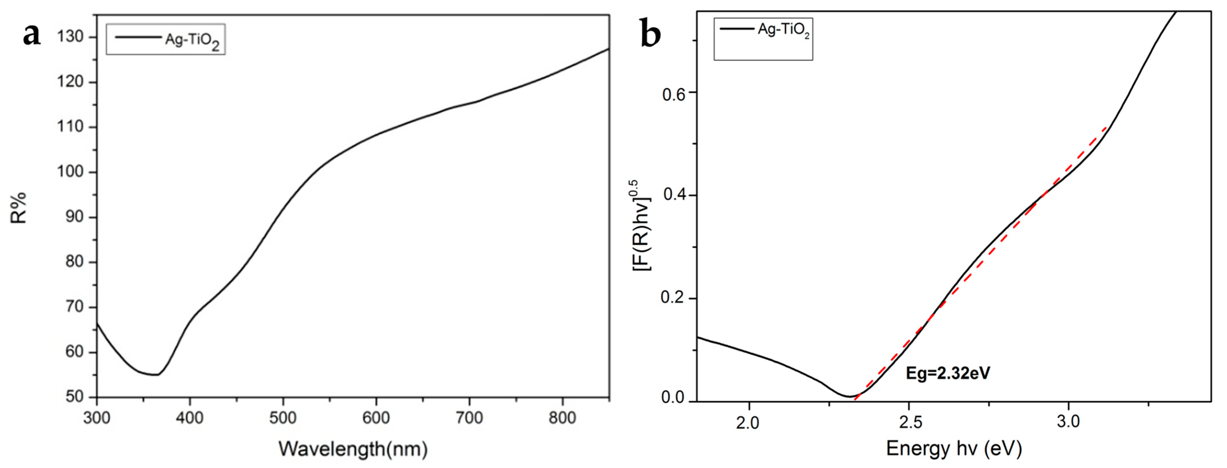
3.1.4. Energy Band Gap Estimation

The energy band gap of a semiconductor indirectly determines the energy that should be provided to excite an electron that leaves the valence band (VB) and transmits to the conduction band (CB). An accurate evaluation of the energy band gap is crucial to predicting the photophysical and photochemical properties of semiconductors [70]. The optical properties were analyzed in the wavelength range of 300–850 nm by UV-Vis diffuse reflectance spectra (DRS). The powder reflectance and the Eg were measured through the Kubelka-Munk (K-M) method, as it was previously described [32] (Figure 7a,b).
The Eg of the commercial pure Evonik P25 was measured, which was Eg = 3.1 eV [32], and the produced Ag-TiO2 NPs revealed that there is a decrease in Eg (Eg = 2.32 eV), in comparison with pure anatase, owing to chemical modification with Ag. This finding is perhaps associated with enhanced photocatalytic behavior visible-light irradiation, because under the chemically modified photocatalyst needed lower energy for its photoactivation, overcoming the Eg [70].

3.1.5. Dynamic Light Scattering (DLS)

The determination of the hydrodynamic diameter (Dh) of the polymeric IP network (microgel PNIPAM/PAA salt) particles was achieved through DLS, within a temperature range of 25–45 °C, at a constant pH value of ~7.4.
A red laser beam that was operating at 633 nm was used for the measurements. An amount of 1 mL of the microgel PNIPAM/PAA salt was placed in a proper DTS1070-type capillary cell (Malvern Instr., Malvern, UK) and measured. Volume phase transition temperature (VPTT) was defined as a direct, perpetual transition in the Dh volume, at the swollen phase and the collapsed phase [26,32,38]. The DLS data that are shown in Figure 8 indicate that the collapsed Dh of the microgel was ~322 nm, whereas the swollen Dh was ~720 nm. The VPTT was evaluated by plotting the first derivative of the particle diameter versus temperature [32]. We estimated the VPTT of the sample to be around 37.6 °C, and thus, a shift toward higher values was observed, compared with the VPTT that was reported for PNIPAM. Hence, this VPTT is considered suitable for applications in drug-delivery systems in biological environments, such as the human body. The selection of the copolymer and the optimal selection of the time interval among the addition of the two monomers of the graft-polymerization process can result in this shift. In temperatures below the VPTT, in an aqueous solution, the chains are still soluble, thanks to the presence of hydrogen bonds that are formed among water and amine chains. In temperatures over the VPTT, a significant amount of water is finally discarded from the microgel; thus, the Dh volume appears to shrink [32].
The aqueous solution of Ag-TiO2 NPs was measured at a pH of 7.4 and the corresponding zeta potential measurements revealed very stable suspensions, as demonstrated in Figure 9 (ZP = (−53.5 ± 6) mV).

3.1.6. TEM Analysis

The observation of the morphology of all the produced composite NPs and the IP network microgel/Ag-TiO2 NPs was performed with transmission electron microscopy (TEM). The images of the PNIPAM/PAA microgel/Ag-TiO2 are shown in Figure 10a–e. PNIPAM/PAA microgel/Ag-TiO2 composite was examined using TEM to visually determine the extent of Ag-TiO2 loading [38]. In the sample, several microgel particles appeared as clear objects, with sizes of approximately ~600 nm, but some particles baffled each other because of the TEM measurement, which led to a concentrated sample of hybrid microgels dispersion, under the intensity of the electron beam [71]. The measured diameter of the hybrid microgel particles might be lower than that was measured by DLS (see Figure 8), which might be attributed to a significant decrease in Dh on the drying microgel sample, placed on the grid for TEM measurement [71]. The observed dark black spots in the TEM image represent Ag-TiO2 nanoparticles that were loaded into the microgel (Figure 10a,c–e). Figure 10a,c,d show the typical morphology and surface of the produced composite Ag-TiO2/microgel NPs, compared with pure TiO2 (Figure 10f) and Ag NPs (Figure 10g). The TEM images of the hybrid microgels showed that Ag-TiO2 nanoparticles are uniformly distributed in the PNIPAM/PAA salt and not aggregated, as is clear in Figure 10a,b. Regarding the Ag-TiO2 NPs, the TEM images (Figure 10a,c–e) clearly show the sphere-like synthesized material. Observation revealed that TiO2 NPs were crystallized well, with a lattice-fringe spacing of 3.55, 2.34, 1.70, 0.24, 1.50 and 1.89 nm, and these findings well match the d-spacing of (101) (112) (105) (004) (213) and (200) planes of anatase TiO2. Ag NPs could be found on the surface TiO2 (in a decorated type) (Figure 10d,e,h,i) [11]. Ag NPs appear with a lattice-fringe spacing of 0.2 nm, which adequately matches the d-spacing of (002). The dopant Ag was well distributed. The Ag-TiO2 has many black–gray colored dots, in contrast. It is important to highlight that these TEM findings might verify the size scale of the Ag-TiO2 NPs, as was previously recorded by XRD [12,50,72]. The energy-dispersive spectroscopy (EDS) analysis of Ag-TiO2/microgel composite is also displayed in Figure 11. It seems that the Ag and Ti elements were incorporated and distributed in the polymer microgels, confirming that Ag-TiO2 NPs were successfully formed. Figure 10c,d clearly indicate the successful embedment of the Ag-TiO2 NPs in the polymeric network.

3.2. Photocatalytic Activity Experiments

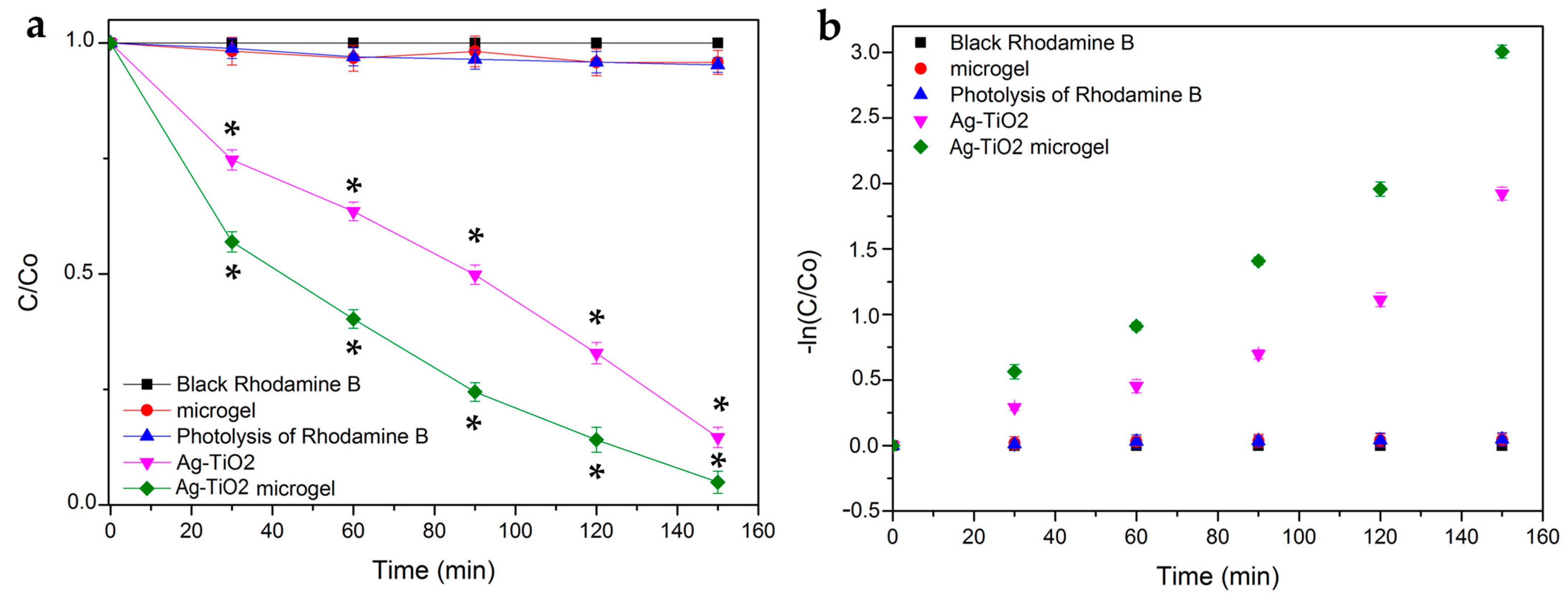
3.2.1. Photocatalytic Efficiency and Kinetics

RhB (C28H31CIN2O3) was chosen as a stable pollutant for the photocatalysis experiments to test the prepared TiO2 basis materials. RhB is a basic red dye of the xanthene class, which has been commonly used as a colorant in foodstuffs and in textiles. It is one of most hazardous types of textile waste and is considered as carcinogenic because it consists of radical compounds that potentially cause damage to the ecosystem [73]. Several inorganic semiconductor materials were used as photocatalysts to degrade wastewater, and many of them have been widely studied [8,9,13,74]. The photocatalytic experiments were implemented at room temperature and at pH of 7.4 (simulating the biological environment), in order to certify the photocatalytic ability of the Ag-TiO2 NPs and composites, before testing their biological effect on cancer cells. The photocatalytic efficiency of the produced materials was estimated under appropriate visible-light illumination.
Figure 12a depicts the photocatalytic activity of all the produced materials under visible irradiation for 150 min. Figure 12 shows that a significant amount of pollutant has been degraded at 150 min, indicated by the removal of RhB by more than over 95% for the case of the composite Ag-TiO2/microgel. Additionally, it is revealed that the produced composites are superior to Ag-TiO2 NPs regarding their photocatalytic activity under visible-light irradiation conditions, and this superiority was statistically significant, according to the Kruskal–Wallis test. This might be because the microgel could act as a bridging mean, keeping the photocatalyst closer to the pollutant compared with the case of its absence. However, pure Ag-TiO2 can also degrade the pollutant, but not as efficiently. Both Ag-TiO2 and Ag-TiO2/microgel show statistically significantly different behavior than the microgel itself, or the light itself (photolysis sample) leaves the pollutant RhB totally unaffected.
First-order kinetics, which were obtained through the Langmuir–Hinshelwood equation, were normalized for the reactions that occurred at the liquid–solid interface, following a standard methodology, which was previously described [18,32,75] (Figure 12b).
For the colored compounds, such as pigments, inks and dyes, their degradation rate commonly increases as the dye concentration increases, but as soon as it attains a critical specific concentration level, it begins to decrease. This reduction in the degradation rate with the increasing concentration of the pollutant could be attributed to the screening of UV-visible-light radiation by the dye molecules, before finally reaching the catalyst surface. Actually, the catalyst concentration can be adjusted for the concentration of the organic compound, so that it can be considerably adsorbed on the photocatalyst surface and thus efficiently degraded [76]. In order to be in accordance with the typical concentrations of the pollutants that are found in real wastewater, the common degradation studies also apply similar concentrations of the organic dyes, in the range of 10–200 mg/L. The same strategy was employed in the present study.
Table 1 gathers the evaluated photoinduced degradation rate constant (kapp) and the coefficient R2 of the linear regression that fit for all the tested materials. As shown through the R2 values, the linear kinetic model seems to satisfactorily fit in all the experimental data. The composite material exhibits the best photocatalytic performance. The intrinsic absorbance of RhB dye measured at 554 nm was used to calculate the removal efficiency by using Equation (1):
Removal   efficiency   ( % ) = C 0 C C 0 · 100

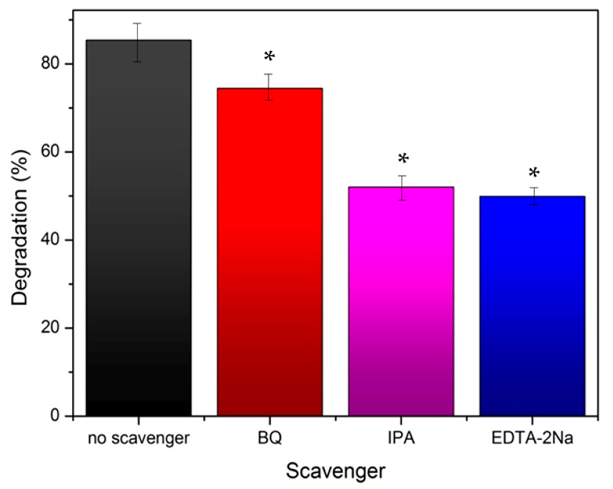
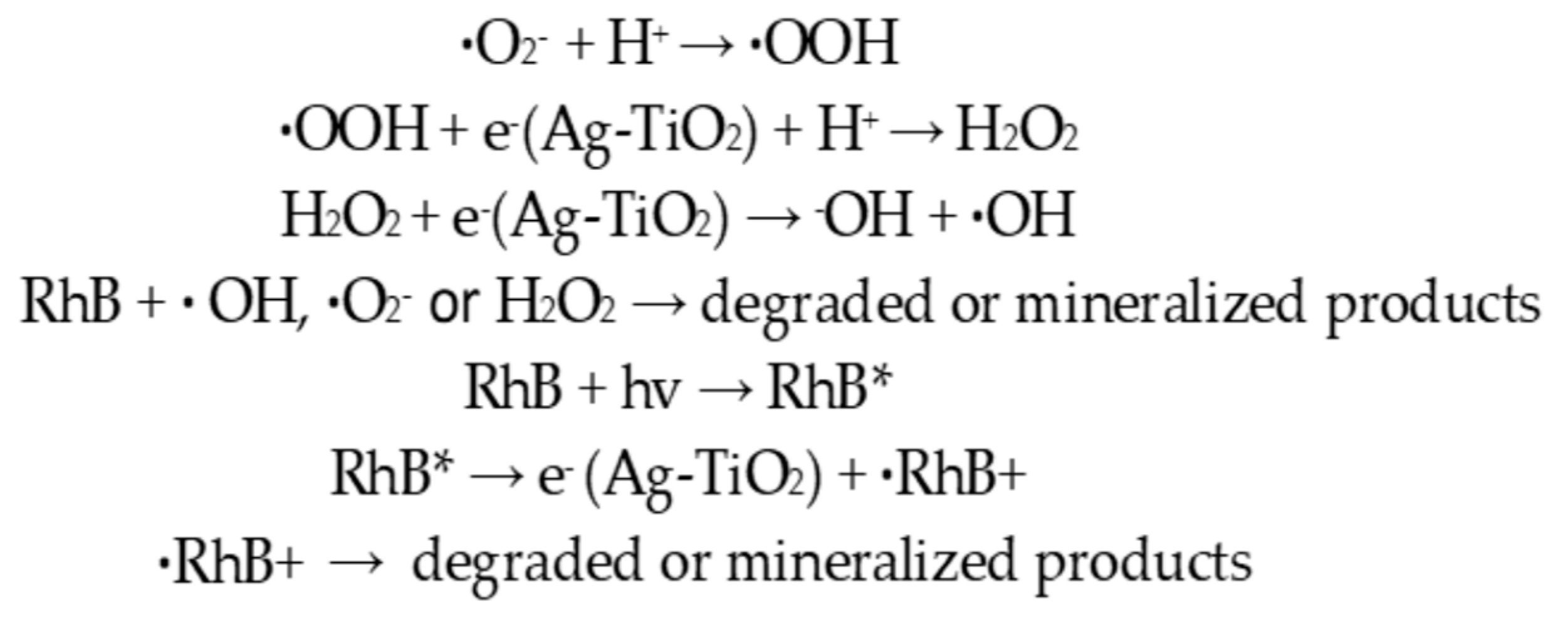
3.2.2. Effect of Radical Scavengers—Proposed Reaction Mechanism

In order to investigate the main reactive species, including h+, ·OH and ·O2, which were produced during the photocatalytic degradation of RhB, the trapping series of experiments were implemented in the presence of the radicals’ scavengers, determining the photocatalytic mechanism of the Ag-TiO2 NPs. Thus, ethylenediaminetetraacetic acid disodium salt (EDTA-2Na), benzoquinone (BQ) and isopropanol (IPA) were introduced, acting as scavengers, to test the effect of ·OH, ·O2 and h+ active species in the photocatalytic experiment, respectively [77,78] (Table 2).
The results demonstrated that the photocatalytic degradation efficiency of Ag-TiO2 NPs decreased in the presence of scavengers, and this decrease is statistically significant [73,78] (Figure 13). The photocatalytic studies with scavengers were performed under the same test conditions as those previously mentioned in the absence of them (see Section 3.2.1). The concentration of RhB/scavenger solutions were set at 0.001 M [53,78]. In Table 3, the calculated photoinduced degradation rate constant (kapp) and the R2 coefficient of the linear regression are presented, fitting for all the scavengers.
The presence of IPA led to a statistically significant reduction in the photodegradation, indicating hydroxyl radical as the dominant ROS species [53,78]. The photocatalytic degradation efficiency of Ag-TiO2 was inhibited more when EDTA-2Na was used, proving that the most reactive species of prepared Ag-TiO2 in the photodegradation process were h+ (Figure 14). In IPA trapping experiments suggested that ·OH was another important photocatalytic active agent in Ag-TiO2. By contrast, the function of ·O2 is the lowest in the presence of all the reactive agents, and it was obtained from the trapping experiments [77,79].

3.2.3. Photocatalytic Mechanism

Photocatalysis is a quite complex process. Generally, the photocatalytic reaction depends on the wavelength, the light (photon) energy and the type of the catalyst. The photocatalytic mechanism includes the following steps, according to the literature [8,13,17]:
(a)
Upon irradiation with an appropriate light source, providing energy at minimum equal to the value of the photocatalyst Eg, the electrons that exist on the VB are agitated and finally move to the CB of the semiconductor. As a consequence, positively charged holes are left in the VB of the semiconductor, oxidizing donor molecules and reacting with the available water molecules in order to generate hydroxyl radicals that have strong oxidizing potential, which is capable of degrading various pollutants or damaging or killing biomolecules, leading cells to undergo apoptosis.
(b)
The electrons of the CB are ready to react with the nearby dissolved oxygen species, forming superoxide ions; thus, these electrons can induce and mediate the redox reactions.
(c)
The electrons and the produced holes undergo consequent oxidation and reduction reactions with any species that are adsorbed on the semiconductor surface, giving the necessary products as separate for a short time.
According to a research report [75], depending on the reaction conditions, the holes and the ·OH radicals, ·O2, H2O2 and O2 play crucial roles during the photocatalytic reaction process. Especially, the increased photocatalytic activity of Ag-TiO2 NPs against RhB dye could be easily explained by the SPR effect of Ag and by the visible-light absorption by RhB on the catalyst surface (Figure 15). The SPR of Ag is responsible for the production of electrons and holes (separated charges) in the presence of visible light. Again, RhB*, excited by visible-light irradiation, can transfer electrons to the CB of the semiconductor (TiO2) and thus lead to the generation of RhB+, which is then degraded through oxidation. Additionally, the electrons (CB electrons of TiO2) are transferred to Ag particles. The electron that is generated by the SPR of Ag and the transferred electron from the CB of TiO2 (from RhB*) are absorbed by the O2, which can further produce ·O2. The superoxide radicals (·O2) further generate ·OOH, ·OH and ·H2O2, which can degrade the dye [13,14]. Figure 16 schematically represents the photocatalytic degradation of RhB, by Ag-TiO2 NPs, under visible-light irradiation.

3.3. Biological Effect

3.3.1. Effect on Cell Proliferation

MDA-MB-231 (cell line with metastatic profile), MCF-7 (low metastatic potential) and HEK293 (normal cells) were treated with increasing concentrations of Ag-TiO2 NPs, PNIPAM/PAA salt and PNIPAM/PAA microgel/Ag-TiO2. In particular, the concentration of the dispersions that was tested was selected as being in the range of 0–0.75 mg/mL. Additionally, the counting of the cells allowed the creation of growth rates, estimating the proliferation rate. There was no significant effect on the cell proliferation of HEK 293 cells in the presence of any of the produced material (Figure 17a–c), even after visible-light irradiation; thus, normal cells were left unaffected. Various other studies have indicated that primary rat hepatocytes, human lung fibroblasts and other types of normal cells were not affected by TiO2 NPs [12]. Moreover, Ag-TiO2 NPs, PNIPAM/PAA salt and PNIPAM/PAA microgel/Ag-TiO2 before and after irradiation had no effect on MCF-7 cells, whereas cisplatin significantly decreased this cell population (Figure 18a–c).
These findings are in accordance with some of our previous studies [3,32,43,44,45]. PNIPAM/PAA salt did not also affect the MDA-MB-231 cells (Figure 19a); thus, PNIPAM/PAA salt is not cytotoxic in any of the cell lines in this range of concentrations (Figure 17a, Figure 18a and Figure 19a). It is well known that PNIPAM and various copolymers based on PNIPAM are biocompatible materials, so our findings are commensurate with previous studies on other biological systems [80]. In the presence of Ag/TiO2 NPs, MDA-MB-231 cells seem to be functional (Figure 19b). It is clear that the cell proliferation of the MDA-MB-231 cells was decreased when 0.75 mg/mL of photoexcited Ag/TiO2 NPs were added (Figure 19b). Hence, a cell-dependent toxicity was observed [32,43,45], perhaps due to the different interactions between the photoactivated materials and cell membranes. Several studies support a similar finding in the anticancer behavior of Ag/TiO2 NPs, such as that of Ahamed et al., who found that A549 human pulmonary cancer cells were unaffected by Ag/TiO2 NPs, whereas there was significant toxicity in human liver cancer (HepG2) cells [12]. Photoactivated PNIPAM/PAA microgel/Ag-TiO2 NPs were more effective and reduced the cell population of MDA-MB-231 cells (Figure 19c). The embedment of Ag-TiO2 in the biocompatible microgel increased the efficacy of the nanoparticles to induce cell death or even to inhibit cell proliferation. The cell population was 50% lower in the presence of 0.50 mg/mL of photoexcited PNIPAM/PAA microgel/Ag-TiO2 NPs. Perhaps the microgel facilitates the release of Ag-TiO2 NPs close to cell membranes and favors the local photocatalytic action of Ag-TiO2 NPs [32]. Thus, the polymeric microgel is actually enhancing the photocatalytic efficiency of Ag-TiO2 NPs.
The selection of the tested materials was based partly on our previous systematic studies focusing on the titania-based materials (TiO2 [43,44], N/TiO2 [3,32] and Ag/TiO2 NPs [45]), optimizing their performance by chemically modifying TiO2 with silver and embedding them inside a thermoresponsive microgel. Thus, among several photocatalysts, TiO2 remains a promising biocompatible material, and for this reason, Ag/TiO2 was developed.
Because the ultimate goal of this research, and for our future plans, is to develop a drug-delivery system that can efficiently kill cancer cells, a polymeric material was critical to be added to the composite particles to provide the required properties and in particular to transfer the material to the target area. Thus, various materials could be used, such as liposomes [81], poly (N-vinyl caprolactam) (PNVCL) [82], chitosan [83] or PEO-b-PCL–DPPC chimeric nanocarriers [84,85]. PNIPAM/PAA microgel combines the required properties of the aforementioned materials, allowing the development of a material with a controlled size [41,42] that can adequately act at 37 °C, a temperature close to that of a human body, and therefore was considered among the most promising biopolymers to be used in our study in order to fulfill our future plans of designing a photoinduced targeted drug-delivery system with anticancer performance.

3.3.2. Effect on Cytotoxicity

MTT colorimetric assay was applied to investigate the viability of HEK293, MCF-7 and MDA-MB-231 cells, as the concentration of Ag-TiO2 NPs, PNIPAM/PAA salt and PNIPAM/PAA microgel/Ag-TiO2 NPs increased before and after of their photoactivation with visible light. By using well-known MTT protocols, the cell viability was estimated as a percentage ratio of the measured optical density of the treated cells to untreated ones. All the types of cells remained unaffected in the presence of PNIPAM/PAA salt or photoactivated PNIPAM/PAA salt, meaning that this material was not cytotoxic, at least in this range of concentrations (Figure 20a,b). Indeed, PNIPAM/PAA and other stimuli-responsive polymers are considered as suitable materials for biomedical applications, as drug carriers [86]. Ag-TiO2 NPs did not induce any cytotoxic effect on these cell lines (Figure 20c). The cell population was also unaffected; thus, this finding was in agreement with that of growth rates (Figure 17b, Figure 18b and Figure 19b). Photoactivated Ag-TiO2 NPs had no biological effect on HEK293 and MCF-7 (Figure 20d), whereas a gradual decrease in cell viability of MDA-MB-231 cells was revealed (Figure 20d). In particular, 0.5 mg/mL of photoactivated Ag/TiO2 NPs reduced MDA-MB-231 cell viability by 20% and 0.75 mg/mL by 30%. As is clear in Figure 16e, cell viability did not change in the presence of PNIPAM/PAA microgel/Ag-TiO2 NPs for all the cell lines. Photoactivated PNIPAM/PAA microgel/Ag-TiO2 NPs had no effect on the cell viability of HEK293 and MCF-7 cells (Figure 20f). Interestingly, under photoenhancement with visible light, the cell viability of MDA-MB-231 cells gradually decreases (Figure 20f). Even 0.25 mg/mL can reduce cell viability by 20%, and 0.75 mg/mL of the composite material finally induced a 50% decrease in cell viability (IC50). Hence, embedment in this thermoresponsive microgel increased the photocatalytic efficiency of Ag-TiO2 NPs. Thus, a cell-type selectivity was observed, where MDA-MB-231 was more vulnerable to these materials.
In order to further develop a drug-delivery system that can be efficient for in vivo treatments, it is crucial to design a 3D tissue model [87]. Our cell-line-based experiments are very promising, but in order to predict the behavior of the produced materials in vivo [88], adding an intermediate step using a 3D model (probably an alginic/gelatin-based scaffold-like system or organoids) is among our future plans.

4. Conclusions

Nanostructured Ag/TiO2 was produced via the sol-gel synthesis method and embedded within PNIPAM/PAA microgel, creating a thermoresponsive composite material. Full characterization, employing XRD, micro-Raman, FT-IR, UV-Vis, DLS and TEM confirmed the physicochemical properties and the morphology of the produced material, verifying the chemical modification of TiO2 with silver and the successful development of the composite material. The photocatalytic efficacy of the produced Ag-TiO2 was broadened, including also the visible-light range of the electromagnetic spectrum, thanks to doping with silver in that the produced Ag-TiO2 nanopowder revealed that there was a decrease in the energy band gap Eg (Eg = 2.32 eV) when it was compared with pure anatase (Eg = 3.1 eV). This decrease indicated that Ag-TiO2 had better photocatalytic activity under irradiation with visible light, because the chemically modified catalyst finally needed lower energy for its photoactivation. Anatase was found to be the dominant crystal phase of the Ag-TiO2 powders, as XRD and Raman analysis results displayed, while the composition of the produced materials was confirmed via FT-IR analysis. The average crystallite size of the produced nanoparticles was estimated to be in the nanoscale, particularly 9.52 nm, as it was obtained through XRD and their zeta potential (ZP = (−53.5 ± 6) mV), indicating their stability. The polymeric network was synthesized following the steps of a precipitation polymerization process, presenting a VPTT at 37.6 °C, which allows the controlled release of a pharmaceutic factor, such as TiO2, inside a biological system. Afterward, the Ag-TiO2 NPs were successfully embedded in the network (as indicated in TEM images). Hence, embedding the nanoparticles resulted in the formation of the composite material (PNIPAM/PAA microgel/Ag-TiO2 NPs), which could act as a thermoresponsive and innovative drug-delivery system.
Photocatalytic tests using RhB pollutant reassured the visible-light activation of the powders, with the composite material succeeding to almost totally degrade the RhB pollutant (over 95%), after 150 min of irradiation with visible light. Composite nanoparticles demonstrated better photocatalytic activity compared with Ag-TiO2 NPs (85.3%), as we expected, because of the structure of microgels. Microgels act like sponges of high porosity that trap the photocatalytic nanoparticles close to the pollutant. In this way, the photocatalytic reaction can take place faster, and the degradation of the pollutant is achievable. The implementation of the same photocatalytic experiments was implemented in the presence of radicals’ scavengers (ethylenediaminetetraacetic acid disodium salt (EDTA-2Na), benzoquinone (BQ) and isopropanol (IPA)), indicating the hydroxyl radical as the dominant reactive oxygen species, shedding light on the mechanism through which this photocatalyst acts.
Furthermore, for the analysis of their anticancer behavior, two breast cancer epithelial cell lines (MCF-7 and MDA-MB-231) and normal human embryonic kidney cells (HEK 293) were cultured and treated with the produced materials (the concentration was in the range of 0–0.75 mg/mL), under irradiation with visible light, and cell proliferation and cytotoxicity assays were employed. All the tested materials did not affect the cell proliferation rate or the cell viability before their photoactivation. This was a very promising result in that it is important to control the anticancer potential of the developed materials. Under visible-light irradiation, PNIPAM/PAA was still harmless for all the cell lines, while Ag-TiO2 NPs gradually decreased the cell proliferation and the MDA-MB-231 cell viability, leaving the MCF-7 and HEK293 cells unaffected. An amount of 0.50 mg/mL of photoactivated PNIPAM/PAA microgel/Ag-TiO2 NPs decreased the cell population of MDA-MB-231 cells by 50%.
Moreover, photoactivated PNIPAM/PAA microgel/Ag-TiO2 NPs had a more significant killing effect on MDA-MB-231 cells, with the IC50 being 0.75 mg/mL for this cell line, while at the same concentration, the cell viability of the MDA-MB-231 cells was decreased by 25–30% in the presence of Ag-TiO2 NPs, meaning that the composite material had enhanced anticancer efficiency.
Further clarification on the interactions between the different cell types would be in our future plans because a cell-dependent toxicity was observed. These promising findings will encourage us to continue to optimize our method in order to develop an alternative photodynamic cancer therapy, one that is based on a drug-delivery system that allows the controlled release of the photocatalytic anticancer material at the target area, maximizing the therapeutic effect on the cancer cells and avoiding the harmful side effects on healthy tissue, while reducing costs to the healthcare system supporting those treatments.

Author Contributions

Conceptualization, E.A.P.; methodology, N.P.-F., N.L. and E.A.P.; validation, N.P.-F., N.L., N.P., E.S., N.B., V.G.G., M.G. and E.A.P.; formal analysis, N.P.-F. and N.L.; investigation, N.P.-F., N.L., N.P., E.S. and N.B.; resources, E.A.P., M.G. and V.G.G.; writing-original draft preparation, N.P.-F and N.L.; writing-review and editing, N.P.-F., N.L., N.P., E.S., N.B., V.G.G., M.G. and E.A.P.; visualization, N.P.-F., N.L., N.P., E.S. and N.B.; supervision, E.A.P.; project administration, E.A.P.; funding acquisition, E.A.P. All authors have read and agreed to the published version of the manuscript.

Funding

This research was funded by the Hellenic Foundation for Research and Innovation (HFRI) (PhD Scholarship—Grant No. 2436 for N.P.-F.) and the General Secretariat for Research and Innovation (GSRI).

Institutional Review Board Statement

Not applicable.

Acknowledgments

The authors gratefully acknowledge useful discussions with Maria-Anna Gatou regarding Raman analysis.

Conflicts of Interest

The authors declare no conflict of interest. Additionally, the funders had no role in the design of the study; in the collection, analyses, or interpretation of data; in the writing of the manuscript; or in the decision to publish the results.

References

  1. Ziental, D.; Czarczynska-Coslinska, B.; Mlynarczyk, D.T.; Glowacka-Sobotta, A.; Stanisz, B.; Goslinski, T.; Sobotta, L. Titanium Dioxide Nanoparticles: Prospects and Applications in Medicine. Nanomaterials 2020, 10, 387. [Google Scholar] [CrossRef] [PubMed] [Green Version]
  2. Thevenot, P.; Cho, J.; Wavhal, D.; Timmons, R.B.; Tang, L. Surface chemistry influences cancer killing effect of TiO2 nanoparticles. Nanomed. Nanotechnol. Biol. Med. 2008, 4, 226–236. [Google Scholar] [CrossRef] [PubMed] [Green Version]
  3. Lagopati, N.; Evangelou, K.; Falaras, P.; Tsilibary, E.C.; Vasileiou, P.; Havaki, S.; Angelopoulou, A.; Pavlatou, E.A.; Gorgoulis, V.G. Nanomedicine: Photo-activated nanostructured titanium dioxide, as a promising anticancer agent. Pharmacol. Ther. 2021, 222, 107795. [Google Scholar] [CrossRef] [PubMed]
  4. Anucha, C.; Altin, I.; Bacaksiz, E.; Stathopoulos, V.N. Titanium dioxide (TiO₂)-based photocatalyst materials activity enhancment for contaminants of emerging concern (CECs) degradation: In the light of modification strategies. CEJ Adv. 2022, 10, 100262. [Google Scholar]
  5. Amin, S.-A.; Pazouki, M.; Hosseinnia, A. Synthesis of TiO2–Ag nanocomposite with sol–gel method and investigation of its antibacterial activity against E. coli. Powder Technol. 2009, 196, 241–245. [Google Scholar] [CrossRef]
  6. Abdelsalam, E.; Mohamed, Y.M.A.; Abdelkhalik, S.; El Nazer, H.A.; Attia Attia, Y. Photocatalytic oxidation of nitrogen oxides (NOx) using Ag and Pt doped TiO2 nanoparticles under visible light irradiation. Environ. Sci. Pollut. Res. 2020, 27, 35828–35836. [Google Scholar] [CrossRef]
  7. Ali, A.; Bano, S.; Poojary, S.S.; Priyadarshi, R.; Choudhary, A.; Kumar, D.; Negi, Y.-S. Comparative analysis of TiO2 and Ag nanoparticles on xylan/chitosan conjugate matrix for wound healing application. Int. J. Polym. Mater. 2020, 75, 376–385. [Google Scholar] [CrossRef]
  8. Ameta, R.; Solanki, M.; Benjamin, S.; Ameta, S.C. Advanced Oxidation Processes for Waste Water Treatment Emerging Green Chemical Technology, 1st ed.; Academic Press: Cambridge, MA, USA, 2018. [Google Scholar]
  9. Khan, F.A. Applications of Nanomaterilas in Human Health, 1st ed.; Springer: Singapore, 2020. [Google Scholar]
  10. Mikhailova, E.O. Silver Nanoparticles: Mechanism of Action and Probable Bio-Application. J. Funct. Biomater. 2020, 11, 84. [Google Scholar] [CrossRef]
  11. Chakhtouna, H.; Benzeid, H.; Zari, N.; El Kacem Qaiss, A.; Bouhfid, R. Recent progress on Ag/TiO2 photocatalysts: Photocatalytic and bactericidal behaviors. Environ. Sci. Pollut. Res. 2021, 28, 44638–44666. [Google Scholar] [CrossRef]
  12. Ahamed, M.; Majeed Khan, M.; Akhtar, M.-J.; Alhadlaq, H.A.; Alshamsan, A. Ag-doping regulates the cytotoxicity of TiO2 nanoparticles via oxidative stress in human cancer cells. Sci. Rep. 2017, 7, 17662. [Google Scholar] [CrossRef] [Green Version]
  13. Khan, M.; Sohn, Y.; Pradhan, D. Nanocomposites for Visible Light-Induced Photocatalysis; Springer International Publishing: Berlin/Heidelberg, Germany, 2017. [Google Scholar]
  14. Bahadur, J.; Agrawal, S.; Panwar, V.; Parveen, A.; Pal, K. Antibacterial Properties of Silver Doped TiO2 Nanoparticles Synthesized via Sol-Gel Technique. Macromol. Res. 2016, 24, 488–493. [Google Scholar] [CrossRef]
  15. Mandari, K.K.; Kwak, B.S.; Kumar, A.; Police, R.; Kang, M. In-situ photo-reduction of silver particles and their SPR effect in enhancing the photocatalytic water splitting of Ag2O/TiO2 photocatalysts under solar light irradiation: A case study. Mat. Res. Bull. 2017, 95, 515–524. [Google Scholar] [CrossRef]
  16. Gupta, K.; Singh, R.; Pandey, A.; Pandey, A. Photocatalytic antibacterial performance of TiO2 and Ag-doped TiO2 against S. aureus. P. aeruginosa and E. coli. Beilstein J. Nanotechnol. 2013, 4, 345–351. [Google Scholar] [CrossRef] [PubMed] [Green Version]
  17. Zhang, J.; Tian, B.; Wang, L.; Xing, M.; Lei, J. Photocatalysis: Fundamentals, Materials and Applications; Springer: Berlin/Heidelberg, Germany, 2018. [Google Scholar]
  18. Ainali, N.M.; Kalaronis, D.; Evgenidou, E.; Bikiaris, D.N.; Lambropoulou, D.A. Insights into Biodegradable Polymer-Supported Titanium Dioxide Photocatalysts for Environmental Remediation. Macromol 2021, 1, 201–233. [Google Scholar] [CrossRef]
  19. Cervantes, F.J.; Ramírez-Montoya, L.A. Immobilized Nanomaterials for Environmental Applications. Molecules 2022, 27, 6659. [Google Scholar] [CrossRef]
  20. Ameta, R.; Solanki, M.; Benjamin, S. Photocatalysis. In Advanced Oxidation Processes for Waste Water Treatment; Digital Science & Research Solutions, Dimensions: London, UK, 2018; pp. 135–175. [Google Scholar]
  21. Wei, M.; Gao, Y.; Li, X.; Serpe, M. Stimuli-responsive polymers and their applications. Polym. Chem. 2017, 8, 127–143. [Google Scholar] [CrossRef] [Green Version]
  22. Ferguson, C.T.J.; Huber, Ν.; Landfester, Κ.; Zhang, Κ. Dual-Responsive Photocatalytic Polymer Nanogels. Angew Chem. 2019, 58, 10677–10681. [Google Scholar] [CrossRef]
  23. Cabane, E.; Zhang, X.; Langowska, K.; Palivan, C.; Meier, W. Stimuli-Responsive Polymers and Their Applications in Nanomedicine. Biointerphases 2012, 7, 9. [Google Scholar] [CrossRef] [Green Version]
  24. Van Gheluwe, L.; Chourpa, I.; Gaigne, C.; Munnier, E. Polymer-Based Smart Drug Delivery Systems for Skin Application and Demonstration of Stimuli-Responsiveness. Polymers 2021, 13, 1285. [Google Scholar] [CrossRef]
  25. Fang, R.; Pi, J.; Wei, T.; Ali, A.; Guo, L. Stimulus-Responsive Polymers Based on Polypeptoid Skeletons. Polymers 2021, 13, 2089. [Google Scholar] [CrossRef]
  26. Zhilin, D.; Pich, A. Nano- and microgels: A review for educators. CTI 2021, 3, 155–167. [Google Scholar] [CrossRef]
  27. Bajpai, A.K.; Shukla, S.K.; Bhanu, S.; Kankane, S. Responsive polymers in controlled drug delivery. Prog. Polym. Sci. 2008, 33, 1088–1118. [Google Scholar] [CrossRef]
  28. Chu, S.; Shi, X.; Tian, Y.; Gao, F. pH-Responsive Polymer Nanomaterials for Tumor Therapy. Front. Oncol. 2022, 12, 855019. [Google Scholar] [CrossRef] [PubMed]
  29. Li, G.; Varga, I.; Kardos, A.; Dobryden, I.; Claesson, P.M. Temperature-Dependent Nanomechanical Properties of Adsorbed Poly-NIPAm Microgel Particles Immersed in Water. Langmuir 2021, 37, 1902–1912. [Google Scholar] [CrossRef]
  30. Aflori, M. Smart Nanomaterials for Biomedical Applications-A Review. Nanomaterials 2021, 11, 396. [Google Scholar] [CrossRef]
  31. Pham, S.H.; Choi, Y.; Choi, J. Stimuli-Responsive Nanomaterials for Application in Antitumor Therapy and Drug Delivery. Pharmaceutics 2020, 12, 630. [Google Scholar] [CrossRef]
  32. Galata, E.; Georgakopoulou, E.A.; Kassalia, M.-E.; Papadopoulou-Fermeli, N.; Pavlatou, E.A. Development of Smart Composites Based on Doped-TiO2 Nanoparticles with Visible Light Anticancer Properties. Materials 2019, 12, 2589. [Google Scholar] [CrossRef] [Green Version]
  33. Wang, D.; Han, X.; Dong, B.; Shi, F. Stimuli responsiveness, propulsion and application of the stimuli-responsive polymer based micromotor. Appl. Mater. Today 2021, 25, 101250. [Google Scholar] [CrossRef]
  34. Chen, H.; Hsieh, Y.-L. Dual Temperature- and pH-Sensitive Hydrogels from Interpenetrating Networks and Copolymerization of N-Isopropylacrylamide. J. Polym. Sci. Part A Polym. Chem. 2004, 42, 3293–3301. [Google Scholar] [CrossRef]
  35. Futscher, M.H.; Philipp, M.; Müller-Buschbaum, P.; Schulte, A. The Role of Backbone Hydration of Poly(N-isopropyl acrylamide) Across the Volume Phase Transition Compared to its Monomer. Sci. Rep. 2017, 7, 17012. [Google Scholar] [CrossRef] [Green Version]
  36. Haq, M.A.; Su, Y.; Wang, D. Mechanical properties of PNIPAM based hydrogels: A review. Mater. Sci. Eng. C Mater. Biol. Appl. 2017, 70 Pt 1, 842–855. [Google Scholar] [CrossRef]
  37. Begum, R.; Naseem, K.; Farooqi, Z. A review of responsive hybrid microgels fabricated with silver nanoparticles: Synthesis, classification, characterization and applications. J. Solgel Sci. Technol. 2016, 77, 497–515. [Google Scholar] [CrossRef]
  38. Coutinho, C.A.; Harrinauth, R.K.; Gupta, V.K. Settling characteristics of composites of PNIPAM microgels and TiO2 nanoparticles. Colloids Surf. A Physicochem. Eng. Asp. 2008, 318, 111–121. [Google Scholar] [CrossRef]
  39. Sun, H.; Kabb, C.P.; Sims, M.B.; Sumerlin, B.S. Architecture-transformable polymers: Reshaping the future ofstimuli-responsive polymers. Prog. Polym. Sci. 2019, 89, 61–75. [Google Scholar] [CrossRef]
  40. Kawaguchi, H. On Going to a New Era of Microgel Exhibiting Volume Phase Transition. Gels 2020, 6, 26. [Google Scholar] [CrossRef] [PubMed]
  41. Agrawal, G.; Agrawal, R. Stimuli-Responsive Microgels and Microgel-Based Systems: Advances in the Exploitation of Microgel Colloidal Properties and Their Interfacial Activity. Polymers 2018, 10, 418. [Google Scholar] [CrossRef] [PubMed] [Green Version]
  42. Gao, Q.; Wang, C.; Liu, H.; Wang, C.; Liu, X.; Tong, Z. Suspension polymerization based on inverse Pickering emulsion droplets for thermo-sensitive hybrid microcapsules with tunable supracolloidal structures. Polymer 2009, 50, 2587–2594. [Google Scholar] [CrossRef]
  43. Lagopati, N.; Tsilibary, E.P.; Falaras, P.; Papazafiri, P.; Pavlatou, E.A.; Kotsopoulou, E.; Kitsiou, P. Effect of nanostructured TiO₂ crystal phase on photoinduced apoptosis of breast cancer epithelial cells. Int. J. Nanomed. 2014, 9, 3219–3230. [Google Scholar]
  44. Lagopati, N.; Kitsiou, P.; Kontos, A.; Venieratos, P.; Kotsopoulou, E.; Kontos, A.; Dionysiou, D.; Pispas, S.; Tsilibary, E.; Falaras, P. Photo-induced treatment of breast epithelial cancer cells using nanostructured titanium dioxide solution. J. Photochem. Photobiol. A Chem. 2010, 214, 215–223. [Google Scholar] [CrossRef]
  45. Lagopati, N.; Kotsinas, A.; Veroutis, D.; Evangelou, K.; Papaspyropoulos, A.; Arfanis, M.; Falaras, P.; Kitsiou, P.V.; Pateras, I.; Bergonzini, A.; et al. Effect of Silver-modified Nanostructured Titanium Dioxide in Cancer. Cancer Genom. Proteom. 2021, 18 (Suppl. S3), 425–439. [Google Scholar] [CrossRef]
  46. Kumar, A.; Pandey, G. A review on the factors affecting the photocatalytic degradation of hazardous materials. Mater. Sci. Eng. Int. J. 2017, 1, 106–114. [Google Scholar] [CrossRef] [Green Version]
  47. Piccinini, F.; Tesei, A.; Arienti, C.; Bevilacqua, A. Cell Counting and Viability Assessment of 2D and 3D Cell Cultures: Expected Reliability of the Trypan Blue Assay. Biol. Proced. Online 2017, 19, 12. [Google Scholar] [CrossRef] [PubMed]
  48. Acar, E.; Sun, L. A generalized Kruskal–Wallis test incorporating group uncertainty with application to genetic association studies. Biometrics 2019, 69, 427–435. [Google Scholar] [CrossRef] [PubMed] [Green Version]
  49. Wu, W.-Y.; Hsu, C.-F.; Wu, M.-J.; Chen, C.-N.; Huang, J.-J. Ag–TiO2 composite photoelectrode for dye-sensitized solar cell. Appl. Phys. A 2017, 123, 357. [Google Scholar] [CrossRef]
  50. Pu, S.; Hou, Y.; Chen, H.; Deng, D.; Yang, Z.; Xue, S.; Zhu, R.; Diao, Z.; Chu, W. An Efficient Photocatalyst for Fast Reduction of Cr(VI) by Ultra-Trace Silver Enhanced Titania in Aqueous Solution. Catalysts 2018, 8, 251. [Google Scholar] [CrossRef] [Green Version]
  51. Duan, Y.; Ma, J.; Liu, J.; Qiang, L.; Xue, J. Facile Synthesis of Thermo-responsive TiO2/PNIPAM Composite with Switchable Photocatalytic Performance. Fibers Polym. 2020, 21, 717–723. [Google Scholar] [CrossRef]
  52. Yu, C.; Tang, J.; Liu, X.; Ren, X.; Zhen, M.; Wang, L. Green Biosynthesis of Silver Nanoparticles Using Eriobotrya japonica (Thunb.) Leaf Extract for Reductive Catalysis. Materials 2019, 12, 189. [Google Scholar] [CrossRef] [Green Version]
  53. Ashkarran, A.; Aghigh, S.; Kavianipour, M.; Farahani, N. Visible light photo-and bioactivity of Ag/TiO2 nanocomposite with various silver contents. Curr. Appl. Phys. 2011, 11, 1048–1055. [Google Scholar] [CrossRef]
  54. Ali, T.; Ahmed, A.; Alam, U.; Uddin, I.; Tripathi, P.; Muneer, M. Enhanced photocatalytic and antibacterial activities of Ag-doped TiO2 nanoparticles under visible light. Mater. Chem. Phys. 2018, 212, 325–335. [Google Scholar] [CrossRef]
  55. Zdravković, A.; Nikolić, L.; Ilić-Stojanović, S.; Nikolić, V.; Savić, S.; Kapor, A. The evaluation of temperature and pH influences on equilibrium. Hem. Ind. 2017, 71, 395–405. [Google Scholar] [CrossRef] [Green Version]
  56. Assaf, S.M.; Abul-Haija, Y.M.; Fares, M.M. Versatile Pectin Grafted Poly (N-isopropylacrylamide); Modulated Targeted Drug Release. J. Macromol. Sci.-Pure Appl. Chem. 2011, 48, 493–502. [Google Scholar] [CrossRef]
  57. Thakur, S.; Arotiba, O. Synthesis, characterization and adsorption studies of an acrylic acid-grafted sodium alginate-based TiO2 hydrogel nanocomposite. Adsorpt. Sci. Technol. 2017, 36, 458–477. [Google Scholar] [CrossRef]
  58. Tang, S.; Zhao, Y.; Wang, H.; Wang, Y.; Zhu, H.; Chen, Y.; Chen, S.; Jin, S.; Yang, Z.; Li, P.; et al. Preparation of the Sodium Alginate-g-(PolyacrylicAcid-co-Allyltrimethylammonium Chloride) Polyampholytic Superabsorbent Polymer and Its Dye Adsorption Property. Mar. Drugs 2018, 476, 1–20. [Google Scholar]
  59. Wang, S.; Lin, L.; Zhu, A. Preparation of multisensitive poly (N-isopropylacrylamide-co-acrylic acid)/TiO2 composites for degradation of methyl orange. Eur. Polym. J. 2011, 47, 1168–1175. [Google Scholar] [CrossRef]
  60. Hirashima, Y.; Suzuki, A. Formation and destruction of hydrogen bonds in gels and in aqueous solutions of N-isopropylacrylamide and sodium acrylate observed by ATR-FTIR spectroscopy. J. Colloid Interface Sci. 2007, 312, 14–20. [Google Scholar] [CrossRef] [PubMed]
  61. Schilli, C.; Zhang, M.; Rizzardo, E.; Thang, S.; Chong, Y.; Edwards, K.; Karlsson, G.; Muller, H. A New Double-Responsive Block Copolymer Synthesized via RAFT Polymerization: Poly(N-isopropylacrylamide)-block-poly(acrylic acid). Macromolecules 2004, 37, 7861–7866. [Google Scholar] [CrossRef]
  62. Yamauc, H.; Maeda, Y. LCST and UCST Behavior of Poly(N-isopropylacrylamide) in DMSO/Water Mixed Solvents Studied by IR and Micro-Raman Spectroscopy. J. Phys. Chem. B 2007, 111, 12964–12968. [Google Scholar] [CrossRef]
  63. Kunnamareddy, M.; Diravidamani, B.; Rajendran, R.; Singaram, B.; Varadharajan, K. Synthesis of silver and sulphur codoped TiO2 nanoparticles for photocatalytic degradation of methylene blue. Mater. Sci. Mater. Electron. 2018, 29, 18111–18119. [Google Scholar] [CrossRef]
  64. Rajkumar, S.; Venkatraman, M.; Suguna, K.; Karuppasamy, P.; Senthil Pandian, M.; Ramasamy, P. Synthesis of Ag-incorporated TiO2 nanoparticles by simple green approach as working electrode for dye sensitized solar cells. J. Mater. Sci. Mater. Electron. 2022, 33, 4965–4973. [Google Scholar] [CrossRef]
  65. Mohamed, H.S.; Rabia, M.; Zhou, X.-G.; Qin, X.-S.; Khabiri, G.; Shaban, M.; Younus, H.; Taha, S.; Hu, Z.-Y.; Liu, J.; et al. Phase-junction Ag/TiO2 nanocomposite as photocathode for H2. J. Mater. Sci. Technol. 2021, 83, 179–187. [Google Scholar] [CrossRef]
  66. Komaraiah, D.; Radha, E.; Sivakumar, J.; Ramana Reddy, M.; Sayanna, R. Photoluminescence and photocatalytic activity of spin coated Ag+ doped anatase TiO2 thin films. Opt. Mater. 2020, 108, 110401. [Google Scholar] [CrossRef]
  67. Nigro, V.; Ripanti, F.; Angelini, R.; Sarra, A.; Bertoldo, M.; Buratti, E.; Postorino, P.; Ruzicka, B. Molecular mechanisms driving the microgels behaviour: A Raman spectroscopy and dynamic light scattering study. J. Mol. Liq. 2019, 284, 718–724. [Google Scholar] [CrossRef] [Green Version]
  68. Nigro, V.; Angelini, R.; Rosi, B.; Bertoldo, M.; Buratti, E.; Casciardi, S.; Sennato, S.; Ruzicka, B. Study of network composition in interpenetrating polymer networks of poly(N isopropylacrylamide) microgels: The role of poly(acrylic acid). J. Colloid Interface Sci. 2019, 545, 210–219. [Google Scholar] [CrossRef] [PubMed]
  69. Sanz, B.; Bilderling, C.; Tuninetti, J.; Pietrasanta, L.; Mijangos, C.; Longo, G.; Azzaroni, O.; Giuss, J. Thermally-Induced Softening of PNIPAm-Based Nanopillar Arrays. Soft Matter 2017, 13, 2453–2464. [Google Scholar] [CrossRef] [PubMed]
  70. Macyk, W. How To Correctly Determine the Band Gap Energy of Modified Semiconductor Photocatalysts Based on UV−Vis Spectra. J. Phys. Chem. Lett. 2018, 9, 6814–6817. [Google Scholar]
  71. Iqbal, S.; Zahoor, C.; Musaddiq, S.; Musaddiq, S.; Hussain, M.; Begum, R.; Irfan, A.; Azam, M.; Farooqi, Z.H. Silver nanoparticles stabilized in polymer hydrogels for catalytic degradation of azo dyes. Ecotoxicol. Environ. Saf. 2020, 202, 110924. [Google Scholar] [CrossRef]
  72. Ren, Y.; Xing, S.; Wang, J.; Ling, Y.; Zhao, D.; Zhao, D.; Wang, H.; Wang, N.; Jiang, W.; Wu, S.; et al. Weak-light-driven Ag–TiO2 photocatalyst and bactericide prepared by coprecipitation with effective Ag doping and deposition. Opt. Mater. 2022, 124, 111993. [Google Scholar] [CrossRef]
  73. Ardila-Leal, L.D.; Poutou-Piñales, R.A.; Pedroza-Rodríguez, A.M.; Quevedo-Hidalgo, B.E. A Brief History of Colour, the Environmental Impact of Synthetic Dyes and Removal by Using Laccases. Molecules 2021, 26, 3813. [Google Scholar] [CrossRef]
  74. Le, T.-T.-T.; Tran, T.-D. Photocatalytic Degradation of Rhodamine B by C and N Co-doped TiO2 Nanoparticles under Visible-Light Irradiation. J. Chem. 2020, 2020, 4310513. [Google Scholar] [CrossRef]
  75. Alhaji, M.-H.; Sanaullah, K.; Salleh, S.-F.; Baini, R.; Lim, S.-F.; Rigit, A.-R.-H.; Said, K.-A.-M.; Khan, A. Photo-oxidation of pre-treated palm oil mill Effluent using cylindrical column immobilized photoreactor. Process Saf. Environ. Prot. 2018, 117, 180–189. [Google Scholar] [CrossRef]
  76. Chiu, Y.-H.; Chang, T.-F.M.; Chen, C.-Y.; Sone, M.; Hsu, Y.-J. Mechanistic Insights into Photodegradation of Organic Dyes Using Heterostructure Photocatalysts. Catalysts 2019, 9, 430. [Google Scholar] [CrossRef] [Green Version]
  77. Jaafar, N.; Jalil, A.; Triwahyono, S.; Efendi, J.; Mukti, R.; Jusoh, R.; Jusoh, N.; Karim, A.; Salleh, N.; Suendo, V. Direct in situ activation of Ag0nanoparticles in synthesis of Ag/TiO2 and its photoactivity. Appl. Surf. Sci. 2015, 338, 75–84. [Google Scholar] [CrossRef]
  78. Ratshiedana, R.; Fakayode, O.; Kumar Mishra, A.; Tawanda Kuvarega, A. Visible-light photocatalytic degradation of tartrazine using hydrothermal synthesized Ag-doped TiO2 nanoparticles. J. Water Process. Eng. 2021, 44, 102372. [Google Scholar] [CrossRef]
  79. Liang, H.; Jia, Z.; Zhang, H.; Wang, X.; Wang, J. Photocatalysis oxidation activity regulation of Ag/TiO2 composites evaluated by the selective oxidation of Rhodamine B. Appl. Surf. Sci. 2017, 442, 1–10. [Google Scholar] [CrossRef]
  80. Lanzalaco, S.; Armelin, E. Poly(N-isopropylacrylamide) and Copolymers: A Review on Recent Progresses in Biomedical Applications. Gels 2017, 3, 36. [Google Scholar] [CrossRef]
  81. Chihara, Y.; Fujimoto, K.; Kondo, H.; Moriwaka, Y.; Sasahira, T.; Hirao, Y.; Kuniyasu, H. Anti-tumor effects of liposome-encapsulated titanium dioxide in nude mice. Pathobiology 2007, 74, 353–358. [Google Scholar] [CrossRef]
  82. Voycheva, C.; Slavkova, M.; Popova, T.; Tzankova, D.; Tosheva, A.; Aluani, D.; Tzankova, V.; Ivanova, I.; Tzankov, S.; Spassova, I.; et al. Synthesis and characterization of PnVCL grafted agar with potential temperature-sensitive delivery of Doxorubicin. J. Drug Deliv. Sci. Technol. 2022, 76, 103725. [Google Scholar] [CrossRef]
  83. Castillo, B.E.; Prokhorov, E.; Luna-Bárcenas, G.; Kovalenko, Y. Potential Use of Chitosan-TiO2 Nanocomposites for the Electroanalytical Detection of Imidacloprid. Polymers 2022, 14, 1686. [Google Scholar] [CrossRef]
  84. Pippa, N.; Naziris, N.; Stellas, D.; Massala, C.; Zouliati, K.; Pispas, S.; Demetzos, C.; Forys, A.; Marcinkowski, A.; Trzebicka, B. PEO-b-PCL grafted niosomes: The cooperativilty of amphiphilic components and their properties in vitro and in vivo. Colloids Surf. B Biointerfaces 2019, 177, 338–345. [Google Scholar] [CrossRef]
  85. Pippa, N.; Kaditi, E.; Pispas, S.; Demetzos, C. PEO-b-PCL–DPPC chimeric nanocarriers: Self-assembly aspects in aqueous and biological media and drug incorporation. Soft Matter 2013, 9, 4073–4082. [Google Scholar] [CrossRef]
  86. Chen, Y.; Gao, Y.; da Silva, L.P.; Pirraco, R.P.; Ma, M.; Yang, L.; Reis, R.L.; Chen, J. A thermo-/pH-responsive hydrogel (PNIPAM-PDMA-PAA) with diverse nanostructure and gel behaviors as a general drug carrier for drug release. Polym. Chem. 2018, 9, 4063–4072. [Google Scholar] [CrossRef]
  87. Nii, T.; Makino, K.; Tabata, Y. Three-Dimensional Culture System of Cancer Cells Combined with Biomaterials for Drug Screening. Cancers 2020, 12, 2754. [Google Scholar] [CrossRef] [PubMed]
  88. Brancato, V.; Oliveira, J.M.; Correlo, V.M.; Reis, R.L.; Kundu, S.C. Could 3D models of cancer enhance drug screening? Biomaterials 2020, 232, 119744. [Google Scholar] [CrossRef] [PubMed]
Figure 1. Schematic representation of the synthesis of PNIPAM/PAA microgel by surfactant-free precipitation polymerization.
Pharmaceutics 15 00135 g001
Figure 2. Schematic representation of the synthesis of Ag-TiO2 nanoparticles.
Pharmaceutics 15 00135 g002
Figure 3. Schematic representation of the synthesis of composite nanoparticles.
Pharmaceutics 15 00135 g003
Figure 4. XRD patterns of Ag-TiO2 nanoparticles (in black), PNIPAM/PAA salt (in red) and PNIPAM/PAA microgel/Ag-TiO2 (in blue).
Pharmaceutics 15 00135 g004
Figure 5. FT-IR spectra of Ag-TiO2 nanoparticles (in black), PNIPAM/PAA salt (in red) and PNIPAM/PAA microgel/Ag-TiO2 composite (in blue).
Pharmaceutics 15 00135 g005
Figure 6. Raman spectra of (a) Ag-TiO2 nanoparticles compared with commercially available Evonik P25 TiO2 anatase and (b) PNIPAM/PAA microgel/Ag-TiO2. The insets depict a part of the Raman spectrum of PNIPAM/PAA salt microgel.
Pharmaceutics 15 00135 g006
Figure 7. (a) F(R) reflectance as a function of wavelength for Ag-TiO2 nanoparticles. (b) Optical energy band gap (Eg) of the Ag-TiO2 nanoparticles.
Pharmaceutics 15 00135 g007
Figure 8. Size (Dh—hydrodynamic diameter) of the microgel PNIPAM/PAA salt, as a function of temperature through DLS. The suspension pH was equal to 7.4. The red dash line underlines the VPTT, which corresponds to 37.6 °C. The data represent means ± standard deviation from three experiments.
Pharmaceutics 15 00135 g008
Figure 9. Zeta potential distribution of Ag-TiO2 nanoparticles via DLS. The suspension pH was equal to 7.4.
Pharmaceutics 15 00135 g009
Figure 10. Transmission electron microscopy images of (a) the spherical particle of microgel with embedded Ag-TiO2 nanoparticles, (b) the PNIPAM/PAA microgel Ag-TiO2 NPs, (c) the outline of a microgel particle, (d) a microgel particle with embedded Ag-TiO2 nanoparticles and lattice measurement spacing, (e) embedded Ag-TiO2 nanoparticles with lattice measurement spacing, (f) TiO2 nanoparticles, (g) Ag nanoparticles and (h,i) TiO2-decorated Ag nanoparticles.
Pharmaceutics 15 00135 g010
Figure 11. Energy-dispersive spectroscopy (EDS) analysis of Ag-TiO2/microgel composite.
Pharmaceutics 15 00135 g011
Figure 12. (a) Degradation curves of rhodamine B for Ag-TiO2, PNIPAM-co-PAA microgel and PNIPAM -co-PAA Ag-TiO2 composite as a function of time, under visible-light irradiation. The phenomenon of rhodamine B photolysis and its degradation in the dark are also studied. The data represent means ± standard deviation from three experiments. In these series of experiments, * p < 0.05 was considered statistically significant. (b) Photocatalytic kinetics of the degradation of rhodamine B for Ag-TiO2, PNIPAM-co-PAA microgel and PNIPAM-co-PAA Ag-TiO2, according to a linear pseudo-first-order model.
Pharmaceutics 15 00135 g012
Figure 13. (a) Photocatalytic degradation curves of RhB in the presence of Ag-TiO2 and the radicals’ scavengers (ethylenediaminetetraacetic acid disodium salt (EDTA-2Na), benzoquinone (BQ) and isopropanol (IPA)). The data represent means ± standard deviation from three experiments. In these series of experiments, * p < 0.05 was considered statistically significant. (b) Photocatalytic kinetics of the same samples, according to a linear pseudo-first-order model.
Pharmaceutics 15 00135 g013
Figure 14. Percentage of degradation of rhodamine B by Ag-TiO2 nanoparticles (in black) and also in the presence of scavengers: ethylenediaminetetraacetic acid disodium salt (EDTA-2Na) (in blue), benzoquinone (BQ) (in red) and isopropanol (IPA) (in purple). The data represent means ± standard deviation from three experiments. In these series of experiments, * p < 0.05 was considered statistically significant.
Pharmaceutics 15 00135 g014
Figure 15. Photocatalytic degradation reactions of RhB.
Pharmaceutics 15 00135 g015
Figure 16. Schematic representation of the photocatalytic degradation of RhB by Ag–TiO2 NPs, under visible-light irradiation.
Pharmaceutics 15 00135 g016
Figure 17. Growth rates of HEK293 cell line, with the addition of (a) PNIPAM/PAA salt, (b) Ag-TiO2 NPs and (c) PNIPAM/PAA microgel/Ag-TiO2. There was no effect on cell proliferation, as the concentration of each of the tested materials increased. Additionally, irradiation with visible light did not affect their proliferation. In these series of experiments, p < 0.05 was considered statistically significant.
Pharmaceutics 15 00135 g017
Figure 18. Growth rates of MCF-7 cell line, with the addition of (a) PNIPAM/PAA salt, (b) Ag-TiO2 NPs and (c) PNIPAM/PAA microgel/Ag-TiO2. There was no detectable effect on cell proliferation in the presence of each of the produced materials in increasing concentrations, even after their photoactivation. Samples treated with cisplatin were used as positive controls in the experiment. In these series of experiments, p < 0.05 was considered statistically significant.
Pharmaceutics 15 00135 g018
Figure 19. Growth rates of MDA-MB-231 cell line, with the addition of (a) PNIPAM/PAA salt, (b) Ag-TiO2 NPs and (c) PNIPAM/PAA microgel/Ag-TiO2. There was no effect on cell proliferation in the presence of increasing concentrations of PNIPAM/PAA salt or Ag-TiO2 before irradiation. Under irradiation with visible light, there was a decrease in cell population when 0.75 mg/mL of Ag/TiO2 NPs were added. The photoexcitement of PNIPAM/PAA microgel/Ag-TiO2 NPs resulted in a significant decrease in this cell proliferation. Samples treated with cisplatin were used as positive controls in the experiment. In these series of experiments, * p < 0.05 was considered statistically significant.
Pharmaceutics 15 00135 g019
Figure 20. Effect of the produced materials on cell viability. MTT colorimetric assay was employed to estimate the percentage of cell viability of HEK293, MCF-7 and MDA-MB-231 cells in the presence of PNIPAM/PAA salt (a,b), Ag-TiO2 NPs (c,d) and PNIPAM/PAA microgel/Ag-TiO2 NPs (e,f) in increasing concentrations and also before and after photoactivation with visible light. Photoactivated Ag-TiO2 NPs gradually decreased the cell viability of MDA-MB-231 cells, and this phenomenon was more intense in the presence of PNIPAM/PAA microgel/Ag-TiO2 NPs. * p < 0.05 vs. negative control, through the Kruskal–Wallis nonparametric test. The obtained data represent means ± standard deviation from four experiments.
Pharmaceutics 15 00135 g020
Table 1. R2—linear correlation coefficients; kapp—photocatalytic degradation rate constant, for the produced materials and pure rhodamine B solution.
MaterialR2Kapp (min−1)Degradation %
Microgel0.862712.96 × 10−44.1%
Rhodamine B0.951073.37 × 10−44.76%
Ag-TiO20.910161.13 × 10−285.3%
Ag-TiO2/microgel0.967341.84 × 10−295%
Table 2. Reactive oxygen species and h+ can be trapped by various scavengers.
ScavengerROS
EDTA-2Nahole (h+)
BQsuperoxide radicals(·O2)
IPAhydroxyl radical (·OH)
Table 3. Linear correlation coefficients (R2) and photocatalytic degradation rate constant (kapp) for all the scavengers.
Ag-TiO2R2Kapp (min−1)
EDTA-2Na0.877983.85 × 10−3
BQ0.941178.86 × 10−3
IPA0.762723.88 × 10−3
Disclaimer/Publisher’s Note: The statements, opinions and data contained in all publications are solely those of the individual author(s) and contributor(s) and not of MDPI and/or the editor(s). MDPI and/or the editor(s) disclaim responsibility for any injury to people or property resulting from any ideas, methods, instructions or products referred to in the content.

