
Comment on Alfassam et al. Development of a Colorimetric Tool for SARS-CoV-2 and Other Respiratory Viruses Detection Using Sialic Acid Fabricated Gold Nanoparticles. Pharmaceutics 2021, 13, 502

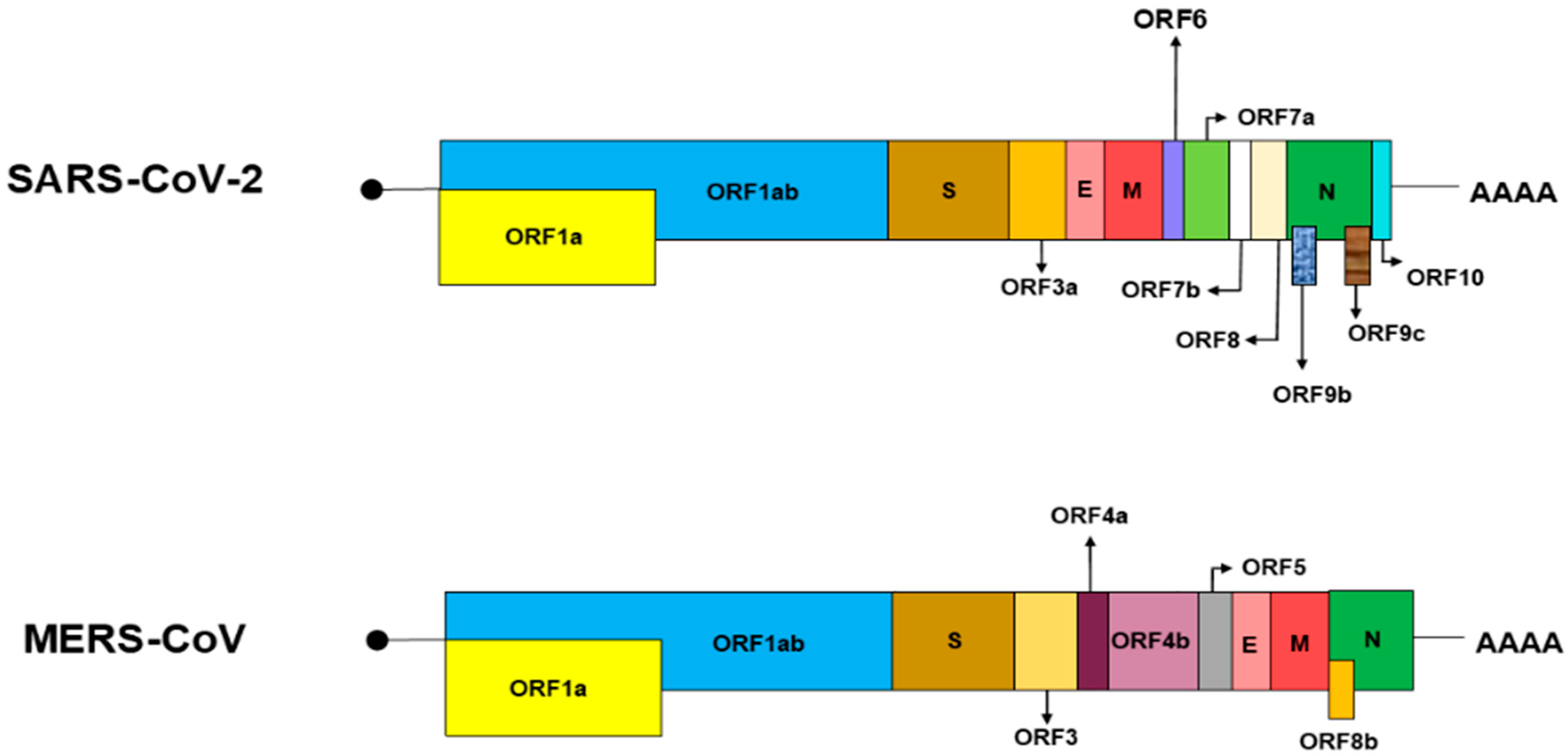
Abstract
:Author Contributions
Funding
Conflicts of Interest
References
- Alfassam, H.A.; Nassar, M.S.; Almusaynid, M.M.; Khalifah, B.A.; Alshahrani, A.S.; Almughem, F.A.; Alshehri, A.A.; Alawad, M.O.; Massadeh, S.; Alaamery, M.; et al. Development of a Colorimetric Tool for SARS-CoV-2 and Other Respiratory Viruses Detection Using Sialic Acid Fabricated Gold Nanoparticles. Pharmaceutics 2021, 13, 502. [Google Scholar] [CrossRef] [PubMed]
- Lang, Y.; Li, W.; Li, Z.; Koerhuis, D.; Van Den Burg, A.C.; Rozemuller, E.; Bosch, B.J.; Van Kuppeveld, F.J.; Boons, G.J.; Huizinga, E.G.; et al. Coronavirus hemagglutinin-esterase and spike proteins coevolve for functional balance and optimal virion avidity. Proc. Natl. Acad. Sci. USA 2020, 117, 25759–25770. [Google Scholar] [CrossRef] [PubMed]
- Yashvardhini, N.; Jha, D.K. Genome organization and pathogenesis of SARS-CoV-2. Int. J. Curr. Microbiol. Appl. Sci. 2020, 9, 2153–2156. [Google Scholar] [CrossRef]
- Yoshimoto, F.K. The proteins of severe acute respiratory syndrome coronavirus-2 (SARS-CoV-2 or n-COV19), the cause of COVID-19. Protein J. 2020, 39, 198–216. [Google Scholar] [CrossRef] [PubMed]
- Zandi, M. Severe acute respiratory syndrome-2 encodes hemagglutinin esterase? Rev. Med. Virol. 2021, 32, e2294. [Google Scholar] [CrossRef] [PubMed]
- Mendonca, P.; Soliman, K.F. Flavonoids activation of the transcription factor Nrf2 as a hypothesis approach for the prevention and modulation of SARS-CoV-2 infection severity. Antioxidants 2020, 9, 659. [Google Scholar] [CrossRef] [PubMed]
- Rabaan, A.A.; Al-Ahmed, S.H.; Haque, S.; Sah, R.; Tiwari, R.; Malik, Y.S.; Dhama, K.; Yatoo, M.I.; Bonilla-Aldana, D.K.; Rodriguez-Morales, A.J. SARS-CoV-2, SARS-CoV, and MERS-COV: A comparative overview. Infez. Med. 2020, 28, 174–184. [Google Scholar] [PubMed]
- Grudlewska-Buda, K.; Wiktorczyk-Kapischke, N.; Wałecka-Zacharska, E.; Kwiecińska-Piróg, J.; Buszko, K.; Leis, K.; Juszczuk, K.; Gospodarek-Komkowska, E.; Skowron, K. SARS-CoV-2—Morphology, Transmission and Diagnosis during Pandemic, Review with Element of Meta-Analysis. J. Clin. Med. 2021, 10, 1962. [Google Scholar] [CrossRef] [PubMed]
- International Committee on Taxonomy of Viruses Executive Committee. The new scope of virus taxonomy: Partitioning the virosphere into 15 hierarchical ranks. Nat. Microbiol. 2020, 5, 668. [Google Scholar] [CrossRef] [PubMed]
- Kim, C.-H. SARS-CoV-2 evolutionary adaptation toward host entry and recognition of receptor O-Acetyl sialylation in virus–host interaction. Int. J. Mol. Sci. 2020, 21, 4549. [Google Scholar]
- Zeng, Q.; Langereis, M.A.; Van Vliet, A.L.; Huizinga, E.G.; De Groot, R.J. Structure of coronavirus hemagglutinin-esterase offers insight into corona and influenza virus evolution. Proc. Natl. Acad. Sci. USA 2008, 105, 9065–9069. [Google Scholar] [CrossRef] [PubMed] [Green Version]
- Chan, J.F.-W.; Kok, K.-H.; Zhu, Z.; Chu, H.; To, K.K.-W.; Yuan, S.; Yuen, K.Y. Genomic characterization of the 2019 novel human-pathogenic coronavirus isolated from a patient with atypical pneumonia after visiting Wuhan. Emerg. Microbes Infect. 2020, 9, 221–236. [Google Scholar] [CrossRef] [PubMed] [Green Version]

Publisher’s Note: MDPI stays neutral with regard to jurisdictional claims in published maps and institutional affiliations. |
© 2022 by the authors. Licensee MDPI, Basel, Switzerland. This article is an open access article distributed under the terms and conditions of the Creative Commons Attribution (CC BY) license (https://creativecommons.org/licenses/by/4.0/).
Share and Cite
Zandi, M.; Soltani, S. Comment on Alfassam et al. Development of a Colorimetric Tool for SARS-CoV-2 and Other Respiratory Viruses Detection Using Sialic Acid Fabricated Gold Nanoparticles. Pharmaceutics 2021, 13, 502. Pharmaceutics 2022, 14, 1871. https://doi.org/10.3390/pharmaceutics14091871
Zandi M, Soltani S. Comment on Alfassam et al. Development of a Colorimetric Tool for SARS-CoV-2 and Other Respiratory Viruses Detection Using Sialic Acid Fabricated Gold Nanoparticles. Pharmaceutics 2021, 13, 502. Pharmaceutics. 2022; 14(9):1871. https://doi.org/10.3390/pharmaceutics14091871
Chicago/Turabian StyleZandi, Milad, and Saber Soltani. 2022. "Comment on Alfassam et al. Development of a Colorimetric Tool for SARS-CoV-2 and Other Respiratory Viruses Detection Using Sialic Acid Fabricated Gold Nanoparticles. Pharmaceutics 2021, 13, 502" Pharmaceutics 14, no. 9: 1871. https://doi.org/10.3390/pharmaceutics14091871