
Therapeutic Cancer Vaccines—Antigen Discovery and Adjuvant Delivery Platforms


Abstract
:1. Introduction
2. Tumor Antigens for Cancer Vaccines
2.1. Classification of Tumor Antigens
2.2. Biochemical and Bioinformatic Approaches for the Identification of Tumor Antigens
2.2.1. Serological Analysis of Recombinant Tumor cDNA Libraries (SEREX)
2.2.2. Computational Prediction Methods for Cancer Antigens
2.3. Delivery of Neoantigens
3. Vaccine Adjuvants
3.1. Delivery Systems
3.2. Immune Potentiators
3.3. Intracellular Delivery of Immune Adjuvants
3.3.1. TLR3
3.3.2. TLR7/8
3.3.3. TLR9
3.3.4. cGAS-STING
3.3.5. Other PRR Agonists
3.4. Combined Delivery of PRR Agonists
4. Future Perspective
Supplementary Materials
Author Contributions
Funding
Institutional Review Board Statement
Informed Consent Statement
Data Availability Statement
Conflicts of Interest
References
- Plotkin, S.A. Vaccines: Past, present and future. Nat. Med. 2005, 11, S5–S11. [Google Scholar] [CrossRef] [PubMed]
- Riedel, S. Edward Jenner and the history of smallpox and vaccination. Proc. (Bayl. Univ. Med. Cent.) 2005, 18, 21–25. [Google Scholar] [CrossRef] [PubMed]
- Tregoning, J.S.; Flight, K.E.; Higham, S.L.; Wang, Z.; Pierce, B.F. Progress of the COVID-19 vaccine effort: Viruses, vaccines and variants versus efficacy, effectiveness and escape. Nat. Rev. Immunol. 2021, 21, 626–636. [Google Scholar] [CrossRef]
- A Phase 1 Study to Evaluate the Safety and Immunogenicity of eOD-GT8 60mer mRNA Vaccine (mRNA-1644) and Core-g28v2 60mer mRNA Vaccine (mRNA-1644v2-Core); 2021. Available online: https://clinicaltrials.gov/ct2/show/results/NCT05001373?view=results (accessed on 30 November 2021).
- Saxena, M.; van der Burg, S.H.; Melief, C.J.M.; Bhardwaj, N. Therapeutic cancer vaccines. Nat. Rev. Cancer 2021, 21, 360–378. [Google Scholar] [CrossRef]
- Prehn, R.T.; Main, J.M. Immunity to Methylcholanthrene-Induced Sarcomas. J. Natl. Cancer Inst. 1957, 18, 769–778. [Google Scholar]
- van der Bruggen, P.; Traversari, C.; Chomez, P.; Lurquin, C.; De Plaen, E.; Van den Eynde, B.; Knuth, A.; Boon, T. A gene encoding an antigen recognized by cytolytic T lymphocytes on a human melanoma. Science 1991, 254, 1643. [Google Scholar] [CrossRef]
- Murphy, K.; Weaver, C.; Janeway, C. Janeway’s Immunobiology, 9th ed.; Garland Science: New York, NY, USA, 2017. [Google Scholar]
- Piertney, S.B.; Oliver, M.K. The evolutionary ecology of the major histocompatibility complex. Heredity 2006, 96, 7–21. [Google Scholar] [CrossRef] [Green Version]
- Klein, J.; Figueroa, F. Evolution of the major histocompatibility complex. Crit. Rev. Immunol. 1986, 6, 295–386. [Google Scholar] [CrossRef]
- Altuvia, Y.; Margalit, H. A structure-based approach for prediction of MHC-binding peptides. Methods 2004, 34, 454–459. [Google Scholar] [CrossRef]
- Wieczorek, M.; Abualrous, E.T.; Sticht, J.; Álvaro-Benito, M.; Stolzenberg, S.; Noé, F.; Freund, C. Major Histocompatibility Complex (MHC) Class I and MHC Class II Proteins: Conformational Plasticity in Antigen Presentation. Front. Immunol. 2017, 8, 292. [Google Scholar] [CrossRef] [Green Version]
- Jeffery, K.J.M.; Bangham, C.R.M. Do infectious diseases drive MHC diversity? Microbes Infect. 2000, 2, 1335–1341. [Google Scholar] [CrossRef]
- Dausset, J. The Major Histocompatibility Complex in Man. Science 1981, 213, 1469–1474. [Google Scholar] [CrossRef] [PubMed]
- Bruggen, P.V.D.; Szikora, J.-P.; Boël, P.; Wildmann, C.; Somville, M.; Sensi, M.; Boon, T. Autologous cytolytic T lymphocytes recognize a MAGE-1 nonapeptide on melanomas expressing HLA-Cw* 1601. Eur. J. Immunol. 1994, 24, 2134–2140. [Google Scholar] [CrossRef] [PubMed]
- Robbins, P.F.; Kawakami, Y. Human tumor antigens recognized by T cells. Curr. Opin. Immunol. 1996, 8, 628–636. [Google Scholar] [CrossRef]
- Boon, T.; Cerottini, J.-C.; Van den Eynde, B.; van der Bruggen, P.; Van Pel, A. Tumor Antigens Recognized by T Lymphocytes. Annu. Rev. Immunol. 1994, 12, 337–365. [Google Scholar] [CrossRef] [PubMed]
- Bennett, S.R.M.; Carbone, F.R.; Karamalis, F.; Flavell, R.A.; Miller, J.F.A.P.; Heath, W.R. Help for cytotoxic-T-cell responses is mediated by CD40 signalling. Nature 1998, 393, 478–480. [Google Scholar] [CrossRef]
- Ridge, J.P.; Di Rosa, F.; Matzinger, P. A conditioned dendritic cell can be a temporal bridge between a CD4+ T-helper and a T-killer cell. Nature 1998, 393, 474–478. [Google Scholar] [CrossRef]
- Schoenberger, S.P.; Toes, R.E.M.; van der Voort, E.I.H.; Offringa, R.; Melief, C.J.M. T-cell help for cytotoxic T lymphocytes is mediated by CD40–CD40L interactions. Nature 1998, 393, 480–483. [Google Scholar] [CrossRef]
- Kennedy, R.; Celis, E. Multiple roles for CD4+ T cells in anti-tumor immune responses. Immunol. Rev. 2008, 222, 129–144. [Google Scholar] [CrossRef]
- Pardoll, D.M.; Topalian, S.L. The role of CD4+ T cell responses in antitumor immunity. Curr. Opin. Immunol. 1998, 10, 588–594. [Google Scholar] [CrossRef]
- Valilou, S.F.; Rezaei, N. Chapter 4—Tumor Antigens. In Vaccines for Cancer Immunotherapy; Rezaei, N., Keshavarz-Fathi, M., Eds.; Academic Press: Cambridge, MA, USA, 2019; pp. 61–74. [Google Scholar] [CrossRef]
- Boon, T.; Coulie, P.G.; Van den Eynde, B. Tumor antigens recognized by T cells. Immunol. Today 1997, 18, 267–268. [Google Scholar] [CrossRef]
- Nagorsen, D.; Marincola, F.M. Analyzing T Cell Responses: How to Analyze Cellular Immune Responses against Tumor Associated Antigens; Springer: Berlin/Heidelberg, Germany, 2006. [Google Scholar]
- Wang, R.-F.; Rosenberg, S.A. Human tumor antigens for cancer vaccine development. Immunol. Rev. 1999, 170, 85–100. [Google Scholar] [CrossRef] [PubMed]
- Richards, J.M.; Mehta, N.; Ramming, K.; Skosey, P. Sequential Chemoimmunotherapy in the Treatment of Metastatic Melanoma. In Proceedings of the Cytokines in Hemopoiesis, Oncology, and AIDS II, Berlin/Heidelberg, Germany, 1 August 1992; pp. 721–727. [Google Scholar]
- Agyemang, A.F.; Odunsi, K.O. Chapter 5—The use of immunotherapy for treatment of chemoresistant ovarian cancer. In Overcoming Ovarian Cancer Chemoresistance; Samimi, G., Annunziata, C., Eds.; Academic Press: Cambridge, MA, USA, 2021; Volume 11, pp. 79–96. [Google Scholar]
- Gjerstorff, M.F.; Andersen, M.H.; Ditzel, H.J. Oncogenic cancer/testis antigens: Prime candidates for immunotherapy. Oncotarget 2015, 6, 15772–15787. [Google Scholar] [CrossRef] [Green Version]
- Smith, C.C.; Selitsky, S.R.; Chai, S.; Armistead, P.M.; Vincent, B.G.; Serody, J.S. Alternative tumour-specific antigens. Nat. Rev. Cancer 2019, 19, 465–478. [Google Scholar] [CrossRef]
- Goodier, J.L.; Kazazian, H.H. Retrotransposons Revisited: The Restraint and Rehabilitation of Parasites. Cell 2008, 135, 23–35. [Google Scholar] [CrossRef] [Green Version]
- Cherkasova, E.; Scrivani, C.; Doh, S.; Weisman, Q.; Takahashi, Y.; Harashima, N.; Yokoyama, H.; Srinivasan, R.; Linehan, W.M.; Lerman, M.I.; et al. Detection of an Immunogenic HERV-E Envelope with Selective Expression in Clear Cell Kidney Cancer. Cancer Res. 2016, 76, 2177–2185. [Google Scholar] [CrossRef] [Green Version]
- Turajlic, S.; Litchfield, K.; Xu, H.; Rosenthal, R.; McGranahan, N.; Reading, J.L.; Wong, Y.N.S.; Rowan, A.; Kanu, N.; Al Bakir, M.; et al. Insertion-and-deletion-derived tumour-specific neoantigens and the immunogenic phenotype: A pan-cancer analysis. Lancet Oncol. 2017, 18, 1009–1021. [Google Scholar] [CrossRef] [Green Version]
- Jayasinghe, R.G.; Cao, S.; Gao, Q.; Wendl, M.C.; Vo, N.S.; Reynolds, S.M.; Zhao, Y.; Climente-González, H.; Chai, S.; Wang, F.; et al. Systematic Analysis of Splice-Site-Creating Mutations in Cancer. Cell Rep. 2018, 23, 270–281.e273. [Google Scholar] [CrossRef] [Green Version]
- Rowley, J.D. A New Consistent Chromosomal Abnormality in Chronic Myelogenous Leukaemia identified by Quinacrine Fluorescence and Giemsa Staining. Nature 1973, 243, 290–293. [Google Scholar] [CrossRef]
- Nowell, P. The minute chromosome (Phl) in chronic granulocytic leukemia. Blut 1962, 8, 65–66. [Google Scholar] [CrossRef]
- Shepherd, P.; Suffolk, R.; Halsey, J.; Allan, N. Analysis of molecular breakpoint and m-RNA transcripts in a prospective randomized trial of interferon in chronic myeloid leukaemia: No correlation with clinical features, cytogenetic response, duration of chronic phase, or survival. Br. J. Haematol. 1995, 89, 546–554. [Google Scholar] [CrossRef] [PubMed]
- Westbrook, C.A.; Hooberman, A.L.; Spino, C.; Dodge, R.K.; Larson, R.A.; Davey, F.; Wurster-Hill, D.H.; Sobol, R.E.; Schiffer, C.; Bloomfield, C.D. Clinical Significance of the BCR-ABL Fusion Gene in Adult Acute Lymphoblastic Leukemia: A Cancer and Leukemia Group B Study (8762). Blood 1992, 80, 2983–2990. [Google Scholar] [CrossRef] [PubMed] [Green Version]
- Russo, C.; Carroll, A.; Kohler, S.; Borowitz, M.; Amylon, M.; Homans, A.; Kedar, A.; Shuster, J.; Land, V.; Crist, W.; et al. Philadelphia Chromosome and Monosomy 7 in Childhood Acute Lymphoblastic Leukemia: A Pediatric Oncology Group Study. Blood 1991, 77, 1050–1056. [Google Scholar] [CrossRef] [PubMed] [Green Version]
- Suryanarayan, K.; Hunger, S.P.; Kohler, S.; Carroll, A.J.; Crist, W.; Link, M.P.; Cleary, M.L. Consistent Involvement of the BCR Gene by 9;22 Breakpoints in Pediatric Acute Leukemias. Blood 1991, 77, 324–330. [Google Scholar] [CrossRef] [Green Version]
- Kurzrock, R.; Shtalrid, M.; Talpaz, M.; Kloetzer, W.S.; Gutterman, J.U. Expression of c-abl in Philadelphia-Positive Acute Myelogenous Leukemia. Blood 1987, 70, 1584–1588. [Google Scholar] [CrossRef] [Green Version]
- Secker-Walker, L.M.; Morgan, G.J.; Min, T.; John Swansbury, G.; Craig, J.; Yamada, T.; Desalvo, L.; Medina, J.W.; Chowdhury, V.; Donahue, R.P.; et al. Inversion of chromosome 16 with the Philadelphia chromosome in acute myelomonocytic leukemia with eosinophilia: Report of two cases. Cancer Genet. Cytogenet. 1992, 58, 29–34. [Google Scholar] [CrossRef]
- Alimena, G.; Cedrone, M.; Nanni, M.; De Cuia, M.; De Sanctis, V.; Cimino, G.; Mancini, M. Acute leukemia presenting a variant Ph chromosome with p190 expression, dup 3q and-7, developed after malignant lymphoma treated with alkylating agents and topoisomerase II inhibitors. Leukemia 1995, 9, 1483–1486. [Google Scholar]
- Fujii, H.; Yashige, H.; Misawa, S.; Tanaka, S.; Urata, Y.; Matuyama, F. Ph chromosome in a patient with non-leukemic non-hodgkin B-cell lymphoma. Am. J. Hematol. 1990, 35, 213–215. [Google Scholar] [CrossRef]
- Mitani, K.; Sato, Y.; Tojo, A.; Ishikawa, F.; Kobayashi, Y.; Miura, Y.; Miyazono, K.; And, A.U.; Takaku, F. Philadelphia chromosome positive B-cell type malignant lymphoma expressing an aberrant 190 kDa bcr-abl protein. Br. J. Haematol. 1990, 76, 221–225. [Google Scholar] [CrossRef]
- Juliusson, G.; Friberg, K.; Gahrton, G. Ph chromosome and B-cell malignancy-associated chromosomal aberrations in non-leukaemic immunoblastic B-cell lymphoma. Acta Haematol. 1985, 74, 171–174. [Google Scholar] [CrossRef]
- Martiat, P.; Mecucci, C.; Nizet, Y.; Stul, M.; Philippe, M.; Cassiman, J.-J.; Michaux, J.L.; Van den Berghe, H.; Sokal, G. P190 BCR/ABL transcript in a case of Philadelphia-positive multiple myeloma. Leukemia 1990, 4, 751–754. [Google Scholar] [PubMed]
- Van Den Berghe, H.a.; Louwagie, A.; Broeckaert-Van Orshoven, A.; David, G.; Verwilghen, R.; Michaux, J.; Sokal, G. Philadelphia chromosome in human multiple myeloma. J. Natl. Cancer Inst. 1979, 63, 11–16. [Google Scholar] [PubMed]
- Karpas, A.; Fischer, P.; Swirsky, D. Human myeloma cell line carrying a Philadelphia chromosome. Science 1982, 216, 997–999. [Google Scholar] [CrossRef]
- Bos, J.L. Ras Oncogenes in Human Cancer: A Review. Cancer Res. 1989, 49, 4682–4689. [Google Scholar] [PubMed]
- Bourgault Villada, I.; Bénéton, N.; Bony, C.; Connan, F.; Monsonego, J.; Bianchi, A.; Saiag, P.; Lévy, J.P.; Guillet, J.G.; Choppin, J. Identification in humans of HPV-16 E6 and E7 protein epitopes recognized by cytolytic T lymphocytes in association with HLA-B18 and determination of the HLA-B18-specific binding motif. Eur. J. Immunol. 2000, 30, 2281–2289. [Google Scholar] [CrossRef]
- Peng, S.; Trimble, C.; Wu, L.; Pardoll, D.; Roden, R.; Hung, C.-F.; Wu, T.C. HLA-DQB1*02–Restricted HPV-16 E7 Peptide–Specific CD4+ T-Cell Immune Responses Correlate with Regression of HPV-16–Associated High-Grade Squamous Intraepithelial Lesions. Clin. Cancer Res. 2007, 13, 2479. [Google Scholar] [CrossRef] [Green Version]
- Kaufmann, A.M.; Nieland, J.; Schinz, M.; Nonn, M.; Gabelsberger, J.; Meissner, H.; Müller, R.T.; Jochmus, I.; Gissmann, L.; Schneider, A.; et al. HPV16 L1E7 chimeric virus-like particles induce specific HLA-restricted T cells in humans after in vitro vaccination. Int. J. Cancer 2001, 92, 285–293. [Google Scholar] [CrossRef]
- Echchakir, H.; Mami-Chouaib, F.; Vergnon, I.; Baurain, J.-F.; Karanikas, V.; Chouaib, S.; Coulie, P.G. A Point Mutation in the α-actinin-4 Gene Generates an Antigenic Peptide Recognized by Autologous Cytolytic T Lymphocytes on a Human Lung Carcinoma. Cancer Res. 2001, 61, 4078. [Google Scholar]
- Wang, H.Y.; Peng, G.; Guo, Z.; Shevach, E.M.; Wang, R.-F. Recognition of a New ARTC1 Peptide Ligand Uniquely Expressed in Tumor Cells by Antigen-Specific CD4+ Regulatory T Cells. J. Immunol. 2005, 174, 2661. [Google Scholar] [CrossRef] [Green Version]
- Sharkey, M.S.; Lizée, G.; Gonzales, M.I.; Patel, S.; Topalian, S.L. CD4+ T-Cell Recognition of Mutated B-RAF in Melanoma Patients Harboring the V599E Mutation. Cancer Res. 2004, 64, 1595. [Google Scholar] [CrossRef] [Green Version]
- Robbins, P.F.; El-Gamil, M.; Li, Y.F.; Kawakami, Y.; Loftus, D.; Appella, E.; Rosenberg, S.A. A mutated beta-catenin gene encodes a melanoma-specific antigen recognized by tumor infiltrating lymphocytes. J. Exp. Med. 1996, 183, 1185–1192. [Google Scholar] [CrossRef] [PubMed] [Green Version]
- Yotnda, P.; Firat, H.; Garcia-Pons, F.; Garcia, Z.; Gourru, G.; Vernant, J.P.; Lemonnier, F.A.; Leblond, V.; Langlade-Demoyen, P. Cytotoxic T cell response against the chimeric p210 BCR-ABL protein in patients with chronic myelogenous leukemia. J. Clin. Investig. 1998, 101, 2290–2296. [Google Scholar] [CrossRef] [PubMed]
- Greco, G.; Fruci, D.; Accapezzato, D.; Barnaba, V.; Nisini, R.; Alimena, G.; Montefusco, E.; Vigneti, E.; Butler, R.; Tanigaki, N.; et al. Two bcr-abl junction peptides bind HLA-A3 molecules and allow specific induction of human cytotoxic T lymphocytes. Leukemia 1996, 10, 693–699. [Google Scholar] [PubMed]
- Bocchia, M.; Korontsvit, T.; Xu, Q.; Mackinnon, S.; Yang, S.; Sette, A.; Scheinberg, D. Specific human cellular immunity to bcr-abl oncogene-derived peptides. Blood 1996, 87, 3587–3592. [Google Scholar] [CrossRef] [PubMed]
- Bosch, G.; Joosten, A.; Kessler, J.; Melief, C.; Leeksma, O. Recognition of BCR-ABL positive leukemic blasts by human CD4+ T cells elicited by primary in vitro immunization with a BCR-ABL breakpoint peptide. Blood 1996, 88, 3522–3527. [Google Scholar] [CrossRef] [Green Version]
- Yasukawa, M.; Ohminami, H.; Kaneko, S.; Yakushijin, Y.; Nishimura, Y.; Inokuchi, K.; Miyakuni, T.; Nakao, S.; Kishi, K.; Kubonishi, I.; et al. CD4+ Cytotoxic T-Cell Clones Specific for bcr-abl b3a2 Fusion Peptide Augment Colony Formation by Chronic Myelogenous Leukemia Cells in a b3a2-Specific and HLA-DR–Restricted Manner. Blood 1998, 92, 3355–3361. [Google Scholar] [CrossRef]
- Pawelec, G.; Max, H.; Halder, T.; Bruserud, O.; Merl, A.; da Silva, P.; Kalbacher, H. BCR/ABL leukemia oncogene fusion peptides selectively bind to certain HLA-DR alleles and can be recognized by T cells found at low frequency in the repertoire of normal donors. Blood 1996, 88, 2118–2124. [Google Scholar] [CrossRef]
- Tanaka, Y.; Takahashi, T.; Nieda, M.; Masuda, S.; Kashiwase, K.; Ogawa, S.; Chiba, S.; Juji, T.; Hirai, H. Generation of HLA-DRB1*1501-restricted p190 minor bcr–abl (e1a2)-specific CD4+ T lymphocytes. Br. J. Haematol. 2000, 109, 435–437. [Google Scholar] [CrossRef]
- ten Bosch, G.J.A.; Kessler, J.H.; Joosten, A.M.; Bres-Vloemans, A.A.; Geluk, A.; Godthelp, B.C.; van Bergen, J.; Melief, C.J.M.; Leeksma, O.C. A BCR-ABL Oncoprotein p210b2a2 Fusion Region Sequence Is Recognized by HLA-DR2a Restricted Cytotoxic T Lymphocytes and Presented by HLA-DR Matched Cells Transfected With an Iib2a2 Construct. Blood 1999, 94, 1038–1045. [Google Scholar] [CrossRef]
- Schwitalle, Y.; Linnebacher, M.; Ripberger, E.; Gebert, J.; von Knebel Doeberitz, M. Immunogenic peptides generated by frameshift mutations in DNA mismatch repair-deficient cancer cells. Cancer Immun. Arch. 2004, 4, 14. [Google Scholar]
- Mandruzzato, S.; Brasseur, F.; Andry, G.; Boon, T.; van der Bruggen, P. A CASP-8 mutation recognized by cytolytic T lymphocytes on a human head and neck carcinoma. J. Exp. Med. 1997, 186, 785–793. [Google Scholar] [CrossRef] [PubMed]
- Wang, R.-F.; Wang, X.; Atwood, A.C.; Topalian, S.L.; Rosenberg, S.A. Cloning Genes Encoding MHC Class II-Restricted Antigens: Mutated CDC27 as a Tumor Antigen. Science 1999, 284, 1351–1354. [Google Scholar] [CrossRef] [PubMed]
- Wolfel, T.; Hauer, M.; Schneider, J.; Serrano, M.; Wolfel, C.; Klehmann-Hieb, E.; De Plaen, E.; Hankeln, T.; Meyer zum Buschenfelde, K.; Beach, D. A p16INK4a-insensitive CDK4 mutant targeted by cytolytic T lymphocytes in a human melanoma. Science 1995, 269, 1281–1284. [Google Scholar] [CrossRef] [PubMed]
- Robbins, P.F.; Lu, Y.-C.; El-Gamil, M.; Li, Y.F.; Gross, C.; Gartner, J.; Lin, J.C.; Teer, J.K.; Cliften, P.; Tycksen, E.; et al. Mining exomic sequencing data to identify mutated antigens recognized by adoptively transferred tumor-reactive T cells. Nat. Med. 2013, 19, 747–752. [Google Scholar] [CrossRef]
- Huang, J.; El-Gamil, M.; Dudley, M.E.; Li, Y.F.; Rosenberg, S.A.; Robbins, P.F. T cells associated with tumor regression recognize frameshifted products of the CDKN2A tumor suppressor gene locus and a mutated HLA class I gene product. J. Immunol. 2004, 172, 6057–6064. [Google Scholar] [CrossRef] [Green Version]
- Corbière, V.; Chapiro, J.; Stroobant, V.; Ma, W.; Lurquin, C.; Lethé, B.; van Baren, N.; Van den Eynde, B.J.; Boon, T.; Coulie, P.G. Antigen Spreading Contributes to MAGE Vaccination-Induced Regression of Melanoma Metastases. Cancer Res. 2011, 71, 1253. [Google Scholar] [CrossRef] [PubMed] [Green Version]
- Maccalli, C.; Pende, D.; Castelli, C.; Mingari, M.C.; Robbins, P.F.; Parmiani, G. NKG2D engagement of colorectal cancer-specific T cells strengthens TCR-mediated antigen stimulation and elicits TCR independent anti-tumor activity. Eur. J. Immunol. 2003, 33, 2033–2043. [Google Scholar] [CrossRef]
- Makita, M.; Azuma, T.; Hamaguchi, H.; Niiya, H.; Kojima, K.; Fujita, S.; Tanimoto, M.; Harada, M.; Yasukawa, M. Leukemia-associated fusion proteins, dek-can and bcr-abl, represent immunogenic HLA-DR-restricted epitopes recognized by fusion peptide-specific CD4+ T lymphocytes. Leukemia 2002, 16, 2400–2407. [Google Scholar] [CrossRef] [Green Version]
- Lennerz, V.; Fatho, M.; Gentilini, C.; Frye, R.A.; Lifke, A.; Ferel, D.; Wölfel, C.; Huber, C.; Wölfel, T. The response of autologous T cells to a human melanoma is dominated by mutated neoantigens. Proc. Natl. Acad. Sci. USA 2005, 102, 16013–16018. [Google Scholar] [CrossRef] [Green Version]
- Hogan, K.T.; Eisinger, D.P.; Cupp, S.B.; Lekstrom, K.J.; Deacon, D.D.; Shabanowitz, J.; Hunt, D.F.; Engelhard, V.H.; Slingluff, C.L.; Ross, M.M. The Peptide Recognized by HLA-A68.2-restricted, Squamous Cell Carcinoma of the Lung-specific Cytotoxic T Lymphocytes Is Derived from a Mutated Elongation Factor 2 Gene. Cancer Res. 1998, 58, 5144. [Google Scholar]
- Yotnda, P.; Garcia, F.; Peuchmaur, M.; Grandchamp, B.; Duval, M.; Lemonnier, F.; Vilmer, E.; Langlade-Demoyen, P. Cytotoxic T cell response against the chimeric ETV6-AML1 protein in childhood acute lymphoblastic leukemia. J. Clin. Investig. 1998, 102, 455–462. [Google Scholar] [CrossRef] [PubMed]
- Graf, C.; Heidel, F.; Tenzer, S.; Radsak, M.P.; Solem, F.K.; Britten, C.M.; Huber, C.; Fischer, T.; Wölfel, T. A neoepitope generated by an FLT3 internal tandem duplication (FLT3-ITD) is recognized by leukemia-reactive autologous CD8+ T cells. Blood 2006, 109, 2985–2988. [Google Scholar] [CrossRef] [PubMed] [Green Version]
- Wang, H.Y.; Zhou, J.; Zhu, K.; Riker, A.I.; Marincola, F.M.; Wang, R.-F. Identification of a mutated fibronectin as a tumor antigen recognized by CD4+ T cells: Its role in extracellular matrix formation and tumor metastasis. J. Exp. Med. 2002, 195, 1397–1406. [Google Scholar] [CrossRef] [Green Version]
- Rajasagi, M.; Shukla, S.A.; Fritsch, E.F.; Keskin, D.B.; DeLuca, D.; Carmona, E.; Zhang, W.; Sougnez, C.; Cibulskis, K.; Sidney, J.; et al. Systematic identification of personal tumor-specific neoantigens in chronic lymphocytic leukemia. Blood 2014, 124, 453–462. [Google Scholar] [CrossRef]
- Wick, D.A.; Webb, J.R.; Nielsen, J.S.; Martin, S.D.; Kroeger, D.R.; Milne, K.; Castellarin, M.; Twumasi-Boateng, K.; Watson, P.H.; Holt, R.A.; et al. Surveillance of the Tumor Mutanome by T Cells during Progression from Primary to Recurrent Ovarian Cancer. Clin. Cancer Res. 2014, 20, 1125. [Google Scholar] [CrossRef] [PubMed] [Green Version]
- Gaudin, C.; Kremer, F.; Angevin, E.; Scott, V.; Triebel, F. A hsp70-2 Mutation Recognized by CTL on a Human Renal Cell Carcinoma. J. Immunol. 1999, 162, 1730. [Google Scholar]
- Guéguen, M.; Patard, J.-J.; Gaugler, B.; Brasseur, F.; Renauld, J.-C.; Van Cangh, P.J.; Boon, T.; Van den Eynde, B.t.J. An Antigen Recognized by Autologous CTLs on a Human Bladder Carcinoma. J. Immunol. 1998, 160, 6188. [Google Scholar]
- Gjertsen, M.K.; Bjørheim, J.; Saeterdal, I.; Myklebust, J.; Gaudernack, G. Cytotoxic CD4+ and CD8+ T lymphocytes, generated by mutant p21-ras (12VAL) peptide vaccination of a patient, recognize 12VAL-dependent nested epitopes present within the vaccine peptide and kill autologous tumour cells carrying this mutation. Int. J. Cancer 1997, 72, 784–790. [Google Scholar] [CrossRef]
- Tran, E.; Robbins, P.F.; Lu, Y.-C.; Prickett, T.D.; Gartner, J.J.; Jia, L.; Pasetto, A.; Zheng, Z.; Ray, S.; Groh, E.M.; et al. T-Cell Transfer Therapy Targeting Mutant KRAS in Cancer. N. Engl. J. Med. 2016, 375, 2255–2262. [Google Scholar] [CrossRef] [PubMed] [Green Version]
- Wang, R.F.; Wang, X.; Rosenberg, S.A. Identification of a novel major histocompatibility complex class II-restricted tumor antigen resulting from a chromosomal rearrangement recognized by CD4(+) T cells. J. Exp. Med. 1999, 189, 1659–1668. [Google Scholar] [CrossRef] [Green Version]
- Kawakami, Y.; Wang, X.; Shofuda, T.; Sumimoto, H.; Tupesis, J.P.; Fitzgerald, E.; Rosenberg, S.A. Isolation of a New Melanoma Antigen, MART-2, Containing a Mutated Epitope Recognized by Autologous Tumor-Infiltrating T Lymphocytes. J. Immunol. 2001, 166, 2871. [Google Scholar] [CrossRef] [PubMed] [Green Version]
- Karanikas, V.; Colau, D.; Baurain, J.-F.; Chiari, R.; Thonnard, J.; Gutierrez-Roelens, I.; Goffinet, C.; Schaftingen, E.V.; Weynants, P.; Boon, T.; et al. High Frequency of Cytolytic T Lymphocytes Directed against a Tumor-specific Mutated Antigen Detectable with HLA Tetramers in the Blood of a Lung Carcinoma Patient with Long Survival. Cancer Res. 2001, 61, 3718. [Google Scholar] [PubMed]
- Coulie, P.G.; Lehmann, F.; Lethé, B.; Herman, J.; Lurquin, C.; Andrawiss, M.; Boon, T. A mutated intron sequence codes for an antigenic peptide recognized by cytolytic T lymphocytes on a human melanoma. Proc. Natl. Acad. Sci. USA 1995, 92, 7976–7980. [Google Scholar] [CrossRef] [PubMed] [Green Version]
- Chiari, R.; Foury, F.; De Plaen, E.; Baurain, J.-F.; Thonnard, J.; Coulie, P.G. Two Antigens Recognized by Autologous Cytolytic T Lymphocytes on a Melanoma Result from a Single Point Mutation in an Essential Housekeeping Gene. Cancer Res. 1999, 59, 5785. [Google Scholar]
- Baurain, J.-F.; Colau, D.; van Baren, N.; Landry, C.; Martelange, V.; Vikkula, M.; Boon, T.; Coulie, P.G. High Frequency of Autologous Anti-Melanoma CTL Directed Against an Antigen Generated by a Point Mutation in a New Helicase Gene. J. Immunol. 2000, 164, 6057. [Google Scholar] [CrossRef] [Green Version]
- Zorn, E.; Hercend, T. A natural cytotoxic T cell response in a spontaneously regressing human melanoma targets a neoantigen resulting from a somatic point mutation. Eur. J. Immunol. 1999, 29, 592–601. [Google Scholar] [CrossRef]
- Linard, B.; Bézieau, S.; Benlalam, H.; Labarrière, N.; Guilloux, Y.; Diez, E.; Jotereau, F. A ras-Mutated Peptide Targeted by CTL Infiltrating a Human Melanoma Lesion. J. Immunol. 2002, 168, 4802. [Google Scholar] [CrossRef] [Green Version]
- Topalian, S.L.; Gonzales, M.I.; Ward, Y.; Wang, X.; Wang, R.-F. Revelation of a Cryptic Major Histocompatibility Complex Class II-restricted Tumor Epitope in a Novel RNA-processing Enzyme. Cancer Res. 2002, 62, 5505. [Google Scholar]
- Takenoyama, M.; Baurain, J.-F.; Yasuda, M.; So, T.; Sugaya, M.; Hanagiri, T.; Sugio, K.; Yasumoto, K.; Boon, T.; Coulie, P.G. A point mutation in the NFYC gene generates an antigenic peptide recognized by autologous cytolytic T lymphocytes on a human squamous cell lung carcinoma. Int. J. Cancer 2006, 118, 1992–1997. [Google Scholar] [CrossRef]
- Ripberger, E.; Linnebacher, M.; Schwitalle, Y.; Gebert, J.; Doeberitz, M.V.K. Identification of an HLA-A0201-Restricted CTL Epitope Generated by a Tumor-Specific Frameshift Mutation in a Coding Microsatellite of the OGT Gene. J. Clin. Immunol. 2003, 23, 415–423. [Google Scholar] [CrossRef]
- Vigneron, N.; Ooms, A.; Morel, S.; Degiovanni, G.; Van Den Eynde, B.J. Identification of a new peptide recognized by autologous cytolytic T lymphocytes on a human melanoma. Cancer Immun. 2002, 2, 9. [Google Scholar] [PubMed]
- Gambacorti-Passerini, C.; Grignani, F.; Arienti, F.; Pandolfi, P.P.; Pelicci, P.G.; Parmiani, G. Human CD4 lymphocytes specifically recognize a peptide representing the fusion region of the hybrid protein pml/RAR alpha present in acute promyelocytic leukemia cells. Blood 1993, 81, 1369–1375. [Google Scholar] [CrossRef] [PubMed] [Green Version]
- Sensi, M.; Nicolini, G.; Zanon, M.; Colombo, C.; Molla, A.; Bersani, I.; Lupetti, R.; Parmiani, G.; Anichini, A. Immunogenicity without Immunoselection: A Mutant but Functional Antioxidant Enzyme Retained in a Human Metastatic Melanoma and Targeted by CD8+ T Cells with a Memory Phenotype. Cancer Res. 2005, 65, 632. [Google Scholar] [CrossRef]
- Novellino, L.; Renkvist, N.; Rini, F.; Mazzocchi, A.; Rivoltini, L.; Greco, A.; Deho, P.; Squarcina, P.; Robbins, P.F.; Parmiani, G.; et al. Identification of a Mutated Receptor-Like Protein Tyrosine Phosphatase κ as a Novel, Class II HLA-Restricted Melanoma Antigen. J. Immunol. 2003, 170, 6363–6370. [Google Scholar] [CrossRef] [Green Version]
- Worley, B.S.; van den Broeke, L.T.; Goletz, T.J.; Pendleton, C.D.; Daschbach, E.M.; Thomas, E.K.; Marincola, F.M.; Helman, L.J.; Berzofsky, J.A. Antigenicity of Fusion Proteins from Sarcoma-associated Chromosomal Translocations. Cancer Res. 2001, 61, 6868. [Google Scholar] [PubMed]
- Sæterdal, I.; Bjørheim, J.; Lislerud, K.; Gjertsen, M.K.; Bukholm, I.K.; Olsen, O.C.; Nesland, J.M.; Eriksen, J.A.; Møller, M.; Lindblom, A.; et al. Frameshift-mutation-derived peptides as tumor-specific antigens in inherited and spontaneous colorectal cancer. Proc. Natl. Acad. Sci. USA 2001, 98, 13255–13260. [Google Scholar] [CrossRef] [PubMed] [Green Version]
- Malekzadeh, P.; Pasetto, A.; Robbins, P.F.; Parkhurst, M.R.; Paria, B.C.; Jia, L.; Gartner, J.J.; Hill, V.; Yu, Z.; Restifo, N.P.; et al. Neoantigen screening identifies broad TP53 mutant immunogenicity in patients with epithelial cancers. J. Clin. Investig. 2019, 129, 1109–1114. [Google Scholar] [CrossRef] [PubMed]
- Pieper, R.; Christian, R.E.; Gonzales, M.I.; Nishimura, M.I.; Gupta, G.; Settlage, R.E.; Shabanowitz, J.; Rosenberg, S.A.; Hunt, D.F.; Topalian, S.L. Biochemical identification of a mutated human melanoma antigen recognized by CD4(+) T cells. J. Exp. Med. 1999, 189, 757–766. [Google Scholar] [CrossRef]
- Li, K.; Adibzadeh, M.; Halder, T.; Kalbacher, H.; Heinzel, S.; Müller, C.; Zeuthen, J.; Pawelec, G. Tumour-specific MHC-class-II-restricted responses after in vitro sensitization to synthetic peptides corresponding to gp100 and Annexin II eluted from melanoma cells. Cancer Immunol. Immunother. 1998, 47, 32–38. [Google Scholar] [CrossRef]
- Sahin, U.; Türeci, O.; Schmitt, H.; Cochlovius, B.; Johannes, T.; Schmits, R.; Stenner, F.; Luo, G.; Schobert, I.; Pfreundschuh, M. Human neoplasms elicit multiple specific immune responses in the autologous host. Proc. Natl. Acad. Sci. USA 1995, 92, 11810–11813. [Google Scholar] [CrossRef] [Green Version]
- Sahin, U.; Pfreundschuh, M. Serological analysis of human tumor antigens: Molecular definition and implications. Mol. Med. Today 1997, 3, 342–349. [Google Scholar] [CrossRef]
- Chen, Y.-T.; Gure, A.O.; Scanlan, M.J. Serological Analysis of Expression cDNA Libraries (SEREX). In Pancreatic Cancer; Springer: Berlin/Heidelberg, Germany, 2005; pp. 207–216. [Google Scholar]
- Türeci, Ö.; Usener, D.; Schneider, S.; Sahin, U. Identification of tumor-associated autoantigens with SEREX. In Adoptive Immunotherapy: Methods and Protocols; Springer: Berlin/Heidelberg, Germany, 2005; pp. 137–154. [Google Scholar]
- Rauch, J.; Gires, O. SEREX, Proteomex, AMIDA, and beyond: Serological screening technologies for target identification. PROTEOMICS—Clin. Appl. 2008, 2, 355–371. [Google Scholar] [CrossRef] [PubMed]
- Whittemore, K.; Sykes, K. A microarray method for identifying tumor antigens by screening a tumor cDNA expression library against cancer sera. Hum. Vaccines Immunother. 2013, 9, 2178–2188. [Google Scholar] [CrossRef] [PubMed] [Green Version]
- Boncheva, V.; Bonney, S.A.; Brooks, S.E.; Tangney, M.; O’Sullivan, G.; Mirnezami, A.; Guinn, B.A. New targets for the immunotherapy of colon cancer—does reactive disease hold the answer? Cancer Gene Ther. 2013, 20, 157–168. [Google Scholar] [CrossRef]
- Roudko, V.; Greenbaum, B.; Bhardwaj, N. Computational Prediction and Validation of Tumor-Associated Neoantigens. Front. Immunol. 2020, 11, 27. [Google Scholar] [CrossRef]
- Koboldt, D.C. Best practices for variant calling in clinical sequencing. Genome Med. 2020, 12, 91. [Google Scholar] [CrossRef]
- Orenbuch, R.; Filip, I.; Rabadan, R. HLA Typing from RNA Sequencing and Applications to Cancer. In Bioinformatics for Cancer Immunotherapy: Methods and Protocols; Boegel, S., Ed.; Springer: New York, NY, USA, 2020; pp. 71–92. [Google Scholar] [CrossRef]
- Díaz-Eufracio, B.I.; Palomino-Hernández, O.; Arredondo-Sánchez, A.; Medina-Franco, J.L. D-Peptide Builder: A Web Service to Enumerate, Analyze, and Visualize the Chemical Space of Combinatorial Peptide Libraries. Mol. Inform. 2020, 39, 2000035. [Google Scholar] [CrossRef]
- Ott, P.A.; Hu, Z.; Keskin, D.B.; Shukla, S.A.; Sun, J.; Bozym, D.J.; Zhang, W.; Luoma, A.; Giobbie-Hurder, A.; Peter, L.; et al. An immunogenic personal neoantigen vaccine for patients with melanoma. Nature 2017, 547, 217–221. [Google Scholar] [CrossRef]
- Bjerregaard, A.-M.; Nielsen, M.; Hadrup, S.R.; Szallasi, Z.; Eklund, A.C. MuPeXI: Prediction of neo-epitopes from tumor sequencing data. Cancer Immunol. Immunother. 2017, 66, 1123–1130. [Google Scholar] [CrossRef]
- Chang, T.-C.; Carter, R.A.; Li, Y.; Li, Y.; Wang, H.; Edmonson, M.N.; Chen, X.; Arnold, P.; Geiger, T.L.; Wu, G.; et al. The neoepitope landscape in pediatric cancers. Genome Med. 2017, 9, 78. [Google Scholar] [CrossRef]
- Hundal, J.; Carreno, B.M.; Petti, A.A.; Linette, G.P.; Griffith, O.L.; Mardis, E.R.; Griffith, M. pVAC-Seq: A genome-guided in silico approach to identifying tumor neoantigens. Genome Med. 2016, 8, 11. [Google Scholar] [CrossRef] [PubMed] [Green Version]
- Kim, S.; Kim, H.S.; Kim, E.; Lee, M.G.; Shin, E.C.; Paik, S.; Kim, S. Neopepsee: Accurate genome-level prediction of neoantigens by harnessing sequence and amino acid immunogenicity information. Ann. Oncol. 2018, 29, 1030–1036. [Google Scholar] [CrossRef] [PubMed]
- Rech, A.J.; Balli, D.; Mantero, A.; Ishwaran, H.; Nathanson, K.L.; Stanger, B.Z.; Vonderheide, R.H. Tumor Immunity and Survival as a Function of Alternative Neopeptides in Human Cancer. Cancer Immunol. Res. 2018, 6, 276–287. [Google Scholar] [CrossRef] [PubMed] [Green Version]
- Rubinsteyn, A.; Kodysh, J.; Hodes, I.; Mondet, S.; Aksoy, B.A.; Finnigan, J.P.; Bhardwaj, N.; Hammerbacher, J. Computational Pipeline for the PGV-001 Neoantigen Vaccine Trial. Front. Immunol. 2018, 8, 1807. [Google Scholar] [CrossRef]
- Zhang, J.; Mardis, E.R.; Maher, C.A. INTEGRATE-neo: A pipeline for personalized gene fusion neoantigen discovery. Bioinformatics 2017, 33, 555–557. [Google Scholar] [CrossRef]
- Zhou, Z.; Lyu, X.; Wu, J.; Yang, X.; Wu, S.; Zhou, J.; Gu, X.; Su, Z.; Chen, S. TSNAD: An integrated software for cancer somatic mutation and tumour-specific neoantigen detection. R. Soc. Open Sci. 2017, 4, 170050. [Google Scholar] [CrossRef] [Green Version]
- Smit, A.; Hubley, R.; Green, P. RepeatMasker Open-4.0. Available online: http://www.repeatmasker.org (accessed on 30 November 2021).
- Smith, C.C.; Beckermann, K.E.; Bortone, D.S.; De Cubas, A.A.; Bixby, L.M.; Lee, S.J.; Panda, A.; Ganesan, S.; Bhanot, G.; Wallen, E.M.; et al. Endogenous retroviral signatures predict immunotherapy response in clear cell renal cell carcinoma. J. Clin. Investig. 2018, 128, 4804–4820. [Google Scholar] [CrossRef] [Green Version]
- Lee, S.-H.; Danishmalik, S.N.; Sin, J.-I. DNA vaccines, electroporation and their applications in cancer treatment. Hum. Vaccines Immunother. 2015, 11, 1889–1900. [Google Scholar] [CrossRef] [Green Version]
- Zhao, Z.; Zheng, L.; Chen, W.; Weng, W.; Song, J.; Ji, J. Delivery strategies of cancer immunotherapy: Recent advances and future perspectives. J. Hematol. Oncol. 2019, 12, 126. [Google Scholar] [CrossRef] [Green Version]
- Pardi, N.; Hogan, M.J.; Porter, F.W.; Weissman, D. mRNA vaccines—A new era in vaccinology. Nat. Rev. Drug Discov. 2018, 17, 261–279. [Google Scholar] [CrossRef] [Green Version]
- Guo, Y.; Lei, K.; Tang, L. Neoantigen Vaccine Delivery for Personalized Anticancer Immunotherapy. Front. Immunol. 2018, 9, 1499. [Google Scholar] [CrossRef] [PubMed] [Green Version]
- Um, W.; Gupta, A.; Song, S.H.; Kim, C.H.; Park, J.H. Biomaterials as Antigen Delivery Carrier for Cancer Immunotherapy. Macromol. Res. 2021, 29, 834–842. [Google Scholar] [CrossRef]
- Cox, J.C.; Coulter, A.R. Adjuvants—A classification and review of their modes of action. Vaccine 1997, 15, 248–256. [Google Scholar] [CrossRef]
- Banzhoff, A.; Gasparini, R.; Laghi-Pasini, F.; Staniscia, T.; Durando, P.; Montomoli, E.; Capecchi, P.L.; di Giovanni, P.; Sticchi, L.; Gentile, C.; et al. MF59-adjuvanted H5N1 vaccine induces immunologic memory and heterotypic antibody responses in non-elderly and elderly adults. PLoS ONE 2009, 4, e4384. [Google Scholar] [CrossRef]
- Coffman, R.L.; Sher, A.; Seder, R.A. Vaccine Adjuvants: Putting Innate Immunity to Work. Immunity 2010, 33, 492–503. [Google Scholar] [CrossRef] [Green Version]
- Didierlaurent, A.M.; Morel, S.; Lockman, L.; Giannini, S.L.; Bisteau, M.; Carlsen, H.; Kielland, A.; Vosters, O.; Vanderheyde, N.; Schiavetti, F.; et al. AS04, an Aluminum Salt- and TLR4 Agonist-Based Adjuvant System, Induces a Transient Localized Innate Immune Response Leading to Enhanced Adaptive Immunity. J. Immunol. 2009, 183, 6186–6197. [Google Scholar] [CrossRef] [PubMed] [Green Version]
- Garçon, N.; Di Pasquale, A. From discovery to licensure, the Adjuvant System story. Hum. Vaccines Immunother. 2016, 13, 19–33. [Google Scholar] [CrossRef] [Green Version]
- Ammi, R.; De Waele, J.; Willemen, Y.; Van Brussel, I.; Schrijvers, D.M.; Lion, E.; Smits, E.L.J. Poly(I:C) as cancer vaccine adjuvant: Knocking on the door of medical breakthroughs. Pharmacol. Ther. 2015, 146, 120–131. [Google Scholar] [CrossRef]
- Apostólico, J.d.S.; Lunardelli, V.A.S.; Coirada, F.C.; Boscardin, S.B.; Rosa, D.S. Adjuvants: Classification, Modus Operandi, and Licensing. J. Immunol. Res. 2016, 2016, 1459394. [Google Scholar] [CrossRef] [Green Version]
- Olive, C. Pattern recognition receptors: Sentinels in innate immunity and targets of new vaccine adjuvants. Expert Rev. Vaccines 2012, 11, 237–256. [Google Scholar] [CrossRef]
- Buonsanti, C.; D’Oro, U. Chapter 5—Discovery of Immune Potentiators as Vaccine Adjuvants. In Immunopotentiators in Modern Vaccines, 2nd ed.; Schijns, V.E.J.C., O’Hagan, D.T., Eds.; Academic Press: Cambridge, MA, USA, 2017; pp. 85–104. [Google Scholar] [CrossRef]
- Kool, M.; Soullié, T.; van Nimwegen, M.; Willart, M.A.M.; Muskens, F.; Jung, S.; Hoogsteden, H.C.; Hammad, H.; Lambrecht, B.N. Alum adjuvant boosts adaptive immunity by inducing uric acid and activating inflammatory dendritic cells. J. Exp. Med. 2008, 205, 869–882. [Google Scholar] [CrossRef] [PubMed] [Green Version]
- Eisenbarth, S.C.; Colegio, O.R.; O’Connor, W.; Sutterwala, F.S.; Flavell, R.A. Crucial role for the Nalp3 inflammasome in the immunostimulatory properties of aluminium adjuvants. Nature 2008, 453, 1122–1126. [Google Scholar] [CrossRef] [PubMed]
- Lambrecht, B.N.; Kool, M.; Willart, M.A.M.; Hammad, H. Mechanism of action of clinically approved adjuvants. Curr. Opin. Immunol. 2009, 21, 23–29. [Google Scholar] [CrossRef] [PubMed]
- Kaneda, Y. Virosome: A novel vector to enable multi-modal strategies for cancer therapy. Adv. Drug Deliv. Rev. 2012, 64, 730–738. [Google Scholar] [CrossRef]
- Montomoli, E.; Piccirella, S.; Khadang, B.; Mennitto, E.; Camerini, R.; De Rosa, A. Current adjuvants and new perspectives in vaccine formulation. Expert Rev. Vaccines 2011, 10, 1053–1061. [Google Scholar] [CrossRef]
- Bowen, W.S.; Svrivastava, A.K.; Batra, L.; Barsoumian, H.; Shirwan, H. Current challenges for cancer vaccine adjuvant development. Expert Rev. Vaccines 2018, 17, 207–215. [Google Scholar] [CrossRef]
- Vermaelen, K. Vaccine Strategies to Improve Anti-cancer Cellular Immune Responses. Front. Immunol. 2019, 10, 8. [Google Scholar] [CrossRef] [Green Version]
- Trinchieri, G.; Sher, A. Cooperation of Toll-like receptor signals in innate immune defence. Nat. Rev. Immunol. 2007, 7, 179–190. [Google Scholar] [CrossRef]
- O’Hagan, D.T.; Valiante, N.M. Recent advances in the discovery and delivery of vaccine adjuvants. Nat. Rev. Drug Discov. 2003, 2, 727–735. [Google Scholar] [CrossRef]
- Xia, Y.; Wu, J.; Wei, W.; Du, Y.; Wan, T.; Ma, X.; An, W.; Guo, A.; Miao, C.; Yue, H.; et al. Exploiting the pliability and lateral mobility of Pickering emulsion for enhanced vaccination. Nat. Mater. 2018, 17, 187. [Google Scholar] [CrossRef]
- Kranz, L.M.; Diken, M.; Haas, H.; Kreiter, S.; Loquai, C.; Reuter, K.C.; Meng, M.; Fritz, D.; Vascotto, F.; Hefesha, H.; et al. Systemic RNA delivery to dendritic cells exploits antiviral defence for cancer immunotherapy. Nature 2016, 534, 396. [Google Scholar] [CrossRef] [PubMed]
- Jabulowsky, R.A.; Loquai, C.; Mitzel-Rink, H.; Utikal, J.; Gebhardt, C.; Hassel, J.C.; Kaufmann, R.; Pinter, A.; Derhovanessian, E.; Anft, C.; et al. Abstract CT156: A first-in-human phase I/II clinical trial assessing novel mRNA-lipoplex nanoparticles encoding shared tumor antigens for immunotherapy of malignant melanoma. Cancer Res. 2018, 78, CT156. [Google Scholar] [CrossRef]
- Luo, M.; Wang, H.; Wang, Z.; Cai, H.; Lu, Z.; Li, Y.; Du, M.; Huang, G.; Wang, C.; Chen, X.; et al. A STING-activating nanovaccine for cancer immunotherapy. Nat. Nanotechnol. 2017, 12, 648–654. [Google Scholar] [CrossRef] [PubMed]
- Bencherif, S.A.; Warren Sands, R.; Ali, O.A.; Li, W.A.; Lewin, S.A.; Braschler, T.M.; Shih, T.-Y.; Verbeke, C.S.; Bhatta, D.; Dranoff, G.; et al. Injectable cryogel-based whole-cell cancer vaccines. Nat. Commun. 2015, 6, 7556. [Google Scholar] [CrossRef]
- Shih, T.-Y.; Blacklow, S.O.; Li, A.W.; Freedman, B.R.; Bencherif, S.; Koshy, S.T.; Darnell, M.C.; Mooney, D.J. Injectable, Tough Alginate Cryogels as Cancer Vaccines. Adv. Healthc. Mater. 2018, 7, 1701469. [Google Scholar] [CrossRef]
- Phuengkham, H.; Song, C.; Um, S.H.; Lim, Y.T. Implantable Synthetic Immune Niche for Spatiotemporal Modulation of Tumor-Derived Immunosuppression and Systemic Antitumor Immunity: Postoperative Immunotherapy. Adv. Mater. 2018, 30, 1706719. [Google Scholar] [CrossRef]
- Park, C.G.; Hartl, C.A.; Schmid, D.; Carmona, E.M.; Kim, H.-J.; Goldberg, M.S. Extended release of perioperative immunotherapy prevents tumor recurrence and eliminates metastases. Sci. Transl. Med. 2018, 10, eaar1916. [Google Scholar] [CrossRef] [Green Version]
- Leach, D.G.; Young, S.; Hartgerink, J.D. Advances in immunotherapy delivery from implantable and injectable biomaterials. Acta Biomater. 2019, 88, 15–31. [Google Scholar] [CrossRef]
- Sun, B.; Yu, S.; Zhao, D.; Guo, S.; Wang, X.; Zhao, K. Polysaccharides as vaccine adjuvants. Vaccine 2018, 36, 5226–5234. [Google Scholar] [CrossRef]
- Highton, A.J.; Girardin, A.; Bell, G.M.; Hook, S.M.; Kemp, R.A. Chitosan gel vaccine protects against tumour growth in an intracaecal mouse model of cancer by modulating systemic immune responses. BMC Immunol. 2016, 17, 39. [Google Scholar] [CrossRef] [Green Version]
- Highton, A.J.; Kojarunchitt, T.; Girardin, A.; Hook, S.; Kemp, R.A. Chitosan hydrogel vaccine generates protective CD8 T cell memory against mouse melanoma. Immunol. Cell Biol. 2015, 93, 634–640. [Google Scholar] [CrossRef] [PubMed]
- Shi, G.-N.; Zhang, C.-N.; Xu, R.; Niu, J.-F.; Song, H.-J.; Zhang, X.-Y.; Wang, W.-W.; Wang, Y.-M.; Li, C.; Wei, X.-Q.; et al. Enhanced antitumor immunity by targeting dendritic cells with tumor cell lysate-loaded chitosan nanoparticles vaccine. Biomaterials 2017, 113, 191–202. [Google Scholar] [CrossRef] [PubMed]
- Zhou, F.; Li, X.; Naylor, M.F.; Hode, T.; Nordquist, R.E.; Alleruzzo, L.; Raker, J.; Lam, S.S.K.; Du, N.; Shi, L.; et al. InCVAX—A novel strategy for treatment of late-stage, metastatic cancers through photoimmunotherapy induced tumor-specific immunity. Cancer Lett. 2015, 359, 169–177. [Google Scholar] [CrossRef] [PubMed] [Green Version]
- Qi, X.; Lam, S.S.; Liu, D.; Kim, D.Y.; Ma, L.; Alleruzzo, L.; Chen, W.; Hode, T.; Henry, C.J.; Kaifi, J.; et al. Development of inCVAX, In situ Cancer Vaccine, and Its Immune Response in Mice with Hepatocellular Cancer. J. Clin. Cell Immunol. 2016, 7, 438. [Google Scholar] [CrossRef] [Green Version]
- Li, X.; Ferrel, G.L.; Guerra, M.C.; Hode, T.; Lunn, J.A.; Adalsteinsson, O.; Nordquist, R.E.; Liu, H.; Chen, W.R. Preliminary safety and efficacy results of laser immunotherapy for the treatment of metastatic breast cancer patients. Photochem. Photobiol. Sci. 2011, 10, 817–821. [Google Scholar] [CrossRef]
- Agier, J.; Żelechowska, P.; Kozłowska, E.; Brzezińska-Błaszczyk, E. Expression of surface and intracellular Toll-like receptors by mature mast cells. Cent. Eur. J. Immunol. 2016, 41, 333–338. [Google Scholar] [CrossRef] [Green Version]
- Schreibelt, G.; Tel, J.; Sliepen, K.H.E.W.J.; Benitez-Ribas, D.; Figdor, C.G.; Adema, G.J.; de Vries, I.J.M. Toll-like receptor expression and function in human dendritic cell subsets: Implications for dendritic cell-based anti-cancer immunotherapy. Cancer Immunol. Immunother. 2010, 59, 1573–1582. [Google Scholar] [CrossRef]
- Juarez, E.; Nuñez, C.; Sada, E.; Ellner, J.J.; Schwander, S.K.; Torres, M. Differential expression of Toll-like receptors on human alveolar macrophages and autologous peripheral monocytes. Respir. Res. 2010, 11, 2. [Google Scholar] [CrossRef] [Green Version]
- Adib-Conquy, M.; Scott-Algara, D.; Cavaillon, J.-M.; Souza-Fonseca-Guimaraes, F. TLR-mediated activation of NK cells and their role in bacterial/viral immune responses in mammals. Immunol. Cell Biol. 2014, 92, 256–262. [Google Scholar] [CrossRef]
- Buchta, C.M.; Bishop, G.A. Toll-like receptors and B cells: Functions and mechanisms. Immunol. Res. 2014, 59, 12–22. [Google Scholar] [CrossRef]
- Kabelitz, D. Expression and function of Toll-like receptors in T lymphocytes. Curr. Opin. Immunol. 2007, 19, 39–45. [Google Scholar] [CrossRef] [PubMed]
- McClure, R.; Massari, P. TLR-Dependent Human Mucosal Epithelial Cell Responses to Microbial Pathogens. Front. Immunol. 2014, 5, 386. [Google Scholar] [CrossRef] [PubMed] [Green Version]
- Khakpour, S.; Wilhelmsen, K.; Hellman, J. Vascular endothelial cell Toll-like receptor pathways in sepsis. Innate Immun. 2015, 21, 827–846. [Google Scholar] [CrossRef] [Green Version]
- Yao, C.; Oh, J.H.; Lee, D.H.; Bae, J.S.; Jin, C.L.; Park, C.H.; Chung, J.H. Toll-like receptor family members in skin fibroblasts are functional and have a higher expression compared to skin keratinocytes. Int. J. Mol. Med. 2015, 35, 1443–1450. [Google Scholar] [CrossRef] [PubMed] [Green Version]
- Gay, N.J.; Symmons, M.F.; Gangloff, M.; Bryant, C.E. Assembly and localization of Toll-like receptor signalling complexes. Nat. Rev. Immunol. 2014, 14, 546. [Google Scholar] [CrossRef] [PubMed]
- Li, A.; Yi, M.; Qin, S.; Song, Y.; Chu, Q.; Wu, K. Activating cGAS-STING pathway for the optimal effect of cancer immunotherapy. J. Hematol. Oncol. 2019, 12, 35. [Google Scholar] [CrossRef] [PubMed]
- Jiang, S.; Li, X.; Hess, N.J.; Guan, Y.; Tapping, R.I. TLR10 Is a Negative Regulator of Both MyD88-Dependent and -Independent TLR Signaling. J. Immunol. 2016, 196, 3834–3841. [Google Scholar] [CrossRef] [Green Version]
- Poltorak, A.; Larkin, B.; Surpris, G.; Ilyuha, V. The role of STING in T lymphocytes (IRM5P.643). J. Immunol. 2015, 194, 58–59. [Google Scholar]
- Corrales, L.; McWhirter, S.M.; Dubensky, T.W., Jr.; Gajewski, T.F. The host STING pathway at the interface of cancer and immunity. J. Clin. Investig. 2016, 126, 2404–2411. [Google Scholar] [CrossRef] [Green Version]
- Demaria, O.; De Gassart, A.; Coso, S.; Gestermann, N.; Di Domizio, J.; Flatz, L.; Gaide, O.; Michielin, O.; Hwu, P.; Petrova, T.V.; et al. STING activation of tumor endothelial cells initiates spontaneous and therapeutic antitumor immunity. Proc. Natl. Acad. Sci. USA 2015, 112, 15408–15413. [Google Scholar] [CrossRef] [Green Version]
- Corrales, L.; Glickman, L.H.; McWhirter, S.M.; Kanne, D.B.; Sivick, K.E.; Katibah, G.E.; Woo, S.-R.; Lemmens, E.; Banda, T.; Leong, J.J.; et al. Direct Activation of STING in the Tumor Microenvironment Leads to Potent and Systemic Tumor Regression and Immunity. Cell Rep. 2015, 11, 1018–1030. [Google Scholar] [CrossRef] [PubMed] [Green Version]
- Fuertes, M.B.; Woo, S.-R.; Burnett, B.; Fu, Y.-X.; Gajewski, T.F. Type I interferon response and innate immune sensing of cancer. Trends Immunol. 2013, 34, 67–73. [Google Scholar] [CrossRef] [PubMed] [Green Version]
- Loo, Y.-M.; Gale, M., Jr. Immune signaling by RIG-I-like receptors. Immunity 2011, 34, 680–692. [Google Scholar] [CrossRef] [PubMed] [Green Version]
- Onoguchi, K.; Yoneyama, M.; Fujita, T. Retinoic acid-inducible gene-I-like receptors. J. Interferon Cytokine Res. 2011, 31, 27–31. [Google Scholar] [CrossRef] [PubMed]
- Maisonneuve, C.; Bertholet, S.; Philpott, D.J.; De Gregorio, E. Unleashing the potential of NOD- and Toll-like agonists as vaccine adjuvants. Proc. Natl. Acad. Sci. USA 2014, 111, 12294–12299. [Google Scholar] [CrossRef] [PubMed] [Green Version]
- Kim, Y.K.; Shin, J.S.; Nahm, M.H. NOD-Like Receptors in Infection, Immunity, and Diseases. Yonsei Med. J. 2016, 57, 5–14. [Google Scholar] [CrossRef] [Green Version]
- Liu, P.; Lu, Z.; Liu, L.; Li, R.; Liang, Z.; Shen, M.; Xu, H.; Ren, D.; Ji, M.; Yuan, S.; et al. NOD-like receptor signaling in inflammation-associated cancers: From functions to targeted therapies. Phytomedicine 2019, 64, 152925. [Google Scholar] [CrossRef]
- Geddes, K.; Magalhães, J.G.; Girardin, S.E. Unleashing the therapeutic potential of NOD-like receptors. Nat. Rev. Drug Discov. 2009, 8, 465–479. [Google Scholar] [CrossRef]
- Sabbah, A.; Chang, T.H.; Harnack, R.; Frohlich, V.; Tominaga, K.; Dube, P.H.; Xiang, Y.; Bose, S. Activation of innate immune antiviral responses by Nod2. Nat. Immunol. 2009, 10, 1073–1080. [Google Scholar] [CrossRef]
- Park, H.-M.; Cho, H.-I.; Shin, C.-A.; Shon, H.-J.; Kim, T.-G. Zoledronic acid induces dose-dependent increase of antigen-specific CD8 T-cell responses in combination with peptide/poly-IC vaccine. Vaccine 2016, 34, 1275–1281. [Google Scholar] [CrossRef]
- Cho, H.-I.; Jung, S.-H.; Sohn, H.-J.; Celis, E.; Kim, T.-G. An optimized peptide vaccine strategy capable of inducing multivalent CD8+ T cell responses with potent antitumor effects. OncoImmunology 2015, 4, e1043504. [Google Scholar] [CrossRef] [PubMed] [Green Version]
- Cho, H.-I.; Barrios, K.; Lee, Y.-R.; Linowski, A.K.; Celis, E. BiVax: A peptide/poly-IC subunit vaccine that mimics an acute infection elicits vast and effective anti-tumor CD8 T-cell responses. Cancer Immunol. Immunother. 2013, 62, 787–799. [Google Scholar] [CrossRef] [PubMed]
- Nagato, T.; Lee, Y.-R.; Harabuchi, Y.; Celis, E. Combinatorial Immunotherapy of Polyinosinic–Polycytidylic Acid and Blockade of Programmed Death-Ligand 1 Induce Effective CD8 T-cell Responses against Established Tumors. Clin. Cancer Res. 2014, 20, 1223–1234. [Google Scholar] [CrossRef] [Green Version]
- Martins, K.A.O.; Bavari, S.; Salazar, A.M. Vaccine adjuvant uses of poly-IC and derivatives. Expert Rev. Vaccines 2015, 14, 447–459. [Google Scholar] [CrossRef] [PubMed]
- Han, H.D.; Byeon, Y.; Jang, J.-H.; Jeon, H.N.; Kim, G.H.; Kim, M.G.; Pack, C.-G.; Kang, T.H.; Jung, I.D.; Lim, Y.T.; et al. In vivo stepwise immunomodulation using chitosan nanoparticles as a platform nanotechnology for cancer immunotherapy. Sci. Rep. 2016, 6, 38348. [Google Scholar] [CrossRef] [PubMed] [Green Version]
- Naumann, K.; Wehner, R.; Schwarze, A.; Petzold, C.; Schmitz, M.; Rohayem, J. Activation of dendritic cells by the novel Toll-like receptor 3 agonist RGC100. Clin. Dev. Immunol. 2013, 2013, 283649. [Google Scholar] [CrossRef]
- Danilenko, E.D.; Belkina, A.O.; Sysoeva, G.M. Development of Drugs Based on High-Polymeric Double-Stranded RNA for Antiviral and Antitumor Therapy. Biochem. (Mosc.) Suppl. Ser. B Biomed. Chem. 2019, 13, 308–323. [Google Scholar] [CrossRef]
- Koerner, J.; Horvath, D.; Herrmann, V.L.; MacKerracher, A.; Gander, B.; Yagita, H.; Rohayem, J.; Groettrup, M. PLGA-particle vaccine carrying TLR3/RIG-I ligand Riboxxim synergizes with immune checkpoint blockade for effective anti-cancer immunotherapy. Nat. Commun. 2021, 12, 2935. [Google Scholar] [CrossRef]
- Urosevic, M.; Dummer, R.; Conrad, C.; Beyeler, M.; Laine, E.; Burg, G.; Gilliet, M. Disease-Independent Skin Recruitment and Activation of Plasmacytoid Predendritic Cells Following Imiquimod Treatment. JNCI J. Natl. Cancer Inst. 2005, 97, 1143–1153. [Google Scholar] [CrossRef] [Green Version]
- Rozenblit, M.; Hendrickx, W.; Heguy, A.; Chiriboga, L.; Loomis, C.; Ray, K.; Darvishian, F.; Egeblad, M.; Demaria, S.; Marincola, F.M.; et al. Transcriptomic profiles conducive to immune-mediated tumor rejection in human breast cancer skin metastases treated with Imiquimod. Sci. Rep. 2019, 9, 8572. [Google Scholar] [CrossRef]
- Hemmi, H.; Kaisho, T.; Takeuchi, O.; Sato, S.; Sanjo, H.; Hoshino, K.; Horiuchi, T.; Tomizawa, H.; Takeda, K.; Akira, S. Small anti-viral compounds activate immune cells via the TLR7 MyD88–dependent signaling pathway. Nat. Immunol. 2002, 3, 196–200. [Google Scholar] [CrossRef] [PubMed]
- Testerman, T.L.; Gerster, J.F.; Imbertson, L.M.; Reiter, M.J.; Miller, R.L.; Gibson, S.J.; Wagner, T.L.; Tomai, M.A. Cytokine induction by the immunomodulators imiquimod and S-27609. J. Leukoc. Biol. 1995, 58, 365–372. [Google Scholar] [CrossRef]
- Le, Q.V.; Suh, J.; Choi, J.J.; Park, G.T.; Lee, J.W.; Shim, G.; Oh, Y.K. In Situ Nanoadjuvant-Assembled Tumor Vaccine for Preventing Long-Term Recurrence. ACS Nano 2019, 13, 7442–7462. [Google Scholar] [CrossRef] [PubMed]
- Dhodapkar, M.V.; Sznol, M.; Zhao, B.; Wang, D.; Carvajal, R.D.; Keohan, M.L.; Chuang, E.; Sanborn, R.E.; Lutzky, J.; Powderly, J.; et al. Induction of Antigen-Specific Immunity with a Vaccine Targeting NY-ESO-1 to the Dendritic Cell Receptor DEC-205. Sci. Transl. Med. 2014, 6, 232ra251. [Google Scholar] [CrossRef]
- Morse, M.A.; Chapman, R.; Powderly, J.; Blackwell, K.; Keler, T.; Green, J.; Riggs, R.; He, L.-Z.; Ramakrishna, V.; Vitale, L.; et al. Phase I Study Utilizing a Novel Antigen-Presenting Cell–Targeted Vaccine with Toll-like Receptor Stimulation to Induce Immunity to Self-antigens in Cancer Patients. Clin. Cancer Res. 2011, 17, 4844–4853. [Google Scholar] [CrossRef] [PubMed] [Green Version]
- Spinetti, T.; Spagnuolo, L.; Mottas, I.; Secondini, C.; Treinies, M.; Rüegg, C.; Hotz, C.; Bourquin, C. TLR7-based cancer immunotherapy decreases intratumoral myeloid-derived suppressor cells and blocks their immunosuppressive function. OncoImmunology 2016, 5, e1230578. [Google Scholar] [CrossRef] [Green Version]
- Nishii, N.; Tachinami, H.; Kondo, Y.; Xia, Y.; Kashima, Y.; Ohno, T.; Nagai, S.; Li, L.; Lau, W.; Harada, H.; et al. Systemic administration of a TLR7 agonist attenuates regulatory T cells by dendritic cell modification and overcomes resistance to PD-L1 blockade therapy. Oncotarget 2018, 9, 13301–13312. [Google Scholar] [CrossRef] [Green Version]
- Dudek, A.Z.; Yunis, C.; Harrison, L.I.; Kumar, S.; Hawkinson, R.; Cooley, S.; Vasilakos, J.P.; Gorski, K.S.; Miller, J.S. First in Human Phase I Trial of 852A, a Novel Systemic Toll-like Receptor 7 Agonist, to Activate Innate Immune Responses in Patients with Advanced Cancer. Clin. Cancer Res. 2007, 13, 7119–7125. [Google Scholar] [CrossRef] [Green Version]
- Smirnov, D.; Schmidt, J.J.; Capecchi, J.T.; Wightman, P.D. Vaccine adjuvant activity of 3M-052: An imidazoquinoline designed for local activity without systemic cytokine induction. Vaccine 2011, 29, 5434–5442. [Google Scholar] [CrossRef]
- Widmer, J.; Thauvin, C.; Mottas, I.; Nguyen, V.N.; Delie, F.; Allémann, E.; Bourquin, C. Polymer-based nanoparticles loaded with a TLR7 ligand to target the lymph node for immunostimulation. Int. J. Pharm. 2018, 535, 444–451. [Google Scholar] [CrossRef] [Green Version]
- Zhou, P.; Qin, J.; Zhou, C.; Wan, G.; Liu, Y.; Zhang, M.; Yang, X.; Zhang, N.; Wang, Y. Multifunctional nanoparticles based on a polymeric copper chelator for combination treatment of metastatic breast cancer. Biomaterials 2019, 195, 86–99. [Google Scholar] [CrossRef] [PubMed]
- David, M.; Ocampo-Martínez, C.; Sanchez-Pena, R. Advances in alkaline water electrolyzers: A review. J. Energy Storage 2019, 23, 392–403. [Google Scholar] [CrossRef] [Green Version]
- Shirota, H.; Klinman, D.M. Chapter 9—CpG Oligodeoxynucleotides as Adjuvants for Clinical Use. In Immunopotentiators in Modern Vaccines, 2nd ed.; Schijns, V.E.J.C., O’Hagan, D.T., Eds.; Academic Press: Cambridge, MA, USA, 2017; pp. 163–198. [Google Scholar] [CrossRef]
- Scheiermann, J.; Klinman, D.M. Clinical evaluation of CpG oligonucleotides as adjuvants for vaccines targeting infectious diseases and cancer. Vaccine 2014, 32, 6377–6389. [Google Scholar] [CrossRef] [PubMed] [Green Version]
- Vollmer, J.; Weeratna, R.; Payette, P.; Jurk, M.; Schetter, C.; Laucht, M.; Wader, T.; Tluk, S.; Liu, M.; Davis, H.L.; et al. Characterization of three CpG oligodeoxynucleotide classes with distinct immunostimulatory activities. Eur. J. Immunol. 2004, 34, 251–262. [Google Scholar] [CrossRef]
- Sinha, A.; Choi, Y.; Nguyen, M.H.; Nguyen, T.L.; Choi, S.W.; Kim, J. A 3D Macroporous Alginate Graphene Scaffold with an Extremely Slow Release of a Loaded Cargo for In Situ Long-Term Activation of Dendritic Cells. Adv. Healthc. Mater. 2019, 8, 1800571. [Google Scholar] [CrossRef]
- Guan, X.; Chen, J.; Hu, Y.; Lin, L.; Sun, P.; Tian, H.; Chen, X. Highly enhanced cancer immunotherapy by combining nanovaccine with hyaluronidase. Biomaterials 2018, 171, 198–206. [Google Scholar] [CrossRef]
- Yu, C.; An, M.; Li, M.; Liu, H. Immunostimulatory Properties of Lipid Modified CpG Oligonucleotides. Mol. Pharm. 2017, 14, 2815–2823. [Google Scholar] [CrossRef]
- Lai, C.; Duan, S.; Ye, F.; Hou, X.; Li, X.; Zhao, J.; Yu, X.; Hu, Z.; Tang, Z.; Mo, F.; et al. The enhanced antitumor-specific immune response with mannose- and CpG-ODN-coated liposomes delivering TRP2 peptide. Theranostics 2018, 8, 1723–1739. [Google Scholar] [CrossRef] [Green Version]
- Liu, H.; Moynihan, K.D.; Zheng, Y.; Szeto, G.L.; Li, A.V.; Huang, B.; Van Egeren, D.S.; Park, C.; Irvine, D.J. Structure-based programming of lymph-node targeting in molecular vaccines. Nature 2014, 507, 519–522. [Google Scholar] [CrossRef] [Green Version]
- Yoshizaki, Y.; Yuba, E.; Sakaguchi, N.; Koiwai, K.; Harada, A.; Kono, K. pH-sensitive polymer-modified liposome-based immunity-inducing system: Effects of inclusion of cationic lipid and CpG-DNA. Biomaterials 2017, 141, 272–283. [Google Scholar] [CrossRef] [Green Version]
- Kapadia, C.H.; Tian, S.; Perry, J.L.; Sailer, D.; Christopher Luft, J.; DeSimone, J.M. Extending antigen release from particulate vaccines results in enhanced antitumor immune response. J. Control. Release 2018, 269, 393–404. [Google Scholar] [CrossRef]
- Kuai, R.; Ochyl, L.J.; Bahjat, K.S.; Schwendeman, A.; Moon, J.J. Designer vaccine nanodiscs for personalized cancer immunotherapy. Nat. Mater. 2016, 16, 489. [Google Scholar] [CrossRef]
- Zhu, G.; Lynn, G.M.; Jacobson, O.; Chen, K.; Liu, Y.; Zhang, H.; Ma, Y.; Zhang, F.; Tian, R.; Ni, Q.; et al. Albumin/vaccine nanocomplexes that assemble in vivo for combination cancer immunotherapy. Nat. Commun. 2017, 8, 1954. [Google Scholar] [CrossRef]
- Gutjahr, A.; Papagno, L.; Nicoli, F.; Kanuma, T.; Kuse, N.; Cabral-Piccin, M.P.; Rochereau, N.; Gostick, E.; Lioux, T.; Perouzel, E.; et al. The STING ligand cGAMP potentiates the efficacy of vaccine-induced CD8+ T cells. JCI Insight 2019, 4, e125107. [Google Scholar] [CrossRef]
- Shae, D.; Becker, K.W.; Christov, P.; Yun, D.S.; Lytton-Jean, A.K.R.; Sevimli, S.; Ascano, M.; Kelley, M.; Johnson, D.B.; Balko, J.M.; et al. Endosomolytic polymersomes increase the activity of cyclic dinucleotide STING agonists to enhance cancer immunotherapy. Nat. Nanotechnol. 2019, 14, 269–278. [Google Scholar] [CrossRef]
- Hanson, M.C.; Crespo, M.P.; Abraham, W.; Moynihan, K.D.; Szeto, G.L.; Chen, S.H.; Melo, M.B.; Mueller, S.; Irvine, D.J. Nanoparticulate STING agonists are potent lymph node–targeted vaccine adjuvants. J. Clin. Investig. 2015, 125, 2532–2546. [Google Scholar] [CrossRef] [Green Version]
- Lara, P.N., Jr.; Douillard, J.-Y.; Nakagawa, K.; Pawel, J.v.; McKeage, M.J.; Albert, I.; Losonczy, G.; Reck, M.; Heo, D.-S.; Fan, X.; et al. Randomized Phase III Placebo-Controlled Trial of Carboplatin and Paclitaxel With or Without the Vascular Disrupting Agent Vadimezan (ASA404) in Advanced Non–Small-Cell Lung Cancer. J. Clin. Oncol. 2011, 29, 2965–2971. [Google Scholar] [CrossRef]
- Conlon, J.; Burdette, D.L.; Sharma, S.; Bhat, N.; Thompson, M.; Jiang, Z.; Rathinam, V.A.K.; Monks, B.; Jin, T.; Xiao, T.S.; et al. Mouse, but not Human STING, Binds and Signals in Response to the Vascular Disrupting Agent 5,6-Dimethylxanthenone-4-Acetic Acid. J. Immunol. 2013, 190, 5216–5225. [Google Scholar] [CrossRef] [Green Version]
- Gao, P.; Ascano, M.; Zillinger, T.; Wang, W.; Dai, P.; Serganov, A.A.; Gaffney, B.L.; Shuman, S.; Jones, R.A.; Deng, L.; et al. Structure-Function Analysis of STING Activation by c[G(2′,5′)pA(3′,5′)p] and Targeting by Antiviral DMXAA. Cell 2013, 154, 748–762. [Google Scholar] [CrossRef] [Green Version]
- Shih, A.Y.; Damm-Ganamet, K.L.; Mirzadegan, T. Dynamic Structural Differences between Human and Mouse STING Lead to Differing Sensitivity to DMXAA. Biophys. J. 2018, 114, 32–39. [Google Scholar] [CrossRef] [Green Version]
- Hwang, J.; Kang, T.; Lee, J.; Choi, B.-S.; Han, S. Design, synthesis, and biological evaluation of C7-functionalized DMXAA derivatives as potential human-STING agonists. Org. Biomol. Chem. 2019, 17, 1869–1874. [Google Scholar] [CrossRef] [PubMed]
- Ramanjulu, J.M.; Pesiridis, G.S.; Yang, J.; Concha, N.; Singhaus, R.; Zhang, S.-Y.; Tran, J.-L.; Moore, P.; Lehmann, S.; Eberl, H.C.; et al. Design of amidobenzimidazole STING receptor agonists with systemic activity. Nature 2018, 564, 439–443. [Google Scholar] [CrossRef] [PubMed]
- Goodwin, T.J.; Huang, L. Investigation of phosphorylated adjuvants co-encapsulated with a model cancer peptide antigen for the treatment of colorectal cancer and liver metastasis. Vaccine 2017, 35, 2550–2557. [Google Scholar] [CrossRef] [Green Version]
- Pavot, V.; Climent, N.; Rochereau, N.; Garcia, F.; Genin, C.; Tiraby, G.; Vernejoul, F.; Perouzel, E.; Lioux, T.; Verrier, B.; et al. Directing vaccine immune responses to mucosa by nanosized particulate carriers encapsulating NOD ligands. Biomaterials 2016, 75, 327–339. [Google Scholar] [CrossRef]
- Pavot, V.; Rochereau, N.; Primard, C.; Genin, C.; Perouzel, E.; Lioux, T.; Paul, S.; Verrier, B. Encapsulation of Nod1 and Nod2 receptor ligands into poly(lactic acid) nanoparticles potentiates their immune properties. J. Control. Release 2013, 167, 60–67. [Google Scholar] [CrossRef]
- Tan, R.S.T.; Ho, B.; Leung, B.P.; Ding, J.L. TLR cross-talk confers specificity to innate immunity. Int. Rev. Immunol. 2014, 33, 443–453. [Google Scholar] [CrossRef] [Green Version]
- Zhu, D.; Hu, C.; Fan, F.; Qin, Y.; Huang, C.; Zhang, Z.; Lu, L.; Wang, H.; Sun, H.; Leng, X.; et al. Co-delivery of antigen and dual agonists by programmed mannose-targeted cationic lipid-hybrid polymersomes for enhanced vaccination. Biomaterials 2019, 206, 25–40. [Google Scholar] [CrossRef]
- Zhang, L.; Wu, S.; Qin, Y.; Fan, F.; Zhang, Z.; Huang, C.; Ji, W.; Lu, L.; Wang, C.; Sun, H.; et al. Targeted Codelivery of an Antigen and Dual Agonists by Hybrid Nanoparticles for Enhanced Cancer Immunotherapy. Nano Lett. 2019, 19, 4237–4249. [Google Scholar] [CrossRef]
- Ebrahimian, M.; Hashemi, M.; Maleki, M.; Hashemitabar, G.; Abnous, K.; Ramezani, M.; Haghparast, A. Co-delivery of Dual Toll-Like Receptor Agonists and Antigen in Poly(Lactic-Co-Glycolic) Acid/Polyethylenimine Cationic Hybrid Nanoparticles Promote Efficient In Vivo Immune Responses. Front. Immunol. 2017, 8, 1077. [Google Scholar] [CrossRef]
- Conniot, J.; Scomparin, A.; Peres, C.; Yeini, E.; Pozzi, S.; Matos, A.I.; Kleiner, R.; Moura, L.I.F.; Zupančič, E.; Viana, A.S.; et al. Immunization with mannosylated nanovaccines and inhibition of the immune-suppressing microenvironment sensitizes melanoma to immune checkpoint modulators. Nat. Nanotechnol. 2019, 14, 891–901. [Google Scholar] [CrossRef]
- Bayyurt, B.; Tincer, G.; Almacioglu, K.; Alpdundar, E.; Gursel, M.; Gursel, I. Encapsulation of two different TLR ligands into liposomes confer protective immunity and prevent tumor development. J. Control. Release 2017, 247, 134–144. [Google Scholar] [CrossRef]
- Jin, J.W.; Tang, S.Q.; Rong, M.Z.; Zhang, M.Q. Synergistic effect of dual targeting vaccine adjuvant with aminated β-glucan and CpG-oligodeoxynucleotides for both humoral and cellular immune responses. Acta Biomater. 2018, 78, 211–223. [Google Scholar] [CrossRef] [PubMed]
- Collier, M.A.; Junkins, R.D.; Gallovic, M.D.; Johnson, B.M.; Johnson, M.M.; Macintyre, A.N.; Sempowski, G.D.; Bachelder, E.M.; Ting, J.P.Y.; Ainslie, K.M. Acetalated Dextran Microparticles for Codelivery of STING and TLR7/8 Agonists. Mol. Pharm. 2018, 15, 4933–4946. [Google Scholar] [CrossRef] [PubMed]
- Ni, Q.; Zhang, F.; Liu, Y.; Wang, Z.; Yu, G.; Liang, B.; Niu, G.; Su, T.; Zhu, G.; Lu, G.; et al. A bi-adjuvant nanovaccine that potentiates immunogenicity of neoantigen for combination immunotherapy of colorectal cancer. Sci. Adv. 2020, 6, eaaw6071. [Google Scholar] [CrossRef] [Green Version]
- Moreno Ayala, M.A.; Gottardo, M.F.; Gori, M.S.; Nicola Candia, A.J.; Caruso, C.; De Laurentiis, A.; Imsen, M.; Klein, S.; Bal de Kier Joffé, E.; Salamone, G.; et al. Dual activation of Toll-like receptors 7 and 9 impairs the efficacy of antitumor vaccines in murine models of metastatic breast cancer. J. Cancer Res. Clin. Oncol. 2017, 143, 1713–1732. [Google Scholar] [CrossRef] [PubMed]
- Re, F.; Strominger, J.L. IL-10 Released by Concomitant TLR2 Stimulation Blocks the Induction of a Subset of Th1 Cytokines That Are Specifically Induced by TLR4 or TLR3 in Human Dendritic Cells. J. Immunol. 2004, 173, 7548–7555. [Google Scholar] [CrossRef] [Green Version]
- Negishi, H.; Yanai, H.; Nakajima, A.; Koshiba, R.; Atarashi, K.; Matsuda, A.; Matsuki, K.; Miki, S.; Doi, T.; Aderem, A.; et al. Cross-interference of RLR and TLR signaling pathways modulates antibacterial T cell responses. Nat. Immunol. 2012, 13, 659–666. [Google Scholar] [CrossRef]
- Underhill, D.M. Collaboration between the innate immune receptors dectin-1, TLRs, and Nods. Immunol. Rev. 2007, 219, 75–87. [Google Scholar] [CrossRef]
- Sun, B.; Hyun, H.; Li, L.-T.; Wang, A.Z. Harnessing nanomedicine to overcome the immunosuppressive tumor microenvironment. Acta Pharmacol. Sin. 2020, 41, 970–985. [Google Scholar] [CrossRef]
- Harada, M.; Li, Y.F.; El-Gamil, M.; Rosenberg, S.A.; Robbins, P.F. Use of an in vitro immunoselected tumor line to identify shared melanoma antigens recognized by HLA-A*0201-restricted T cells. Cancer Res. 2001, 61, 1089. [Google Scholar]
- Tsang, K.Y.; Zaremba, S.; Nieroda, C.A.; Zhu, M.Z.; Hamilton, J.M.; Schlom, J. Generation of Human Cytotoxic T Cells Specific for Human Carcinoembryonic Antigen Epitopes from Patients Immunized with Recombinant Vaccinia-CEA Vaccine. JNCI J. Natl. Cancer Inst. 1995, 87, 982–990. [Google Scholar] [CrossRef] [PubMed]
- Ras, E.; van der Burg, S.H.; Zegveld, S.T.; Brandt, R.M.; Kuppen, P.J.; Offringa, R.; Warnarr, S.O.; van de Velde, C.J.; Melief, C.J. Identification of Potential HLA-A *0201 Restricted CTL Epitopes Derived from the Epithelial Cell Adhesion Molecule (Ep-CAM) and the Carcinoembryonic Antigen (CEA). Hum. Immunol. 1997, 53, 81–89. [Google Scholar] [CrossRef]
- Kawashima, I.; Tsai, V.; Southwood, S.; Takesako, K.; Sette, A.; Celis, E. Identification of HLA-A3-restricted Cytotoxic T Lymphocyte Epitopes from Carcinoembryonic Antigen and HER-2/neu by Primary in Vitro Immunization with Peptide-pulsed Dendritic Cells. Cancer Res. 1999, 59, 431–435. [Google Scholar]
- Bremers, A.J.A.; Van Der Burg, S.H.; Kuppen, P.J.K.; Kast, W.M.; Van De Velde, C.J.H.; Melief, C.J.M. The Use of Epstein-Barr Virus-Transformed B Lymphocyte Cell Lines in a Peptide-Reconstitution Assay: Identification of CEA-Related HLA-A*0301-Restricted Potential Cytotoxic T-Lymphocyte Epitopes. J. Immunother. 1995, 18, 77–85. [Google Scholar] [CrossRef]
- Nukaya, I.; Yasumoto, M.; Iwasaki, T.; Ideno, M.; Sette, A.; Celis, E.; Takesako, K.; Kato, I. Identification of HLA-A24 epitope peptides of carcinoembryonic antigen which induce tumor-reactive cytotoxic T lymphocyte. Int. J. Cancer 1999, 80, 92–97. [Google Scholar] [CrossRef]
- Huarte, E.; Sarobe, P.; Lasarte, J.J.; Brem, G.; Weiss, E.H.; Prieto, J.; Borrás-Cuesta, F. Identification of HLA-B27-restricted cytotoxic T lymphocyte epitope from carcinoembryonic antigen. Int. J. Cancer 2001, 97, 58–63. [Google Scholar] [CrossRef] [Green Version]
- Crosti, M.; Longhi, R.; Consogno, G.; Melloni, G.; Zannini, P.; Protti, M.P. Identification of Novel Subdominant Epitopes on the Carcinoembryonic Antigen Recognized by CD4+ T Cells of Lung Cancer Patients. J. Immunol. 2006, 176, 5093–5099. [Google Scholar] [CrossRef] [Green Version]
- Ruiz, M.; Kobayashi, H.; Lasarte, J.J.; Prieto, J.; Borrás-Cuesta, F.; Celis, E.; Sarobe, P. Identification and Characterization of a T-Helper Peptide from Carcinoembryonic Antigen. Clin. Cancer Res. 2004, 10, 2860–2867. [Google Scholar] [CrossRef] [Green Version]
- Kobayashi, H.; Omiya, R.; Ruiz, M.; Huarte, E.; Sarobe, P.; Lasarte, J.J.; Herraiz, M.; Sangro, B.; Prieto, J.; Borras-Cuesta, F.; et al. Identification of an antigenic epitope for helper T lymphocytes from carcinoembryonic antigen. Clin. Cancer Res. 2002, 8, 3219. [Google Scholar]
- Campi, G.; Crosti, M.; Consogno, G.; Facchinetti, V.; Conti-Fine, B.M.; Longhi, R.; Casorati, G.; Dellabona, P.; Protti, M.P. CD4(+) T cells from healthy subjects and colon cancer patients recognize a carcinoembryonic antigen-specific immunodominant epitope. Cancer Res. 2003, 63, 8481–8486. [Google Scholar]
- Kawakami, Y.; Eliyahu, S.; Delgado, C.H.; Robbins, P.F.; Sakaguchi, K.; Appella, E.; Yannelli, J.R.; Adema, G.J.; Miki, T.; Rosenberg, S.A. Identification of a human melanoma antigen recognized by tumor-infiltrating lymphocytes associated with in vivo tumor rejection. Proc. Natl. Acad. Sci. USA 1994, 91, 6458–6462. [Google Scholar] [CrossRef] [PubMed] [Green Version]
- Barker, A.B.H.; Schreurs, M.W.J.; Tafazzul, G.; De Boer, A.J.; Kawakami, Y.; Adema, G.J.; Figdor, C.G. Identification of a novel peptide derived from the melanocyte-specific gp100 antigen as the dominant epitope recognized by an HLA-A2.1-restricted anti-melanoma CTL line. Int. J. Cancer 1995, 62, 97–102. [Google Scholar] [CrossRef] [PubMed] [Green Version]
- Kawashima, I.; Tsai, V.; Southwood, S.; Takesako, K.; Celis, E.; Sette, A. Identification of GP100-derived, melanoma-specific cytotoxic T-lymphocyte epitopes restricted by HLA-A3 supertype molecules by primary in vitro immunization with peptide-pulsed dendritic cells. Int. J. Cancer 1998, 78, 518–524. [Google Scholar] [CrossRef]
- Robbins, P.F.; El-Gamil, M.; Li, Y.F.; Fitzgerald, E.B.; Kawakami, Y.; Rosenberg, S.A. The intronic region of an incompletely spliced gp100 gene transcript encodes an epitope recognized by melanoma-reactive tumor-infiltrating lymphocytes. J. Immunol. 1997, 159, 303. [Google Scholar] [PubMed]
- Sensi, M.; Pellegatta, S.; Vegetti, C.; Nicolini, G.; Parmiani, G.; Anichini, A. Identification of a novel gp100/pMel17 peptide presented by HLA-A*6801 and recognized on human melanoma by cytolytic T cell clones. Tissue Antigens 2002, 59, 273–279. [Google Scholar] [CrossRef] [PubMed]
- Benlalam, H.; Linard, B.; Guilloux, Y.; Moreau-Aubry, A.; Derré, L.; Diez, E.; Dreno, B.; Jotereau, F.; Labarrière, N. Identification of Five New HLA-B*3501-Restricted Epitopes Derived from Common Melanoma-Associated Antigens, Spontaneously Recognized by Tumor-Infiltrating Lymphocytes. J. Immunol. 2003, 171, 6283–6289. [Google Scholar] [CrossRef] [Green Version]
- Castelli, C.; Tarsini, P.; Mazzocchi, A.; Rini, F.; Rivoltini, L.; Ravagnani, F.; Gallino, F.; Belli, F.; Parmiani, G. Novel HLA-Cw8-restricted T cell epitopes derived from tyrosinase-related protein-2 and gp100 melanoma antigens. J. Immunol. 1999, 162, 1739–1748. [Google Scholar]
- Kobayashi, H.; Lu, J.; Celis, E. Identification of helper T-cell epitopes that encompass or lie proximal to cytotoxic T-cell epitopes in the gp100 melanoma tumor antigen. Cancer Res. 2001, 61, 7577–7584. [Google Scholar]
- Touloukian, C.E.; Leitner, W.; Topalian, S.L.; Li, Y.F.; Robbins, P.F.; Rosenberg, S.A.; Restifo, N.P. Identification of a MHC Class II-Restricted Human gp100 Epitope Using DR4-IE Transgenic Mice. J. Immunol. 2000, 164, 3535–3542. [Google Scholar] [CrossRef] [Green Version]
- Lapointe, R.; Royal, R.E.; Reeves, M.E.; Altomare, I.; Robbins, P.F.; Hwu, P. Retrovirally Transduced Human Dendritic Cells Can Generate T Cells Recognizing Multiple MHC Class I and Class II Epitopes from the Melanoma Antigen Glycoprotein 100. J. Immunol. 2001, 167, 4758–4764. [Google Scholar] [CrossRef]
- Jaramillo, A.; Majumder, K.; Manna, P.P.; Fleming, T.P.; Doherty, G.; Dipersio, J.F.; Mohanakumar, T. Identification of HLA-A3-restricted CD8+ T cell epitopes derived from mammaglobin-A, a tumor-associated antigen of human breast cancer. Int. J. Cancer 2002, 102, 499–506. [Google Scholar] [CrossRef] [PubMed]
- Kawakami, Y.; Eliyahu, S.; Sakaguchi, K.; Robbins, P.F.; Rivoltini, L.; Yannelli, J.R.; Appella, E.; Rosenberg, S.A. Identification of the immunodominant peptides of the MART-1 human melanoma antigen recognized by the majority of HLA-A2-restricted tumor infiltrating lymphocytes. J. Exp. Med. 1994, 180, 347–352. [Google Scholar] [CrossRef] [Green Version]
- Kawakami, Y.; Eliyahu, S.; Delgado, C.H.; Robbins, P.F.; Rivoltini, L.; Topalian, S.L.; Miki, T.; Rosenberg, S.A. Cloning of the gene coding for a shared human melanoma antigen recognized by autologous T cells infiltrating into tumor. Proc. Natl. Acad. Sci. USA 1994, 91, 3515–3519. [Google Scholar] [CrossRef] [PubMed] [Green Version]
- Schneider, J.; Brichard, V.; Boon, T.; Meyer Zum Büschenfelde, K.-H.; Wölfel, T. Overlapping peptides of melanocyte differentiation antigen melan-A/MART-1 recognized by autologous cytolytic T lymphocytes in association with HLA-B45.1 and HLA-A2.1. Int. J. Cancer 1998, 75, 451–458. [Google Scholar] [CrossRef]
- Larrieu, P.; Renaud, V.; Godet, Y.; Jotereau, F.; Fonteneau, J.-F. A HLA-Cw*0701 restricted Melan-A/MART1 epitope presented by melanoma tumor cells to CD8+ tumor infiltrating lymphocytes. Cancer Immunol. Immunother. 2008, 57, 745–752. [Google Scholar] [CrossRef] [PubMed]
- Bioley, G.; Jandus, C.; Tuyaerts, S.; Rimoldi, D.; Kwok, W.W.; Speiser, D.E.; Tiercy, J.M.; Thielemans, K.; Cerottini, J.C.; Romero, P. Melan-A/MART-1-Specific CD4 T Cells in Melanoma Patients: Identification of New Epitopes and Ex Vivo Visualization of Specific T Cells by MHC Class II Tetramers. J. Immunol. 2006, 177, 6769. [Google Scholar] [CrossRef] [Green Version]
- Zarour, H.M.; Kirkwood, J.M.; Kierstead, L.S.; Herr, W.; Brusic, V.; Slingluff, C.L., Jr.; Sidney, J.; Sette, A.; Storkus, W.J. Melan-A/MART-1(51-73) represents an immunogenic HLA-DR4-restricted epitope recognized by melanoma-reactive CD4(+) T cells. Proc. Natl. Acad. Sci. USA 2000, 97, 400–405. [Google Scholar] [CrossRef] [Green Version]
- Godefroy, E.; Scotto, L.; Souleimanian, N.E.; Ritter, G.; Old, L.J.; Jotereau, F.; Valmori, D.; Ayyoub, M. Identification of two Melan-A CD4+ T cell epitopes presented by frequently expressed MHC class II alleles. Clin. Immunol. 2006, 121, 54–62. [Google Scholar] [CrossRef]
- Salazar-Onfray, F.; Nakazawa, T.; Chhajlani, V.; Petersson, M.; Kärre, K.; Masucci, G.; Celis, E.; Sette, A.; Southwood, S.; Appella, E.; et al. Synthetic peptides derived from the melanocyte-stimulating hormone receptor MC1R can stimulate HLA-A2-restricted cytotoxic T lymphocytes that recognize naturally processed peptides on human melanoma cells. Cancer Res. 1997, 57, 4348. [Google Scholar]
- Wang, W.; Epler, J.; Salazar, L.G.; Riddell, S.R. Recognition of Breast Cancer Cells by CD8+ Cytotoxic T-Cell Clones Specific for NY-BR-1. Cancer Res. 2006, 66, 6826–6833. [Google Scholar] [CrossRef] [Green Version]
- Touloukian, C.E.; Leitner, W.W.; Schnur, R.E.; Robbins, P.F.; Li, Y.; Southwood, S.; Sette, A.; Rosenberg, S.A.; Restifo, N.P. Normal Tissue Depresses While Tumor Tissue Enhances Human T Cell Responses In Vivo to a Novel Self/Tumor Melanoma Antigen, OA1. J. Immunol. 2003, 170, 1579–1585. [Google Scholar] [CrossRef] [PubMed] [Green Version]
- Olson, B.M.; Frye, T.P.; Johnson, L.E.; Fong, L.; Knutson, K.L.; Disis, M.L.; McNeel, D.G. HLA-A2-restricted T-cell epitopes specific for prostatic acid phosphatase. Cancer Immunol. Immunother. 2010, 59, 943–953. [Google Scholar] [CrossRef] [PubMed] [Green Version]
- Correale, P.; Nieroda, C.; Zaremba, S.; Zhu, M.; Schlom, J.; Tsang, K.Y.; Konstantin, W. In Vitro Generation of Human Cytotoxic T Lymphocytes Specific for Peptides Derived from Prostate-Specific Antigen. JNCI J. Natl. Cancer Inst. 1997, 89, 293–300. [Google Scholar] [CrossRef] [PubMed] [Green Version]
- Corman, J.M.; Sercarz, E.E.; Nanda, N.K. Recognition of prostate-specific antigenic peptide determinants by human CD4 and CD8 T cells. Clin. Exp. Immunol. 1998, 114, 166–172. [Google Scholar] [CrossRef]
- Touloukian, C.E.; Leitner, W.W.; Robbins, P.F.; Rosenberg, S.A.; Restifo, N.P. Mining the melanosome for tumor vaccine targets: P.polypeptide is a novel tumor-associated antigen. Cancer Res. 2001, 61, 8100. [Google Scholar]
- Walton, S.M.; Gerlinger, M.; de la Rosa, O.; Nuber, N.; Knights, A.; Gati, A.; Laumer, M.; Strauss, L.; Exner, C.; Schäfer, N.; et al. Spontaneous CD8 T Cell Responses against the Melanocyte Differentiation Antigen RAB38/NY-MEL-1 in Melanoma Patients. J. Immunol. 2006, 177, 8212. [Google Scholar] [CrossRef] [Green Version]
- Khong, H.T.; Rosenberg, S.A. The Waardenburg syndrome type 4 gene, SOX10, is a novel tumor-associated antigen identified in a patient with a dramatic response to immunotherapy. Cancer Res. 2002, 62, 3020–3023. [Google Scholar]
- Wang, R.F.; Parkhurst, M.R.; Kawakami, Y.; Robbins, P.F.; Rosenberg, S.A. Utilization of an alternative open reading frame of a normal gene in generating a novel human cancer antigen. J. Exp. Med. 1996, 183, 1131–1140. [Google Scholar] [CrossRef]
- Touloukian, C.E.; Leitner, W.W.; Robbins, P.F.; Li, Y.F.; Kang, X.; Lapointe, R.; Hwu, P.; Rosenberg, S.A.; Restifo, N.P. Expression of a “self-”antigen by human tumor cells enhances tumor antigen-specific CD4(+) T-cell function. Cancer Res. 2002, 62, 5144–5147. [Google Scholar]
- Robbins, P.F.; El-Gamil, M.; Li, Y.F.; Zeng, G.; Dudley, M.; Rosenberg, S.A. Multiple HLA Class II-Restricted Melanocyte Differentiation Antigens Are Recognized by Tumor-Infiltrating Lymphocytes from a Patient with Melanoma. J. Immunol. 2002, 169, 6036–6047. [Google Scholar] [CrossRef]
- Osen, W.; Soltek, S.; Song, M.; Leuchs, B.; Steitz, J.; Tüting, T.; Eichmüller, S.B.; Nguyen, X.D.; Schadendorf, D.; Paschen, A. Screening of human tumor antigens for CD4 T cell epitopes by combination of HLA-transgenic mice, recombinant adenovirus and antigen peptide libraries. PLoS ONE 2010, 5, e14137. [Google Scholar] [CrossRef] [PubMed]
- Parkhurst, M.R.; Fitzgerald, E.B.; Southwood, S.; Sette, A.; Rosenberg, S.A.; Kawakami, Y. Identification of a Shared HLA-A*0201-restricted T-Cell Epitope from the Melanoma Antigen Tyrosinase-related Protein 2 (TRP2). Cancer Res. 1998, 58, 4895. [Google Scholar] [PubMed]
- Wang, R.F.; Johnston, S.L.; Southwood, S.; Sette, A.; Rosenberg, S.A. Recognition of an antigenic peptide derived from tyrosinase-related protein-2 by CTL in the context of HLA-A31 and -A33. J. Immunol. 1998, 160, 890. [Google Scholar] [PubMed]
- Paschen, A.; Song, M.; Osen, W.; Nguyen, X.D.; Mueller-Berghaus, J.; Fink, D.; Daniel, N.; Donzeau, M.; Nagel, W.; Kropshofer, H.; et al. Detection of Spontaneous CD4+ T-Cell Responses in Melanoma Patients against a Tyrosinase-Related Protein-2–Derived Epitope Identified in HLA-DRB1*0301 Transgenic Mice. Clin. Cancer Res. 2005, 11, 5241–5247. [Google Scholar] [CrossRef] [PubMed] [Green Version]
- Kawakami, Y.; Robbins, P.F.; Wang, X.; Tupesis, J.P.; Parkhurst, M.R.; Kang, X.; Sakaguchi, K.; Appella, E.; Rosenberg, S.A. Identification of new melanoma epitopes on melanosomal proteins recognized by tumor infiltrating T lymphocytes restricted by HLA-A1, -A2, and -A3 alleles. J. Immunol. 1998, 161, 6985. [Google Scholar]
- Wölfel, T.; Van Pel, A.; Brichard, V.; Schneider, J.; Seliger, B.; Büschenfelde, K.-H.M.Z.; Boon, T. Two tyrosinase nonapeptides recognized on HLA-A2 melanomas by autologous cytolytic T lymphocytes. Eur. J. Immunol. 1994, 24, 759–764. [Google Scholar] [CrossRef]
- Brichard, V.; Van Pel, A.; Wolfel, T.; Wolfel, C.; De Plaen, E.; Lethé, B.; Coulie, P.; Boon, T. The tyrosinase gene codes for an antigen recognized by autologous cytolytic T lymphocytes on HLA-A2 melanomas. J. Exp. Med. 1993, 178, 489–495. [Google Scholar] [CrossRef]
- Kang, X.; Kawakami, Y.; El-Gamil, M.; Wang, R.; Sakaguchi, K.; Yannelli, J.R.; Appella, E.; Rosenberg, S.A.; Robbins, P.F. Identification of a tyrosinase epitope recognized by HLA-A24-restricted, tumor-infiltrating lymphocytes. J. Immunol. 1995, 155, 1343–1348. [Google Scholar]
- Morel, S.; Ooms, A.; Van Pel, A.; Brichard, V.G.; Van der Bruggen, P. A tyrosinase peptide presented by HLA-B35 is recognized on a human melanoma by autologous cytotoxic T lymphocytes. Int. J. Cancer 1999, 83, 755–759. [Google Scholar] [CrossRef]
- Brichard, V.G.; Herman, J.; Van Pel, A.; Wildmann, C.; Gaugler, B.; Wölfel, T.; Boon, T.; Lethé, B. A tyrosinase nonapeptide presented by HLA-B44 is recognized on a human melanoma by autologous cytolytic T lymphocytes. Eur. J. Immunol. 1996, 26, 224–230. [Google Scholar] [CrossRef]
- Topalian, S.L.; Gonzales, M.I.; Parkhurst, M.R.; Li, Y.F.; Southwood, S.; Sette, A.; Rosenberg, S.A.; Robbins, P.F. Melanoma-specific CD4+ T cells recognize nonmutated HLA-DR-restricted tyrosinase epitopes. J. Exp. Med. 1996, 183, 1965–1971. [Google Scholar] [CrossRef] [PubMed] [Green Version]
- Kobayashi, H.; Kokubo, T.; Sato, K.; Kimura, S.; Asano, K.; Takahashi, H.; Iizuka, H.; Miyokawa, N.; Katagiri, M. CD4+ T cells from peripheral blood of a melanoma patient recognize peptides derived from nonmutated tyrosinase. Cancer Res. 1998, 58, 296. [Google Scholar] [PubMed]
- Schmidt, S.M.; Schag, K.; Müller, M.R.; Weinschenk, T.; Appel, S.; Schoor, O.; Weck, M.M.; Grünebach, F.; Kanz, L.; Stevanovic, S.; et al. Induction of Adipophilin-Specific Cytotoxic T Lymphocytes Using a Novel HLA-A2-Binding Peptide That Mediates Tumor Cell Lysis. Cancer Res. 2004, 64, 1164–1170. [Google Scholar] [CrossRef] [Green Version]
- Harada, M.; Li, Y.F.; El-Gamil, M.; Ohnmacht, G.A.; Rosenberg, S.A.; Robbins, P.F. Melanoma-Reactive CD8 + T Cells Recognize a Novel Tumor Antigen Expressed in a Wide Variety of Tumor Types. J. Immunother. 2001, 24, 323–333. [Google Scholar] [CrossRef] [PubMed]
- Visus, C.; Ito, D.; Amoscato, A.; Maciejewska-Franczak, M.; Abdelsalem, A.; Dhir, R.; Shin, D.M.; Donnenberg, V.S.; Whiteside, T.L.; DeLeo, A.B. Identification of Human Aldehyde Dehydrogenase 1 Family Member A1 as a Novel CD8+ T-Cell–Defined Tumor Antigen in Squamous Cell Carcinoma of the Head and Neck. Cancer Res. 2007, 67, 10538–10545. [Google Scholar] [CrossRef] [PubMed] [Green Version]
- Butterfield, L.H.; Koh, A.; Meng, W.; Vollmer, C.M.; Ribas, A.; Dissette, V.; Lee, E.; Glaspy, J.A.; McBride, W.H.; Economou, J.S. Generation of Human T-cell Responses to an HLA-A2.1-restricted Peptide Epitope Derived from α-Fetoprotein. Cancer Res. 1999, 59, 3134–3142. [Google Scholar]
- Alisa, A.; Ives, A.; Pathan, A.A.; Navarrete, C.V.; Williams, R.; Bertoletti, A.; Behboudi, S. Analysis of CD4+ T-Cell Responses to a Novel α-Fetoprotein-Derived Epitope in Hepatocellular Carcinoma Patients. Clin. Cancer Res. 2005, 11, 6686–6694. [Google Scholar] [CrossRef] [Green Version]
- Kawano, K.; Gomi, S.; Tanaka, K.; Tsuda, N.; Kamura, T.; Itoh, K.; Yamada, A. Identification of a new endoplasmic reticulum-resident protein recognized by HLA-A24-restricted tumor-infiltrating lymphocytes of lung cancer. Cancer Res. 2000, 60, 3550. [Google Scholar]
- Sørensen, R.B.; Hadrup, S.R.; Køllgaard, T.; Svane, I.M.; Thor Straten, P.; Andersen, M.H. Efficient tumor cell lysis mediated by a Bcl-X(L) specific T cell clone isolated from a breast cancer patient. Cancer Immunol. Immunother. 2007, 56, 527–533. [Google Scholar] [CrossRef]
- Rosenberg, S.A.; Tong-On, P.; Li, Y.; Riley, J.P.; El-Gamil, M.; Parkhurst, M.R.; Robbins, P.F. Identification of BING-4 Cancer Antigen Translated From an Alternative Open Reading Frame of a Gene in the Extended MHC Class II Region Using Lymphocytes From a Patient With a Durable Complete Regression Following Immunotherapy. J. Immunol. 2002, 168, 2402–2407. [Google Scholar] [CrossRef]
- Durgeau, A.; Virk, Y.; Gros, G.; Voilin, E.; Corgnac, S.; Djenidi, F.; Salmon, J.; Adam, J.; De Montpréville, V.; Validire, P.; et al. Human preprocalcitonin self-antigen generates TAP-dependent and -independent epitopes triggering optimised T-cell responses toward immune-escaped tumours. Nat. Commun. 2018, 9, 5097. [Google Scholar] [CrossRef] [PubMed] [Green Version]
- Tomita, Y.; Imai, K.; Senju, S.; Irie, A.; Inoue, M.; Hayashida, Y.; Shiraishi, K.; Mori, T.; Daigo, Y.; Tsunoda, T.; et al. A novel tumor-associated antigen, cell division cycle 45-like can induce cytotoxic T-lymphocytes reactive to tumor cells. Cancer Sci. 2011, 102, 697–705. [Google Scholar] [CrossRef]
- Munir, S.; Andersen, G.H.; Met, Ö.; Donia, M.; Frøsig, T.M.; Larsen, S.K.; Klausen, T.W.; Svane, I.M.; Andersen, M.H. HLA-Restricted CTL That Are Specific for the Immune Checkpoint Ligand PD-L1 Occur with High Frequency in Cancer Patients. Cancer Res. 2013, 73, 1764–1776. [Google Scholar] [CrossRef] [PubMed] [Green Version]
- Konopitzky, R.; Koenig, U.; Meyer, R.G.; Sommergruber, W.; Wölfel, T.; Schweighoffer, T. Identification of HLA-A*0201-Restricted T Cell Epitopes Derived from the Novel Overexpressed Tumor Antigen Calcium-Activated Chloride Channel 2. J. Immunol. 2002, 169, 540–547. [Google Scholar] [CrossRef] [Green Version]
- Maeda, Y.; Ito, M.; Harashima, N.; Nakatsura, T.; Hida, N.; Imai, N.; Sato, Y.; Shichijo, S.; Todo, S.; Itoh, K. Cleavage and polyadenylation specificity factor (CPSF)-derived peptides can induce HLA-A2-restricted and tumor-specific CTLs in the majority of gastrointestinal cancer patients. Int. J. Cancer 2002, 99, 409–417. [Google Scholar] [CrossRef] [PubMed]
- Kondo, E.; Maecker, B.; Weihrauch, M.R.; Wickenhauser, C.; Zeng, W.; Nadler, L.M.; Schultze, J.L.; von Bergwelt-Baildon, M.S. Cyclin D1–Specific Cytotoxic T Lymphocytes Are Present in the Repertoire of Cancer Patients: Implications for Cancer Immunotherapy. Clin. Cancer Res. 2008, 14, 6574–6579. [Google Scholar] [CrossRef] [Green Version]
- Dengjel, J.; Decker, P.; Schoor, O.; Altenberend, F.; Weinschenk, T.; Rammensee, H.G.; Stevanovic, S. Identification of a naturally processed cyclin D1 T-helper epitope by a novel combination of HLA class II targeting and differential mass spectrometry. Eur. J. Immunol. 2004, 34, 3644–3651. [Google Scholar] [CrossRef]
- Gomi, S.; Nakao, M.; Niiya, F.; Imamura, Y.; Kawano, K.; Nishizaka, S.; Hayashi, A.; Sobao, Y.; Oizumi, K.; Itoh, K. A cyclophilin B gene encodes antigenic epitopes recognized by HLA-A24-restricted and tumor-specific CTLs. J. Immunol. 1999, 163, 4994. [Google Scholar]
- Qian, J.; Xie, J.; Hong, S.; Yang, J.; Zhang, L.; Han, X.; Wang, M.; Zhan, F.; Shaughnessy, J.D.; Epstein, J.; et al. Dickkopf-1 (DKK1) is a widely expressed and potent tumor-associated antigen in multiple myeloma. Blood 2007, 110, 1587–1594. [Google Scholar] [CrossRef]
- Di Modugno, F.; Bronzi, G.; Scanlan, M.J.; Del Bello, D.; Cascioli, S.; Venturo, I.; Botti, C.; Nicotra, M.R.; Mottolese, M.; Natali, P.G.; et al. Human mena protein, a serex-defined antigen overexpressed in breast cancer eliciting both humoral and CD8+ T-cell immune response. Int. J. Cancer 2004, 109, 909–918. [Google Scholar] [CrossRef]
- Tajima, K.; Demachi, A.; Ito, Y.; Nishida, K.; Akatsuka, Y.; Tsujimura, K.; Kuwano, H.; Mitsudomi, T.; Takahashi, T.; Kuzushima, K. Identification of an epitope from the epithelial cell adhesion molecule eliciting HLA-A*2402-restricted cytotoxic T-lymphocyte responses. Tissue Antigens 2004, 64, 650–659. [Google Scholar] [CrossRef] [PubMed]
- Chiari, R.; Hames, G.; Stroobant, V.; Texier, C.; Maillère, B.; Boon, T.; Coulie, P.G. Identification of a tumor-specific shared antigen derived from an Eph receptor and presented to CD4 T cells on HLA class II molecules. Cancer Res. 2000, 60, 4855. [Google Scholar] [PubMed]
- Itoh, Y.; Komohara, Y.; Komatsu, N.; Minami, T.; Saito, K.; Noguchi, M.; Itoh, K.; Harada, M. New peptides of the polycomb group protein enhancer of zeste homolog 2 with the potential to induce cancer-reactive cytotoxic T lymphocytes in human leukocyte antigen-A2+ prostate cancer patients. Oncol. Rep. 2007, 18, 1231–1237. [Google Scholar] [CrossRef] [Green Version]
- Ogata, R.; Matsueda, S.; Yao, A.; Noguchi, M.; Itoh, K.; Harada, M. Identification of polycomb group protein enhancer of zeste homolog 2 (EZH2)-derived peptides immunogenic in HLA-A24+ prostate cancer patients. Prostate 2004, 60, 273–281. [Google Scholar] [CrossRef] [PubMed]
- Hanada, K.-I.; Yewdell, J.W.; Yang, J.C. Immune recognition of a human renal cancer antigen through post-translational protein splicing. Nature 2004, 427, 252–256. [Google Scholar] [CrossRef]
- Suzuki, K.; Sahara, H.; Okada, Y.; Yasoshima, T.; Hirohashi, Y.; Nabeta, Y.; Hirai, I.; Torigoe, T.; Takahashi, S.; Matsuura, A.; et al. Identification of natural antigenic peptides of a human gastric signet ring cell carcinoma recognized by HLA-A31-restricted cytotoxic T lymphocytes. J. Immunol. 1999, 163, 2783. [Google Scholar] [PubMed]
- Vissers, J.L.; De Vries, I.J.; Schreurs, M.W.; Engelen, L.P.; Oosterwijk, E.; Figdor, C.G.; Adema, G.J. The renal cell carcinoma-associated antigen G250 encodes a human leukocyte antigen (HLA)-A2.1-restricted epitope recognized by cytotoxic T lymphocytes. Cancer Res. 1999, 59, 5554. [Google Scholar]
- Komori, H.; Nakatsura, T.; Senju, S.; Yoshitake, Y.; Motomura, Y.; Ikuta, Y.; Fukuma, D.; Yokomine, K.; Harao, M.; Beppu, T.; et al. Identification of HLA-A2- or HLA-A24-Restricted CTL Epitopes Possibly Useful for Glypican-3-Specific Immunotherapy of Hepatocellular Carcinoma. Clin. Cancer Res. 2006, 12, 2689–2697. [Google Scholar] [CrossRef] [Green Version]
- Neidert, M.C.; Kowalewski, D.J.; Silginer, M.; Kapolou, K.; Backert, L.; Freudenmann, L.K.; Peper, J.K.; Marcu, A.; Wang, S.S.-Y.; Walz, J.S.; et al. The natural HLA ligandome of glioblastoma stem-like cells: Antigen discovery for T cell-based immunotherapy. Acta Neuropathol. 2018, 135, 923–938. [Google Scholar] [CrossRef] [Green Version]
- Guo, J.; Li, G.; Tang, J.; Cao, X.-B.; Zhou, Q.-Y.; Fan, Z.-J.; Zhu, B.; Pan, X.-H. HLA-A2-restricted Cytotoxic T Lymphocyte Epitopes from Human Hepsin as Novel Targets for Prostate Cancer Immunotherapy. Scand. J. Immunol. 2013, 78, 248–257. [Google Scholar] [CrossRef]
- Rongcun, Y.; Salazar-Onfray, F.; Charo, J.; Malmberg, K.J.; Evrin, K.; Maes, H.; Kono, K.; Hising, C.; Petersson, M.; Larsson, O.; et al. Identification of New HER2/neu Derived Peptide Epitopes That Can Elicit Specific CTL Against Autologous and Allogeneic Carcinomas and Melanomas. J. Immunol. 1999, 163, 1037. [Google Scholar] [PubMed]
- Tanaka, H.; Tsunoda, T.; Nukaya, I.; Sette, A.; Matsuda, K.; Umano, Y.; Yamaue, H.; Takesako, K.; Tanimura, H. Mapping the HLA-A24-restricted T-cell epitope peptide from a tumour-associated antigen HER2 / neu: Possible immunotherapy for colorectal carcinomas. Br. J. Cancer 2001, 84, 94–99. [Google Scholar] [CrossRef] [PubMed] [Green Version]
- Anderson, B.W.; Kudelka, A.P.; Honda, T.; Pollack, M.S.; Gershenson, D.M.; Gillogly, M.A.; Murray, J.L.; Ioannides, C.G. Induction of determinant spreading and of Th1 responses by in vitro stimulation with HER-2 peptides. Cancer Immunol. Immunother. 2000, 49, 459–468. [Google Scholar] [CrossRef] [PubMed]
- Kang, Y.J.; Zeng, W.; Song, W.; Reinhold, B.; Choi, J.; Brusic, V.; Yamashita, T.; Munshi, A.; Li, C.; Minvielle, S.; et al. Identification of human leucocyte antigen (HLA)-A*0201-restricted cytotoxic T lymphocyte epitopes derived from HLA-DOβ as a novel target for multiple myeloma. Br. J. Haematol. 2013, 163, 343–351. [Google Scholar] [CrossRef] [Green Version]
- Ishibashi, K.; Kumai, T.; Ohkuri, T.; Kosaka, A.; Nagato, T.; Hirata, Y.; Ohara, K.; Oikawa, K.; Aoki, N.; Akiyama, N.; et al. Epigenetic modification augments the immunogenicity of human leukocyte antigen G serving as a tumor antigen for T cell-based immunotherapy. OncoImmunology 2016, 5, e1169356. [Google Scholar] [CrossRef] [Green Version]
- Shimizu, Y.; Yoshikawa, T.; Kojima, T.; Shoda, K.; Nosaka, K.; Mizuno, S.; Wada, S.; Fujimoto, Y.; Sasada, T.; Kohashi, K.; et al. Heat shock protein 105 peptide vaccine could induce antitumor immune reactions in a phase I clinical trial. Cancer Sci. 2019, 110, 3049–3060. [Google Scholar] [CrossRef] [Green Version]
- Ronsin, C.; Chung-Scott, V.; Poullion, I.; Aknouche, N.; Gaudin, C.; Triebel, F. A non-AUG-defined alternative open reading frame of the intestinal carboxyl esterase mRNA generates an epitope recognized by renal cell carcinoma-reactive tumor-infiltrating lymphocytes in situ. J. Immunol. 1999, 163, 483. [Google Scholar]
- Sørensen, R.B.; Berge-Hansen, L.; Junker, N.; Hansen, C.A.; Hadrup, S.R.; Schumacher, T.; Svane, I.M.; Becker, J.C.; Straten, P.T.; Andersen, M.H. The Immune System Strikes Back: Cellular Immune Responses against Indoleamine 2,3-dioxygenase. PLoS ONE 2009, 4, e6910. [Google Scholar] [CrossRef]
- Tomita, Y.; Harao, M.; Senju, S.; Imai, K.; Hirata, S.; Irie, A.; Inoue, M.; Hayashida, Y.; Yoshimoto, K.; Shiraishi, K.; et al. Peptides derived from human insulin-like growth factor-II mRNA binding protein 3 can induce human leukocyte antigen-A2-restricted cytotoxic T lymphocytes reactive to cancer cells. Cancer Sci. 2010, 102, 71–78. [Google Scholar] [CrossRef]
- Okano, F.; Storkus, W.J.; Chambers, W.H.; Pollack, I.F.; Okada, H. Identification of a novel HLA-A*0201-restricted, cytotoxic T lymphocyte epitope in a human glioma-associated antigen, interleukin 13 receptor alpha2 chain. Clin. Cancer Res. 2002, 8, 2851–2855. [Google Scholar]
- Hirayama, M.; Tomita, Y.; Yuno, A.; Tsukamoto, H.; Senju, S.; Imamura, Y.; Abu Sayem, M.; Irie, A.; Yoshitake, Y.; Fukuma, D.; et al. An oncofetal antigen, IMP-3-derived long peptides induce immune responses of both helper T cells and CTLs. OncoImmunology 2016, 5, e1123368. [Google Scholar] [CrossRef]
- Wilkinson, R.; Woods, K.; D’Rozario, R.; Prue, R.; Vari, F.; Hardy, M.; Dong, Y.; Clements, J.; Hart, D.N.J.; Radford, K. Human kallikrein 4 signal peptide induces cytotoxic T cell responses in healthy donors and prostate cancer patients. Cancer Immunol. Immunother. 2011, 61, 169–179. [Google Scholar] [CrossRef] [PubMed] [Green Version]
- Hural, J.A.; Friedman, R.S.; McNabb, A.; Steen, S.S.; Henderson, R.A.; Kalos, M. Identification of Naturally Processed CD4 T Cell Epitopes from the Prostate-Specific Antigen Kallikrein 4 Using Peptide-Based In Vitro Stimulation. J. Immunol. 2002, 169, 557–565. [Google Scholar] [CrossRef] [Green Version]
- Imai, K.; Hirata, S.; Irie, A.; Senju, S.; Ikuta, Y.; Yokomine, K.; Harao, M.; Inoue, M.; Tomita, Y.; Tsunoda, T.; et al. Identification of HLA-A2-restricted CTL epitopes of a novel tumour-associated antigen, KIF20A, overexpressed in pancreatic cancer. Br. J. Cancer 2010, 104, 300–307. [Google Scholar] [CrossRef] [PubMed] [Green Version]
- Nakatsura, T.; Nakatsugawa, M.; Horie, K.; Yoshikawa, T.; Shimomura, M.; Kikuchi, Y.; Sakemura, N.; Suzuki, S.; Nobuoka, D.; Hirohashi, Y.; et al. Identification of an HLA-A*0201-restricted cytotoxic T lymphocyte epitope from the lung carcinoma antigen, Lengsin. Int. J. Oncol. 2011, 39, 1041–1049. [Google Scholar] [CrossRef] [PubMed]
- Schmollinger, J.C.; Vonderheide, R.H.; Hoar, K.M.; Maecker, B.; Schultze, J.L.; Hodi, F.S.; Soiffer, R.J.; Jung, K.; Kuroda, M.J.; Letvin, N.L.; et al. Melanoma inhibitor of apoptosis protein (ML-IAP) is a target for immune-mediated tumor destruction. Proc. Natl. Acad. Sci. USA 2003, 100, 3398–3403. [Google Scholar] [CrossRef] [Green Version]
- Probst-Kepper, M.; Stroobant, V.; Kridel, R.; Gaugler, B.; Landry, C.; Brasseur, F.; Cosyns, J.-P.; Weynand, B.; Boon, T.; Eynde, B.J.V.D. An Alternative Open Reading Frame of the Human Macrophage Colony-Stimulating Factor Gene Is Independently Translated and Codes for an Antigenic Peptide of 14 Amino Acids Recognized by Tumor-Infiltrating Cd8 T Lymphocytes. J. Exp. Med. 2001, 193, 1189–1198. [Google Scholar] [CrossRef] [Green Version]
- Erfurt, C.; Sun, Z.; Haendle, I.; Schuler-Thurner, B.; Heirman, C.; Thielemans, K.; van der Bruggen, P.; Schuler, G.; Schultz, E.S. Tumor-Reactive CD4+ T Cell Responses to the Melanoma-Associated Chondroitin Sulphate Proteoglycan in Melanoma Patients and Healthy Individuals in the Absence of Autoimmunity. J. Immunol. 2007, 178, 7703–7709. [Google Scholar] [CrossRef] [Green Version]
- Asai, T.; Storkus, W.J.; Mueller-Berghaus, J.; Knapp, W.; DeLeo, A.B.; Chikamatsu, K.; Whiteside, T.L. In vitro generated cytolytic T lymphocytes reactive against head and neck cancer recognize multiple epitopes presented by HLA-A2, including peptides derived from the p53 and MDM-2 proteins. Cancer Immun. 2002, 2, 3. [Google Scholar]
- Godet, Y.; Moreau-Aubry, A.; Guilloux, Y.; Vignard, V.; Khammari, A.; Dreno, B.; Jotereau, F.; Labarriere, N. MELOE-1 is a new antigen overexpressed in melanomas and involved in adoptive T cell transfer efficiency. J. Exp. Med. 2008, 205, 2673–2682. [Google Scholar] [CrossRef] [Green Version]
- Bobinet, M.; Vignard, V.; Rogel, A.; Khammari, A.; Dreno, B.; Lang, F.; Labarriere, N. MELOE-1 Antigen Contains Multiple HLA Class II T Cell Epitopes Recognized by Th1 CD4+ T Cells from Melanoma Patients. PLoS ONE 2012, 7, e51716. [Google Scholar] [CrossRef] [PubMed] [Green Version]
- Rogel, A.; Vignard, V.; Bobinet, M.; Labarriere, N.; Lang, F. A long peptide from MELOE-1 contains multiple HLA class II T cell epitopes in addition to the HLA-A*0201 epitope: An attractive candidate for melanoma vaccination. Cancer Immunol. Immunother. 2010, 60, 327–337. [Google Scholar] [CrossRef] [PubMed]
- Kerzerho, J.; Adotevi, O.; Castelli, F.A.; Dosset, M.; Bernardeau, K.; Szely, N.; Lang, F.; Tartour, E.; Maillere, B. The Angiogenic Growth Factor and Biomarker Midkine Is a Tumor-Shared Antigen. J. Immunol. 2010, 185, 418–423. [Google Scholar] [CrossRef] [PubMed]
- Kerzerho, J.; Schneider, A.; Favry, E.; Castelli, F.A.; Maillère, B. The Signal Peptide of the Tumor-shared Antigen Midkine Hosts CD4+ T Cell Epitopes. J. Biol. Chem. 2013, 288, 13370–13377. [Google Scholar] [CrossRef] [Green Version]
- Godefroy, E.; Moreau-Aubry, A.; Diez, E.; Dreno, B.; Jotereau, F.; Guilloux, Y. alpha v beta3-dependent cross-presentation of matrix metalloproteinase-2 by melanoma cells gives rise to a new tumor antigen. J. Exp. Med. 2005, 202, 61–72. [Google Scholar] [CrossRef] [Green Version]
- Yokoyama, Y.; Grünebach, F.; Schmidt, S.M.; Heine, A.; Häntschel, M.; Stevanovic, S.; Rammensee, H.-G.; Brossart, P. Matrilysin (MMP-7) Is a Novel Broadly Expressed Tumor Antigen Recognized by Antigen-Specific T Cells. Clin. Cancer Res. 2008, 14, 5503–5511. [Google Scholar] [CrossRef] [Green Version]
- Brossart, P.; Heinrich, K.S.; Stuhler, G.; Behnke, L.; Reichardt, V.L.; Stevanovic, S.; Muhm, A.; Rammensee, H.G.; Kanz, L.; Brugger, W. Identification of HLA-A2–Restricted T-Cell Epitopes Derived from the MUC1 Tumor Antigen for Broadly Applicable Vaccine Therapies. Blood 1999, 93, 4309–4317. [Google Scholar] [CrossRef]
- Hiltbold, E.M.; Ciborowski, P.; Finn, O.J. Naturally processed class II epitope from the tumor antigen MUC1 primes human CD4+ T cells. Cancer Res. 1998, 58, 5066. [Google Scholar]
- Yamazoe, S.; Tanaka, H.; Iwauchi, T.; Yoshii, M.; Ito, G.; Amano, R.; Yamada, N.; Sawada, T.; Ohira, M.; Hirakawa, K. Identification of HLA-A*0201- and A*2402-Restricted Epitopes of Mucin 5AC Expressed in Advanced Pancreatic Cancer. Pancreas 2011, 40, 896–904. [Google Scholar] [CrossRef]
- Labarrière, N.; Bretaudeau, L.; Gervois, N.; Bodinier, M.; Bougras, G.; Diez, E.; Lang, F.; Gregoire, M.; Jotereau, F. Apoptotic body–loaded dendritic cells efficiently cross-prime cytotoxic T lymphocytes specific for NA17-A antigen but not for Melan-A/MART-1 antigen. Int. J. Cancer 2002, 101, 280–286. [Google Scholar] [CrossRef]
- Lopez, M.; Ghidouche, A.; Rochas, C.; Godelaine, D.; Carrasco, J.; Colau, D.; Hames, G.; Montero-Julian, F.A.; Coulie, P.G.; Olive, D. Identification of a naturally processed HLA-A*02:01-restricted CTL epitope from the human tumor-associated antigen Nectin-4. Cancer Immunol. Immunother. 2016, 65, 1177–1188. [Google Scholar] [CrossRef] [PubMed]
- Robbins, P.F.; El-Gamil, M.; Li, Y.F.; Topalian, S.L.; Rivoltini, L.; Sakaguchi, K.; Appella, E.; Kawakami, Y.; Rosenberg, S.A. Cloning of a new gene encoding an antigen recognized by melanoma-specific HLA-A24-restricted tumor-infiltrating lymphocytes. J. Immunol. 1995, 154, 5944. [Google Scholar] [PubMed]
- Röpke, M.; Hald, J.; Guldberg, P.; Zeuthen, J.; Nørgaard, L.; Fugger, L.; Svejgaard, A.; Van Der Burg, S.; Nijman, H.W.; Melief, C.J.M.; et al. Spontaneous human squamous cell carcinomas are killed by a human cytotoxic T lymphocyte clone recognizing a wild-type p53-derived peptide. Proc. Natl. Acad. Sci. USA 1996, 93, 14704–14707. [Google Scholar] [CrossRef] [Green Version]
- Eura, M.; Chikamatsu, K.; Katsura, F.; Obata, A.; Sobao, Y.; Takiguchi, M.; Song, Y.; Appella, E.; Whiteside, T.L.; DeLeo, A.B. A wild-type sequence p53 peptide presented by HLA-A24 induces cytotoxic T lymphocytes that recognize squamous cell carcinomas of the head and neck. Clin. Cancer Res. 2000, 6, 979–986. [Google Scholar]
- Azuma, K.; Shichijo, S.; Maeda, Y.; Nakatsura, T.; Nonaka, Y.; Fujii, T.; Koike, K.; Itoh, K. Mutated p53 gene encodes a nonmutated epitope recognized by HLA-B*4601-restricted and tumor cell-reactive CTLs at tumor site. Cancer Res. 2003, 63, 854. [Google Scholar]
- Fujita, H.; Senju, S.; Yokomizo, H.; Saya, H.; Ogawa, M.; Matsushita, S.; Nishimura, Y. Evidence that HLA class II-restricted human CD4+ T cells specific to p53 self peptides respond to p53 proteins of both wild and mutant forms. Eur. J. Immunol. 1998, 28, 305–316. [Google Scholar] [CrossRef]
- Chikamatsu, K.; Albers, A.E.; Stanson, J.; Kwok, W.W.; Appella, E.; Whiteside, T.L.; DeLeo, A.B. P53(110-124)-specific human CD4+ T-helper cells enhance in vitro generation and antitumor function of tumor-reactive CD8+ T cells. Cancer Res. 2003, 63, 3675. [Google Scholar] [PubMed]
- Yan, M.; Himoudi, N.; Pule, M.; Sebire, N.; Poon, E.; Blair, A.; Williams, O.; Anderson, J. Development of Cellular Immune Responses against PAX5, a Novel Target for Cancer Immunotherapy. Cancer Res. 2008, 68, 8058–8065. [Google Scholar] [CrossRef] [Green Version]
- Tsukahara, T.; Nabeta, Y.; Kawaguchi, S.; Ikeda, H.; Sato, Y.; Shimozawa, K.; Ida, K.; Asanuma, H.; Hirohashi, Y.; Torigoe, T.; et al. Identification of Human Autologous Cytotoxic T-Lymphocyte-Defined Osteosarcoma Gene That Encodes a Transcriptional Regulator, Papillomavirus Binding Factor. Cancer Res. 2004, 64, 5442–5448. [Google Scholar] [CrossRef] [Green Version]
- Li, Q.; Liu, M.; Wu, M.; Zhou, X.; Wang, S.; Hu, Y.; Wang, Y.; He, Y.; Zeng, X.; Chen, J.; et al. PLAC1-specific TCR-engineered T cells mediate antigen-specific antitumor effects in breast cancer. Oncol. Lett. 2018, 15, 5924–5932. [Google Scholar] [CrossRef] [Green Version]
- Kessler, J.H.; Beekman, N.J.; Bres-Vloemans, S.A.; Verdijk, P.; van Veelen, P.; Kloosterman-Joosten, A.M.; Vissers, D.C.; Bosch, G.J.T.; Kester, M.G.; Sijts, A.; et al. Efficient Identification of Novel Hla-A*0201–Presented Cytotoxic T Lymphocyte Epitopes in the Widely Expressed Tumor Antigen Prame by Proteasome-Mediated Digestion Analysis. J. Exp. Med. 2001, 193, 73–88. [Google Scholar] [CrossRef] [PubMed]
- Ikeda, H.; Lethé, B.; Lehmann, F.; Van Baren, N.; Baurain, J.-F.; De Smet, C.; Chambost, H.; Vitale, M.; Moretta, A.; Boon, T.; et al. Characterization of an Antigen That Is Recognized on a Melanoma Showing Partial HLA Loss by CTL Expressing an NK Inhibitory Receptor. Immunity 1997, 6, 199–208. [Google Scholar] [CrossRef] [Green Version]
- Murphy, G.; Tjoa, B.; Ragde, H.; Kenny, G.; Boynton, A. Phase I clinical trial: T-cell therapy for prostate cancer using autologous dendritic cells pulsed with HLA-A0201-specific peptides from prostate-specific membrane antigen. Prostate 1996, 29, 371–380. [Google Scholar] [CrossRef]
- Horiguchi, Y.; Nukaya, I.; Okazawa, K.; Kawashima, I.; Fikes, J.; Sette, A.; Tachibana, M.; Takesako, K.; Murai, M. Screening of HLA-A24-restricted epitope peptides from prostate-specific membrane antigen that induce specific antitumor cytotoxic T lymphocytes. Clin. Cancer Res. 2002, 8, 3885. [Google Scholar]
- Schroers, R.; Shen, L.; Rollins, L.; Xiao, Z.; Sonderstrup, G.; Slawin, K.; Huang, X.F.; Chen, S.-Y. Identification of MHC class II-restricted T-cell epitopes in prostate-specific membrane antigen. Clin. Cancer Res. 2003, 9, 3260. [Google Scholar] [PubMed]
- Gaugler, B.; Brouwenstijn, N.; Vantomme, V.; Szikora, J.P.; Van der Spek, C.W.; Patard, J.J.; Boon, T.; Schrier, P.; Van den Eynde, B.J. A new gene coding for an antigen recognized by autologous cytolytic T lymphocytes on a human renal carcinoma. Immunogenetics 1996, 44, 323–330. [Google Scholar] [CrossRef] [PubMed]
- Boß, C.N.; Grünebach, F.; Brauer, K.; Häntschel, M.; Mirakaj, V.; Weinschenk, T.; Stevanovic, S.; Rammensee, H.G.; Brossart, P. Identification and Characterization of T-Cell Epitopes Deduced from RGS5, a Novel Broadly Expressed Tumor Antigen. Clin. Cancer Res. 2007, 13, 3347. [Google Scholar] [CrossRef] [Green Version]
- Wenandy, L.; Sørensen, R.B.; Svane, I.M.; Straten, P.T.; Andersen, M.H. RhoC a new target for therapeutic vaccination against metastatic cancer. Cancer Immunol. Immunother. 2008, 57, 1871–1878. [Google Scholar] [CrossRef] [Green Version]
- Uchida, N.; Tsunoda, T.; Wada, S.; Furukawa, Y.; Nakamura, Y.; Tahara, H. Ring Finger Protein 43 as a New Target for Cancer Immunotherapy. Clin. Cancer Res. 2004, 10, 8577–8586. [Google Scholar] [CrossRef] [Green Version]
- Morel, S.; Lévy, F.; Schiltz, O.; Brasseur, F.; Probst-Kepper, M.; Peitrequin, A.-L.; Monsarrat, B.; Van Velthoven, R.; Cerottini, J.-C.; Boon, T.; et al. Processing of Some Antigens by the Standard Proteasome but Not by the Immunoproteasome Results in Poor Presentation by Dendritic Cells. Immunity 2000, 12, 107–117. [Google Scholar] [CrossRef]
- Van den Eynde, B.J.; Gaugler, B.; Probst-Kepper, M.; Michaux, L.; Devuyst, O.; Lorge, F.; Weynants, P.; Boon, T. A new antigen recognized by cytolytic T lymphocytes on a human kidney tumor results from reverse strand transcription. J. Exp. Med. 1999, 190, 1793–1800. [Google Scholar] [CrossRef] [PubMed]
- Kikuchi, M.; Nakao, M.; Inoue, Y.; Matsunaga, K.; Shichijo, S.; Yamana, H.; Itoh, K. Identification of a SART-1-derived peptide capable of inducing HLA-A24-restricted and tumor-specific cytotoxic T lymphocytes. Int. J. Cancer 1999, 81, 459–466. [Google Scholar] [CrossRef]
- Shichijo, S.; Nakao, M.; Imai, Y.; Takasu, H.; Kawamoto, M.; Niiya, F.; Yang, D.; Toh, Y.; Yamana, H.; Itoh, K. A Gene Encoding Antigenic Peptides of Human Squamous Cell Carcinoma Recognized by Cytotoxic T Lymphocytes. J. Exp. Med. 1998, 187, 277–288. [Google Scholar] [CrossRef] [PubMed]
- Nakao, M.; Shichijo, S.; Imaizumi, T.; Inoue, Y.; Matsunaga, K.; Yamada, A.; Kikuchi, M.; Tsuda, N.; Ohta, K.; Takamori, S.; et al. Identification of a gene coding for a new squamous cell carcinoma antigen recognized by the CTL. J. Immunol. 2000, 164, 2565–2574. [Google Scholar] [CrossRef] [Green Version]
- Ito, M.; Shichijo, S.; Miyagi, Y.; Kobayashi, T.; Tsuda, N.; Yamada, A.; Saito, N.; Itoh, K. Identification of SART3-derived peptides capable of inducing HLA-A2-restricted and tumor-specific CTLs in cancer patients with different HLA-A2 subtypes. Int. J. Cancer 2000, 88, 633–639. [Google Scholar] [CrossRef]
- Yang, D.; Nakao, M.; Shichijo, S.; Sasatomi, T.; Takasu, H.; Matsumoto, H.; Mori, K.; Hayashi, A.; Yamana, H.; Shirouzu, K.; et al. Identification of a Gene Coding for a Protein Possessing Shared Tumor Epitopes Capable of Inducing HLA-A24-restricted Cytotoxic T Lymphocytes inCancer Patients. Cancer Res. 1999, 59, 4056. [Google Scholar]
- Suda, T.; Tsunoda, T.; Uchida, N.; Watanabe, T.; Hasegawa, S.; Satoh, S.; Ohgi, S.; Furukawa, Y.; Nakamura, Y.; Tahara, H. Identification of secernin 1 as a novel immunotherapy target for gastric cancer using the expression profiles of cDNA microarray. Cancer Sci. 2006, 97, 411–419. [Google Scholar] [CrossRef]
- Rodeberg, D.A.; Nuss, R.A.; Elsawa, S.F.; Celis, E. Recognition of Six-Transmembrane Epithelial Antigen of the Prostate–Expressing Tumor Cells by Peptide Antigen–Induced Cytotoxic T Lymphocytes. Clin. Cancer Res. 2005, 11, 4545–4552. [Google Scholar] [CrossRef] [Green Version]
- Schmitz, M.; Diestelkoetter, P.; Weigle, B.; Schmachtenberg, F.; Stevanovic, S.; Ockert, D.; Rammensee, H.G.; Rieber, E.P. Generation of survivin-specific CD8+ T effector cells by dendritic cells pulsed with protein or selected peptides. Cancer Res. 2000, 60, 4845. [Google Scholar]
- Hirohashi, Y.; Torigoe, T.; Maeda, A.; Nabeta, Y.; Kamiguchi, K.; Sato, T.; Yoda, J.; Ikeda, H.; Hirata, K.; Yamanaka, N.; et al. An HLA-A24-restricted cytotoxic T lymphocyte epitope of a tumor-associated protein, survivin. Clin. Cancer Res. 2002, 8, 1731. [Google Scholar]
- Vonderheide, R.H.; Hahn, W.C.; Schultze, J.L.; Nadler, L.M. The Telomerase Catalytic Subunit Is a Widely Expressed Tumor-Associated Antigen Recognized by Cytotoxic T Lymphocytes. Immunity 1999, 10, 673–679. [Google Scholar] [CrossRef] [Green Version]
- Schroers, R.; Huang, X.F.; Hammer, J.; Zhang, J.; Chen, S.-Y. Identification of HLA DR7-restricted epitopes from human telomerase reverse transcriptase recognized by CD4+ T-helper cells. Cancer Res. 2002, 62, 2600–2605. [Google Scholar] [PubMed]
- Schroers, R.; Shen, L.; Rollins, L.; Rooney, C.M.; Slawin, K.; Sonderstrup, G.; Huang, X.F.; Chen, S.-Y. Human telomerase reverse transcriptase-specific T-helper responses induced by promiscuous major histocompatibility complex class II-restricted epitopes. Clin. Cancer Res. 2003, 9, 4743. [Google Scholar] [PubMed]
- Tykodi, S.S.; Satoh, S.; Deming, J.; Chou, J.; Harrop, R.; Warren, E. CD8+ T-cell Clones Specific for the 5T4 Antigen Target Renal Cell Carcinoma Tumor-initiating Cells in a Murine Xenograft Model. J. Immunother. 2012, 35, 523–533. [Google Scholar] [CrossRef] [PubMed] [Green Version]
- Ohkouchi, S.; Kawamoto, N.; Koga, M.; Sakanashi, F.; Shichijo, S.; Saijo, Y.; Nukiwa, T.; Itoh, K.; Yamada, A. Identification of a CTL-directed epitope encoded by an intron of the putative tumor suppressor gene Testin of the common fragile site 7G region: A peptide vaccine candidate for HLA-B52+ and HLA-62+ cancer patients. Eur. J. Immunol. 2003, 33, 2964–2973. [Google Scholar] [CrossRef]
- Weinzierl, A.O.; Maurer, D.; Altenberend, F.; Schneiderhan-Marra, N.; Klingel, K.; Schoor, O.; Wernet, D.; Joos, T.; Rammensee, H.-G.; Stevanović, S. A Cryptic Vascular Endothelial Growth Factor T-Cell Epitope: Identification and Characterization by Mass Spectrometry and T-Cell Assays. Cancer Res. 2008, 68, 2447–2454. [Google Scholar] [CrossRef] [Green Version]
- Oka, Y.; Elisseeva, O.A.; Tsuboi, A.; Ogawa, H.; Tamaki, H.; Li, H.; Oji, Y.; Kim, E.H.; Soma, T.; Asada, M.; et al. Human cytotoxic T-lymphocyte responses specific for peptides of the wild-type Wilms’ tumor gene (WT1) product. Immunogenetics 2000, 51, 99–107. [Google Scholar] [CrossRef]
- Azuma, T.; Makita, M.; Ninomiya, K.; Fujita, S.; Harada, M.; Yasukawa, M. Identification of a novel WT1-derived peptide which induces human leucocyte antigen-A24-restricted anti-leukaemia cytotoxic T lymphocytes. Br. J. Haematol. 2002, 116, 601–603. [Google Scholar] [CrossRef]
- Guo, Y.; Niiya, H.; Azuma, T.; Uchida, N.; Yakushijin, Y.; Sakai, I.; Hato, T.; Takahashi, M.; Senju, S.; Nishimura, Y.; et al. Direct recognition and lysis of leukemia cells by WT1-specific CD4+ T lymphocytes in an HLA class II-restricted manner. Blood 2005, 106, 1415–1418. [Google Scholar] [CrossRef]
- Knights, A.J.; Zaniou, A.; Rees, R.C.; Pawelec, G.; Müller, L. Prediction of an HLA-DR-binding peptide derived from Wilms’ tumour 1 protein and demonstration of in vitro immunogenicity of WT1(124–138)-pulsed dendritic cells generated according to an optimised protocol. Cancer Immunol. Immunother. 2002, 51, 271–281. [Google Scholar] [CrossRef]
- Boël, P.; Wildmann, C.; Sensi, M.L.; Brasseur, R.; Renauld, J.-C.; Coulie, P.; Boon, T.; van der Bruggen, P. BAGE: A new gene encoding an antigen recognized on human melanomas by cytolytic T lymphocytes. Immunity 1995, 2, 167–175. [Google Scholar] [CrossRef] [Green Version]
- Flores-Villanueva, P.O.; Ganachari, M.; Guio, H.; Mejia, J.A.; Granados, J. An Isolated TCR αβ Restricted by HLA-A*02:01/CT37 Peptide Redirecting CD8(+) T Cells to Kill and Secrete IFN-γ in Response to Lung Adenocarcinoma Cell Lines. J. Immunol. 2018, 200, 2965–2977. [Google Scholar] [CrossRef] [PubMed] [Green Version]
- Ochsenreither, S.; Majeti, R.; Schmitt, T.; Stirewalt, D.; Keilholz, U.; Loeb, K.R.; Wood, B.; Choi, Y.E.; Bleakley, M.; Warren, E.; et al. Cyclin-A1 represents a new immunogenic targetable antigen expressed in acute myeloid leukemia stem cells with characteristics of a cancer-testis antigen. Blood 2012, 119, 5492–5501. [Google Scholar] [CrossRef]
- Fleischhauer, K.; Gattinoni, L.; Dalerba, P.; Lauvau, G.; Zanaria, E.; Dabovic, B.; Van Endert, P.M.; Bordignon, C.; Traversari, C. The DAM gene family encodes a new group of tumor-specific antigens recognized by human leukocyte antigen A2-restricted cytotoxic T lymphocytes. Cancer Res. 1998, 58, 2969. [Google Scholar]
- Vauchy, C.; Gamonet, C.; Ferrand, C.; Daguindau, E.; Galaine, J.; Beziaud, L.; Chauchet, A.; Dunand, C.J.H.; Deschamps, M.; Rohrlich, P.S.; et al. CD20 alternative splicing isoform generates immunogenic CD4 helper T epitopes. Int. J. Cancer 2014, 137, 116–126. [Google Scholar] [CrossRef]
- Van Den Eynde, B.; Peeters, O.; De Backer, O.; Gaugler, B.; Lucas, S.; Boon, T. A new family of genes coding for an antigen recognized by autologous cytolytic T lymphocytes on a human melanoma. J. Exp. Med. 1995, 182, 689–698. [Google Scholar] [CrossRef] [PubMed] [Green Version]
- De Backer, O.; Arden, K.C.; Boretti, M.; Vantomme, V.; De Smet, C.; Czekay, S.; Viars, C.S.; De Plaen, E.; Brasseur, F.; Chomez, P.; et al. Characterization of the GAGE Genes That Are Expressed in Various Human Cancers and in Normal Testis. Cancer Res. 1999, 59, 3157. [Google Scholar] [PubMed]
- Guilloux, Y.; Lucas, S.; Brichard, V.G.; Van Pel, A.; Viret, C.; De Plaen, E.; Brasseur, F.; Lethé, B.; Jotereau, F.; Boon, T. A peptide recognized by human cytolytic T lymphocytes on HLA-A2 melanomas is encoded by an intron sequence of the N-acetylglucosaminyltransferase V gene. J. Exp. Med. 1996, 183, 1173–1183. [Google Scholar] [CrossRef]
- Takahashi, Y.; Harashima, N.; Kajigaya, S.; Yokoyama, H.; Cherkasova, E.; McCoy, J.P.; Hanada, K.-I.; Mena, O.; Kurlander, R.; Abdul, T.; et al. Regression of human kidney cancer following allogeneic stem cell transplantation is associated with recognition of an HERV-E antigen by T cells. J. Clin. Investig. 2008, 118, 1099–1109. [Google Scholar] [CrossRef] [Green Version]
- Schiavetti, F.; Thonnard, J.; Colau, D.; Boon, T.; Coulie, P.G. A human endogenous retroviral sequence encoding an antigen recognized on melanoma by cytolytic T lymphocytes. Cancer Res. 2002, 62, 5510. [Google Scholar]
- Fukuyama, T.; Hanagiri, T.; Takenoyama, M.; Ichiki, Y.; Mizukami, M.; So, T.; Sugaya, M.; So, T.; Sugio, K.; Yasumoto, K. Identification of a New Cancer/Germline Gene, KK-LC-1, Encoding an Antigen Recognized by Autologous CTL Induced on Human Lung Adenocarcinoma. Cancer Res. 2006, 66, 4922–4928. [Google Scholar] [CrossRef] [PubMed] [Green Version]
- Monji, M.; Nakatsura, T.; Senju, S.; Yoshitake, Y.; Sawatsubashi, M.; Shinohara, M.; Kageshita, T.; Ono, T.; Inokuchi, A.; Nishimura, Y. Identification of a Novel Human Cancer/Testis Antigen, KM-HN-1, Recognized by Cellular and Humoral Immune Responses. Clin. Cancer Res. 2004, 10, 6047–6057. [Google Scholar] [CrossRef] [PubMed] [Green Version]
- Aarnoudse, C.A.; Van Den Doel, P.B.; Heemskerk, B.; Schrier, P.I. Interleukin-2-induced, melanoma-specific T cells recognize camel, an unexpected translation product of LAGE-1. Int. J. Cancer 1999, 82, 442–448. [Google Scholar] [CrossRef]
- Wang, R.F.; Johnston, S.L.; Zeng, G.; Topalian, S.L.; Schwartzentruber, D.J.; Rosenberg, S.A. A breast and melanoma-shared tumor antigen: T cell responses to antigenic peptides translated from different open reading frames. J. Immunol. 1998, 161, 3596. [Google Scholar]
- Sun, Z.; Lethé, B.; Zhang, Y.; Russo, V.; Colau, D.; Stroobant, V.; Boon, T.; van der Bruggen, P. A new LAGE-1 peptide recognized by cytolytic T lymphocytes on HLA-A68 tumors. Cancer Immunol. Immunother. 2005, 55, 644–652. [Google Scholar] [CrossRef]
- SSlager, E.H.; van der Minne, C.E.; Goudsmit, J.; van Oers, J.M.; Kostense, S.; Havenga, M.J.; Osanto, S.; Griffioen, M. Induction of CAMEL/NY-ESO-ORF2-specific CD8+ T cells upon stimulation with dendritic cells infected with a modified Ad5 vector expressing a chimeric Ad5/35 fiber. Cancer Gene Ther. 2004, 11, 227–236. [Google Scholar] [CrossRef] [Green Version]
- Zeng, G.; Wang, X.; Robbins, P.F.; Rosenberg, S.A.; Wang, R.-F. CD4+ T cell recognition of MHC class II-restricted epitopes from NY-ESO-1 presented by a prevalent HLA DP4 allele: Association with NY-ESO-1 antibody production. Proc. Natl. Acad. Sci. USA 2001, 98, 3964–3969. [Google Scholar] [CrossRef] [Green Version]
- Slager, E.H.; Van Der Minne, C.E.; Krüse, M.; Krueger, D.D.; Griffioen, M.; Osanto, S. Identification of Multiple HLA-DR-Restricted Epitopes of the Tumor-Associated Antigen CAMEL by CD4+Th1/Th2 Lymphocytes. J. Immunol. 2004, 172, 5095. [Google Scholar] [CrossRef] [Green Version]
- Jäger, E.; Jäger, D.; Karbach, J.; Chen, Y.-T.; Ritter, G.; Nagata, Y.; Gnjatic, S.; Stockert, E.; Arand, M.; Old, L.J.; et al. Identification of Ny-Eso-1 Epitopes Presented by Human Histocompatibility Antigen (Hla)-Drb4*0101–0103 and Recognized by CD4+T Lymphocytes of Patients with Ny-Eso-1–Expressing Melanoma. J. Exp. Med. 2000, 191, 625–630. [Google Scholar] [CrossRef] [Green Version]
- Slager, E.H.; Borghi, M.; van der Minne, C.E.; Aarnoudse, C.A.; Havenga, M.J.E.; Schrier, P.I.; Osanto, S.; Griffioen, M. CD4+Th2 Cell Recognition of HLA-DR-Restricted Epitopes Derived from CAMEL: A Tumor Antigen Translated in an Alternative Open Reading Frame. J. Immunol. 2003, 170, 1490–1497. [Google Scholar] [CrossRef] [Green Version]
- Hasegawa, K.; Noguchi, Y.; Koizumi, F.; Uenaka, A.; Tanaka, M.; Shimono, M.; Nakamura, H.; Shiku, H.; Gnjatic, S.; Murphy, R.; et al. In vitro Stimulation of CD8 and CD4 T Cells by Dendritic Cells Loaded with a Complex of Cholesterol-Bearing Hydrophobized Pullulan and NY-ESO-1 Protein: Identification of a New HLA-DR15–Binding CD4 T-Cell Epitope. Clin. Cancer Res. 2006, 12, 1921–1927. [Google Scholar] [CrossRef] [PubMed] [Green Version]
- Marijt, K.A.; Blijleven, L.; Verdegaal, E.M.; Kester, M.G.; Kowalewski, D.J.; Rammensee, H.-G.; Stevanović, S.; Heemskerk, M.H.; Van Der Burg, S.H.; Van Hall, T. Identification of non-mutated neoantigens presented by TAP-deficient tumors. J. Exp. Med. 2018, 215, 2325–2337. [Google Scholar] [CrossRef] [PubMed]
- Suda, T.; Tsunoda, T.; Daigo, Y.; Nakamura, Y.; Tahara, H. Identification of human leukocyte antigen-A24-restricted epitope peptides derived from gene products upregulated in lung and esophageal cancers as novel targets for immunotherapy. Cancer Sci. 2007, 98, 1803–1808. [Google Scholar] [CrossRef] [PubMed]
- Tomita, Y.; Yuno, A.; Tsukamoto, H.; Senju, S.; Kuroda, Y.; Hirayama, M.; Imamura, Y.; Yatsuda, J.; Sayem, M.A.; Irie, A.; et al. Identification of immunogenic LY6K long peptide encompassing both CD4(+) and CD8(+) T-cell epitopes and eliciting CD4(+) T-cell immunity in patients with malignant disease. Oncoimmunology 2014, 3, e28100. [Google Scholar] [CrossRef] [Green Version]
- Pascolo, S.; Schirle, M.; Gückel, B.; Dumrese, T.; Stumm, S.; Kayser, S.; Moris, A.; Wallwiener, D.; Rammensee, H.G.; Stevanovic, S. A MAGE-A1 HLA-A A*0201 epitope identified by mass spectrometry. Cancer Res. 2001, 61, 4072. [Google Scholar]
- McIntyre, C.A.; Rees, R.C.; Platts, K.E.; Cooke, C.J.; Smith, M.O.; Mulcahy, K.A.; Murray, A.K. Identification of peptide epitopes of MAGE-1, -2, -3 that demonstrate HLA-A3-specific binding. Cancer Immunol. Immunother. 1996, 42, 246–250. [Google Scholar] [CrossRef]
- Fujie, T.; Tahara, K.; Tanaka, F.; Mori, M.; Takesako, K.; Akiyoshi, T. A MAGE-1-encoded HLA-A24-binding synthetic peptide induces specific anti-tumor cytotoxic T lymphocytes. Int. J. Cancer 1999, 80, 169–172. [Google Scholar] [CrossRef]
- Chaux, P.; Luiten, R.; Demotte, N.; Vantomme, V.; Stroobant, V.; Traversari, C.; Russo, V.; Schultz, E.; Cornelis, G.R.; Boon, T.; et al. Identification of five MAGE-A1 epitopes recognized by cytolytic T lymphocytes obtained by in vitro stimulation with dendritic cells transduced with MAGE-A1. J. Immunol. 1999, 163, 2928. [Google Scholar]
- Luiten, R.; Van Der Bruggen, P. A MAGE-A1 peptide is recognized on HLA-B7 human tumors by cytolytic T lymphocytes. Tissue Antigens 2000, 55, 149–152. [Google Scholar] [CrossRef]
- Tanzarella, S.; Russo, V.; Lionello, I.; Dalerba, P.; Rigatti, D.; Bordignon, C.; Traversari, C. Identification of a promiscuous T-cell epitope encoded by multiple members of the MAGE family. Cancer Res. 1999, 59, 2668–2674. [Google Scholar]
- Stroobant, V.; Demotte, N.; Luiten, R.; Leonhardt, R.M.; Cresswell, P.; Bonehill, A.; Michaux, A.; Ma, W.; Mulder, A.; Eynde, B.J.V.D.; et al. Inefficient exogenous loading of a tapasin-dependent peptide onto HLA-B*44:02 can be improved by acid treatment or fixation of target cells. Eur. J. Immunol. 2012, 42, 1417–1428. [Google Scholar] [CrossRef] [PubMed]
- Corbiere, V.; Nicolay, H.; Russo, V.; Stroobant, V.; Brichard, V.; Boon, T.; Van Der Bruggen, P. Identification of a MAGE-1 peptide recognized by cytolytic T lymphocytes on HLA-B*5701 tumors. Tissue Antigens 2004, 63, 453–457. [Google Scholar] [CrossRef] [PubMed]
- Goodyear, O.C.; Pearce, H.; Pratt, G.; Moss, P. Dominant responses with conservation of T-cell receptor usage in the CD8+ T-cell recognition of a cancer testis antigen peptide presented through HLA-Cw7 in patients with multiple myeloma. Cancer Immunol. Immunother. 2011, 60, 1751–1761. [Google Scholar] [CrossRef] [PubMed]
- Wang, X.-F.; Cohen, W.M.; Castelli, F.A.; Almunia, C.; Lethé, B.; Pouvelle-Moratille, S.; Munier, G.; Charron, D.; Menez, A.; Zarour, H.M.; et al. Selective identification of HLA-DP4 binding T cell epitopes encoded by the MAGE-A gene family. Cancer Immunol. Immunother. 2006, 56, 807–818. [Google Scholar] [CrossRef]
- Goodyear, O.C.; Pratt, G.; Mclarnon, A.; Cook, M.; Piper, K.; Moss, P. Differential pattern of CD4+ and CD8+ T-cell immunity to MAGE-A1/A2/A3 in patients with monoclonal gammopathy of undetermined significance (MGUS) and multiple myeloma. Blood 2008, 112, 3362–3372. [Google Scholar] [CrossRef]
- Chaux, P.; Lethé, B.; Van Snick, J.; Corthals, J.; Schultz, E.S.; Cambiaso, C.L.; Boon, T.; Van Der Bruggen, P. A MAGE-1 peptide recognized on HLA-DR15 by CD4+ T cells. Eur. J. Immunol. 2001, 31, 1910–1916. [Google Scholar] [CrossRef]
- Visseren, M.J.; van der Burg, S.H.; van der Voort, E.I.; Brandt, R.M.; Schrier, P.I.; van der Bruggen, P.; Boon, T.; Melief, C.J.; Kast, W.M. Identification of HLA-A*0201-restricted CTL epitopes encoded by the tumor-specificMAGE-2 gene product. Int. J. Cancer 1997, 73, 125–130. [Google Scholar] [CrossRef]
- Tahara, K.; Takesako, K.; Sette, A.; Celis, E.; Kitano, S.; Akiyoshi, T. Identification of a MAGE-2-encoded human leukocyte antigen-A24-binding synthetic peptide that induces specific antitumor cytotoxic T lymphocytes. Clin. Cancer Res. 1999, 5, 2236. [Google Scholar]
- Breckpot, K.; Heirman, C.; De Greef, C.; Van Der Bruggen, P.; Thielemans, K. Identification of New Antigenic Peptide Presented by HLA-Cw7 and Encoded by Several MAGE Genes Using Dendritic Cells Transduced with Lentiviruses. J. Immunol. 2004, 172, 2232–2237. [Google Scholar] [CrossRef] [Green Version]
- Gaugler, B.; Eynde, B.V.D.; Van Der Bruggen, P.; Romero, P.; Gaforio, J.J.; De Plaen, E.; Lethe, B.; Brasseur, F.; Boon, T. Human gene MAGE-3 codes for an antigen recognized on a melanoma by autologous cytolytic T lymphocytes. J. Exp. Med. 1994, 179, 921–930. [Google Scholar] [CrossRef]
- van der Bruggen, P.; Bastin, J.; Gajewski, T.; Coulie, P.G.; Boël, P.; De Smet, C.; Traversari, C.; Townsend, A.; Boon, T. A peptide encoded by human gene MAGE-3 and presented by HLA-A2 induces cytolytic T lymphocytes that recognize tumor cells expressing MAGE-3. Eur. J. Immunol. 1994, 24, 3038–3043. [Google Scholar] [CrossRef] [PubMed]
- Tanaka, F.; Fujie, T.; Tahara, K.; Mori, M.; Takesako, K.; Sette, A.; Celis, E.; Akiyoshi, T. Induction of antitumor cytotoxic T lymphocytes with a MAGE-3-encoded synthetic peptide presented by human leukocytes antigen-A24. Cancer Res. 1997, 57, 4465. [Google Scholar] [PubMed]
- Bilsborough, J.; Panichelli, C.; Duffour, M.-T.; Warnier, G.; Lurquin, C.; Schultz, E.; Thielemans, K.; Corthals, J.; Boon, T.; Van Der Bruggen, P. A MAGE-3 peptide presented by HLA-B44 is also recognized by cytolytic T lymphocytes on HLA-B18. Tissue Antigens 2002, 60, 16–24. [Google Scholar] [CrossRef]
- Schultz, E.S.; Zhang, Y.; Knowles, R.; Tine, J.; Traversari, C.; Boon, T.; Van Der Bruggen, P. A MAGE-3 peptide recognized on HLA-B35 and HLA-A1 by cytolytic T lymphocytes. Tissue Antigens 2001, 57, 103–109. [Google Scholar] [CrossRef] [PubMed] [Green Version]
- Schultz, E.S.; Chapiro, J.; Lurquin, C.; Claverol, S.; Schiltz, O.; Warnier, G.; Russo, V.; Morel, S.; Lévy, F.; Boon, T.; et al. The Production of a New MAGE-3 Peptide Presented to Cytolytic T Lymphocytes by HLA-B40 Requires the Immunoproteasome. J. Exp. Med. 2002, 195, 391–399. [Google Scholar] [CrossRef] [PubMed]
- Herman, J.; Van Der Bruggen, P.; Luescher, I.F.; Mandruzzato, S.; Romero, P.; Thonnard, J.; Fleischhauer, K.; Boon, T.; Coulie, P.G. A peptide encoded by the human MAGE3 gene and presented by HLA-1344 induces cytolytic T lymphocytes that recognize tumor cells expressing MAGE3. Immunogenetics 1996, 43, 377–383. [Google Scholar] [CrossRef]
- Russo, V.; Tanzarella, S.; Dalerba, P.; Rigatti, D.; Rovere, P.; Villa, A.; Bordignon, C.; Traversari, C. Dendritic cells acquire the MAGE-3 human tumor antigen from apoptotic cells and induce a class I-restricted T cell response. Proc. Natl. Acad. Sci. USA 2000, 97, 2185–2190. [Google Scholar] [CrossRef] [Green Version]
- Schultz, E.S.; Lethé, B.; Cambiaso, C.L.; Van Snick, J.; Chaux, P.; Corthals, J.; Heirman, C.; Thielemans, K.; Boon, T.; Van Der Bruggen, P. A MAGE-A3 peptide presented by HLA-DP4 is recognized on tumor cells by CD4+ cytolytic T lymphocytes. Cancer Res. 2000, 60, 6272. [Google Scholar]
- Schultz, E.S.; Schuler-Thurner, B.; Stroobant, V.; Jenne, L.; Berger, T.G.; Thielemanns, K.; Van Der Bruggen, P.; Schuler, G. Functional Analysis of Tumor-Specific Th Cell Responses Detected in Melanoma Patients after Dendritic Cell-Based Immunotherapy. J. Immunol. 2004, 172, 1304–1310. [Google Scholar] [CrossRef] [Green Version]
- Zhang, Y.; Chaux, P.; Stroobant, V.; Eggermont, A.M.M.; Corthals, J.; Maillère, B.; Thielemans, K.; Marchand, M.; Boon, T.; van der Bruggen, P. A MAGE-3 Peptide Presented by HLA-DR1 to CD4+T Cells That Were Isolated from a Melanoma Patient Vaccinated with a MAGE-3 Protein. J. Immunol. 2003, 171, 219–225. [Google Scholar] [CrossRef] [Green Version]
- Kobayashi, H.; Song, Y.; Hoon, D.S.; Appella, E.; Celis, E. Tumor-reactive T helper lymphocytes recognize a promiscuous MAGE-A3 epitope presented by various major histocompatibility complex class II alleles. Cancer Res. 2001, 61, 4773. [Google Scholar] [PubMed]
- Manici, S.; Sturniolo, T.; Imro, M.A.; Hammer, J.; Sinigaglia, F.; Noppen, C.; Spagnoli, G.; Mazzi, B.; Bellone, M.; Dellabona, P.; et al. Melanoma Cells Present a MAGE-3 Epitope to CD4+ Cytotoxic T Cells in Association with Histocompatibility Leukocyte Antigen DR11. J. Exp. Med. 1999, 189, 871–876. [Google Scholar] [CrossRef] [PubMed]
- Kobayashi, T.; Lonchay, C.; Colau, D.; Demotte, N.; Boon, T.; Van Der Bruggen, P. New MAGE-4 antigenic peptide recognized by cytolytic T lymphocytes on HLA-A1 tumor cells. Tissue Antigens 2003, 62, 426–432. [Google Scholar] [CrossRef] [PubMed]
- Duffour, M.-T.; Chaux, P.; Lurquin, C.; Cornelis, G.; Boon, T.; Van Der Bruggen, P. A MAGE-A4 peptide presented by HLA-A2 is recognized by cytolytic T lymphocytes. Eur. J. Immunol. 1999, 29, 3329–3337. [Google Scholar] [CrossRef]
- Miyahara, Y.; Naota, H.; Wang, L.; Hiasa, A.; Goto, M.; Watanabe, M.; Kitano, S.; Okumura, S.; Takemitsu, T.; Yuta, A.; et al. Determination of Cellularly Processed HLA-A2402-Restricted Novel CTL Epitopes Derived from Two Cancer Germ Line Genes, MAGE-A4 and SAGE. Clin. Cancer Res. 2005, 11, 5581–5589. [Google Scholar] [CrossRef] [Green Version]
- Zhang, Y.; Stroobant, V.; Russo, V.; Boon, T.; Van Der Bruggen, P. A MAGE-A4 peptide presented by HLA-B37 is recognized on human tumors by cytolytic T lymphocytes. Tissue Antigens 2002, 60, 365–371. [Google Scholar] [CrossRef]
- Zorn, E.; Hercend, T. A MAGE-6-encoded peptide is recognized by expanded lymphocytes infiltrating a spontaneously regressing human primary melanoma lesion. Eur. J. Immunol. 1999, 29, 602–607. [Google Scholar] [CrossRef]
- Vantomme, V.; Boël, P.; De Plaen, E.; Boon, T.; Van Der Bruggen, P. A new tumor-specific antigenic peptide encoded by MAGE-6 is presented to cytolytic T lymphocytes by HLA-Cw16. Cancer Immun. 2003, 3, 17. [Google Scholar]
- Tatsumi, T.; Kierstead, L.S.; Ranieri, E.; Gesualdo, L.; Schena, F.P.; Finke, J.H.; Bukowski, R.M.; Brusic, V.; Sidney, J.; Sette, A.; et al. MAGE-6 Encodes HLA-DRβ1*0401-presented Epitopes Recognized by CD4+ T Cells from Patients with Melanoma or Renal Cell Carcinoma. Clin. Cancer Res. 2003, 9, 947–954. [Google Scholar]
- Oehlrich, N.; Devitt, G.; Linnebacher, M.; Schwitalle, Y.; Groβkinski, S.; Stevanovic, S.; Zöller, M. Generation of RAGE-1 and MAGE-9 peptide-specific cytotoxic T-Lymphocyte lines for transfer in patients with renal cell carcinoma. Int. J. Cancer 2005, 117, 256–264. [Google Scholar] [CrossRef]
- Huang, L.Q.; Brasseur, F.; Serrano, A.; De Plaen, E.; Van Der Bruggen, P.; Boon, T.; Van Pel, A. Cytolytic T lymphocytes recognize an antigen encoded by MAGE-A10 on a human melanoma. J. Immunol. 1999, 162, 6849. [Google Scholar] [PubMed]
- Panelli, M.C.; Bettinotti, M.P.; Lally, K.; Ohnmacht, G.A.; Li, Y.; Robbins, P.; Riker, A.; Rosenberg, S.A.; Marincola, F.M. A Tumor-Infiltrating Lymphocyte from a Melanoma Metastasis with Decreased Expression of Melanoma Differentiation Antigens Recognizes MAGE-12. J. Immunol. 2000, 164, 4382–4392. [Google Scholar] [CrossRef] [PubMed] [Green Version]
- Anderson, L.D.; Cook, D.R.; Yamamoto, T.N.; Berger, C.; Maloney, D.G.; Riddell, S.R. Identification of MAGE-C1 (CT-7) epitopes for T-cell therapy of multiple myeloma. Cancer Immunol. Immunother. 2011, 60, 985–997. [Google Scholar] [CrossRef] [PubMed] [Green Version]
- Nuber, N.; Curioni-Fontecedro, A.; Matter, C.; Soldini, D.; Tiercy, J.M.; von Boehmer, L.; Moch, H.; Dummer, R.; Knuth, A.; van den Broek, M. Fine analysis of spontaneous MAGE-C1/CT7-specific immunity in melanoma patients. Proc. Natl. Acad. Sci. USA 2010, 107, 15187–15192. [Google Scholar] [CrossRef] [Green Version]
- Ma, W.; Germeau, C.; Vigneron, N.; Maernoudt, A.-S.; Morel, S.; Boon, T.; Coulie, P.G. Two new tumor-specific antigenic peptides encoded by geneMAGE-C2 and presented to cytolytic T lymphocytes by HLA-A2. Int. J. Cancer 2004, 109, 698–702. [Google Scholar] [CrossRef]
- Godelaine, D.; Carrasco, J.; Brasseur, F.; Neyns, B.; Thielemans, K.; Boon, T.; Van Pel, A. A new tumor-specific antigen encoded by MAGE-C2 and presented to cytolytic T lymphocytes by HLA-B44. Cancer Immunol. Immunother. 2006, 56, 753–759. [Google Scholar] [CrossRef]
- Ma, W.; Vigneron, N.; Chapiro, J.; Stroobant, V.; Germeau, C.; Boon, T.; Coulie, P.G.; Eynde, B.J.V.D. A MAGE-C2 antigenic peptide processed by the immunoproteasome is recognized by cytolytic T cells isolated from a melanoma patient after successful immunotherapy. Int. J. Cancer 2011, 129, 2427–2434. [Google Scholar] [CrossRef]
- Wen, W.; Zhang, L.; Peng, J.; Chen, J.; Hao, J.; Li, X.; Qian, X.; Zeng, P.; Zhang, Y.; Yin, Y. Identification of promiscuous HLA-DR-restricted CD4+ T-cell epitopes on the cancer-testis antigen HCA587. Cancer Sci. 2011, 102, 1455–1461. [Google Scholar] [CrossRef]
- Moreau-Aubry, A.; Le Guiner, S.; Labarrière, N.; Gesnel, M.C.; Jotereau, F.; Breathnach, R. A processed pseudogene codes for a new antigen recognized by a CD8(+) T cell clone on melanoma. J. Exp. Med. 2000, 191, 1617–1624. [Google Scholar] [CrossRef] [Green Version]
- Jäger, E.; Chen, Y.-T.; Drijfhout, J.W.; Karbach, J.; Ringhoffer, M.; Jäger, D.; Arand, M.; Wada, H.; Noguchi, Y.; Stockert, E.; et al. Simultaneous Humoral and Cellular Immune Response against Cancer–Testis Antigen NY-ESO-1: Definition of Human Histocompatibility Leukocyte Antigen (HLA)-A2–binding Peptide Epitopes. J. Exp. Med. 1998, 187, 265–270. [Google Scholar] [CrossRef]
- Eikawa, S.; Kakimi, K.; Isobe, M.; Kuzushima, K.; Luescher, I.; Ohue, Y.; Ikeuchi, K.; Uenaka, A.; Nishikawa, H.; Udono, H.; et al. Induction of CD8 T-cell responses restricted to multiple HLA class I alleles in a cancer patient by immunization with a 20-mer NY-ESO-1f (NY-ESO-1 91-110) peptide. Int. J. Cancer 2012, 132, 345–354. [Google Scholar] [CrossRef] [PubMed] [Green Version]
- Matsuzaki, J.; Qian, F.; Luescher, I.; Lele, S.; Ritter, G.; Shrikant, P.A.; Gnjatic, S.; Old, L.J.; Odunsi, K. Recognition of naturally processed and ovarian cancer reactive CD8+ T cell epitopes within a promiscuous HLA class II T-helper region of NY-ESO-1. Cancer Immunol. Immunother. 2008, 57, 1185–1195. [Google Scholar] [CrossRef] [PubMed]
- Ebert, L.M.; Liu, Y.C.; Clements, C.S.; Robson, N.C.; Jackson, H.M.; Markby, J.L.; Dimopoulos, N.; Tan, B.S.; Luescher, I.F.; Davis, I.D.; et al. A Long, Naturally Presented Immunodominant Epitope from NY-ESO-1 Tumor Antigen: Implications for Cancer Vaccine Design. Cancer Res. 2009, 69, 1046–1054. [Google Scholar] [CrossRef] [PubMed] [Green Version]
- Knights, A.J.; Nuber, N.; Thomson, C.W.; De La Rosa, O.; Jäger, E.; Tiercy, J.-M.; Broek, M.V.D.; Pascolo, S.; Knuth, A.; Zippelius, A. Modified tumour antigen-encoding mRNA facilitates the analysis of naturally occurring and vaccine-induced CD4 and CD8 T cells in cancer patients. Cancer Immunol. Immunother. 2008, 58, 325–338. [Google Scholar] [CrossRef]
- Jäger, E.; Karbach, J.; Gnjatic, S.; Jäger, D.; Maeurer, M.; Atmaca, A.; Arand, M.; Skipper, J.; Stockert, E.; Chen, Y.-T.; et al. Identification of a naturally processed NY-ESO-1 peptide recognized by CD8+ T cells in the context of HLA-B51. Cancer Immun. 2002, 2, 12. [Google Scholar] [PubMed]
- Gnjatic, S.; Nagata, Y.; Jäger, E.; Stockert, E.; Shankara, S.; Roberts, B.L.; Mazzara, G.P.; Lee, S.Y.; Dunbar, P.R.; Dupont, B.; et al. Strategy for monitoring T cell responses to NY-ESO-1 in patients with any HLA class I allele. Proc. Natl. Acad. Sci. USA 2000, 97, 10917–10922. [Google Scholar] [CrossRef] [PubMed] [Green Version]
- Neumann, F.; Wagner, C.; Kubuschok, B.; Stevanovic, S.; Rammensee, H.-G.; Pfreundschuh, M. Identification of an antigenic peptide derived from the cancer-testis antigen NY-ESO-1 binding to a broad range of HLA-DR subtypes. Cancer Immunol. Immunother. 2004, 53, 589–599. [Google Scholar] [CrossRef]
- Mizote, Y.; Taniguchi, T.; Tanaka, K.; Isobe, M.; Wada, H.; Saika, T.; Kita, S.; Koide, Y.; Uenaka, A.; Nakayama, E. Three novel NY-ESO-1 epitopes bound to DRB1*0803, DQB1*0401 and DRB1*0901 recognized by CD4 T cells from CHP-NY-ESO-1-vaccinated patients. Vaccine 2010, 28, 5338–5346. [Google Scholar] [CrossRef] [Green Version]
- Bioley, G.; Dousset, C.; Yeh, A.; Dupont, B.; Bhardwaj, N.; Mears, G.; Old, L.J.; Ayyoub, M.; Valmori, D. Vaccination with Recombinant NY-ESO-1 Protein Elicits Immunodominant HLA-DR52b-restricted CD4+ T Cell Responses with a Conserved T Cell Receptor Repertoire. Clin. Cancer Res. 2009, 15, 4467–4474. [Google Scholar] [CrossRef] [Green Version]
- Chiriva-Internati, M.; Wang, Z.; Pochopien, S.; Salati, E.; Lim, S.H. Identification of a sperm protein 17 CTL epitope restricted by HLA-A1. Int. J. Cancer 2003, 107, 863–865. [Google Scholar] [CrossRef]
- Ayyoub, M.; Stevanovic, S.; Sahin, U.; Guillaume, P.; Servis, C.; Rimoldi, D.; Valmori, D.; Romero, P.; Cerottini, J.-C.; Rammensee, H.-G.; et al. Proteasome-assisted identification of a SSX-2-derived epitope recognized by tumor-reactive CTL infiltrating metastatic melanoma. J. Immunol. 2002, 168, 1717–1722. [Google Scholar] [CrossRef] [PubMed] [Green Version]
- Ayyoub, M.; Hesdorffer, C.S.; Metthez, G.; Stevanovic, S.; Ritter, G.; Chen, Y.-T.; Old, L.J.; Speiser, D.; Cerottini, J.-C.; Valmori, D. Identification of an SSX-2 Epitope Presented by Dendritic Cells to Circulating Autologous CD4+ T Cells. J. Immunol. 2004, 172, 7206–7211. [Google Scholar] [CrossRef] [PubMed] [Green Version]
- Neumann, F.; Kubuschok, B.; Ertan, K.; Schormann, C.; Stevanovic, S.; Preuss, K.D.; Schmidt, W.; Pfreundschuh, M. A peptide epitope derived from the cancer testis antigen HOM-MEL-40/SSX2 capable of inducing CD4+ and CD8+ T-cell as well as B-cell responses. Cancer Immunol. Immunother. 2011, 60, 1333. [Google Scholar] [CrossRef] [PubMed]
- Ayyoub, M.; Merlo, A.; Hesdorffer, C.S.; Speiser, D.; Rimoldi, D.; Cerottini, J.-C.; Ritter, G.; Chen, Y.-T.; Old, L.J.; Stevanovic, S.; et al. Distinct but overlapping T helper epitopes in the 37–58 region of SSX-2. Clin. Immunol. 2005, 114, 70–78. [Google Scholar] [CrossRef]
- Neumann, F.; Wagner, C.; Stevanovic, S.; Kubuschok, B.; Schormann, C.; Mischo, A.; Ertan, K.; Schmidt, W.; Pfreundschuh, M. Identification of an HLA-DR-restricted peptide epitope with a promiscuous binding pattern derived from the cancer testis antigen HOM-MEL-40/SSX2. Int. J. Cancer 2004, 112, 661–668. [Google Scholar] [CrossRef] [PubMed]
- Ayyoub, M.; Hesdorffer, C.S.; Montes, M.; Merlo, A.; Speiser, D.; Rimoldi, D.; Cerottini, J.C.; Ritter, G.; Scanlan, M.; Old, L.J.; et al. An immunodominant SSX-2-derived epitope recognized by CD4+ T cells in association with HLA-DR. J. Clin. Investig. 2004, 113, 1225–1233. [Google Scholar] [CrossRef]
- Ayyoub, M.; Merlo, A.; Hesdorffer, C.S.; Rimoldi, D.; Speiser, D.; Cerottini, J.-C.; Chen, Y.-T.; Old, L.J.; Stevanovic, S.; Valmori, D. CD4+ T Cell Responses to SSX-4 in Melanoma Patients. J. Immunol. 2005, 174, 5092–5099. [Google Scholar] [CrossRef] [Green Version]
- Valmori, D.; Qian, F.; Ayyoub, M.; Renner, C.; Merlo, A.; Gjnatic, S.; Stockert, E.; Driscoll, D.; Lele, S.; Old, L.J.; et al. Expression of Synovial Sarcoma X (SSX) Antigens in Epithelial Ovarian Cancer and Identification of SSX-4 Epitopes Recognized by CD4+ T Cells. Clin. Cancer Res. 2006, 12, 398–404. [Google Scholar] [CrossRef] [Green Version]
- Adair, S.J.; Carr, T.M.; Fink, M.J.; Slingluff, C.L.; Hogan, K.T. The TAG Family of Cancer/Testis Antigens is Widely Expressed in a Variety of Malignancies and Gives Rise to HLA-A2–Restricted Epitopes. J. Immunother. 2008, 31, 7–17. [Google Scholar] [CrossRef]
- Zhu, B.; Chen, Z.; Cheng, X.; Lin, Z.; Guo, J.; Jia, Z.; Zou, L.; Wang, Z.; Hu, Y.; Wang, N.; et al. Identification of HLA-A*0201-restricted cytotoxic T lymphocyte epitope from TRAG-3 antigen. Clin. Cancer Res. 2003, 9, 1850. [Google Scholar]
- Lupetti, R.; Pisarra, P.; Verrecchia, A.; Farina, C.; Nicolini, G.; Anichini, A.; Bordignon, C.; Sensi, M.; Parmiani, G.; Traversari, C. Translation of a Retained Intron in Tyrosinase-related Protein (TRP) 2 mRNA Generates a New Cytotoxic T Lymphocyte (CTL)-defined and Shared Human Melanoma Antigen Not Expressed in Normal Cells of the Melanocytic Lineage. J. Exp. Med. 1998, 188, 1005–1016. [Google Scholar] [CrossRef] [PubMed]
- Ohue, Y.; Eikawa, S.; Okazaki, N.; Mizote, Y.; Isobe, M.; Uenaka, A.; Fukuda, M.; Old, L.J.; Oka, M.; Nakayama, E. Spontaneous antibody, and CD4 and CD8 T-cell responses against XAGE-1b (GAGED2a) in non-small cell lung cancer patients. Int. J. Cancer 2011, 131, E649–E658. [Google Scholar] [CrossRef] [PubMed]
- Shimono, M.; Uenaka, A.; Noguchi, Y.; Sato, S.; Okumura, H.; Nakagawa, K.; Kiura, K.; Tanimoto, M.; Nakayama, E. Identification of DR9-restricted XAGE antigen on lung adenocarcinoma recognized by autologous CD4 T-cells. Int. J. Oncol. 2007, 30, 835–840. [Google Scholar] [CrossRef] [PubMed] [Green Version]


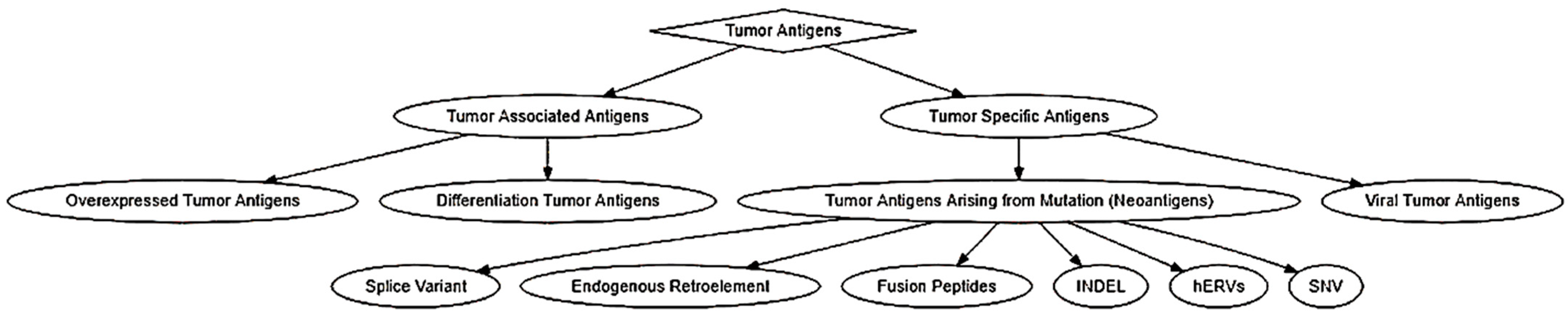
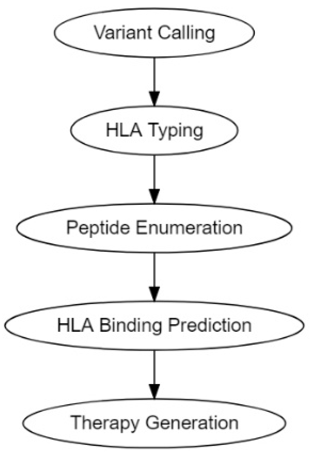
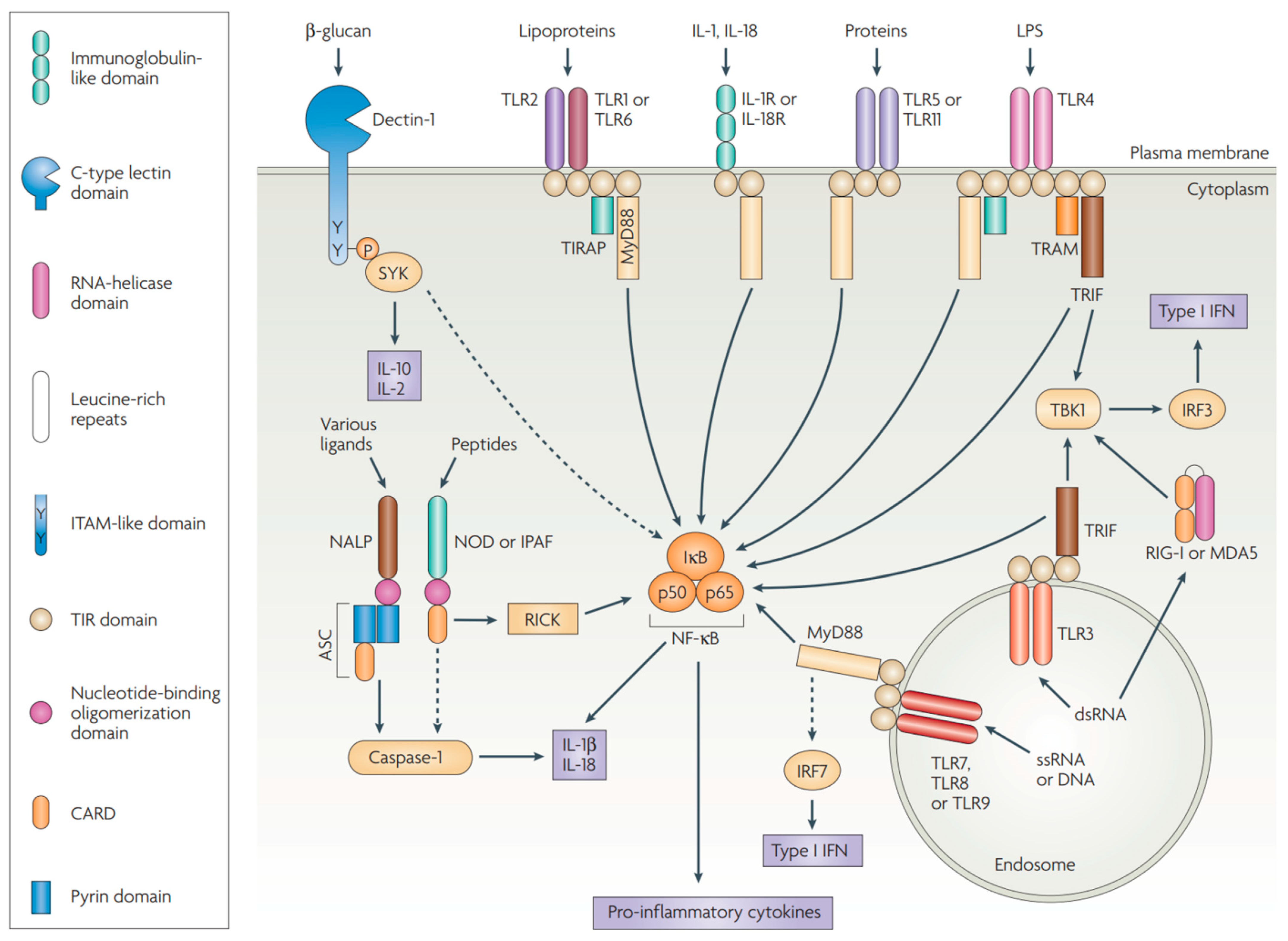
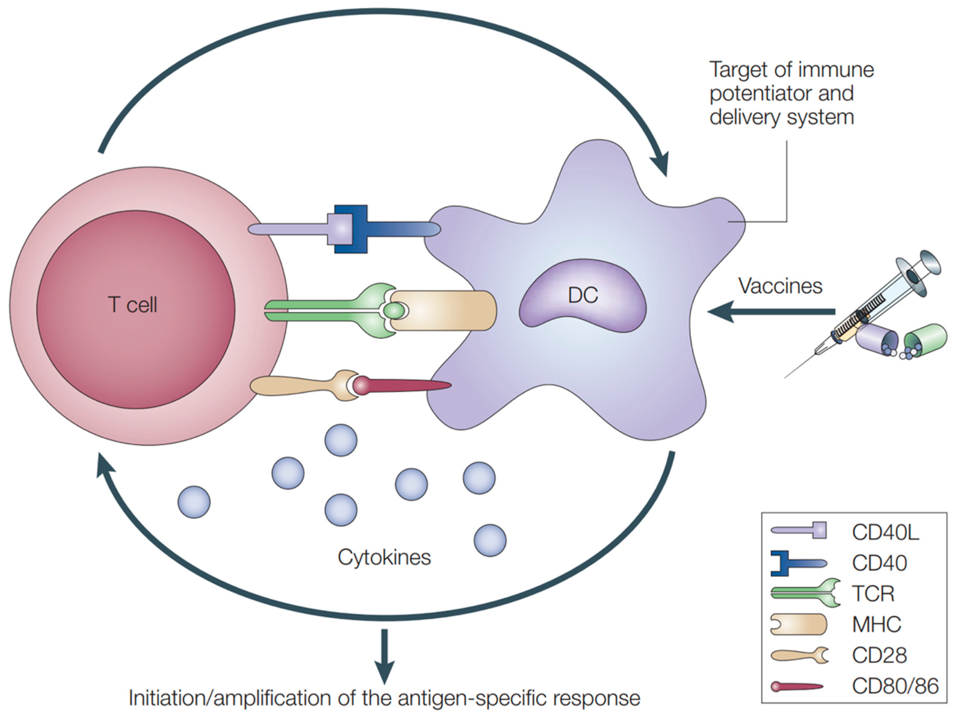
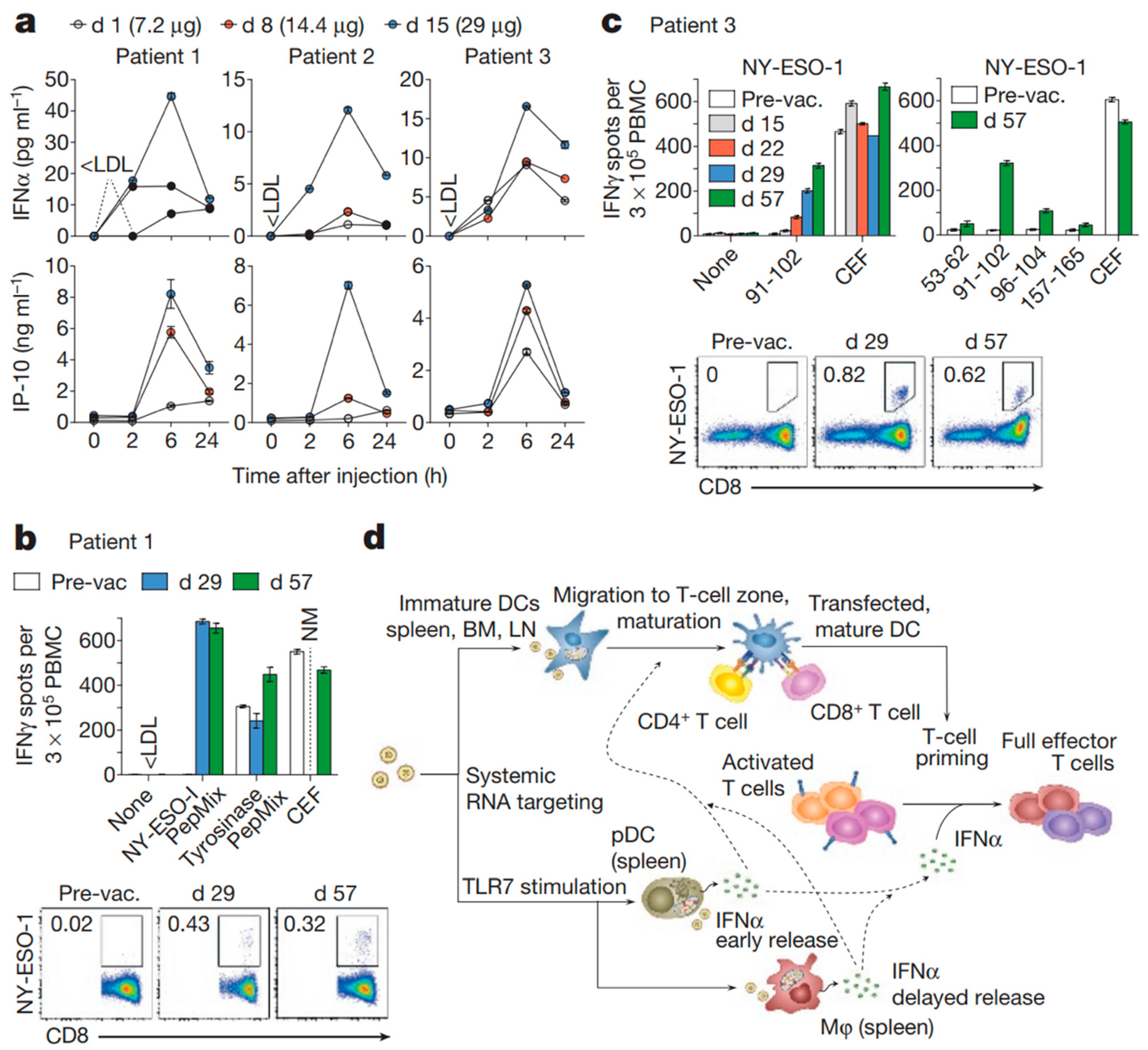
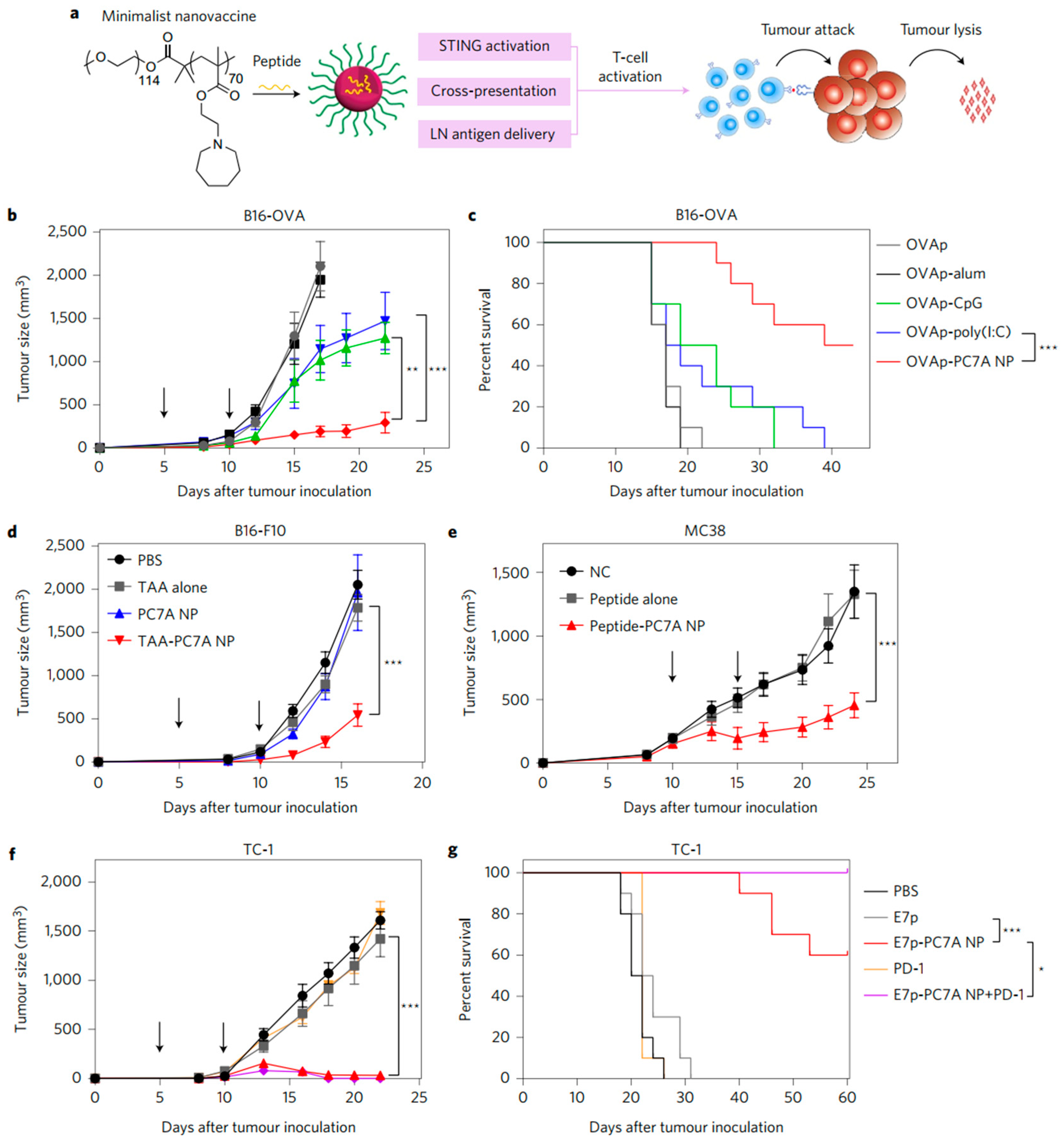
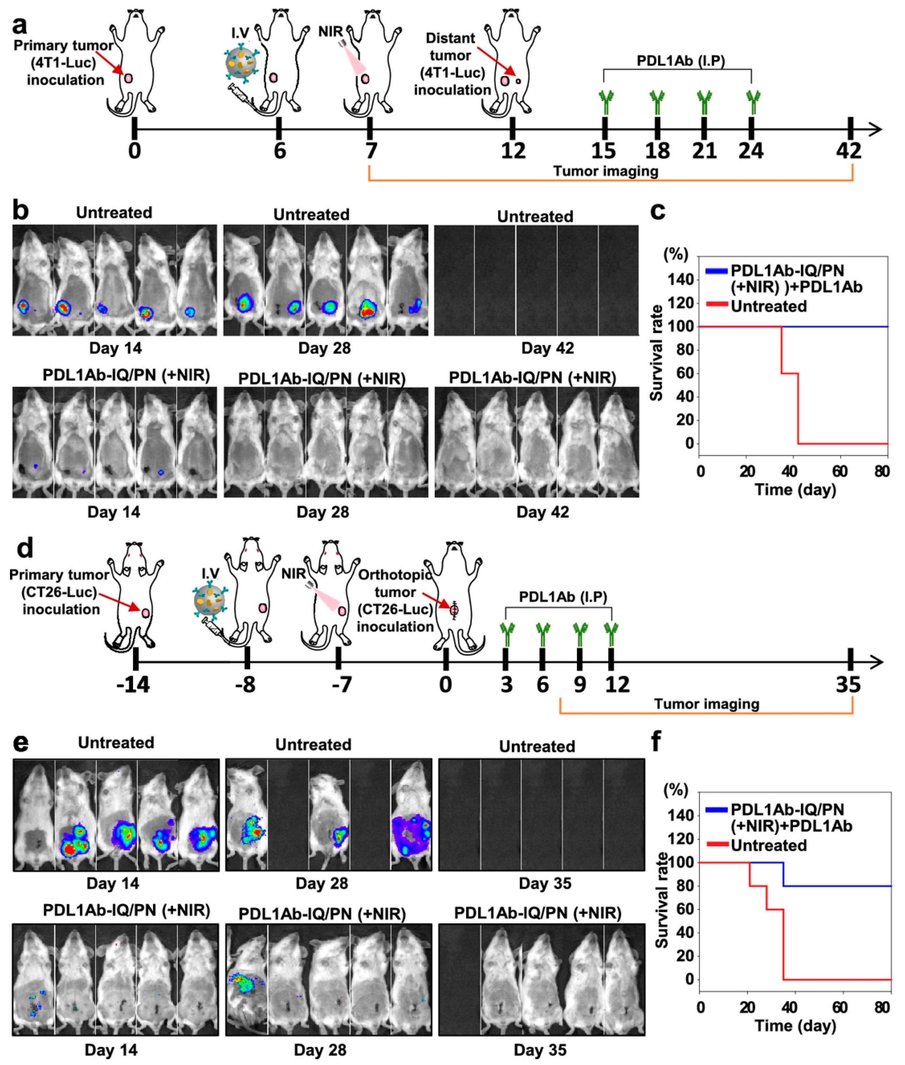
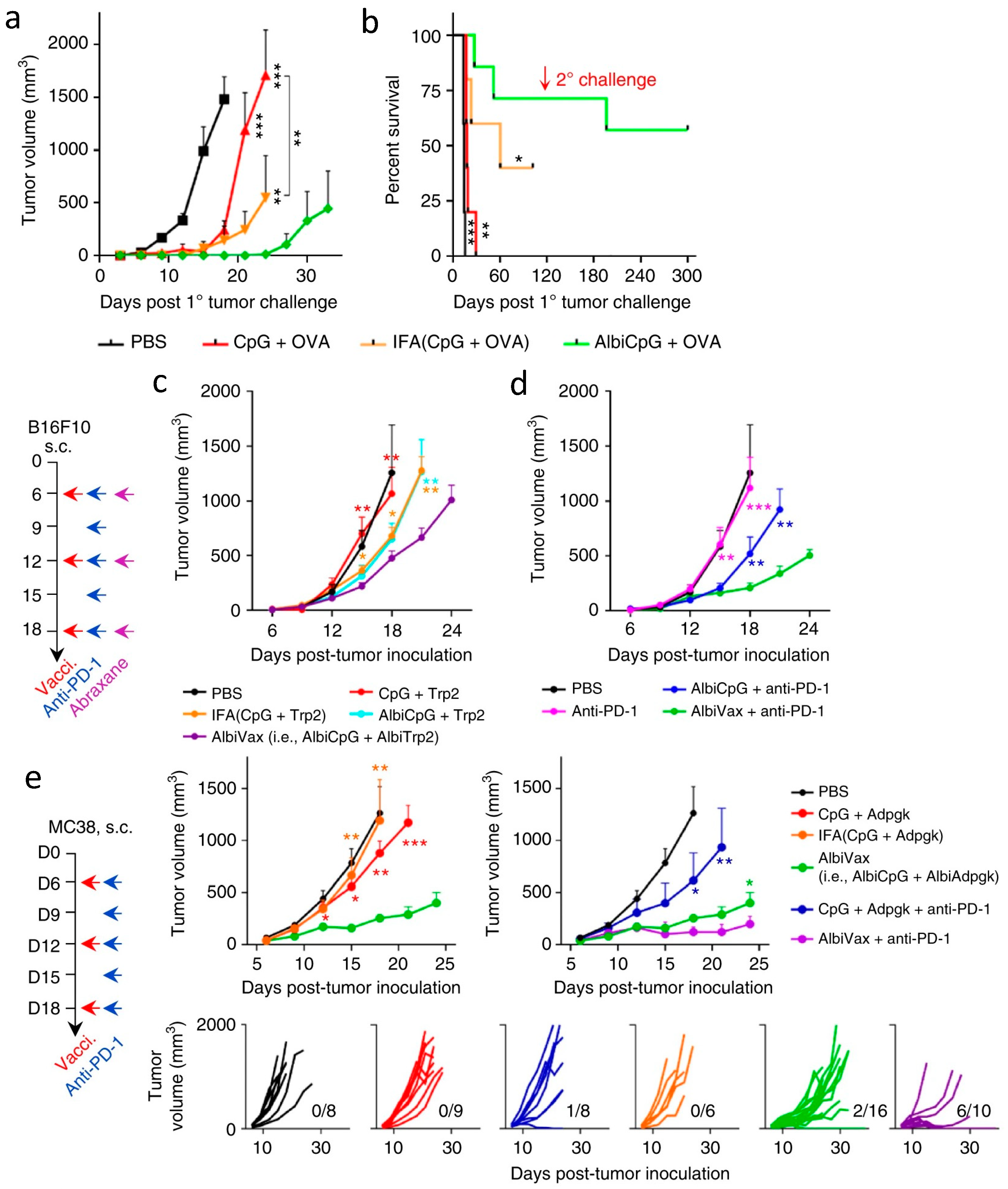
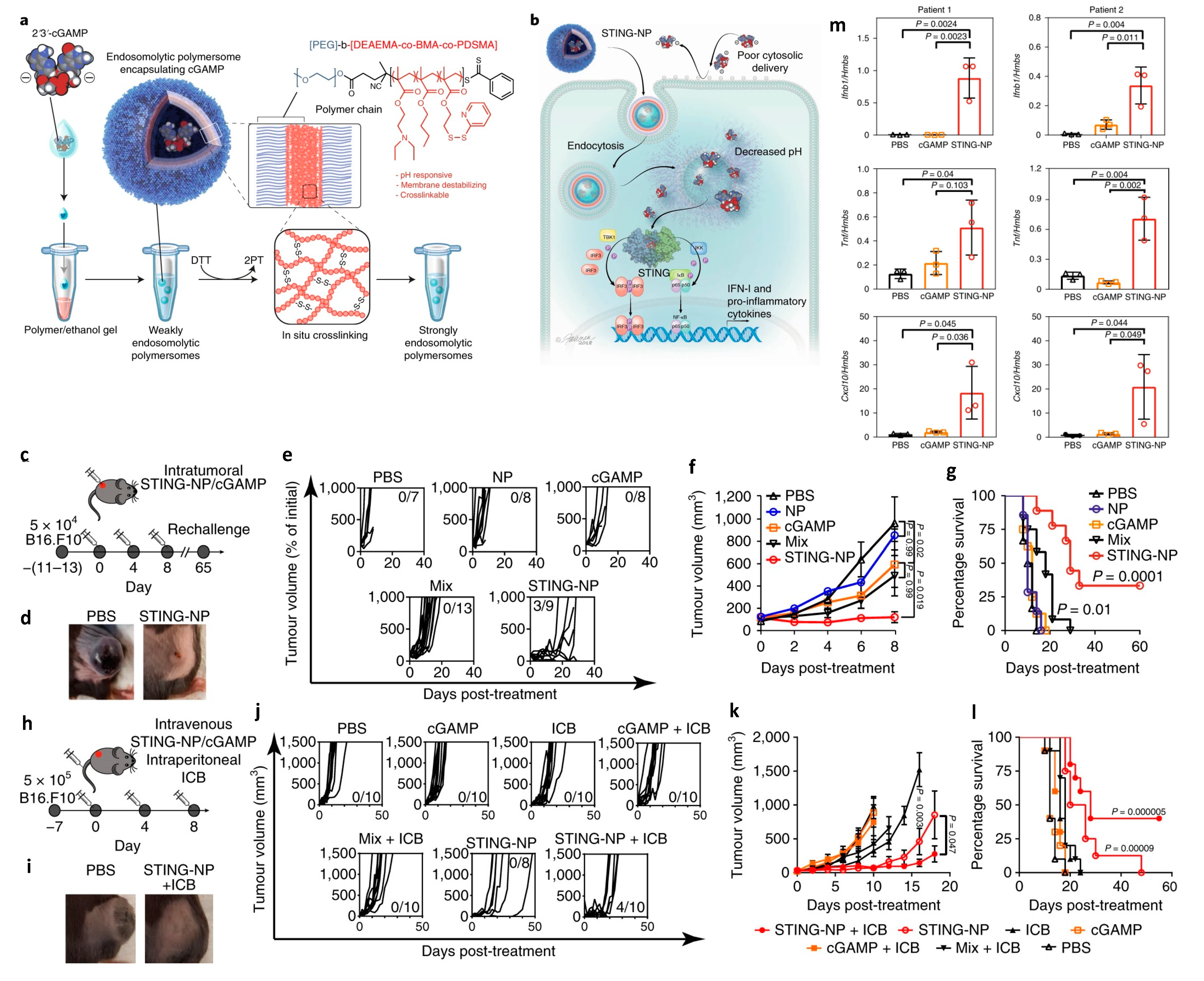
| Antigen | MHC-I | MHC-II |
|---|---|---|
| α-actinin | HLA-A2 [54] | |
| ARTC1 | HLA-DR1 [55] | |
| B-RAF | HLA-DR4 [56] | |
| β-catenin | HLA-A24 [57] | |
| BCR-ABL | HLA-A2 [58] HLA-A3 [59] HLA-A11 [60] HLA-B8 [58] | HLA-DR4 [61] HLA-DR9 [62] HLA-DR11 [63] HLA-DR15 [64] HLA-DR1 [65] |
| Caspase-5 | HLA-A2 [66] | |
| Caspase-8 | HLA-B35 [67] | |
| CDC27 | HLA-DR4 [68] | |
| CDK-4 | HLA-A2 [69] | |
| CDK-12 | HLA-A11 [70] | |
| CDK2NA | HLA-A11 [71] | |
| CLPP | HLA-A2 [72] | |
| COA-1 | HLA-DR4 [73] HLA-DR13 [73] | |
| CSNK1A1 | HLA-A2 [70] | |
| dek-can | HLA-DR53 [74] | |
| EFTUD2 | HLA-A3 [75] | |
| ELF2M | HLA-A68 [76] | |
| ETV6-AML1 | HLA-A2 [77] | |
| FLT3-ITD | HLA-A1 [78] | |
| fibronectin | HLA-DR15 [79] | |
| FNDC3B | HLA-A2 [80] | |
| GAS7 | HLA-A2 [70] | |
| GPNMB | HLA-A3 [75] | |
| HAUS3 | HLA-A2 [70] | |
| HSDL1 | HLA-Cw14 [81] | |
| HSP70-2 | HLA-A2 [82] | |
| KIA A0205 | HLA-B44 [83] | |
| K-ras | HLA-B35 [84] HLA-Cw8 [85] | |
| LDLR-FUT | HLA-DR1 [86] | |
| MART-2 | HLA-A1 [87] | |
| MATN | HLA-A11 [70] | |
| ME1 | HLA-A2 [88] | |
| MUM-1 | HLA-B44 [89] | |
| MUM-2 | HLA-B44 [90] HLA-Cw6 [90] | |
| MUM-3 | HLA-A68 [91] | |
| Myosin-m | HLA-A3 [92] | |
| N-ras | HLA-A1 [93] | |
| neo-PAP | HLA-DR7 [94] | |
| NFYC | HLA-B52 [95] | |
| OGT | HLA-A2 [96] | |
| OS-9 | HLA-B44 [97] | |
| p14ARF | HLA-A11 [71] | |
| p16INK | HLA-A11 [71] | |
| pml-RARalpha fusion protein | HLA-DR11 [98] | |
| PPP1R3B | HLA-A1 [70] | |
| PRDX5 | HLA-A2 [99] | |
| PTPRK | HLA-DR10 [100] | |
| RBAF600 | HLA-B7 [75] | |
| SIRT2 | HLA-A3 [75] | |
| SNDRP1 | HLA-B38 | |
| SYT-SSX1 or -SSX2 fusion protein | HLA-B7 [101] | |
| TGFBRII | HLA-DR3 [102] | |
| TP53 | HLA-A2 [103] | |
| TPI | HLA-DR1 [104] | |
| Annexin II | HLA-DR4 [105] |
| Adjuvants | Major Immunostimulatory Component(s) | Innate Receptors or Pathway Activated | Status |
|---|---|---|---|
| Alum | Aluminum salts | NLRP3/NALP3 inflammasome complex [142,143,144] | Licensed (Diphtheria, tetanus, pneumococcus, MenC, MenB, and many others) |
| MF59, AS03 | Squalene-in-water emulsions | MyD88 | Licensed (influenza) |
| AS04 | MPL adsorbed to alum | TLR4 | Licensed (HBV, HPV) |
| Virosomes | Viral glycoproteins and membrane lipids | Receptor-mediated endocytosis by APCs [140,145,146] | Licensed (influenza) |
| RC529 | Synthetic TLR4 ligand adsorbed to alum | TLR4 | Licensed (HBV) |
| Flagellin | Flagellin from S. typhimurium | TLR5 | Phase II (influenza, NCT00921947) |
| Monophosphoryl lipid A (MPL) and formulations (AS01, AS02) | MPL, Saponin (QS21) | TLR4 | Phase II (TB, NCT00146744; cancer, NCT00290355) Phase III (malaria, NCT00866619; herpes zoster, NCT01165177) |
| Imiquimod and Resiquimod | Imidazoquinoline derivatives | TLR7, TLR8 or both | Licensed (skin cancer) |
| CpG ODN and formulations (IC31) | Synthetic phophorothioate-linked DNA oligonucleotides with optimized CpG motifs | TLR9 | Phase I/IIa (malaria, NCT00984763; influenza, NCT03945825) Phase II/III (HBV, NCT01005407; breast cancer, NCT00043394; anthrax, NCT03877926; TB, NCT02075203) |
| AS15 | Liposomes, MPL, CpG, saponin (QS21) | TLR4 and 9 | Phase III (melanoma, NCT00796445) |
| GLA-SE | Oil-in-water emulsion with synthetic TLR4 ligand (GLA) | TLR4 | Phase II (influenza, NCT01991561) |
| Alum/TLR7 | Small-molecule synthetic TLR7 ligand adsorbed to alum | TLR7 | Phase I (MenC, NCT02639351) |
| RNAdjuvant® (CV8102) | Single-stranded, non-coding U-rich RNA complexed with crosslinked cationic peptide | TLR7/8/RIG-I | Phase I/II (skin cancer, NCT03291002; hepatocellular carcinoma, NCT03203005) |
| Adjuvant Combination | Delivery System | Outcome | Ref. |
|---|---|---|---|
| MPLA (TLR4) Imiquimod (TLR7) | Mannose-functionalized lipid hybrid polymersomes formed with DOTAP, PCL-PEG-PCL and Mannose-PEG-DSPE | Greater prophylactic effect against E.G7-OVA tumor challenge than free OVA plus adjuvants. Greater therapeutic effect when used together with anti-PD1 against E.G7-OVA tumor, comparing to untargeted/targeted polymersomes alone or free OVA plus adjuvants. | [239,240] |
| CpG (TLR9) with MPLA (TLR4) or R848 (TLR7/8) | PLGA/PEI nanoparticles | After in vivo administration, either combination could induce a significantly higher IFN-γ secretion comparing to single adjuvant or alum; MPLA and CpG could induce more IL-4 secretion than R848 and CpG. As for antibody responses, R848 and CpG could induce a high titer of IgG2a while MPLA and CpG could induce the highest titer of IgG1, which were comparable to alum. | [241] |
| MPLA (TLR4) CpG (TLR9) | Mannosylated PLGA/PLA nanoparticles | Superior prophylactic effect against melanoma challenge when combined with anti-PD1 and anti-OX40, comparing to nanoparticles alone or other free formulations. Ibrutinib, a MDSC inhibitor, was needed to inhibit melanoma growth at the therapeutic regimen. | [242] |
| Poly(I:C) (TLR3) CpG (TLR9) | Liposomes prepared with phospholipid and cholesterol | Dual TLR-adjuvant vaccine protected 6 out of 8 mice from tumor challenge of OVA expressing E.G7 in 8 months after primary and booster injections. | [243] |
| β-glucan (TLR2) CpG (TLR9) | Aminated glucan nanoparticles (AG NP) | CpG-AG NP elevated antigen-specific antibody titers in serum (IgG1 and IgG2a) and the production of IL-4 and IFN-γ in immunized mice, which were comparable to Freund’s adjuvant without causing any significant toxicity. | [244] |
| R848 (TLR7/8) cGAMP (STING) | Acetalated dextran microparticles (Ace-DEX MP) | Dual-adjuvant Ace-DEX MP induced a higher production of IL-2 and IFN-γ than Alum in immunized mice. This combination also induced elevated overall antibody titers with a near even ratio of IgG2c/IgG1, achieving a balanced Th1/Th2 humoral responses. | [245] |
| R848 (TLR7/8) CpG (TLR9) | PEG-PLA micelles | Bi-adjuvant nanovaccine loaded with neoantigen Adpgk induced potent antitumor immunity against MC38 colorectal cancer without causing acute systemic toxicity. | [246] |
Publisher’s Note: MDPI stays neutral with regard to jurisdictional claims in published maps and institutional affiliations. |
© 2022 by the authors. Licensee MDPI, Basel, Switzerland. This article is an open access article distributed under the terms and conditions of the Creative Commons Attribution (CC BY) license (https://creativecommons.org/licenses/by/4.0/).
Share and Cite
Alarcon, N.O.; Jaramillo, M.; Mansour, H.M.; Sun, B. Therapeutic Cancer Vaccines—Antigen Discovery and Adjuvant Delivery Platforms. Pharmaceutics 2022, 14, 1448. https://doi.org/10.3390/pharmaceutics14071448
Alarcon NO, Jaramillo M, Mansour HM, Sun B. Therapeutic Cancer Vaccines—Antigen Discovery and Adjuvant Delivery Platforms. Pharmaceutics. 2022; 14(7):1448. https://doi.org/10.3390/pharmaceutics14071448
Chicago/Turabian StyleAlarcon, Neftali Ortega, Maddy Jaramillo, Heidi M. Mansour, and Bo Sun. 2022. "Therapeutic Cancer Vaccines—Antigen Discovery and Adjuvant Delivery Platforms" Pharmaceutics 14, no. 7: 1448. https://doi.org/10.3390/pharmaceutics14071448