
Metallic Engineered Nanomaterials and Ocular Toxicity: A Current Perspective

 , , , and
Abstract
:1. Introduction
2. Characteristics and General Toxicity of Metallic ENMs
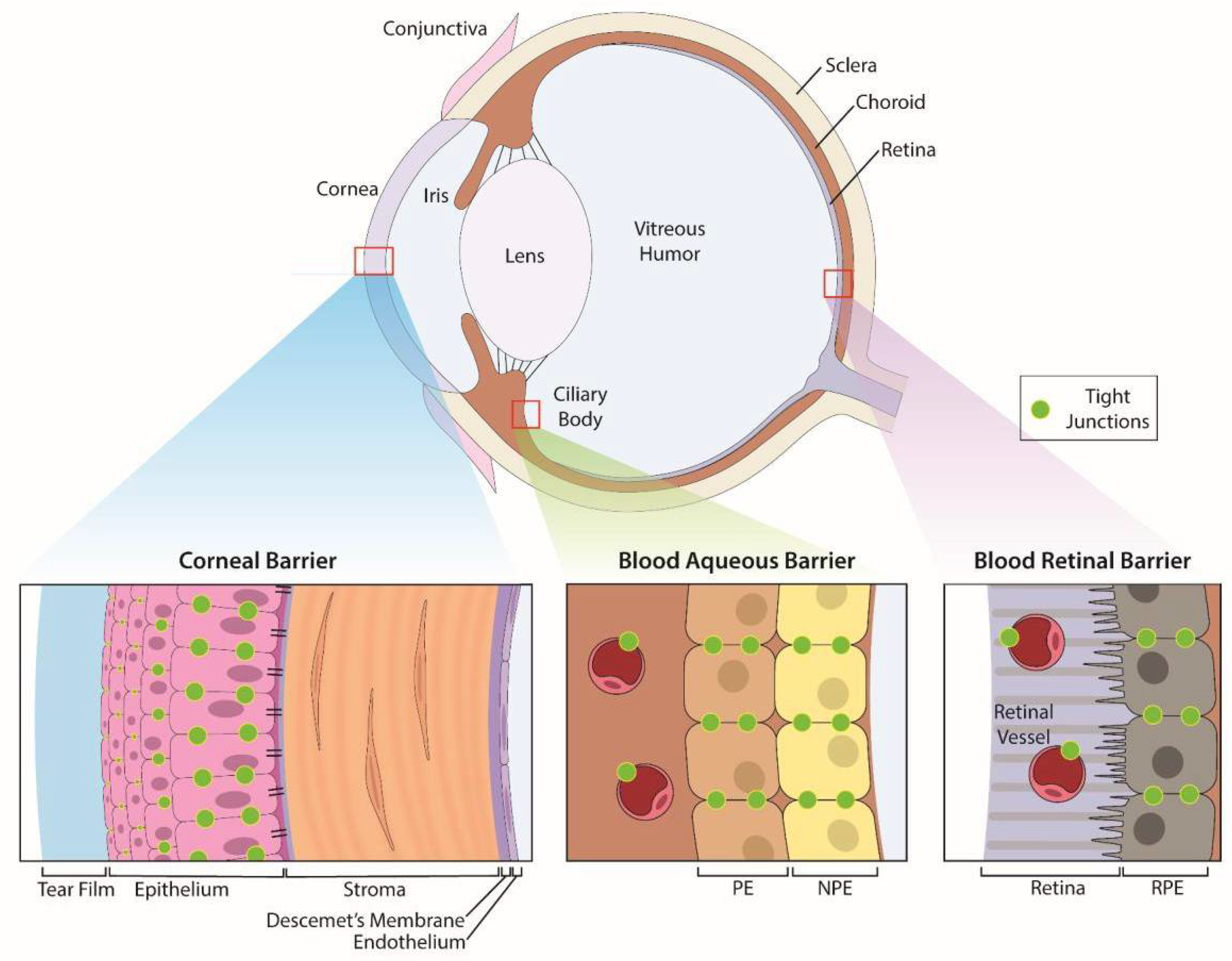
3. The Eye Is a Formidable Barrier
3.1. Surface Barriers Prevent Exogenous Agents from Entering the Eye
3.2. Blood Ocular Barriers Limit Access of Systemic Agents to the Eye
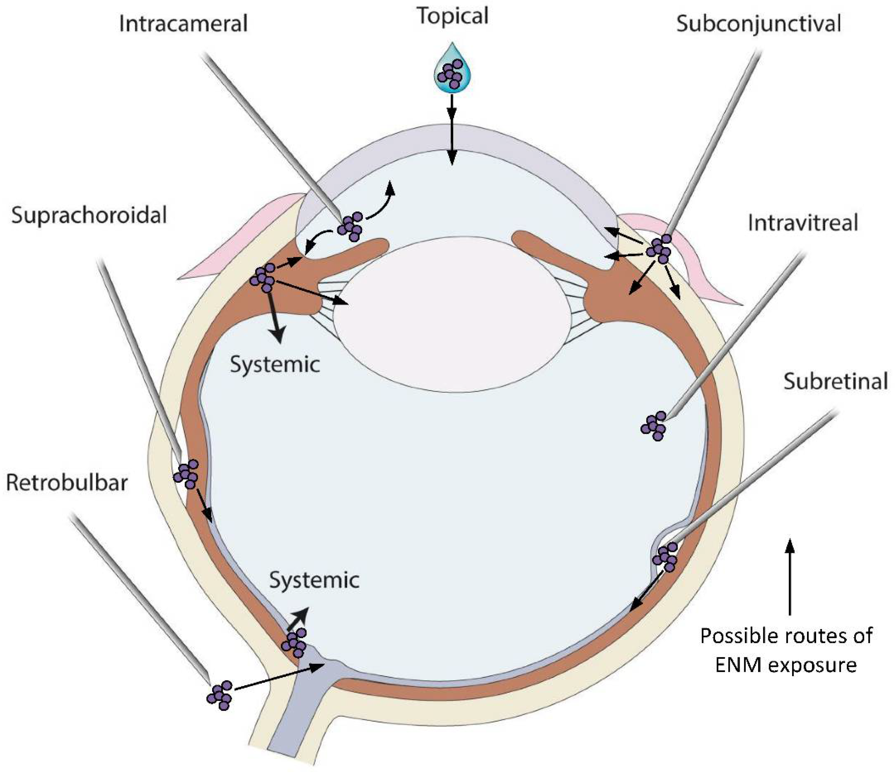
4. Toxicity and Utilization of Metallic ENMs in Ophthalmology
4.1. Gold ENMs
4.1.1. Anterior Ocular Toxicity
4.1.2. Posterior Ocular Toxicity
4.2. Silver ENMs
4.2.1. Anterior Ocular Toxicity
4.2.2. Posterior Ocular Toxicity
4.3. Metal Oxide ENMs
4.3.1. Titanium Dioxide ENMs
4.3.2. Zinc Oxide ENM
4.3.3. Cerium ENMs
4.3.4. Other Metallic ENMs
4.3.5. Magnetic ENMs
5. Conclusions
Author Contributions
Funding
Institutional Review Board Statement
Informed Consent Statement
Data Availability Statement
Conflicts of Interest
References
- Albalawi, F.; Hussein, M.Z.; Fakurazi, S.; Masarudin, M.J. Engineered Nanomaterials: The Challenges and Opportunities for Nanomedicines. Int. J. Nanomed. 2021, 16, 161–184. [Google Scholar] [CrossRef] [PubMed]
- Johnston, L.J.; Gonzalez-Rojano, N.; Wilkinson, K.J.; Xing, B.S. Key challenges for evaluation of the safety of engineered nanomaterials. Nanoimpact 2020, 18, 100129. [Google Scholar] [CrossRef]
- Giese, B.; Klaessig, F.; Park, B.; Kaegi, R.; Steinfeldt, M.; Wigger, H.; von Gleich, A.; Gottschalk, F. Risks, Release and Concentrations of Engineered Nanomaterial in the Environment. Sci. Rep. 2018, 8, 1565. [Google Scholar] [CrossRef]
- Wigger, H.; Kägi, R.; Wiesner, M.; Nowack, B. Exposure and Possible Risks of Engineered Nanomaterials in the Environment—Current Knowledge and Directions for the Future. Rev. Geophys. 2020, 58, e2020RG000710. [Google Scholar] [CrossRef]
- Kumar, A.; Kumar, P.; Anandan, A.; Fernandes, T.F.; Ayoko, G.A.; Biskos, G. Engineered Nanomaterials: Knowledge Gaps in Fate, Exposure, Toxicity, and Future Directions. J. Nanomater. 2014, 2014, 130198. [Google Scholar] [CrossRef] [Green Version]
- Duncan, R. The dawning era of polymer therapeutics. Nat. Rev. Drug Discov. 2003, 2, 347–360. [Google Scholar] [CrossRef] [PubMed]
- Pinnell, S.R.; Fairhurst, D.; Gillies, R.; Mitchnick, M.A.; Kollias, N. Microfine zinc oxide is a superior sunscreen ingredient to microfine titanium dioxide. Dermatol. Surg. 2000, 26, 309–313. [Google Scholar] [CrossRef]
- Hadrup, N.; Lam, H.R. Oral toxicity of silver ions, silver nanoparticles and colloidal silver—A review. Regul. Toxicol. Pharm. 2014, 68, 1–7. [Google Scholar] [CrossRef]
- Liu, S.Y.; Dozois, M.D.; Chang, C.N.; Ahmad, A.; Ng, D.L.T.; Hileeto, D.; Liang, H.Y.; Reyad, M.M.; Boyd, S.; Jones, L.W.; et al. Prolonged Ocular Retention of Mucoadhesive Nanoparticle Eye Drop Formulation Enables Treatment of Eye Diseases Using Significantly Reduced Dosage. Mol. Pharm. 2016, 13, 2897–2905. [Google Scholar] [CrossRef]
- Roh, Y.J.; Rho, C.R.; Cho, W.K.; Kang, S. The Antiangiogenic Effects of Gold Nanoparticles on Experimental Choroidal Neovascularization in Mice. Investig. Ophthalmol. Vis. Sci. 2016, 57, 6561–6567. [Google Scholar] [CrossRef] [Green Version]
- Silva, R.M.; Xu, J.Y.; Saiki, C.; Anderson, D.S.; Franzi, L.M.; Vulpe, C.D.; Gilbert, B.; Van Winkle, L.S.; Pinkerton, K.E. Short versus long silver nanowires: A comparison of in vivo pulmonary effects post instillation. Part. Fibre Toxicol. 2014, 11, 52. [Google Scholar] [CrossRef] [PubMed] [Green Version]
- Billi, F.; Campbell, P. Nanotoxicology of metal wear particles in total joint arthroplasty: A review of current concepts. J. Appl. Biomater. Biom. 2010, 8, 1–6. [Google Scholar]
- Yah, C.S.; Simate, G.S.; Iyuke, S.E. Nanoparticles toxicity and their routes of exposures. Pak. J. Pharm. Sci. 2012, 25, 477–491. [Google Scholar] [PubMed]
- Choi, S.J.; Choy, J.H. Biokinetics of zinc oxide nanoparticles: Toxicokinetics, biological fates, and protein interaction. Int J. Nanomed. 2014, 9 (Suppl. 2), 261–269. [Google Scholar]
- Kim, Y.R.; Park, J.I.; Lee, E.J.; Park, S.H.; Seong, N.W.; Kim, J.H.; Kim, G.Y.; Meang, E.H.; Hong, J.S.; Kim, S.H.; et al. Toxicity of 100 nm zinc oxide nanoparticles: A report of 90-day repeated oral administration in Sprague Dawley rats. Int. J. Nanomed. 2014, 9 (Suppl. 2), 109–126. [Google Scholar]
- Kunzmann, A.; Andersson, B.; Thurnherr, T.; Krug, H.; Scheynius, A.; Fadeel, B. Toxicology of engineered nanomaterials: Focus on biocompatibility, biodistribution and biodegradation. BBA-Gen. Subj. 2011, 1810, 361–373. [Google Scholar] [CrossRef]
- Frick, K.D.; Gower, E.W.; Kempen, J.H.; Wolff, J.L. Economic impact of visual impairment and blindness in the United States. Arch. Ophthalmol. 2007, 125, 544–550. [Google Scholar] [CrossRef] [Green Version]
- Koberlein, J.; Beifus, K.; Schaffert, C.; Finger, R.P. The economic burden of visual impairment and blindness: A systematic review. BMJ Open 2013, 3, e003471. [Google Scholar] [CrossRef]
- Liu, S.; Jones, L.; Gu, F.X. Nanomaterials for ocular drug delivery. Macromol. Biosci. 2012, 12, 608–620. [Google Scholar] [CrossRef]
- Burnett, M.E.; Wang, S.Q. Current sunscreen controversies: A critical review. Photodermatol. Photoimmunol. Photomed. 2011, 27, 58–67. [Google Scholar] [CrossRef]
- Bachu, R.D.; Chowdhury, P.; Al-Saedi, Z.H.F.; Karla, P.K.; Boddu, S.H.S. Ocular Drug Delivery Barriers-Role of Nanocarriers in the Treatment of Anterior Segment Ocular Diseases. Pharmaceutics 2018, 10, 28. [Google Scholar] [CrossRef] [PubMed] [Green Version]
- Janagam, D.R.; Wu, L.; Lowe, T.L. Nanoparticles for drug delivery to the anterior segment of the eye. Adv. Drug Deliv. Rev. 2017, 122, 31–64. [Google Scholar] [CrossRef] [PubMed]
- Rodrigues, F.S.C.; Campos, A.; Martins, J.; Ambrosio, A.F.; Campos, E.J. Emerging Trends in Nanomedicine for Improving Ocular Drug Delivery: Light-Responsive Nanoparticles, Mesoporous Silica Nanoparticles, and Contact Lenses. ACS Biomater. Sci. Eng. 2020, 6, 6587–6597. [Google Scholar] [CrossRef] [PubMed]
- Dubald, M.; Bourgeois, S.; Andrieu, V.; Fessi, H. Ophthalmic Drug Delivery Systems for Antibiotherapy—A Review. Pharmaceutics 2018, 10, 10. [Google Scholar] [CrossRef] [PubMed] [Green Version]
- Sahoo, S.K.; Dilnawaz, F.; Krishnakumar, S. Nanotechnology in ocular drug delivery. Drug Discov. Today 2008, 13, 144–151. [Google Scholar] [CrossRef]
- B.S. Institution. Terminology for Nanomaterials. Publicly Available Specification 2007. Volume 136. Available online: https://www.iso.org/obp/ui/#iso:std:iso:ts:12901:-1:ed-1:v1:en (accessed on 21 April 2022).
- Guo, T.; Yao, M.S.; Lin, Y.H.; Nan, C.W. A comprehensive review on synthesis methods for transition-metal oxide nanostructures. CrystEngComm 2015, 17, 3551–3585. [Google Scholar] [CrossRef]
- Roduner, E. Size matters: Why nanomaterials are different. Chem. Soc. Rev. 2006, 35, 583–592. [Google Scholar] [CrossRef]
- Moore, T.L.; Rodriguez-Lorenzo, L.; Hirsch, V.; Balog, S.; Urban, D.; Jud, C.; Rothen-Rutishauser, B.; Lattuada, M.; Petri-Fink, A. Nanoparticle colloidal stability in cell culture media and impact on cellular interactions. Chem. Soc. Rev. 2015, 44, 6287–6305. [Google Scholar] [CrossRef] [Green Version]
- Guerrini, L.; Alvarez-Puebla, R.A.; Pazos-Perez, N. Surface Modifications of Nanoparticles for Stability in Biological Fluids. Materials 2018, 11, 1154. [Google Scholar] [CrossRef] [Green Version]
- Tso, C.P.; Zhung, C.M.; Shih, Y.H.; Tseng, Y.M.; Wu, S.C.; Doong, R.A. Stability of metal oxide nanoparticles in aqueous solutions. Water Sci. Technol. 2010, 61, 127–133. [Google Scholar] [CrossRef] [Green Version]
- Song, Y.X.; Zhao, K.; Li, M.Q.; Pan, X.X.; Li, D.Q. A novel method for measuring zeta potentials of solid-liquid interfaces. Anal. Chim. Acta 2015, 853, 689–695. [Google Scholar] [CrossRef] [PubMed]
- Worthen, A.J.; Tran, V.; Cornell, K.A.; Truskett, T.M.; Johnston, K.P. Steric stabilization of nanoparticles with grafted low molecular weight ligands in highly concentrated brines including divalent ions. Soft Matter 2016, 12, 2025–2039. [Google Scholar] [CrossRef] [PubMed]
- Mu, Q.; Jiang, G.; Chen, L.; Zhou, H.; Fourches, D.; Tropsha, A.; Yan, B. Chemical Basis of Interactions Between Engineered Nanoparticles and Biological Systems. Chem. Rev. 2014, 114, 7740–7781. [Google Scholar] [CrossRef] [PubMed] [Green Version]
- Bondarenko, O.; Juganson, K.; Ivask, A.; Kasemets, K.; Mortimer, M.; Kahru, A. Toxicity of Ag, CuO and ZnO nanoparticles to selected environmentally relevant test organisms and mammalian cells in vitro: A critical review. Arch. Toxicol. 2013, 87, 1181–1200. [Google Scholar] [CrossRef] [Green Version]
- Gaillet, S.; Rouanet, J.M. Silver nanoparticles: Their potential toxic effects after oral exposure and underlying mechanisms—A review. Food Chem. Toxicol. 2015, 77, 58–63. [Google Scholar] [CrossRef]
- Antony, J.J.; Sivalingam, P.; Chen, B.A. Toxicological effects of silver nanoparticles. Environ. Toxicol. Pharmacol. 2015, 40, 729–732. [Google Scholar] [CrossRef]
- Ivask, A.; Titma, T.; Visnapuu, M.; Vija, H.; Kakinen, A.; Sihtmae, M.; Pokhrel, S.; Madler, L.; Heinlaan, M.; Kisand, V.; et al. Toxicity of 11 Metal Oxide Nanoparticles to Three Mammalian Cell Types In Vitro. Curr. Top. Med. Chem. 2015, 15, 1914–1929. [Google Scholar] [CrossRef]
- Ema, M.; Okuda, H.; Gamo, M.; Honda, K. A review of reproductive and developmental toxicity of silver nanoparticles in laboratory animals. Reprod. Toxicol. 2017, 67, 149–164. [Google Scholar] [CrossRef]
- Park, H.S.; Kim, S.J.; Lee, T.J.; Kim, G.Y.; Meang, E.; Hong, J.S.; Kim, S.H.; Koh, S.B.; Hong, S.G.; Sun, Y.S.; et al. A 90-day study of sub-chronic oral toxicity of 20 nm positively charged zinc oxide nanoparticles in Sprague Dawley rats. Int. J. Nanomed. 2014, 9, 93–107. [Google Scholar]
- Ebabe Elle, R.; Gaillet, S.; Vidé, J.; Romain, C.; Lauret, C.; Rugani, N.; Cristol, J.P.; Rouanet, J.M. Dietary exposure to silver nanoparticles in Sprague-Dawley rats: Effects on oxidative stress and inflammation. Food Chem. Toxicol. 2013, 60, 297–301. [Google Scholar] [CrossRef]
- Stankic, S.; Suman, S.; Haque, F.; Vidic, J. Pure and multi metal oxide nanoparticles: Synthesis, antibacterial and cytotoxic properties. J. Nanobiotechnol. 2016, 14, 73. [Google Scholar] [CrossRef] [PubMed] [Green Version]
- Ivask, A.; Juganson, K.; Bondarenko, O.; Mortimer, M.; Aruoja, V.; Kasemets, K.; Blinova, I.; Heinlaan, M.; Slaveykova, V.; Kahru, A. Mechanisms of toxic action of Ag, ZnO and CuO nanoparticles to selected ecotoxicological test organisms and mammalian cells in vitro: A comparative review. Nanotoxicology 2014, 8 (Suppl. 1), 57–71. [Google Scholar] [CrossRef] [PubMed]
- Lanone, S.; Boczkowski, J. Biomedical applications and potential health risks of nanomaterials: Molecular mechanisms. Curr. Mol. Med. 2006, 6, 651–663. [Google Scholar] [CrossRef] [PubMed]
- Aillon, K.L.; Xie, Y.; El-Gendy, N.; Berkland, C.J.; Forrest, M.L. Effects of nanomaterial physicochemical properties on in vivo toxicity. Adv. Drug Deliv. Rev. 2009, 61, 457–466. [Google Scholar] [CrossRef] [Green Version]
- Raghunathan, V.K.; Devey, M.; Hawkins, S.; Hails, L.; Davis, S.A.; Mann, S.; Chang, I.T.; Ingham, E.; Malhas, A.; Vaux, D.J.; et al. Influence of particle size and reactive oxygen species on cobalt chrome nanoparticle-mediated genotoxicity. Biomaterials 2013, 34, 3559–3570. [Google Scholar] [CrossRef]
- Riaz Ahmed, K.B.; Nagy, A.M.; Brown, R.P.; Zhang, Q.; Malghan, S.G.; Goering, P.L. Silver nanoparticles: Significance of physicochemical properties and assay interference on the interpretation of in vitro cytotoxicity studies. Toxicol. In Vitro 2017, 38, 179–192. [Google Scholar] [CrossRef]
- Ahamed, M.; Karns, M.; Goodson, M.; Rowe, J.; Hussain, S.M.; Schlager, J.J.; Hong, Y. DNA damage response to different surface chemistry of silver nanoparticles in mammalian cells. Toxicol. Appl. Pharmacol. 2008, 233, 404–410. [Google Scholar] [CrossRef]
- Lynch, I.; Salvati, A.; Dawson, K.A. Protein-Nanoparticle Interactions What does the cell see? Nat. Nanotechnol. 2009, 4, 546–547. [Google Scholar] [CrossRef]
- Mahmoudi, M.; Lynch, I.; Ejtehadi, M.R.; Monopoli, M.P.; Bombelli, F.B.; Laurent, S. Protein-Nanoparticle Interactions: Opportunities and Challenges. Chem. Rev. 2011, 111, 5610–5637. [Google Scholar] [CrossRef]
- Kaur, I.P.; Kanwar, M. Ocular preparations: The formulation approach. Drug Dev. Ind. Pharm. 2002, 28, 473–493. [Google Scholar] [CrossRef]
- Urtti, A. Challenges and obstacles of ocular pharmacokinetics and drug delivery. Adv. Drug Deliv. Rev. 2006, 58, 1131–1135. [Google Scholar] [CrossRef] [PubMed]
- Han, J.Y.; Kang, B.; Eom, Y.; Kim, H.M.; Song, J.S. Comparing the Effects of Particulate Matter on the Ocular Surfaces of Normal Eyes and a Dry Eye Rat Model. Cornea 2017, 36, 605–610. [Google Scholar] [CrossRef] [PubMed]
- Li, Y.J.; Luo, L.J.; Harroun, S.G.; Wei, S.C.; Unnikrishnan, B.; Chang, H.T.; Huang, Y.F.; Lai, J.Y.; Huang, C.C. Synergistically dual-functional nano eye-drops for simultaneous anti-inflammatory and anti-oxidative treatment of dry eye disease. Nanoscale 2019, 11, 5580–5594. [Google Scholar] [CrossRef] [PubMed]
- Yan, J.; Peng, X.; Cai, Y.; Cong, W. Development of facile drug delivery platform of ranibizumab fabricated PLGA-PEGylated magnetic nanoparticles for age-related macular degeneration therapy. J. Photochem. Photobiol. B 2018, 183, 133–136. [Google Scholar] [CrossRef]
- Jemni-Damer, N.; Guedan-Duran, A.; Fuentes-Andion, M.; Serrano-Bengoechea, N.; Alfageme-Lopez, N.; Armada-Maresca, F.; Guinea, G.V.; Perez-Rigueiro, J.; Rojo, F.; Gonzalez-Nieto, D.; et al. Biotechnology and Biomaterial-Based Therapeutic Strategies for Age-Related Macular Degeneration. Part I: Biomaterials-Based Drug Delivery Devices. Front. Bioeng. Biotechnol. 2020, 8, 549089. [Google Scholar] [CrossRef]
- Sharma, S.; Bhatia, V. Nanoscale Drug Delivery Systems for Glaucoma: Experimental and In Silico Advances. Curr. Top. Med. Chem. 2020, 21, 115–125. [Google Scholar] [CrossRef]
- Weng, Y.; Liu, J.; Jin, S.; Guo, W.; Liang, X.; Hu, Z. Nanotechnology-based strategies for treatment of ocular disease. Acta Pharm. Sin. B 2017, 7, 281–291. [Google Scholar] [CrossRef] [Green Version]
- Puglia, C.; Santonocito, D.; Ostacolo, C.; Maria Sommella, E.; Campiglia, P.; Carbone, C.; Drago, F.; Pignatello, R.; Bucolo, C. Ocular Formulation Based on Palmitoylethanolamide-Loaded Nanostructured Lipid Carriers: Technological and Pharmacological Profile. Nanomaterials 2020, 10, 287. [Google Scholar] [CrossRef] [Green Version]
- Mehra, N.K.; Cai, D.; Kuo, L.; Hein, T.; Palakurthi, S. Safety and toxicity of nanomaterials for ocular drug delivery applications. Nanotoxicology 2016, 10, 836–860. [Google Scholar] [CrossRef]
- Wang, F.; Wang, D.; Song, M.; Zhou, Q.; Liao, R.; Wang, Y. MiRNA-155-5p Reduces Corneal Epithelial Permeability by Remodeling Epithelial Tight Junctions during Corneal Wound Healing. Curr. Eye Res. 2020, 45, 904–913. [Google Scholar] [CrossRef]
- Steed, E.; Balda, M.S.; Matter, K. Dynamics and functions of tight junctions. Trends Cell. Biol. 2010, 20, 142–149. [Google Scholar] [CrossRef] [PubMed]
- Anderson, J.M.; Van Itallie, C.M. Physiology and function of the tight junction. Cold Spring Harb. Perspect. Biol. 2009, 1, a002584. [Google Scholar] [CrossRef] [PubMed]
- Hou, J. Chapter 6—Paracellular Water Channel. In The Paracellular Channel; Hou, J., Ed.; Academic Press: Cambridge, MA, USA, 2019; pp. 83–92. [Google Scholar]
- Tervonen, A.; Ihalainen, T.O.; Nymark, S.; Hyttinen, J. Structural dynamics of tight junctions modulate the properties of the epithelial barrier. PLoS ONE 2019, 14, e0214876. [Google Scholar] [CrossRef] [PubMed]
- Zihni, C.; Mills, C.; Matter, K.; Balda, M.S. Tight junctions: From simple barriers to multifunctional molecular gates. Nat. Rev. Mol. Cell Biol. 2016, 17, 564–580. [Google Scholar] [CrossRef] [PubMed]
- Xiang, P.; Jia, Y.; Wang, K.; Li, M.Y.; Qin, Y.S.; He, R.W.; Gao, P.; Liu, Y.; Liu, X.; Ma, L.Q. Water extract of indoor dust induces tight junction disruption in normal human corneal epithelial cells. Environ. Pollut. 2018, 243 Pt A, 301–307. [Google Scholar] [CrossRef]
- Huang, C.; Liao, R.; Wang, F.; Tang, S. Characteristics of Reconstituted Tight Junctions after Corneal Epithelial Wounds and Ultrastructure Alterations of Corneas in Type 2 Diabetic Rats. Curr. Eye Res. 2016, 41, 783–790. [Google Scholar] [CrossRef]
- Antcliff, R.J.; Marshall, J. The pathogenesis of edema in diabetic maculopathy. Semin. Ophthalmol. 1999, 14, 223–232. [Google Scholar] [CrossRef]
- Rudraraju, M.; Narayanan, S.P.; Somanath, P.R. Regulation of blood-retinal barrier cell-junctions in diabetic retinopathy. Pharmacol. Res. 2020, 161, 105115. [Google Scholar] [CrossRef]
- Chen, M.L.; Ge, Z.; Fox, J.G.; Schauer, D.B. Disruption of tight junctions and induction of proinflammatory cytokine responses in colonic epithelial cells by Campylobacter jejuni. Infect. Immun. 2006, 74, 6581–6589. [Google Scholar] [CrossRef] [Green Version]
- Wittekindt, O.H. Tight junctions in pulmonary epithelia during lung inflammation. Pflüg. Arch. Eur. J. Physiol. 2017, 469, 135–147. [Google Scholar] [CrossRef] [Green Version]
- Bhat, A.A.; Uppada, S.; Achkar, I.W.; Hashem, S.; Yadav, S.K.; Shanmugakonar, M.; Al-Naemi, H.A.; Haris, M.; Uddin, S. Tight Junction Proteins and Signaling Pathways in Cancer and Inflammation: A Functional Crosstalk. Front. Physiol. 2018, 9, 1942. [Google Scholar] [CrossRef] [PubMed] [Green Version]
- Cong, X.; Kong, W. Endothelial tight junctions and their regulatory signaling pathways in vascular homeostasis and disease. Cell Signal. 2020, 66, 109485. [Google Scholar] [CrossRef] [PubMed]
- Semeraro, F.; Russo, A.; Gambicorti, E.; Duse, S.; Morescalchi, F.; Vezzoli, S.; Costagliola, C. Efficacy and vitreous levels of topical NSAIDs. Expert Opin. Drug Deliv. 2015, 12, 1767–1782. [Google Scholar] [CrossRef]
- Barar, J.; Aghanejad, A.; Fathi, M.; Omidi, Y. Advanced drug delivery and targeting technologies for the ocular diseases. Bioimpacts 2016, 6, 49–67. [Google Scholar] [CrossRef] [PubMed] [Green Version]
- Souza, J.G.; Dias, K.; Pereira, T.A.; Bernardi, D.S.; Lopez, R.F. Topical delivery of ocular therapeutics: Carrier systems and physical methods. J. Pharm. Pharmacol. 2014, 66, 507–530. [Google Scholar] [CrossRef] [PubMed]
- Anne, S.; Kerstin, R.; Sarah, S.; Peter, V.; Thomas, S.; Iliyana, P. In vitro wound healing assays—State of the art. BioNanoMaterials 2016, 17, 79–87. [Google Scholar]
- Gonzalez-Andrades, M.; Alonso-Pastor, L.; Mauris, J.; Cruzat, A.; Dohlman, C.H.; Argueso, P. Establishment of a novel in vitro model of stratified epithelial wound healing with barrier function. Sci. Rep. 2016, 6, 19395. [Google Scholar] [CrossRef] [Green Version]
- Wang, Y.C.; Zhu, J.C.; Chen, P.; Li, Y.W.; Yan, S.Q.; Wang, J.; Du, W.; Liu, B.F. Wound-on-a-chip: High-throughput 3D wound healing assay with a novel SU-8 mesh chip. Sens. Actuat B-Chem. 2019, 280, 86–93. [Google Scholar] [CrossRef]
- Biglari, S.; Le, T.Y.L.; Tan, R.P.; Wise, S.G.; Zambon, A.; Codolo, G.; De Bernard, M.; Warkiani, M.; Schindeler, A.; Naficy, S.; et al. Simulating Inflammation in a Wound Microenvironment Using a Dermal Wound-on-a-Chip Model. Adv. Healthc. Mater. 2019, 8, 1801307. [Google Scholar] [CrossRef] [Green Version]
- Kim, S.; Gates, B.L.; Chang, M.; Pinkerton, K.E.; Van Winkle, L.S.; Murphy, C.J.; Leonard, B.C.; Demokritou, P.; Thomasy, S.M. Transcorneal delivery of topically applied silver nanoparticles does not delay epithelial wound healing. NanoImpact 2021, 24, 100352. [Google Scholar] [CrossRef]
- Occhiutto, M.L.; Freitas, F.R.; Maranhao, R.C.; Costa, V.P. Breakdown of the Blood-Ocular Barrier as a Strategy for the Systemic Use of Nanosystems. Pharmaceutics 2012, 4, 252–275. [Google Scholar] [CrossRef] [Green Version]
- Freddo, T.F. A contemporary concept of the blood-aqueous barrier. Prog. Retin. Eye Res. 2013, 32, 181–195. [Google Scholar] [CrossRef] [PubMed] [Green Version]
- Krishnaswami, V.; Ponnusamy, C.; Sankareswaran, S.; Paulsamy, M.; Madiyalakan, R.; Palanichamy, R.; Kandasamy, R.; Natesan, S. Development of copolymeric nanoparticles of hypocrellin B: Enhanced phototoxic effect and ocular distribution. Eur. J. Pharm. Sci. 2018, 116, 26–36. [Google Scholar] [CrossRef] [PubMed]
- Kim, J.H.; Kim, J.H.; Kim, K.W.; Kim, M.H.; Yu, Y.S. Intravenously administered gold nanoparticles pass through the blood-retinal barrier depending on the particle size, and induce no retinal toxicity. Nanotechnology 2009, 20, 8288–8301. [Google Scholar] [CrossRef] [PubMed]
- Jo, D.H.; Lee, T.G.; Kim, J.H. Nanotechnology and Nanotoxicology in Retinopathy. Int. J. Mol. Sci. 2011, 12, 8288–8301. [Google Scholar] [CrossRef] [PubMed] [Green Version]
- De Matteis, V.; Rizzello, L. Noble Metals and Soft Bio-Inspired Nanoparticles in Retinal Diseases Treatment: A Perspective. Cells 2020, 9, 679. [Google Scholar] [CrossRef] [Green Version]
- Kamaleddin, M.A. Nano-ophthalmology: Applications and considerations. Nanomedicine 2017, 13, 1459–1472. [Google Scholar] [CrossRef]
- Masse, F.; Ouellette, M.; Lamoureux, G.; Boisselier, E. Gold nanoparticles in ophthalmology. Med. Res. Rev. 2019, 39, 302–327. [Google Scholar] [CrossRef] [Green Version]
- Li, Q.; Weng, J.; Wong, S.N.; Thomas Lee, W.Y.; Chow, S.F. Nanoparticulate Drug Delivery to the Retina. Mol. Pharm. 2020, 18, 506–521. [Google Scholar] [CrossRef]
- Bakri, S.J.; Pulido, J.S.; Mukherjee, P.; Marler, R.J.; Mukhopadhyay, D. Absence of histologic retinal toxicity of intravitreal nanogold in a rabbit model. Retina 2008, 28, 147–149. [Google Scholar] [CrossRef]
- Kim, J.H.; Kim, M.H.; Jo, D.H.; Yu, Y.S.; Lee, T.G.; Kim, J.H. The inhibition of retinal neovascularization by gold nanoparticles via suppression of VEGFR-2 activation. Biomaterials 2011, 32, 1865–1871. [Google Scholar] [CrossRef]
- Fiorani, L.; Passacantando, M.; Santucci, S.; Di Marco, S.; Bisti, S.; Maccarone, R. Cerium Oxide Nanoparticles Reduce Microglial Activation and Neurodegenerative Events in Light Damaged Retina. PLoS ONE 2015, 10, e0140387. [Google Scholar] [CrossRef] [PubMed] [Green Version]
- Wu, T.; Tang, M. The inflammatory response to silver and titanium dioxide nanoparticles in the central nervous system. Nanomedicine 2018, 13, 233–249. [Google Scholar] [CrossRef] [PubMed]
- Khlebtsov, N.; Dykman, L. Biodistribution and toxicity of engineered gold nanoparticles: A review of In Vitro and In Vivo studies. Chem. Soc. Rev. 2011, 40, 1647–1671. [Google Scholar] [CrossRef]
- Apaolaza, P.S.; Busch, M.; Asin-Prieto, E.; Peynshaert, K.; Rathod, R.; Remaut, K.; Dunker, N.; Gopferich, A. Hyaluronic acid coating of gold nanoparticles for intraocular drug delivery: Evaluation of the surface properties and effect on their distribution. Exp. Eye Res. 2020, 198, 108151. [Google Scholar] [CrossRef] [PubMed]
- Karakocak, B.B.; Raliya, R.; Davis, J.T.; Chavalmane, S.; Wang, W.N.; Ravi, N.; Biswas, P. Biocompatibility of gold nanoparticles in retinal pigment epithelial cell line. Toxicol. In Vitro 2016, 37, 61–69. [Google Scholar] [CrossRef] [PubMed]
- Boca, S.; Rugina, D.; Pintea, A.; Barbu-Tudoran, L.; Astilean, S. Flower-shaped gold nanoparticles: Synthesis, characterization and their application as SERS-active tags inside living cells. Nanotechnology 2011, 22, 055702. [Google Scholar] [CrossRef]
- Hayashi, A.; Naseri, A.; Pennesi, M.E.; de Juan, E. Subretinal delivery of immunoglobulin G with gold nanoparticles in the rabbit eye. JPN J. Ophthalmol. 2009, 53, 249–256. [Google Scholar] [CrossRef] [PubMed]
- Soderstjerna, E.; Bauer, P.; Cedervall, T.; Abdshill, H.; Johansson, F.; Johansson, U.E. Silver and Gold Nanoparticles Exposure to In Vitro Cultured Retina—Studies on Nanoparticle Internalization, Apoptosis, Oxidative Stress, Glial- and Microglial Activity. PLoS ONE 2014, 9, e105359. [Google Scholar]
- Uddin, M.I.; Jayagopal, A.; Wong, A.; McCollum, G.W.; Wright, D.W.; Penn, J.S. Real-time imaging of VCAM-1 mRNA in TNF-alpha activated retinal microvascular endothelial cells using antisense hairpin-DNA functionalized gold nanoparticles. Nanomedicine 2018, 14, 63–71. [Google Scholar] [CrossRef]
- Sharma, A.; Tandon, A.; Tovey, J.C.K.; Gupta, R.; Robertson, J.D.; Fortune, J.A.; Klibanov, A.M.; Cowden, J.W.; Rieger, F.G.; Mohan, R.R. Polyethylenimine-conjugated gold nanoparticles: Gene transfer potential and low toxicity in the cornea. Nanomed. Nanotechnol. 2011, 7, 505–513. [Google Scholar] [CrossRef] [Green Version]
- Tandon, A.; Sharma, A.; Rodier, J.T.; Klibanov, A.M.; Rieger, F.G.; Mohan, R.R. BMP7 Gene Transfer via Gold Nanoparticles into Stroma Inhibits Corneal Fibrosis In Vivo. PLoS ONE 2013, 8, e66434. [Google Scholar] [CrossRef] [PubMed] [Green Version]
- Kim, S.; Gates, B.; Leonard, B.C.; Gragg, M.; Pinkerton, K.E.; Winkle, L.V.; Murphy, C.J.; Pyrgiotakis, G.; Zhang, Z.; Demokritou, P.; et al. Engineered metal oxide nanomaterials inhibit corneal epithelial wound healing In Vitro and In Vivo. NanoImpact 2020, 17, 100198. [Google Scholar] [CrossRef] [PubMed]
- Zoroddu, M.A.; Medici, S.; Ledda, A.; Nurchi, V.M.; Lachowicz, J.I.; Peana, M. Toxicity of nanoparticles. Curr. Med. Chem. 2014, 21, 3837–3853. [Google Scholar] [CrossRef] [PubMed]
- Choi, O.; Hu, Z. Size dependent and reactive oxygen species related nanosilver toxicity to nitrifying bacteria. Environ. Sci. Technol. 2008, 42, 4583–4588. [Google Scholar] [CrossRef]
- Dunn, K.C.; Aotaki-Keen, A.E.; Putkey, F.R.; Hjelmeland, L.M. ARPE-19, a human retinal pigment epithelial cell line with differentiated properties. Exp. Eye Res. 1996, 62, 155–169. [Google Scholar] [CrossRef]
- Song, H.B.; Wi, J.S.; Jo, D.H.; Kim, J.H.; Lee, S.W.; Lee, T.G.; Kim, J.H. Intraocular application of gold nanodisks optically tuned for optical coherence tomography: Inhibitory effect on retinal neovascularization without unbearable toxicity. Nanomedicine 2017, 13, 1901–1911. [Google Scholar] [CrossRef]
- Flores-Lopez, L.Z.; Espinoza-Gomez, H.; Somanathan, R. Silver nanoparticles: Electron transfer, reactive oxygen species, oxidative stress, beneficial and toxicological effects. Mini review. J. Appl. Toxicol. 2019, 39, 16–26. [Google Scholar] [CrossRef] [Green Version]
- Alarcon, E.I.; Vulesevic, B.; Argawal, A.; Ross, A.; Bejjani, P.; Podrebarac, J.; Ravichandran, R.; Phopase, J.; Suuronen, E.J.; Griffith, M. Coloured cornea replacements with anti-infective properties: Expanding the safe use of silver nanoparticles in regenerative medicine. Nanoscale 2016, 8, 6484–6489. [Google Scholar] [CrossRef]
- Nguyen, D.D.; Luo, L.-J.; Lai, J.-Y. Toward understanding the purely geometric effects of silver nanoparticles on potential application as ocular therapeutics via treatment of bacterial keratitis. Mater. Sci. Eng. C 2021, 119, 111497. [Google Scholar] [CrossRef]
- Kim, J.S.; Song, K.S.; Sung, J.H.; Ryu, H.R.; Choi, B.G.; Cho, H.S.; Lee, J.K.; Yu, I.J. Genotoxicity, acute oral and dermal toxicity, eye and dermal irritation and corrosion and skin sensitisation evaluation of silver nanoparticles. Nanotoxicology 2013, 7, 953–960. [Google Scholar] [CrossRef]
- Butler, M.R.; Prospero Ponce, C.M.; Weinstock, Y.E.; Orengo-Nania, S.; Chevez-Barrios, P.; Frankfort, B.J. Topical silver nanoparticles result in improved bleb function by increasing filtration and reducing fibrosis in a rabbit model of filtration surgery. Investig. Ophthalmol. Vis. Sci. 2013, 54, 4982–4990. [Google Scholar] [CrossRef] [PubMed] [Green Version]
- Kalishwaralal, K.; Banumathi, E.; Pandian, S.R.K.; Deepak, V.; Muniyandi, J.; Eom, S.H.; Gurunathan, S. Silver nanoparticles inhibit VEGF induced cell proliferation and migration in bovine retinal endothelial cells. Colloid. Surf. B 2009, 73, 51–57. [Google Scholar] [CrossRef] [PubMed]
- Sriram, M.I.; Kalishwaralal, K.; Barathmanikanth, S.; Gurunathani, S. Size-based cytotoxicity of silver nanoparticles in bovine retinal endothelial cells. Nanosci. Methods 2012, 1, 56–77. [Google Scholar] [CrossRef]
- Quan, J.H.; Gao, F.F.; Ismail, H.A.H.A.; Yuk, J.M.; Cha, G.H.; Chu, J.Q.; Lee, Y.H. Silver Nanoparticle-Induced Apoptosis in ARPE-19 Cells Is Inhibited by Toxoplasma gondii Pre-Infection Through Suppression of NOX4-Dependent ROS Generation. Int. J. Nanomed. 2020, 15, 3695–3716. [Google Scholar] [CrossRef]
- Roizenblatt, R.; Weiland, J.D.; Carcieri, S.; Qiu, G.; Behrend, M.; Humayun, M.S.; Chow, R.H. Nanobiolistic delivery of indicators to the living mouse retina. J. Neurosci. Methods 2006, 153, 154–161. [Google Scholar] [CrossRef]
- Zhou, E.H.; Watson, C.; Pizzo, R.; Cohen, J.; Dang, Q.; de Barros, P.M.F.; Park, C.Y.; Chen, C.; Brain, J.D.; Butler, J.P.; et al. Assessing the impact of engineered nanoparticles on wound healing using a novel In Vitro bioassay. Nanomedicine 2014, 9, 2803–2815. [Google Scholar] [CrossRef] [Green Version]
- Wu, Q.X.; Guo, D.D.; Du, Y.X.; Liu, D.M.; Wang, D.G.; Bi, H.S. UVB Irradiation Enhances TiO2 Nanoparticle-induced Disruption of Calcium Homeostasis in Human Lens Epithelial Cells. Photochem. Photobiol. 2014, 90, 1324–1331. [Google Scholar] [CrossRef]
- Warheit, D.B.; Hoke, R.A.; Finlay, C.; Donner, E.M.; Reed, K.L.; Sayes, C.M. Development of a base set of toxicity tests using ultrafine TiO2 particles as a component of nanoparticle risk management. Toxicol. Lett. 2007, 171, 99–110. [Google Scholar] [CrossRef]
- Eom, Y.; Song, J.S.; Lee, D.Y.; Kim, M.K.; Kang, B.R.; Heo, J.H.; Lee, H.K.; Kim, H.M. Effect of Titanium Dioxide Nanoparticle Exposure on the Ocular Surface: An Animal Study. Ocul. Surf. 2016, 14, 224–232. [Google Scholar] [CrossRef]
- Wang, Y.J.; He, Z.Z.; Fang, Y.W.; Xu, Y.; Chen, Y.N.; Wang, G.Q.; Yang, Y.Q.; Yang, Z.; Li, Y.H. Effect of titanium d ioxide nanoparticles on zebrafish embryos and developing retina. Int. J. Ophthalmol. 2014, 7, 917–923. [Google Scholar]
- Jo, D.H.; Kim, J.H.; Son, J.G.; Song, N.W.; Kim, Y.I.; Yu, Y.S.; Lee, T.G.; Kim, J.H. Anti-angiogenic effect of bare titanium dioxide nanoparticles on pathologic neovascularization without unbearable toxicity. Nanomedicine 2014, 10, 1109–1117. [Google Scholar] [CrossRef] [PubMed]
- Zucker, R.M.; Massaro, E.J.; Sanders, K.M.; Degn, L.L.; Boyes, W.K. Detection of TiO2 nanoparticles in cells by flow cytometry. Cytom. A 2010, 77, 677–685. [Google Scholar] [CrossRef] [PubMed]
- Chan, Y.J.; Liao, P.L.; Tsai, C.H.; Cheng, Y.W.; Lin, F.L.; Ho, J.D.; Chen, C.Y.; Li, C.H. Titanium dioxide nanoparticles impair the inner blood-retinal barrier and retinal electrophysiology through rapid ADAM17 activation and claudin-5 degradation. Part. Fibre Toxicol. 2021, 18, 4. [Google Scholar] [CrossRef] [PubMed]
- Wang, L.; Chen, C.; Guo, L.; Li, Q.; Ding, H.; Bi, H.; Guo, D. Zinc oxide nanoparticles induce murine photoreceptor cell death via mitochondria-related signaling pathway. Artif. Cells Nanomed. Biotechnol. 2018, 46 (Suppl. 1), 1102–1113. [Google Scholar] [CrossRef] [PubMed]
- Olivieri, M.; Cristaldi, M.; Pezzino, S.; Rusciano, D.; Tomasello, B.; Anfuso, C.D.; Lupo, G. Phenotypic characterization of the SIRC (Statens Seruminstitut Rabbit Cornea) cell line reveals a mixed epithelial and fibroblastic nature. Exp. Eye Res. 2018, 172, 123–127. [Google Scholar] [CrossRef]
- Lee, H.; Park, K. In Vitro Cytotoxicity of Zinc Oxide Nanoparticles in Cultured Statens Seruminstitut Rabbit Cornea Cells. Toxicol. Res. 2019, 35, 287–294. [Google Scholar] [CrossRef] [Green Version]
- Yin, X.; Li, Q.; Wei, H.; Chen, N.; Wu, S.; Yuan, Y.; Liu, B.; Chen, C.; Bi, H.; Guo, D. Zinc oxide nanoparticles ameliorate collagen lattice contraction in human tenon fibroblasts. Arch. Biochem. Biophys. 2019, 669, 1–10. [Google Scholar] [CrossRef]
- Wang, L.; Guo, D.D.; Wang, Z.; Yin, X.W.; Wei, H.X.; Hu, W.L.; Chen, R.H.; Chen, C. Zinc oxide nanoparticles induce human tenon fibroblast apoptosis through reactive oxygen species and caspase signaling pathway. Arch. Biochem. Biophys. 2020, 683, 108324. [Google Scholar] [CrossRef]
- Shayani Rad, M.; Sabeti, Z.; Mohajeri, S.A.; Fazly Bazzaz, B.S. Preparation, Characterization, and Evaluation of Zinc Oxide Nanoparticles Suspension as an Antimicrobial Media for Daily Use Soft Contact Lenses. Curr. Eye Res. 2020, 45, 931–939. [Google Scholar] [CrossRef]
- Agban, Y.; Lian, J.X.; Prabakar, S.; Seyfoddin, A.; Rupenthal, I.D. Nanoparticle cross-linked collagen shields for sustained delivery of pilocarpine hydrochloride. Int. J. Pharm. 2016, 501, 96–101. [Google Scholar] [CrossRef]
- Guo, D.D.; Li, Q.N.; Li, C.M.; Bi, H.S. Zinc oxide nanoparticles inhibit murine photoreceptor-derived cell proliferation and migration via reducing TGF-beta and MMP-9 expression in vitro. Cell Proliferat. 2015, 48, 198–208. [Google Scholar] [CrossRef] [PubMed]
- Bose, K.; Lakshminarasimhan, H.; Sundar, K.; Kathiresan, T. Cytotoxic effect of ZnS nanoparticles on primary mouse retinal pigment epithelial cells. Artif. Cell Nanomed. B 2016, 44, 1764–1773. [Google Scholar] [CrossRef] [PubMed]
- Xia, T.; Kovochich, M.; Liong, M.; Madler, L.; Gilbert, B.; Shi, H.B.; Yeh, J.I.; Zink, J.I.; Nel, A.E. Comparison of the Mechanism of Toxicity of Zinc Oxide and Cerium Oxide Nanoparticles Based on Dissolution and Oxidative Stress Properties. ACS Nano 2008, 2, 2121–2134. [Google Scholar] [CrossRef] [PubMed] [Green Version]
- Azari, A.; Shokrzadeh, M.; Zamani, E.; Amani, N.; Shaki, F. Cerium oxide nanoparticles protects against acrylamide induced toxicity in HepG2 cells through modulation of oxidative stress. Drug Chem. Toxicol. 2019, 42, 54–59. [Google Scholar] [CrossRef]
- Kyosseva, S.V.; Chen, L.J.; Seal, S.; McGinnis, J.F. Nanoceria inhibit expression of genes associated with inflammation and angiogenesis in the retina of Vldlr null mice. Exp. Eye Res. 2013, 116, 63–74. [Google Scholar] [CrossRef] [Green Version]
- Najafi, R.; Hosseini, A.; Ghaznavi, H.; Mehrzadi, S.; Sharifi, A.M. Neuroprotective effect of cerium oxide nanoparticles in a rat model of experimental diabetic neuropathy. Brain Res. Bull. 2017, 131, 117–122. [Google Scholar] [CrossRef]
- Yang, J.; Cai, L.; Zhang, S.; Zhu, X.J.; Zhou, P.; Lu, Y. Silica-based cerium (III) chloride nanoparticles prevent the fructose-induced glycation of alpha-crystallin and H2O2- induced oxidative stress in human lens epithelial cells. Arch. Pharm. Res. 2014, 37, 404–411. [Google Scholar] [CrossRef]
- Yang, J.; Gong, X.; Fang, L.; Fan, Q.; Cai, L.; Qiu, X.; Zhang, B.; Chang, J.; Lu, Y. Potential of CeCl3@mSiO2 nanoparticles in alleviating diabetic cataract development and progression. Nanomedicine 2017, 13, 1147–1155. [Google Scholar] [CrossRef]
- Tang, W.H.; Martin, K.A.; Hwa, J. Aldose reductase, oxidative stress, and diabetic mellitus. Front. Pharmacol 2012, 3, 87. [Google Scholar] [CrossRef] [Green Version]
- Zheng, Q.; Fang, Y.; Zeng, L.; Li, X.; Chen, H.; Song, H.; Huang, J.; Shi, S. Cytocompatible cerium oxide-mediated antioxidative stress in inhibiting ocular inflammation- associated corneal neovascularization. J. Mater. Chem. B 2019, 7, 6759–6769. [Google Scholar] [CrossRef]
- Pierscionek, B.K.; Li, Y.B.; Yasseen, A.A.; Colhoun, L.M.; Schachar, R.A.; Chen, W. Nanoceria have no genotoxic effect on human lens epithelial cells. Nanotechnology 2010, 21, 035102. [Google Scholar] [CrossRef] [PubMed]
- Pierscionek, B.K.; Li, Y.B.; Schachar, R.A.; Chen, W. The effect of high concentration and exposure duration of nanoceria on human lens epithelial cells. Nanomed. Nanotechnol. 2012, 8, 383–390. [Google Scholar] [CrossRef] [PubMed]
- Snider, E.J.; Kubelick, K.P.; Tweed, K.; Kim, R.K.; Li, Y.; Gao, K.; Read, A.T.; Emelianov, S.; Ethier, C.R. Improving Stem Cell Delivery to the Trabecular Meshwork Using Magnetic Nanoparticles. Sci. Rep.-UK 2018, 8, 1–13. [Google Scholar] [CrossRef] [PubMed] [Green Version]
- Prow, T.; Smith, J.N.; Grebe, R.; Salazar, J.H.; Wang, N.; Kotov, N.; Lutty, G.; Leary, J. Construction, gene delivery, and expression of DNA tethered nanoparticles. Mol. Vis. 2006, 12, 606–615. [Google Scholar] [PubMed]
- Raju, H.B.; Hu, Y.; Vedula, A.; Dubovy, S.R.; Goldberg, J.L. Evaluation of Magnetic Micro- and Nanoparticle Toxicity to Ocular Tissues. PLoS ONE 2011, 6, e17452. [Google Scholar] [CrossRef] [PubMed]
- Giannaccini, M.; Giannini, M.; Calatayud, M.P.; Goya, G.F.; Cuschieri, A.; Dente, L.; Raffa, V. Magnetic Nanoparticles as Intraocular Drug Delivery System to Target Retinal Pigmented Epithelium (RPE). Int. J. Mol. Sci. 2014, 15, 1590–1605. [Google Scholar] [CrossRef] [Green Version]
- Giannaccini, M.; Pedicini, L.; De Matienzo, G.; Chiellini, F.; Dente, L.; Raffa, V. Magnetic nanoparticles: A strategy to target the choroidal layer in the posterior segment of the eye. Sci. Rep.-UK 2017, 7, srep43092. [Google Scholar] [CrossRef] [Green Version]
- Amato, R.; Giannaccini, M.; Dal Monte, M.; Cammalleri, M.; Pini, A.; Raffa, V.; Lulli, M.; Casini, G. Association of the Somatostatin Analog Octreotide with Magnetic Nanoparticles for Intraocular Delivery: A Possible Approach for the Treatment of Diabetic Retinopathy. Front. Bioeng. Biotechnol. 2020, 8, 144. [Google Scholar] [CrossRef]
- Bi, Y.L.; Wu, M.F.; Lu, L.X.; Zhou, Q.; Du, F.; Sun, X.T.; Tang, S.F.; Xu, G.T. Functions of corneal endothelial cells do not change after uptake of superparamagnetic iron oxide nanoparticles. Mol. Med. Rep. 2013, 7, 1767–1772. [Google Scholar] [CrossRef]
- Cornell, L.E.; Wehmeyer, J.L.; Johnson, A.J.; Desilva, M.N.; Zamora, D.O. Magnetic Nanoparticles as a Potential Vehicle for Corneal Endothelium Repair. Mil. Med. 2016, 181, 232–239. [Google Scholar] [CrossRef] [Green Version]


| Metallic ENMs | Size (nm) | Synthesis/Stabilization | Characterization Methods | Concentrations | Cell Types/Animals | Treatment Times/Details | Experimental Design | Toxicology | Reference |
|---|---|---|---|---|---|---|---|---|---|
| In vitro | |||||||||
| IgG-absorbed AuENMs | 12 | Citrate reduction of HAuCl4 | Spectrophotometer (maximum absorption at 520 nm), TEM | 10 and 100 μM, 1 mM | Human RPE cells (ARPE-19) | 24, 48, 72 and 96 h | Proliferation Curve (cell count with hemocytometer) | No significant differences in proliferation at all concentrations | Hayashi et al., 2009 |
| AuENMs | 20 and 100 | Commercially purchased | Not specified | 1, 10 and 100 μM/L | HRMECs and human retinoblastoma cells | 48 h | MTT, ICC, Western blotting (ZO-1, glut-1, neurofilament) | No effect on cell viability or change in expression of representative biological molecules (ZO-1, glut-1, neurofilament) | Kim et al., 2009 |
| Au-Nanoflower | 40 (gold core) with 10 nm protrusions | Synthesized with L-ascorbic acid and HAuCl4 | Spectrophotometer, TEM | 0.47–5.64 × 10⁻¹³ M | Human RPE cell line | 24 h | MTT | Significantly lower cell viability at ≥0.47 × 10⁻¹³ M | Boca et al., 2011 |
| AuENMs | 20 | Commercially purchased | Not specified | 0.1–10 μM | HRMECs | 48 h | MTT, Wound migration, tube formation assay, Western blot (VEGFR-2, ERK1/2) | No toxicity observed with all assays | Kim et al., 2011 |
| PEI2-AuENMs | Not specified | Synthesized by conjugation of thiol modified 2-kDa PEI to AuNPs | Not specified | 150 mM (1.9 to 6.5 μL) | Primary human corneal fibroblasts | 1 h treatment/24 h without NPs | Trypan blue exclusion assay, transfection AuNP-plasmid | Significant transgene delivery without altering the viability or phenotype of cells | Kim et al., 2011 |
| AuENMs | 20 | Commercially purchased | Not specified | 0.1, 1 and 10 μM | Human RPE cells | 24 h | Apoptosis (cytotoxicity) | No cytotoxicity against RPE cells | Roh et al., 2016 |
| Au-Nanodisks | 160 in diameters; 20 in thickness | Top-down synthesis | SEM, Seta potential analysis, UV (830 nm) -vis measurement | 1 and 3 pM/1–104 particles per cell | HRMECs | 12–48 h | WST-1, wound migration assay | No cellular toxicity; suppressed VEGF- induced migration of endothelial cells | Song et al., 2017 |
| AuENMs | 50 | Synthesized by employing HAuCl4-gold halides | TEM, spectrophotometer | 50–600 μg/mL | Melanoma cells (extracted from malignant choroidal melanoma patient) | 24, 72 and 168 h | MTT, imaging and apoptosis detection after irradiation (30 Gy radiation) | Induce cytotoxicity a ≥200 μg/mL; AuNPs with irradiation induced melanoma cell apoptosis | Kanavi et al., 2018 |
| Au-Nanorods | 11 × 43 | Commercially purchased | FESEM | Not specified | Y79 retinoblastoma cells and fetal retinal cells | 1 h | MTS, Calcein-AM, propidium iodide fluorescence microscopy after scanned with femtosecond laser pulses (35 fs laser pulses at a central wavelength of 800 nm) | Au-nanorods induced cell ablation | Katchinskiy et al., 2018 |
| Antisense hairpin DNA- functionalized AuENMs | 37 ± 4 | Synthesized | DLS | 0–5 nM | Retinal microvascular endothelial cells | 1–24 h | Live-dead assay, TEM | Detect and monitor VCAM-1 mRNA activity by TNF-α without acute toxicity | Uddin et al., 2018 |
| Au-nanospheres with HA | 20 nm gold core | Citrate reduction of HAuCl4. | UV-vis spectroscopy, PCS, LDV, TEM | 25 µM and 50 µM | Adult retinal pigment epithelial cell line ARPE-19 | 2, 4, 6 and 24 h | Cellular uptake and distribution MTT measuring activity against AGE cytotoxicity | HA modified NPs do not inhibit AGE induced cytotoxicity compared to bare AuNPs | Apaolaza et al., 2020 |
| In vivo | |||||||||
| AuENMs | Not specified | Adding sodium borohydride to HAuCl4 under vigorous stirring | Not specified | 67 and 670 μM/0.1 mL | Dutch-belted rabbits | IVT; once; 1 week and 1 month | Histopathology (retinotoxicity) | No signs of retinal or optic nerve toxicity | Bakri et al., 2008 |
| IgG-absorbed AuENMs | 12 | Citrate reduction of HAuCl4 | Spectrophotometer (maximum absorption at 520 nm), TEM | 10 and 100 μM, 1 mM | Rabbits | Subretinal inj.; once; 1 and 3 months | Fundus photo, IHC, TEM | Injected AuNPs were observed in the outer segments of photoreceptors at 1 month after the injection and were accumulated in the lysosomes in the cytoplasm of the RPE at 1 and 3 months after injection. Mild retinal degeneration and pigmentation with no cytotoxicity | Hayashi et al., 2009 |
| AuENMs | 20 and 100 | Commercially purchased | Not specified | 1 g/kg | C57BL/6 mice | IV (diluted in PBS); once; euthanize at 1 and 7 days after the injection | TEM, TUNEL, H&E | 20 nm NPs passed through the BRB and were distributed in all retinal layers | Kim et al., 2009 |
| AuENMs | 20 | Commercially purchased | Not specified | 1 μM in 1 μL PBS | C57BL/6 mice | IVT; once; on P14; 3 days | Oxygen-inducedretinopathy; fluorescein angiography, TUNEL, H&E | Inhibit retinal neovascularization | Kim et al., 2011 |
| PEI2-AuENMs | Not specified | Synthesized by conjugation of thiol modified 2-kDa PEI to AuNPs | Not specified | 150 mM/100 μL | New Zealand White rabbits | Topical; 5 min at the central 7 mm cornea after epithelial debridement; 12 and 72 h or 7 days | Clinical exam, TUNEL, silver staining (distribution), instrumental neutron activation analysis (quantify the amount of AuNP uptake) | The PEI2-AuNPs were detected in the keratocytes and the extracellular matrix up to 7 days after topical application with no inflammation or redness and only moderate cell death and immune reactions | Sharma et al., 2011 |
| AuENMs | 30 | Sodium citrate with HAuCl4 solution | Spectrophotometer (520 nm), TEM, XRD | 40 mg/mL | Wister rats | Topical; q6 h; 24 h | Endotoxin (LPS) induced uveitis model; ELISA (TNF-α level), western blot (TLR4, NF-κB) | Anti-inflammatory effects (down- regulation of the TLR4-NF-κB pathway) | Pereira et al., 2012 |
| TMAT-AuENMs | 1.3 ± 0.4 | Cation ligand (triphenylphosphine) stabilization | Proton nuclear magnetic resonance, UV-vis, TEM, small-angle X-ray scattering | 0.08–50 mg/L | Zebrafish | 0 to 120 hpf | Developmental toxicity, in vivo cell death (IHC, WISH, TUNEL, PCR), behaviour testing | Behavioural and neuronal damage in the developing zebrafish | Kim et al., 2013 |
| PEI2_AuENMs | Not specified | Synthesized by conjugation of thiol modified 2-kDa PEI to AuNPs | Not specified | 150 mM (with 10 μg of plasmid DNA) | New Zealand White rabbits | Topical; 5 min; 4 weeks | Photorefractive keratectomy (PRK); clinical exam, immunofluorescence staining (α-SMA), TUNEL, | PEI2-AuNPs showed substantial BMP7 gene delivery into keratocytes. Localized BMP7 gene therapy showed a significant corneal haze decrease and inhibits fibrosis without immunogenic effects and calcification | Tandon et al., 2013 |
| AuENMs | 20 | Commercially purchased | Not specified | 5 μL/drop | Balb/c mice | Topical; q6 h; 7 days | Alkali burn model; corneal neovasculazation analysis, Western blot (VEGFR2, ERK1/2) | Significantly reduced inflammatory corneal neovascularization by inhibiting the ERK pathway | Cho et al., 2015 |
| AuENMs | 20 | Commercially purchased | Not specified | 10 μM/1 μL | C57BL/6 mice | IVT; once; 2 weeks | CNV model; choroidal flat-mounts, IF (isolectin B4) | Inhibited CNV | Roh et al., 2016 |
| AuENMs | 30 | Sodium citrate with HAuCl4 solution | UV-vis spectroscopy, XRD diffractometry, TEM | 40 mg/mL | Wister rats | Topical; q6 h; for 24 h | Endotoxin (LPS) induced uveitis model l; ELISA, western blot for VEGFR2 | No decrease in VEGF and VEGFR2 concentrations in the rat retina | Pereira et al., 2017 |
| Au-Nanodisks | 160 in diameters; 20 in thickness | Top-down synthesis (re) | SEM, Seta potential analysis, UV (830 nm) -vis measurement | 1 and 3 pM | C57BL/6 J mice | IVT; once (P14); 3 days (P17) | Oxygen-induced retinopathy; VEGF measurement (ELISA), isolectin-B4 (retinal neovascularization), toxicity evaluation (Histology, TUNEL, ERG) | Attenuate neovascularization of oxygen- induced retinopathy without histologic or electrophysiologic toxicity | Song et al., 2017 |
| AuENMs | 50–100 nm | Citrate reduction of HAuCl4 | TEM, zetasizer | 0.025 mM loaded into contact lens | New Zealand white rabbits | GNP-modified contact lenses in both eyes, timolol-soaked lens in left, control in right for 4 days | Analyzing release of timolol in tear film, histopathology after 4 days (hematoxylin stain) | Normal nonkeratinizing epithelium observed | Maulvi et al., 2019 |
| Ex vivo, etc. | |||||||||
| Au-Nanorods | 10–15 in diameter/40–60 in lengths | Synthesized in a seed mediated approach | Spectrophotometer, TEM | 10 nM colloids (<10% w/v) | Ex vivo porcine anterior lens capsule | Sandwich laser-welding | Photothermal effects of laser activated ENMs | Fusion of lens capsules with thermal damage | Ratto et al., 2009 |
| Au-Nanocages | 5 × 65 | Synthesized by microwave assisted polyol methods | SEM, TEM, XRD, EDS | 17–100% | Ex vivo porcine eye | High-contrast imaging conducted using tubing filled with solutions of different concentrations of Au-nanocages | Biological photoacoustic imaging and ultrasound imaging | Potential utility for diagnostic imaging of ocular disease | Raveendran et al., 2018 |
| Au-nanospheres with HA | 20 nm gold core | Citrate reduction of HAuCl4 | UV-vis spectroscopy, PCS, LDV, TEM | 0.5 mM | Ex vivo porcine eye | Vitreous separated and injected with 100 µL NPs for 24 h Applied to retinal explants for 24 h | Diffusion and localization of NPs observed with bright field camera (vitreous) or microscopy (retina, 12 µm cryosections) and TEM for retinal explants | Vitreous: aggregation 4 h post administration, no diffusion outside injection site Retina: distributed from ganglion cell layer to photoreceptors | Apaolaza et al., 2020 |
| Metallic ENMs | Size (nm) | Synthesis/Stabilization | Characterization Methods | Concentrations | Cell Types/Animals | Treatment Times/Details | Experimental Design | Results | References |
|---|---|---|---|---|---|---|---|---|---|
| In vitro | |||||||||
| AgENMs | 80 | Not specified | EM, optical microscopy | 40 mg/15μL | Retinal progenitor cells | NPs were propelled under 75–250 psi of pressure | Live/Dead Cell Viability/Cytotoxicity Kit | AgNPs were delivered rapidly and efficiently with minimal cell damage | Roizenblatt et al., 2006 |
| AgENMs | 20, 40 and 60 | Commercially purchased | Not specified | 2–10 μM (7 × 10¹¹, 9 × 10¹⁰ and 2.6 × 10¹⁰ particles/mL suspensions) | Murine RAW264.7 cell line Transformed human corneal epithelial cells | 1, 2 and 3 weeks | ToxiLight® bioluminescence assay (toxicity), Bacterial viability, ELISA (IL-1β, IL-4, IL-6, IL-8) | Minimal microcidal and cytotoxic effects | Santoro et al., 2007 |
| AgENMs | 40–50 | Synthesized using wet B. licheniformis biomass and 1 mM AgNO3 solution | DLS, spectrophotometer | 100–500 nM | Bovine retinal endothelial cells | 24 h | MTT, cell migration assay, Western blots, caspase-3-enzyme activity, DNA ladder analysis | AgNPs inhibit cell survival via PI3K.Akt dependent pathway | Kalishwaralal et al.., 2009 |
| AgENMs | 20–30 | Commercially purchased | Not specified | 0.0156 to 8 µg/mL | 216 fungi strains (Fusarium spp., Aspergillus spp., and Al. alternate) | 48 h at 35 °C | Antifungal susceptibility test | AgNPs exhibits potent in vitro activity against ocular pathogenic filamentous fungi | Xu et al., 2013 |
| AgENMs (green and blue) | 10–100 | Synthesized using a modification of the photochemical preparation (Green AgNPs) or LED- mediated re-shaping methods (Blue AgNPs) | TEM, DLS | 500 µM | Human corneal epithelial cells | 12 h, 1, 3 and 5 days | Cell proliferation assay | No cytotoxicity observed | Alarcon et al., 2016 |
| AgENMs nanorods | 96 × 12 nm | Detailed synthesis for all shapes in publication | TEM, ICP, XRD | 10 ppm and 5 × 10¹⁰ particles/mL | Rabbit Corneal Keratocytes | 48 h | Morphology, MTS assay, Comet assay, DCFH-DA assay | Rod—lowest biocompatibility Sphere—highest biocompatibility | Nguyen et al., 2020 |
| AgENMs (green and blue) | 10–100 | Synthesized using a modification of the photochemical preparation (Green AgNPs) or LED- mediated re-shaping methods (Blue AgNPs) | TEM, DLS | 500 µM | Cornea-shaped collagen hydrogels (500 μm thickness) Incubation with Pseudomonas aeruginosa | Coating with Green or Blue AgNPs (12, 24, 72 h) 24 h | Mechanical testing (tensile strength, elongation), Light absorption, transparency, silver releasing rates (spectrometry) Measure survival colonies cultured after 24 h incubation | Blue AgNPs more transparent than normal yellowed colored AgNP in the hydrogel Survival colonies were reduced after exposure to Green-1 and Blue AgNPs | Alarcon et al., 2016 |
| AgENMs nanorods nanotriangles nanospheres | Detailed synthesis for all shapes in publication | TEM, ICP, XRD | 10 ppm | New Zealand white rabbits | 72 h | Anti-corneal neovascularization with slit-lamp microscopy (maximum vessel length) | Rod—highest antiangiogenic activity Sphere—lowest antiangiogenic activity | Nguyen et al., 2020 | |
| 5 × 10¹⁰ particles/mL | 72 h | Bacterial Keratitis clearing | Spherical AgNP induced complete clearing by day 3 postoperatively |
| Metallic ENMs | Size (nm) | Synthesis/Stabilization | Characterization Methods | Concentrations | Cell Types/Animals | Treatment Times/Details | Experimental Designs | Results | References |
|---|---|---|---|---|---|---|---|---|---|
| In vitro | |||||||||
| CeO2 ENMs | 6.3 | Synthesized by adding H2O2 to cerium (III) acetate hydrate solution with the mixture being continuously stirred | TEM, High-resolution spectrophotometer | 5 and 10 μg/mL | HLE cells (ATCC-LGC CRL- 11421) | 24 h | Alkaline COMET assay (DNA damage) | No genotoxicity or DNA damage | Pierscionek et al., 2010 |
| CeO2 ENMs | 6 | Synthesized by adding H2O2 to cerium (III) acetate hydrate solution | TEM, High-resolution spectrophotometer | 10, 20, and 100 μg/mL | HLE cells | 72 h | Alkaline COMET assay, Live cell imaging for cell growth | Potential genotoxicity at higher exposures; no impact on cell growth | Pierscionek et al., 2012 |
| CeO2 ENMs | 20 nm | Ce(NO3)3 added to buffer with Sodium acetate and ethylic acid and stirred before dilution, heating, and five cycles of centrifugation and resuspension | TEM, XPS | 0-100 mg/mL | HCECs | 24 h | MTT, migration, ROS (DCFDA assay), NO (Griess reagent) | CeNPs inhibited migration but exhibited no toxicity. Reduced ROS and NO production. | Zheng et al., 2019 |
| 10 and 100 nm | Purchased | ||||||||
| Silica-CeCl3 ENMs | 130 | Stirred the mixture solution of micro-porous silica power material and CeCl3 powder by magnetic stirring | SEM, DLS | 6 and 12 mg/mL | HLE cells | 24 h | Intracellular ROS and GSH assay | Inhibited formation of advanced glycation end-products and reduced oxidative stress | Yang et al., 2014 |
| TiO2 ENMs | 60 | Commercially purchased | TEM | 2.5–10 µg/mL | HLE cells (HLE B-3) | 24–72 h | MTT assay, measurement of ROS and intracellular Ca2+ level with UVB irradiation | Inhibit cell proliferation, generate excessive ROS and elevate the intracellular Ca2+ level; potential for the application of PCO treatment under UVB irradiation | Wu et al., 2014 |
| TiO2 ENMs | 36–97 nm | Commercially purchased | BET test, TEM, DLS, XRD (contracted outside lab) | 0.1–30 µg/mL | ARPE-19 cells | 24 h | Calcein-AM and propidium iodide, flow cytometry, and fixed cells stained with DAPI, HO3342, YoPro1, SYTOX green, and SYTOX orange | NPs localized to ER and surrounded nucleus and concentration dependent aggregates within cytoplasm. ~2% decrease in cell viability at highest dose. TiO2 NPs showed dose dependent changes in FSC and SSC intensity in flow cytometry. | Zucker et al., 2010 |
| TiO2 ENMs | 42 nm | Commercially purchased | TEM, DLS, zeta-potential | 10–1000 ng/mL | HREC, ARPE-19 | 24 h | MTT | HREC cytotoxicity observed in dose dependent fashion, ARPE-19 not effected | Chan et al., 2021 |
| TiO2 ENMs CuO ENMs ZnO ENMs | 25 <50 40–100 | Commercially purchased | Nitrogen adsorption/Bruanuer–Emmet–Teller (BET) method (characterize specific surface area), X-ray diffraction, DLS | ≤108 μg/mL | HCLE cell line, HCFs | 18 h | MTT and Alamar Blue assay (cell viability), CyQUANT® assay (Cell proliferation), Circular wound healing bioassay, Single cell migration assay, Cellular uptake | CuO impeded wound healing of HCLEs and HCFs while ZnO had was less cytotoxic to HCFs versus HCLEs in comparison to CuO; | Zhou et al., 2014 |
| TiO2 ENMs ZnO ENMs ZnO/PVP ENMs | 19 ± 0.8 5 ± 0.32 6 ± 1.74 | Continuous stirring with titanium tetraisopropoxide solution or zinc acetate dehydrate and then hydrolyzed by adding potassium hydroxide in ethanol | Light scattering spectroscopy; particle size, zeta potential, PDI | 0.625–60 μL/mL | HCECs, ARPE-19 cells | 24 h | MTT | ZnO/PVP NPs had a protective effect and the highest IC50 (24 μg/mL) | Agban et al., 2016 |
| ZnO ENMs | 15–50 | Commercially purchased | Field emission scanning electron microscope | 31.5–125.0 µmol/L | Murine photoreceptor cell line | 6 or 24 h | Cytotoxic effect (LDH release assay, ROS, mitochondria membrane potential) | Induced cytotoxicity via potassium channel block and ATPase inhibition | Chen et al., 2017 |
| ZnO ENMs | 10–35 | Provided by a company | SEM, Zeta-potential | 0–125 μmol/L in DMEM | Murine photoreceptor cell line (661 W) | 6 h | Cytochrome-c ELISA, flow cytometry for mitochondrial membrane potential and ROS, apoptosis/necrosis, proteomic analysis | Induced mitochondria-induced murine photoreceptor cell death (collapse the mitochondrial membrane potential, generate excessive ROS, etc.) | Wang et al., 2018 |
| ZnO ENMs | 20–90 nm | Commercially purchased | SEM, zeta potential | 1–16 µg/mL | Human Tenon Fibroblasts | 24, 48, and 72 h | MTT, CCK8 | Dose-dependent cytotoxicity | Yin et al., 2019 |
| Moderate time dependent cytotoxicity | Wang et al., 2020 | ||||||||
| ZnO ENMs | 60 nm | Commercially purchased | TEM | 2.5–10 µg/m | Murine photoreceptor cells (661 W cell line) | 72 h | RT-CES | Dose-dependent cytotoxicity | Guo et al., 2015 |
| ZnS ENMs | 50–200 | Synthesized using the biomass of bacterium Brevibacterium casei incubated with 5 mM ZnSO4 | UV-visible spectrophotometer, XRD, FTIR spectrum, TEM and DLS | 10–1000 nM | Primary mouse RPE cells | 24, 48 and 72 h | MTT, intracellular ROS measurement, Flow cytometric analysis for live/dead cell assay with PI, Western blots with Akt antibody | Cytotoxicity over 600 nM and enhancing Akt activity in a dose-dependent manner | Bose et al., 2016 |
| ZnO ENMs Al2O3 ENMs Fe2O3 ENMs CeO2 ENMs CuO ENMs TiO2 ENMs V2O5 ENMs MgO ENMs WO3 ENMs | 50 nm 30 nm 10 nm 10 and 30 nm 50 nm 25 and 100 nm 100 nm 20 nm 15 nm | Procured from Engineered Nanomaterials Coordination Core as part of NHIR consortium | DLS | 0.05–250 µg/mL | Human telomerase reverse transcriptase-immortalized corneal epithelial cells | 24 h | Calcein AM, MTT, OrisTM migration assay | V2O5, WO3, and ZnO ENMs markedly decreased cell viability at 50 µg/mL or less. Al2O3 CeO2 (10 and 30 nm), CuO, Fe2O3 and MgO significantly impacted viability only at highest concentration tested. Migration was significantly reduced by Al2O3 CeO2 10 nm, CuO, Fe2O3 at ≥50 µg/mL. V2O2 and ZnO reduced migration at ≥5 µg/mL. | Kim et al., 2020 |
| MENMs | 10 nm core | Commercially purchased, coated in house | TEM | 0–4 OD | MSC | 24 h | Propidium Iodide staining and flow cytometry | No decrease in cell viability or multipotency | Snider et al., 2018 |
| MENMs | 100 | Commercially purchased (iron oxide core coated with dextran bioconjugated to streptavidin) | DNA tethered and lipid coating | ≤400 million/µL | Adult dog and human RECs | 24 and 48 h | Cytotoxicity morphological analysis; CM-H2DCFDA staining, transfection efficiency (fluorescence microscopy), ROS and necrosis (flow cytometry) | High transfection efficiencies without ROS formation or necrosis | Prow et al., 2006 |
| MENMs | 50 nm | Commercially purchased Covalently functionalized via EDC chemistry | UV-vis spectroscopy (thiocyanate assay) | 0.001 µM–1 µM | HRECs | 24 h | Dose-response analysis, MTT, cell migration with and without 80 ng/mL VEGF | No decrease in cell viability or migration due to MNPs | Amato et al., 2020 |
| SPIO ENMs | 50 | Commercially purchased | TEM, Zeta potential | 4–46 μg/mL | |||||
| SPIO ENMs | 50 | Commercially purchased | DLS, Zeta potential | 1, 10 and 100 × 106 SPIONPs/cells | Primary rabbit CECs | 3 and 6 h | MTT, TEM, Homotypic adhesion assay, immunocytochemistry (ZO-1 and anti-Ki67), flow cytometry analysis (Ki67), measurement of corneal endothelial cell pump function | SPIONPs labelling of rabbit CECs does not affect cell functions at 16 μg/mL for 36 h | Bi et al., 2013 |
| Fe3O4 ENMs MSIO nanofluid | 7.2 ± 0.76 | Synthesized using a modified high temperature thermal decomposition method | FTIR spectrometer, vibrating sample magnetometer | 1–700 μg/mL | Primary bovine CECs | 24, 48 and 72 h | MTT, live/dead cell assay, Cellular uptake after magnetic exposure | Significant differences in the metabolic activity of the CECs at 100 × 106 SPIONPs/cell without cytoskeletal changes | Cornell et al., 2016 |
| Transformed rat RGC-5 cells | 24 h | MTT, inductive-coupled plasma mass spectroscopy, induction of HPSs 72 | No cytotoxicity up to 30 μg/mL with high cellular uptake up to a 52.5%. successful induction of HPSs 72. | Bae et al., 2016 | |||||
| In vivo | |||||||||
| CeO2 ENMs | Not specified | Not specified | Not specified | 1 μL of 1 mM (172 ng) | Mutant mice with targeted deletion of the Vldlr gene (B6; 129S7- Vldlrtm [1]Her/J; Vldlr−/−) | IVT; once (at P28); 7 days | Expression of cytokine genes (PCR array, Western blots), functional network analysis | Inhibited pro-inflammatory cytokines, pro-angiogenic growth factor and up- regulation of several cytokines and anti-angiogenic genes. CeO2 NPs inhibited the activation of ERK1/2, JNK, p38 MAP kinase, and Akt. | Kyosseva et al., 2013 |
| CeO2 ENMs | 18.2–50.7 | Commercially purchased | FE-SEM, zeta potential and size distribution | 65 and 85 mg/kg | Wister rats | PO; twice (one week before and after of STZ injection); 8 weeks | STZ induced diabetic rat model; antioxidant properties (measurement of lipid peroxidation), change (H&E), morphological | CeO2 NPs reduced oxidative stress and improved the histopathology and morphological abnormalities of dorsal root ganglion neurons | Najafi et al., 2017 |
| CeCl3@mSiO2 | 87.6 ± 8.9 | Mixed both mSiO2 NPs and CeCl3 power in acetone solution | TEM, DLS, spectrophotometer, UV-Vis | 10 and 20 mg/kg | Wister rats | IP; twice a week; 8 weeks | STZ induced diabetic rat model; clinical exam, H&E, Biochemical analyses (MDA, GSH, SOD and GPx levels) | Antioxidant activity and antiglycation effect in the lens | Yang et al., 2017 |
| CeO2 ENMs | 20 nm | Ce(NO3)3 added to buffer with Sodium acetate and ethylic acid and stirred before dilution, heating, and 5 cycles of centrifugation and resuspension | TEM, XPS | 80 µg/mL | Japanese white rabbit SD rat | 1, 6, 12, 24 h 3, 7, 14 days | Slit lamp at each time point, modified Draize test, fluorescein staining 6 h post treatment alkali burn (0.9 M sodium hydroxide) antineovascularization and induced inflammation | No abnormal changes reported, fluorescein confirmed normal epithelium. Neovascularization decreased after treatment with CeNPs | Zheng et al., 2019 |
| 10 and 100 nm | Purchased | ||||||||
| TiO2 ENMs (P25) | 21 | Commercial type | Spectrophotometer | 1 mg/L | New Zealand White rabbits | Topical; once; 72 h | Acute eye irritation test (USEPA, 1998, and OECD405, 2002, guidelines) | Minimal irritation (conjunctival redness) | Warheit et al., 2007 |
| TiO2 ENMs | <75 | Commercially purchased | Not specified | 0.5 mg/mL | Zebrafish embryos (Danio rerio, AB strain) | Exposed unit postfertilization; 72 h | Evaluation eye development and retina (IHC, whole mount in situ hybridization) | No embryonic development or retinal neurotoxicity | Wang et al., 2014 |
| TiO2 ENMs | <75 nm | Commercially purchased | N/A | 100 µg/mL | New Zealand white rabbits | Topical installation for 1 and 4 days | Ocular surface staining, phenol red thread test, tear sample, impression cytology, SEM | TiO2 treated groups had higher surface staining, no difference in tear secretion before and after exposure but LDH activity was 2-fold higher and MUC5AC conc was higher for 1 day | Eom et al., 2016 |
| treated rabbits. TiO2 treated eyes had lower PAS-positive conjunctival goblet cell density and the median (IQR) goblet cell area per unit Area for TiO2 group was lower than control | |||||||||
| TiO2 ENMs | 42 nm | Commercially purchased | TEM, DLS, zeta-potential | 0.25 and 0.5 ng per eye, 1 µL volume | C57BL/6 mice | Intravitreal, once | Retinal function, IOP, fundus photography, fundus fluorescein angiography, laser speckle flowgraphy, optical coherence tomography, electroretinogram | TiO2 diffuses from injection site and observed various injuries to retinal structure and function | Chan et al., 2021 |
| ZnO ENMs | 100 | Commercially purchased | Not specified | 500 mg/kg | Lewis rats | Topical; once | Dry eye model (scopolamine hydrobromide SC); Clinical scoring, phenol-red cotton thread test, tear evaluation (TUNEL, TNF- a, mucin) | The tear LDH level, TNUEL positive cells, TNF-a level and inflammatory cell infiltration on the ocular surface were higher in the dry eye model than the normal eyes | Han et al., 2017 |
| ZnO ENMs | 30 | Commercially purchased | XRD, Fourier transform infrared spectroscopy | 500 mg/kg | Sprague Dawley rats | Oral; once; 90 days | Histopathological changes with H&E stain | Retinal atrophy | Kim et al., 2014 |
| ZnO ENMs V2O5 ENMs | 50 nm 100 nm | Procured from Engineered Nanomaterials Coordination Core as part of NHIR consortium | DLS | 50 µg/mL | New Zealand white rabbits | Topical six times daily final timepoint 105 h | Mechanical wound healing model | Corneal epithelial wound healing was significantly delayed by ZnO. Hyperspectral darkfield microscopy showed transcorneal penetration of ZnO and V2O5 in wounded and unwounded corneas. | Kim et al., 2020 |
| Fe3O4 ferrofluid | 10 | Monodispersed Fe3O4 particles suspended in a fluorocarbon carrier oil | XRD | 0.1 μM | Sprague Dawley rats | Oral; once; 90 days | Histopathological changes with H&E stain | Retinal atrophy | Park et al., 2014 |
| MSIO nanofluid | 7.2 ± 0.76 | Synthesized using a modified high temperature thermal | FTIR spectrometer, vibrating sample magnetometer | 30 μL/mL | Sprague Dawley rats | IVT and AC injection; once; 5 months | Functional and morphological changes (ERG, endothelial cell count, IOP, Histology and IHC) | No toxicity on the retina and no IOP changes | Raju et al., 2011 |
| 5 mg/mL | |||||||||
| Ex vivo, etc. | |||||||||
| ZnO/PVP ENMs | 6 ± 1.74 | Continuous stirring with titanium tetraisopropoxide | Light scattering spectroscopy; particle size, zeta potential, PDI | ZnO/PVP:collagen ratios; 0.25, 0.5 and 1:1 w/w | Sprague Dawley rats New Zealand White rabbits | Intravitreal infusion for 30 min; once; | Diffusion behaviour, histology, TEM | Locally induce HSPs 72 in RGCs | Bae et al., 2016 |
| solution or zinc acetate dehydrate | |||||||||
| TiO2 ENMs | 25 | Mix titanium- diisopropoxide- bis(acetylacetonate) and 2-hydroxyethyl methacrylate (HEMA) under constant stirring | Powder diffractometer (XPD) | - | NP cross-linked collagen shields (for sustained delivery of pilocarpine hydrochloride) | 14 days | Shield transparency, mechanical strength, swelling capacity and bioadhesive properties, release of zinc ions and PHCl | Collagen shields cross-linked with ZnO/PVP NPs released pilocarpine over 14 days offering a sustained release treatment option for glaucoma | Agban et al., 2016 |
| MENMs | 50 nm | Commercially purchased Covalently functionalized via EDC chemistry | UV-vis spectroscopy (thiocyanate assay) | 0.001 µM–1 µM | C57BL/6J Mouse Retinal Explants | 3 days | Biocompatibility and dose- response with drug-functionalized MNPs | MNPs did not induce apoptosis. MNPs loaded with octreotide showed increased bioactivity | Amato et al., 2020 |
| Hybrid materials of TiO2 NPs and poly-HEMA for IOL | In situ generated TiO2 NPs to enhance the refractive index of poly-HEMA hydrogels | TiO2 hydrogel were obtained flexible polymer lenses with high surface quality, shape memory and superior optical properties | Hampp et al., 2017 |
Publisher’s Note: MDPI stays neutral with regard to jurisdictional claims in published maps and institutional affiliations. |
© 2022 by the authors. Licensee MDPI, Basel, Switzerland. This article is an open access article distributed under the terms and conditions of the Creative Commons Attribution (CC BY) license (https://creativecommons.org/licenses/by/4.0/).
Share and Cite
Cosert, K.M.; Kim, S.; Jalilian, I.; Chang, M.; Gates, B.L.; Pinkerton, K.E.; Van Winkle, L.S.; Raghunathan, V.K.; Leonard, B.C.; Thomasy, S.M. Metallic Engineered Nanomaterials and Ocular Toxicity: A Current Perspective. Pharmaceutics 2022, 14, 981. https://doi.org/10.3390/pharmaceutics14050981
Cosert KM, Kim S, Jalilian I, Chang M, Gates BL, Pinkerton KE, Van Winkle LS, Raghunathan VK, Leonard BC, Thomasy SM. Metallic Engineered Nanomaterials and Ocular Toxicity: A Current Perspective. Pharmaceutics. 2022; 14(5):981. https://doi.org/10.3390/pharmaceutics14050981
Chicago/Turabian StyleCosert, Krista M., Soohyun Kim, Iman Jalilian, Maggie Chang, Brooke L. Gates, Kent E. Pinkerton, Laura S. Van Winkle, Vijay Krishna Raghunathan, Brian C. Leonard, and Sara M. Thomasy. 2022. "Metallic Engineered Nanomaterials and Ocular Toxicity: A Current Perspective" Pharmaceutics 14, no. 5: 981. https://doi.org/10.3390/pharmaceutics14050981