
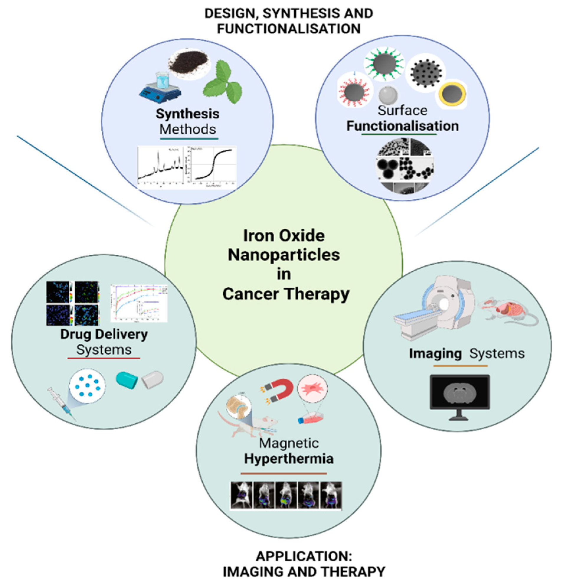
Advances in the Synthesis and Application of Magnetic Ferrite Nanoparticles for Cancer Therapy


Abstract
:1. Introduction
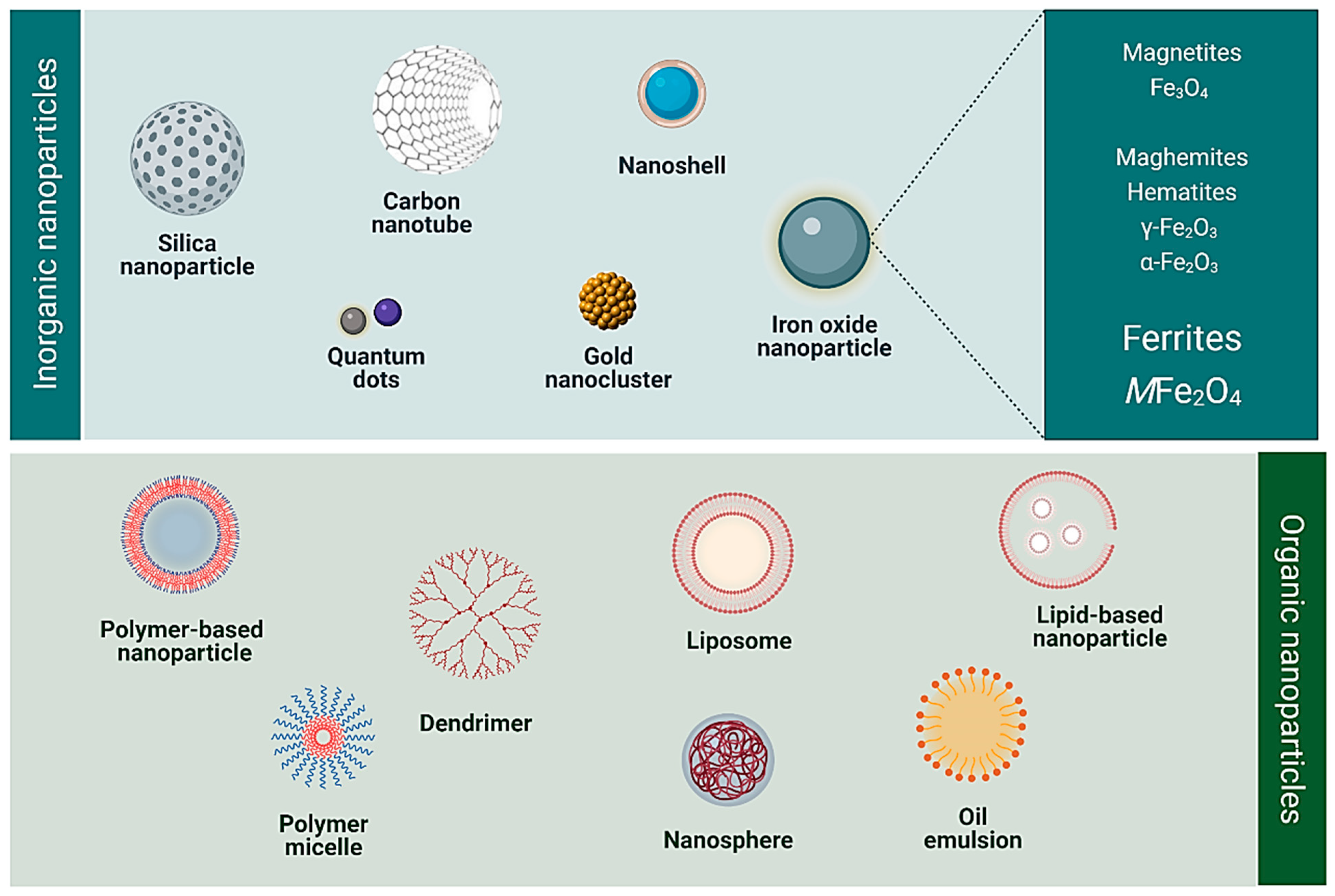
2. Iron Oxide Nanoparticles in Cancer Diagnostics and Therapy
2.1. Magnetic Hyperthermia
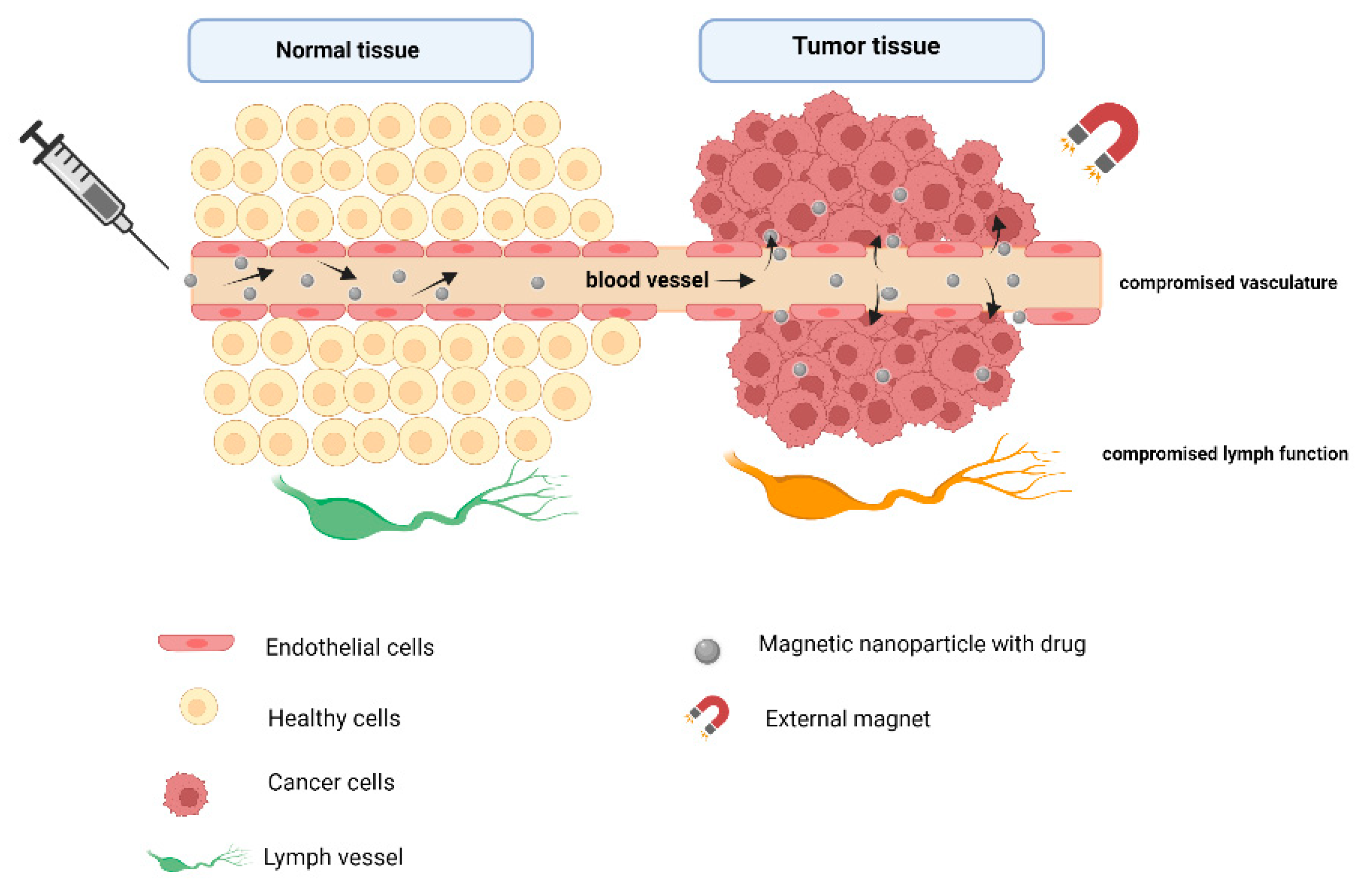
2.2. Targeted Drug Delivery
2.3. Imaging Systems
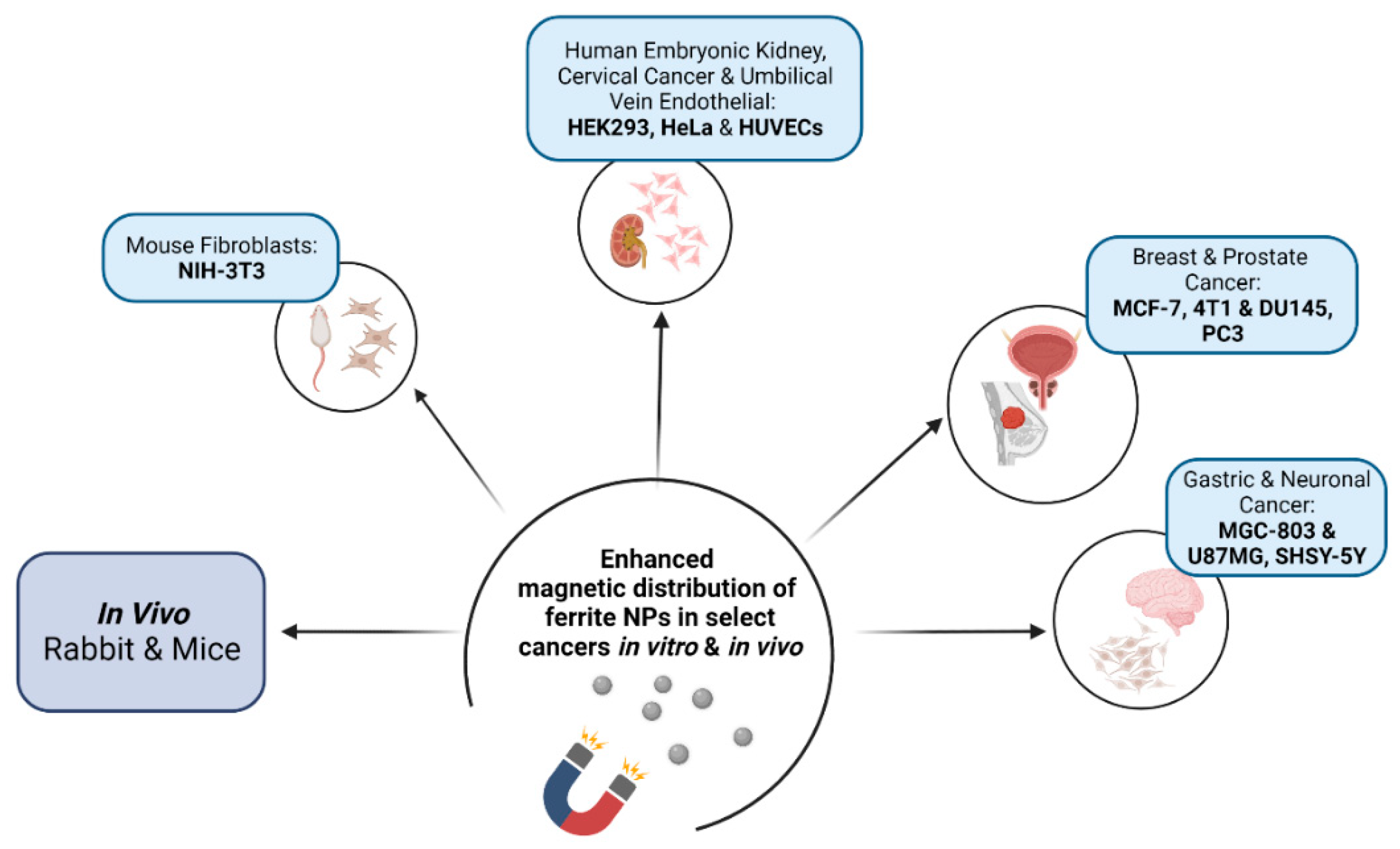
3. Ferrites (MFe2O4)—Nanocarriers of Choice in Biomedicine
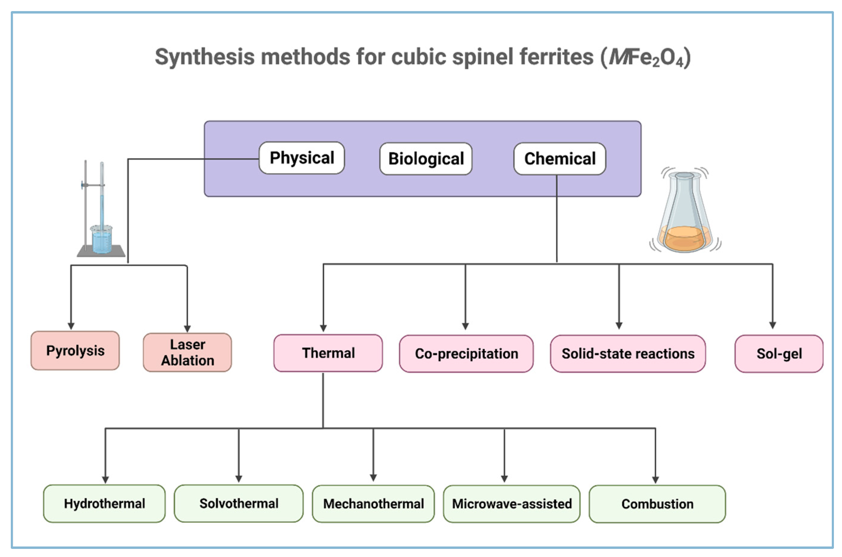
4. Synthesis Methods of Ferrites
4.1. Biological Methods
4.2. Physical Methods
4.3. Wet Chemistry Methods
4.3.1. Co-Precipitation
4.3.2. Thermal Methods
Hydrothermal and Solvothermal Methods
Microwave Method
Mechano-Chemical Method
The Combustion Method
4.3.3. The Sol–Gel Method
4.3.4. Solid-State Method
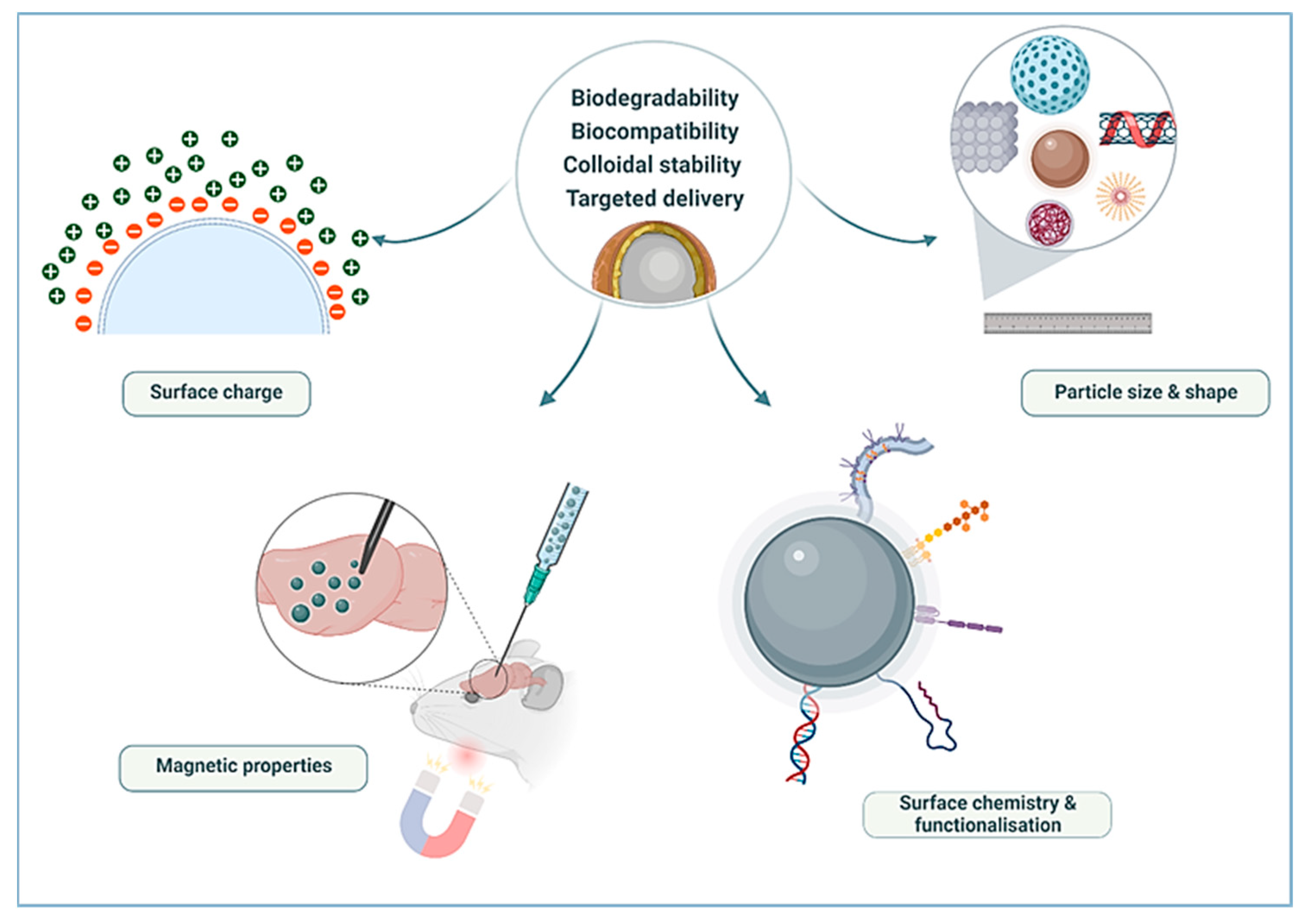
5. Potential of Ferrites in Biomedical Applications
5.1. Particle Size and Shape
5.2. Surface Chemistry and Functionalisation
5.2.1. Organic Polymers
5.2.2. Inorganic Compounds
6. Conclusions
Author Contributions
Funding
Institutional Review Board Statement
Informed Consent Statement
Data Availability Statement
Conflicts of Interest
References
- Senapati, S.; Mahanta, A.K.; Kumar, S.; Maiti, P. Controlled drug delivery vehicles for cancer treatment and their performance. Signal Transduct. Target. Ther. 2018, 3, 7. [Google Scholar] [CrossRef] [PubMed] [Green Version]
- Peng, J.; Liang, X.; Calderon, L. Progress in research on gold nanoparticles in cancer management. Medicine 2019, 98, e15311. [Google Scholar] [CrossRef] [PubMed]
- Ferlay, J.; Colombet, M.; Soerjomataram, I.; Parkin, D.M.; Piñeros, M.; Znaor, A.; Bray, F. Cancer statistics for the year 2020: An overview. Int. J. Cancer 2021, 149, 778–789. [Google Scholar] [CrossRef] [PubMed]
- Auría-Soro, C.; Nesma, T.; Juanes-Velasco, P.; Landeira-Viñuela, A.; Fidalgo-Gomez, H.; Acebes-Fernandez, V.; Gongora, R.; Parra, M.J.A.; Manzano-Roman, R.; Fuentes, M. Interactions of Nanoparticles and Biosystems: Microenvironment of Nanoparticles and Biomolecules in Nanomedicine. Nanomaterials 2019, 9, 1365. [Google Scholar] [CrossRef] [PubMed] [Green Version]
- Fornaguera, C.; García-Celma, M.J. Personalized nanomedicine: A revolution at the nanoscale. J. Pers. Med. 2017, 7, 12. [Google Scholar] [CrossRef] [PubMed] [Green Version]
- Navya, P.N.; Kaphle, A.; Srinivas, S.P.; Bhargava, S.K.; Rotello, V.M.; Daima, H.K. Current trends and challenges in cancer management and therapy using designer nanomaterials. Nano Converg. 2019, 6, 23. [Google Scholar] [CrossRef] [PubMed] [Green Version]
- Hermawan, H.; Ramdan, D.; Djuansjah, J.R.P. Metals for Biomedical Applications. In Biomedical Engineering—From Theory to Applications; Fazal-Rezai, R., Ed.; IntechOpen Ltd.: London, UK, 2011; pp. 411–430. [Google Scholar] [CrossRef] [Green Version]
- Gao, Y.; Xie, J.; Chen, H.; Gu, S.; Zhao, R.; Shao, J.; Jia, L. Nanotechnology-based intelligent drug design for cancer metastasis treatment. Biotechnol. Adv. 2014, 32, 761–777. [Google Scholar] [CrossRef] [PubMed]
- Martinelli, C.; Pucci, C.; Ciofani, G. Nanostructured carriers as innovative tools for cancer diagnosis and therapy. APL Bioeng. 2019, 3, 011502. [Google Scholar] [CrossRef] [Green Version]
- Srinivasan, M.; Rajabi, M.; Mousa, S.A. Multifunctional nanomaterials and their applications in drug delivery and cancer therapy. Nanomaterials 2015, 5, 1690–1703. [Google Scholar] [CrossRef]
- Dulińska-Litewka, J.; Łazarczyk, A.; Hałubiec, P.; Szafrański, O.; Karnas, K.; Karewicz, A. Superparamagnetic iron oxide nanoparticles-current and prospective medical applications. Materials 2019, 12, 617. [Google Scholar] [CrossRef] [Green Version]
- Daniels, A.N.; Singh, M. Sterically stabilized siRNA:gold nanocomplexes enhance c-MYC silencing in a breast cancer cell model. Nanomedicine 2019, 14, 1387–1401. [Google Scholar] [CrossRef] [PubMed]
- Dizaj, S.M.; Jafari, S.; Khosroushahi, A.Y. A sight on the current nanoparticle-based gene delivery vectors. Nanoscale Res. Lett. 2014, 9, 252. [Google Scholar] [CrossRef] [PubMed] [Green Version]
- Dobson, J. Gene therapy progress and prospects: Magnetic nanoparticle-based gene delivery. Gene Ther. 2006, 13, 283–287. [Google Scholar] [CrossRef] [PubMed] [Green Version]
- Singh, M.; Ariatti, M. A cationic cytofectin with long spacer mediates favourable transfection in transformed human epithelial cells. Int. J. Pharm. 2006, 309, 189–198. [Google Scholar] [CrossRef] [PubMed]
- Masserini, M. Nanoparticles for Brain Drug Delivery. ISRN Biochem. 2013, 2013, 238428. [Google Scholar] [CrossRef] [PubMed] [Green Version]
- Bertrand, Y.; Currie, J.-C.; Demeule, M.; Régina, A.; Ché, C.; Abulrob, A.; Fatehi, D.; Sartelet, H.; Gabathuler, R.; Castaigne, J.-P.; et al. Transport characteristics of a novel peptide platform for CNS therapeutics. J. Cell. Mol. Med. 2010, 14, 2827–2839. [Google Scholar] [CrossRef] [PubMed] [Green Version]
- Anselmo, A.C.; Mitragotri, S. A Review of Clinical Translation of Inorganic Nanoparticles. AAPS J. 2015, 17, 1041–1054. [Google Scholar] [CrossRef] [Green Version]
- Kandasamy, G.; Maity, D. Recent advances in superparamagnetic iron oxide nanoparticles (SPIONs) for in vitro and in vivo cancer nanotheranostics. Int. J. Pharm. 2015, 496, 191–218. [Google Scholar] [CrossRef]
- Bhattacharyya, S.; Kudgus, R.A.; Bhattacharya, R.; Mukherjee, P. Inorganic nanoparticles in cancer therapy. Pharm. Res. 2011, 28, 237–259. [Google Scholar] [CrossRef] [Green Version]
- Dadfar, S.M.; Roemhild, K.; Drude, N.I.; von Stillfried, S.; Knüchel, R.; Kiessling, F.; Lammers, T. Iron oxide nanoparticles: Diagnostic, therapeutic and theranostic applications. Adv. Drug Deliv. Rev. 2019, 138, 302–325. [Google Scholar] [CrossRef]
- Wu, W.; Wu, Z.; Yu, T.; Jiang, C.; Kim, W.-S. Recent progress on magnetic iron oxide nanoparticles: Synthesis, surface functional strategies and biomedical applications. Sci. Technol. Adv. Mater. 2015, 16, 023501. [Google Scholar] [CrossRef] [PubMed]
- Kharisov, B.I.; Dias, H.V.R.; Kharissova, O.V. Mini-review: Ferrite nanoparticles in the catalysis. Arab. J. Chem. 2019, 12, 1234–1246. [Google Scholar] [CrossRef] [Green Version]
- Ramnandan, D.; Mokhosi, S.; Daniels, A.; Singh, M. Chitosan, polyethylene glycol and polyvinyl alcohol modified MgFe2O4 ferrite magnetic nanoparticles in doxorubicin delivery: A comparative study in vitro. Molecules 2021, 26, 3893. [Google Scholar] [CrossRef] [PubMed]
- Mngadi, S.; Mokhosi, S.; Singh, M. Chitosan-functionalized Mg0.5Co0.5Fe2O4 magnetic nanoparticles enhance delivery of 5-fluorouracil in vitro. Coatings 2020, 10, 446. [Google Scholar] [CrossRef]
- Moise, S.; Céspedes, E.; Soukup, D.; Byrne, J.M.; El Haj, A.J.; Telling, N.D. The cellular magnetic response and biocompatibility of biogenic zinc- and cobalt-doped magnetite nanoparticles. Sci. Rep. 2017, 7, 39922. [Google Scholar] [CrossRef] [Green Version]
- Yang, H.W.; Hua, M.Y.; Liu, H.L.; Huang, C.Y.; Wei, K.C. Potential of magnetic nanoparticles for targeted drug delivery. Nanotechnol. Sci. Appl. 2012, 5, 73–86. [Google Scholar] [CrossRef] [Green Version]
- Wang, Y.; Cui, H.; Li, K.; Sun, C.; Du, W.; Cui, J.; Zhao, X.; Chen, W. A magnetic nanoparticle-based multiple-gene delivery system for transfection of porcine kidney cells. PLoS ONE 2014, 9, e102886. [Google Scholar] [CrossRef]
- Arias, L.S.; Pessan, J.P.; Vieira, A.P.M.; De Lima, T.M.T.; Delbem, A.C.B.; Monteiro, D.R. Iron oxide nanoparticles for biomedical applications: A perspective on synthesis, drugs, antimicrobial activity, and toxicity. Antibiotics 2018, 7, 46. [Google Scholar] [CrossRef] [Green Version]
- Tombácz, E.; Turcu, R.; Socoliuc, V.; Vékás, L. Magnetic iron oxide nanoparticles: Recent trends in design and synthesis of magnetoresponsive nanosystems. Biochem. Biophys. Res. Commun. 2015, 468, 442–453. [Google Scholar] [CrossRef] [Green Version]
- Revia, R.A.; Zhang, M. Magnetite nanoparticles for cancer diagnosis, treatment, and treatment monitoring: Recent advances. Mater. Today 2016, 19, 157–168. [Google Scholar] [CrossRef]
- Popescu, R.C.; Andronescu, E.; Vasile, B.S. Recent advances in magnetite nanoparticle functionalization for nanomedicine. Nanomaterials 2019, 9, 1791. [Google Scholar] [CrossRef] [PubMed] [Green Version]
- Senyei, A.; Widder, K.; Czerlinski, C. Magnetic guidance of drug-carrying microspheres. J. Appl. Phys. 1978, 49, 3578–3583. [Google Scholar] [CrossRef]
- Khalkhali, M.; Rostamizadeh, K.; Sadighian, S.; Khoeini, F.; Naghibi, M.; Hamidi, M. The impact of polymer coatings on magnetite nanoparticles performance as MRI contrast agents: A comparative study. DARU J. Pharm. Sci. 2015, 23, 45. [Google Scholar] [CrossRef] [Green Version]
- Zhang, Y.; Zhang, L.; Song, X.; Gu, X.; Sun, H.; Fu, C.; Meng, F. Synthesis of superparamagnetic iron oxide nanoparticles modified with MPEG-PEI via photochemistry as new MRI contrast agent. J. Nanomater. 2015, 2015, 417389. [Google Scholar] [CrossRef] [Green Version]
- Unterweger, H.; Dézsi, L.; Matuszak, J.; Janko, C.; Poettler, M.; Jordan, J.; Bäuerle, T.; Szebeni, J.; Fey, T.; Boccaccini, A.R.; et al. Dextran-coated superparamagnetic iron oxide nanoparticles for magnetic resonance imaging: Evaluation of size-dependent imaging properties, storage stability and safety. Int. J. Nanomed. 2018, 13, 1899–1915. [Google Scholar] [CrossRef] [PubMed] [Green Version]
- Vallabani, N.V.S.; Singh, S. Recent advances and future prospects of iron oxide nanoparticles in biomedicine and diagnostics. 3 Biotech 2018, 8, 279. [Google Scholar] [CrossRef] [PubMed] [Green Version]
- De Paula, L.B.; Primo, F.L.; Pinto, M.R.; Morais, P.C.; Tedesco, A.C. Evaluation of a chloroaluminium phthalocyanine-loaded magnetic nanoemulsion as a drug delivery device to treat glioblastoma using hyperthermia and photodynamic therapy. RSC Adv. 2017, 7, 9115–9122. [Google Scholar] [CrossRef] [Green Version]
- Liu, X.; Zhang, H.; Zhang, T.; Wang, Y.; Jiao, W.; Lu, X.; Gao, X.; Xie, M.; Shan, Q.; Wen, N.; et al. Magnetic nanomaterials-mediated cancer diagnosis and therapy. Prog. Biomed. Eng. 2022, 4, 012005. [Google Scholar] [CrossRef]
- Liang, C.; Zhang, X.; Cheng, Z.; Yang, M.; Huang, W.; Dong, X. Magnetic iron oxide nanomaterials: A key player in cancer nanomedicine. View 2020, 1, 20200046. [Google Scholar] [CrossRef]
- Ching-Chien, H.; Mo, C.-C.; Hung, Y.-H.; Zuo, W.-Z.; Huang, J.-Y. Effect of particle size of as-milled powders on microstructural and magnetic properties of Y3MnxAl 0.8−xFe4.2O12 ferrites. J. Am. Ceram. Soc. 2019, 102, 3525–3534. [Google Scholar] [CrossRef]
- Mahmoudi, M.; Hofmann, H.; Rothen-Rutishauser, B.; Petri-Fink, A. Assessing the in vitro and in vivo toxicity of superparamagnetic iron oxide nanoparticles. Chem. Rev. 2012, 112, 2323–2338. [Google Scholar] [CrossRef] [PubMed] [Green Version]
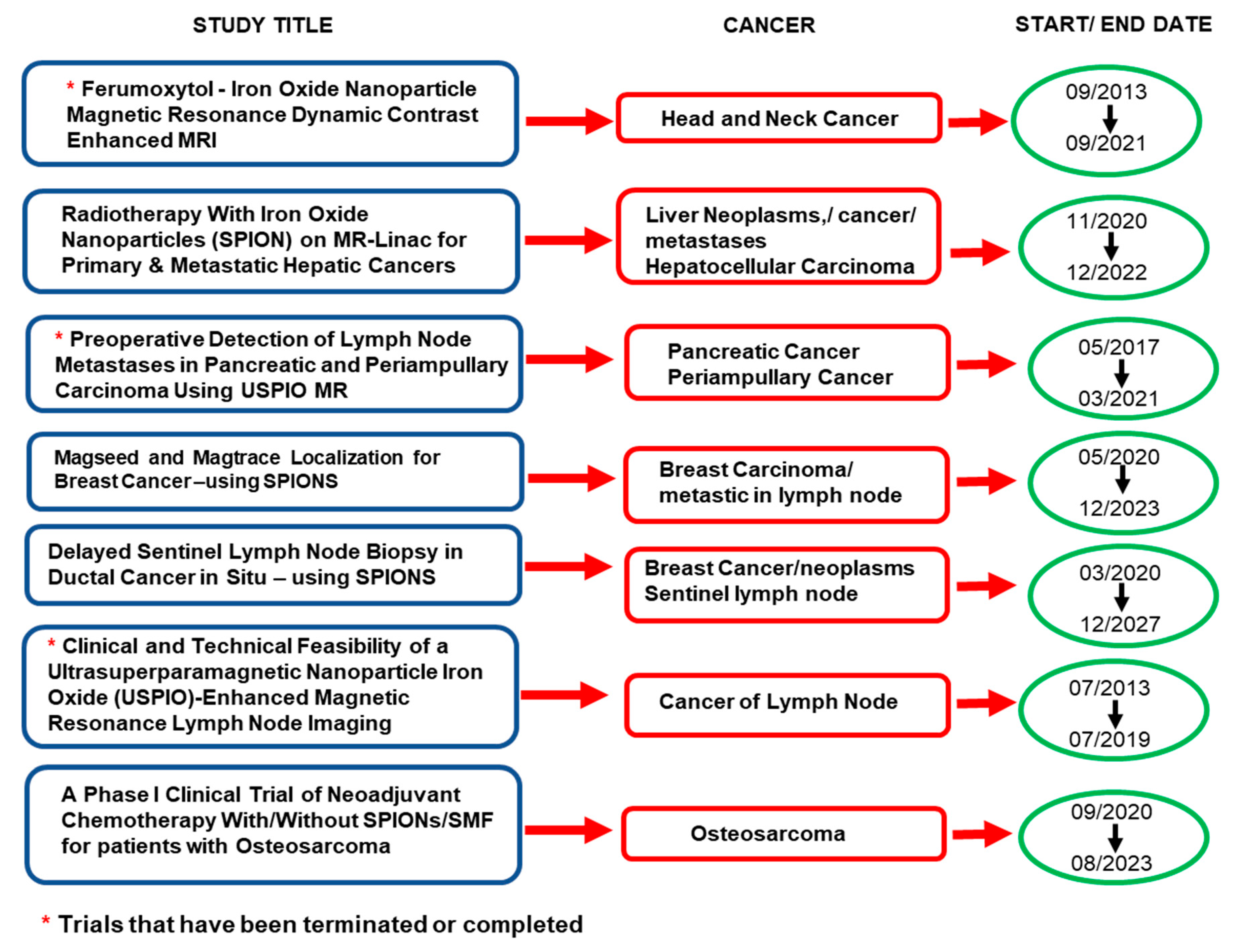
- ClinicalTrials.gov. Available online: https://www.clinicaltrials.gov/ct2/results?cond+cancer&term=iron+oxide+nanoparticles (accessed on 12 April 2021).
- Zhu, N.; Ji, H.; Yu, P.; Niu, J.; Farooq, M.U.; Waseem Akram, M.; Udego, I.O.; Li, H.; Niu, X. Surface modification of magnetic iron oxide nanoparticles. Nanomaterials 2018, 8, 810. [Google Scholar] [CrossRef] [PubMed] [Green Version]
- Wu, M.; Huang, S. Magnetic nanoparticles in cancer diagnosis, drug delivery and treatment. Mol. Clin. Oncol. 2017, 7, 738–746. [Google Scholar] [CrossRef] [PubMed] [Green Version]
- Kudr, J.; Haddad, Y.; Richtera, L.; Heger, Z.; Cernak, M.; Adam, V.; Zitka, O. Magnetic nanoparticles: From design and synthesis to real world applications. Nanomaterials 2017, 7, 243. [Google Scholar] [CrossRef]
- Chang, D.; Lim, M.; Goos, J.A.C.M.; Qiao, R.; Ng, Y.Y.; Mansfeld, F.M.; Jackson, M.; Davis, T.P.; Kavallari, M. Biologically targeted magnetic hyperthermia: Potential and limitations. Front. Pharmacol. 2018, 9, 831. [Google Scholar] [CrossRef] [Green Version]
- Jeyadevan, B. Present status and prospects of magnetite nanoparticles-based hyperthermia. J. Ceram. Soc. Jpn. 2010, 118, 391–401. [Google Scholar] [CrossRef] [Green Version]
- Gul, S.; Khan, S.B.; Rehman, I.U.; Khan, M.A.; Khan, M.I. A Comprehensive Review of Magnetic Nanomaterials Modern Day Theranostics. Front. Mater. 2019, 6, 179. [Google Scholar] [CrossRef] [Green Version]
- Williams, H.M. The application of magnetic nanoparticles in the treatment and monitoring of cancer and infectious diseases. Biosci. Horiz. 2017, 10, hzx009. [Google Scholar] [CrossRef] [Green Version]
- Shirazi, H.; Daneshpour, M.; Kashanian, S.; Omidfar, K. Synthesis, characterization and in vitro biocompatibility study of Au/TMC/Fe3O4 nanocomposites as a promising, nontoxic system for biomedical applications. Beilstein J. Nanotechnol. 2015, 6, 1677–1689. [Google Scholar] [CrossRef] [Green Version]
- Patil, R.M.; Shete, P.B.; Thorat, N.D.; Otari, S.V.; Barick, K.C.; Prasad, A.; Ningthoujam, R.S.; Tiwale, B.M.; Pawar, S.H. Superparamagnetic iron oxide/chitosan core/shells for hyperthermia application: Improved colloidal stability and biocompatibility. J. Magn. Magn. Mater. 2014, 355, 22–30. [Google Scholar] [CrossRef]
- Suciu, M.; Ionescu, C.M.; Ciorita, A.; Tripon, S.C.; Nica, D.; Al-Salami, H.; Barbu-Tudoran, L. Applications of superparamagnetic iron oxide nanoparticles in drug and therapeutic delivery, and biotechnological advancements. Beilstein J. Nanotechnol. 2020, 11, 1092–1109. [Google Scholar] [CrossRef] [PubMed]
- Zhang, H.; Wang, J.; Zeng, Y.; Wang, G.; Han, S.; Yang, Z.; Li, B.; Wang, X.; Gao, J.; Zheng, L.; et al. Leucine-coated cobalt ferrite nanoparticles: Synthesis, characterization and potential biomedical applications for drug delivery. Phys. Lett. A 2020, 384, 126600. [Google Scholar] [CrossRef]
- Verma, J.; Lal, S.; Van Noorden, C.J.F. Nanoparticles for hyperthermic therapy: Synthesis strategies and applications in glioblastoma. Int. J. Nanomed. 2014, 9, 2863–2877. [Google Scholar] [CrossRef] [Green Version]
- Ulbrich, K.; Holá, K.; Šubr, V.; Bakandritsos, A.; Tuček, J.; Zbořil, R. Targeted Drug Delivery with Polymers and Magnetic Nanoparticles: Covalent and Noncovalent Approaches, Release Control, and Clinical Studies. Chem. Rev. 2016, 116, 5338–5431. [Google Scholar] [CrossRef]
- Li, Z.; Tan, S.; Li, S.; Shen, Q.; Wang, K. Cancer drug delivery in the nano era: An overview and perspectives. Oncol. Rep. 2017, 38, 611–624. [Google Scholar] [CrossRef] [Green Version]
- Golovin, Y.I.; Golovin, D.Y.; Vlasova, K.Y.; Veselov, M.M.; Usvaliev, A.D.; Kabanov, A.V.; Klyachko, N.L. Non-Heating Alternating Magnetic Field Nanomechanical Stimulation of Biomolecule Structures via Magnetic Nanoparticles as the Basis for Future Low-Toxic Biomedical Applications. Nanomaterials 2021, 11, 2255. [Google Scholar] [CrossRef]
- Gribanovsky, S.L.; Zhigacheva, A.O.; Golovin, D.Y.; Golovin, Y.I.; Klyachko, N.L. Mechanisms and conditions for mechanical activation of magnetic nanoparticles by external magnetic field for biomedical applications. J. Magn. Magn. Mater. 2022, 553, 169278. [Google Scholar] [CrossRef]
- Broders-Bondon, F.; Ho-Bouldoires, T.H.N.; Fernandez-Sanchez, M.-E.; Farge, E. Mechanotransduction in tumor progression: The dark side of the force. J. Cell Biol. 2018, 217, 1571–1587. [Google Scholar] [CrossRef] [Green Version]
- García, R.S.; Stafford, S.; Gun’ko, Y.K. Recent progress in synthesis and functionalization of multimodal fluorescent-magnetic nanoparticles for biological applications. Appl. Sci. 2018, 8, 172. [Google Scholar] [CrossRef] [Green Version]
- Bertrand, N.; Wu, J.; Xu, X.; Kamaly, N.; Farokhzad, O.C. Cancer nanotechnology: The impact of passive and active targeting in the era of modern cancer biology. Adv. Drug Deliv. Rev. 2014, 66, 2–25. [Google Scholar] [CrossRef] [Green Version]
- Huang, J.; Li, Y.; Orza, A.; Lu, Q.; Guo, P.; Wang, L.; Yang, L.; Mao, H. Magnetic Nanoparticle Facilitated Drug Delivery for Cancer Therapy with Targeted and Image-Guided Approaches. Adv. Funct. Mater. 2016, 26, 3818–3836. [Google Scholar] [CrossRef] [PubMed] [Green Version]
- Mirza, A.Z.; Siddiqui, F.A. Nanomedicine and drug delivery: A mini review. Int. Nano Lett. 2014, 4, 94. [Google Scholar] [CrossRef] [Green Version]
- Estelrich, J.; Escribano, E.; Queralt, J.; Busquets, M.A. Iron oxide nanoparticles for magnetically-guided and magnetically-responsive drug delivery. Int. J. Mol. Sci. 2015, 16, 8070–8101. [Google Scholar] [CrossRef] [PubMed] [Green Version]
- Xu, Y.; Zhu, Y. Synthesis of Magnetic Nanoparticles for Biomedical Applications. Nano Adv. 2016, 2, 25–38. [Google Scholar] [CrossRef]
- Mahmoudi, M.; Sant, S.; Wang, B.; Laurent, S.; Sen, T. Superparamagnetic iron oxide nanoparticles (SPIONs): Development, surface modification and applications in chemotherapy. Adv. Drug Deliv. Rev. 2011, 63, 24–46. [Google Scholar] [CrossRef] [Green Version]
- Gao, H. Progress and perspectives on targeting nanoparticles for brain drug delivery. Acta Pharm. Sin. B 2016, 6, 268–286. [Google Scholar] [CrossRef]
- Rosenblum, D.; Joshi, N.; Tao, W.; Karp, J.M.; Peer, D. Progress and challenges towards targeted delivery of cancer therapeutics. Nat. Commun. 2018, 9, 1410. [Google Scholar] [CrossRef] [Green Version]
- Arum, Y.; Oh, Y.O.; Kang, H.W.; Ahn, S.H.; Oh, J. Chitosan-coated Fe3O4 magnetic nanoparticles as carrier of cisplatin for drug delivery. Fish. Aquat. Sci. 2015, 18, 89–98. [Google Scholar] [CrossRef] [Green Version]
- Saif, B.; Wang, C.; Chuan, D.; Shuang, S. Synthesis and Characterization of of Fe3O4 magnetic nanofluid coated on APTES as Carriers for Morin-Anticancer Drug. J. Biomater. Nanobiotechnol. 2015, 6, 267–275. [Google Scholar] [CrossRef]
- Kariminia, S.; Shamsipur, A.; Shamsipur, M. Analytical characteristics and application of novel chitosan coated magnetic nanoparticles as an efficient drug delivery system for ciprofloxacin. Enhanced drug release kinetics by low-frequency ultrasounds. J. Pharm. Biomed. Anal. 2016, 129, 450–457. [Google Scholar] [CrossRef]
- Karimi, Z.; Abbasi, S.; Shokrollahi, H.; Yousefi, G.; Fahham, M.; Karimi, L.; Firuzi, O. Pegylated and amphiphilic Chitosan coated manganese ferrite nanoparticles for pH-sensitive delivery of methotrexate: Synthesis and characterization. Mater. Sci. Eng. C 2017, 71, 504–511. [Google Scholar] [CrossRef] [PubMed]
- Jose, R.; Rinita, J.; Jothi, N.S.N. Synthesis and characterisation of stimuli-responsive drug delivery system using ZnFe2O4 and Ag1−XZnxFe2O4 nanoparticles. Mater. Technol. 2020, 36, 347–355. [Google Scholar] [CrossRef]
- Nigam, A.; Pawar, S.J. Structural, magnetic, and antimicrobial properties of zinc doped magnesium ferrite for drug delivery applications. Ceram. Int. 2020, 46, 4058–4064. [Google Scholar] [CrossRef]
- Javed, F.; Abbas, M.A.; Asad, M.I.; Ahmed, N.; Naseer, N.; Saleem, H.; Errachid, A.; Lebaz, N.; Elaissari, A.; Ahmad, N.M. Gd3+ Doped CoFe2O4 Nanoparticles for Targeted Drug Delivery and Magnetic Resonance Imaging. Magnetochemistry 2021, 7, 47. [Google Scholar] [CrossRef]
- Malik, A.R.; Aziz, M.H.; Atif, M.; Irshad, M.S.; Ullah, H.; Gia, T.N.; Ahmed, H.; Ahmad, S.; Botmart, T. Lime peel extract induced NiFe2O4 NPs: Synthesis to applications and oxidative stress mechanism for anticancer, antibiotic activity. J. Saudi Chem. Soc. 2022, 26, 101422. [Google Scholar] [CrossRef]
- Vigneswari, T.; Thiruramanathan, P. Magnetic Targeting Carrier Applications of Bismuth-Doped Nickel Ferrites Nanoparticles by Co-precipitation Method. Trans. Indian Inst. Met. 2021, 74, 2255–2265. [Google Scholar] [CrossRef]
- Veiseh, O.; Gunn, J.; Zhang, M. Design and fabrication of magnetic nanoparticles for targeted drug delivery and imaging. Adv. Drug Deliv. Rev. 2011, 62, 284–304. [Google Scholar] [CrossRef] [Green Version]
- Kefeni, K.K.; Msagati, T.A.M.; Nkambule, T.T.; Mamba, B.B. Spinel ferrite nanoparticles and nanocomposites for biomedical applications and their toxicity. Mater. Sci. Eng. C 2020, 107, 110314. [Google Scholar] [CrossRef]
- Zhang, Q.; Yin, T.; Gao, G.; Shapter, J.G.; Lai, W.; Huang, P.; Qi, W.; Song, J.; Cui, D. Multifunctional core @ shell magnetic nanoprobes for enhancing targeted magnetic resonance imaging and Fluorescent Labeling in Vitro and in Vivo. ACS Appl. Mater. Interfaces 2017, 9, 17777–17785. [Google Scholar] [CrossRef]
- Jauhar, S.; Kaur, J.; Goyal, A.; Singhal, S. Tuning the properties of cobalt ferrite: A road towards diverse applications. RSC Adv. 2016, 6, 97694–97719. [Google Scholar] [CrossRef]
- El-Dek, S.I.; Ali, M.A.; El-Zanaty, S.M.; Ahmed, S.E. Comparative investigations on ferrite nanocomposites for magnetic hyperthermia applications. J. Magn. Magn. Mater. 2018, 458, 147–155. [Google Scholar] [CrossRef]
- Darwish, M.S.A.; Kim, H.; Lee, H.; Ryu, C.; Lee, J.Y.; Yoon, J. Synthesis of Magnetic Ferrite Nanoparticles with High Hyperthermia Performance via a Controlled Co-Precipitation Method. Nanomaterials 2019, 9, 1176. [Google Scholar] [CrossRef] [PubMed] [Green Version]
- Petrova, E.; Kotsikau, D.; Pankov, V.; Fahmi, A. Influence of Synthesis Methods on Structural and Magnetic Characteristics of Mg-Zn-Ferrite Nanopowders. J. Magn. Magn. Mater. 2019, 473, 85–91. [Google Scholar] [CrossRef]
- Amiri, M.; Salavati-Niasari, M.; Akbari, A. Magnetic nanocarriers: Evolution of spinel ferrites for medical applications. Adv. Colloid Interface Sci. 2019, 265, 29–44. [Google Scholar] [CrossRef] [PubMed]
- Nadeem, M.; Ahmad, M.; Akthar, M.S.; Shaari, S.; Riaz, S.; Naseem, S.; Masood, M.; Saaeed, M.A. Magnetic properties of polyvinyl alcohol and doxorubicine loaded iron oxide nanoparticles for anticancer drug delivery applications. PLoS ONE 2016, 11, e01580842016. [Google Scholar] [CrossRef] [PubMed]
- Ehi-Eromosele, C.O.; Ita, B.I.; Iweala, E.E.J. The effect of polyethylene glycol (PEG) coating on the magneto-structural properties and colloidal stability of CO0.8Mg0.2Fe2O4 nanoparticles for potential biomedical applications. Dig. J. Nanomater. Biostruct. 2016, 11, 7–14. [Google Scholar]
- Kückelhaus, S.; Reis, S.C.; Carneiro, M.F.; Tedesco, A.C.; .Oliveira, D.M.; Lima, E.C.D.; Morais, P.C.; Azevedo, R.B.; .Lacava, Z.G.M. In vivo investigation of cobalt ferrite-based magnetic fluid and magnetoliposomes using morphological tests. J. Magn. Magn. Mater. 2004, 272–276, 2402–2403. [Google Scholar] [CrossRef]
- Baldi, G.; Bonacchi, D.; Innocenti, C.; Lorenzi, G.; Sangregorio, C. Cobalt ferrite nanoparticles: The control of the particle size and surface state and their effects on magnetic properties. J. Magn. Magn. Mater. 2007, 311, 10–16. [Google Scholar] [CrossRef]
- Lin, M.; Zhang, D.; Huang, J.; Zhang, J.; Xiao, W.; Yu, H.; Zhang, L.; Ye, J. The anti-hepatoma effect of nanosized Mn-Zn ferrite magnetic fluid hyperthermia associated with radiation in vitro and in vivo. Nanotechnology 2013, 24, 255101. [Google Scholar] [CrossRef]
- Momin, N.; Deshmukh, A.; Radha, S. Synthesis and characterization of CoFe2O4 & NiFe2O4 magnetic nanoparticles for various biomedical applications: Cell viability and cell death evaluations. J. Nano Res. 2015, 34, 1–8. [Google Scholar] [CrossRef]
- Shi, Z.; Zeng, Y.; Chen, X.; Zhou, F.; Zheng, L.; Wang, G.; Gao, J.; Ma, Y.; Zheng, L.; Fu, B.; et al. Mesoporous superparamagnetic cobalt ferrite nanoclusters: Synthesis, characterization and application in drug delivery. J. Magn. Magn. Mater. 2020, 498, 166222. [Google Scholar] [CrossRef]
- Humbe, A.V.; Birajdar, S.D.; Bhandari, J.M.; Waghule, N.N.; Bhagwat, V.R.; Jadhav, K.M. Polyethylene glycol coated CoFe2O4 nanoparticles: A potential spinel ferrite for biomedical applications. AIP Conf. Proc. 2015, 1665, 2–5. [Google Scholar] [CrossRef]
- Dey, C.; Ghosh, A.; Ahir, M.; Ghosh, A.; Goswami, M.M. Improvement of Anticancer Drug Release by Cobalt Ferrite Magnetic Nanoparticles through Combined pH and Temperature Responsive Technique. ChemPhysChem 2018, 19, 2872–2878. [Google Scholar] [CrossRef] [PubMed]
- Sangeetha, K.; Ashok, M.; Girija, E.K. Development of multifunctional cobalt ferrite/hydroxyapatite nanocomposites by microwave assisted wet precipitation method: A promising platform for synergistic chemo-hyperthermia therapy. Ceram. Int. 2019, 45, 12860–12869. [Google Scholar] [CrossRef]
- Gandhi, S.; Issar, S.; Mahapatro, A.K.; Roy, I. Cobalt ferrite nanoparticles for bimodal hyperthermia and their mechanistic interactions with lysozyme. J. Mol. Liq. 2020, 310, 113194. [Google Scholar] [CrossRef]
- Nasiri, M.; Hassanzadeh-Tabrizi, S.A. Synthesis and Characterization of Folate-decorated Cobalt Ferrite Nanoparticles Coated with Poly (Ethylene Glycol) for Biomedical Applications. J. Chin. Chem. Soc. 2018, 65, 231–242. [Google Scholar] [CrossRef]
- Manohar, A.; Geleta, D.D.; Krishnamoorthi, C.; Lee, J. Synthesis, characterization and magnetic hyperthermia properties of nearly monodisperse CoFe2O4 nanoparticles. Ceram. Int. 2020, 46, 28035–28041. [Google Scholar] [CrossRef]
- Mushtaq, M.W.; Kanwal, F.; Batool, A.; Jamil, T.; Zia-ul-Haq, M.; Ijaz, B.; Huang, Q.; Ullah, Z. Polymer-coated CoFe2O4 nanoassemblies as biocompatible magnetic nanocarriers for anticancer drug delivery. J. Mater. Sci. 2017, 52, 9282–9293. [Google Scholar] [CrossRef]
- Mushtaq, M.W.; Kanwal, F.; Islam, A.; Ahmed, K.; Zia-ul-Haq, M.; Jamil, T.; Imran, M.; Abbas, S.M.; Huang, Q. Synthesis and characterisation of doxorubicin-loaded functionalised cobalt ferrite nanoparticles and their in vitro anti-tumour activity under an AC-magnetic field. Trop. J. Pharm. Res. 2017, 16, 1663–1674. [Google Scholar] [CrossRef] [Green Version]
- Shyamaldas; Bououdina, M.; Manoharan, C. Dependence of structure/morphology on electrical/magnetic properties of hydrothermally synthesised cobalt ferrite nanoparticles. J. Magn. Magn. Mater. 2020, 493, 165703. [Google Scholar] [CrossRef]
- Jermy, R.; Ravinayagam, V.; Alamoudi, W.; Almohazey, D.; Elanthikkal, S.; Dafalla, H.; Rehman, S.; Chandrasekar, G.; Baykal, A. Tuning pH sensitive chitosan and cisplatin over spinel ferrite/silica nanocomposite for anticancer activity in MCF-7 cell line. J. Drug Deliv. Sci. Technol. 2020, 57, 101711. [Google Scholar] [CrossRef]
- Ghasemian, Z.; Shahbazi-Gahrouei, D.; Manouchehri, S. Cobalt zinc ferrite nanoparticles as a potential magnetic resonance imaging agent: An in vitro study. Avicenna J. Med. Biotechnol. 2015, 7, 64–68. [Google Scholar] [PubMed]
- Kamta Tedjieukeng, H.M.; Tsobnang, P.K.; Fomekong, R.L.; Etape, E.P.; Joy, P.A.; Delcorte, A.; Lambi, J.N. Structural characterization and magnetic properties of undoped and copper-doped cobalt ferrite nanoparticles prepared by the octanoate coprecipitation route at very low dopant concentrations. RSC Adv. 2018, 8, 38621–38630. [Google Scholar] [CrossRef] [Green Version]
- Margabandhu, M.; Sendhilnathan, S.; Senthilkumar, S.; Gajalakshmi, D. Investigation of Structural, Morphological, Magnetic Properties and Biomedical applications of Cu2+ Substituted Uncoated Cobalt Ferrite Nanoparticles. Braz. Arch. Biol. Technol. 2016, 59, e16161046. [Google Scholar] [CrossRef] [Green Version]
- Ehi-Eromosele, C.O.; Ita, B.I.; Iweala, E.E.; Ogunniran, K.O.; Adekoya, J.A.; Siyanbola, T.O. Silica functionalized magnesium ferrite nanocomposites for potential biomedical applications: Preparation, characterization and enhanced colloidal stability studies. J. Nano Res. 2016, 40, 146–157. [Google Scholar] [CrossRef]
- Mngadi, S.M.; Mokhosi, S.R.; Singh, M. Surface-coating of Mg0.5Co0.5Fe2O4 nanoferrites and their in vitro cytotoxicity. Inorg. Chem. Commun. 2019, 108, 107525. [Google Scholar] [CrossRef]
- Mokhosi, S.R.; Mdlalose, W.; Mngadi, S.; Singh, M.; Moyo, T. Assessing the structural, morphological and magnetic properties of polymer-coated magnesium-doped cobalt ferrite (CoFe2O4) nanoparticles for biomedical application. J. Phys. Conf. Ser. 2019, 1310, 012014. [Google Scholar] [CrossRef] [Green Version]
- De-León-Prado, L.E.; Cortés-Hernández, D.A.; Almanza-Robles, J.M.; Escobedo-Bocardo, J.C.; Sánchez, J.; Reyes-Rdz, P.Y.; Jasso-Terán, R.A.; Hurtado-López, G.F. Synthesis and characterization of nanosized MgxMn1−xFe2O4 ferrites by both sol-gel and thermal decomposition methods. J. Magn. Magn. Mater. 2017, 427, 230–234. [Google Scholar] [CrossRef]
- Mdlalose, W.B.; Mokhosi, S.R.; Dlamini, S.; Moyo, T.; Singh, M. Effect of chitosan coating on the structural and magnetic properties of MnFe2O4 and Mn0.5Co0.5Fe2O4 nanoparticles. AIP Adv. 2018, 8, 056726. [Google Scholar] [CrossRef] [Green Version]
- Wang, G.; Ma, Y.; Zhang, L.; Mu, J.; Zhang, Z.; Zhang, X.; Che, H.; Bai, Y.; Hou, J. Facile synthesis of manganese ferrite/graphene oxide nanocomposites for controlled targeted drug delivery. J. Magn. Magn. Mater. 2016, 401, 647–650. [Google Scholar] [CrossRef]
- Abbasi Pour, S.; Shaterian, H.R.; Afradi, M.; Yazdani-Elah-Abadi, A. Carboxymethyl cellulose (CMC)-loaded Co-Cu doped manganese ferrite nanorods as a new dual-modal simultaneous contrast agent for magnetic resonance imaging and nanocarrier for drug delivery system. J. Magn. Magn. Mater. 2017, 438, 85–94. [Google Scholar] [CrossRef]
- Sánchez, J.; Cortés-Hernández, D.A.; Escobedo-Bocardo, J.C.; Almanza-Robles, J.M.; Reyes-Rodríguez, P.Y.; Jasso-Terán, R.A.; Bartolo, P.; De-León-Prado, L.E. Sol-gel synthesis of Mn x Ga 1−x Fe 2 O 4 nanoparticles as candidates for hyperthermia treatment. Ceram. Int. 2016, 42, 13755–13760. [Google Scholar] [CrossRef]
- Kanagesan, S.; Aziz, S.B.A.; Hashim, M.; Ismail, I.; Tamilselvan, S.; Alitheen, N.B.B.M.; Swamy, M.K.; Rao, B.P.C. Synthesis, characterization and in vitro evaluation of manganese ferrite (MnFe2O4) nanoparticles for their biocompatibility with murine breast cancer cells (4T1). Molecules 2016, 21, 312. [Google Scholar] [CrossRef] [PubMed]
- Jesudoss, S.K.; Vijaya, J.J.; Kennedy, L.J.; Rajan, P.I.; Al-Lohedan, H.A.; Ramalingam, R.J.; Kaviyarasu, K.; Bououdina, M. Studies on the efficient dual performance of Mn1–xNixFe2O4 spinel nanoparticles in photodegradation and antibacterial activity. J. Photochem. Photobiol. B Biol. 2016, 165, 121–132. [Google Scholar] [CrossRef]
- Andersen, H.L.; Saura-Múzquiz, M.; Granados-Miralles, C.; Canévet, E.; Lock, N.; Christensen, M. Crystalline and magnetic structure–property relationship in spinel ferrite nanoparticles. Nanoscale 2018, 10, 14902–14914. [Google Scholar] [CrossRef] [Green Version]
- Bhosale, S.V.; Ekambe, P.S.; Bhoraskar, S.V.; Mathe, V.L. Effect of surface properties of NiFe2O4 nanoparticles synthesized by dc thermal plasma route on antimicrobial activity. Appl. Surf. Sci. 2018, 441, 724–733. [Google Scholar] [CrossRef]
- Manjura Hoque, S.; Tariq, M.; Liba, S.I.; Mahmood, Z.H.; Khan, M.N.I.; Chattopadhayay, K.; Isalm, R.; Akhter, S. Thermo-therapeutic applications of chitosan- and PEG-coated NiFe2O4 nanoparticles. Nanotechnology 2016, 27, 285702. [Google Scholar] [CrossRef]
- Amiri, M.; Pardakhti, A.; Ahmadi-Zeidabadi, M.; Akbari, A.; Salavati-Niasari, M. Magnetic nickel ferrite nanoparticles: Green synthesis by Urtica and therapeutic effect of frequency magnetic field on creating cytotoxic response in neural cell lines. Colloids Surf. B Biointerfaces 2018, 172, 244–253. [Google Scholar] [CrossRef]
- Andjelković, L.; Šuljagić, M.; Lakić, M.; Jeremić, D.; Vulić, P.; Nikolić, A.S. A study of the structural and morphological properties of Ni–ferrite, Zn–ferrite and Ni–Zn–ferrites functionalized with starch. Ceram. Int. 2018, 44, 14163–14168. [Google Scholar] [CrossRef]
- Hanini, A.; El Massoudi, M.; Gavard, J.; Kacem, K.; Ammar, S.; Souilem, O. Nanotoxicological study of polyol-made cobalt-zinc ferrite nanoparticles in rabbit. Environ. Toxicol. Pharmacol. 2016, 45, 321–327. [Google Scholar] [CrossRef]
- Naik, M.M.; Naik, H.S.B.; Nagaraju, G.; Vinuth, M.; Naika, H.R.; Vinu, K. Green synthesis of zinc ferrite nanoparticles in Limonia acidissima juice: Characterization and their application as photocatalytic and antibacterial activities. Microchem. J. 2019, 146, 1227–1235. [Google Scholar] [CrossRef]
- Sattarahmady, N.; Zare, T.; Mehdizadeh, A.R.; Azarpira, N.; Heidari, M.; Lotfi, M.; Heli, H. Dextrin-coated zinc substituted cobalt-ferrite nanoparticles as an MRI contrast agent: In vitro and in vivo imaging studies. Colloid Surf. B Biointerfaces 2015, 129, 15–20. [Google Scholar] [CrossRef] [PubMed]
- Masina, P.; Moyo, T.; Abdallah, H.M.I. Synthesis, structural and magnetic properties of ZnxMg1−xFe2O4nanoferrites. J. Magn. Magn. Mater. 2015, 381, 41–49. [Google Scholar] [CrossRef]
- Msomi, J.Z.; Nhlapo, T.A.; Moyo, T.; Snyman, J.; Strydom, A.M. Grain size effects on the magnetic properties of ZnxMn1−xFe2O4 nanoferrites. J. Magn. Magn. Mater. 2015, 373, 74–77. [Google Scholar] [CrossRef]
- Bansode, J.S.; Patil, V.C. Applications of Spinel Ferrite Nanoparticles: A Short- Review. Int. J. Adv. Sci. Res. Eng. Trends 2021, 6, 78–82. [Google Scholar] [CrossRef]
- Rana, G.; Dhiman, P.; Kumar, A.; Vo, D.-V.N.; Sharma, G.; Sharma, S.; Naushad, M. Recent advances on nickel nano-ferrite: A review on processing techniques, properties and diverse applications. Chem. Eng. Res. Des. 2012, 175, 182–208. [Google Scholar] [CrossRef]
- Mondal, R.; Sarkar, K.; Dey, S.; Majumdar, D.; Bhattacharya, S.K.; Sen, P.; Kumar, S. Magnetic, pseudocapacitive, and H2O2-electrosensing properties of self-assembled superparamagnetic Co0.3Zn0.7Fe2O4 with enhanced saturation magnetization. ACS Omega 2019, 4, 12632–12646. [Google Scholar] [CrossRef] [Green Version]
- Khan, I.; Saeed, K.; Khan, I. Nanoparticles: Properties, applications and toxicities. Arab. J. Chem. 2019, 12, 908–931. [Google Scholar] [CrossRef]
- Hakeem, A.; Alshahrani, T.; Muhammed, G.; Alhossainy, M.H.; Laref, A.; Khan, A.R.; Ali, I.; Farid, H.M.T.; Ghrib, T.; Ejaz, S.R.; et al. Magnetic, dielectric and structural properties of spinel ferrites synthesized by sol-gel method. J. Mater. Res. Technol. 2021, 11, 158–169. [Google Scholar] [CrossRef]
- Loganathan, A.; Kumar, K. Effects on structural, optical, and magnetic properties of pure and Sr-substituted MgFe2O4 nanoparticles at different calcination temperatures. Appl. Nanosci. 2016, 6, 629–639. [Google Scholar] [CrossRef] [Green Version]
- Jha, A.K.; Prasad, K. Biological synthesis of cobalt ferrite nanoparticles. Nanotechnol. Dev. 2012, 2, e9. [Google Scholar] [CrossRef]
- Thakur, P.; Taneja, S.; Chahar, D.; Ravelo, B.; Thakur, A. Recent advances on synthesis, characterization and high frequency applications of Ni-Zn ferrite nanoparticles. J. Magn. Magn. Mater. 2021, 530, 167925. [Google Scholar] [CrossRef]
- Iravani, S.; Korbekandi, H.; Mirmohammadi, S.V.; Zolfaghari, B. Synthesis of silver nanoparticles: Chemical, physical and biological methods. Res. Pharm. Sci. 2014, 9, 385–406. [Google Scholar]
- Peng, Y.; Tang, H.; Yao, B.; Gao, X.; Yang, X.; Zhou, Y. Activation of peroxymonosulfate (PMS) by spinel ferrite and their composites in degradation of organic pollutants: A Review. Chem. Eng. J. 2021, 414, 128800. [Google Scholar] [CrossRef]
- Narang, S.B.; Pubby, K. Nickel Spinel Ferrites: A review. J. Magn. Magn. Mater. 2021, 519, 167163. [Google Scholar] [CrossRef]
- Sorescu, M.; Diamandescu, L.; Swaminathan, R.; McHenry, M.E.; Feder, M. Structural and magnetic properties of NiZn and Zn ferrite thin films obtained by laser ablation deposition. J. Appl. Phys. 2005, 97, 10G105. [Google Scholar] [CrossRef]
- Özçelik, S.; Yalçın, B.; Arda, L.; Santos, H.; Sáez-Puche, R.; Angurel, L.A.; de la Fuente, G.F.; Özçelik, B. Structure, magnetic, photocatalytic and blood compatibility studies of nickel nanoferrites prepared by laser ablation technique in distilled water. J. Alloys Compd. 2021, 854, 57279. [Google Scholar] [CrossRef]
- Almessiere, M.A.; Güner, S.; Slimani, Y.; Hassan, M.; Baykal, A.; Gondal, M.A.; Baig, U.; Trukhanov, S.V.; Trukhanov, A.V. Structural and magnetic properties of Co0.5Ni0.5Ga0.01Gd0.01Fe1.98O4/ZnFe2O4 spinel ferrite nanocomposites: Comparative study between sol-gel and pulsed laser ablation in liquid approaches. Nanomaterials 2021, 11, 2461. [Google Scholar] [CrossRef]
- Msomi, J.Z.; Dlamini, W.B.; Moyo, T.; Ezekiel, P. Investigation of phase formation of (Zn, Mg)0.5Co0.5Fe2O4 nanoferrites. J. Magn. Magn. Mater. 2015, 373, 68–73. [Google Scholar] [CrossRef]
- Osman, N.S.E.; Moyo, T. Structural and magnetic properties of CoFe2O4 nanoferrite simultaneously and symmetrically substituted by Mg, Sr and Mn. Mater. Chem. Phys. 2015, 164, 138–144. [Google Scholar] [CrossRef]
- Peng, J.; Hojamberdiev, M.; Xu, Y.; Cao, B.; Wang, J.; Wu, H. Hydrothermal synthesis and magnetic properties of gadolinium-doped CoFe2O4 nanoparticles. J. Magn. Magn. Mater. 2011, 323, 133–137. [Google Scholar] [CrossRef]
- Suresh, S.; Prakash, A.; Bahadur, D. The role of reduced graphene oxide on the electrochemical activity of MFe2O4 (M = Fe, Co, Ni and Zn) nanohybrids. J. Magn. Magn. Mater. 2018, 448, 43–51. [Google Scholar] [CrossRef]
- Džunuzović, A.S.; Ilić, N.I.; Vijatović Petrović, M.M.; Bobić, J.D.; Stojadinović, B.; Dohčević-Mitrović, Z.; Stojanović, B.D. Structure and properties of Ni-Zn ferrite obtained by auto-combustion method. J. Magn. Magn. Mater. 2015, 374, 245–251. [Google Scholar] [CrossRef]
- Kiran, V.S.; Sumathi, S. Comparison of catalytic activity of bismuth substituted cobalt ferrite nanoparticles synthesized by combustion and co-precipitation method. J. Magn. Magn. Mater. 2017, 421, 113–119. [Google Scholar] [CrossRef]
- Agarwal, A.A.; Aghamkar, P.; Lal, B. Structural and multiferroic properties of barium substituted bismuth ferrite nanocrystallites prepared by sol–gel method. J. Magn. Magn. Mater. 2017, 426, 800–805. [Google Scholar] [CrossRef]
- Duong, G.V.; Turtelli, R.S.; Hanh, N.; Linh, D.V.; Reissner, M.; Michor, H.; Fidler, J.; Wiesinger, G.; Grössinger, R. Magnetic properties of nanocrystalline Co1−xZnxFe2O4 prepared by forced hydrolysis method. J. Magn. Magn. Mater. 2006, 307, 313–317. [Google Scholar] [CrossRef]
- Baykal, A.; Kasapoǧlu, N.; Köseoǧlu, Y.; Toprak, M.S.; Bayrakdar, H. CTAB-assisted hydrothermal synthesis of NiFe2O4 and its magnetic characterization. J. Alloys Compd. 2008, 464, 514–518. [Google Scholar] [CrossRef]
- Nguyet, D.T.T.; Duong, N.P.; Hung, L.T.; Hien, T.D.; Satoh, T. Crystallization and magnetic behavior of nanosized nickel ferrite prepared by citrate precursor method. J. Alloys Compd. 2011, 509, 6621–6625. [Google Scholar] [CrossRef]
- Nivetha, R.; Chella, S.; Kollu, P.; Jeong, S.K.; Bhatnagar, A.; Andrews, N.G. Cobalt and nickel ferrites based graphene nanocomposites for electrochemical hydrogen evolution. J. Magn. Magn. Mater. 2018, 448, 165–171. [Google Scholar] [CrossRef] [Green Version]
- Chen, D.; Meng, Y. Nanomed Synthesis of Magnetic Oxide Nanoparticles for Biomedical Applications. Glob. J. Nanomed. 2017, 2, 0051–0054. [Google Scholar] [CrossRef]
- Kumari, N.; Kour, S.; Singh, G.; Sharma, R.K. A brief review on synthesis, properties and applications of ferrites. AIP Conf. Proc. 2020, 2220, 020164. [Google Scholar] [CrossRef]
- Vadivelan, S.; Sowmiya, S. Structural and magnetic studies of nickel doped barium ferrite via Co-Precipitation method. Phys. Open 2021, 9, 100094. [Google Scholar] [CrossRef]
- Vatsalya, V.L.S.; Sundari, G.S.; Sridhar, C.S.L.N.; Lakshmi, C.S. Evidence of Superparamagnetism in nano phased copper doped nickel zinc ferrites synthesized by Hydrothermal Method. Optik 2021, 247, 167874. [Google Scholar] [CrossRef]
- Al-Rawi, N.N.; Anwer, B.A.; Al-Rawi, N.H.; Uthman, A.T.; Ahmed, I.S. Magnetism in drug delivery: The marvels of iron oxides and substituted ferrites nanoparticles. Saudi Pharm. J. 2020, 28, 876–887. [Google Scholar] [CrossRef]
- Kaur, H.; Singh, A.; Kumar, V.; Ahlawat, D.S. Structural, thermal and magnetic investigations of cobalt ferrite doped with Zn2+ and Cd2+ synthesized by auto combustion method. J. Magn. Magn. Mater. 2019, 474, 505–511. [Google Scholar] [CrossRef]
- Allaedini, G.; Tasirin, S.M.; Aminayi, P. Magnetic properties of cobalt ferrite synthesized by hydrothermal method. Int. Nano Lett. 2015, 5, 183–186. [Google Scholar] [CrossRef] [Green Version]
- Nhlapo, T.A.; Msomi, J.Z.; Moyo, T. Temperature-dependent magnetic behavior of Mn-Mg spinel ferrites with substituted Co, Ni & Zn, synthesized by hydrothermal method. J. Mol. Struct. 2021, 1245, 131042. [Google Scholar] [CrossRef]
- Liu, G.; Dai, B.; Ren, Y.; Zhang, W. Rapid synthesis and characterization of spinel manganese ferrite nanopowder by microwave-assisted hydrothermal method. Results Phys. 2021, 26, 104441. [Google Scholar] [CrossRef]
- Gan, Y.X.; Jayatissa, A.H.; Yu, Z.; Chen, X.; Li, M. Hydrothermal Synthesis of Nanomaterials. J. Nanomater. 2020, 2020, 8917013. [Google Scholar] [CrossRef]
- Li, Y.; Yang, W. Microwave synthesis of zeolite membranes: A review. J. Memb. Sci. 2008, 316, 3–17. [Google Scholar] [CrossRef]
- Gurgel, A.L.; Martinelli, A.E.; de Aquino Conceição, O.L.; Xavier, M.M.; Morales Torres, M.A.; de Araújo Melo, D.M. Microwave-assisted hydrothermal synthesis and magnetic properties of nanostructured cobalt ferrite. J. Alloys Compd. 2019, 799, 36–42. [Google Scholar] [CrossRef]
- Shu, R.; Zhang, J.; Guo, C.; Wu, Y.; Wan, Z.; Shi, J.; Liu, Y.; Zheng, M. Facile synthesis of nitrogen-doped reduced graphene oxide/nickel-zinc ferrite composites as high-performance microwave absorbers in the X-band. Chem. Eng. J. 2020, 384, 123266. [Google Scholar] [CrossRef]
- Houbi, A.; Aldashevich, Z.A.; Atassi, Y.; Bagasharova Telmanovna, Z.; Saule, M.; Kubanych, K. Microwave absorbing properties of ferrites and their composites: A review. J. Magn. Magn. Mater. 2021, 529, 167839. [Google Scholar] [CrossRef]
- Thakur, P.; Chahar, D.; Taneja, S.; Bhalla, N.; Thakur, A. A review on MnZn ferrites: Synthesis, characterization and applications. Ceram. Int. 2020, 46, 15740–15763. [Google Scholar] [CrossRef] [PubMed]
- Melo, R.S.; Silva, F.C.; Moura, K.R.M.; De Menezes, A.S.; Sinfrônio, F.S.M. Magnetic ferrites synthesised using the microwave-hydrothermal method. J. Magn. Magn. Mater. 2015, 381, 109–115. [Google Scholar] [CrossRef] [Green Version]
- Bhongale, S.R.; Ingawale, H.R.; Shinde, T.J.; Vasambekar, P.N. Effect of Nd3+ substitution on structural and magnetic properties of Mg–Cd ferrites synthesized by microwave sintering technique. J. Rare Earths 2018, 36, 390–397. [Google Scholar] [CrossRef]
- Gennari, F.C.; Andrade-Gamboa, J.J. A systematic approach to the synthesis, thermal stability and hydrogen storage properties of rare-earth borohydrides. In Emerging Materials for Energy Conversion and Storage; Cheong, K.W., Impellizzeri, G., Fraga, M.A., Eds.; Elsevier Inc.: Amsterdam, The Netherlands, 2018; pp. 429–459. [Google Scholar] [CrossRef]
- Tsuzuki, T.; McCormick, P.G. Mechanochemical synthesis of nanoparticles. J. Mater. Sci. 2004, 39, 5143–5146. [Google Scholar] [CrossRef]
- Lazarević, Z.Ž.; Jovalekić, Č.; Milutinović, A.; Sekulić, D.; Ivanovski, V.N.; Rečnik, A.; Cekić, B.; Romčević, N.Ž. Nanodimensional spinel NiFe2O4 and ZnFe2O4 ferrites prepared by soft mechanochemical synthesis. J. Appl. Phys. 2013, 113, 187221. [Google Scholar] [CrossRef]
- Castrillón Arango, J.A.; Cristóbal, A.A.; Ramos, C.P.; Bercoff, P.G.; Botta, P.M. Mechanochemical synthesis and characterization of nanocrystalline Ni1−xCoxFe2O4 (0 ≤ x ≤ 1) ferrites. J. Alloys Compd. 2019, 811, 152044. [Google Scholar] [CrossRef]
- Aruna, S.T.; Mukasyan, A.S. Combustion synthesis and nanomaterials. Curr. Opin. Solid State Mater. Sci. 2008, 12, 44–50. [Google Scholar] [CrossRef]
- Tholkappiyan, R.; Vishista, K. Influence of lanthanum on the optomagnetic properties of zinc ferrite prepared by combustion method. Phys. B Condens. Matter 2014, 448, 177–183. [Google Scholar] [CrossRef]
- Salunkhe, A.B.; Khot, V.M.; Phadatare, M.R.; Pawar, S.H. Combustion synthesis of cobalt ferrite nanoparticles—Influence of fuel to oxidizer ratio. J. Alloys Compd. 2012, 514, 91–96. [Google Scholar] [CrossRef]
- Jagadeesha Angadi, V.; Manjunatha, K.; Praveena, K.; Pattar, V.K.; Fernandes, B.J.; Manjunatha, S.O.; Husain, J.; Angadi, S.V.; Horakeri, L.D.; Ramesh, K.P. Magnetic properties of larger ionic radii samarium and gadalonium doped manganese zinc ferrite nanoparticles prepared by solution combustion method. J. Magn. Magn. Mater. 2021, 529, 167899. [Google Scholar] [CrossRef]
- Pathak, S.S.; Khanna, A.S. Sol–gel nanocoatings for corrosion protection. In Corrosion Protection and Control Using Nanomaterials; Saji, V.S., Cook, R., Eds.; Woodhead Publishing Limited: Cambridge, UK, 2012; pp. 302–329. [Google Scholar] [CrossRef]
- Parashar, M.; Shukla, V.K.; Singh, R. Metal oxides nanoparticles via sol–gel method: A review on synthesis, characterization and applications. J. Mater. Sci. Mater. Electron. 2020, 31, 3729–3749. [Google Scholar] [CrossRef]
- de Souza Nunes, G.C.; Biondo, V.; Ferreira, R.F.; Tupan, L.; Nicolodi, S.; Ivashita, F.F.; Isnard, O.; Junior, A.P. Structural and magnetic characterization of the Nd2Fe14B+ 10%wt.Fe system subjected to high-energy milling. Hyperfine Interact. 2019, 240, 18–23. [Google Scholar] [CrossRef]
- Wei, M.; Wang, B.; Chen, M.; Lyu, H.; Lee, X.; Wang, S.; Yu, Z.; Zhang, X. Recent advances in the treatment of contaminated soils by ball milling technology: Classification, mechanisms, and applications. J. Clean. Prod. 2022, 340, 130821. [Google Scholar] [CrossRef]
- Yadav, R.S.; Havlica, J.; Hnatko, J.; Šajgalík, P.; Alexander, C.; Palou, M.; Bartoníčková, E.; Boháč, M.; Frajkorová, F.; Masilko, J.; et al. Magnetic properties of Co1−xZnxFe2O4 spinel ferrite nanoparticles synthesized by starch-assisted sol-gel autocombustion method and its ball milling. J. Magn. Magn. Mater. 2015, 378, 190–199. [Google Scholar] [CrossRef]
- Neuberger, T.; Schöpf, B.; Hofmann, H.; Hofmann, M.; Von Rechenberg, B. Superparamagnetic nanoparticles for biomedical applications: Possibilities and limitations of a new drug delivery system. J. Magn. Magn. Mater. 2005, 293, 483–496. [Google Scholar] [CrossRef]
- Issa, B.; Obaidat, I.M.; Albiss, B.A.; Haik, Y. Magnetic nanoparticles: Surface effects and properties related to biomedicine applications. Int. J. Mol. Sci. 2013, 14, 21266–21305. [Google Scholar] [CrossRef] [Green Version]
- Wang, Y.; Miao, Y.; Su, M.; Chen, X.; Zhang, H.; Zhang, Y.; Jiao, Y.; He, Y.; Yi, J.; Liu, X.; et al. Engineering ferrite nanoparticles with enhanced magnetic response for advanced biomedical applications. Mater. Today Adv. 2020, 8, 100119. [Google Scholar] [CrossRef]
- Bustamante-Torres, M.; Romero-Fierro, D.; Estrella-Nuñez, J.; Arcentales-Vera, B.; Chichande-Proaño, E.; Bucio, E. Polymeric Composite of Magnetite Iron Oxide Nanoparticles and Their Application in Biomedicine: A Review. Polymers 2022, 14, 752. [Google Scholar] [CrossRef] [PubMed]
- Bohara, R.A.; Yadav, H.M.; Thorat, N.D.; Mali, S.S.; Hong, C.K.; Nanaware, S.G.; Pawar, S.H. Synthesis of functionalized Co0.5Zn0.5Fe2O4 nanoparticles for biomedical applications. J. Magn. Magn. Mater. 2015, 378, 397–401. [Google Scholar] [CrossRef]
- Krishnan, K.M. Biomedical nanomagnetics: A spin through possibilities in imaging, diagnostics, and therapy. IEEE Trans. Magn. 2010, 46, 2523–2558. [Google Scholar] [CrossRef] [PubMed] [Green Version]
- Ha, Y.; Ko, S.; Kim, I.; Huang, Y.; Mohanty, K.; Huh, C.; Maynard, J. Recent Advances Incorporating Superparamagnetic Nanoparticles into Immunoassays. ACS Appl. Nano Mater. 2018, 1, 512–521. [Google Scholar] [CrossRef] [Green Version]
- Hole, P.; Sillence, K.; Hannell, C.; Maguire, C.N.; Roesslein, M.; Suarez, G.; Capracotta, S.; Magdolenova, Z.; Horev-Azaria, L.; Dybowska, A.; et al. Interlaboratory comparison of size measurements on nanoparticles using nanoparticle tracking analysis (NTA). J. Nanopart. Res. 2013, 15, 2101. [Google Scholar] [CrossRef] [Green Version]
- Kolhatkar, A.G.; Jamison, A.C.; Litvinov, D.; Willson, R.C.; Lee, T.R. Tuning the magnetic properties of nanoparticles. Int. J. Mol. Sci. 2013, 14, 15977–16009. [Google Scholar] [CrossRef] [Green Version]
- Baaziz, W.; Pichon, B.P.; Fleutot, S.; Liu, Y.; Lefevre, C.; Greneche, J.-M.; Toumi, M.; Mhiri, T.; Begin-Colin, S. Magnetic iron oxide nanoparticles: Reproducible tuning of the size and nanosized-dependent composition, defects, and spin canting. J. Phys. Chem. C 2014, 118, 3795–3810. [Google Scholar] [CrossRef]
- Hoque, S.M.; Islam, M.K.; Hoq, A.; Haque, M.M.; Maritim, S.; Coman, D.; Hyder, F. Comparative Study of Specific Loss Power and Transverse Relaxivity of Spinel Ferrite Nanoensembles Coated With Chitosan and Polyethylene Glycol. Front. Nanotechnol. 2021, 3, 644080. [Google Scholar] [CrossRef]
- Nejati, S.; Mohseni Vadeghani, E.; Khorshidi, S.; Karkhaneh, A. Role of particle shape on efficient and organ-based drug delivery. Eur. Polym.J. 2020, 122, 109353. [Google Scholar] [CrossRef]
- Christopher, A.M.L.S.M. Principles of nanoparticle design for overcoming biological. Physiol. Behav. 2016, 176, 100–106. [Google Scholar] [CrossRef]
- Salazar-Alvarez, G.; Qin, J.; Sepelák, V.; Bergmann, I.; Vasilakaki, M.; Trohidou, K.N.; Ardisson, J.D.; Macedo, W.A.A.; Mikhaylova, M.; Muhammed, M.; et al. Cubic versus spherical magnetic nanoparticles: The role of surface anisotropy. J. Am. Chem. Soc. 2008, 130, 13234–13239. [Google Scholar] [CrossRef] [PubMed]
- Nguyen, C.T.; Kim, C.R.; Le, T.H.; Koo, K.I.; Hwang, C.H. Magnetically guided targeted delivery of erythropoietin using magnetic nanoparticles: Proof of concept. Medicine 2020, 99, e19972. [Google Scholar] [CrossRef] [PubMed]
- Chenthamara, D.; Subramaniam, S.; Ramakrishnan, S.G.; Krishnaswamy, S.; Essa, M.M.; Lin, F.-H.; Qoronfleh, M.W. Therapeutic efficacy of nanoparticles and routes of administration. Biomater. Res. 2019, 23, 20. [Google Scholar] [CrossRef] [PubMed]
- Jiang, Z.; Shan, K.; Song, J.; Liu, J.; Rajendran, S.; Pugazhendhi, A.; Jacob, J.A.; Chen, B. Toxic effects of magnetic nanoparticles on normal cells and organs. Life Sci. 2019, 220, 156–161. [Google Scholar] [CrossRef]
- Anik, M.I.; Hossain, M.K.; Hossain, I.; Mahfuz, A.M.U.B.; Rahman, M.T.; Ahmed, I. Recent progress of magnetic nanoparticles in biomedical applications: A review. Nano Sel. 2021, 2, 1146–1186. [Google Scholar] [CrossRef]
- Wallyn, J.; Anton, N.; Vandamme, T.F. Synthesis, principles, and properties of magnetite nanoparticles for in vivo imaging applications—A review. Pharmaceutics 2019, 11, 601. [Google Scholar] [CrossRef] [Green Version]
- Varanda, L.C.; Souza, C.G.S.; Moraes, D.A.; Neves, H.R.; Souza Junior, J.B.; Silva, M.F.; Bini, R.A.; Albers, R.F.; Silva, T.L.; Beck Junior, W. Size and shape-controlled nanomaterials based on modified polyol and thermal decomposition approaches. A brief review. An. Acad. Bras. Ciênc. 2019, 91, e20181180. [Google Scholar] [CrossRef]
- Mngadi, S.; Singh, M.; Mokhosi, S. PVA coating of ferrite nanoparticles triggers pH-responsive release of 5-fluorouracil in cancer cells. J. Polym. Eng. 2021, 41, 597–606. [Google Scholar] [CrossRef]
- Wang, Y.; Li, P.; Kong, L. Chitosan-Modified PLGA Nanoparticles with Versatile Surface for Improved Drug Delivery. AAPS PharmSciTech 2013, 14, 585–592. [Google Scholar] [CrossRef] [Green Version]
- Sandler, S.E.; Fellows, B.; Thompson Mefford, O. Best Practices for Characterization of Magnetic Nanoparticles for Biomedical Applications. Anal. Chem. 2019, 91, 14159–14169. [Google Scholar] [CrossRef] [Green Version]
- Mahdavi, M.; Ahmad, M.B.; Haron, M.J.; Namvar, F.; Nadi, B.; Rahman, M.Z.A.; Amin, J. Synthesis, surface modification and characterisation of biocompatible magnetic iron oxide nanoparticles for biomedical applications. Molecules 2013, 18, 7533–7548. [Google Scholar] [CrossRef] [PubMed] [Green Version]
- Yang, Z.; Duan, J.; Wang, J.; Liu, Q.; Shang, R.; Yang, X.; Lu, P.; Xia, C.; Wang, L.; Dou, K. Superparamagnetic iron oxide nanoparticles modified with polyethylenimine and galactose for siRNA targeted delivery in hepatocellular carcinoma therapy. Int. J. Nanomed. 2018, 13, 1851–1865. [Google Scholar] [CrossRef] [PubMed] [Green Version]
- Srinivas, C.; Tirupanyam, B.V.; Satish, A.; Seshubai, V.; Sastry, D.L.; Caltun, O.F. Effect of Ni2+ substitution on structural and magnetic properties of Ni-Zn ferrite nanoparticles. J. Magn. Magn. Mater. 2015, 382, 15–19. [Google Scholar] [CrossRef]
- Mdlalose, W.B.; Dlamini, S.; Moyo, T.; Mokhosi, S.R.; Singh, M. Chitosan coating by mechanical milling of MnFe2O4 and Mn0.5Co0.5Fe2O4: Effect of milling. J. Phys. Conf. Ser. 2019, 1310, 012016–012022. [Google Scholar] [CrossRef]
- Thanh, N.T.K.; Green, L.A.W. Functionalisation of nanoparticles for biomedical applications. Nano Today 2010, 5, 213–230. [Google Scholar] [CrossRef]
- Singh, D.; Singh, M. Hepatocellular-Targeted mRNA Delivery using functionalized Selenium Nanoparticles in vitro. Pharmaceutics 2021, 13, 298. [Google Scholar] [CrossRef]
- Naicker, K.; Ariatti, M.; Singh, M. PEGylated Galactosylated Cationic Liposomes for Hepatocytic Gene Delivery. Colloid Surf. B: Biointerfaces 2014, 122, 482–490. [Google Scholar] [CrossRef]
- Natarajan, S.; Harini, K.; Gajula, G.P.; Sarmento, B.; Neves-Petersen, M.T.; Thiagarajan, V. Multifunctional magnetic iron oxide nanoparticles: Diverse synthetic approaches, surface modifications, cytotoxicity towards biomedical and industrial applications. BMC Mater. 2019, 1, 2. [Google Scholar] [CrossRef]
- Hoskins, C.; Min, Y.; Gueorguieva, M.; McDougall, C.; Volovick, A.; Prentice, P.; Wang, Z.; Melzer, A.; Cuschieri, A.; Wang, L. Hybrid gold-iron oxide nanoparticles as a multifunctional platform for biomedical application. J. Nanobiotechnol. 2012, 10, 27. [Google Scholar] [CrossRef] [Green Version]
- Tarkistani, M.A.M.; Komalla, V.; Kayser, V. Recent advances in the use of iron–gold hybrid nanoparticles for biomedical applications. Nanomaterials 2021, 11, 1227. [Google Scholar] [CrossRef]
- Wani, T.U.; Raza, S.N.; Khan, N.A. Nanoparticle opsonization: Forces involved and protection by long chain polymers. Polym. Bull. 2020, 77, 3865–3889. [Google Scholar] [CrossRef]


| Trade/Generic Name/Clinical Trial ID | Nanocomposite Material | Application (Cancer Type) |
|---|---|---|
| Abdoscan®/Ferristene/OMP (Nycomed) | Polystyrene-coated iron oxide NPs | MRI imaging: gastrointestinal tract |
| Combidex® (USA), Sinerem® (EU), Ferumoxtran-10/AMI-277 (Guerbet/AMAG Pharmaceuticals Inc) | Iron oxide coated with dextran (T10) | MRI imaging: prostate, breast, bladder, genitourinary cancers, and lymph node metastases |
| Feraheme® (USA), Rienso® (EU)/Ferumoxytol (AMAG Pharmaceutical Inc.) | Polyglucose-sorbitol-carboxymethyl-ether-coated iron oxide (γ-Fe2O3) | Imaging: rectal, oesophageal, bone, colorectal, prostate, bladder, kidney, lymph node, head and neck, breast, non-small cell lung, and pancreatic cancers; osteonecrosis, soft tissue sarcoma, chondrosarcoma, glioblastoma; melanoma |
| Feridex I.V. (USA), Endorem™ (EU), AMI-25/ferumoxides (AMAG Pharmaceuticals) | Iron oxide coated with dextran (T10) | MRI—liver/spleen imaging |
| Lumirem® (USA), GastroMARK® (EU), AMI- 121 (AMAG Pharmaceuticals Inc/Guerbet) | Siloxane-coated iron oxide NPs | MRI Imaging: gastrointestinal tract |
| Resovist® (USA, Japan, EU) Cliavist® (France), Ferucarbotran/ SHU555A (Bayer Schering Pharma) | Carboxydextran-coated iron oxide (γ-Fe2O3) | MRI imaging: liver/spleen tumours |
| Nanotherm™ (Magforce Nanotech AG) | Aminosilane-coated iron oxide NPs | Thermal ablation, hyperthermia local ablation in glioblastoma. |
| MagProbeTM (University of New Mexico) | Magnetic iron oxide NPs | Leukaemia |
| Magnablate I (University College London) | Iron NPs | Prostate cancer |
| NC100150/Clarisan/Feruglose/PEG-fero (Nycomed) | Carbohydrate-polyethylene-glycol-coated ultra-superparamagnetic iron oxide NPs | MRI imaging: tumour microvasculature |
| Sienna+® (Endomagnetics Ltd.) | Carboxydextran-coated iron oxide NPs | Breast and rectal cancer |
| NCT01895829 NTC03179449 NTC04369560 | Polyglucose sorbitol carboxy methyl ether coated SPIONs | MRI detection for the spread of head and neck cancer MRI detection of inflammation (macrophage) in childhood brain neoplasm MRI detection for urinary bladder neoplasms |
| NCT01749280 NCT04316091 | USPIONs | MRI to predict the growth of abdominal aortic aneurysms Neoadjuvant chemotherapy+ SPIONs/spinning magnetic field; evaluate tolerability, safety, and efficacy of the treatment: osteosarcoma |
| Ferumoxytol USPIO-MRI | Enhanced MRI | Enhanced MRI in imaging lymph nodes in patients with locally advanced rectal cancer: head and neck cancer |
| Ferumoxytol MIONs | Ferumoxytol | Pilot feasibility study of MIONs MR dynamic contrast-enhanced MRI for primary and nodal tumour imaging in locally advanced head and neck squamous cell carcinomas |
| Ferrites | Synthesis Method | Surface Functionalisation | Application | Reference |
|---|---|---|---|---|
| Iron oxide | Coprecipitation; sono-chemical | PEG | Potential bioapplication | [83] |
| Cobalt core @ manganese shell | Thermal decomposition | PEG | MRI and fluorescent labeling in vitro and in vivo | [81] |
| Cobalt and nickel | Solvothermal | Amine | Drug delivery | [92] |
| Cobalt and zinc–cobalt | Co-precipitation | Sodium citrate | Cytotoxicity in NIH-3T3 cell line | [84] |
| Cobalt | Solvothermal | L-Arginine | Drug delivery | [93] |
| Cobalt | Solvothermal | Leucine | Drug delivery | [54] |
| Cobalt | Sol–gel autocombustion | PEG | Potential bioapplication | [94] |
| Cobalt | Solvothermal | Folic acid | Hyperthermia | [95] |
| Cobalt | Microwave-assisted | Hydroxyapatite | Hyperthermia | [96] |
| Cobalt | Co-precipitation | - | Hyperthermia | [97] |
| Cobalt | Co-precipitation | PEG | Potential bioapplication | [98] |
| Cobalt | Solvothermal | - | Potential hyperthermia | [99] |
| Cobalt | Co-precipitation | Xantham gum, poly-methacrylic acid (PMAA) | Drug delivery | [100] |
| Cobalt | Co-precipitation | Xantham gum | Drug delivery | [101] |
| Cobalt | Hydrothermal | - | Potential bioapplication | [102] |
| Cobalt, copper, manganese, and nickel | Chitosan | Anti-cancer activity in MCF-7 cell line | [103] | |
| Cobalt–manganese | Combustion | PEG | Potential bioapplication | [88] |
| Cobalt–zinc | Co-precipitation | DMSA | MRI in human prostate cancer cells | [104] |
| Copper–cobalt | Co-precipitation | - | Potential bioapplication | [105] |
| Copper–cobalt | Co-precipitation | - | Potential bioapplication | [106] |
| Magnesium | Combustion | Silica | Potential bioapplication | [107] |
| Magnesium–cobalt | Glycol-thermal | Chitosan, PEG, PVA | Cytotoxicities in HEK293 and HeLa cell lines | [108] |
| Magnesium–cobalt | Glycol-thermal | Chitosan, PEG, PVA | Cytotoxicities in HeLa cell lines | [109] |
| Magnesium–cobalt | Glycol-thermal | Chitosan | 5-FU delivery fin HEK293, HeLa, and MCF-7 cell lines | [25] |
| Magnesium–manganese | Sol–gel, thermal decomposition | - | Potential bioapplication | [110] |
| Manganese–cobalt | Glycol-thermal | Chitosan | Potential bioapplication | [111] |
| Manganese | Sonochemical | Graphene oxide | Drug delivery | [112] |
| Manganese | Co-precipitation | Chitosan, PEG | Drug delivery | [73] |
| Cobalt and copper-doped manganese | Co-precipitation | Carboxymethyl cellulose | MRI, drug delivery | [113] |
| Manganese, gallium | Sol–gel | - | Potential hyperthermia | [114] |
| Manganese | Sol–gel self-combustion | - | Cancer therapy for murine breast cancer cell line (4T1) | [115] |
| Manganese–nickel | Microwave combustion | - | Potential bioapplication | [116] |
| Manganese, zinc, nickel, and cobalt | Hydrothermal | - | Potential bioapplication | [117] |
| Nickel | Co-precipitation, gas-phase condensation | - | Potential bioapplication | [118] |
| Nickel | Co-precipitation | Chitosan, PEG | Thermo-therapeutic applications | [119] |
| Nickel | Green synthesis, hydrothermal | - | Anti-cancer in neuronal cells | [120] |
| Nickel, zinc, and nickel–zinc | Thermal decomposition | Starch | Potential bioapplication | [121] |
| Zinc | Polyol | - | In vitro hyperthermia | [122] |
| Zinc | Green synthesis | - | Potential bioapplication | [123] |
| Zinc–cobalt | Co-precipitation | Dextrin | MRI | [124] |
| Zinc–magnesium | Glycol-thermal | - | Potential bioapplication | [125] |
| Zinc–manganese | Glycol-thermal | - | Potential bioapplication | [126] |
Publisher’s Note: MDPI stays neutral with regard to jurisdictional claims in published maps and institutional affiliations. |
© 2022 by the authors. Licensee MDPI, Basel, Switzerland. This article is an open access article distributed under the terms and conditions of the Creative Commons Attribution (CC BY) license (https://creativecommons.org/licenses/by/4.0/).
Share and Cite
Mokhosi, S.R.; Mdlalose, W.; Nhlapo, A.; Singh, M. Advances in the Synthesis and Application of Magnetic Ferrite Nanoparticles for Cancer Therapy. Pharmaceutics 2022, 14, 937. https://doi.org/10.3390/pharmaceutics14050937
Mokhosi SR, Mdlalose W, Nhlapo A, Singh M. Advances in the Synthesis and Application of Magnetic Ferrite Nanoparticles for Cancer Therapy. Pharmaceutics. 2022; 14(5):937. https://doi.org/10.3390/pharmaceutics14050937
Chicago/Turabian StyleMokhosi, Seipati Rosemary, Wendy Mdlalose, Amos Nhlapo, and Moganavelli Singh. 2022. "Advances in the Synthesis and Application of Magnetic Ferrite Nanoparticles for Cancer Therapy" Pharmaceutics 14, no. 5: 937. https://doi.org/10.3390/pharmaceutics14050937