
Multifunctional Nanoplatforms as a Novel Effective Approach in Photodynamic Therapy and Chemotherapy, to Overcome Multidrug Resistance in Cancer


Abstract
:1. Introduction
2. Nanoparticles—General Systematization
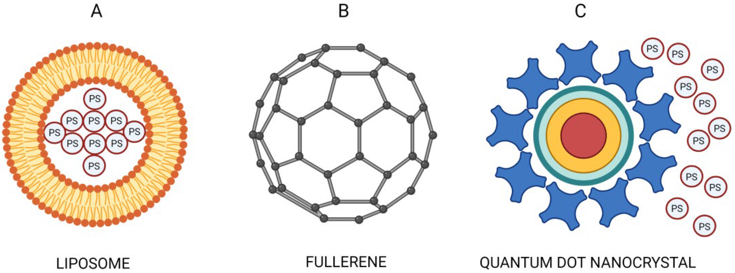
2.1. The First Class of NPs—Carriers of PSs
2.2. The Second Class of NPs—PSs by Themselves
2.3. The Third Class of NPs—Energy Transducers of PSs
3. Problematic Attributes and Limitations of PDT
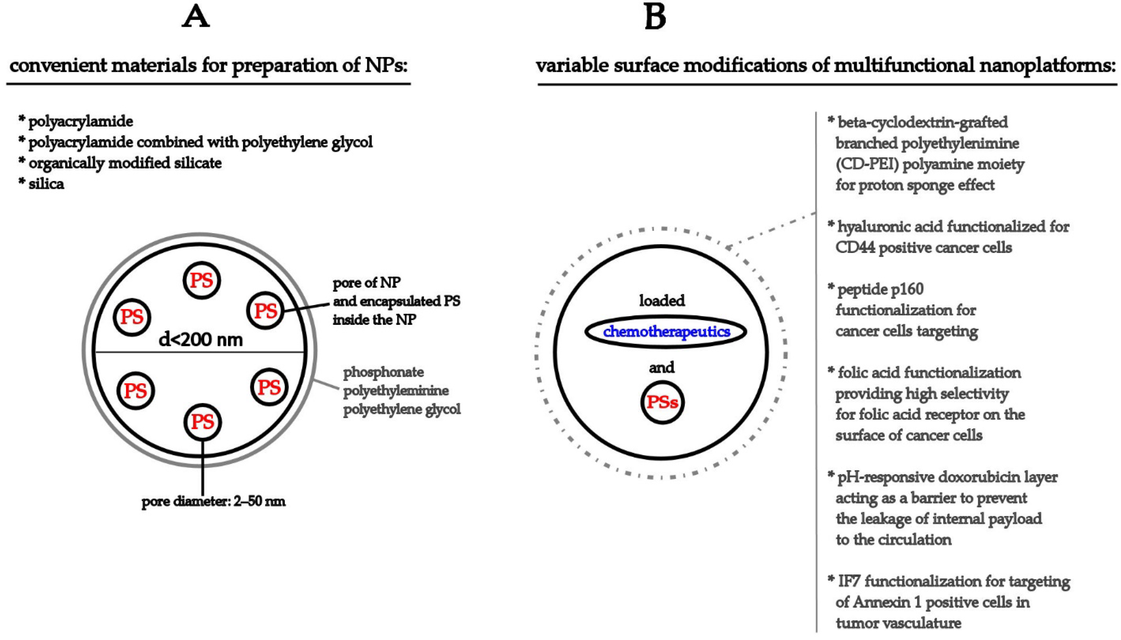
4. Nanoparticles as a Possible Solution for Reducing the MDR Effect in Cancer Treatment
4.1. Dynamic Nanoplatform (DNP)—The Concept of 1O2 Release to Target Cells Rather than the PS Itself
4.2. Multifunctional Therapeutic Nanoplatforms as a Highly Effective Novel Treatment Modality to Reduce the MDR Effect in Tumors
Author Contributions
Funding
Acknowledgments
Conflicts of Interest
Abbreviations
| 5ALA | 5-aminolevulinic acid |
| ABC transporters | ATB-binding casette transporters |
| AlPcS4Cl | phthalocyanine chloride tetra sulfonic acid |
| BCRP/ABCG2 | breast cancer resistant protein |
| BPD | benzoporphyrin derivative |
| BPD-MA | benzoporphyrin derivative monoacid ring A |
| CD-PEI | beta-cyclodextrin-grafted branched polyethylenimine |
| Ce6 | chlorin e6 |
| CSC | cancer stem cell |
| DEB/TQR@PMP | DEB-BDTO/tariquidar and polymeric prodrug micelles |
| DMSO | dimethyl sulfoxide |
| DNP | dynamic nanoplatform |
| DOX | doxorubicin |
| EPR | enhanced permeability and retention effect |
| FA | folic acid |
| FDA | Food and Drug Administration |
| HA | hypocrellin A |
| HAC | hyaluronic acid |
| HB | hypocrellin B |
| hCTR1 | human copper transporter protein 1 |
| HMSNPs | hollow-type mesoporous silica NPs |
| HPPH | 2-[1-hexyloxyethyl]-2-devinyl pyropheophorbide-a |
| HY-PDT | photodynamic therapy with hypericin |
| ICG | indocyanine green |
| MB | methylene blue |
| MDR | multidrug resistance |
| MIT-PFP-PPP | MIT-poly(ε-caprolactone)-pluronic F68-poly(ε-caprolactone)/poly(D,L-lactide-co-glycolide)–poly(ethylene glycol)–poly(D,L-lactide-co-glycolide) |
| MRP1/ABCC1 | MDR-associated protein-1 |
| MSNs | nesoporous silica nanoparticles |
| mTHPC | meso-tetra-hydroxyphenyl-chlorin |
| NPs | nanoparticles |
| NS | nanosystem |
| PARP | poly (ADP-ribose) polymerase |
| Pc4 | silicon phthalocyanine |
| PDT | photodynamic therapy |
| PEG | polyethylene glycol |
| P-gp/ABCB1 | P-glycoprotein |
| PHPP | 2,7,12,18-Tetra-methyl-3,8-di-(1-propoxyethyl)-13,17-bis-(3-hydroxypropyl) porphyrin |
| PLA | polyactic acid polymeric nanoparticles |
| PLGA | paclitaxel-loaded poly(DL-lactide-co-glycolide) |
| PMP | polymeric prodrug |
| PpIX | protoporphyrin IX |
| PSs | photosensitizers |
| PT | photofrin |
| RB | rose bengal |
| ROS | reactive oxygen species |
| Ru | ruthenium |
| SiPcCl2 | silicon phthalocyanine dichloride |
| SLNs | solid lipid NPs |
| SP | side population |
| TPPS2a | disulfonated meso-tetraphenylporphine |
| TQR | tariquidar |
| ZnO | zinc oxide |
References
- Sung, H.; Ferlay, J.; Siegel, R.L.; Laversanne, M.; Soerjomataram, I.; Jemal, A.; Bray, F. Global Cancer Statistics 2020: GLOBOCAN Estimates of Incidence and Mortality Worldwide for 36 Cancers in 185 Countries. CA. Cancer J. Clin. 2021, 71, 209–249. [Google Scholar] [CrossRef] [PubMed]
- Bray, F.; Ferlay, J.; Soerjomataram, I.; Siegel, R.L.; Torre, L.A.; Jemal, A. Global cancer statistics 2018: GLOBOCAN estimates of incidence and mortality worldwide for 36 cancers in 185 countries. CA. Cancer J. Clin. 2018, 68, 394–424. [Google Scholar] [CrossRef] [PubMed] [Green Version]
- Slamon, D.J.; Godolphin, W.; Jones, L.A.; Holt, J.A.; Wong, S.G.; Keith, D.E.; Levin, W.J.; Stuart, S.G.; Udove, J.; Ullrich, A.; et al. Studies of the HER-2/ neu Proto-Oncogene in Human Breast and Ovarian Cancer. Science 1989, 244, 707–712. [Google Scholar] [CrossRef] [PubMed]
- Pakkala, S.; Ramalingam, S.S. Personalized therapy for lung cancer: Striking a moving target. JCI Insight 2018, 3, e120858. [Google Scholar] [CrossRef] [PubMed] [Green Version]
- Majidinia, M.; Mirza-Aghazadeh-Attari, M.; Rahimi, M.; Mihanfar, A.; Karimian, A.; Safa, A.; Yousefi, B. Overcoming multidrug resistance in cancer: Recent progress in nanotechnology and new horizons. IUBMB Life 2020, 72, 855–871. [Google Scholar] [CrossRef] [PubMed]
- Rodriguez-Pascual, J.; Ayuso-Sacido, A.; Belda-Iniesta, C. Drug resistance in cancer immunotherapy: New strategies to improve checkpoint inhibitor therapies. Cancer Drug Resist 2019, 2, 980–993. [Google Scholar] [CrossRef] [Green Version]
- Liu, J.; Guo, B. RNA-based therapeutics for colorectal cancer: Updates and future directions. Pharmacol. Res. 2020, 152, 104550. [Google Scholar] [CrossRef]
- Murayama, T.; Gotoh, N. Drug resistance mechanisms of cancer stem-like cells and their therapeutic potential as drug targets. Cancer Drug Resist. 2019, 2, 457–470. [Google Scholar] [CrossRef] [Green Version]
- Babinčák, M.; Jendželovský, R.; Košuth, J.; Majerník, M.; Vargová, J.; Mikulášek, K.; Zdráhal, Z.; Fedoročko, P. Death Receptor 5 (TNFRSF10B) Is Upregulated and TRAIL Resistance Is Reversed in Hypoxia and Normoxia in Colorectal Cancer Cell Lines after Treatment with Skyrin, the Active Metabolite of Hypericum spp. Cancers 2021, 13, 1646. [Google Scholar] [CrossRef]
- Keating, P.; Cambrosio, A.; Nelson, N.C.; Mogoutov, A.; Cointet, J.-P. Therapy’s Shadow: A Short History of the Study of Resistance to Cancer Chemotherapy. Front. Pharmacol. 2013, 4, 58. [Google Scholar] [CrossRef] [Green Version]
- Tsai, T.; Hong, R.-L.; Tsai, J.-C.; Lou, P.-J.; Ling, I.-F.; Chen, C.-T. Effect of 5-aminolevulinic acid-mediated photodynamic therapy on MCF-7 and MCF-7/ADR cells. Lasers Surg. Med. 2004, 34, 62–72. [Google Scholar] [CrossRef]
- Kessel, D.; Woodburn, K.; Skalkos, D. Impaired accumulation of a cationic photosensitizing agent by a cell line exhibiting multidrug resistance. Photochem. Photobiol. 1994, 60, 61–63. [Google Scholar] [CrossRef] [PubMed]
- Kessel, D.; Woodburn, K. Selective photodynamic inactivation of a multidrug transporter by a cationic photosensitising agent. Br. J. Cancer 1995, 71, 306–310. [Google Scholar] [CrossRef] [PubMed] [Green Version]
- Hill, J.E.; Linder, M.K.; Davies, K.S.; Sawada, G.A.; Morgan, J.; Ohulchanskyy, T.Y.; Detty, M.R. Selenorhodamine Photosensitizers for Photodynamic Therapy of P-Glycoprotein-Expressing Cancer Cells. J. Med. Chem. 2014, 57, 8622–8634. [Google Scholar] [CrossRef] [PubMed] [Green Version]
- Kuchárová, B.; Mikeš, J.; Jendželovský, R.; Vargová, J.; Mikešová, L.; Jendželovská, Z.; Kovaľ, J.; Fedoročko, P. Potentiation of hypericin-mediated photodynamic therapy cytotoxicity by MK-886: Focus on ABC transporters, GDF-15 and redox status. Photodiagnosis Photodyn. Ther. 2015, 12, 490–503. [Google Scholar] [CrossRef] [PubMed]
- Morgan, J.; Jackson, J.D.; Zheng, X.; Pandey, S.K.; Pandey, R.K. Substrate Affinity of Photosensitizers Derived from Chlorophyll-a: The ABCG2 Transporter Affects the Phototoxic Response of Side Population Stem Cell-like Cancer Cells to Photodynamic Therapy. Mol. Pharm. 2010, 7, 1789–1804. [Google Scholar] [CrossRef] [PubMed] [Green Version]
- Robey, R.W.; Steadman, K.; Polgar, O.; Bates, S.E. ABCG2-mediated transport of photosensitizers: Potential impact on photodynamic therapy. Cancer Biol. Ther. 2005, 4, 187–194. [Google Scholar] [CrossRef] [Green Version]
- Liu, W.; Baer, M.R.; Bowman, M.J.; Pera, P.; Zheng, X.; Morgan, J.; Pandey, R.A.; Oseroff, A.R. The Tyrosine Kinase Inhibitor Imatinib Mesylate Enhances the Efficacy of Photodynamic Therapy by Inhibiting ABCG2. Clin. Cancer Res. 2007, 13, 2463–2470. [Google Scholar] [CrossRef] [Green Version]
- Robey, R.W.; Fetsch, P.A.; Polgar, O.; Dean, M.; Bates, S.E. The livestock photosensitizer, phytoporphyrin (phylloerythrin), is a substrate of the ATP-binding cassette transporter ABCG2. Res. Vet. Sci. 2006, 81, 345–349. [Google Scholar] [CrossRef]
- Jendželovský, R.; Mikeš, J.; Koval’, J.; Souček, K.; Procházková, J.; Kello, M.; Sačková, V.; Hofmanová, J.; Kozubík, A.; Fedoročko, P. Drug efflux transporters, MRP1 and BCRP, affect the outcome of hypericin-mediated photodynamic therapy in HT-29 adenocarcinoma cells. Photochem. Photobiol. Sci. 2009, 8, 1716–1723. [Google Scholar] [CrossRef]
- Goler-Baron, V.; Assaraf, Y.G. Overcoming multidrug resistance via photodestruction of ABCG2-rich extracellular vesicles sequestering photosensitive chemotherapeutics. PLoS ONE 2012, 7, e35487. [Google Scholar] [CrossRef] [Green Version]
- Bram, E.E.; Adar, Y.; Mesika, N.; Sabisz, M.; Skladanowski, A.; Assaraf, Y.G. Structural determinants of imidazoacridinones facilitating antitumor activity are crucial for substrate recognition by ABCG2. Mol. Pharmacol. 2009, 75, 1149–1159. [Google Scholar] [CrossRef] [Green Version]
- Jendželovská, Z.; Jendželovský, R.; Hiľovská, L.; Kovaľ, J.; Mikeš, J.; Fedoročko, P. Single pre-treatment with hypericin, a St. John’s wort secondary metabolite, attenuates cisplatin- and mitoxantrone-induced cell death in A2780, A2780cis and HL-60 cells. Toxicol. Vitr. 2014, 28, 1259–1273. [Google Scholar] [CrossRef] [PubMed]
- Roy, I.; Ohulchanskyy, T.Y.; Pudavar, H.E.; Bergey, E.J.; Oseroff, A.R.; Morgan, J.; Dougherty, T.J.; Prasad, P.N. Ceramic-based nanoparticles entrapping water-insoluble photosensitizing anticancer drugs: A novel drug-carrier system for photodynamic therapy. J. Am. Chem. Soc. 2003, 125, 7860–7865. [Google Scholar] [CrossRef] [PubMed]
- Ross, B.; Rehemtulla, A.; Koo, Y.-E.L.; Reddy, R.; Kim, G.; Behrend, C.; Buck, S.; Schneider, R.J.; Philbert, M.A.; Weissleder, R.; et al. Photonic and magnetic nanoexplorers for biomedical use: From subcellular imaging to cancer diagnostics and therapy. In Nanobiophotonics and Biomedical Applications; Cartwright, A.N., Ed.; SPIE: Bellingham, WA, USA, 2004; p. 76. [Google Scholar]
- Zhu, J.; Wang, H.; Liao, L.; Zhao, L.; Zhou, L.; Yu, M.; Wang, Y.; Liu, B.; Yu, C. Small Mesoporous Silica Nanoparticles as Carriers for Enhanced Photodynamic Therapy. Chem. Asian J. 2011, 6, 2332–2338. [Google Scholar] [CrossRef]
- Tu, J.; Wang, T.; Shi, W.; Wu, G.; Tian, X.; Wang, Y.; Ge, D.; Ren, L. Multifunctional ZnPc-loaded mesoporous silica nanoparticles for enhancement of photodynamic therapy efficacy by endolysosomal escape. Biomaterials 2012, 33, 7903–7914. [Google Scholar] [CrossRef] [PubMed]
- Chen, Z.-L.; Sun, Y.; Huang, P.; Yang, X.-X.; Zhou, X.-P. Studies on Preparation of Photosensitizer Loaded Magnetic Silica Nanoparticles and Their Anti-Tumor Effects for Targeting Photodynamic Therapy. Nanoscale Res. Lett. 2009, 4, 400. [Google Scholar] [CrossRef] [PubMed] [Green Version]
- Zhou, J.; Zhou, L.; Dong, C.; Feng, Y.; Wei, S.; Shen, J.; Wang, X. Preparation and photodynamic properties of water-soluble hypocrellin A-silica nanospheres. Mater. Lett. 2008, 62, 2910–2913. [Google Scholar] [CrossRef]
- Zhou, L.; Liu, J.-H.; Zhang, J.; Wei, S.-H.; Feng, Y.-Y.; Zhou, J.-H.; Yu, B.-Y.; Shen, J. A new sol–gel silica nanovehicle preparation for photodynamic therapy in vitro. Int. J. Pharm. 2010, 386, 131–137. [Google Scholar] [CrossRef]
- Qian, J.; Gharibi, A.; He, S. Colloidal mesoporous silica nanoparticles with protoporphyrin IX encapsulated for photodynamic therapy. J. Biomed. Opt. 2009, 14, 014012. [Google Scholar] [CrossRef]
- Compagnin, C.; Baù, L.; Mognato, M.; Celotti, L.; Miotto, G.; Arduini, M.; Moret, F.; Fede, C.; Selvestrel, F.; Echevarria, I.M.R.; et al. The cellular uptake of meta-tetra(hydroxyphenyl)chlorin entrapped in organically modified silica nanoparticles is mediated by serum proteins. Nanotechnology 2009, 20, 345101. [Google Scholar] [CrossRef] [PubMed]
- Zhao, B.; Yin, J.-J.; Bilski, P.J.; Chignell, C.F.; Roberts, J.E.; He, Y.-Y. Enhanced photodynamic efficacy towards melanoma cells by encapsulation of Pc4 in silica nanoparticles. Toxicol. Appl. Pharmacol. 2009, 241, 163–172. [Google Scholar] [CrossRef] [PubMed] [Green Version]
- Simon, V.; Devaux, C.; Darmon, A.; Donnet, T.; Thiénot, E.; Germain, M.; Honnorat, J.; Duval, A.; Pottier, A.; Borghi, E.; et al. Pp IX Silica Nanoparticles Demonstrate Differential Interactions with In Vitro Tumor Cell Lines and In Vivo Mouse Models of Human Cancers. Photochem. Photobiol. 2010, 86, 213–222. [Google Scholar] [CrossRef] [PubMed]
- Kim, S.; Ohulchanskyy, T.Y.; Pudavar, H.E.; Pandey, R.K.; Prasad, P.N. Organically Modified Silica Nanoparticles Co-encapsulating Photosensitizing Drug and Aggregation-Enhanced Two-Photon Absorbing Fluorescent Dye Aggregates for Two-Photon Photodynamic Therapy. J. Am. Chem. Soc. 2007, 129, 2669–2675. [Google Scholar] [CrossRef] [Green Version]
- He, X.; Wu, X.; Wang, K.; Shi, B.; Hai, L. Methylene blue-encapsulated phosphonate-terminated silica nanoparticles for simultaneous in vivo imaging and photodynamic therapy. Biomaterials 2009, 30, 5601–5609. [Google Scholar] [CrossRef]
- Allhoff, F.; Lin, P.; Moore, D. What is Nanotechnology and why does it Matter? Wiley: Hoboken, NJ, USA, 2010; ISBN 9781405175456. [Google Scholar]
- Taniguchi, N.; Arakawa, C.; Kobayashi, T. On the Basic concept of Nanotechnology. Proc. ICPE 1974, 2, 18–23. [Google Scholar]
- McNeil, S.E. Nanotechnology for the biologist. J. Leukoc. Biol. 2005, 78, 585–594. [Google Scholar] [CrossRef]
- Salata, O. Applications of nanoparticles in biology and medicine. J. Nanobiotechnol. 2004, 2, 3. [Google Scholar] [CrossRef] [Green Version]
- Yang, Y.; Yu, M.; Song, H.; Wang, Y.; Yu, C. Preparation of fluorescent mesoporous hollow silica–fullerene nanoparticles via selective etching for combined chemotherapy and photodynamic therapy. Nanoscale 2015, 7, 11894–11898. [Google Scholar] [CrossRef]
- Yang, Y.; Wang, A.; Jia, Y.; Brezesinski, G.; Dai, L.; Zhao, J.; Li, J. Peptide p160-Coated Silica Nanoparticles Applied in Photodynamic Therapy. Chem. Asian J. 2014, 9, 2126–2131. [Google Scholar] [CrossRef]
- Don, T.-M.; Lu, K.-Y.; Lin, L.-J.; Hsu, C.-H.; Wu, J.-Y.; Mi, F.-L. Temperature/pH/Enzyme Triple-Responsive Cationic Protein/PAA- b -PNIPAAm Nanogels for Controlled Anticancer Drug and Photosensitizer Delivery against Multidrug Resistant Breast Cancer Cells. Mol. Pharm. 2017, 14, 4648–4660. [Google Scholar] [CrossRef]
- Ji, Y.; Zhao, J.; Chu, C.-C. Biodegradable nanocomplex from hyaluronic acid and arginine based poly(ester amide)s as the delivery vehicles for improved photodynamic therapy of multidrug resistant tumor cells: An in vitro study of the performance of chlorin e6 photosensitizer. J. Biomed. Mater. Res. Part A 2017, 105, 1487–1499. [Google Scholar] [CrossRef] [PubMed]
- Khdair, A.; Handa, H.; Mao, G.; Panyam, J. Nanoparticle-mediated combination chemotherapy and photodynamic therapy overcomes tumor drug resistance in vitro. Eur. J. Pharm. Biopharm. 2009, 71, 214–222. [Google Scholar] [CrossRef] [PubMed]
- Li, H.; Liu, C.; Zeng, Y.-P.; Hao, Y.-H.; Huang, J.-W.; Yang, Z.-Y.; Li, R. Nanoceria-Mediated Drug Delivery for Targeted Photodynamic Therapy on Drug-Resistant Breast Cancer. ACS Appl. Mater. Interfaces 2016, 8, 31510–31523. [Google Scholar] [CrossRef] [PubMed]
- Zhen, S.; Yi, X.; Zhao, Z.; Lou, X.; Xia, F.; Tang, B.Z. Drug delivery micelles with efficient near-infrared photosensitizer for combined image-guided photodynamic therapy and chemotherapy of drug-resistant cancer. Biomaterials 2019, 218, 119330. [Google Scholar] [CrossRef]
- Li, Z.; Cai, Y.; Zhao, Y.; Yu, H.; Zhou, H.; Chen, M. Polymeric mixed micelles loaded mitoxantrone for overcoming multidrug resistance in breast cancer via photodynamic therapy. Int. J. Nanomed. 2017, 12, 6595–6604. [Google Scholar] [CrossRef] [Green Version]
- Luo, Z.; Li, M.; Zhou, M.; Li, H.; Chen, Y.; Ren, X.; Dai, Y. O2-evolving and ROS-activable nanoparticles for treatment of multi-drug resistant Cancer by combination of photodynamic therapy and chemotherapy. Nanomed. Nanotechnol. Biol. Med. 2019, 19, 49–57. [Google Scholar] [CrossRef]
- Ma, X.; Qu, Q.; Zhao, Y. Targeted Delivery of 5-Aminolevulinic Acid by Multifunctional Hollow Mesoporous Silica Nanoparticles for Photodynamic Skin Cancer Therapy. ACS Appl. Mater. Interfaces 2015, 7, 10671–10676. [Google Scholar] [CrossRef]
- Wang, D.; Li, X.; Li, X.; Kang, A.; Sun, L.; Sun, M.; Yang, F.; Xu, C. Magnetic And pH Dual-Responsive Nanoparticles For Synergistic Drug-Resistant Breast Cancer Chemo/Photodynamic Therapy. Int. J. Nanomed. 2019, 14, 7665–7679. [Google Scholar] [CrossRef] [Green Version]
- Yan, T.; Cheng, J.; Liu, Z.; Cheng, F.; Wei, X.; He, J. pH-Sensitive mesoporous silica nanoparticles for chemo-photodynamic combination therapy. Colloids Surf. B Biointerfaces 2018, 161, 442–448. [Google Scholar] [CrossRef]
- Zhang, W.; Shen, J.; Su, H.; Mu, G.; Sun, J.-H.; Tan, C.-P.; Liang, X.-J.; Ji, L.-N.; Mao, Z.-W. Co-Delivery of Cisplatin Prodrug and Chlorin e6 by Mesoporous Silica Nanoparticles for Chemo-Photodynamic Combination Therapy to Combat Drug Resistance. ACS Appl. Mater. Interfaces 2016, 8, 13332–13340. [Google Scholar] [CrossRef]
- Zhou, Y.; Chang, C.; Liu, Z.; Zhao, Q.; Xu, Q.; Li, C.; Chen, Y.; Zhang, Y.; Lu, B. Hyaluronic Acid-Functionalized Hollow Mesoporous Silica Nanoparticles as pH-Sensitive Nanocarriers for Cancer Chemo-Photodynamic Therapy. Langmuir 2021, 37, 2619–2628. [Google Scholar] [CrossRef] [PubMed]
- Sun, J.H.; Zhang, W.; Zhang, D.Y.; Shen, J.; Tan, C.P.; Ji, L.N.; Mao, Z.W. Multifunctional mesoporous silica nanoparticles as efficient transporters of doxorubicin and chlorin e6 for chemo-photodynamic combinatorial cancer therapy. J. Biomater. Appl. 2018, 32, 1253–1264. [Google Scholar] [CrossRef]
- Ellahioui, Y.; Patra, M.; Mari, C.; Kaabi, R.; Karges, J.; Gasser, G.; Gómez-Ruiz, S. Mesoporous silica nanoparticles functionalised with a photoactive ruthenium (II) complex: Exploring the formulation of a metal-based photodynamic therapy photosensitiser. Dalt. Trans. 2019, 48, 5940–5951. [Google Scholar] [CrossRef] [PubMed]
- Baglo, Y.; Liang, B.J.; Robey, R.W.; Ambudkar, S.V.; Gottesman, M.M.; Huang, H.-C. Porphyrin-lipid assemblies and nanovesicles overcome ABC transporter-mediated photodynamic therapy resistance in cancer cells. Cancer Lett. 2019, 457, 110–118. [Google Scholar] [CrossRef] [PubMed]
- Usacheva, M.; Swaminathan, S.K.; Kirtane, A.R.; Panyam, J. Enhanced Photodynamic Therapy and Effective Elimination of Cancer Stem Cells Using Surfactant–Polymer Nanoparticles. Mol. Pharm. 2014, 11, 3186–3195. [Google Scholar] [CrossRef]
- Crous, A.; Abrahamse, H. Effective Gold Nanoparticle-Antibody-Mediated Drug Delivery for Photodynamic Therapy of Lung Cancer Stem Cells. Int. J. Mol. Sci. 2020, 21, 3742. [Google Scholar] [CrossRef]
- Wang, H.; Agarwal, P.; Zhao, S.; Yu, J.; Lu, X.; He, X. Combined cancer therapy with hyaluronan-decorated fullerene-silica multifunctional nanoparticles to target cancer stem-like cells. Biomaterials 2016, 97, 62–73. [Google Scholar] [CrossRef] [Green Version]
- Yang, B.; Liu, H.; Yang, H.; Chen, W.; Wu, J.; Feng, X.; Tong, R.; Yu, H.; Chen, Y.; Lv, Z.; et al. Combinatorial photochemotherapy on liver cancer stem cells with organoplatinum (ii) metallacage-based nanoparticles. J. Mater. Chem. B 2019, 7, 6476–6487. [Google Scholar] [CrossRef]
- Bayda, S.; Adeel, M.; Tuccinardi, T.; Cordani, M.; Rizzolio, F. The History of Nanoscience and Nanotechnology: From Chemical–Physical Applications to Nanomedicine. Molecules 2019, 25, 112. [Google Scholar] [CrossRef] [Green Version]
- Talebian, S.; Rodrigues, T.; das Neves, J.; Sarmento, B.; Langer, R.; Conde, J. Facts and Figures on Materials Science and Nanotechnology Progress and Investment. ACS Nano 2021, 15, 15940–15952. [Google Scholar] [CrossRef]
- Lucky, S.S.; Soo, K.C.; Zhang, Y. Nanoparticles in Photodynamic Therapy. Chem. Rev. 2015, 115, 1990–2042. [Google Scholar] [CrossRef] [PubMed]
- Couleaud, P.; Morosini, V.; Frochot, C.; Richeter, S.; Raehm, L.; Durand, J.-O. Silica-based nanoparticles for photodynamic therapy applications. Nanoscale 2010, 2, 1083. [Google Scholar] [CrossRef] [PubMed]
- Pechanova, O.; Barta, A.; Koneracka, M.; Zavisova, V.; Kubovcikova, M.; Klimentova, J.; Tӧrӧk, J.; Zemancikova, A.; Cebova, M. Protective Effects of Nanoparticle-Loaded Aliskiren on Cardiovascular System in Spontaneously Hypertensive Rats. Molecules 2019, 24, 2710. [Google Scholar] [CrossRef] [PubMed] [Green Version]
- Antosova, A.; Bednarikova, Z.; Koneracka, M.; Antal, I.; Marek, J.; Kubovcikova, M.; Zavisova, V.; Jurikova, A.; Gazova, Z. Amino Acid Functionalized Superparamagnetic Nanoparticles Inhibit Lysozyme Amyloid Fibrillization. Chem.—An Eur. J. 2019, 25, 7501–7514. [Google Scholar] [CrossRef]
- Antal, I.; Strbak, O.; Khmara, I.; Koneracka, M.; Kubovcikova, M.; Zavisova, V.; Kmetova, M.; Baranovicova, E.; Dobrota, D. MRI Relaxivity Changes of the Magnetic Nanoparticles Induced by Different Amino Acid Coatings. Nanomaterials 2020, 10, 394. [Google Scholar] [CrossRef] [PubMed] [Green Version]
- Konan, Y.N.; Gurny, R.; Allémann, E. State of the art in the delivery of photosensitizers for photodynamic therapy. J. Photochem. Photobiol. B Biol. 2002, 66, 89–106. [Google Scholar] [CrossRef]
- Bechet, D.; Couleaud, P.; Frochot, C.; Viriot, M.-L.; Guillemin, F.; Barberi-Heyob, M. Nanoparticles as vehicles for delivery of photodynamic therapy agents. Trends Biotechnol. 2008, 26, 612–621. [Google Scholar] [CrossRef]
- Paszko, E.; Ehrhardt, C.; Senge, M.O.; Kelleher, D.P.; Reynolds, J.V. Nanodrug applications in photodynamic therapy. Photodiagnosis Photodyn. Ther. 2011, 8, 14–29. [Google Scholar] [CrossRef]
- Birrenbach, G.; Speiser, P.P. Polymerized Micelles and Their Use as Adjuvants in Immunology. J. Pharm. Sci. 1976, 65, 1763–1766. [Google Scholar] [CrossRef]
- Kang, C.; Sun, Y.; Zhu, J.; Li, W.; Zhang, A.; Kuang, T.; Xie, J.; Yang, Z. Delivery of Nanoparticles for Treatment of Brain Tumor. Curr. Drug Metab. 2016, 17, 745–754. [Google Scholar] [CrossRef]
- Sun, J.; Kormakov, S.; Liu, Y.; Huang, Y.; Wu, D.; Yang, Z. Recent Progress in Metal-Based Nanoparticles Mediated Photodynamic Therapy. Molecules 2018, 23, 1704. [Google Scholar] [CrossRef] [Green Version]
- Wu, C.; Chen, Z.; Hu, Y.; Rao, Z.; Wu, W.; Yang, Z. Nanocrystals: The Preparation, Precise Control and Application Toward the Pharmaceutics and Food Industry. Curr. Pharm. Des. 2018, 24, 2425–2431. [Google Scholar] [CrossRef] [PubMed]
- Yang, Z.; Xie, J.; Zhu, J.; Kang, C.; Chiang, C.; Wang, X.; Wang, X.; Kuang, T.; Chen, F.; Chen, Z.; et al. Functional exosome-mimic for delivery of siRNA to cancer: In vitro and in vivo evaluation. J. Control. Release 2016, 243, 160–171. [Google Scholar] [CrossRef] [PubMed] [Green Version]
- Bangham, A.D.; Standish, M.M.; Watkins, J.C. Diffusion of univalent ions across the lamellae of swollen phospholipids. J. Mol. Biol. 1965, 13, 238–352. [Google Scholar] [CrossRef]
- Gregoriadis, G. Liposomes and mRNA: Two technologies together create a COVID-19 vaccine. Med. Drug Discov. 2021, 12, 100104. [Google Scholar] [CrossRef]
- Gregoriadis, G.; Leathwood, P.D.; Ryman, B.E. Enzyme entrapment in liposomes. FEBS Lett. 1971, 14, 95–99. [Google Scholar] [CrossRef] [Green Version]
- Gregoriadis, G.; Ryman, B.E. Fate of Protein-Containing Liposomes Injected into Rats. An Approach to the Treatment of Storage Diseases. Eur. J. Biochem. 1972, 24, 485–491. [Google Scholar] [CrossRef] [PubMed]
- Gregoriadis, G.; Ryman, B.E. Lysosomal localization of β-fructofuranosidase-containing liposomes injected into rats. Some implications in the treatment of genetic disorders. Biochem. J. 1972, 129, 123–133. [Google Scholar] [CrossRef] [Green Version]
- Bánó, G.; Staničová, J.; Jancura, D.; Marek, J.; Bánó, M.; Uličný, J.; Strejčková, A.; Miškovský, P. On the Diffusion of Hypericin in Dimethylsulfoxide/Water Mixtures—The Effect of Aggregation. J. Phys. Chem. B 2011, 115, 2417–2423. [Google Scholar] [CrossRef]
- Suváková, M.; Majerník, M.; Jendželovský, R.; Hovan, A.; Bánó, G.; Fedoročko, P.; Antalík, M. In vitro study of disodium cromoglicate as a novel effective hydrotrope solvent for hypericin utilisation in photodynamic therapy. J. Photochem. Photobiol. B Biol. 2020, 206, 111855. [Google Scholar] [CrossRef]
- Timm, M.; Saaby, L.; Moesby, L.; Hansen, E.W. Considerations regarding use of solvents in in vitro cell based assays. Cytotechnology 2013, 65, 887–894. [Google Scholar] [CrossRef] [Green Version]
- Hall, M.D.; Telma, K.A.; Chang, K.-E.; Lee, T.D.; Madigan, J.P.; Lloyd, J.R.; Goldlust, I.S.; Hoeschele, J.D.; Gottesman, M.M. Say No to DMSO: Dimethylsulfoxide Inactivates Cisplatin, Carboplatin, and Other Platinum Complexes. Cancer Res. 2014, 74, 3913–3922. [Google Scholar] [CrossRef] [PubMed] [Green Version]
- Kollerup Madsen, B.; Hilscher, M.; Zetner, D.; Rosenberg, J. Adverse reactions of dimethyl sulfoxide in humans: A systematic review. F1000 Res. 2019, 7, 1746. [Google Scholar] [CrossRef] [PubMed]
- Pezzuoli, D.; Cozzolino, M.; Montali, C.; Brancaleon, L.; Bianchini, P.; Zantedeschi, M.; Bonardi, S.; Viappiani, C.; Abbruzzetti, S. Serum albumins are efficient delivery systems for the photosensitizer hypericin in photosensitization-based treatments against Staphylococcus aureus. Food Control 2018, 94, 254–262. [Google Scholar] [CrossRef]
- Roelants, M.; Van Cleynenbreugel, B.; Lerut, E.; Van Poppel, H.; de Witte, P.A.M. Human serum albumin as key mediator of the differential accumulation of hypericin in normal urothelial cell spheroids versusurothelial cell carcinoma spheroids. Photochem. Photobiol. Sci. 2011, 10, 151–159. [Google Scholar] [CrossRef]
- Blascakova, L.; Horvath, D.; Belej, D.; Wagnieres, G.; Miskovsky, P.; Jancura, D.; Huntosova, V. Hypericin can cross barriers in the chicken’s chorioallantoic membrane model when delivered in low-density lipoproteins. Photodiagnosis Photodyn. Ther. 2018, 23, 306–313. [Google Scholar] [CrossRef]
- de Morais, F.A.P.; Gonçalves, R.S.; Vilsinski, B.H.; de Oliveira, É.L.; Rocha, N.L.; Hioka, N.; Caetano, W. Hypericin photodynamic activity in DPPC liposome. PART I: Biomimetism of loading, location, interactions and thermodynamic properties. J. Photochem. Photobiol. B Biol. 2019, 190, 118–127. [Google Scholar] [CrossRef]
- Plenagl, N.; Duse, L.; Seitz, B.S.; Goergen, N.; Pinnapireddy, S.R.; Jedelska, J.; Brüßler, J.; Bakowsky, U. Photodynamic therapy —Hypericin tetraether liposome conjugates and their antitumor and antiangiogenic activity. Drug Deliv. 2019, 26, 23–33. [Google Scholar] [CrossRef] [Green Version]
- Penjweini, R.; Smisdom, N.; Deville, S.; Ameloot, M. Transport and accumulation of PVP-Hypericin in cancer and normal cells characterized by image correlation spectroscopy techniques. Biochim. Biophys. Acta—Mol. Cell Res. 2014, 1843, 855–865. [Google Scholar] [CrossRef] [Green Version]
- Penjweini, R.; Deville, S.; Haji Maghsoudi, O.; Notelaers, K.; Ethirajan, A.; Ameloot, M. Investigating the effect of poly-l-lactic acid nanoparticles carrying hypericin on the flow-biased diffusive motion of HeLa cell organelles. J. Pharm. Pharmacol. 2018, 71, 104–116. [Google Scholar] [CrossRef] [Green Version]
- Shao, C.; Shang, K.; Xu, H.; Zhang, Y.; Pei, Z.; Pei, Y. Facile fabrication of hypericin-entrapped glyconanoparticles for targeted photodynamic therapy. Int. J. Nanomed. 2018, 13, 4319–4331. [Google Scholar] [CrossRef] [Green Version]
- Torchilin, V.P. Recent advances with liposomes as pharmaceutical carriers. Nat. Rev. Drug Discov. 2005, 4, 145–160. [Google Scholar] [CrossRef] [PubMed]
- Ghosh, S.; Carter, K.A.; Lovell, J.F. Liposomal formulations of photosensitizers. Biomaterials 2019, 218, 119341. [Google Scholar] [CrossRef] [PubMed]
- Lin, M.-W.; Huang, Y.-B.; Chen, C.-L.; Wu, P.-C.; Chou, C.-Y.; Wu, P.-C.; Hung, S.-Y. A Formulation Study of 5-Aminolevulinic Encapsulated in DPPC Liposomes in Melanoma Treatment. Int. J. Med. Sci. 2016, 13, 483–489. [Google Scholar] [CrossRef] [PubMed] [Green Version]
- Derycke, A. Liposomes for photodynamic therapy. Adv. Drug Deliv. Rev. 2004, 56, 17–30. [Google Scholar] [CrossRef]
- Lasic, D.D.; Martin, F.J.; Gabizon, A.; Huang, S.K.; Papahadjopoulos, D. Sterically stabilized liposomes: A hypothesis on the molecular origin of the extended circulation times. Biochim. Biophys. Acta—Biomembr. 1991, 1070, 187–192. [Google Scholar] [CrossRef]
- Sharma, S.K.; Chiang, L.Y.; Hamblin, M.R. Photodynamic therapy with fullerenes in vivo: Reality or a dream? Nanomedicine 2011, 6, 1813–1825. [Google Scholar] [CrossRef] [Green Version]
- Rozhkova, E.A.; Ulasov, I.; Lai, B.; Dimitrijevic, N.M.; Lesniak, M.S.; Rajh, T. A High-Performance Nanobio Photocatalyst for Targeted Brain Cancer Therapy. Nano Lett. 2009, 9, 3337–3342. [Google Scholar] [CrossRef] [Green Version]
- Cai, R.; Hashimoto, K.; Itoh, K.; Kubota, Y.; Fujishima, A. Photokilling of Malignant Cells with Ultrafine TiO2 Powder. Bull. Chem. Soc. Jpn. 1991, 64, 1268–1273. [Google Scholar] [CrossRef] [Green Version]
- Cai, R.; Kubota, Y.; Shuin, T.; Sakai, H.; Hashimoto, K.; Fujishima, A. Induction of cytotoxicity by photoexcited TiO2 particles. Cancer Res. 1992, 52, 2346–2348. [Google Scholar]
- Wamer, W.G.; Yin, J.-J.; Wei, R.R. Oxidative Damage to Nucleic Acids Photosensitized by Titanium Dioxide. Free Radic. Biol. Med. 1997, 23, 851–858. [Google Scholar] [CrossRef]
- Liao, F.; Saitoh, Y.; Miwa, N. Anticancer Effects of Fullerene [C60] Included in Polyethylene Glycol Combined With Visible Light Irradiation Through ROS Generation and DNA Fragmentation on Fibrosarcoma Cells With Scarce Cytotoxicity to Normal Fibroblasts. Oncol. Res. Featur. Preclin. Clin. Cancer Ther. 2011, 19, 203–216. [Google Scholar] [CrossRef]
- Tabata, Y.; Murakami, Y.; Ikada, Y. Photodynamic Effect of Polyethylene Glycol-modified Fullerene on Tumor. Jpn. J. Cancer Res. 1997, 88, 1108–1116. [Google Scholar] [CrossRef] [PubMed]
- Ikeda, A.; Doi, Y.; Nishiguchi, K.; Kitamura, K.; Hashizume, M.; Kikuchi, J.; Yogo, K.; Ogawa, T.; Takeya, T. Induction of cell death by photodynamic therapy with water-soluble lipid-membrane-incorporated [60]fullerene. Org. Biomol. Chem. 2007, 5, 1158. [Google Scholar] [CrossRef] [PubMed]
- Akiyama, M.; Ikeda, A.; Shintani, T.; Doi, Y.; Kikuchi, J.; Ogawa, T.; Yogo, K.; Takeya, T.; Yamamoto, N. Solubilisation of [60]fullerenes using block copolymers and evaluation of their photodynamic activities. Org. Biomol. Chem. 2008, 6, 1015. [Google Scholar] [CrossRef]
- Metanawin, T.; Tang, T.; Chen, R.; Vernon, D.; Wang, X. Cytotoxicity and photocytotoxicity of structure-defined water-soluble C 60/micelle supramolecular nanoparticles. Nanotechnology 2011, 22, 235604. [Google Scholar] [CrossRef]
- Kwag, D.S.; Oh, N.M.; Oh, Y.T.; Oh, K.T.; Youn, Y.S.; Lee, E.S. Photodynamic therapy using glycol chitosan grafted fullerenes. Int. J. Pharm. 2012, 431, 204–209. [Google Scholar] [CrossRef]
- Jacques, S.L.; Weaver, D.R.; Reppert, S.M. Penetration of light into the uterus of pregnant mammals. Photochem. Photobiol. 1987, 45, 637–641. [Google Scholar] [CrossRef]
- Liu, Q.; Xu, L.; Zhang, X.; Li, N.; Zheng, J.; Guan, M.; Fang, X.; Wang, C.; Shu, C. Enhanced Photodynamic Efficiency of an Aptamer-Guided Fullerene Photosensitizer toward Tumor Cells. Chem.—An Asian J. 2013, 8, 2370–2376. [Google Scholar] [CrossRef]
- Zhou, J.; Xu, N.S.; Wang, Z.L. Dissolving Behavior and Stability of ZnO Wires in Biofluids: A Study on Biodegradability and Biocompatibility of ZnO Nanostructures. Adv. Mater. 2006, 18, 2432–2435. [Google Scholar] [CrossRef]
- Jones, N.; Ray, B.; Ranjit, K.T.; Manna, A.C. Antibacterial activity of ZnO nanoparticle suspensions on a broad spectrum of microorganisms. FEMS Microbiol. Lett. 2008, 279, 71–76. [Google Scholar] [CrossRef] [PubMed] [Green Version]
- Sirelkhatim, A.; Mahmud, S.; Seeni, A.; Kaus, N.H.M.; Ann, L.C.; Bakhori, S.K.M.; Hasan, H.; Mohamad, D. Review on Zinc Oxide Nanoparticles: Antibacterial Activity and Toxicity Mechanism. Nano-Micro Lett. 2015, 7, 219–242. [Google Scholar] [CrossRef] [Green Version]
- Zhou, G.; Li, Y.; Xiao, W.; Zhang, L.; Zuo, Y.; Xue, J.; Jansen, J.A. Synthesis, characterization, and antibacterial activities of a novel nanohydroxyapatite/zinc oxide complex. J. Biomed. Mater. Res. Part A 2008, 85A, 929–937. [Google Scholar] [CrossRef] [PubMed] [Green Version]
- Raghupathi, K.R.; Koodali, R.T.; Manna, A.C. Size-Dependent Bacterial Growth Inhibition and Mechanism of Antibacterial Activity of Zinc Oxide Nanoparticles. Langmuir 2011, 27, 4020–4028. [Google Scholar] [CrossRef] [PubMed]
- Ostrovsky, S.; Kazimirsky, G.; Gedanken, A.; Brodie, C. Selective cytotoxic effect of ZnO nanoparticles on glioma cells. Nano Res. 2009, 2, 882–890. [Google Scholar] [CrossRef] [Green Version]
- Choi, K.-H.; Nam, K.; Lee, S.-Y.; Cho, G.; Jung, J.-S.; Kim, H.-J.; Park, B. Antioxidant Potential and Antibacterial Efficiency of Caffeic Acid-Functionalized ZnO Nanoparticles. Nanomaterials 2017, 7, 148. [Google Scholar] [CrossRef] [Green Version]
- Wang, J.; Lee, J.S.; Kim, D.; Zhu, L. Exploration of Zinc Oxide Nanoparticles as a Multitarget and Multifunctional Anticancer Nanomedicine. ACS Appl. Mater. Interfaces 2017, 9, 39971–39984. [Google Scholar] [CrossRef]
- Sivakumar, P.; Lee, M.; Kim, Y.-S.; Shim, M.S. Photo-triggered antibacterial and anticancer activities of zinc oxide nanoparticles. J. Mater. Chem. B 2018, 6, 4852–4871. [Google Scholar] [CrossRef]
- Chen, W.; Zhang, J. Using Nanoparticles to Enable Simultaneous Radiation and Photodynamic Therapies for Cancer Treatment. J. Nanosci. Nanotechnol. 2006, 6, 1159–1166. [Google Scholar] [CrossRef]
- Chen, W. Nanoparticle Self-Lighting Photodynamic Therapy for Cancer Treatment. J. Biomed. Nanotechnol. 2008, 4, 369–376. [Google Scholar] [CrossRef]
- Samia, A.C.S.; Chen, X.; Burda, C. Semiconductor Quantum Dots for Photodynamic Therapy. J. Am. Chem. Soc. 2003, 125, 15736–15737. [Google Scholar] [CrossRef] [PubMed]
- Gomes, A.J.; Lunardi, L.O.; Marchetti, J.M.; Lunardi, C.N.; Tedesco, A.C. Indocyanine Green Nanoparticles Useful for Photomedicine. Photomed. Laser Surg. 2006, 24, 514–521. [Google Scholar] [CrossRef] [PubMed] [Green Version]
- Zheng, Y.; Tang, Y.; Bao, Z.; Wang, H.; Ren, F.; Guo, M.; Quan, H.; Jiang, C. FePt nanoparticles as a potential X-ray activated chemotherapy agent for HeLa cells. Int. J. Nanomed. 2015, 10, 6435–6444. [Google Scholar] [CrossRef] [Green Version]
- Ahirwar, S.; Mallick, S.; Bahadur, D. Photodynamic therapy using graphene quantum dot derivatives. J. Solid State Chem. 2020, 282, 121107. [Google Scholar] [CrossRef]
- Shen, Y.; Shuhendler, A.J.; Ye, D.; Xu, J.-J.; Chen, H.-Y. Two-photon excitation nanoparticles for photodynamic therapy. Chem. Soc. Rev. 2016, 45, 6725–6741. [Google Scholar] [CrossRef]
- Osuchowski, M.; Osuchowski, F.; Latos, W.; Kawczyk-Krupka, A. The Use of Upconversion Nanoparticles in Prostate Cancer Photodynamic Therapy. Life 2021, 11, 360. [Google Scholar] [CrossRef]
- Lipson, R.L.; Baldes, E.J.; Olsen, A.M. The Use of a Derivative of Hematoporphyrin in Tumor Detection. JNCI J. Natl. Cancer Inst. 1961, 26, 1–11. [Google Scholar] [CrossRef]
- Dougherty, T.J.; Gomer, C.J.; Henderson, B.W.; Jori, G.; Kessel, D.; Korbelik, M.; Moan, J.; Peng, Q. Photodynamic Therapy. JNCI J. Natl. Cancer Inst. 1998, 90, 889–905. [Google Scholar] [CrossRef] [Green Version]
- Majerník, M.; Jendželovský, R.; Fedoročko, P. Potentiality, Limitations, and Consequences of Different Experimental Models to Improve Photodynamic Therapy for Cancer Treatment in Relation to Antiangiogenic Mechanism. Cancers 2020, 12, 2118. [Google Scholar] [CrossRef]
- Mikeš, J.; Jendželovský, R.; Fedoročko, P. Cellular Aspects of Photodynamic Therapy with Hypericin; Nova Science Publishers: New York, NY, USA, 2013; ISBN 9781624176357. [Google Scholar]
- Rossi, R.; Bruscino, N.; Ricceri, F.; Grazzini, M.; Dindelli, M.; Lotti, T. Photodynamic treatment for viral infections of the skin. G. Ital. Dermatol. Venereol. 2009, 144, 79–83. [Google Scholar]
- Kharkwal, G.B.; Sharma, S.K.; Huang, Y.-Y.; Dai, T.; Hamblin, M.R. Photodynamic therapy for infections: Clinical applications. Lasers Surg. Med. 2011, 43, 755–767. [Google Scholar] [CrossRef] [PubMed] [Green Version]
- Harris, F.; Pierpoint, L. Photodynamic therapy based on 5-aminolevulinic acid and its use as an antimicrobial Agent. Med. Res. Rev. 2012, 32, 1292–1327. [Google Scholar] [CrossRef] [PubMed]
- Jacobson, J.M.; Feinman, L.; Liebes, L.; Ostrow, N.; Koslowski, V.; Tobia, A.; Cabana, B.E.; Lee, D.-H.; Spritzler, J.; Prince, A.M. Pharmacokinetics, Safety, and Antiviral Effects of Hypericin, a Derivative of St. John’s Wort Plant, in Patients with Chronic Hepatitis C Virus Infection. Antimicrob. Agents Chemother. 2001, 45, 517–524. [Google Scholar] [CrossRef] [Green Version]
- Lim, M.E.; Lee, Y.; Zhang, Y.; Chu, J.J.H. Photodynamic inactivation of viruses using upconversion nanoparticles. Biomaterials 2012, 33, 1912–1920. [Google Scholar] [CrossRef]
- Moan, J.; Berg, K. The photodegradation of porphyrins in cells can be used to estimate the lifetime of singlet oxygen. Photochem. Photobiol. 1991, 53, 549–553. [Google Scholar] [CrossRef] [PubMed]
- Oleinick, N.L.; Morris, R.L.; Belichenko, I. The role of apoptosis in response to photodynamic therapy: What, where, why, and how. Photochem. Photobiol. Sci. 2002, 1, 1–21. [Google Scholar] [CrossRef] [PubMed]
- Kessel, D.; Luo, Y.; Deng, Y.; Chang, C.K. The role of subcellular localization in initiation of apoptosis by photodynamic therapy. Photochem. Photobiol. 1997, 65, 422–426. [Google Scholar] [CrossRef] [Green Version]
- Mesquita, M.Q.; Dias, C.J.; Gamelas, S.; Fardilha, M.; Neves, M.G.P.M.S.; Faustino, M.A.F. An insight on the role of photosensitizer nanocarriers for Photodynamic Therapy. An. Acad. Bras. Cienc. 2018, 90, 1101–1130. [Google Scholar] [CrossRef] [Green Version]
- Tang, W.; Xu, H.; Kopelman, R.A.; Philbert, M. Photodynamic Characterization and In Vitro Application of Methylene Blue-containing Nanoparticle Platforms. Photochem. Photobiol. 2005, 81, 242. [Google Scholar] [CrossRef] [Green Version]
- Williams, J.L.; Stamp, J.; Devonshire, R.; Fowler, G.J.S. Methylene blue and the photodynamic therapy of superficial bladder cancer. J. Photochem. Photobiol. B Biol. 1989, 4, 229–232. [Google Scholar] [CrossRef]
- Orth, K.; Beck, G.; Genze, F.; Rück, A. Methylene blue mediated photodynamic therapy in experimental colorectal tumors in mice. J. Photochem. Photobiol. B Biol. 2000, 57, 186–192. [Google Scholar] [CrossRef]
- Wainwright, M.; Phoenix, D.A.; Rice, L.; Burrow, S.M.; Waring, J. Increased cytotoxicity and phototoxicity in the methylene blue series via chromophore methylation. J. Photochem. Photobiol. B Biol. 1997, 40, 233–239. [Google Scholar] [CrossRef]
- Bhuvaneswari, R.; Gan, Y.Y.Y.; Yee, K.K.L.; Soo, K.C.; Olivo, M. Effect of hypericin-mediated photodynamic therapy on the expression of vascular endothelial growth factor in human nasopharyngeal carcinoma. Int. J. Mol. Med. 2007, 20, 421–428. [Google Scholar] [CrossRef] [PubMed] [Green Version]
- Bhuvaneswari, R.; Gan, Y.K.; Lucky, S.S.; Chin, W.W.L.; Ali, S.M.; Soo, K.C.; Olivo, M. Molecular profiling of angiogenesis in hypericin mediated photodynamic therapy. Mol. Cancer 2008, 7, 1–14. [Google Scholar] [CrossRef] [PubMed] [Green Version]
- Petersen, B.; Wiegell, S.R.; Wulf, H.C. Light protection of the skin after photodynamic therapy reduces inflammation: An unblinded randomized controlled study. Br. J. Dermatol. 2014, 171, 175–178. [Google Scholar] [CrossRef] [PubMed]
- Nowell, P.C. The Clonal Evolution of Tumor Cell Populations. Science 1976, 194, 23–28. [Google Scholar] [CrossRef]
- Kocibalova, Z.; Guzyova, M.; Borovska, I.; Messingerova, L.; Copakova, L.; Sulova, Z.; Breier, A. Development of Multidrug Resistance in Acute Myeloid Leukemia Is Associated with Alterations of the LPHN1/GAL-9/TIM-3 Signaling Pathway. Cancers 2021, 13, 3629. [Google Scholar] [CrossRef]
- Gibalová, L.; Sedlák, J.; Labudová, M.; Barancík, M.; Reháková, A.; Breier, A.; Sulová, Z. Multidrug resistant P-glycoprotein positive L1210/VCR cells are also cross-resistant to cisplatin via a mechanism distinct from P-glycoprotein-mediated drug efflux activity. Gen. Physiol. Biophys. 2009, 28, 391–403. [Google Scholar] [CrossRef] [Green Version]
- Gottesman, M.M.; Ambudkar, S.V. Overview: ABC transporters and human disease. J. Bioenerg. Biomembr. 2001, 33, 453–458. [Google Scholar] [CrossRef]
- Vlaming, M.L.H.; Lagas, J.S.; Schinkel, A.H. Physiological and pharmacological roles of ABCG2 (BCRP): Recent findings in Abcg2 knockout mice. Adv. Drug Deliv. Rev. 2009, 61, 14–25. [Google Scholar] [CrossRef]
- Tian, Y.; Bian, Y.; Jiang, Y.; Qian, S.; Yu, A.; Zeng, S. Interplay of Breast Cancer Resistance Protein (BCRP) and Metabolizing Enzymes. Curr. Drug Metab. 2015, 16, 877–893. [Google Scholar] [CrossRef] [PubMed]
- Halwachs, S.; Kneuer, C.; Gohlsch, K.; Müller, M.; Ritz, V.; Honscha, W. The ABCG2 efflux transporter from rabbit placenta: Cloning and functional characterization. Placenta 2016, 38, 8–15. [Google Scholar] [CrossRef] [PubMed] [Green Version]
- Filia, M.F.; Marchini, T.; Minoia, J.M.; Roma, M.I.; De Fino, F.T.; Rubio, M.C.; Copello, G.J.; Evelson, P.A.; Peroni, R.N. Induction of ABCG2/BCRP restricts the distribution of zidovudine to the fetal brain in rats. Toxicol. Appl. Pharmacol. 2017, 330, 74–83. [Google Scholar] [CrossRef] [PubMed]
- Ding, X.; Wu, J.; Jiang, C. ABCG2: A potential marker of stem cells and novel target in stem cell and cancer therapy. Life Sci. 2010, 86, 631–637. [Google Scholar] [CrossRef]
- Vargová, J.; Mikeš, J.; Jendželovský, R.; Mikešová, L.; Kuchárová, B.; Čulka, Ľ.; Fedr, R.; Remšík, J.; Souček, K.; Kozubík, A.; et al. Hypericin affects cancer side populations via competitive inhibition of BCRP. Biomed. Pharmacother. 2018, 99, 511–522. [Google Scholar] [CrossRef]
- Robey, R.W.; Polgar, O.; Deeken, J.; To, K.W.; Bates, S.E. ABCG2: Determining its relevance in clinical drug resistance. Cancer Metastasis Rev. 2007, 26, 39–57. [Google Scholar] [CrossRef] [Green Version]
- Jendželovský, R.; Jendželovská, Z.; Kuchárová, B.; Fedoročko, P. Breast cancer resistance protein is the enemy of hypericin accumulation and toxicity of hypericin-mediated photodynamic therapy. Biomed. Pharmacother. 2019, 109, 2173–2181. [Google Scholar] [CrossRef]
- Barron, G.A.; Moseley, H.; Woods, J.A. Differential sensitivity in cell lines to photodynamic therapy in combination with ABCG2 inhibition. J. Photochem. Photobiol. B Biol. 2013, 126, 87–96. [Google Scholar] [CrossRef]
- Ogino, T.; Kobuchi, H.; Munetomo, K.; Fujita, H.; Yamamoto, M.; Utsumi, T.; Inoue, K.; Shuin, T.; Sasaki, J.; Inoue, M.; et al. Serum-dependent export of protoporphyrin IX by ATP-binding cassette transporter G2 in T24 cells. Mol. Cell. Biochem. 2011, 358, 297–307. [Google Scholar] [CrossRef]
- Bebes, A.; Nagy, T.; Bata-Csörgő, Z.; Kemény, L.; Dobozy, A.; Széll, M. Specific inhibition of the ABCG2 transporter could improve the efficacy of photodynamic therapy. J. Photochem. Photobiol. B Biol. 2011, 105, 162–166. [Google Scholar] [CrossRef]
- Abdel Gaber, S.A.; Müller, P.; Zimmermann, W.; Hüttenberger, D.; Wittig, R.; Abdel Kader, M.H.; Stepp, H. ABCG2-mediated suppression of chlorin e6 accumulation and photodynamic therapy efficiency in glioblastoma cell lines can be reversed by KO143. J. Photochem. Photobiol. B. 2018, 178, 182–191. [Google Scholar] [CrossRef] [PubMed]
- Jonker, J.W.; Buitelaar, M.; Wagenaar, E.; Van Der Valk, M.A.; Scheffer, G.L.; Scheper, R.J.; Kuipers, F.; Plosch, T.; Elferink, R.P.J.O.; Rosing, H.; et al. The breast cancer resistance protein protects against a major chlorophyll-derived dietary phototoxin and protoporphyria. Proc. Natl. Acad. Sci. USA 2002, 99, 15649–15654. [Google Scholar] [CrossRef] [PubMed] [Green Version]
- Kim, J.H.; Park, J.M.; Roh, Y.J.; Kim, I.-W.; Hasan, T.; Choi, M.-G. Enhanced efficacy of photodynamic therapy by inhibiting ABCG2 in colon cancers. BMC Cancer 2015, 15, 504. [Google Scholar] [CrossRef] [PubMed] [Green Version]
- Robey, R.W.; Steadman, K.; Polgar, O.; Morisaki, K.; Blayney, M.; Mistry, P.; Bates, S.E. Pheophorbide a is a specific probe for ABCG2 function and inhibition. Cancer Res. 2004, 64, 1242–1246. [Google Scholar] [CrossRef] [PubMed] [Green Version]
- Palasuberniam, P.; Yang, X.; Kraus, D.; Jones, P.; Myers, K.A.; Chen, B. ABCG2 transporter inhibitor restores the sensitivity of triple negative breast cancer cells to aminolevulinic acid-mediated photodynamic therapy. Sci. Rep. 2015, 5, 13298. [Google Scholar] [CrossRef]
- Usuda, J.; Tsunoda, Y.; Ichinose, S.; Ishizumi, T.; Ohtani, K.; Maehara, S.; Ono, S.; Tsutsui, H.; Ohira, T.; Okunaka, T.; et al. Breast cancer resistant protein (BCRP) is a molecular determinant of the outcome of photodynamic therapy (PDT) for centrally located early lung cancer. Lung Cancer 2010, 67, 198–204. [Google Scholar] [CrossRef]
- Chen, B.; Delaey, E.; Vandenheede, J.R.; Agostinis, P.; Xu, Y.; de Witte, P.; Roskams, T. Efficacy of antitumoral photodynamic therapy with hypericin: Relationship between biodistribution and photodynamic effects in the RIF-1 mouse tumor model. Int. J. Cancer 2001, 93, 275–282. [Google Scholar] [CrossRef]
- Chen, B.; Roskams, T.; Xu, Y.; Agostinis, P.; De Witte, P.A.M. Photodynamic therapy with hypericin induces vascular damage and apoptosis in the RIF-1 mouse tumor model. Int. J. Cancer 2002, 98, 284–290. [Google Scholar] [CrossRef] [Green Version]
- Sanovic, R.; Verwanger, T.; Hartl, A.; Krammer, B. Low dose hypericin-PDT induces complete tumor regression in BALB/c mice bearing CT26 colon carcinoma. Photodiagnosis Photodyn. Ther. 2011, 8, 291–296. [Google Scholar] [CrossRef]
- Weiswald, L.-B.; Bellet, D.; Dangles-Marie, V. Spherical cancer models in tumor biology. Neoplasia 2015, 17, 1–15. [Google Scholar] [CrossRef] [Green Version]
- Kirby, C.; Clarke, J.; Gregoriadis, G. Cholesterol content of small unilamellar liposomes controls phospholipid loss to high density lipoproteins in the presence of serum. FEBS Lett. 1980, 111, 324–328. [Google Scholar] [CrossRef] [Green Version]
- Børresen, B.; Hansen, A.E.; Kjaer, A.; Andresen, T.L.; Kristensen, A.T. Liposome-encapsulated chemotherapy: Current evidence for its use in companion animals. Vet. Comp. Oncol. 2018, 16, E1–E15. [Google Scholar] [CrossRef] [PubMed] [Green Version]
- Jiang, F.; Lilge, L.; Logie, B.; Li, Y.; Chopp, M. Photodynamic therapy of 9L gliosarcoma with liposome-delivered photofrin. Photochem. Photobiol. 1997, 65, 701–706. [Google Scholar] [CrossRef]
- Jiang, F.; Lilge, L.; Grenier, J.; Li, Y.; Wilson, M.D.; Chopp, M. Photodynamic therapy of U87 human glioma in nude rat using liposome-delivered photofrin. Lasers Surg. Med. 1998, 22, 74–80. [Google Scholar] [CrossRef]
- Takeuchi, Y.; Ichikawa, K.; Yonezawa, S.; Kurohane, K.; Koishi, T.; Nango, M.; Namba, Y.; Oku, N. Intracellular target for photosensitization in cancer antiangiogenic photodynamic therapy mediated by polycation liposome. J. Control. Release 2004, 97, 231–240. [Google Scholar] [CrossRef] [PubMed]
- Wang, Z.J.; He, Y.Y.; Huang, C.G.; Huang, J.S.; Huang, Y.C.; An, J.Y.; Gu, Y.; Jiang, L.J. Pharmacokinetics, tissue distribution and photodynamic therapy efficacy of liposomal-delivered hypocrellin A, a potential photosensitizer for tumor therapy. Photochem. Photobiol. 1999, 70, 773–780. [Google Scholar] [CrossRef] [PubMed]
- Ma, H.L.; Varanda, L.C.; Perussi, J.R.; Carrilho, E. Hypericin-loaded oil-in-water nanoemulsion synthesized by ultrasonication process enhances photodynamic therapy efficiency. J. Photochem. Photobiol. B Biol. 2021, 223, 112303. [Google Scholar] [CrossRef]
- Lima, A.M.; Pizzol, C.D.; Monteiro, F.B.F.; Creczynski-Pasa, T.B.; Andrade, G.P.; Ribeiro, A.O.; Perussi, J.R. Hypericin encapsulated in solid lipid nanoparticles: Phototoxicity and photodynamic efficiency. J. Photochem. Photobiol. B. 2013, 125, 146–154. [Google Scholar] [CrossRef]
- Lou, G.; Anderluzzi, G.; Schmidt, S.T.; Woods, S.; Gallorini, S.; Brazzoli, M.; Giusti, F.; Ferlenghi, I.; Johnson, R.N.; Roberts, C.W.; et al. Delivery of self-amplifying mRNA vaccines by cationic lipid nanoparticles: The impact of cationic lipid selection. J. Control. Release 2020, 325, 370–379. [Google Scholar] [CrossRef]
- Panigaj, M.; Dobrovolskaia, M.A.; Afonin, K.A. 2021: An immunotherapy odyssey and the rise of nucleic acid nanotechnology. Nanomedicine 2021, 16, 1635–1640. [Google Scholar] [CrossRef]
- Tenchov, R.; Bird, R.; Curtze, A.E.; Zhou, Q. Lipid Nanoparticles─From Liposomes to mRNA Vaccine Delivery, a Landscape of Research Diversity and Advancement. ACS Nano 2021, 15, 16982–17015. [Google Scholar] [CrossRef] [PubMed]
- Youssef, T.; Fadel, M.; Fahmy, R.; Kassab, K. Evaluation of hypericin-loaded solid lipid nanoparticles: Physicochemical properties, photostability and phototoxicity. Pharm. Dev. Technol. 2012, 17, 177–186. [Google Scholar] [CrossRef] [PubMed]
- Zeisser-Labouèbe, M.; Lange, N.; Gurny, R.; Delie, F. Hypericin-loaded nanoparticles for the photodynamic treatment of ovarian cancer. Int. J. Pharm. 2006, 326, 174–181. [Google Scholar] [CrossRef] [PubMed]
- Zhu, R.; Cheng, K.-W.; Mackenzie, G.; Huang, L.; Sun, Y.; Xie, G.; Vrankova, K.; Constantinides, P.P.; Rigas, B. Phospho-Sulindac (OXT-328) Inhibits the Growth of Human Lung Cancer Xenografts in Mice: Enhanced Efficacy and Mitochondria Targeting by its Formulation in Solid Lipid Nanoparticles. Pharm. Res. 2012, 29, 3090–3101. [Google Scholar] [CrossRef] [Green Version]
- Mu, L.; Feng, S.-S. PLGA/TPGS nanoparticles for controlled release of paclitaxel: Effects of the emulsifier and drug loading ratio. Pharm. Res. 2003, 20, 1864–1872. [Google Scholar] [CrossRef]
- Görner, T.; Gref, R.; Michenot, D.; Sommer, F.; Tran, M.N.; Dellacherie, E. Lidocaine-loaded biodegradable nanospheres. I. Optimization Of the drug incorporation into the polymer matrix. J. Control. Release 1999, 57, 259–268. [Google Scholar] [CrossRef]
- Dillon, J.; Kennedy, J.C.; Pottier, R.H.; Roberts, J.E. In vitro and in vivo protection against phototoxic side effects of photodynamic therapy by radioprotective agentswr–2721 andwr–77913. Photochem. Photobiol. 1988, 48, 235–238. [Google Scholar] [CrossRef]
- Xu, H.; Buck, S.M.; Kopelman, R.; Philbert, M.A.; Brasuel, M.; Monson, E.; Behrend, C.; Ross, B.; Rehemtulla, A.; Koo, Y.-E.L. Fluorescent Pebble Nano-Sensors and Nanoexplorers for Real-Time Intracellular and Biomedical Applications. In Advanced Concepts in Fluorescence Sensing; Springer: Boston, MA, USA, 2005; pp. 69–126. [Google Scholar]
- Ghaferi, M.; Esfahani, M.K.M.; Raza, A.; Al Harthi, S.; Ebrahimi Shahmabadi, H.; Alavi, S.E. Mesoporous silica nanoparticles: Synthesis methods and their therapeutic use-recent advances. J. Drug Target. 2021, 29, 131–154. [Google Scholar] [CrossRef]
- Qiao, Z.-A.; Zhang, L.; Guo, M.; Liu, Y.; Huo, Q. Synthesis of Mesoporous Silica Nanoparticles via Controlled Hydrolysis and Condensation of Silicon Alkoxide. Chem. Mater. 2009, 21, 3823–3829. [Google Scholar] [CrossRef]
- Huynh, E.; Zheng, G. Cancer nanomedicine: Addressing the dark side of the enhanced permeability and retention effect. Nanomedicine 2015, 10, 1993–1995. [Google Scholar] [CrossRef]
- Berners-Price, S.J. Activating Platinum Anticancer Complexes with Visible Light. Angew. Chem. Int. Ed. 2011, 50, 804–805. [Google Scholar] [CrossRef] [PubMed]
- Wong, E.; Giandomenico, C.M. Current Status of Platinum-Based Antitumor Drugs. Chem. Rev. 1999, 99, 2451–2466. [Google Scholar] [CrossRef] [PubMed]
- Piao, Y.; Burns, A.; Kim, J.; Wiesner, U.; Hyeon, T. Designed Fabrication of Silica-Based Nanostructured Particle Systems for Nanomedicine Applications. Adv. Funct. Mater. 2008, 18, 3745–3758. [Google Scholar] [CrossRef]
- Spring, B.Q.; Rizvi, I.; Xu, N.; Hasan, T. The role of photodynamic therapy in overcoming cancer drug resistance. Photochem. Photobiol. Sci. 2015, 14, 1476–1491. [Google Scholar] [CrossRef] [PubMed] [Green Version]
- Wheate, N.J.; Walker, S.; Craig, G.E.; Oun, R. The status of platinum anticancer drugs in the clinic and in clinical trials. Dalt. Trans. 2010, 39, 8113. [Google Scholar] [CrossRef] [PubMed] [Green Version]
- Kelland, L. The resurgence of platinum-based cancer chemotherapy. Nat. Rev. Cancer 2007, 7, 573–584. [Google Scholar] [CrossRef]
- Siddik, Z.H. Cisplatin: Mode of cytotoxic action and molecular basis of resistance. Oncogene 2003, 22, 7265–7279. [Google Scholar] [CrossRef] [Green Version]
- Köberle, B.; Tomicic, M.T.; Usanova, S.; Kaina, B. Cisplatin resistance: Preclinical findings and clinical implications. Biochim. Biophys. Acta—Rev. Cancer 2010, 1806, 172–182. [Google Scholar] [CrossRef]
- Ishida, S.; Lee, J.; Thiele, D.J.; Herskowitz, I. Uptake of the anticancer drug cisplatin mediated by the copper transporter Ctr1 in yeast and mammals. Proc. Natl. Acad. Sci. USA 2002, 99, 14298–14302. [Google Scholar] [CrossRef] [Green Version]
- Holzer, A.K.; Samimi, G.; Katano, K.; Naerdemann, W.; Lin, X.; Safaei, R.; Howell, S.B. The Copper Influx Transporter Human Copper Transport Protein 1 Regulates the Uptake of Cisplatin in Human Ovarian Carcinoma Cells. Mol. Pharmacol. 2004, 66, 817–823. [Google Scholar] [CrossRef] [Green Version]
- Song, I.-S.; Savaraj, N.; Siddik, Z.H.; Liu, P.; Wei, Y.; Wu, C.J.; Kuo, M.T. Role of human copper transporter Ctr1 in the transport of platinum-based antitumor agents in cisplatin-sensitive and cisplatin-resistant cells. Mol. Cancer Ther. 2004, 3, 1543–1549. [Google Scholar] [PubMed]
- Xue, X.; Hall, M.D.; Zhang, Q.; Wang, P.C.; Gottesman, M.M.; Liang, X.-J. Nanoscale Drug Delivery Platforms Overcome Platinum-Based Resistance in Cancer Cells Due to Abnormal Membrane Protein Trafficking. ACS Nano 2013, 7, 10452–10464. [Google Scholar] [CrossRef] [PubMed] [Green Version]
- Christowitz, C.; Davis, T.; Isaacs, A.; van Niekerk, G.; Hattingh, S.; Engelbrecht, A.-M. Mechanisms of doxorubicin-induced drug resistance and drug resistant tumour growth in a murine breast tumour model. BMC Cancer 2019, 19, 757. [Google Scholar] [CrossRef] [PubMed] [Green Version]
- Hu, C.-M.J.; Zhang, L. Nanoparticle-based combination therapy toward overcoming drug resistance in cancer. Biochem. Pharmacol. 2012, 83, 1104–1111. [Google Scholar] [CrossRef] [PubMed]
- Xu, X.; Ho, W.; Zhang, X.; Bertrand, N.; Farokhzad, O. Cancer nanomedicine: From targeted delivery to combination therapy. Trends Mol. Med. 2015, 21, 223–232. [Google Scholar] [CrossRef] [Green Version]
- Bozic, I.; Reiter, J.G.; Allen, B.; Antal, T.; Chatterjee, K.; Shah, P.; Moon, Y.S.; Yaqubie, A.; Kelly, N.; Le, D.T.; et al. Evolutionary dynamics of cancer in response to targeted combination therapy. Elife 2013, 2, e00747. [Google Scholar] [CrossRef] [PubMed]
- MacDonagh, L.; Gray, S.G.; Breen, E.; Cuffe, S.; Finn, S.P.; O’Byrne, K.J.; Barr, M.P. Lung cancer stem cells: The root of resistance. Cancer Lett. 2016, 372, 147–156. [Google Scholar] [CrossRef]


| In Vitro/In Vivo Model | PS/Chemotherapeutic Agent | PS Administration | NS or Solvent | Irradiation Conditions | Observed Effects | References |
|---|---|---|---|---|---|---|
| ovarian serous carcinoma (UCI 107), human cervical adenocarcinoma (HeLa) | 2-devinyl-2-(1-hexyloxyethyl) pyropheophorbide | silica NPs | 650 nm; 1.4 mW/cm2; 10 min | * average size of NPs = 30 nm * NPs were accumulated in cytoplasm *↓ toxicity in dark conditions * significant increase in cell death was observed, if NPs with PS were applied | [24] | |
| human breast adenocarcinoma (MCF-7), melanoma (MDA 435); rat experimental model | PT | i.v. | * RGD peptide modified PAA NPs coated with PEG (RGD peptide modified polyacrylamide (PAA) core with a surface consisting of PEG) | 630 ± 3 nm; 700 mW; 3 min | * average size of NPs: 40 nm * massive necrosis after PDT was observed, if NPs with bound PS were applied * NPs had a potential to selectively bind to αvβ3 integrin on the surface of cancer cells * no toxicity was detected in experimental animals four weeks after NPs application | [25] |
| human cervical adenocarcinoma (HeLa) | HA | silica NPs | ------- | * average size of NPs = 130 nm * micro-hole size (<0.53 nm) makes possible to release only the 1O2 from the NPs, molecules of PS were retained *↑ quantum yield of encapsulated HA * HA detected in HeLa cells * in dark condition no toxicity of encapsulated HA was detected *↑ effectivity of PDT with encapsulated HA | [29] | |
| human cervical adenocarcinoma (HeLa) | HA | silica NPs | 0–25 J/cm2 | * average size of NPs: 110 nm *↑ fluorescence intensity of encapsulated HA *↑ photostability and 1O2 generation of encapsulated HA * active uptake of HA if NPs were used * low dark toxicity of encapsulated HA * apoptosis was observed and ↑ photodamage after PDT with NPs utilization | [30] | |
| human cervical adenocarcinoma (HeLa) | PpIX | silica NPs | 532 nm, 2 mW/cm2; 2 min | * average size of NPs: 25 nm * encapsulated PpIX emitted ↑ fluorescence than free PpIX * encapsulated PpIX had net cationic charge and the HeLa cells had anionic charges on their membranes. This interaction facilitated the uptake of the cationic amino-functionalized NPs by the HeLa cells * HeLa cells were successfully destroyed 8 min after PDT with encapsulated PpIX * after PDT necrosis was detected | [31] | |
| esophageal squamous cell carcinoma (KYSE 510) | mTHPC | silica NPs | 600–700 nm; 2 mW cm−2; 0.12 J cm−2 | * average size of NPs: 24–47 nm * spherically shaped NPs * the molecules of mTHPC were included inside the NP in monomeric form * complete loss of viability after PDT in cells treated with 1.25 uM of encapsulated mTHPC was detected * encapsulation did not affect intracellular fluorescence distribution of PS * mTHPC was largely localized in GA and ER * free mTHPC was taken up by the cells more efficiently than mTHPC in NPs but the cytotoxic effect was equal | [32] | |
| melanoma (A375; B16-F10) | Pc4 | silica NPs | 600–700 nm; 6.6 J cm−2; 15 min | * average size of NPs: 25–30 nm *↑ fluorescence lifetime, photostability and reduced photobleaching rate of encapsulated Pc4 * no dark toxicity of encapsulated and free Pc4 *↑ phototoxicity and apoptosis rate detected, if Pc4 was encapsulated in comparison to free solution *↑ Pc4 was localized in mitochondria, if NPs were utilized | [33] | |
| human colon cancer (HCT 116, HT-29), human breast adenocarcinoma (MCF7, MDA-MB-231), human skin carcinoma (A431), LLBC37, human lung carcinoma (A549); female athymic Swiss nude mice | PpIX | i.v. | silica NPs | 630 nm; 4 mW cm−2 | * average size of NPs: 10–60 nm * in all tumor types, encapsulated PpIX was more efficient than free PpIX *↑ ROS production in HCT 116 and HT-29 cells, if encapsulated PPIX was used * tumor models reached maximal accumulation of NPs at different time points: 2 h for glioblastoma, 16 h for A549 and 20 h for HCT 116 *↑ NPs accumulation was detected in the liver than in the tumor | [34] |
| human cervical adenocarcinoma (HeLa) | HPPH | silica NPs | 850 nm | * average size of NPs: ≤30 nm * encapsulated HPPH produced 1O2 in water * active uptake of encapsulated PS by tumor cells was observed and fluorescence of NPs in cytoplasm was detected * cell necrosis after PDT with encapsulated HPPH was detected | [35] | |
| human cervical adenocarcinoma (HeLa); male athymic BALB/c nude mice | MB | i.v. | phosphonate coated silica NPs | in vitro: 635 nm; 27.5 mW/cm2; 0–45 min, in vivo: 635 nm; 500 mW/cm2; 5 min | * average size of NPs: 105 ± 6.8 nm * phosphonate coated NPs with encapsulated MB have 8.6 time ↑ emission signal than non phosphonated NPs * encapsulation of MB effectively prolonged the fluorescence properties of MB in water and serum * >80% cytotoxicity of encapsulated MB in vitro even in the lower concentration (0.7 mg/mL) * in vivo: after PDT with encapsulated MB necrosis of tumors was detected | [36] |
| human colon adenocarcinoma (SW480) | PHPP | silica coated magnetic NPs | 488 nm; 4.35 J/cm2 | * average size of NPs: 20–30 nm * concentration dependent cytotoxicity of encapsulated PHPP * significant 1O2 generation with NPs utilization was observed * no dark toxicity | [28] | |
| human cervical adenocarcinoma (HeLa) | SiPcCl2 | MSNs | 600–710 nm, 0.8 mW cm−2 | * average size of NPs: 37 nm * encapsulated PS can double the efficiency of 1O2 generation * in the dark almost no cytotoxicity of encapsulated PS was detected * 6.3 to 7.0 fold ↑ photocytotoxic effect and fluorescence intensity of encapsulated PS in comparison to free PS * encapsulated PS was detected not only in cytoplasm, but also in cell nucleus *↑ loading capacity of PS (82.6%) | [26] | |
| murine hepatocellular carcinoma (H22); female BALB/c nude mice | phthalocyanine | i.v. | polyethylenimine and polyethyleneglycol functionalized MSNs (PEG-PEI-MSNs/ZnPc) | in vitro: 680 nm; 3–36 J/cm2; 10 mW/cm2; 5–60 min, in vivo: 680 nm; 12 J/cm2; 200 min | * average size of NPs: 50 nm and pore: 3.3 nm * encapsulated PS effectively produces 1O2 * functionalized NPs have a high efficiency to escape from the lysosome into the cytosol *↑ cell death in PEG-PEi-MSNs/ZnPc was in comparison to other experimental groups * PEG-PEI-MSNs/ZnPc, could produce ↑ (>80%) phototoxicity with a final concentration of ZnPC at ≥0.26 µM * NPs with ZnPc were prevalently accumulated in tumor * PEGylation of MSNs ↑ accumulation in comparison to non-PEGylated NPs * tumor growth was significantly suppressed in the PEG-PEI-MSNs/ZnPC-PDT experimental group | [27] |
| In Vitro/In Vivo Model | PS/Chemotherapeutic Agent | PS Administration | NS or Solvent | Irradiation Conditions | Observed Effects | References |
|---|---|---|---|---|---|---|
| human breast adenocarcinoma (MCF-7) | fullerene (C60)/DOX | mesoporous hollow silica-fullerene NPs (MHSF); silica-fullerene NPs (SSF) | UV irradiation; 5 min | * average size of NPs: 50 ± 7 nm *↑ generation of 1O2 by MHSF in comparison to SSF due to enhanced porosity in the silica framework of MHSF * 10× ↑ loading capacity of MHSF in comparison to SSF *↑ DOX release rate from MHSF in acidic environment than in neutral environment * excellent biocompatibility of MHSF * PDT with MHSF induced ↑ cell inhibition in comparison to SSF * silica framework effectively minimizes 1O2 quenching | [41] | |
| human breast adenocarcinoma (MCF-7), human embryo skin fibroblast (ESF) | HB | p160-MSN-HB | 480 nm; 10 min | * functionalization of MSN on p160 leads to significantly ↑ accumulation in MCF-7 cells in comparison to non-functionalized MSNs * significant ↓ cell viability in p160-MSNs-HB-treated group in both cell lines in comparison to other experimental groups | [42] | |
| murine melanoma (B16-F10) | 5ALA | hollow MSNs functionalized by folic acid | 635 nm; 25 mW cm−2; 15 min | * average size of NPs: 150 nm * functionalization of NPs with folic acid leads to NPs internalization by endosomal route | [50] | |
| human breast adenocarcinoma (MCF-7), DOX resistant human breast adenocarcinoma (MCF-7/ADR); BALB/c nude mice | Ce6/DOX | i.v. | magnetic mesoporous silica-based nanocomposite (MMSN) | in vitro: 660 nm; 3 min, in vivo: 660 nm; 10 J/cm2; 5 min | * average size of NPs: 135–145 nm *↑ production of 1O2 in free Ce6 NPs * MMSNs produce sufficient level of 1O2 * significantly ↑ releasing of DOX from NPs in acidic pH *↑ intracellular uptake of encapsulated DOX in comparison to free DOX *↑ cytotoxicity of photoactivated MMSNs in MCF-7/ADR cells in comparison to free DOX * no dark toxicity of MMSN *↓ migration and invasion and apoptosis detected after irradiation with MMSN in MCF-7/ADR cells * good biocompatibility of MMSNs | [51] |
| human breast adenocarcinoma cancer (MCF-7) | ICG/DOX | pH-sensitive MSNs | 532 nm; 0.5 W/cm2; 10 min | * average size of NPs: 218 nm * in pH 5.5 cumulative release of DOX was observed in contrast to pH 7.4 * releasing of RB remained low at both pH 5.5 and 7.4 *↑ 1O2 production by NPs in acidic than in neutral environment * sufficient 1O2 production by encapsulated RB *↑ intracellular accumulation and cytotoxicity of encapsulated DOX/RB in comparison to free solution * synergistic effect of encapsulated DOX/RB was detected | [52] | |
| human lung adenocarcinoma (A549), cisplatin resistant human lung adenocarcinoma (A549R) | Ce6/cisplatin | MSNs | 660 nm; 10 mW/cm2; 5 min | * average size of NPs: 100 nm * CD-PEI polyamine moiety on the surface of NPs facilitates NPs releasing from lysosomes to cytoplasm * MSNs with Ce6 and cisplatin showed significantly ↑ cytotoxicity and intracellular accumulation in comparison to free cisplatin * synergistic effect of Ce6 and cisplatin in MSNs was detected * utilization of MSNs leads to bypassing the traditional way of cisplatin transport to the cell including hCTR1 * intracellular transport of MSNs is mediated by endocytosis | [53] | |
| murine breast carcinoma (4T1); human renal epithelial cells (293T) | ICG/DOX | CD44 functionalized HMSNs | 808 nm; 5 min | * average size of NPs: 170 nm * ↑ release of drug under acidic pH * almost no fluorescence of NPs in 293T cells and detected fluorescence in 4T1 cells * excellent targeting ability against CD44 * NPs showed good biosafety in the dark conditions * ↑ cytotoxicity in experimental groups treated with functionalized NPs in comparison to other experimental groups | [54] | |
| human lung carcinoma (A549) | Ce6/DOX | MCM-41 type MSNs | 660 nM; 10 mW/cm2; 2 min | * average size: 100 nm * combination of DOX/Ce6 led to ↑ intracellular accumulation of drugs * free DOX was prevalently located in cell nucleus and encapsulated DOX was prevalently detected in cytoplasm * PDT leads to endosome destruction and DOX releasing to cytoplasm * NPs showed good biosafety in the dark conditions * synergistic effect of encapsulated DOX/Ce6 was detected | [55] | |
| human cervical adenocarcinoma (HeLa) | Ru complexes | MSNs: MSN-CL-Ru, MSN-CNO-Ru, MSN-TRI-Ru | 350 nm; 2.58 J cm−2; 10 min | * average size: 64–90 nm * cytotoxicity of MSNs-Ru was not significant in comparison to free Ru complexes that could be associated with low loading efficiency of MSNs | [56] | |
| human breast adenocarcinoma (MCF-7; MCF-7 TX400—P-gp overexpressing; MCF-7 MX100—ABCG2 overexpressing; MCF-7/VP—MRP1 overexpressing) | BPD | porphyrin-lipid nanovesicles | 690 nm, 0–20 J/cm2, 10 W/cm2 | * BPD loaded in NPs is no longer a substrate for ABCG2 and becomes a weaker substrate for P-gp * significantly ↑ BPD accumulation and intracellular retention in MCF-7 MX100 and MCF-7 TX400 cells was observed if NPs were used in comparison to experimental groups treated with free BPD | [57] | |
| DOX resistant human breast adenocarcinoma (MCF-7/ADR) | RB/DOX | triple-responsive nanogels | 550 nm, 50 mW/cm2; 8 min | * average size: 153.5–244.9 nm * decreased temperature, reducing pH and enzyme treating promotes DOX releasing from NPs * loading of RB and DOX ↓ cell viability after PDT | [43] | |
| multidrug resistant human melanoma cells (MDA-MB-435/MDR) | Ce6 | biodegradable nanocomplex HA-Arg-PEA from (HAC) and arginine based poly(ester amide)s (ARG-PEA) | 660 nm with light dose of 3 J/cm2, 2 min | * Arg-PEA component facilitated the formation of Ce6 monomer with ↑ photosensitizing efficiency * HA component achieved targeted delivery in CD44 positive tumor cells * monomerization of Ce6 loaded in NPs was observed in acidic pH compared to free Ce6 and ↑ generation of 1O2 was detected *↑ photocytotoxicity after PDT in tumor cells treated with Ce6 loaded in NPs in comparison to free Ce6 | [44] | |
| drug resistant human ovarian serous adenocarcinoma (NCI/ADR-RES) | MB/DOX | aerosol OT (AOT)-alginate nanoparticles | 665 nm; 2400 mJ/cm2 | * encapsulation in NPs was able to overcome resistance mechanisms and ↑ the cytotoxicity in resistant tumor cells *↑ ROS production, if combined therapy with NPs was applied in comparison to single drug treatment * combined therapy with NPs was able to overcome resistance mechanisms and resulted in ↑ cytotoxicity in drug-resistant tumor cells | [45] | |
| MCF-7;MCF-7/ADR; female athymic nude mice | Ce6/DOX | inorganic ceria (cerium oxide NPs) nanocomposites | 600 nm; 100 mW/cm2, 3 min | * NPs functionalized with FA ↑ cellular uptake of Ce6 * Ce6 loaded in NPs selectively accumulated in lysosomes and triggered ROS production after PDT *↓ expression of P-gp after PDT with Ce6 loaded in NPs detected *↑ chemotherapeutic efficacy of DOX and ↑ phototoxicity in drug-resistant cancer cells detected * apoptosis, autophagy and oncosis detected after PDT * significant tumor targeting and tumor growth inhibition observed | [46] | |
| human cervical adenocarcinoma (HeLa), human ovarian serous cystadenocarcinoma (SKOV-3 and multidrug resistant SKOV-3/MDR); murine experimental model | NIR fluorophore (DEB-BDTO)/polymeric prodrug (PMP) | DEB/TQR@PMP | ------- | * NPs exhibit synergistic effect of PDT and chemotherapy upon light irradiation to all 3 cell lines * in the tumor bearing mouse model, the DEB/TQR@PMP preferentially accumulated in the tumor tissue and overcame MDR and displayed ↑ inhibition of the tumor growth | [47] | |
| DOX resistant human breast adenocarcinoma (MCF-7/ADR) | mitoxantrone | MIT-PFP/PPP | 660 nm, 6, 12, 24 mW, 30 min | *↑ROS production, ↓ P-gp activity and ↑ cellular uptake of mitoxantrone after PDT MIT-PFP/PPP * after PDT MIT-PFP/PPP were able to ↑ ROS level, ↓ P-gp activity and ↑ cellular uptake of mitoxantrone * apoptotic cell death detected * reversed MDR detected | [48] | |
| DOX resistant human breast adenocarcinoma (MCF-7/ADR); murine experimental model | disulfonated meso-tetraphenylporphine (TPPS2a)/DOX | IF7-ROSPCNP | 10 J/cm2 | * NPs underwent a dramatic structure disruption after exposing to a certain intensity of laser and then released free DOX *↑ cellular uptake of TPPS2a and DOX mediated by IF7 (specifically binds to annexin 1) improved cytotoxicity to tumor cells *↑ antitumor activity after functionalization with IF7 | [49] |
Publisher’s Note: MDPI stays neutral with regard to jurisdictional claims in published maps and institutional affiliations. |
© 2022 by the authors. Licensee MDPI, Basel, Switzerland. This article is an open access article distributed under the terms and conditions of the Creative Commons Attribution (CC BY) license (https://creativecommons.org/licenses/by/4.0/).
Share and Cite
Majerník, M.; Jendželovský, R.; Vargová, J.; Jendželovská, Z.; Fedoročko, P. Multifunctional Nanoplatforms as a Novel Effective Approach in Photodynamic Therapy and Chemotherapy, to Overcome Multidrug Resistance in Cancer. Pharmaceutics 2022, 14, 1075. https://doi.org/10.3390/pharmaceutics14051075
Majerník M, Jendželovský R, Vargová J, Jendželovská Z, Fedoročko P. Multifunctional Nanoplatforms as a Novel Effective Approach in Photodynamic Therapy and Chemotherapy, to Overcome Multidrug Resistance in Cancer. Pharmaceutics. 2022; 14(5):1075. https://doi.org/10.3390/pharmaceutics14051075
Chicago/Turabian StyleMajerník, Martin, Rastislav Jendželovský, Jana Vargová, Zuzana Jendželovská, and Peter Fedoročko. 2022. "Multifunctional Nanoplatforms as a Novel Effective Approach in Photodynamic Therapy and Chemotherapy, to Overcome Multidrug Resistance in Cancer" Pharmaceutics 14, no. 5: 1075. https://doi.org/10.3390/pharmaceutics14051075