Review

Mesenchymal Stem/Stromal Cells in Organ Transplantation

Personalized Transplant Medicine Institute, 691 Central Avenue, New Providence, NJ 07974, USA
*
Author to whom correspondence should be addressed.
Pharmaceutics 2022, 14(4), 791; https://doi.org/10.3390/pharmaceutics14040791
Submission received: 28 February 2022 / Revised: 2 April 2022 / Accepted: 4 April 2022 / Published: 4 April 2022

Abstract

:
Organ transplantation is essential and crucial for saving and enhancing the lives of individuals suffering from end-stage organ failure. Major challenges in the medical field include the shortage of organ donors, high rates of organ rejection, and long wait times. To address the current limitations and shortcomings, cellular therapy approaches have been developed using mesenchymal stem/stromal cells (MSC). MSC have been isolated from various sources, have the ability to differentiate to important cell lineages, have anti-inflammatory and immunomodulatory properties, allow immunosuppressive drug minimization, and induce immune tolerance towards the transplanted organ. Additionally, rapid advances in the fields of tissue engineering and regenerative medicine have emerged that focus on either generating new organs and organ sources or maximizing the availability of existing organs. This review gives an overview of the various properties of MSC that have enabled its use as a cellular therapy for organ preservation and transplant. We also highlight emerging fields of tissue engineering and regenerative medicine along with their multiple sub-disciplines, underlining recent advances, widespread clinical applications, and potential impact on the future of tissue and organ transplantation.

1. Introduction

Organ transplantation is unquestionably the preferred standard of care for patients with end-stage organ failure. It has been largely reported that organ transplantation not only increases the overall survival of patients experiencing organ failure but also improves the quality of life of these transplant recipients [1]. Although medicine and technology have shown considerable advancement over the past years, organ transplantation still faces substantial hurdles as the number of candidates listed for transplantation has increased dramatically over the years. As per the United Network of Organ Sharing (UNOS), currently there are over 106,000 patients waiting for a transplant. In 2021, there were 41,354 transplants performed using donated organs from 20,401 living and deceased donors combined [2]. This large disparity has led to long wait times and increased mortality with 17 patients dying every day waiting for a life-saving transplant. Additional challenges for organ donation and transplant include ethical concerns, lack of awareness, logistical issues, availability of specialized equipment, and high cost of organ transplantation and post-transplant immunosuppression medication.
In an attempt to overcome donor organ shortage and effects of long-term immunosuppression, the quest for alternative strategies to allogeneic organ transplantation is gaining traction. One of the most widely studied cell types having a broad-ranging clinical potential are mesenchymal stem/stromal cells (MSC). They act as a central building block in the rapidly growing field of tissue engineering. MSC can be grown rapidly in a culture dish, secrete an abundance of growth factors and cytokines through their paracrine mechanisms, and have been pursued for their ability to induce transplant tolerance [3]. Infusion of MSC from both autologous and allogeneic sources as a cellular therapy have been carried out for their ability to reduce the use of immunosuppressive drugs in organ transplant recipients [4].
Recent advances in the field of tissue engineering and regenerative medicine have introduced new methods and techniques to replace and regenerate functional tissues of clinical relevance. Regeneration of tissue substitutes using an extracellular matrix as a biological scaffold and having a simple architecture such as flat two-dimensional or hollow tubular structures has been developed [5]. The cells used to generate these structures are usually taken from the same patient (autologous) to avoid rejection of the transplanted tissue or organ by the patient’s own immune system. However, cells from other sources (allogeneic), or even stem cells, have been used to generate functional tissue substitutes and protect them from rejection by suppressing the host immune response using immunosuppressant drugs [6].
Creating specific organs using tissue engineering techniques starts with the construction of a scaffold made up of biomaterials such as growth factors, organ specific cells, endothelial cells, and stem cells. However, the most favorable scaffold is that from the original organ itself. This scaffold is generated by decellularization of the target organ thus retaining only the extracellular matrix. The decellularization process removes immune cells and offers a scaffold that has a natural physical and mechanical structure. Recellularization of this scaffold with stem cells allows for their growth, maturation, and differentiation, generating a more mature and fully personalized organ that is ready for transplant [7]. Recently, 3D bioprinting technology has been used in tissue engineering and regenerative medicine to print tissues or organs using additive manufacturing techniques. Three-dimensional structures are created by using appropriate bioinks that are combined with cells and growth factors, and adding these materials in a layer-by-layer process on the scaffold.
The present review aims to systematically assess the potential application of MSC in the context of organ transplantation. Evaluation of unique properties of MSC, including their paracrine and immunomodulatory properties, and ability to induce transplant tolerance and minimization of immunomodulatory drugs are noted. Furthermore, advances in the field of tissue engineering and regenerative medicine technologies that use MSC to develop complex 3D structures suitable for transplantation of functional tissues and organs are discussed.

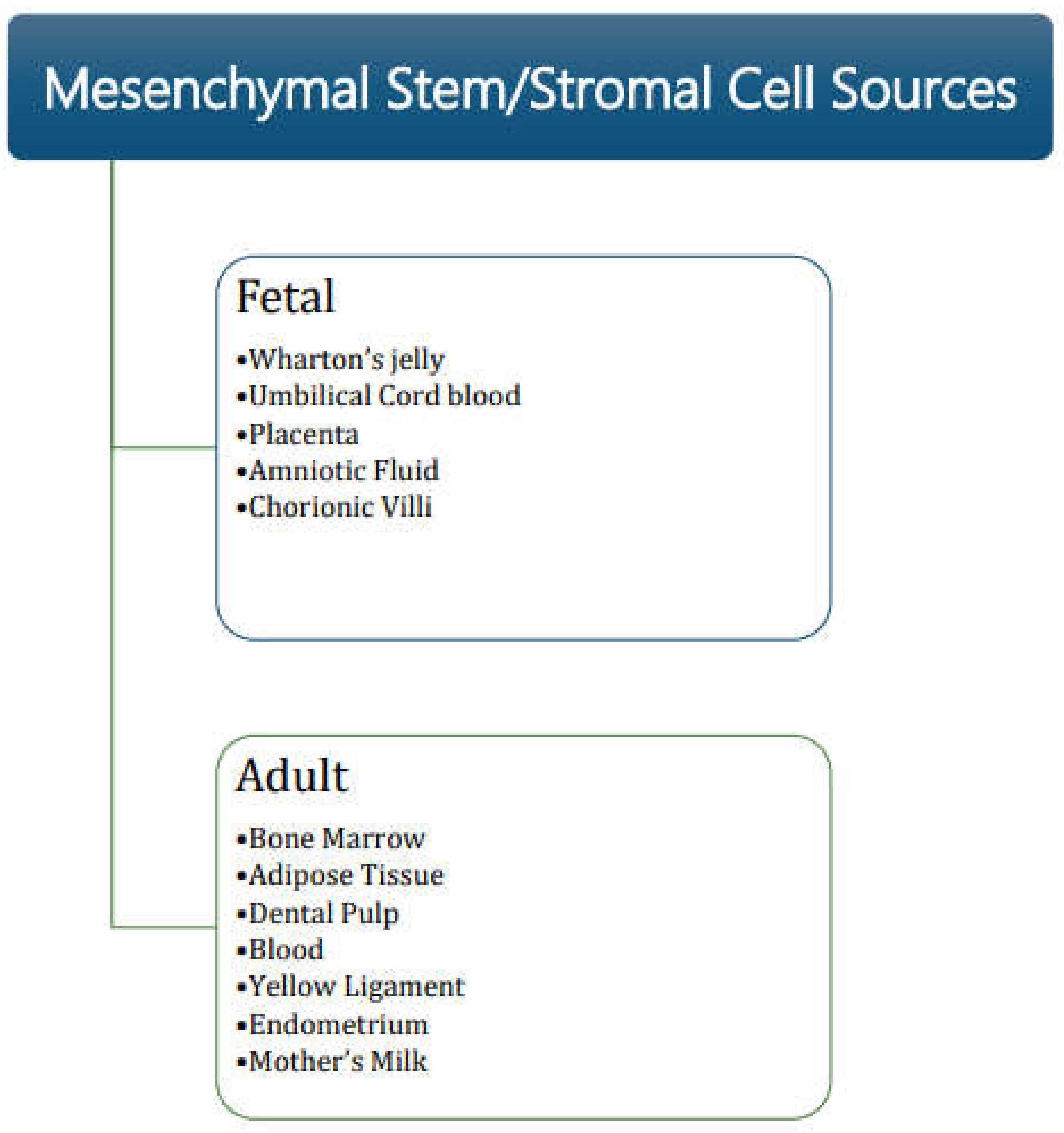
2. Properties of MSC

Historically, bone marrow was the first source from which MSC were isolated, showing their plastic adherence property. Reports of isolation of MSC from various developmental stages (including fetal, young, adult and older population) from various sources having similar properties have since been published [8,9]. Sources of fetal tissues from where MSC have been isolated include Wharton’s jelly (umbilical cord), umbilical cord blood, placenta, amniotic fluid, and chorionic villi. Adult sources of MSC include tissues and secretions such as adipose tissue, dental pulp, menstrual blood, peripheral blood, yellow ligament, endometrium, and mother’s milk [10,11,12,13,14,15,16,17,18,19,20,21,22,23] (Figure 1).
We have isolated and characterized MSC from solid adipose tissue obtained from research consented deceased donors. Our results demonstrate that an increased number of MSC can be obtained from deceased donor adipose tissue using our newly developed nonenzymatic technique as compared to the conventional enzymatic method. Deceased donor adipose tissue can be recovered during the routine deceased donor process, substantially increasing the supply and access to MSC without the pain, morbidity, and mortality associated with living donor stem cell collection [24].
The main criterion for the identification of MSC is their ability to grow in vitro as a population adhering to the substrate. These cells are phenotypically characterized by the expression of CD73, CD90, CD105 surface antigens and the lack of expression of CD4, CD34, CD14, CD11b, CD79a, CD19 or class II human leukocyte antigens (HLA II) [25,26]. Some additional markers such as stromal-1 antigen (STRO-1), vascular cell adhesion molecule (VCAM/CD106), and melanoma cell adhesion molecule (MCAM/CD146) were also identified as being useful during the isolation of MSC having multidirectional differentiation ability and high degree of clonogenicity [27,28,29].
It has been observed that the rate of proliferation of MSC differs amongst the different sources from which these cells have been isolated. MSC isolated from fetal origin tissues have a faster rate of proliferation and a greater number of in vitro passages until senescence than those isolated from adult sources [30]. A higher degree of ‘stemness’ is demonstrated by the ability of cells to create a large number of CFU-F colonies. This is a characteristic of MSC isolated from bone marrow and adipose tissues [31,32]. MSC isolated from umbilical cord blood lack the ability to differentiate into adipocytes whereas bone marrow and adipose tissue MSC have a greater tendency to differentiate to osteoblasts [33]. Age of the donor also plays an important part in the proliferation ability of MSC. A greater percentage of apoptotic cells, slower rate of proliferation, and weaker differentiation potential have been observed when MSC have been isolated from older donors [34,35,36].
The morphology of cultured MSC isolated from the same tissue can be differentiated into three sub-populations: (i) cells resembling fibroblasts with characteristic spindle-shaped proliferating cells, (ii) multi-granular large flat cells with a clearly marked cytoskeleton, and (iii) small round cells having self-renewal capacity [37]. Thus, the morphology of MSC suggests that these cells are multipotent having multi-directional differentiation potential which is the most critical characteristic of MSC. The differentiation of MSC into different end-stage lineage cells highly depends on the tissue source. Bone-marrow-derived MSC have the greatest capacity to differentiate into the three mesenchymal cell lineages (osteoblast, chondrocyte, and adipocyte) [38]. On the other hand, adipose-derived MSC have been shown to differentiate into cardiomyocytes, hepatocytes, and islet cells [39,40,41]. Umbilical cord blood-derived MSC have a biological advantage over MSC isolated from other tissues. They have the ability of large-scale expansion, high anti-inflammatory effects, significant retardation of senescence, and capacity of longer culture times. These properties enable umbilical cord blood MSC to differentiate into neuronal cells, cardiomyocytes, endothelial cells, skeletal cells, and the three mesenchymal cell lineages (osteoblast, chondrocyte, and adipocyte). MSC from liver have a strong differentiation potential towards chondrogenic and osteogenic lineages, but less towards adipogenic lineages. MSC isolated from Wharton’s jelly have been successfully differentiated into endothelial cells after the addition of vascular endothelial growth factor (VEGF) and skeletal muscle lineages such as bone, cartilage, and skeletal muscle cells [42,43,44]. Thus, selecting the source of MSC for clinical transplantation depends on the capacity of MSC to differentiate towards certain lineages that is relevant to the specific transplantation site. We have shown the ability of adipose-derived MSC, obtained from research consented deceased donors by a novel nonenzymatic technique, to successfully differentiate into beta cells. These cells demonstrate secretion of c-peptide and insulin under glucose challenge thus confirming their functionality. The availability of an abundant supply of adipose-derived MSC from deceased donors and their subsequent differentiation into functional beta cells renders them as a promising cellular therapeutic approach for treating patients with type 1 diabetes (T1D) [45].

3. Functions of MSC in Organ Transplantation

MSC have unique functional properties that have been used to enable better engraftment and acceptance of the transplanted organ (Figure 2).

3.1. Paracrine Signaling

The resolution of adult tissue damage is a complex process that is not likely resolved by MSC alone. Harnessing the ability of MSC to produce factors and cytokines that can modulate immune responses and inflammation, as well as stimulate tissue repair, has been the focus of many recent studies. Clinical trials are ongoing to test how to utilize the paracrine activity of MSC in addition to the ability to differentiate to mesenchymal lineages. The paracrine secretions of MSC, commonly referred to as ‘secretome’ have the ability to support regenerative processes in damaged tissues. The secreted factors have a distinct impact on various regulatory processes [46,47]. The major components that make up the MSC secretome include growth factors and cytokines. Some of the growth factors secreted by MSC include VEGF, bFGF, NGF, TGF, and KGF [48,49,50]. During the process of regeneration, these growth factors help in the reduction of fibrosis of tissues. Growth factors secreted by MSC having an anti-apoptotic effect include HGF, CICN-3, TIMP-1, TIMP-2, and BDNF [51,52,53,54]. Factors in the secretome that stimulate the proliferation of cells include NGF, bFGF, IGFBP-1, and M-CSF. Chemokines secreted by MSC have the function of blocking or stimulating cell chemotaxis and include CCL5 (RANTES), CCL8 (MCP-2), and CXCL12 (SDF-1) [55]. Factors promoting angiogenesis in the secretome include VEGF, whereas anti-angiogenesis factors such as IFN are also present within the secretome. MSC secretome also contains factors that have anti-bacterial, anti-viral, and anti-parasitic activity [56,57,58,59]. It has been shown that some of the factors within the MSC secretome have anti-cancer activity and interact with cancer cells to reduce their proliferation, migration, and viability [60]. We have identified various cytokines, growth factors, and signaling proteins in the supernatant of MSC grown in tissue culture flasks. Maximum secretion of these factors was observed on day 4 of MSC culture that decreased by day 6 culture. The paracrine secretions of MSC were also observed in the secretome that separates as a byproduct during the isolation of adipose-derived MSC. Further studies are needed to evaluate the levels of these factors secreted among different donor age groups and their ability to enhance the tissue repair functionality of MSC (recent, unpublished observations).
In recent years, the paracrine activity of MSC has expanded to include a rapidly developing field of secreted extracellular vesicles (EVs). These include apoptotic bodies, microvesicles, and exosomes. These EVs play an important role in the regulation of immune response, homeostasis, and other biological functions. The EVs have a composition that coincides with the cellular components of MSC from which they originate [61,62,63]. Supernatants from the in vitro culture of MSC have been shown to protect other cells from apoptosis, have immunomodulatory effects, induce proliferation of cells, stimulate angiogenesis, prevent fibrosis of tissues, and induce stem cell differentiation [64,65,66].

3.2. Immunomodulation

MSC have a major advantage in regulating the immune response due to their ability to secrete various regulatory factors. All these multiple factors acting in unison enable the immune-modulatory and anti-inflammatory properties of MSC thus acting as a master regulator of the immune system. Both primary and acquired immune responses are regulated by the interaction of MSC with various effector cells [67]. The anti-inflammatory properties of MSC include blocking the differentiation of CD34+ cells into mature dendritic cells using secreted factors as well as direct contact of MSC with dendritic cells [68], therefore limiting the mobilization of the mature dendritic cells to the tissues [69]. T-cell proliferation is inhibited by IL-10 that is secreted by MSC while the pro-inflammatory M1 macrophages are transformed into anti-inflammatory M2 macrophages phenotype by interaction with MSC [70]. In vitro studies have shown that MSC suppress the activation of CD4+ T cells and CD8+ B cells [71], reduce levels of pro-inflammatory TNF and IFN cytokines, and increase the synthesis of anti-inflammatory IL-4 cytokines [72]. MSC are able to skew the maturing immune cell populations by increasing the levels of regulatory T cells (Treg), anti-inflammatory Th2 cells, and dendritic DC2 cells, while reducing the levels of pro-inflammatory Th1 cells, DC1 cells, and NK cells [73]. The synthesis of immunoglobulins such as IgM, IgG, and IgA that are secreted by B cells and their differentiation to plasma cells can be blocked by MSC, reducing the levels of circulating antibodies. The ability of B cells to migrate is also negatively affected by MSC as they can reduce the expression of chemokine receptors on the surface of B cells [74].
Among the immunomodulatory properties, MSC are able to block apoptosis of activated neutrophils, limit their mobilization to the area of damage, and reduce their binding to vascular endothelial cells [75]. MSC also inhibit the complement-mediated effects of peripheral blood mononuclear cell proliferation [76]. Pro-inflammatory cytokines secreted by MSC are able to limit mast cell degranulation and migration towards chemotactic factors and stimulate neutrophil chemotaxis [77,78]. MSC also block the proliferation of induced NK cells and aid in the reduction of the cytotoxic activity of NK cells [79].

3.3. Transplant Tolerance

Organ transplantation is the standard of care for end-stage organ failure patients [80]. While conventional immunosuppression protocols have improved short-term outcomes of transplant recipients, the long-term outcomes are not as favorable, leading to chronic rejection and death. In addition, prolonged immunosuppression leads to an increase in side effects such as malignancies, infections, toxicities, and other metabolic diseases. The survival rate for a functioning heart, liver, and kidney is approximately 50%, while that for lung is only 30%, ten years post-transplantation [81]. The most desirable outcome of transplantation, often described as the “Holy Grail” of transplantation, is the establishment of transplant tolerance. Tolerance in the setting of organ and tissue transplantation would abrogate the need for chronic immunosuppression, eliminate drug-related side effects, and extend organ half-life thereby addressing the critical shortage of organ availability for transplant [82].
After organ or tissue transplantation, interplay between the host immune system and the transplanted organ has led to the development of T-cell immunosuppressive agents which has abated the risk of acute rejection. To achieve long-term graft survival, the identification of innovative strategies that lead to allograft tolerance should be developed. MSC are a heterogeneous population of non-hematopoietic cells that are able to differentiate into tissues of mesodermal lineages. In the pursuit of transplant tolerance induction, MSC seem a very promising cellular therapy for the minimization or even discontinuation of lifelong immunosuppression. MSC can dampen the activation of cells of both the adaptive and innate immune systems, reprogramming them into regulatory cells inhibiting the immune alloresponse at different levels. Infusion of MSC has proven to be effective in prolonging graft survival and controlling autoimmunity [83]. MSC reduce the host-vs-graft response by secreting soluble immunomodulatory factors through paracrine mechanisms and through contact-dependent regulation [84,85] tipping the balance of alloresponse from effector to regulatory function. Therefore, acute and chronic alloimmune response can be reduced using MSC that not only target T cells which are the main players of alloimmunity, but also regulate B cells, dendritic cells, and macrophages.
Autologous and allogeneic MSC have been shown to regulate the T-cell response to the transplanted organ by suppressing T-cell cytotoxicity and proliferation caused by antigen priming and polyclonal activators [86]. The prevalence of a high frequency of alloreactive memory T cells before transplantation is a barrier to tolerance induction, especially in the context of T-cell depletion therapy, and has a deleterious effect on allograft survival. Human autologous and allogeneic MSC have been shown to suppress and inhibit these memory T cells, including CD8+ memory T cells, leading to allograft survival [87,88]. MSC are able to modulate the activity of helper T cells by suppressing the proliferation of Th1 cells and rewiring their polarization to the favorable Th2 cells. This shift in the polarization of helper T cells also helps in an increase in the secretion of IL-10 and other Th2 specific cytokines [89]. In addition to the suppressing effector T cells, MSC have been shown to expand a pool of regulatory T cells thus modulating the immunological response towards the transplanted organ. Human MSC promote the generation of FoxP3+ regulatory T cells (TREG) and induce tolerance to the allograft by mediating immunosuppression in vivo [85,90].
B cells are known to secrete antibodies that bind to the donor organ leading to antibody-mediated rejection (AMR). MSC have been shown to inhibit the formation of donor-specific antibodies (DSA) [91] and block B-cell proliferation through cell cycle arrest [74]. Inhibition of the proliferation of B cells by MSC prevents them from maturing into plasmablasts causing a steep decline in antibody secretion [92]. MSC infusion modulates the humoral response in vivo by expanding the number of regulatory B cells (BREG) along with IL-10 production [93]. Spontaneous operational tolerance has been shown in kidney allograft recipients receiving MSC infusion through the increase in ‘transitional’ B-cell subsets [62,94].
Dendritic cells (DC) are responsible for the direct alloantigen presentation to CD4+ T cells and cross-presentation of allopeptides to CD8+ T cells. Exposure to MSC interferes with DC proliferation, maturation, impairs DC homing to secondary lymphoid organs, and downregulates MHC Class II and costimulatory molecules [95]. This has a direct effect on alloantigen presentation on both recipient and donor DC leading to the inhibition of alloresponse and the induction of regulatory phenotype by increasing the abundance of TREG compared to effector T cells. Kidney transplant recipients receiving MSC infusion showed an increased suppressive TREG population in the graft and secondary lymphoid organs, impaired donor-specific T-cell proliferation, and a high frequency of immature tolerogenic DC [96].
Macrophage proliferation and migration have been shown to increase in the presence of MSC along with a pro-tolerogenic shift in polarization of the macrophages to the M2 phenotype [97]. In a corneal transplant model, infusion of MSC conferred protection towards allograft rejection by redirecting the macrophages towards the M2 phenotype [98]. The macrophage M2 is a pro-tolerogenic phenotype that reduces TNF, IFN, and IL-12 secretion and increases the production of IL-10 that promotes TREG proliferation and inhibits effector T-cell responses [70].

3.4. Timing of MSC Infusion to Develop Transplant Tolerance

The lifespan of MSC is limited both in tissue culture system in vitro and after in vivo administration raising the question of the optimal timing for MSC infusion in transplant recipients. Development of long-term tolerance to the transplanted organ after a single infusion of MSC would only be possible if multilevel protolerogenic effects could be developed in the short period of time the MSC are actively engaging with their in vivo environment [62,99]. MSC infusion in rats carried out for four days before heart transplantation induced transplant tolerance and acceptance of the transplanted heart. However, rejection of the transplanted heart was observed when MSC infusion was performed three days after performing the heart transplant [63,100]. Timing of MSC infusion may also impact their localization. MSC have been shown to migrate to the graft rather than to the secondary lymphoid organs after transplantation. This difference in the localization dictates the immunomodulatory properties of MSC. Migration of MSC to the transplanted organ after post-transplant infusion leads to the stimulation of proinflammatory phenotype characterized by complement deposition, neutrophil infiltration, and graft rejection. On the other hand, pre-transplant infusion leads to the localization of MSC to the secondary lymphoid organs promoting the protolerogenic effects and prolonged graft survival [101]. Furthermore, the infusion of MSC post-transplant was unable to convert conventional T cells to the immunoregulatory TREG population and reduce the inhibitory effect of MSC on DC maturation, therefore decreasing the immunomodulatory ability of MSC [102].

3.5. Minimization of Immunosuppressive Drugs

Development of a pro-tolerogenic environment and repair of chronic allograft damage by MSC therapy in kidney transplantation has enabled the minimization of induction and maintenance immunosuppressive drugs [103,104]. A clinical trial showed that induction therapy using basiliximab could be replaced safely with the administration of BM-MSC infusion, resulting in a 50% reduction of tacrolimus used as maintenance therapy. Patients receiving MSC therapy displayed incidence of acute rejection, graft survival and function, similar to the patients receiving a full dose of tacrolimus maintenance immunosuppression [105,106]. In another clinical trial, the timing of BM-MSC infusion was shown to be critical in living donor kidney transplant recipients. Patients were given an autologous MSC infusion a day before kidney transplantation. At first, basiliximab was removed from the induction therapy to avoid inhibition of TREG expansion by this drug. There was no acute renal insufficiency, but the absence of this drug led to an increased risk of acute rejection. The protocol was modified and basiliximab induction therapy was re-introduced in the pre-transplant MSC-infused patients. The 5–7-year follow-up showed stable graft function with no major side effects. Extensive longitudinal immunological studies in these patients displayed a long-lasting increase in the levels of TREG cells compared to the CD8+ T cells and persistent reduction of T-cell cytotoxicity. In one of the patients showing no evidence of de novo donor-specific antibodies and normal histology at 1-year post-transplant, gradual tapering and withdrawal of immunosuppressive drugs cyclosporin and mycophenolate mofetil led to the patient being free from immunosuppression and stable graft function [103,104,106]. Thus, MSC therapy has shown to create a pro-tolerogenic environment and complement the tolerogenic potential of induction therapies for prevention of acute graft rejection. Identification of MSC therapy response biomarkers would aid in the selection of patients who are amenable to safe immunosuppressive drug withdrawal.

4. Regenerative Approach as ‘Bridge to Transplant’

The field of transplant medicine has seen major advances in surgical techniques and immunosuppressive therapy saving lives through organ and tissue transplants. However, according to the Health Resources and Services Administration (HRSA), there is a major imbalance between organ supply and demand. Over 100,000 people are on the national transplant waitlist; with a patient being added to this list every 9 minutes, while 17 people die each day while waiting for a life-saving transplant. Of the limited number of eligible donors, less than 1% of all deaths meet the specific medical criteria to be a donor [107]. Additional factors posing a barrier include a narrow timeframe of organ viability after procurement, inadequate organ preservation/storage, and complex transplant logistics. In the past decade, the fields of regenerative medicine (RM) and tissue engineering have emerged offering different approaches and novel strategies as a ‘bridge to transplant’ to overcome these hurdles and tackle unsolved problems [108]. Transplant medicine has benefited a great deal from the various aspects of RM as they offer flexibility and planning of transplantation, lower immunogenicity, generation of universally accepted ‘off-the-shelf organs-on-demand’, increase in organ utilization, expansion of the donor pool, reduction in disparities, and enabling faster access to transplantation [109] (Figure 3).

4.1. Organ Preservation

Soon after organ procurement, the lack of blood supply leads to oxygen deprivation, electrolyte imbalance, anaerobic respiration, and metabolic waste accumulation leading to ischemic injury. Reperfusion of the organ leads to epithelial cell damage and generation of reactive oxygen species (ROS), aggravating the already damaged organ called reperfusion injury. Prolonged ex vivo preservation of organs and tissues has been made possible by the development of machine perfusion techniques. Organ perfusion using an oxygenated cold preservation solution (hypothermic oxygenated perfusion) focuses on slowing down metabolic rates [110,111], whereas ex vivo perfusion at physiological temperature mimics the in vivo environment (normothermic perfusion) [112]. Both these approaches allow for the measurement of predictive biomarkers including but not limited to neutrophil gelatinase-associated lipocalin (NGAL), liver-type fatty acid binding protein 1, kidney injury molecule-1 (KIM-1), endothelin-1, and (L-FABP) micro ribonucleic acids (miRNAs) that give valuable information of organ quality [113]. Machine perfusion offers a perfect platform for the delivery of treatments solely to the graft, bypassing complications arising from systemic delivery. This is beneficial for ECD and DCD donation, facilitating organ storage, transportation, tissue repair, and organ regeneration [114]. Delivery of stem cells using machine perfusion as a treatment option is gaining importance. Several preclinical studies are being conducted to investigate the effect of paracrine factors secreted by MSC and MSC-derived extracellular vesicles in kidney and liver perfusion [115,116]. Results demonstrated that there was significantly less global ischemic damage in DCD rat kidneys that were perfused with MSC or MSC-derived extracellular vesicles [117,118].

4.2. Decellularization and Recellularization

The process of isolation of the extracellular matrix (ECM) from any given tissue or organ maximizing the removal of cellular debris with minimal loss, damage, or disruption to the tissue or organ, is known as decellularization. DNA content in the decellularized tissue is reduced to less than 4% while structural proteins such as collagen, laminin, and fibronectin remain in the scaffold [119,120]. The decellularized scaffold can influence cell migration, proliferation, and differentiation, and therefore serves as more than just a framework for cells [121]. The use of decellularized scaffolds for organ bioengineering has focused on organs such as heart, lungs, kidney, liver, and pancreas [122]. Sufficient decellularization is essential to reduce immunogenicity [123]. The final goal of decellularization is to ultimately repopulate the scaffold with patient-specific pluripotent cells to generate a personalized ‘organ-on-demand’. This process of recellularization necessitates seeding of terminally differentiated somatic cells or stem cells onto the scaffold via perfusion of cells through the vasculature, ureter, or trachea, and further maturation in special bioreactors [124,125,126]. Neo-bladders were generated by Atala et al. [127] by first decellularized bladder submucosa followed by recellularization with donor-derived muscle and urothelial cells. After ex vivo culture, these bioengineered bladders were implanted in patients who showed no postoperative complications and normal bladder function. Recellularization of a whole organ remains a challenge due to the need of seeding and precise positioning of different cell types, thromboembolic events during perfusion, inadequate supply of oxygen and nutrients, and improper vascularization of recellularized organs [122]. Preclinical studies in mice have shown promising results. Decellularized mouse hearts that were repopulated with induced pluripotent stem cell (iPSC)-derived cardiovascular cells exhibited spontaneous contractions after 20 days [128]. Repopulation of lung scaffolds with lung epithelial cells and vascular endothelial cells showed survival and differentiation of the epithelium and clearance of secretions [126]. Bioengineered mouse kidneys prepared after decellularization followed by recellularization with human umbilical vein endothelial cells (HUVEC), neonatal kidney cells, and mouse embryonic stem cells, displayed increased creatinine clearance, albumin retention, and increased reabsorption of glucose and electrolyte [125]. Seeding of hepatic stem cells into decellularized liver matrix exhibited markers of hepatocyte and cholangiocytic differentiation and high engraftment rates [129].

4.3. Organoids

Most two-dimensional studies carried out in vitro fail to replicate the in vivo interactions among cells and between cells and the extracellular matrix. The development of 3D culture systems allowed the mimicking of in vivo conditions involving interactions between cells and the surrounding matrix leading to the dynamic regulation of signaling pathways and paracrine signals [130]. Organoids are 3D structures created in this culture system typically originating from stem cells having multiple cell types that self-organize in culture [131]. They are miniaturized version of organs having native architecture and morphology, and display several biological interactions that occur in vivo [132]. Several controlled parameters are used to induce iPSCs to differentiate into specific lineages to form tissue specific organoids. These include endogenous and exogenous signals that stimulate the differentiation of iPSCs and self-organization into the 3D structures. Organoids can also be developed by seeding differentiated stem cells along with endothelial cells and MSC in combination to form self-assembled 3D structures [133]. At present, organoid technology is used for drug screening and disease modeling. However, the ultimate goal is to evaluate the transplantation of tissue-specific organoids and access their engraftment, biocompatibility, and tissue specific functionality in vivo. Most of the studies on the efficacy of transplantation of organoids is carried out using nude mouse model. Kidney organoids derived from human pluripotent stem cell (hPSC) differentiation were transplanted under the renal capsule of immunodeficient mice. These organoids exhibited glomerular vascularization and connection with preexisting host vascular networks, functional glomerular perfusion, maturation of podocytes, and tubular reabsorption [134].
Immune deficiency caused by the destruction of pancreatic beta cells leads to the development of type 1 diabetes (T1D). Pancreatic organoids have been developed by differentiation of hPSC into acinar and ductal cells, and inducing them to self-organize into pancreatic organoids. These organoids can express pancreatic markers and are functionally and structurally similar to the pancreas. Pancreatic organoids were placed in a 3D-printed tissue trapper and implanted into the peritoneal cavity of immunodeficient mice. Results indicated that the implanted organoids exhibited engraftment, neo-vascularization, an increased number of insulin-positive cells, and improved c-peptide secretion suggesting their applicability in the treatment of T1D [135].
Liver organoids were formed by inducing the coculture of hPSC, MSC, and HUVEC to self-organize into 3D structures resembling liver buds. These liver organoids, when transplanted into nude mice, displayed functional vascularization, connections among donor and host cells, and drug metabolism activity; all essential functional components of liver function [133]. However, there are practical challenges to the use of organoids for transplantation purposes. Organoids are typically 10 mm to 1 mm in diameter. The small size of organoids is a major concern for their use as a substitute for larger organs such as the kidney. Production of bigger organoids developed using more precursor cells or assembling large number of organoids could possibly alleviate this issue [136]. Most of the studies rely on transplantation studies carried out in immunodeficient mouse model. Translation of these studies into human trials to establish the use of organoids as medical devices to replace or improve organ function is far from reality at present. Use of larger humanized animal models for preclinical studies could help overcome this limitation [137]. Tracking the ongoing in vivo engraftment, vascularization, behavior, and function of transplanted organoids is an important aspect of organoid transplantation technology. The use of iPSC expression fluorescent biosensors could help in creating an informative tracking system [138].

4.4. 3D Bioprinting

One of the latest approaches to meet the increasing demand of organs for critical human organ transplantation is the use of 3D bioprinting technology. Bioprinting is an additive fabrication process that uses layer-by-layer addition of living cells and growth factors suspended in an appropriate bioink to create three-dimensional structures [139]. There are three basic steps in the 3D bioprinting process: model the 3D structure of tissue or organ using a computer modelling program, printing the 3D structure using bioink, and post-processing assessment of the physical, mechanical, and biological functions before transplanting into patients. Recent advances in 3D bioprinting have the potential of meeting the demands of tissues and organs for transplant [140]. The critical components of the 3D bioprinting process are the selection of a suitable bioink and appropriate cell type selection. The bioink material should provide appropriate growth and adhesion factors, signaling proteins, and mechanical and structural properties of the extracellular cell matrix [141]. Cell selection for 3D organ bioprinting involves the selection of cells possessing proliferation and differentiation capacity in the printed scaffold. These cells should interact with signaling molecules and be able to survive and remain viable during and after the printing process [142]. Recent advances in the field of 3D bioprinting have gained importance as this process can improve the quality of life and also save lives of patients. Burn victims are greatly benefited by the use of 3D bioprinting technology through the development of 3D-printed artificial skin that contains multiple cell types such as keratinocytes, melanocytes, and fibroblasts layered on a desired scaffold [143]. Restoration of damaged cartilage has been possible by incorporating 3D printing technology using cartilaginous tissue scaffolds with chondrocytes, MSC, and bone marrow cells [144]. Using a CT scan of the patient, 3D-printed bone implants using MSC-differentiated osteoblasts were designed to perfectly fit the broken bone fragment [144]. Three-dimensional bioprinting of the heart involves the initial CT scan of the donor’s heart followed by using stem cells, growth factors, and appropriate bioink mixed with hydrogel. The scaffold that is used to layer the bioink provides mechanical support and the exact shape of the patient’s heart where the cells grow and proliferate. The synchronous beating of the cells is soon observed and the scaffold is then ready for implantation [145].

5. Conclusions and Future Perspectives

MSC have been recognized as a major player in the field of transplant medicine as they play an active role in homeostasis of tissues and organs. They are the driving force behind developing new technologies in biomedical research as they display their therapeutic effects through their ability to differentiate into different tissue types and stimulate the regeneration of damaged tissues. Although MSC compose a negligible fraction of cells in vivo, they can be isolated from various tissues and body fluids and subject to in vitro expansion and storage, known in clinical terms as biomanufacturing and biobanking. The critical importance of paracrine effects of MSC are now being recognized and advocated to explain their functional benefits. MSC secrete many different growth factors and cytokines that have immunomodulatory properties, regulate inflammation and migration of cells to the damaged tissue, influence wound healing, promote angiogenesis, and protect host cells from apoptosis. These complex paracrine mechanisms lead to improved quality and functionality of tissue repair. MSC cell therapy is especially impactful for transplant patients as they shift the risk:benefit ratio by inducing immune tolerance thus alleviating the burden of lifelong immunosuppression, associated morbidity, and chronic rejection, significantly improving transplant outcomes.
Maximizing the use of available tissue and organs has been achieved by focusing on new technologies in tissue engineering and regenerative medicine. Fabrication of new organs by optimizing discarded donor organs as a source of organ scaffold using machine perfusion and decellularization techniques is gaining importance. Recellularization of tissue or organ scaffolds using 3D printing technology employing sequential layering of a mixture of lineage-specific differentiated cells and appropriate bioink has shown promising results. Three-dimensional culture systems have been developed that allow self-aggregation of MSC, endothelial cells, and terminally differentiated cells to produce organoids that reproduce the structural complexity of a real organ. Organoids have differentiated and functional cells capable of interacting with host cells. Organoids have also been used as promising tools for drug screening, disease modeling, and personalized medicine thus reducing whole organ transplant requirement.
In summary, it appears that MSC are becoming a powerful tool in the field of transplant medicine by increasing organ preservation, utilization, and immune tolerance. Increase in the scientific knowledge and clinical applications of MSC is beginning to contribute towards the transformation of the therapeutic applications in organ and tissue transplantation. Major discoveries in the tissue engineering and regenerative medicine fields, originally thought as ‘illusions’ or ‘science fiction’, have enabled significant advances of truly disruptive approaches and technologies that could forever change the field of transplantation as we know it today. It would be prudent to speculate the next milestone in tissue engineering and regenerative medicine: the transplantation of an artificial, bioengineered functioning whole organ in humans.

Author Contributions

D.D. performed the literature search, wrote, and revised the manuscript. M.M. and P.R. critically revised the manuscript. All authors have read and agreed to the published version of the manuscript.

Funding

This research received no external funding.

Institutional Review Board Statement

Not applicable.

Informed Consent Statement

Not applicable.

Conflicts of Interest

The authors declare no conflict of interest.

References

  1. Nathan, H.M.; Conrad, S.L.; Held, P.J.; McCullough, K.P.; Pietroski, R.E.; Siminoff, L.A.; Ojo, A.O. Organ donation in the United States. Am. J. Transplant. 2003, 3 (Suppl. S4), 29–40. [Google Scholar] [CrossRef] [PubMed]
  2. Transplant Trends. Available online: https://unos.org/data/transplant-trends/ (accessed on 23 February 2022).
  3. Pileggi, A.; Xu, X.; Tan, J.; Ricordi, C. Mesenchymal stromal (stem) cells to improve solid organ transplant outcome: Lessons from the initial clinical trials. Curr. Opin. Organ. Transplant. 2013, 18, 672–681. [Google Scholar] [CrossRef] [PubMed] [Green Version]
  4. Buron, F.; Perrin, H.; Malcus, C.; Hequet, O.; Thaunat, O.; Kholopp-Sarda, M.N.; Moulin, F.T.; Morelon, E. Human mesenchymal stem cells and immunosuppressive drug interactions in allogeneic responses: An in vitro study using human cells. Transplant. Proc. 2009, 41, 3347–3352. [Google Scholar] [CrossRef] [PubMed]
  5. Badylak, S.F.; Freytes, D.O.; Gilbert, T.W. Extracellular matrix as a biological scaffold material: Structure and function. Acta Biomater. 2009, 5, 1–13. [Google Scholar] [CrossRef]
  6. Gao, G.; Cui, X. Three-dimensional bioprinting in tissue engineering and regenerative medicine. Biotechnol. Lett. 2016, 38, 203–211. [Google Scholar] [CrossRef]
  7. Porzionato, A.; Stocco, E.; Barbon, S.; Grandi, F.; Macchi, V.; De Caro, R. Tissue-Engineered Grafts from Human Decellularized Extracellular Matrices: A Systematic Review and Future Perspectives. Int. J. Mol. Sci. 2018, 19, 4117. [Google Scholar] [CrossRef] [Green Version]
  8. Liu, J.; Yu, F.; Sun, Y.; Jiang, B.; Zhang, W.; Yang, J.; Xu, G.T.; Liang, A.; Liu, S. Concise reviews: Characteristics and potential applications of human dental tissue-derived mesenchymal stem cells. Stem Cells 2015, 33, 627–638. [Google Scholar] [CrossRef]
  9. Muhammad, G.; Jablonska, A.; Rose, L.; Walczak, P.; Janowski, M. Effect of MRI tags: SPIO nanoparticles and 19F nanoemulsion on various populations of mouse mesenchymal stem cells. Acta Neurobiol. Exp. 2015, 75, 144–159. [Google Scholar]
  10. Girdlestone, J.; Limbani, V.A.; Cutler, A.J.; Navarrete, C.V. Efficient expansion of mesenchymal stromal cells from umbilical cord under low serum conditions. Cytotherapy 2009, 11, 738–748. [Google Scholar] [CrossRef]
  11. Erices, A.; Conget, P.; Minguell, J.J. Mesenchymal progenitor cells in human umbilical cord blood. Br. J. Haematol. 2000, 109, 235–242. [Google Scholar] [CrossRef]
  12. Zeddou, M.; Briquet, A.; Relic, B.; Josse, C.; Malaise, M.G.; Gothot, A.; Lechanteur, C.; Beguin, Y. The umbilical cord matrix is a better source of mesenchymal stem cells (MSC) than the umbilical cord blood. Cell Biol. Int. 2010, 34, 693–701. [Google Scholar] [CrossRef]
  13. Miao, Z.; Jin, J.; Chen, L.; Zhu, J.; Huang, W.; Zhao, J.; Qian, H.; Zhang, X. Isolation of mesenchymal stem cells from human placenta: Comparison with human bone marrow mesenchymal stem cells. Cell Biol. Int. 2006, 30, 681–687. [Google Scholar] [CrossRef]
  14. Marongiu, F.; Gramignoli, R.; Sun, Q.; Tahan, V.; Miki, T.; Dorko, K.; Ellis, E.; Strom, S.C. Isolation of amniotic mesenchymal stem cells. Curr. Protoc. Stem Cell Biol. 2010. Chapter 1, Unit 1E 5. [Google Scholar] [CrossRef]
  15. Roubelakis, M.G.; Pappa, K.I.; Bitsika, V.; Zagoura, D.; Vlahou, A.; Papadaki, H.A.; Antsaklis, A.; Anagnou, N.P. Molecular and proteomic characterization of human mesenchymal stem cells derived from amniotic fluid: Comparison to bone marrow mesenchymal stem cells. Stem Cells Dev. 2007, 16, 931–952. [Google Scholar] [CrossRef] [Green Version]
  16. Poloni, A.; Rosini, V.; Mondini, E.; Maurizi, G.; Mancini, S.; Discepoli, G.; Biasio, S.; Battaglini, G.; Berardinelli, E.; Serrani, F.; et al. Characterization and expansion of mesenchymal progenitor cells from first-trimester chorionic villi of human placenta. Cytotherapy 2008, 10, 690–697. [Google Scholar] [CrossRef]
  17. Katz, A.J.; Tholpady, A.; Tholpady, S.S.; Shang, H.; Ogle, R.C. Cell surface and transcriptional characterization of human adipose-derived adherent stromal (hADAS) cells. Stem Cells 2005, 23, 412–423. [Google Scholar] [CrossRef]
  18. Agha-Hosseini, F.; Jahani, M.A.; Jahani, M.; Mirzaii-Dizgah, I.; Ali-Moghaddam, K. In vitro isolation of stem cells derived from human dental pulp. Clin. Transplant. 2010, 24, E23–E28. [Google Scholar] [CrossRef]
  19. Macias, M.I.; Grande, J.; Moreno, A.; Dominguez, I.; Bornstein, R.; Flores, A.I. Isolation and characterization of true mesenchymal stem cells derived from human term decidua capable of multilineage differentiation into all 3 embryonic layers. Am. J. Obstet. Gynecol. 2010, 203, 495.e9–495.e23. [Google Scholar] [CrossRef]
  20. Kassis, I.; Zangi, L.; Rivkin, R.; Levdansky, L.; Samuel, S.; Marx, G.; Gorodetsky, R. Isolation of mesenchymal stem cells from G-CSF-mobilized human peripheral blood using fibrin microbeads. Bone Marrow Transplant. 2006, 37, 967–976. [Google Scholar] [CrossRef] [Green Version]
  21. Chen, Y.T.; Wei, J.D.; Wang, J.P.; Lee, H.H.; Chiang, E.R.; Lai, H.C.; Chen, L.L.; Lee, Y.T.; Tsai, C.C.; Liu, C.L.; et al. Isolation of mesenchymal stem cells from human ligamentum flavum: Implicating etiology of ligamentum flavum hypertrophy. Spine 2011, 36, E1193–E1200. [Google Scholar] [CrossRef]
  22. Meng, X.; Ichim, T.E.; Zhong, J.; Rogers, A.; Yin, Z.; Jackson, J.; Wang, H.; Ge, W.; Bogin, V.; Chan, K.W.; et al. Endometrial regenerative cells: A novel stem cell population. J. Transl. Med. 2007, 5, 57. [Google Scholar] [CrossRef] [PubMed] [Green Version]
  23. Patki, S.; Kadam, S.; Chandra, V.; Bhonde, R. Human breast milk is a rich source of multipotent mesenchymal stem cells. Hum. Cell 2010, 23, 35–40. [Google Scholar] [CrossRef] [PubMed]
  24. Rao, P.N.; Deo, D.D.; Marchioni, M.A.; Taghizadeh, R.R.; Cetrulo, K.; Sawczak, S.; Myrick, J. Structural and Functional Characterization of Deceased Donor Stem Cells: A Viable Alternative to Living Donor Stem Cells. Stem Cells Int. 2019, 2019, 5841587. [Google Scholar] [CrossRef] [PubMed]
  25. Horwitz, E.M.; Le Blanc, K.; Dominici, M.; Mueller, I.; Slaper-Cortenbach, I.; Marini, F.C.; Deans, R.J.; Krause, D.S.; Keating, A. Clarification of the nomenclature for MSC: The International Society for Cellular Therapy position statement. Cytotherapy 2005, 7, 393–395. [Google Scholar] [CrossRef]
  26. Dominici, M.; Le Blanc, K.; Mueller, I.; Slaper-Cortenbach, I.; Marini, F.; Krause, D.; Deans, R.; Keating, A.; Prockop, D.; Horwitz, E. Minimal criteria for defining multipotent mesenchymal stromal cells. The International Society for Cellular Therapy position statement. Cytotherapy 2006, 8, 315–317. [Google Scholar] [CrossRef]
  27. Sacchetti, B.; Funari, A.; Michienzi, S.; Di Cesare, S.; Piersanti, S.; Saggio, I.; Tagliafico, E.; Ferrari, S.; Robey, P.G.; Riminucci, M.; et al. Self-renewing osteoprogenitors in bone marrow sinusoids can organize a hematopoietic microenvironment. Cell 2007, 131, 324–336. [Google Scholar] [CrossRef] [Green Version]
  28. Simmons, P.J.; Torok-Storb, B. Identification of stromal cell precursors in human bone marrow by a novel monoclonal antibody, STRO-1. Blood 1991, 78, 55–62. [Google Scholar] [CrossRef] [Green Version]
  29. Gronthos, S.; Zannettino, A.C.; Hay, S.J.; Shi, S.; Graves, S.E.; Kortesidis, A.; Simmons, P.J. Molecular and cellular characterisation of highly purified stromal stem cells derived from human bone marrow. J. Cell Sci. 2003, 116, 1827–1835. [Google Scholar] [CrossRef] [Green Version]
  30. Hass, R.; Kasper, C.; Bohm, S.; Jacobs, R. Different populations and sources of human mesenchymal stem cells (MSC): A comparison of adult and neonatal tissue-derived MSC. Cell Commun. Signal. 2011, 9, 12. [Google Scholar] [CrossRef] [Green Version]
  31. Heo, J.S.; Choi, Y.; Kim, H.S.; Kim, H.O. Comparison of molecular profiles of human mesenchymal stem cells derived from bone marrow, umbilical cord blood, placenta and adipose tissue. Int. J. Mol. Med. 2016, 37, 115–125. [Google Scholar] [CrossRef] [Green Version]
  32. Saler, M.; Caliogna, L.; Botta, L.; Benazzo, F.; Riva, F.; Gastaldi, G. hASC and DFAT, Multipotent Stem Cells for Regenerative Medicine: A Comparison of Their Potential Differentiation In Vitro. Int. J. Mol. Sci. 2017, 18, 2699. [Google Scholar] [CrossRef] [Green Version]
  33. Kern, S.; Eichler, H.; Stoeve, J.; Kluter, H.; Bieback, K. Comparative analysis of mesenchymal stem cells from bone marrow, umbilical cord blood, or adipose tissue. Stem Cells 2006, 24, 1294–1301. [Google Scholar] [CrossRef]
  34. Zhou, S.; Greenberger, J.S.; Epperly, M.W.; Goff, J.P.; Adler, C.; Leboff, M.S.; Glowacki, J. Age-related intrinsic changes in human bone-marrow-derived mesenchymal stem cells and their differentiation to osteoblasts. Aging Cell 2008, 7, 335–343. [Google Scholar] [CrossRef] [Green Version]
  35. Phinney, D.G.; Kopen, G.; Righter, W.; Webster, S.; Tremain, N.; Prockop, D.J. Donor variation in the growth properties and osteogenic potential of human marrow stromal cells. J. Cell Biochem. 1999, 75, 424–436. [Google Scholar] [CrossRef]
  36. Pineiro-Ramil, M.; Sanjurjo-Rodriguez, C.; Rodriguez-Fernandez, S.; Castro-Vinuelas, R.; Hermida-Gomez, T.; Blanco-Garcia, F.J.; Fuentes-Boquete, I.; Diaz-Prado, S. Generation of Mesenchymal Cell Lines Derived from Aged Donors. Int. J. Mol. Sci. 2021, 22, 667. [Google Scholar] [CrossRef]
  37. Colter, D.C.; Sekiya, I.; Prockop, D.J. Identification of a subpopulation of rapidly self-renewing and multipotential adult stem cells in colonies of human marrow stromal cells. Proc. Natl. Acad. Sci. USA 2001, 98, 7841–7845. [Google Scholar] [CrossRef] [Green Version]
  38. Charbord, P.; Livne, E.; Gross, G.; Haupl, T.; Neves, N.M.; Marie, P.; Bianco, P.; Jorgensen, C. Human bone marrow mesenchymal stem cells: A systematic reappraisal via the genostem experience. Stem Cell Rev. Rep. 2011, 7, 32–42. [Google Scholar] [CrossRef] [Green Version]
  39. Neshati, V.; Mollazadeh, S.; Fazly Bazzaz, B.S.; de Vries, A.A.; Mojarrad, M.; Naderi-Meshkin, H.; Neshati, Z.; Kerachian, M.A. Cardiomyogenic differentiation of human adipose-derived mesenchymal stem cells transduced with Tbx20-encoding lentiviral vectors. J. Cell. Biochem. 2018, 119, 6146–6153. [Google Scholar] [CrossRef]
  40. Yin, L.; Zhu, Y.; Yang, J.; Ni, Y.; Zhou, Z.; Chen, Y.; Wen, L. Adipose tissue-derived mesenchymal stem cells differentiated into hepatocyte-like cells in vivo and in vitro. Mol. Med. Rep. 2015, 11, 1722–1732. [Google Scholar] [CrossRef]
  41. Gao, J.; Yuan, Y.; Chen, Y. PDX1 transfection induces human adipose derived stem cells differentiation into islet-like cells: What is the benefit for diabetic rats? Pharmazie 2018, 73, 213–217. [Google Scholar] [CrossRef]
  42. Jin, H.J.; Bae, Y.K.; Kim, M.; Kwon, S.J.; Jeon, H.B.; Choi, S.J.; Kim, S.W.; Yang, Y.S.; Oh, W.; Chang, J.W. Comparative analysis of human mesenchymal stem cells from bone marrow, adipose tissue, and umbilical cord blood as sources of cell therapy. Int. J. Mol. Sci. 2013, 14, 17986–18001. [Google Scholar] [CrossRef]
  43. Wu, K.H.; Zhou, B.; Lu, S.H.; Feng, B.; Yang, S.G.; Du, W.T.; Gu, D.S.; Han, Z.C.; Liu, Y.L. In vitro and in vivo differentiation of human umbilical cord derived stem cells into endothelial cells. J. Cell. Biochem. 2007, 100, 608–616. [Google Scholar] [CrossRef]
  44. Conconi, M.T.; Burra, P.; Di Liddo, R.; Calore, C.; Turetta, M.; Bellini, S.; Bo, P.; Nussdorfer, G.G.; Parnigotto, P.P. CD105(+) cells from Wharton's jelly show in vitro and in vivo myogenic differentiative potential. Int. J. Mol. Med. 2006, 18, 1089–1096. [Google Scholar] [CrossRef] [Green Version]
  45. Rao, P.; Deo, D.; Marchioni, M. Differentiation of Human Deceased Donor, Adipose-Derived, Mesenchymal Stem Cells into Functional Beta Cells. J. Stem Cells Regen. Med. 2020, 16, 63–72. [Google Scholar] [CrossRef]
  46. Kupcova, S. Proteomic techniquies for characterisation of mesenchymal stem cell secretome. Biochimie 2013, 95, 2196–2211. [Google Scholar] [CrossRef]
  47. Han, T.; Song, P.; Wu, Z.; Xiang, X.; Liu, Y.; Wang, Y.; Fang, H.; Niu, Y.; Shen, C. MSC secreted extracellular vesicles carrying TGF-beta upregulate Smad 6 expression and promote the regrowth of neurons in spinal cord injured rats. Stem Cell Rev. Rep. 2022, 18, 1078–1096. [Google Scholar] [CrossRef]
  48. Alvarez, D.; Levine, M.; Rojas, M. Regenerative medicine in the treatment of idiopathic pulmonary fibrosis: Current position. Stem Cells Cloning 2015, 8, 61–65. [Google Scholar] [CrossRef] [PubMed] [Green Version]
  49. Paganelli, A.; Trubiani, O.; Diomede, F.; Pisciotta, A.; Paganelli, R. Immunomodulating Profile of Dental Mesenchymal Stromal Cells: A Comprehensive Overview. Front. Oral Health 2021, 2, 635055. [Google Scholar] [CrossRef] [PubMed]
  50. Shin, S.; Lee, J.; Kwon, Y.; Park, K.S.; Jeong, J.H.; Choi, S.J.; Bang, S.I.; Chang, J.W.; Lee, C. Comparative Proteomic Analysis of the Mesenchymal Stem Cells Secretome from Adipose, Bone Marrow, Placenta and Wharton's Jelly. Int. J. Mol. Sci. 2021, 22, 845. [Google Scholar] [CrossRef] [PubMed]
  51. Zanotti, L.; Angioni, R.; Cali, B.; Soldani, C.; Ploia, C.; Moalli, F.; Gargesha, M.; D'Amico, G.; Elliman, S.; Tedeschi, G.; et al. Mouse mesenchymal stem cells inhibit high endothelial cell activation and lymphocyte homing to lymph nodes by releasing TIMP-1. Leukemia 2016, 30, 1143–1154. [Google Scholar] [CrossRef] [PubMed]
  52. Cantinieaux, D.; Quertainmont, R.; Blacher, S.; Rossi, L.; Wanet, T.; Noel, A.; Brook, G.; Schoenen, J.; Franzen, R. Conditioned medium from bone marrow-derived mesenchymal stem cells improves recovery after spinal cord injury in rats: An original strategy to avoid cell transplantation. PLoS ONE 2013, 8, e69515. [Google Scholar] [CrossRef]
  53. Gonzalez-Rey, E.; Gonzalez, M.A.; Varela, N.; O’Valle, F.; Hernandez-Cortes, P.; Rico, L.; Buscher, D.; Delgado, M. Human adipose-derived mesenchymal stem cells reduce inflammatory and T cell responses and induce regulatory T cells in vitro in rheumatoid arthritis. Ann. Rheum. Dis. 2010, 69, 241–248. [Google Scholar] [CrossRef] [Green Version]
  54. Wang, P.; Cui, Y.; Wang, J.; Liu, D.; Tian, Y.; Liu, K.; Wang, X.; Liu, L.; He, Y.; Pei, Y.; et al. Mesenchymal stem cells protect against acetaminophen hepatotoxicity by secreting regenerative cytokine hepatocyte growth factor. Stem Cell Res. Ther. 2022, 13, 94. [Google Scholar] [CrossRef]
  55. Eliopoulos, N.; Zhao, J.; Bouchentouf, M.; Forner, K.; Birman, E.; Yuan, S.; Boivin, M.N.; Martineau, D. Human marrow-derived mesenchymal stromal cells decrease cisplatin renotoxicity in vitro and in vivo and enhance survival of mice post-intraperitoneal injection. Am. J. Physiol. Renal Physiol. 2010, 299, F1288–F1298. [Google Scholar] [CrossRef] [Green Version]
  56. Meisel, R.; Brockers, S.; Heseler, K.; Degistirici, O.; Bulle, H.; Woite, C.; Stuhlsatz, S.; Schwippert, W.; Jager, M.; Sorg, R.; et al. Human but not murine multipotent mesenchymal stromal cells exhibit broad-spectrum antimicrobial effector function mediated by indoleamine 2,3-dioxygenase. Leukemia 2011, 25, 648–654. [Google Scholar] [CrossRef]
  57. Xu, R.; Feng, Z.; Wang, F.S. Mesenchymal stem cell treatment for COVID-19. EBioMedicine 2022, 77, 103920. [Google Scholar] [CrossRef]
  58. Navard, S.H.; Rezvan, H.; Haddad, M.H.F.; Ali, S.A.; Nourian, A.; Eslaminejad, M.B.; Behmanesh, M.A. Therapeutic effects of mesenchymal stem cells on cutaneous leishmaniasis lesions caused by Leishmania major. J. Glob. Antimicrob. Resist. 2020, 23, 243–250. [Google Scholar] [CrossRef]
  59. Harrell, C.R.; Popovska Jovicic, B.; Djonov, V.; Volarevic, V. Molecular Mechanisms Responsible for Mesenchymal Stem Cell-Based Treatment of Viral Diseases. Pathogens 2021, 10, 409. [Google Scholar] [CrossRef]
  60. Attar-Schneider, O.; Zismanov, V.; Drucker, L.; Gottfried, M. Secretome of human bone marrow mesenchymal stem cells: An emerging player in lung cancer progression and mechanisms of translation initiation. Tumour Biol. 2016, 37, 4755–4765. [Google Scholar] [CrossRef]
  61. Van Balkom, B.W.M.; Gremmels, H.; Giebel, B.; Lim, S.K. Proteomic Signature of Mesenchymal Stromal Cell-Derived Small Extracellular Vesicles. Proteomics 2019, 19, e1800163. [Google Scholar] [CrossRef] [Green Version]
  62. Kaundal, U.; Ramachandran, R.; Arora, A.; Kenwar, D.B.; Sharma, R.R.; Nada, R.; Minz, M.; Jha, V.; Rakha, A. Mesenchymal Stromal Cells Mediate Clinically Unpromising but Favourable Immune Responses in Kidney Transplant Patients. Stem Cells Int. 2022, 2022, 2154544. [Google Scholar] [CrossRef]
  63. Casiraghi, F.; Todeschini, M.; Azzollini, N.; Cravedi, P.; Cassis, P.; Solini, S.; Fiori, S.; Rota, C.; Karachi, A.; Carrara, C.; et al. Effect of Timing and Complement Receptor Antagonism on Intragraft Recruitment and Protolerogenic Effects of Mesenchymal Stromal Cells in Murine Kidney Transplantation. Transplantation 2019, 103, 1121–1130. [Google Scholar] [CrossRef]
  64. Tang, Y.L.; Zhao, Q.; Qin, X.; Shen, L.; Cheng, L.; Ge, J.; Phillips, M.I. Paracrine action enhances the effects of autologous mesenchymal stem cell transplantation on vascular regeneration in rat model of myocardial infarction. Ann. Thorac. Surg. 2005, 80, 229–236, discussion 236–227. [Google Scholar] [CrossRef] [PubMed]
  65. Maggini, J.; Mirkin, G.; Bognanni, I.; Holmberg, J.; Piazzon, I.M.; Nepomnaschy, I.; Costa, H.; Canones, C.; Raiden, S.; Vermeulen, M.; et al. Mouse bone marrow-derived mesenchymal stromal cells turn activated macrophages into a regulatory-like profile. PLoS ONE 2010, 5, e9252. [Google Scholar] [CrossRef] [PubMed]
  66. Patel, S.A.; Meyer, J.R.; Greco, S.J.; Corcoran, K.E.; Bryan, M.; Rameshwar, P. Mesenchymal stem cells protect breast cancer cells through regulatory T cells: Role of mesenchymal stem cell-derived TGF-beta. J. Immunol. 2010, 184, 5885–5894. [Google Scholar] [CrossRef] [PubMed]
  67. Krampera, M.; Galipeau, J.; Shi, Y.; Tarte, K.; Sensebe, L.; MSC Committee of the International Society for Cellular Therapy (ISCT). Immunological characterization of multipotent mesenchymal stromal cells—The International Society for Cellular Therapy (ISCT) working proposal. Cytotherapy 2013, 15, 1054–1061. [Google Scholar] [CrossRef] [PubMed] [Green Version]
  68. Nauta, A.J.; Kruisselbrink, A.B.; Lurvink, E.; Willemze, R.; Fibbe, W.E. Mesenchymal stem cells inhibit generation and function of both CD34+-derived and monocyte-derived dendritic cells. J. Immunol. 2006, 177, 2080–2087. [Google Scholar] [CrossRef] [PubMed] [Green Version]
  69. Su, W.R.; Zhang, Q.Z.; Shi, S.H.; Nguyen, A.L.; Le, A.D. Human gingiva-derived mesenchymal stromal cells attenuate contact hypersensitivity via prostaglandin E2-dependent mechanisms. Stem Cells 2011, 29, 1849–1860. [Google Scholar] [CrossRef] [PubMed]
  70. Gao, S.; Mao, F.; Zhang, B.; Zhang, L.; Zhang, X.; Wang, M.; Yan, Y.; Yang, T.; Zhang, J.; Zhu, W.; et al. Mouse bone marrow-derived mesenchymal stem cells induce macrophage M2 polarization through the nuclear factor-kappaB and signal transducer and activator of transcription 3 pathways. Exp. Biol. Med. 2014, 239, 366–375. [Google Scholar] [CrossRef] [PubMed]
  71. Glennie, S.; Soeiro, I.; Dyson, P.J.; Lam, E.W.; Dazzi, F. Bone marrow mesenchymal stem cells induce division arrest anergy of activated T cells. Blood 2005, 105, 2821–2827. [Google Scholar] [CrossRef] [PubMed]
  72. Yanez, R.; Lamana, M.L.; Garcia-Castro, J.; Colmenero, I.; Ramirez, M.; Bueren, J.A. Adipose tissue-derived mesenchymal stem cells have in vivo immunosuppressive properties applicable for the control of the graft-versus-host disease. Stem Cells 2006, 24, 2582–2591. [Google Scholar] [CrossRef]
  73. Ghannam, S.; Pene, J.; Moquet-Torcy, G.; Jorgensen, C.; Yssel, H. Mesenchymal stem cells inhibit human Th17 cell differentiation and function and induce a T regulatory cell phenotype. J. Immunol. 2010, 185, 302–312. [Google Scholar] [CrossRef] [Green Version]
  74. Corcione, A.; Benvenuto, F.; Ferretti, E.; Giunti, D.; Cappiello, V.; Cazzanti, F.; Risso, M.; Gualandi, F.; Mancardi, G.L.; Pistoia, V.; et al. Human mesenchymal stem cells modulate B-cell functions. Blood 2006, 107, 367–372. [Google Scholar] [CrossRef] [Green Version]
  75. Munir, H.; Rainger, G.E.; Nash, G.B.; McGettrick, H. Analyzing the effects of stromal cells on the recruitment of leukocytes from flow. J. Vis. Exp. 2015, e52480. [Google Scholar] [CrossRef] [Green Version]
  76. Tu, Z.; Li, Q.; Bu, H.; Lin, F. Mesenchymal stem cells inhibit complement activation by secreting factor H. Stem Cells Dev. 2010, 19, 1803–1809. [Google Scholar] [CrossRef]
  77. Brown, J.M.; Nemeth, K.; Kushnir-Sukhov, N.M.; Metcalfe, D.D.; Mezey, E. Bone marrow stromal cells inhibit mast cell function via a COX2-dependent mechanism. Clin. Exp. Allergy 2011, 41, 526–534. [Google Scholar] [CrossRef] [Green Version]
  78. Brandau, S.; Jakob, M.; Bruderek, K.; Bootz, F.; Giebel, B.; Radtke, S.; Mauel, K.; Jager, M.; Flohe, S.B.; Lang, S. Mesenchymal stem cells augment the anti-bacterial activity of neutrophil granulocytes. PLoS ONE 2014, 9, e106903. [Google Scholar] [CrossRef] [Green Version]
  79. Spaggiari, G.M.; Capobianco, A.; Abdelrazik, H.; Becchetti, F.; Mingari, M.C.; Moretta, L. Mesenchymal stem cells inhibit natural killer-cell proliferation, cytotoxicity, and cytokine production: Role of indoleamine 2,3-dioxygenase and prostaglandin E2. Blood 2008, 111, 1327–1333. [Google Scholar] [CrossRef]
  80. Rana, A.; Gruessner, A.; Agopian, V.G.; Khalpey, Z.; Riaz, I.B.; Kaplan, B.; Halazun, K.J.; Busuttil, R.W.; Gruessner, R.W. Survival benefit of solid-organ transplant in the United States. JAMA Surg. 2015, 150, 252–259. [Google Scholar] [CrossRef]
  81. Lodhi, S.A.; Lamb, K.E.; Meier-Kriesche, H.U. Solid organ allograft survival improvement in the United States: The long-term does not mirror the dramatic short-term success. Am. J. Transplant. 2011, 11, 1226–1235. [Google Scholar] [CrossRef]
  82. Madariaga, M.L.; Spencer, P.J.; Shanmugarajah, K.; Crisalli, K.A.; Chang, D.C.; Markmann, J.F.; Elias, N.; Cosimi, A.B.; Sachs, D.H.; Kawai, T. Effect of tolerance versus chronic immunosuppression protocols on the quality of life of kidney transplant recipients. JCI Insight 2016, 1, e87019. [Google Scholar] [CrossRef] [Green Version]
  83. Pistoia, V.; Raffaghello, L. Mesenchymal stromal cells and autoimmunity. Int. Immunol. 2017, 29, 49–58. [Google Scholar] [CrossRef] [Green Version]
  84. Gao, F.; Chiu, S.M.; Motan, D.A.; Zhang, Z.; Chen, L.; Ji, H.L.; Tse, H.F.; Fu, Q.L.; Lian, Q. Mesenchymal stem cells and immunomodulation: Current status and future prospects. Cell Death Dis. 2016, 7, e2062. [Google Scholar] [CrossRef] [Green Version]
  85. English, K.; Ryan, J.M.; Tobin, L.; Murphy, M.J.; Barry, F.P.; Mahon, B.P. Cell contact, prostaglandin E(2) and transforming growth factor beta 1 play non-redundant roles in human mesenchymal stem cell induction of CD4+CD25(High) forkhead box P3+ regulatory T cells. Clin. Exp. Immunol. 2009, 156, 149–160. [Google Scholar] [CrossRef] [Green Version]
  86. Mareschi, K.; Castiglia, S.; Sanavio, F.; Rustichelli, D.; Muraro, M.; Defedele, D.; Bergallo, M.; Fagioli, F. Immunoregulatory effects on T lymphocytes by human mesenchymal stromal cells isolated from bone marrow, amniotic fluid, and placenta. Exp. Hematol. 2016, 44, 138–150.e1. [Google Scholar] [CrossRef] [Green Version]
  87. Heeger, P.S.; Greenspan, N.S.; Kuhlenschmidt, S.; Dejelo, C.; Hricik, D.E.; Schulak, J.A.; Tary-Lehmann, M. Pretransplant frequency of donor-specific, IFN-gamma-producing lymphocytes is a manifestation of immunologic memory and correlates with the risk of posttransplant rejection episodes. J. Immunol. 1999, 163, 2267–2275. [Google Scholar]
  88. De Martino, M.; Zonta, S.; Rampino, T.; Gregorini, M.; Frassoni, F.; Piotti, G.; Bedino, G.; Cobianchi, L.; Dal Canton, A.; Dionigi, P.; et al. Mesenchymal stem cells infusion prevents acute cellular rejection in rat kidney transplantation. Transplant. Proc. 2010, 42, 1331–1335. [Google Scholar] [CrossRef]
  89. Jia, Z.; Jiao, C.; Zhao, S.; Li, X.; Ren, X.; Zhang, L.; Han, Z.C.; Zhang, X. Immunomodulatory effects of mesenchymal stem cells in a rat corneal allograft rejection model. Exp. Eye Res. 2012, 102, 44–49. [Google Scholar] [CrossRef] [PubMed]
  90. Gregorini, M.; Bosio, F.; Rocca, C.; Corradetti, V.; Valsania, T.; Pattonieri, E.F.; Esposito, P.; Bedino, G.; Collesi, C.; Libetta, C.; et al. Mesenchymal stromal cells reset the scatter factor system and cytokine network in experimental kidney transplantation. BMC Immunol. 2014, 15, 44. [Google Scholar] [CrossRef] [PubMed] [Green Version]
  91. Comoli, P.; Ginevri, F.; Maccario, R.; Avanzini, M.A.; Marconi, M.; Groff, A.; Cometa, A.; Cioni, M.; Porretti, L.; Barberi, W.; et al. Human mesenchymal stem cells inhibit antibody production induced in vitro by allostimulation. Nephrol. Dial. Transplant. 2008, 23, 1196–1202. [Google Scholar] [CrossRef] [PubMed]
  92. Rosado, M.M.; Bernardo, M.E.; Scarsella, M.; Conforti, A.; Giorda, E.; Biagini, S.; Cascioli, S.; Rossi, F.; Guzzo, I.; Vivarelli, M.; et al. Inhibition of B-cell proliferation and antibody production by mesenchymal stromal cells is mediated by T cells. Stem Cells Dev. 2015, 24, 93–103. [Google Scholar] [CrossRef] [Green Version]
  93. Luk, F.; Carreras-Planella, L.; Korevaar, S.S.; de Witte, S.F.H.; Borras, F.E.; Betjes, M.G.H.; Baan, C.C.; Hoogduijn, M.J.; Franquesa, M. Inflammatory Conditions Dictate the Effect of Mesenchymal Stem or Stromal Cells on B Cell Function. Front. Immunol. 2017, 8, 1042. [Google Scholar] [CrossRef] [Green Version]
  94. Newell, K.A.; Asare, A.; Kirk, A.D.; Gisler, T.D.; Bourcier, K.; Suthanthiran, M.; Burlingham, W.J.; Marks, W.H.; Sanz, I.; Lechler, R.I.; et al. Identification of a B cell signature associated with renal transplant tolerance in humans. J. Clin. Investig. 2010, 120, 1836–1847. [Google Scholar] [CrossRef] [Green Version]
  95. English, K.; Barry, F.P.; Mahon, B.P. Murine mesenchymal stem cells suppress dendritic cell migration, maturation and antigen presentation. Immunol. Lett. 2008, 115, 50–58. [Google Scholar] [CrossRef] [Green Version]
  96. Ge, W.; Jiang, J.; Arp, J.; Liu, W.; Garcia, B.; Wang, H. Regulatory T-cell generation and kidney allograft tolerance induced by mesenchymal stem cells associated with indoleamine 2,3-dioxygenase expression. Transplantation 2010, 90, 1312–1320. [Google Scholar] [CrossRef]
  97. Melief, S.M.; Schrama, E.; Brugman, M.H.; Tiemessen, M.M.; Hoogduijn, M.J.; Fibbe, W.E.; Roelofs, H. Multipotent stromal cells induce human regulatory T cells through a novel pathway involving skewing of monocytes toward anti-inflammatory macrophages. Stem Cells 2013, 31, 1980–1991. [Google Scholar] [CrossRef]
  98. Ko, J.H.; Lee, H.J.; Jeong, H.J.; Kim, M.K.; Wee, W.R.; Yoon, S.O.; Choi, H.; Prockop, D.J.; Oh, J.Y. Mesenchymal stem/stromal cells precondition lung monocytes/macrophages to produce tolerance against allo- and autoimmunity in the eye. Proc. Natl. Acad. Sci. USA 2016, 113, 158–163. [Google Scholar] [CrossRef] [Green Version]
  99. Eggenhofer, E.; Benseler, V.; Kroemer, A.; Popp, F.C.; Geissler, E.K.; Schlitt, H.J.; Baan, C.C.; Dahlke, M.H.; Hoogduijn, M.J. Mesenchymal stem cells are short-lived and do not migrate beyond the lungs after intravenous infusion. Front. Immunol. 2012, 3, 297. [Google Scholar] [CrossRef] [Green Version]
  100. Popp, F.C.; Eggenhofer, E.; Renner, P.; Slowik, P.; Lang, S.A.; Kaspar, H.; Geissler, E.K.; Piso, P.; Schlitt, H.J.; Dahlke, M.H. Mesenchymal stem cells can induce long-term acceptance of solid organ allografts in synergy with low-dose mycophenolate. Transpl. Immunol. 2008, 20, 55–60. [Google Scholar] [CrossRef]
  101. Casiraghi, F.; Azzollini, N.; Todeschini, M.; Cavinato, R.A.; Cassis, P.; Solini, S.; Rota, C.; Morigi, M.; Introna, M.; Maranta, R.; et al. Localization of mesenchymal stromal cells dictates their immune or proinflammatory effects in kidney transplantation. Am. J. Transplant. 2012, 12, 2373–2383. [Google Scholar] [CrossRef]
  102. Spaggiari, G.M.; Abdelrazik, H.; Becchetti, F.; Moretta, L. MSCs inhibit monocyte-derived DC maturation and function by selectively interfering with the generation of immature DCs: Central role of MSC-derived prostaglandin E2. Blood 2009, 113, 6576–6583. [Google Scholar] [CrossRef]
  103. Perico, N.; Casiraghi, F.; Introna, M.; Gotti, E.; Todeschini, M.; Cavinato, R.A.; Capelli, C.; Rambaldi, A.; Cassis, P.; Rizzo, P.; et al. Autologous mesenchymal stromal cells and kidney transplantation: A pilot study of safety and clinical feasibility. Clin. J. Am. Soc. Nephrol. 2011, 6, 412–422. [Google Scholar] [CrossRef] [Green Version]
  104. Perico, N.; Casiraghi, F.; Gotti, E.; Introna, M.; Todeschini, M.; Cavinato, R.A.; Capelli, C.; Rambaldi, A.; Cassis, P.; Rizzo, P.; et al. Mesenchymal stromal cells and kidney transplantation: Pretransplant infusion protects from graft dysfunction while fostering immunoregulation. Transpl. Int. 2013, 26, 867–878. [Google Scholar] [CrossRef]
  105. Tan, J.; Wu, W.; Xu, X.; Liao, L.; Zheng, F.; Messinger, S.; Sun, X.; Chen, J.; Yang, S.; Cai, J.; et al. Induction therapy with autologous mesenchymal stem cells in living-related kidney transplants: A randomized controlled trial. JAMA 2012, 307, 1169–1177. [Google Scholar] [CrossRef] [PubMed] [Green Version]
  106. Pan, G.H.; Chen, Z.; Xu, L.; Zhu, J.H.; Xiang, P.; Ma, J.J.; Peng, Y.W.; Li, G.H.; Chen, X.Y.; Fang, J.L.; et al. Low-dose tacrolimus combined with donor-derived mesenchymal stem cells after renal transplantation: A prospective, non-randomized study. Oncotarget 2016, 7, 12089–12101. [Google Scholar] [CrossRef] [PubMed] [Green Version]
  107. Organ Donation Statistics. Available online: https://www.organdonor.gov/learn/organ-donation-statistics (accessed on 23 February 2022).
  108. Dzobo, K.; Thomford, N.E.; Senthebane, D.A.; Shipanga, H.; Rowe, A.; Dandara, C.; Pillay, M.; Motaung, K. Advances in Regenerative Medicine and Tissue Engineering: Innovation and Transformation of Medicine. Stem Cells Int. 2018, 2018, 2495848. [Google Scholar] [CrossRef] [PubMed] [Green Version]
  109. Orlando, G.; Murphy, S.V.; Bussolati, B.; Clancy, M.; Cravedi, P.; Migliaccio, G.; Murray, P. Rethinking Regenerative Medicine From a Transplant Perspective (and Vice Versa). Transplantation 2019, 103, 237–249. [Google Scholar] [CrossRef] [PubMed]
  110. Dondossola, D.; Ravaioli, M.; Lonati, C.; Maroni, L.; Pini, A.; Accardo, C.; Germinario, G.; Antonelli, B.; Odaldi, F.; Zanella, A.; et al. The Role of Ex Situ Hypothermic Oxygenated Machine Perfusion and Cold Preservation Time in Extended Criteria Donation After Circulatory Death and Donation After Brain Death. Liver Transpl. 2021, 27, 1130–1143. [Google Scholar] [CrossRef]
  111. Ravaioli, M.; De Pace, V.; Angeletti, A.; Comai, G.; Vasuri, F.; Baldassarre, M.; Maroni, L.; Odaldi, F.; Fallani, G.; Caraceni, P.; et al. Hypothermic Oxygenated New Machine Perfusion System in Liver and Kidney Transplantation of Extended Criteria Donors:First Italian Clinical Trial. Sci. Rep. 2020, 10, 6063. [Google Scholar] [CrossRef]
  112. Ploeg, R.J.; Friend, P.J. New strategies in organ preservation: Current and future role of machine perfusion in organ transplantation. Transpl. Int. 2015, 28, 633. [Google Scholar] [CrossRef]
  113. Holzscheiter, L.; Beck, C.; Rutz, S.; Manuilova, E.; Domke, I.; Guder, W.G.; Hofmann, W. NGAL, L-FABP, and KIM-1 in comparison to established markers of renal dysfunction. Clin. Chem. Lab. Med. 2014, 52, 537–546. [Google Scholar] [CrossRef]
  114. Karimian, N.; Yeh, H. Opportunities for Therapeutic Intervention During Machine Perfusion. Curr. Transplant. Rep. 2017, 4, 141–148. [Google Scholar] [CrossRef]
  115. De Stefano, N.; Navarro-Tableros, V.; Roggio, D.; Calleri, A.; Rigo, F.; David, E.; Gambella, A.; Bassino, D.; Amoroso, A.; Patrono, D.; et al. Human liver stem cell-derived extracellular vesicles reduce injury in a model of normothermic machine perfusion of rat livers previously exposed to a prolonged warm ischemia. Transpl. Int. 2021, 34, 1607–1617. [Google Scholar] [CrossRef]
  116. Rampino, T.; Gregorini, M.; Germinario, G.; Pattonieri, E.F.; Erasmi, F.; Grignano, M.A.; Bruno, S.; Alomari, E.; Bettati, S.; Asti, A.; et al. Extracellular Vesicles Derived from Mesenchymal Stromal Cells Delivered during Hypothermic Oxygenated Machine Perfusion Repair Ischemic/Reperfusion Damage of Kidneys from Extended Criteria Donors. Biology 2022, 11, 350. [Google Scholar] [CrossRef]
  117. Cantz, T.; Manns, M.P.; Ott, M. Stem cells in liver regeneration and therapy. Cell Tissue Res. 2008, 331, 271–282. [Google Scholar] [CrossRef] [Green Version]
  118. Gregorini, M.; Corradetti, V.; Pattonieri, E.F.; Rocca, C.; Milanesi, S.; Peloso, A.; Canevari, S.; De Cecco, L.; Dugo, M.; Avanzini, M.A.; et al. Perfusion of isolated rat kidney with Mesenchymal Stromal Cells/Extracellular Vesicles prevents ischaemic injury. J. Cell Mol. Med. 2017, 21, 3381–3393. [Google Scholar] [CrossRef]
  119. Ott, H.C.; Matthiesen, T.S.; Goh, S.K.; Black, L.D.; Kren, S.M.; Netoff, T.I.; Taylor, D.A. Perfusion-decellularized matrix: Using nature’s platform to engineer a bioartificial heart. Nat. Med. 2008, 14, 213–221. [Google Scholar] [CrossRef]
  120. Aubin, H.; Kranz, A.; Hulsmann, J.; Lichtenberg, A.; Akhyari, P. Decellularized whole heart for bioartificial heart. Methods Mol. Biol. 2013, 1036, 163–178. [Google Scholar] [CrossRef]
  121. Crapo, P.M.; Gilbert, T.W.; Badylak, S.F. An overview of tissue and whole organ decellularization processes. Biomaterials 2011, 32, 3233–3243. [Google Scholar] [CrossRef] [Green Version]
  122. Guruswamy Damodaran, R.; Vermette, P. Tissue and organ decellularization in regenerative medicine. Biotechnol. Prog. 2018, 34, 1494–1505. [Google Scholar] [CrossRef]
  123. Montoya, C.V.; McFetridge, P.S. Preparation of ex vivo-based biomaterials using convective flow decellularization. Tissue Eng. Part C Methods 2009, 15, 191–200. [Google Scholar] [CrossRef] [Green Version]
  124. Wang, Y.; Nicolas, C.T.; Chen, H.S.; Ross, J.J.; De Lorenzo, S.B.; Nyberg, S.L. Recent Advances in Decellularization and Recellularization for Tissue-Engineered Liver Grafts. Cells Tissues Organs 2017, 204, 125–136. [Google Scholar] [CrossRef]
  125. Song, J.J.; Guyette, J.P.; Gilpin, S.E.; Gonzalez, G.; Vacanti, J.P.; Ott, H.C. Regeneration and experimental orthotopic transplantation of a bioengineered kidney. Nat. Med. 2013, 19, 646–651. [Google Scholar] [CrossRef]
  126. Ott, H.C.; Clippinger, B.; Conrad, C.; Schuetz, C.; Pomerantseva, I.; Ikonomou, L.; Kotton, D.; Vacanti, J.P. Regeneration and orthotopic transplantation of a bioartificial lung. Nat. Med. 2010, 16, 927–933. [Google Scholar] [CrossRef]
  127. Atala, A.; Bauer, S.B.; Soker, S.; Yoo, J.J.; Retik, A.B. Tissue-engineered autologous bladders for patients needing cystoplasty. Lancet 2006, 367, 1241–1246. [Google Scholar] [CrossRef]
  128. Lu, T.Y.; Lin, B.; Kim, J.; Sullivan, M.; Tobita, K.; Salama, G.; Yang, L. Repopulation of decellularized mouse heart with human induced pluripotent stem cell-derived cardiovascular progenitor cells. Nat. Commun. 2013, 4, 2307. [Google Scholar] [CrossRef]
  129. Wang, Y.; Cui, C.B.; Yamauchi, M.; Miguez, P.; Roach, M.; Malavarca, R.; Costello, M.J.; Cardinale, V.; Wauthier, E.; Barbier, C.; et al. Lineage restriction of human hepatic stem cells to mature fates is made efficient by tissue-specific biomatrix scaffolds. Hepatology 2011, 53, 293–305. [Google Scholar] [CrossRef]
  130. Yin, X.; Mead, B.E.; Safaee, H.; Langer, R.; Karp, J.M.; Levy, O. Engineering Stem Cell Organoids. Cell Stem Cell 2016, 18, 25–38. [Google Scholar] [CrossRef] [PubMed] [Green Version]
  131. Shah, S.B.; Singh, A. Cellular self-assembly and biomaterials-based organoid models of development and diseases. Acta Biomater. 2017, 53, 29–45. [Google Scholar] [CrossRef] [PubMed] [Green Version]
  132. Li, M.; Izpisua Belmonte, J.C. Organoids-Preclinical Models of Human Disease. N. Engl. J. Med. 2019, 380, 569–579. [Google Scholar] [CrossRef] [PubMed]
  133. Takebe, T.; Enomura, M.; Yoshizawa, E.; Kimura, M.; Koike, H.; Ueno, Y.; Matsuzaki, T.; Yamazaki, T.; Toyohara, T.; Osafune, K.; et al. Vascularized and Complex Organ Buds from Diverse Tissues via Mesenchymal Cell-Driven Condensation. Cell Stem Cell 2015, 16, 556–565. [Google Scholar] [CrossRef] [Green Version]
  134. Van den Berg, C.W.; Ritsma, L.; Avramut, M.C.; Wiersma, L.E.; van den Berg, B.M.; Leuning, D.G.; Lievers, E.; Koning, M.; Vanslambrouck, J.M.; Koster, A.J.; et al. Renal Subcapsular Transplantation of PSC-Derived Kidney Organoids Induces Neo-vasculogenesis and Significant Glomerular and Tubular Maturation In Vivo. Stem Cell Rep. 2018, 10, 751–765. [Google Scholar] [CrossRef] [Green Version]
  135. Soltanian, A.; Ghezelayagh, Z.; Mazidi, Z.; Halvaei, M.; Mardpour, S.; Ashtiani, M.K.; Hajizadeh-Saffar, E.; Tahamtani, Y.; Baharvand, H. Generation of functional human pancreatic organoids by transplants of embryonic stem cell derivatives in a 3D-printed tissue trapper. J. Cell. Physiol. 2019, 234, 9564–9576. [Google Scholar] [CrossRef]
  136. Xinaris, C.; Benedetti, V.; Rizzo, P.; Abbate, M.; Corna, D.; Azzollini, N.; Conti, S.; Unbekandt, M.; Davies, J.A.; Morigi, M.; et al. In vivo maturation of functional renal organoids formed from embryonic cell suspensions. J. Am. Soc. Nephrol. 2012, 23, 1857–1868. [Google Scholar] [CrossRef] [Green Version]
  137. Walsh, N.C.; Kenney, L.L.; Jangalwe, S.; Aryee, K.E.; Greiner, D.L.; Brehm, M.A.; Shultz, L.D. Humanized Mouse Models of Clinical Disease. Annu. Rev. Pathol. 2017, 12, 187–215. [Google Scholar] [CrossRef] [Green Version]
  138. Jung, K.B.; Lee, H.; Son, Y.S.; Lee, J.H.; Cho, H.S.; Lee, M.O.; Oh, J.H.; Lee, J.; Kim, S.; Jung, C.R.; et al. In vitro and in vivo imaging and tracking of intestinal organoids from human induced pluripotent stem cells. FASEB J. 2018, 32, 111–122. [Google Scholar] [CrossRef] [Green Version]
  139. Cui, H.; Nowicki, M.; Fisher, J.P.; Zhang, L.G. 3D Bioprinting for Organ Regeneration. Adv. Healthc. Mater. 2017, 6. [Google Scholar] [CrossRef] [Green Version]
  140. Ozbolat, I.T. Bioprinting scale-up tissue and organ constructs for transplantation. Trends Biotechnol. 2015, 33, 395–400. [Google Scholar] [CrossRef]
  141. Gopinathan, J.; Noh, I. Recent trends in bioinks for 3D printing. Biomater. Res. 2018, 22, 11. [Google Scholar] [CrossRef] [Green Version]
  142. Murphy, S.V.; Atala, A. 3D bioprinting of tissues and organs. Nat. Biotechnol. 2014, 32, 773–785. [Google Scholar] [CrossRef]
  143. Nguyen, D.; Hagg, D.A.; Forsman, A.; Ekholm, J.; Nimkingratana, P.; Brantsing, C.; Kalogeropoulos, T.; Zaunz, S.; Concaro, S.; Brittberg, M.; et al. Cartilage Tissue Engineering by the 3D Bioprinting of iPS Cells in a Nanocellulose/Alginate Bioink. Sci. Rep. 2017, 7, 658. [Google Scholar] [CrossRef] [PubMed]
  144. Tsimbouri, P.M.; Childs, P.G.; Pemberton, G.D.; Yang, J.; Jayawarna, V.; Orapiriyakul, W.; Burgess, K.; Gonzalez-Garcia, C.; Blackburn, G.; Thomas, D.; et al. Stimulation of 3D osteogenesis by mesenchymal stem cells using a nanovibrational bioreactor. Nat. Biomed. Eng. 2017, 1, 758–770. [Google Scholar] [CrossRef] [PubMed] [Green Version]
  145. Lee, A.; Hudson, A.R.; Shiwarski, D.J.; Tashman, J.W.; Hinton, T.J.; Yerneni, S.; Bliley, J.M.; Campbell, P.G.; Feinberg, A.W. 3D bioprinting of collagen to rebuild components of the human heart. Science 2019, 365, 482–487. [Google Scholar] [CrossRef] [PubMed]
Figure 1. Sources of mesenchymal stromal/stem cells.
Pharmaceutics 14 00791 g001
Figure 2. Functional properties of MSC that enable better engraftment of the transplanted organ.
Pharmaceutics 14 00791 g002
Figure 3. Schematic representation of novel technologies in tissue engineering and regenerative medicine using MSC as a bridge to organ transplantation.
Pharmaceutics 14 00791 g003
Publisher’s Note: MDPI stays neutral with regard to jurisdictional claims in published maps and institutional affiliations.

