
Next-Generation 3D Scaffolds for Nano-Based Chemotherapeutics Delivery and Cancer Treatment
 ,
,  and
and
Abstract
:1. Introduction
2. Biomaterials for 3D Scaffolds
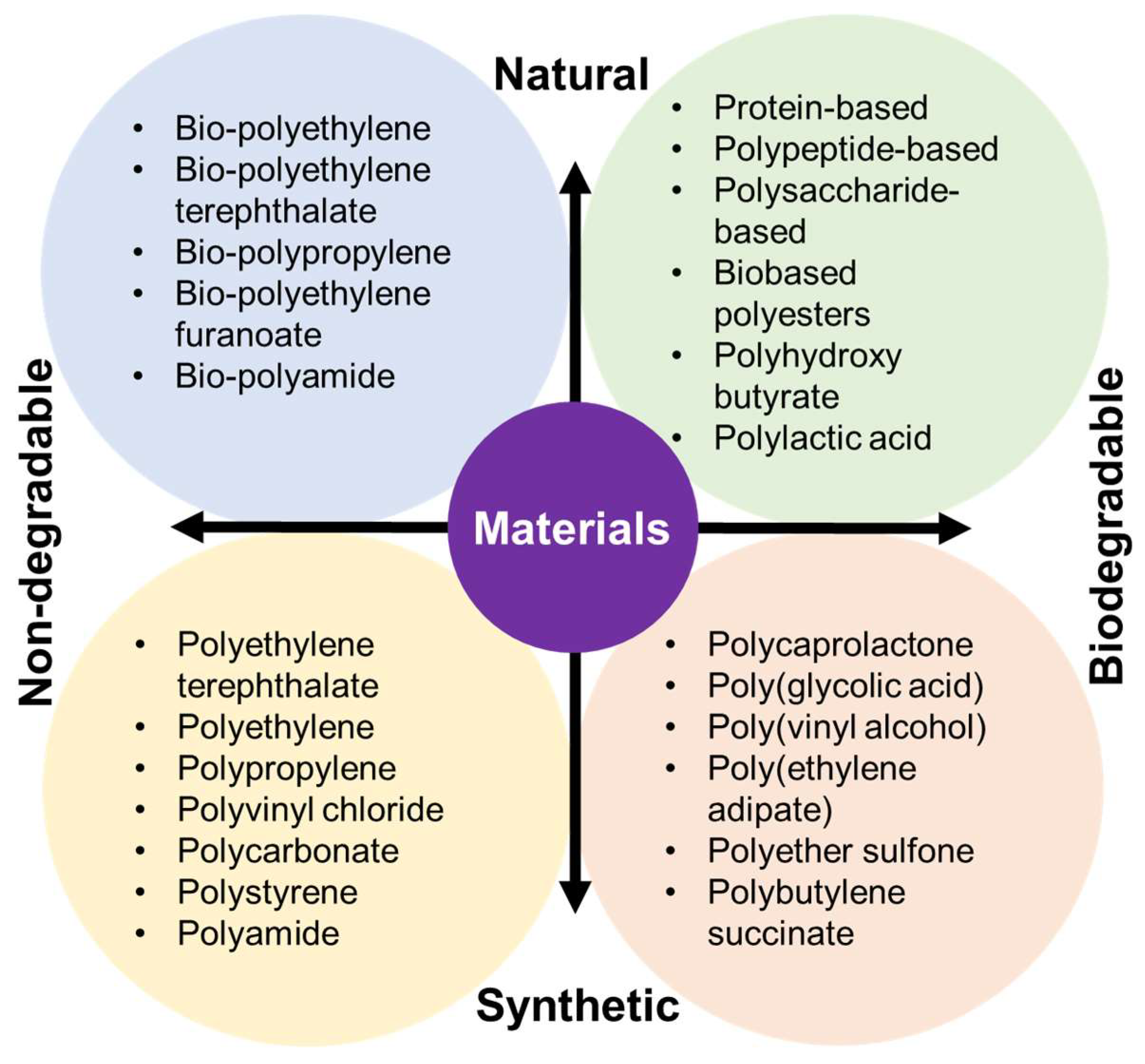
2.1. Natural Biomaterials
2.2. Synthetic Biomaterials
3. 3D Scaffolds for Chemotherapeutic Delivery and Cancer Treatment
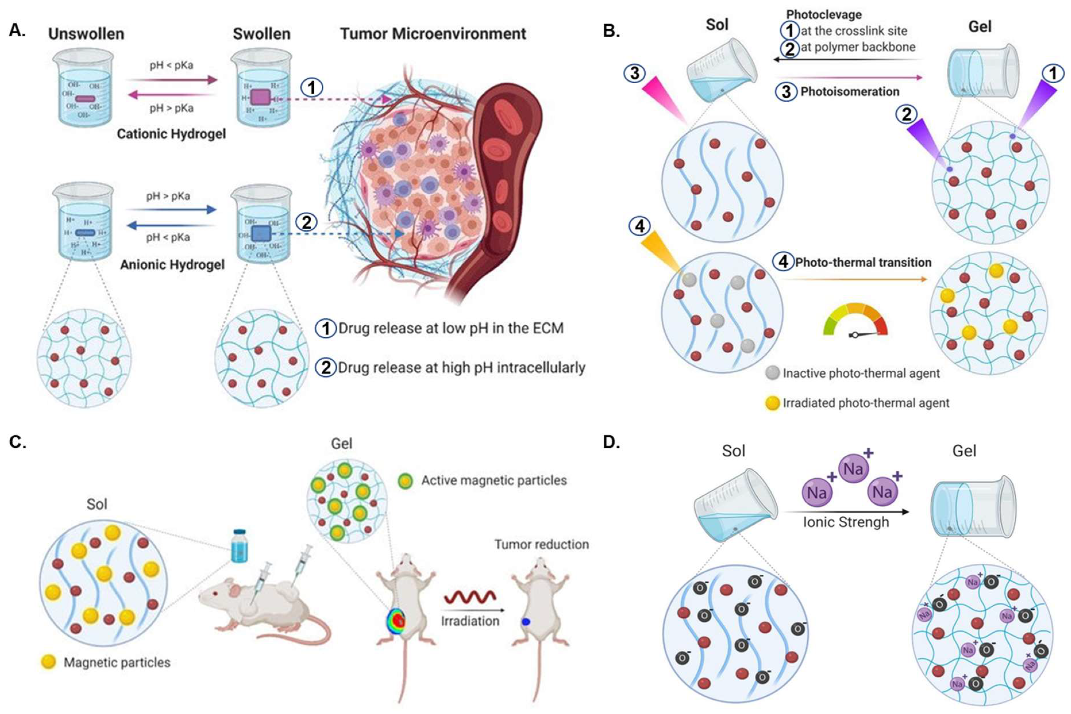
3.1. Smart Scaffolds
3.2. Expandable Scaffolds
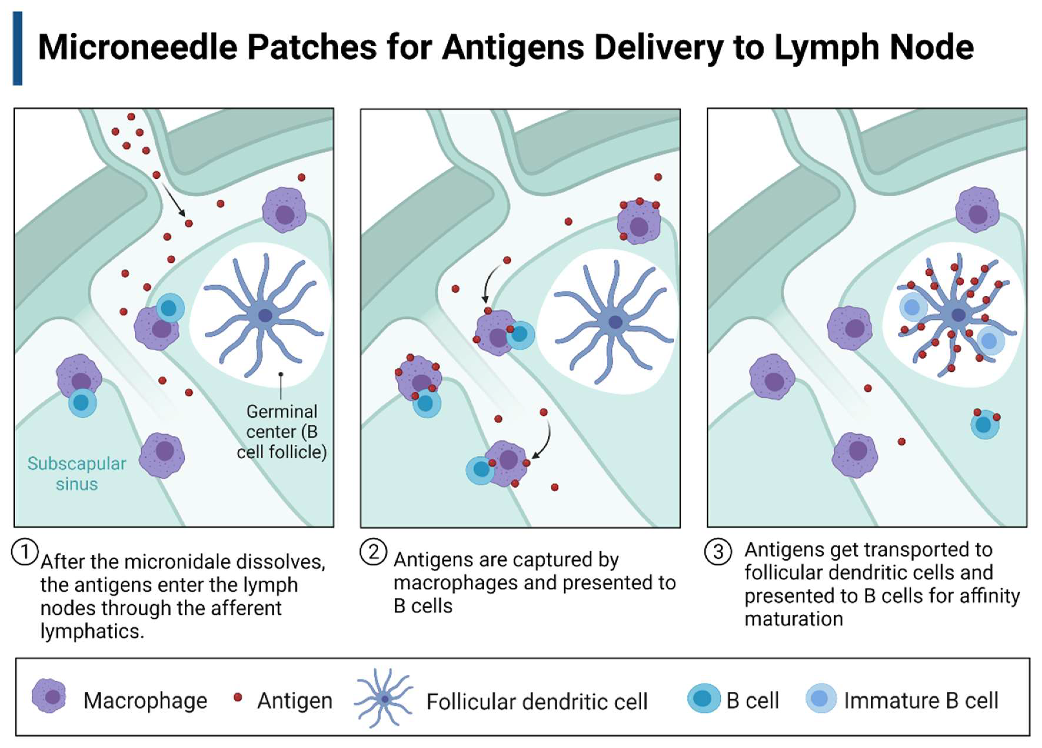
3.3. Microneedle Patch
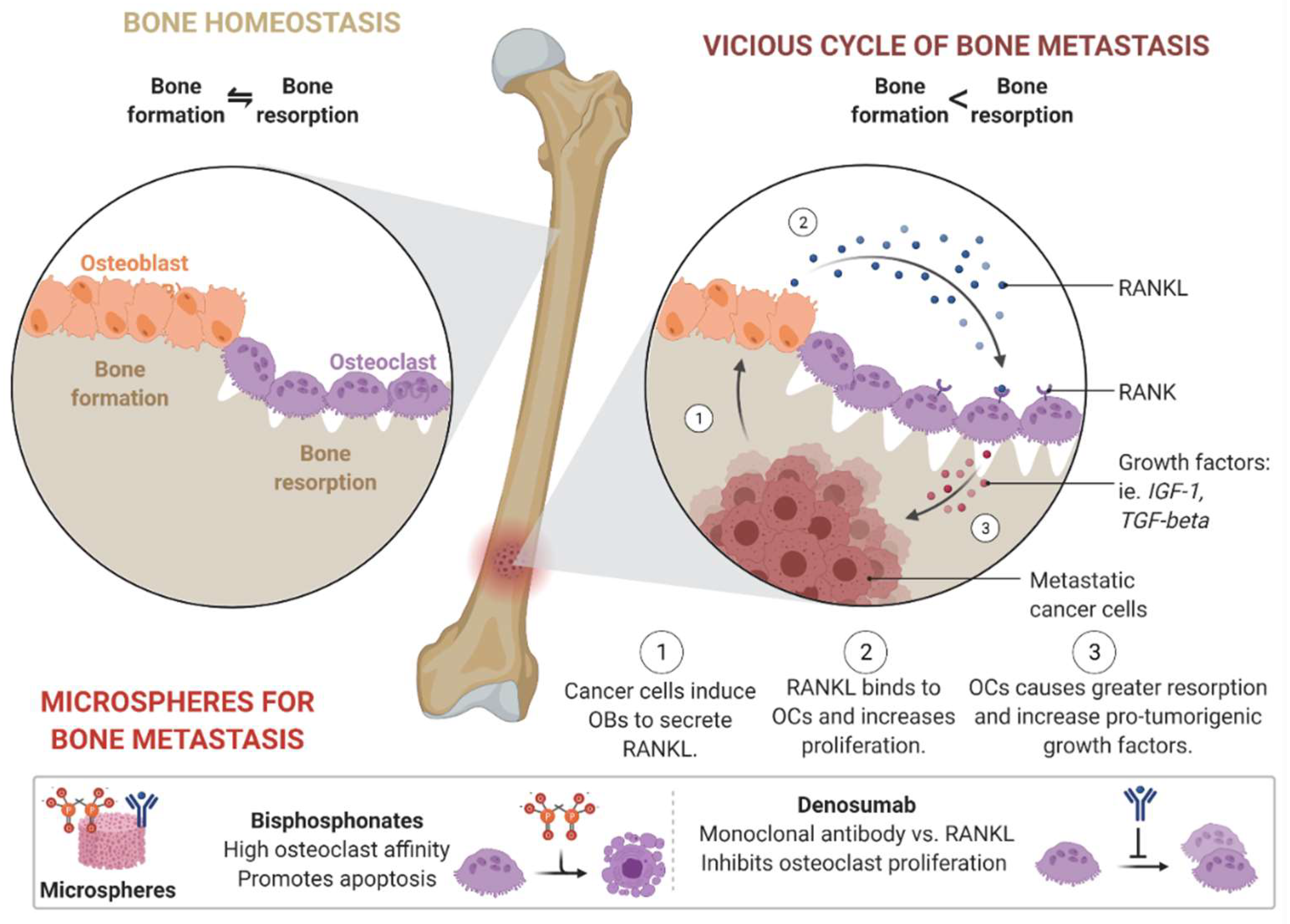
3.4. Microspheres
4. Nanotechnology-Based Treatment Approach
4.1. Nanoparticles (NPs)
4.2. Nanospheres
4.3. Exosomes
4.4. Nanogels
5. Bacteriophage
6. Conclusions and Perspectives
Author Contributions
Funding
Institutional Review Board Statement
Conflicts of Interest
References
- Safarzadeh, E.; Shotorbani, S.S.; Baradaran, B. Herbal medicine as inducers of apoptosis in cancer treatment. Adv. Pharm. Bull. 2014, 4, 421. [Google Scholar] [PubMed]
- Senapati, S.; Mahanta, A.K.; Kumar, S.; Maiti, P. Controlled drug delivery vehicles for cancer treatment and their performance. Signal Transduct. Target. Ther. 2018, 3, 7. [Google Scholar] [CrossRef] [PubMed] [Green Version]
- Bray, F.; Ferlay, J.; Soerjomataram, I.; Siegel, R.; Torre, L.; Jemal, A. Erratum: Global cancer statistics 2018: GLOBOCAN estimates of incidence and mortality worldwide for 36 cancers in 185 countries. CA Cancer J. Clin. 2020, 70, 313. [Google Scholar]
- Schirrmacher, V. From chemotherapy to biological therapy: A review of novel concepts to reduce the side effects of systemic cancer treatment. Int. J. Oncol. 2019, 54, 407–419. [Google Scholar] [PubMed]
- Lowenthal, R.M.; Eaton, K. Toxicity of chemotherapy. Hematol./Oncol. Clin. 1996, 10, 967–990. [Google Scholar] [CrossRef] [PubMed]
- Chakraborty, S.; Rahman, T. The difficulties in cancer treatment. Ecancermedicalscience 2012, 6, ed16. [Google Scholar]
- Amjad, M.T.; Chidharla, A.; Kasi, A. Cancer Chemotherapy; StatPearls Publishing: Treasure Island, FL, USA, 2022. [Google Scholar]
- Andrade, F.; Roca-Melendres, M.M.; Durán-Lara, E.F.; Rafael, D.; Schwartz, S., Jr. Stimuli-responsive hydrogels for cancer treatment: The role of pH, light, ionic strength and magnetic field. Cancers 2021, 13, 1164. [Google Scholar] [CrossRef]
- Tewabe, A.; Abate, A.; Tamrie, M.; Seyfu, A.; Siraj, E.A. Targeted drug delivery—From magic bullet to nanomedicine: Principles, challenges, and future perspectives. J. Multidiscip. Healthc. 2021, 14, 1711. [Google Scholar] [CrossRef]
- Layek, B.; Sadhukha, T.; Panyam, J.; Prabha, S. Nano-engineered mesenchymal stem cells increase therapeutic efficacy of anticancer drug through true active tumor targeting. Mol. Cancer Ther. 2018, 17, 1196–1206. [Google Scholar] [CrossRef] [Green Version]
- Huang, Z.; Xiao, H.; Lu, X.; Yan, W.; Ji, Z. Enhanced photo/chemo combination efficiency against bladder tumor by encapsulation of DOX and ZnPC into in situ-formed thermosensitive polymer hydrogel. Int. J. Nanomed. 2018, 13, 7623. [Google Scholar] [CrossRef] [PubMed] [Green Version]
- Chew, S.A.; Danti, S. Biomaterial-based implantable devices for cancer therapy. Adv. Healthc. Mater. 2017, 6, 1600766. [Google Scholar] [CrossRef]
- Alsuraifi, A.; Curtis, A.; Lamprou, D.A.; Hoskins, C. Stimuli responsive polymeric systems for cancer therapy. Pharmaceutics 2018, 10, 136. [Google Scholar] [CrossRef]
- Nosrati, R.; Abnous, K.; Alibolandi, M.; Mosafer, J.; Dehghani, S.; Taghdisi, S.M.; Ramezani, M. Targeted SPION siderophore conjugate loaded with doxorubicin as a theranostic agent for imaging and treatment of colon carcinoma. Sci. Rep. 2021, 11, 13065. [Google Scholar] [CrossRef] [PubMed]
- Zhang, K.; Fang, Y.; He, Y.; Yin, H.; Guan, X.; Pu, Y.; Zhou, B.; Yue, W.; Ren, W.; Du, D. Extravascular gelation shrinkage-derived internal stress enables tumor starvation therapy with suppressed metastasis and recurrence. Nat. Commun. 2019, 10, 5380. [Google Scholar] [CrossRef] [PubMed] [Green Version]
- Chen, K.; Chen, X. Integrin targeted delivery of chemotherapeutics. Theranostics 2011, 1, 189. [Google Scholar] [CrossRef] [PubMed] [Green Version]
- Taherian, A.; Esfandiari, N.; Rouhani, S. Breast cancer drug delivery by novel drug-loaded chitosan-coated magnetic nanoparticles. Cancer Nanotechnol. 2021, 12, 15. [Google Scholar] [CrossRef]
- Lei, N.; Gong, C.; Qian, Z.; Luo, F.; Wang, C.; Wang, H.; Wei, Y. Therapeutic application of injectable thermosensitive hydrogel in preventing local breast cancer recurrence and improving incision wound healing in a mouse model. Nanoscale 2012, 4, 5686–5693. [Google Scholar] [CrossRef]
- Yang, Y.; Chen, S.; Liu, L.; Li, S.; Zeng, Q.; Zhao, X.; Li, H.; Zhang, Z.; Bouchard, L.-S.; Liu, M. Increasing cancer therapy efficiency through targeting and localized light activation. ACS Appl. Mater. Interfaces 2017, 9, 23400–23408. [Google Scholar] [CrossRef]
- Babu, A.; Templeton, A.K.; Munshi, A.; Ramesh, R. Nanodrug delivery systems: A promising technology for detection, diagnosis, and treatment of cancer. AAPS PharmSciTech 2014, 15, 709–721. [Google Scholar] [CrossRef] [Green Version]
- Parveen, S.; Sahoo, S.K. Polymeric nanoparticles for cancer therapy. J. Drug Target. 2008, 16, 108–123. [Google Scholar] [CrossRef]
- Kumar, R.; Sarkar, C.; Panja, S.; Khatua, C.; Gugulothu, K.; Sil, D. Biomimetic Nanocomposites for Biomedical Applications. In Biorenewable Nanocomposite Materials, Vol. 1: Electrocatalysts and Energy Storage; ACS Publications: Washington, DC, USA, 2022; pp. 163–196. [Google Scholar]
- Nunes, D.; Andrade, S.; Ramalho, M.J.; Loureiro, J.A.; Pereira, M.C. Polymeric Nanoparticles-Loaded Hydrogels for Biomedical Applications: A Systematic Review on in Vivo Findings. Polymers 2022, 14, 1010. [Google Scholar] [CrossRef]
- Nichols, J.W.; Bae, Y.H. EPR: Evidence and fallacy. J. Control. Release 2014, 190, 451–464. [Google Scholar] [CrossRef]
- Caro, C.; Avasthi, A.; Paez-Muñoz, J.M.; Leal, M.P.; García-Martín, M.L. Passive targeting of high-grade gliomas via the EPR effect: A closed path for metallic nanoparticles? Biomater. Sci. 2021, 9, 7984–7995. [Google Scholar] [CrossRef] [PubMed]
- Golombek, S.K.; May, J.-N.; Theek, B.; Appold, L.; Drude, N.; Kiessling, F.; Lammers, T. Tumor targeting via EPR: Strategies to enhance patient responses. Adv. Drug Deliv. Rev. 2018, 130, 17–38. [Google Scholar] [CrossRef] [PubMed]
- Huynh, E.; Zheng, G. Cancer nanomedicine: Addressing the dark side of the enhanced permeability and retention effect. Nanomedicine 2015, 10, 1993–1995. [Google Scholar] [CrossRef] [PubMed]
- Ovais, M.; Mukherjee, S.; Pramanik, A.; Das, D.; Mukherjee, A.; Raza, A.; Chen, C. Designing stimuli-responsive upconversion nanoparticles that exploit the tumor microenvironment. Adv. Mater. 2020, 32, 2000055. [Google Scholar] [CrossRef]
- Chen, Y.; Gao, D.-Y.; Huang, L. In vivo delivery of miRNAs for cancer therapy: Challenges and strategies. Adv. Drug Deliv. Rev. 2015, 81, 128–141. [Google Scholar] [CrossRef] [Green Version]
- Perez-Ruiz, A.G.; Ganem, A.; Olivares-Corichi, I.M.; García-Sánchez, J.R. Lecithin–chitosan–TPGS nanoparticles as nanocarriers of (−)-epicatechin enhanced its anticancer activity in breast cancer cells. RSC Adv. 2018, 8, 34773–34782. [Google Scholar] [CrossRef] [Green Version]
- Qi, F.-l.; Wang, M.-f.; Li, B.-z.; Lu, Z.-f.; Nie, G.-j.; Li, S.-p. Reversal of the immunosuppressive tumor microenvironment by nanoparticle-based activation of immune-associated cells. Acta Pharmacol. Sin. 2020, 41, 895–901. [Google Scholar] [CrossRef]
- Yao, Y.; Zhou, Y.; Liu, L.; Xu, Y.; Chen, Q.; Wang, Y.; Wu, S.; Deng, Y.; Zhang, J.; Shao, A. Nanoparticle-based drug delivery in cancer therapy and its role in overcoming drug resistance. Front. Mol. Biosci. 2020, 7, 193. [Google Scholar] [CrossRef]
- Mendez, F.B.; Medrano, F.J.E.; Pariona, N.; de Leon Gomez, R.D.; Romero, J.L.O. Recent Progress in Antitumoral Nanotechnology. Int. J. Nanopart. Nanotech. 2015, 1, 1. [Google Scholar]
- Basak, D.; Arrighi, S.; Darwiche, Y.; Deb, S. Comparison of Anticancer Drug Toxicities: Paradigm Shift in Adverse Effect Profile. Life 2021, 12, 48. [Google Scholar] [CrossRef] [PubMed]
- Fan, D.-y.; Tian, Y.; Liu, Z.-j. Injectable hydrogels for localized cancer therapy. Front. Chem. 2019, 7, 675. [Google Scholar] [CrossRef] [PubMed]
- Sengupta, P.; Agrawal, V.; Prasad, B.L. Development of a Smart Scaffold for Sequential Cancer Chemotherapy and Tissue Engineering. ACS Omega 2020, 5, 20724–20733. [Google Scholar] [CrossRef] [PubMed]
- Shin, G.R.; Kim, H.E.; Kim, J.H.; Choi, S.; Kim, M.S. Advances in injectable in situ-forming hydrogels for intratumoral treatment. Pharmaceutics 2021, 13, 1953. [Google Scholar] [CrossRef] [PubMed]
- Wang, H.; Najibi, A.J.; Sobral, M.C.; Seo, B.R.; Lee, J.Y.; Wu, D.; Li, A.W.; Verbeke, C.S.; Mooney, D.J. Biomaterial-based scaffold for in situ chemo-immunotherapy to treat poorly immunogenic tumors. Nat. Commun. 2020, 11, 5696. [Google Scholar] [CrossRef] [PubMed]
- Conde, J.; Shomron, N.; Artzi, N. Biomaterials for abrogating metastasis: Bridging the gap between basic and translational research. Adv. Healthc. Mater. 2016, 5, 2312–2319. [Google Scholar] [CrossRef]
- Lanza, R.; Langer, R.; Vacanti, J. Preface. Principles of Tissue Engineering; Elsevier: Amsterdam, The Netherlands, 2014. [Google Scholar]
- Rajangam, T.; An, S.S.A. Fibrinogen and fibrin based micro and nano scaffolds incorporated with drugs, proteins, cells and genes for therapeutic biomedical applications. Int. J. Nanomed. 2013, 8, 3641. [Google Scholar]
- Pawar, R.; Jadhav, W.; Bhusare, S.; Borade, R.; Farber, S.; Itzkowitz, D.; Domb, A. Polysaccharides as carriers of bioactive agents for medical applications. In Natural-Based Polymers for Biomedical Applications; Elsevier: Amsterdam, The Netherlands, 2008; pp. 3–53. [Google Scholar]
- Abbasian, M.; Massoumi, B.; Mohammad-Rezaei, R.; Samadian, H.; Jaymand, M. Scaffolding polymeric biomaterials: Are naturally occurring biological macromolecules more appropriate for tissue engineering? Int. J. Biol. Macromol. 2019, 134, 673–694. [Google Scholar]
- Xu, S.; Xu, H.; Wang, W.; Li, S.; Li, H.; Li, T.; Zhang, W.; Yu, X.; Liu, L. The role of collagen in cancer: From bench to bedside. J. Transl. Med. 2019, 17, 309. [Google Scholar]
- Chen, H.; Wang, X.; Sutrisno, L.; Zeng, T.; Kawazoe, N.; Yang, Y.; Chen, G. Folic acid–functionalized composite scaffolds of gelatin and gold nanoparticles for photothermal ablation of breast cancer cells. Front. Bioeng. Biotechnol. 2020, 8, 589905. [Google Scholar] [CrossRef] [PubMed]
- Aldana, A.A.; Abraham, G.A. Current advances in electrospun gelatin-based scaffolds for tissue engineering applications. Int. J. Pharm. 2017, 523, 441–453. [Google Scholar] [CrossRef] [PubMed] [Green Version]
- Rouse, J.G.; Van Dyke, M.E. A review of keratin-based biomaterials for biomedical applications. Materials 2010, 3, 999–1014. [Google Scholar] [CrossRef] [Green Version]
- Roslan, M.R.; Nasir, N.F.M.; Cheng, E.M.; Amin, N.A.M. Tissue engineering scaffold based on starch: A review. In Proceedings of the 2016 International Conference on Electrical, Electronics, and Optimization Techniques (ICEEOT), Chennai, India, 3–5 March 2016; pp. 1857–1860. [Google Scholar]
- Ahmad, M.; Manzoor, K.; Ikram, S. 2—Chitosan based nanocomposites for drug, gene delivery, and bioimaging applications. In Applications of Nanocomposite Materials in Drug Delivery; Inamuddin Asiri, A.M., Mohammad, A., Eds.; Woodhead Publishing: Sawston, UK, 2018; pp. 27–38. [Google Scholar]
- Yassue-Cordeiro, P.H.; Severino, P.; Souto, E.B.; Gomes, E.L.; Yoshida, C.M.P.; de Moraes, M.A.; da Silva, C.F. 1—Chitosan-based nanocomposites for drug delivery. In Applications of Nanocomposite Materials in Drug Delivery; Inamuddin Asiri, A.M., Mohammad, A., Eds.; Woodhead Publishing: Sawston, UK, 2018; pp. 1–26. [Google Scholar]
- Liu, L.; Bai, L.; Tripathi, A.; Yu, J.; Wang, Z.; Borghei, M.; Fan, Y.; Rojas, O.J. High axial ratio nanochitins for ultrastrong and shape-recoverable hydrogels and cryogels via ice templating. ACS Nano 2019, 13, 2927–2935. [Google Scholar] [CrossRef] [PubMed] [Green Version]
- Zarrintaj, P.; Manouchehri, S.; Ahmadi, Z.; Saeb, M.R.; Urbanska, A.M.; Kaplan, D.L.; Mozafari, M. Agarose-based biomaterials for tissue engineering. Carbohydr. Polym. 2018, 187, 66–84. [Google Scholar] [CrossRef]
- Roberts, J.J.; Martens, P.J. 9—Engineering biosynthetic cell encapsulation systems. In Biosynthetic Polymers for Medical Applications; Poole-Warren, L., Martens, P., Green, R., Eds.; Woodhead Publishing: Sawston, UK, 2016; pp. 205–239. [Google Scholar]
- Hasnain, M.S.; Nayak, A.K. Alginate-inorganic composite particles as sustained drug delivery matrices. In Applications of Nanocomposite Materials in Drug Delivery; Elsevier: Amsterdam, The Netherlands, 2018; pp. 39–74. [Google Scholar]
- An, J.M.; Shahriar, S.S.; Hasan, M.N.; Cho, S.; Lee, Y.-k. Carboxymethyl cellulose, pluronic, and pullulan-based compositions efficiently enhance antiadhesion and tissue regeneration properties without using any drug molecules. ACS Appl. Mater. Interfaces 2021, 13, 15992–16006. [Google Scholar] [CrossRef]
- Luo, Z.; Dai, Y.; Gao, H. Development and application of hyaluronic acid in tumor targeting drug delivery. Acta Pharm. Sin. B 2019, 9, 1099–1112. [Google Scholar] [CrossRef]
- Köwitsch, A.; Zhou, G.; Groth, T. Medical application of glycosaminoglycans: A review. J. Tissue Eng. Regen. Med. 2018, 12, e23–e41. [Google Scholar] [CrossRef]
- Chakraborty, I.; Chatterjee, K. Polymers and composites derived from castor oil as sustainable materials and degradable biomaterials: Current status and emerging trends. Biomacromolecules 2020, 21, 4639–4662. [Google Scholar] [CrossRef]
- Ouellette, R.J.; Rawn, J.D. 15—Synthetic Polymers. In Principles of Organic Chemistry; Ouellette, R.J., Rawn, J.D., Eds.; Elsevier: Boston, MA, USA, 2015; pp. 397–419. [Google Scholar]
- Ouellette, R.J.; Rawn, J.D. Principles of Organic Chemistry; Academic Press: Cambridge, MA, USA, 2015. [Google Scholar]
- Salerno, A.; Domingo, C.; Saurina, J. PCL foamed scaffolds loaded with 5-fluorouracil anti-cancer drug prepared by an eco-friendly route. Mater. Sci. Eng. C 2017, 75, 1191–1197. [Google Scholar] [CrossRef]
- Chi, H.Y.; Chan, V.; Li, C.; Hsieh, J.H.; Lin, P.H.; Tsai, Y.-H.; Chen, Y. Fabrication of polylactic acid/paclitaxel nano fibers by electrospinning for cancer therapeutics. BMC Chem. 2020, 14, 63. [Google Scholar] [CrossRef]
- Ghalia, M.A.; Dahman, Y. Biodegradable poly(lactic acid)-based scaffolds: Synthesis and biomedical applications. J. Polym. Res. 2017, 24, 74. [Google Scholar] [CrossRef]
- Astete, C.E.; Sabliov, C.M. Synthesis and characterization of PLGA nanoparticles. J. Biomater. Sci. Polym. Ed. 2006, 17, 247–289. [Google Scholar] [CrossRef]
- Zhang, J.; Yang, S.; Yang, X.; Xi, Z.; Zhao, L.; Cen, L.; Lu, E.; Yang, Y. Novel fabricating process for porous polyglycolic acid scaffolds by melt-foaming using supercritical carbon dioxide. ACS Biomater. Sci. Eng. 2018, 4, 694–706. [Google Scholar] [CrossRef]
- Park, W.; Shin, H.; Choi, B.; Rhim, W.-K.; Na, K.; Keun Han, D. Advanced hybrid nanomaterials for biomedical applications. Prog. Mater. Sci. 2020, 114, 100686. [Google Scholar] [CrossRef]
- Cha, C.; Shin, S.R.; Annabi, N.; Dokmeci, M.R.; Khademhosseini, A. Carbon-based nanomaterials: Multifunctional materials for biomedical engineering. ACS Nano 2013, 7, 2891–2897. [Google Scholar] [CrossRef]
- Khatoon, H.; Iqbal, S.; Ahmad, S. Covalently functionalized ethylene diamine modified graphene oxide poly-paraphenylene diamine dispersed polyurethane anticorrosive nanocomposite coatings. Prog. Org. Coat. 2021, 150, 105966. [Google Scholar] [CrossRef]
- Kumar, A.; Han, S.S. PVA-based hydrogels for tissue engineering: A review. Int. J. Polym. Mater. Polym. Biomater. 2017, 66, 159–182. [Google Scholar] [CrossRef]
- Sasanuma, Y.; Takahashi, Y. Structure–Property Relationships of Poly (ethylene carbonate) and Poly (propylene carbonate). ACS Omega 2017, 2, 4808–4819. [Google Scholar] [CrossRef] [Green Version]
- Ren, S.; Dai, Y.; Li, C.; Qiu, Z.; Wang, X.; Tian, F.; Zhou, S.; Liu, Q.; Xing, H.; Lu, Y.; et al. Pharmacokinetics and pharmacodynamics evaluation of a thermosensitive chitosan based hydrogel containing liposomal doxorubicin. Eur. J. Pharm. Sci. 2016, 92, 137–145. [Google Scholar] [CrossRef]
- Sheu, M.-T.; Jhan, H.-J.; Su, C.-Y.; Chen, L.-C.; Chang, C.-E.; Liu, D.-Z.; Ho, H.-O. Codelivery of doxorubicin-containing thermosensitive hydrogels incorporated with docetaxel-loaded mixed micelles enhances local cancer therapy. Colloids Surf. B Biointerfaces 2016, 143, 260–270. [Google Scholar] [CrossRef]
- Ma, H.; He, C.; Cheng, Y.; Li, D.; Gong, Y.; Liu, J.; Tian, H.; Chen, X. PLK1shRNA and doxorubicin co-loaded thermosensitive PLGA-PEG-PLGA hydrogels for osteosarcoma treatment. Biomaterials 2014, 35, 8723–8734. [Google Scholar] [CrossRef]
- Pesoa, J.I.; Rico, M.J.; Rozados, V.R.; Scharovsky, O.G.; Luna, J.A.; Mengatto, L.N. Paclitaxel delivery system based on poly(lactide-co-glycolide) microparticles and chitosan thermo-sensitive gel for mammary adenocarcinoma treatment. J. Pharm. Pharmacol. 2018, 70, 1494–1502. [Google Scholar] [CrossRef]
- Dong, X.; Wei, C.; Liu, T.; Lv, F.; Qian, Z. Real-time fluorescence tracking of protoporphyrin incorporated thermosensitive hydrogel and its drug release in vivo. ACS Appl. Mater. Interfaces 2016, 8, 5104–5113. [Google Scholar] [CrossRef]
- Lin, Z.; Xu, S.; Gao, W.; Hu, H.; Chen, M.; Wang, Y.; He, B.; Dai, W.; Zhang, H.; Wang, X. A comparative investigation between paclitaxel nanoparticle-and nanocrystal-loaded thermosensitive PECT hydrogels for peri-tumoural administration. Nanoscale 2016, 8, 18782–18791. [Google Scholar] [CrossRef]
- Fathi, M.; Alami-Milani, M.; Geranmayeh, M.H.; Barar, J.; Erfan-Niya, H.; Omidi, Y. Dual thermo-and pH-sensitive injectable hydrogels of chitosan/(poly (N-isopropylacrylamide-co-itaconic acid)) for doxorubicin delivery in breast cancer. Int. J. Biol. Macromol. 2019, 128, 957–964. [Google Scholar] [CrossRef]
- Liang, Y.; Zhao, X.; Ma, P.X.; Guo, B.; Du, Y.; Han, X. pH-responsive injectable hydrogels with mucosal adhesiveness based on chitosan-grafted-dihydrocaffeic acid and oxidized pullulan for localized drug delivery. J. Colloid Interface Sci. 2019, 536, 224–234. [Google Scholar] [CrossRef]
- Liu, Z.; Xu, G.; Wang, C.; Li, C.; Yao, P. Shear-responsive injectable supramolecular hydrogel releasing doxorubicin loaded micelles with pH-sensitivity for local tumor chemotherapy. Int. J. Pharm. 2017, 530, 53–62. [Google Scholar] [CrossRef]
- Qu, J.; Zhao, X.; Ma, P.X.; Guo, B. pH-responsive self-healing injectable hydrogel based on N-carboxyethyl chitosan for hepatocellular carcinoma therapy. Acta Biomater. 2017, 58, 168–180. [Google Scholar] [CrossRef]
- Yue, Z.; Che, Y.; Jin, Z.; Wang, S.; Ma, Q.; Zhang, Q.; Tan, Y.; Meng, F. A facile method to fabricate thermo-and pH-sensitive hydrogels with good mechanical performance based on poly (ethylene glycol) methyl ether methacrylate and acrylic acid as a potential drug carriers. J. Biomater. Sci. Polym. Ed. 2019, 30, 1375–1398. [Google Scholar] [CrossRef]
- Zhang, W.; Jin, X.; Li, H.; Zhang, R.-r.; Wu, C.-w. Injectable and body temperature sensitive hydrogels based on chitosan and hyaluronic acid for pH sensitive drug release. Carbohydr. Polym. 2018, 186, 82–90. [Google Scholar] [CrossRef] [PubMed]
- Kang, H.; Liu, H.; Zhang, X.; Yan, J.; Zhu, Z.; Peng, L.; Yang, H.; Kim, Y.; Tan, W. Photoresponsive DNA-cross-linked hydrogels for controllable release and cancer therapy. Langmuir 2011, 27, 399–408. [Google Scholar] [CrossRef] [PubMed] [Green Version]
- Guo, D.; Xu, S.; Huang, Y.; Jiang, H.; Yasen, W.; Wang, N.; Su, Y.; Qian, J.; Li, J.; Zhang, C.; et al. Platinum(IV) complex-based two-in-one polyprodrug for a combinatorial chemo-photodynamic therapy. Biomaterials 2018, 177, 67–77. [Google Scholar] [CrossRef]
- Cimen, Z.; Babadag, S.; Odabas, S.; Altuntas, S.; Demirel, G.; Demirel, G.B. Injectable and Self-Healable pH-Responsive Gelatin–PEG/Laponite Hybrid Hydrogels as Long-Acting Implants for Local Cancer Treatment. ACS Appl. Polym. Mater. 2021, 3, 3504–3518. [Google Scholar] [CrossRef]
- Bordbar-Khiabani, A.; Gasik, M. Smart Hydrogels for Advanced Drug Delivery Systems. Int. J. Mol. Sci. 2022, 23, 3665. [Google Scholar] [CrossRef]
- Liu, J.; Huang, Y.; Kumar, A.; Tan, A.; Jin, S.; Mozhi, A.; Liang, X.-J. pH-Sensitive nano-systems for drug delivery in cancer therapy. Biotechnol. Adv. 2014, 32, 693–710. [Google Scholar] [CrossRef]
- Rizwan, M.; Yahya, R.; Hassan, A.; Yar, M.; Azzahari, A.D.; Selvanathan, V.; Sonsudin, F.; Abouloula, C.N. pH sensitive hydrogels in drug delivery: Brief history, properties, swelling, and release mechanism, material selection and applications. Polymers 2017, 9, 137. [Google Scholar] [CrossRef] [Green Version]
- Raza, F.; Zhu, Y.; Chen, L.; You, X.; Zhang, J.; Khan, A.; Khan, M.W.; Hasnat, M.; Zafar, H.; Wu, J. Paclitaxel-loaded pH responsive hydrogel based on self-assembled peptides for tumor targeting. Biomater. Sci. 2019, 7, 2023–2036. [Google Scholar] [CrossRef]
- Yuan, Z.; Zhao, X.; Zhao, J.; Pan, G.; Qiu, W.; Wang, X.; Zhu, Y.; Zheng, Q.; Cui, W. Synergistic mediation of tumor signaling pathways in hepatocellular carcinoma therapy via dual-drug-loaded pH-responsive electrospun fibrous scaffolds. J. Mater. Chem. B 2015, 3, 3436–3446. [Google Scholar] [CrossRef]
- Rezk, A.I.; Obiweluozor, F.O.; Choukrani, G.; Park, C.H.; Kim, C.S. Drug release and kinetic models of anticancer drug (BTZ) from a pH-responsive alginate polydopamine hydrogel: Towards cancer chemotherapy. Int. J. Biol. Macromol. 2019, 141, 388–400. [Google Scholar] [CrossRef]
- Wang, M.; Chen, M.; Niu, W.; Winston, D.D.; Cheng, W.; Lei, B. Injectable biodegradation-visual self-healing citrate hydrogel with high tissue penetration for microenvironment-responsive degradation and local tumor therapy. Biomaterials 2020, 261, 120301. [Google Scholar] [CrossRef] [PubMed]
- Martinez, G.V.; Zhang, X.; García-Martín, M.L.; Morse, D.L.; Woods, M.; Sherry, A.D.; Gillies, R.J. Imaging the extracellular pH of tumors by MRI after injection of a single cocktail of T1 and T2 contrast agents. NMR Biomed. 2011, 24, 1380–1391. [Google Scholar] [CrossRef] [PubMed] [Green Version]
- Bai, G.; Yuan, P.; Cai, B.; Qiu, X.; Jin, R.; Liu, S.; Li, Y.; Chen, X. Stimuli-Responsive Scaffold for Breast Cancer Treatment Combining Accurate Photothermal Therapy and Adipose Tissue Regeneration. Adv. Funct. Mater. 2019, 29, 1904401. [Google Scholar] [CrossRef]
- Li, L.; Scheiger, J.M.; Levkin, P.A. Design and applications of photoresponsive hydrogels. Adv. Mater. 2019, 31, 1807333. [Google Scholar] [CrossRef] [PubMed] [Green Version]
- Wang, Z.J.; Li, C.Y.; Zhao, X.Y.; Wu, Z.L.; Zheng, Q. Thermo-and photo-responsive composite hydrogels with programmed deformations. J. Mater. Chem. B 2019, 7, 1674–1678. [Google Scholar] [CrossRef]
- Gulfam, M.; Jo, S.-H.; Jo, S.-W.; Vu, T.T.; Park, S.-H.; Lim, K.T. Highly porous and injectable hydrogels derived from cartilage acellularized matrix exhibit reduction and NIR light dual-responsive drug release properties for application in antitumor therapy. NPG Asia Mater. 2022, 14, 8. [Google Scholar] [CrossRef]
- Tomatsu, I.; Peng, K.; Kros, A. Photoresponsive hydrogels for biomedical applications. Adv. Drug Deliv. Rev. 2011, 63, 1257–1266. [Google Scholar]
- Mallidi, S.; Anbil, S.; Bulin, A.-L.; Obaid, G.; Ichikawa, M.; Hasan, T. Beyond the barriers of light penetration: Strategies, perspectives and possibilities for photodynamic therapy. Theranostics 2016, 6, 2458. [Google Scholar] [CrossRef] [Green Version]
- Majerník, M.; Jendželovský, R.; Vargová, J.; Jendželovská, Z.; Fedoročko, P. Multifunctional Nanoplatforms as a Novel Effective Approach in Photodynamic Therapy and Chemotherapy, to Overcome Multidrug Resistance in Cancer. Pharmaceutics 2022, 14, 1075. [Google Scholar] [CrossRef]
- Jin, R.; Yang, J.; Zhao, D.; Hou, X.; Li, C.; Chen, W.; Zhao, Y.; Yin, Z.; Liu, B. Hollow gold nanoshells-incorporated injectable genetically engineered hydrogel for sustained chemo-photothermal therapy of tumor. J. Nanobiotechnol. 2019, 17, 99. [Google Scholar] [CrossRef] [Green Version]
- Wang, X.; Yuan, Z.; Tao, A.; Wang, P.; Xie, W.; Yang, S.; Huang, J.; Wen, N. Hydrogel-based patient-friendly photodynamic therapy of oral potentially malignant disorders. Biomaterials 2022, 281, 121377. [Google Scholar] [CrossRef] [PubMed]
- Zhang, Q.; Wang, X.; Kuang, G.; Yu, Y.; Zhao, Y. Photopolymerized 3D Printing Scaffolds with Pt (IV) Prodrug Initiator for Postsurgical Tumor Treatment. Research 2022, 2022, 9784510. [Google Scholar] [CrossRef] [PubMed]
- Leganes Bayon, J.; Sánchez-Migallón, A.; Díaz-Ortiz, Á.; Castillo, C.A.; Ballesteros-Yáñez, I.; Merino, S.; Vázquez, E. On-Demand Hydrophobic Drug Release Based on Microwave-Responsive Graphene Hydrogel Scaffolds. Chem.–A Eur. J. 2020, 26, 17069–17080. [Google Scholar] [CrossRef] [PubMed]
- Mishra, B.; Singh, J. Hydrogel-based drug delivery systems for cancer therapy. In Advanced Drug Delivery Systems in the Management of Cancer; Elsevier: Amsterdam, The Netherlands, 2021; pp. 63–74. [Google Scholar]
- Kasiński, A.; Zielińska-Pisklak, M.; Oledzka, E.; Sobczak, M. Smart hydrogels–synthetic stimuli-responsive antitumor drug release systems. Int. J. Nanomed. 2020, 15, 4541. [Google Scholar] [CrossRef] [PubMed]
- Rock, K.L.; Kono, H. The inflammatory response to cell death. Annu. Rev. Pathol. 2008, 3, 99. [Google Scholar] [CrossRef]
- Baghban, R.; Afarid, M.; Soleymani, J.; Rahimi, M. Were magnetic materials useful in cancer therapy? Biomed. Pharmacother. 2021, 144, 112321. [Google Scholar]
- Xiang, T.; Lu, T.; Zhao, W.-F.; Zhao, C.-S. Ionic-strength responsive zwitterionic copolymer hydrogels with tunable swelling and adsorption behaviors. Langmuir 2018, 35, 1146–1155. [Google Scholar] [CrossRef]
- Ma, G.; Lin, W.; Yuan, Z.; Wu, J.; Qian, H.; Xu, L.; Chen, S. Development of ionic strength/pH/enzyme triple-responsive zwitterionic hydrogel of the mixed l-glutamic acid and l-lysine polypeptide for site-specific drug delivery. J. Mater. Chem. B 2017, 5, 935–943. [Google Scholar] [CrossRef]
- Zhang, C.; Ni, D.; Liu, Y.; Yao, H.; Bu, W.; Shi, J. Magnesium silicide nanoparticles as a deoxygenation agent for cancer starvation therapy. Nat. Nanotechnol. 2017, 12, 378–386. [Google Scholar] [CrossRef]
- Du, L.; Yang, S.; Li, W.; Li, H.; Feng, S.; Zeng, R.; Yu, B.; Xiao, L.; Nie, H.-Y.; Tu, M. Scaffold composed of porous vancomycin-loaded poly (lactide-co-glycolide) microspheres: A controlled-release drug delivery system with shape-memory effect. Mater. Sci. Eng. C 2017, 78, 1172–1178. [Google Scholar] [CrossRef]
- Chen, Y.; Shafiq, M.; Liu, M.; Morsi, Y.; Mo, X. Advanced fabrication for electrospun three-dimensional nanofiber aerogels and scaffolds. Bioact. Mater. 2020, 5, 963–979. [Google Scholar] [CrossRef] [PubMed]
- Costantini, M.; Barbetta, A. Gas foaming technologies for 3D scaffold engineering. In Functional 3D Tissue Engineering Scaffolds; Elsevier: Amsterdam, The Netherlands, 2018; pp. 127–149. [Google Scholar]
- Cavo, M.; Serio, F.; Kale, N.R.; D’Amone, E.; Gigli, G.; Loretta, L. Electrospun nanofibers in cancer research: From engineering of in vitro 3D cancer models to therapy. Biomater. Sci. 2020, 8, 4887–4905. [Google Scholar] [PubMed]
- Zhang, Y.; Xu, J.; Fei, Z.; Dai, H.; Fan, Q.; Yang, Q.; Chen, Y.; Wang, B.; Wang, C. 3D printing scaffold vaccine for antitumor immunity. Adv. Mater. 2021, 33, 2106768. [Google Scholar] [CrossRef] [PubMed]
- Anselmo, A.C.; Gokarn, Y.; Mitragotri, S. Non-invasive delivery strategies for biologics. Nat. Rev. Drug Discov. 2019, 18, 19–40. [Google Scholar] [CrossRef]
- Sun, Z.; Song, C.; Wang, C.; Hu, Y.; Wu, J. Hydrogel-based controlled drug delivery for cancer treatment: A review. Mol. Pharm. 2019, 17, 373–391. [Google Scholar] [CrossRef] [PubMed]
- Zhao, X.; Li, X.; Zhang, P.; Du, J.; Wang, Y. Tip-loaded fast-dissolving microneedle patches for photodynamic therapy of subcutaneous tumor. J. Control. Release 2018, 286, 201–209. [Google Scholar] [CrossRef]
- Yang, H.; Wu, X.; Zhou, Z.; Chen, X.; Kong, M. Enhanced transdermal lymphatic delivery of doxorubicin via hyaluronic acid based transfersomes/microneedle complex for tumor metastasis therapy. Int. J. Biol. Macromol. 2019, 125, 9–16. [Google Scholar] [CrossRef]
- Paulson, K.G.; Lahman, M.C.; Chapuis, A.G.; Brownell, I. Immunotherapy for skin cancer. Int. Immunol. 2019, 31, 465–475. [Google Scholar]
- Lan, X.; Zhu, W.; Huang, X.; Yu, Y.; Xiao, H.; Jin, L.; Pu, J.J.; Xie, X.; She, J.; Lui, V.W.Y. Microneedles loaded with anti-PD-1–cisplatin nanoparticles for synergistic cancer immuno-chemotherapy. Nanoscale 2020, 12, 18885–18898. [Google Scholar] [CrossRef]
- Wang, C.; Ye, Y.; Hochu, G.M.; Sadeghifar, H.; Gu, Z. Enhanced cancer immunotherapy by microneedle patch-assisted delivery of anti-PD1 antibody. Nano Lett. 2016, 16, 2334–2340. [Google Scholar] [CrossRef]
- Chen, G.; Chen, Z.; Wen, D.; Wang, Z.; Li, H.; Zeng, Y.; Dotti, G.; Wirz, R.E.; Gu, Z. Transdermal cold atmospheric plasma-mediated immune checkpoint blockade therapy. Proc. Natl. Acad. Sci. USA 2020, 117, 3687–3692. [Google Scholar] [CrossRef] [PubMed]
- Kim, N.W.; Kim, S.-Y.; Lee, J.E.; Yin, Y.; Lee, J.H.; Lim, S.Y.; Kim, E.S.; Duong, H.T.T.; Kim, H.K.; Kim, S. Enhanced cancer vaccination by in situ nanomicelle-generating dissolving microneedles. ACS Nano 2018, 12, 9702–9713. [Google Scholar] [CrossRef] [PubMed]
- Abuchowski, A.; Van Es, T.; Palczuk, N.; Davis, F. Alteration of immunological properties of bovine serum albumin by covalent attachment of polyethylene glycol. J. Biol. Chem. 1977, 252, 3578–3581. [Google Scholar] [CrossRef] [PubMed]
- Song, G.; Jiang, G.; Liu, T.; Zhang, X.; Zeng, Z.; Wang, R.; Li, P.; Yang, Y. Separable microneedles for synergistic chemo-photothermal therapy against superficial skin tumors. ACS Biomater. Sci. Eng. 2020, 6, 4116–4125. [Google Scholar] [CrossRef]
- Rohani Shirvan, A.; Nouri, A.; Wen, C. 12—Structural polymer biomaterials. In Structural Biomaterials; Wen, C., Ed.; Woodhead Publishing: Sawston, UK, 2021; pp. 395–439. [Google Scholar]
- Kanda, Y.; Kakutani, K.; Yurube, T.; Zhang, Z.; Miyazaki, S.; Kakiuchi, Y.; Takeoka, Y.; Tsujimoto, R.; Miyazaki, K.; Kawamoto, T. A novel topical treatment for bone metastases using a gelatin hydrogel incorporating cisplatin as a sustained release system. J. Orthop. Res. 2021, 39, 525–535. [Google Scholar] [CrossRef] [PubMed]
- Holen, I.; E Coleman, R. Bisphosphonates as treatment of bone metastases. Curr. Pharm. Des. 2010, 16, 1262–1271. [Google Scholar] [CrossRef] [PubMed]
- Gül, G.; Sendur, M.A.; Aksoy, S.; Sever, A.R.; Altundag, K. A comprehensive review of denosumab for bone metastasis in patients with solid tumors. Curr. Med. Res. Opin. 2016, 32, 133–145. [Google Scholar] [CrossRef]
- Ni, G.; Yang, G.; He, Y.; Li, X.; Du, T.; Xu, L.; Zhou, S. Uniformly sized hollow microspheres loaded with polydopamine nanoparticles and doxorubicin for local chemo-photothermal combination therapy. Chem. Eng. J. 2020, 379, 122317. [Google Scholar] [CrossRef]
- Fortin, S.; Bérubé, G. Advances in the development of hybrid anticancer drugs. Expert Opin. Drug Discov. 2013, 8, 1029–1047. [Google Scholar] [CrossRef]
- Kennedy, L.C.; Bickford, L.R.; Lewinski, N.A.; Coughlin, A.J.; Hu, Y.; Day, E.S.; West, J.L.; Drezek, R.A. A new era for cancer treatment: Gold-nanoparticle-mediated thermal therapies. Small 2011, 7, 169–183. [Google Scholar] [CrossRef]
- Misra, R.; Acharya, S.; Sahoo, S.K. Cancer nanotechnology: Application of nanotechnology in cancer therapy. Drug Discov. Today 2010, 15, 842–850. [Google Scholar] [CrossRef] [PubMed]
- Ferrari, M. Cancer nanotechnology: Opportunities and challenges. Nat. Rev. Cancer 2005, 5, 161–171. [Google Scholar] [CrossRef] [PubMed]
- Park, J.W. Liposome-based drug delivery in breast cancer treatment. Breast Cancer Res. 2002, 4, 95–99. [Google Scholar] [CrossRef] [Green Version]
- Farooq, M.A.; Aquib, M.; Farooq, A.; Haleem Khan, D.; Joelle Maviah, M.B.; Sied Filli, M.; Kesse, S.; Boakye-Yiadom, K.O.; Mavlyanova, R.; Parveen, A. Recent progress in nanotechnology-based novel drug delivery systems in designing of cisplatin for cancer therapy: An overview. Artif. Cells Nanomed. Biotechnol. 2019, 47, 1674–1692. [Google Scholar] [CrossRef] [PubMed] [Green Version]
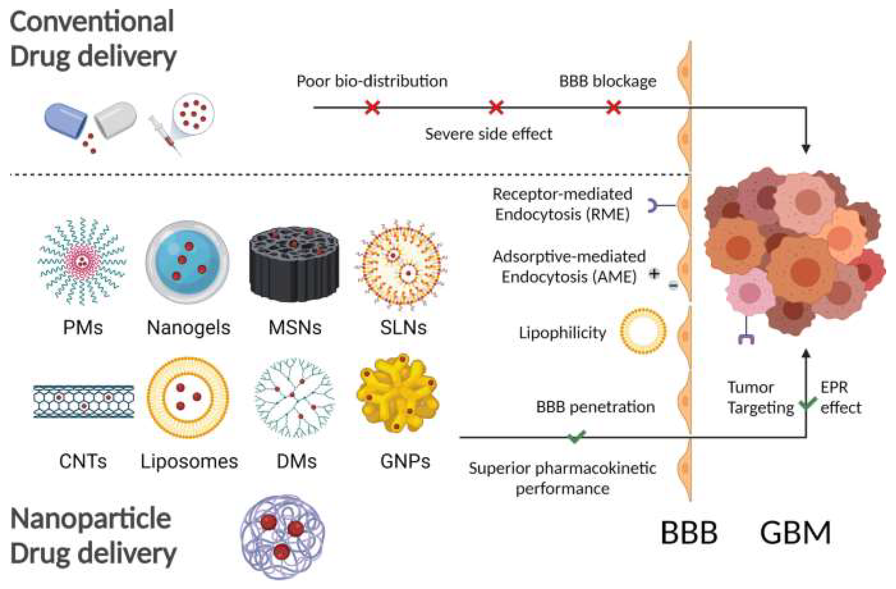
- Liu, Z.; Ji, X.; He, D.; Zhang, R.; Liu, Q.; Xin, T. Nanoscale Drug Delivery Systems in Glioblastoma. Nanoscale Res. Lett. 2022, 17, 27. [Google Scholar] [CrossRef] [PubMed]
- Rosenberger, I.; Strauss, A.; Dobiasch, S.; Weis, C.; Szanyi, S.; Gil-Iceta, L.; Alonso, E.; González Esparza, M.; Gómez-Vallejo, V.; Szczupak, B.; et al. Targeted diagnostic magnetic nanoparticles for medical imaging of pancreatic cancer. J. Control. Release 2015, 214, 76–84. [Google Scholar] [CrossRef] [PubMed]
- Luo, Y.; Yang, J.; Yan, Y.; Li, J.; Shen, M.; Zhang, G.; Mignani, S.; Shi, X. RGD-functionalized ultrasmall iron oxide nanoparticles for targeted T1-weighted MR imaging of gliomas. Nanoscale 2015, 7, 14538–14546. [Google Scholar] [CrossRef]
- Wang, Z.; Tong, M.; Chen, X.; Hu, S.; Yang, Z.; Zhang, Y.; Zhou, H.; Wu, Y.; Li, X.; Li, D. Survivin-targeted nanoparticles for pancreatic tumor imaging in mouse model. Nanomed. Nanotechnol. Biol. Med. 2016, 12, 1651–1661. [Google Scholar] [CrossRef]
- Guibert, C.; Dupuis, V.; Peyre, V.; Fresnais, J. Hyperthermia of Magnetic Nanoparticles: Experimental Study of the Role of Aggregation. J. Phys. Chem. C 2015, 119, 28148–28154. [Google Scholar] [CrossRef]
- Cheng, Y.; Muroski, M.E.; Petit, D.C.M.C.; Mansell, R.; Vemulkar, T.; Morshed, R.A.; Han, Y.; Balyasnikova, I.V.; Horbinski, C.M.; Huang, X.; et al. Rotating magnetic field induced oscillation of magnetic particles for in vivo mechanical destruction of malignant glioma. J. Control. Release 2016, 223, 75–84. [Google Scholar] [CrossRef] [Green Version]
- Sekar, V.; Rajendran, K.; Vallinayagam, S.; Deepak, V.; Mahadevan, S. Synthesis and characterization of chitosan ascorbate nanoparticles for therapeutic inhibition for cervical cancer and their in silico modeling. J. Ind. Eng. Chem. 2018, 62, 239–249. [Google Scholar] [CrossRef]
- Khodaverdi, S.; Jafari, A.; Movahedzadeh, F.; Madani, F.; Yousefi Avarvand, A.; Falahatkar, S. Evaluating Inhibitory Effects of Paclitaxel and Vitamin D(3) Loaded Poly Lactic Glycolic Acid Co-Delivery Nanoparticles on the Breast Cancer Cell Line. Adv. Pharm. Bull. 2020, 10, 30–38. [Google Scholar] [CrossRef] [PubMed]
- Dang, Y.; Guan, J. Nanoparticle-based drug delivery systems for cancer therapy. Smart Mater. Med. 2020, 1, 10–19. [Google Scholar] [CrossRef] [PubMed]
- Maier-Hauff, K.; Ulrich, F.; Nestler, D.; Niehoff, H.; Wust, P.; Thiesen, B.; Orawa, H.; Budach, V.; Jordan, A. Efficacy and safety of intratumoral thermotherapy using magnetic iron-oxide nanoparticles combined with external beam radiotherapy on patients with recurrent glioblastoma multiforme. J. Neuro-Oncol. 2011, 103, 317–324. [Google Scholar] [CrossRef] [Green Version]
- Ho, D.; Wang, C.-H.K.; Chow, E.K.-H. Nanodiamonds: The intersection of nanotechnology, drug development, and personalized medicine. Sci. Adv. 2015, 1, e1500439. [Google Scholar] [CrossRef] [Green Version]
- Sarkar, S.; Konar, S.; Prasad, P.N.; Rajput, S.; Kumar, B.N.P.; Rao, R.R.; Pathak, A.; Fisher, P.B.; Mandal, M. Micellear Gold Nanoparticles as Delivery Vehicles for Dual Tyrosine Kinase Inhibitor ZD6474 for Metastatic Breast Cancer Treatment. Langmuir 2017, 33, 7649–7659. [Google Scholar] [CrossRef]
- Tsai, L.-C.; Hsieh, H.-Y.; Lu, K.-Y.; Wang, S.-Y.; Mi, F.-L. EGCG/gelatin-doxorubicin gold nanoparticles enhance therapeutic efficacy of doxorubicin for prostate cancer treatment. Nanomedicine 2016, 11, 9–30. [Google Scholar] [CrossRef]
- Barabadi, H.; Ovais, M.; Shinwari, Z.K.; Saravanan, M. Anti-cancer green bionanomaterials: Present status and future prospects. Green Chem. Lett. Rev. 2017, 10, 285–314. [Google Scholar] [CrossRef] [Green Version]
- Boca-Farcau, S.; Potara, M.; Simon, T.; Juhem, A.; Baldeck, P.; Astilean, S. Folic Acid-Conjugated, SERS-Labeled Silver Nanotriangles for Multimodal Detection and Targeted Photothermal Treatment on Human Ovarian Cancer Cells. Mol. Pharm. 2014, 11, 391–399. [Google Scholar] [CrossRef]
- Nima, Z.A.; Mahmood, M.; Xu, Y.; Mustafa, T.; Watanabe, F.; Nedosekin, D.A.; Juratli, M.A.; Fahmi, T.; Galanzha, E.I.; Nolan, J.P.; et al. Circulating tumor cell identification by functionalized silver-gold nanorods with multicolor, super-enhanced SERS and photothermal resonances. Sci. Rep. 2014, 4, 4752. [Google Scholar] [CrossRef] [Green Version]
- Müller, R.H.; Petersen, R.D.; Hommoss, A.; Pardeike, J. Nanostructured lipid carriers (NLC) in cosmetic dermal products. Adv. Drug Deliv. Rev. 2007, 59, 522–530. [Google Scholar] [CrossRef] [PubMed]
- Haider, N.; Fatima, S.; Taha, M.; Rizwanullah, M.; Firdous, J.; Ahmad, R.; Mazhar, F.; Khan, M.A. Nanomedicines in diagnosis and treatment of cancer: An update. Curr. Pharm. Des. 2020, 26, 1216–1231. [Google Scholar] [CrossRef] [PubMed]
- Prasad, R.; Jain, N.K.; Yadav, A.S.; Chauhan, D.S.; Devrukhkar, J.; Kumawat, M.K.; Shinde, S.; Gorain, M.; Thakor, A.S.; Kundu, G.C.; et al. Liposomal nanotheranostics for multimode targeted in vivo bioimaging and near-infrared light mediated cancer therapy. Commun. Biol. 2020, 3, 284. [Google Scholar] [CrossRef] [PubMed]
- Mussi, S.V.; Torchilin, V.P. Recent trends in the use of lipidic nanoparticles as pharmaceutical carriers for cancer therapy and diagnostics. J. Mater. Chem. B 2013, 1, 5201–5209. [Google Scholar] [CrossRef]
- Huang, H.; Dong, Y.; Zhang, Y.; Ru, D.; Wu, Z.; Zhang, J.; Shen, M.; Duan, Y.; Sun, Y. GSH-sensitive Pt(IV) prodrug-loaded phase-transitional nanoparticles with a hybrid lipid-polymer shell for precise theranostics against ovarian cancer. Theranostics 2019, 9, 1047–1065. [Google Scholar] [CrossRef]
- Yan, L.; Shen, J.; Wang, J.; Yang, X.; Dong, S.; Lu, S. Nanoparticle-based drug delivery system: A patient-friendly chemotherapy for oncology. Dose-Response 2020, 18, 1559325820936161. [Google Scholar] [CrossRef]
- Teleanu, D.M.; Chircov, C.; Grumezescu, A.M.; Volceanov, A.; Teleanu, R.I. Blood-brain delivery methods using nanotechnology. Pharmaceutics 2018, 10, 269. [Google Scholar] [CrossRef] [Green Version]
- Tu, C.; He, J.; Chen, R.; Li, Z. The emerging role of exosomal non-coding RNAs in musculoskeletal diseases. Curr. Pharm. Des. 2019, 25, 4523–4535. [Google Scholar] [CrossRef]
- Gebeyehu, A.; Kommineni, N.; Meckes, D.G., Jr.; Sachdeva, M.S. Role of exosomes for delivery of chemotherapeutic drugs. Crit. Rev. Ther. Drug Carr. Syst. 2021, 38, 53–97. [Google Scholar] [CrossRef]
- Massey, A.E.; Malik, S.; Sikander, M.; Doxtater, K.A.; Tripathi, M.K.; Khan, S.; Yallapu, M.M.; Jaggi, M.; Chauhan, S.C.; Hafeez, B.B. Clinical Implications of Exosomes: Targeted Drug Delivery for Cancer Treatment. Int. J. Mol. Sci. 2021, 22, 5278. [Google Scholar] [CrossRef]
- Xin, Y.; Yin, M.; Zhao, L.; Meng, F.; Luo, L. Recent progress on nanoparticle-based drug delivery systems for cancer therapy. Cancer Biol. Med. 2017, 14, 228. [Google Scholar] [CrossRef] [Green Version]
- Schindler, C.; Collinson, A.; Matthews, C.; Pointon, A.; Jenkinson, L.; Minter, R.R.; Vaughan, T.J.; Tigue, N.J. Exosomal delivery of doxorubicin enables rapid cell entry and enhanced in vitro potency. PLoS ONE 2019, 14, e0214545. [Google Scholar] [CrossRef] [PubMed] [Green Version]
- Liang, G.; Zhu, Y.; Ali, D.J.; Tian, T.; Xu, H.; Si, K.; Sun, B.; Chen, B.; Xiao, Z. Engineered exosomes for targeted co-delivery of miR-21 inhibitor and chemotherapeutics to reverse drug resistance in colon cancer. J. Nanobiotechnol. 2020, 18, 10. [Google Scholar] [CrossRef] [PubMed]
- Kim, M.S.; Haney, M.J.; Zhao, Y.; Mahajan, V.; Deygen, I.; Klyachko, N.L.; Inskoe, E.; Piroyan, A.; Sokolsky, M.; Okolie, O.; et al. Development of exosome-encapsulated paclitaxel to overcome MDR in cancer cells. Nanomed. Nanotechnol. Biol. Med. 2016, 12, 655–664. [Google Scholar] [CrossRef] [PubMed] [Green Version]
- Srivastava, A.; Amreddy, N.; Babu, A.; Panneerselvam, J.; Mehta, M.; Muralidharan, R.; Chen, A.; Zhao, Y.D.; Razaq, M.; Riedinger, N. Nanosomes carrying doxorubicin exhibit potent anticancer activity against human lung cancer cells. Sci. Rep. 2016, 6, 38541. [Google Scholar] [CrossRef] [Green Version]
- Yallapu, M.M.; Reddy, M.K.; Labhasetwar, V. Nanogels: Chemistry to drug delivery. In Biomedical Applications of Nanotechnology; Wiley: Hoboken, NJ, USA, 2007; pp. 131–171. [Google Scholar]
- Mohammadi, M.; Arabi, L.; Alibolandi, M. Doxorubicin-loaded composite nanogels for cancer treatment. J. Control. Release 2020, 328, 171–191. [Google Scholar] [CrossRef]
- Huang, S.-J.; Sun, S.-L.; Feng, T.-H.; Sung, K.-H.; Lui, W.-L.; Wang, L.-F. Folate-mediated chondroitin sulfate-Pluronic® 127 nanogels as a drug carrier. Eur. J. Pharm. Sci. 2009, 38, 64–73. [Google Scholar] [CrossRef]
- Oh, N.M.; Oh, K.T.; Baik, H.J.; Lee, B.R.; Lee, A.H.; Youn, Y.S.; Lee, E.S. A self-organized 3-diethylaminopropyl-bearing glycol chitosan nanogel for tumor acidic pH targeting: In vitro evaluation. Colloids Surf. B Biointerfaces 2010, 78, 120–126. [Google Scholar] [CrossRef]
- Elmowafy, E.M.; Tiboni, M.; Soliman, M.E. Biocompatibility, biodegradation and biomedical applications of poly (lactic acid)/poly (lactic-co-glycolic acid) micro and nanoparticles. J. Pharm. Investig. 2019, 49, 347–380. [Google Scholar] [CrossRef]
- Jain, A.K.; Das, M.; Swarnakar, N.K.; Jain, S. Engineered PLGA nanoparticles: An emerging delivery tool in cancer therapeutics. Crit. Rev. Ther. Drug Carr. Syst. 2011, 28, 1–45. [Google Scholar] [CrossRef]
- Manzoor, A.A.; Lindner, L.H.; Landon, C.D.; Park, J.-Y.; Simnick, A.J.; Dreher, M.R.; Das, S.; Hanna, G.; Park, W.; Chilkoti, A. Overcoming limitations in nanoparticle drug delivery: Triggered, intravascular release to improve drug penetration into tumors. Cancer Res. 2012, 72, 5566–5575. [Google Scholar] [CrossRef] [PubMed] [Green Version]
- Jafari, D.; Shajari, S.; Jafari, R.; Mardi, N.; Gomari, H.; Ganji, F.; Forouzandeh Moghadam, M.; Samadikuchaksaraei, A. Designer exosomes: A new platform for biotechnology therapeutics. BioDrugs 2020, 34, 567–586. [Google Scholar] [CrossRef] [PubMed]
- Haraszti, R.A.; Miller, R.; Stoppato, M.; Sere, Y.Y.; Coles, A.; Didiot, M.-C.; Wollacott, R.; Sapp, E.; Dubuke, M.L.; Li, X. Exosomes produced from 3D cultures of MSCs by tangential flow filtration show higher yield and improved activity. Mol. Ther. 2018, 26, 2838–2847. [Google Scholar] [CrossRef] [PubMed] [Green Version]
- Mandal, A.; Clegg, J.R.; Anselmo, A.C.; Mitragotri, S. Hydrogels in the clinic. Bioeng. Transl. Med. 2020, 5, e10158. [Google Scholar] [CrossRef] [PubMed] [Green Version]
- Veeranarayanan, S.; Azam, A.H.; Kiga, K.; Watanabe, S.; Cui, L. Bacteriophages as solid tumor theragnostic agents. Int. J. Mol. Sci. 2021, 23, 402. [Google Scholar] [CrossRef] [PubMed]
- Duyvesteyn, H.M.; Santos-Pérez, I.; Peccati, F.; Martinez-Castillo, A.; Walter, T.S.; Reguera, D.; Goñi, F.M.; Jiménez-Osés, G.; Oksanen, H.M.; Stuart, D.I. Bacteriophage PRD1 as a nanoscaffold for drug loading. Nanoscale 2021, 13, 19875–19883. [Google Scholar] [CrossRef] [PubMed]
- Kim, K.P.; Cha, J.D.; Jang, E.H.; Klumpp, J.; Hagens, S.; Hardt, W.D.; Lee, K.Y.; Loessner, M.J. PEGylation of bacteriophages increases blood circulation time and reduces T-helper type 1 immune response. Microb. Biotechnol. 2008, 1, 247–257. [Google Scholar] [CrossRef] [Green Version]
- Wu, L.-P.; Ahmadvand, D.; Su, J.; Hall, A.; Tan, X.; Farhangrazi, Z.S.; Moghimi, S.M. Crossing the blood-brain-barrier with nanoligand drug carriers self-assembled from a phage display peptide. Nat. Commun. 2019, 10, 4635. [Google Scholar] [CrossRef] [Green Version]
- Ashley, C.E.; Carnes, E.C.; Phillips, G.K.; Durfee, P.N.; Buley, M.D.; Lino, C.A.; Padilla, D.P.; Phillips, B.; Carter, M.B.; Willman, C.L. Cell-specific delivery of diverse cargos by bacteriophage MS2 virus-like particles. ACS Nano 2011, 5, 5729–5745. [Google Scholar] [CrossRef]
- Bar, H.; Yacoby, I.; Benhar, I. Killing cancer cells by targeted drug-carrying phage nanomedicines. BMC Biotechnol. 2008, 8, 37. [Google Scholar] [CrossRef]


| Natural Materials and Their Chemical Structure | SPR | Merits | Demerits | References |
|---|---|---|---|---|
Collagen  | Hydrogen bonds hold the structure. Presence of glycine, proline, and hydroxyproline. | Biocompatible and biodegradable. Non-toxic. Less immunogenic. Extracellular matrix secretion. | Poor mechanical properties. Less stable. | [44] |
Fibrinogen | Presence primary and secondary amines in the structure. It consists of polypeptide chains. | High cellular uptake. Hemostatic properties. High cell adhesion properties. High surface-to-volume ratio. | Fast degradation. Poorly stable. | [41] |
Gelatin [45,46] | Consists of glycine, proline, and 4-hydroxyproline. | It can be used as a crosslinking agent. It helps to enhance the expansion ratios of other polymers. Excellent cell adhesion, proliferation, and differentiation properties. Less immunogenic. Biodegradable. Biocompatible. | Low stability. | [45,46] |
| Keratin | Presence of cysteine residues. Structural stability comes from intermediate filaments. | Excellent cell proliferation properties. Self-assemble. High cell viability. Controlled release properties. Time-dependent degradation profile. | Poor structural integrity at biological environment. | [47] |
Starch  | Consists of α-glycans. Carbohydrates. | Cytocompatibility. Excellent cell adhesion profiles. Highly hydrophilic Biodegradable. Suitable for photothermal therapy. | High water absorption ability. Poor mechanical properties. Difficult to chemical modification. | [48] |
Chitosan  | Linear polysaccharides. Beta-(1→4)-linked D-glucosamine | Highly porous structure. Hemostatic properties. High thermal stability. Inhibits liver metastasis. Inhibits growth factor-based proliferation of tumor cells. | Poor solubility in water. Susceptible to proteolytic enzymes. | [49,50] |
Chitin  | Presence of N-acetylglucosamine and N-glucosamine | It can be used for tissue repairing after breast cancer surgery. Non-toxic. Anti-inflammatory. Inhibits angiogenesis in tumors. | Poor stability. Poor solubility. | [51] |
Agarose  | Agarobiose units are linked by hydrogen bonds. | Injectable in liquid form that later forms gel at body temperature. Excellent for cell delivery to target organs. It does not enhance immunogenicity. Biocompatible and biodegradable. | Non-degradable. Poor cell attachment. | [52,53] |
Alginate | Different units of alginate have different properties. Presence of -COOH groups that can be chemically linked with anticancer drugs. Presence of guluronate units that inhibit metastasis. | It can mimic natural ECM. Inhibits tumor cell proliferations due to gel-forming properties at body temperature. Highly hydrophilic. Biocompatible and biodegradable. | Poor mechanical strength. Difficult to use in cell-based anticancer therapy due to poor cell adhesion properties. | [54] |
Cellulose  | The glucose units are linked by glycosidic bonds and thereby form a polysaccharide structure. | Excellent mechanical properties. Hydrophilic in nature. Non-toxic. | Non-degradable. | [55] |
Hyaluronic acid (HA)  | It consists of repeating disaccharide units. Presence of -OH and -COOH groups on the surface that can be chemically crosslinked with anticancer drugs. | High drug-loading properties. Facilitates tumor cell targeting properties. High degradable profile. Non-immunogenic. | Poor degradation profile. Unstable structure due to poor mechanical properties. | [56] |
Glycosaminoglycans  | Individual disaccharide units are linked together by glycosidic bonds. | Anticancer activity. Prevents blood clots. Inhibits inflammatory pathway. Inhibition of metastasis. | Microbial Contamination. | [57] |
| Synthetic Materials | SPR | Merits | Demerits | References |
|---|---|---|---|---|
Polycaprolactone | Presence of aliphatic ester chains. | It can block angiogenesis. High tensile strength. Plasticity. Biocompatible. Highly stable. | Low degradation profile. Hydrophobic. | [61] |
Polylactic acid  | It contains -COOH as a functional group. | Excellent elastic properties. High mechanical properties. Thermally stable. Non-toxic. Hemocompatibility. | Hydrophobic. Non-degradable. | [62,63] |
Polylactic-co-glycolic acid  | It is a block polymer of polylactic acid and polyglycolic acid. | High cellular interaction and migration. High mechanical properties. Tissue regeneration properties. Wound-healing properties. Enhance anticancer activities with doxorubicin. | Fragile structure. Poor tensile strength. | [64] |
Polyglycolic acid  | Linear polyester. | Hydrophilic. Can form nanoparticles. Thermal stability. Excellent tensile modulus. | Hydrolysis-based degradation. | [65] |
Polypropylene fumarate | It consists of fumaric acid. | High mechanical strength. It can arrest the cell cycle in an abnormally grown cell lines. Biostable. | Viscous liquid. | [66] |
Polyethylene glycol | Derived from ethylene oxide | Highly elastic. Hydrophilic. Non-inflammatory. Mucoadhesive. Highly porous. Excellent polymers for targeted drug delivery system. | Poor cell interaction properties. | [67] |
Polyurethane | It consists of urethane groups. | High mechanical properties. Non-allergic. Thermally conductive. Heat resistance. | High hemolytic ratio. Less stable in biological environment. | [68] |
Polyvinyl alcohol | Polyhydroxy backbone. | It can mimic articular cartilages. Non-immunogenic. Hydrophilic. Hemocompatible. | Poor cell adhesion properties | [69] |
Polypropylene Carbonate  | Block polymer of carbon dioxide and CH3CHCH2O. | High biodegradability. No inflammation. Structural stability. Non-toxic. | Rigid and fragile structure. Poor cell attachment. | [70] |
Polyhydroxy butyrate | Beta-hydroxy acid. High crystallin structure. | Controlled release properties. Time-dependent degradation. Excellent candidate for drug delivery systems. | Highly rigid. Heat-induced instability. | [66] |
| Types of Scaffolds and Polymers | Drugs | Route of Administration | Cell Line | Types of Cancer | Outcomes | References |
|---|---|---|---|---|---|---|
| LMW Chitosan and β-glycerophosphate | Doxorubicin | Intratumoral | H22 and SMMC 7721 | Hepatoma | Consistent chemotherapy drug delivery to tumor tissue. Less toxicity to normal tissues. | [71] |
| Hyaluronic acid, Pluronic L121, and F127 | Doxorubicin and Docetaxel | Intratumoral and peritumoral | CT-26 | Colorectal carcinoma | Tumor inhibition. Reduce chemoresistance. | [72] |
| Polylactic-co-glycolic acid and polyethylene glycol | PLK1shRNA and Doxorubicin | Injection: beside tumors | Saos-2 and MG63 | Osteosarcoma | Complete inhibition of cancer within 2 weeks. Higher apoptosis compared to single therapy. No systemic toxicity. | [73] |
| Poly(lactide-co-glycolide) and chitosan | Paclitaxel | Intratumor | M234-p | Mammary cancer | Crystal of paclitaxel decreases its action. A single dose of this scaffold is equal to four IP injections of paclitaxel. 63% of tumors suppressed. Non-toxic delivery system. | [74] |
| Polycaprolactone and polyethylene glycol | Porphyrin | Intravenous | HepG-2 | Hepatocellular carcinoma | Excellent tumor targeting capability. Noninvasive. Biocompatible. | [75] |
| Polycaprolactone, 1,4,8-trioxa-spiro-9-undecanone, and polyethylene glycol | Doxorubicin, thermos-responsive NPs, and zinc phthalocyanine | Peritumoral | 5637 cells | Bladder tumor | Less than 20% tumor cell viability after treatment. Less toxicity. Inhibits tumor growth. | [11] |
| Poly(ε-caprolactone) and polyethylene glycol | Paclitaxel | Subcutaneous | 4T1 | Breast cancer | Preventing primary breast cancer. Inhibits distal metastasis. Wound-healing properties. | [18] |
| Pluronic F127 and PECT | Nanocrystal of paclitaxel | Peritumoral injection | MCF-7 | Breast tumor | High drug-loading efficiency. Long-time stable at peritumoral site. Comparable anticancer effects. | [76] |
| Chitosan, poly (N-isopropyl acrylamide-co-itaconic acid), and glycerophosphate | Doxorubicin | N/A | MCF-7 | Breast cancer | Sustained drug release. Anti-proliferative effect. | [77] |
| Chitosan, dihydrocaffeic acid, and pullulan | Doxorubicin and amoxicillin | N/A | HCT116 | Colon cancer and bacterial infections | Inhibits the proliferation of tumor cells. Antimicrobial properties. Good candidate for mucosal drug delivery. | [78] |
| LMW chitosan, cyclodextrin, and F127 | Doxorubicin. | Intravenous | H22 | Breast tumor | Complete regression of tumor. Target delivery to H22 tumor. No doxorubicin accumulation in healthy tissues. | [79] |
| Carboxyethyl chitosan and di-benzaldehyde polyethylene glycol | Doxorubicin | N/A | HepG2 and I929 | Hepatocellular carcinoma | Self-healing properties. High drug-loading capacity. Long stability. Good cytocompatibility. | [80] |
| Polyethylene glycol methyl ether methacrylate and acrylic acid | 5-Fluorouracil | N/A | HepG2 and LO2 | Liver cancer | Controlled delivery of 5-Fluorouracil. Thermal, pH, and salinity sensitives. | [81] |
| Glycol chitosan, hyaluronic acid, and β-sodium glycerophosphate. | Doxorubicin | N/A | Hela | Cervical carcinoma | Excellent cancer cell adhesion. pH-sensitive drug release. | [82] |
| Polyacrylamide and DNA complex | Complementary DNA and doxorubicin | N/A | CEM | Lymphocytic leukemia | Maximum therapeutic response. | [83] |
| Poly-PPM | Platinum (IV) complex-mediated prodrug | Intravenous | A549 | Lung cancer | Sustained drug release properties. Prolongs half-life. Oxygen-independent reactive oxygen species generation. High accumulation of drug in cancer cells. Downregulates the expression of multidrug resistance protein 1. | [84] |
| Name (Sponsor Company/University) | Hydrogel Material/Payload (Gelation Mechanism) | Injection/Implant | Indications | Accessed on 1 October 2022 (http://clinicaltrials.gov) Identifier (Phase) |
|---|---|---|---|---|
| Absorbable Radiopaque Tissue Marker (Sidney Kimmel Comprehensive Cancer Center at Johns Hopkins) | Polyethylene glycol/TraceIT® (chemical reaction) | Between pancreas and duodenum | Imaging of pancreatic adenocarcinoma | NCT03307564 |
| Memorial Sloan Kettering Cancer Center | Polyethylene glycol (chemical reaction) | Visceral pleura | Lung biopsy | NCT02224924 (Ph III) |
| Absorbable Radiopaque Tissue Marker (Washington University School of Medicine) | Polyethylene glycol/TraceIT® (chemical reaction) | Resection bed | Imaging of oropharyngeal cancer | NCT03713021 (Ph I) |
| Absorbable Radiopaque Hydrogel Spacer (Thomas Zilli, University Hospital, Geneva) | Polyethylene glycol/TraceIT® (chemical reaction) | Between the target (prostate/vagina) and the organ (rectum) | Spacing in radiation therapy for rectal cancer | NCT03258541 (NA) |
| Augmenix, Inc. | Polyethylene glycol/SpaceOAR® (chemical reaction) | Between the rectum and prostate | Spacing in radiation therapy for prostate cancer | NCT01538628 (Ph III) |
| Royal North Shore Hospital | Polyethylene glycol/SpaceOAR® (chemical reaction) | Between the rectum and prostate | Spacing in radiation therapy for prostate cancer | NCT02212548 (NA) |
| University of Washington | Polyethylene glycol/TraceIT® (chemical reaction) | Around circumference of the tumor bed | Imaging of bladder carcinoma | NCT03125226 |
| Icahn School of Medicine at Mount Sinai | Polyethylene glycol/SpaceOAR® | Between the rectum and prostate | Spacing in radiation therapy for prostate cancer | NCT05224869 (Ph II) |
| Cancer applications: natural | ||||
| Gut Guarding Gel (National Cheng-Kung University Hospital) | Sodium alginate/calcium lactate (physical interaction) | Submucosal | Gastroenterological tumor and polyps | NCT03321396 (NA) |
| Smart Matrix Limited (Welsh Centre for Burns and Plastic Surgery, Swansea, UK Queen Victoria Hospital NHS Foundation Trust) | Human fibrin/alginate porous matrix | Surgical wound site | Basal Cell Carcinoma Squamous Cell Carcinoma | NCT02059252 (Ph I) (Ph II) |
| Smart Matrix Limited | Human fibrin/alginate porous matrix | Full-thickness wounds arising from surgical excision of basal cell or squamous cell carcinomas | Basal Cell Carcinoma Squamous Cell Carcinoma | NCT03742726 (NA) |
| Fibralign Corporation (University of Chicago Stanford University) | BioBridge® Collagen Matrix | Upper limb lymphedema secondary to breast cancer treatment | Breast Cancer-Associated Lymphedema | NCT04606030 (NA) |
| Memorial Sloan Kettering Cancer Center (Integra LifeSciences Corporation) | (MatriStem PSM) A porcine-derived, extracellular matrix | Esophagus | Esophageal Adenocarcinoma | NCT01970306 (Ph II) |
| Nano-Chemotherapeutics | Type of Cancer | ClinicalTrials.gov Identifier |
|---|---|---|
| Carbon nanoparticles | Thyroid Cancer | NCT02724176 |
| Docetaxel-PNP | Advanced Solid Malignancies | NCT01103791 |
| Magnetic Nanoparticle Injection | Prostate Cancer | NCT02033447 |
| TKM-080301 | Colorectal Cancer with Hepatic Metastases | NCT01437007 |
| Pancreas Cancer with Hepatic Metastases | ||
| Gastric Cancer with Hepatic Metastases | ||
| ExoIntelliScore Prostate | Prostate Cancer | NCT02702856 |
| Dex2 | Non-small Cell Lung Cancer | NCT01159288 |
| ExoDx Prostate (IntelliScore) | Urologic Cancer | NCT04720599 |
| IGF-1R/AS ODN | Malignant Glioma of Brain | NCT01550523 |
| Etuximab nanoparticles | Colon Cancer, Colo-rectal Cancer | NCT03774680 |
| Quercetin-encapsulated PLGA-PEG nanoparticles | Oral Cancer | NCT05456022 |
| BIND-014 | KRAS Positive Patients With Non-small Cell Lung Cancer | NCT02283320 |
| SN-38 liposome | Colorectal Cancer | NCT00311610 |
| Liposome Entrapped Docetaxel (LE-DT | Pancreatic Cancer | NCT01186731 |
| Pegylated Liposomal Doxorubicin | Ovarian Neoplasms | NCT02751918 |
| Rastuzumab and non-pegylated liposomal doxorubicin | Breast Cancer | NCT02562378 |
Publisher’s Note: MDPI stays neutral with regard to jurisdictional claims in published maps and institutional affiliations. |
© 2022 by the authors. Licensee MDPI, Basel, Switzerland. This article is an open access article distributed under the terms and conditions of the Creative Commons Attribution (CC BY) license (https://creativecommons.org/licenses/by/4.0/).
Share and Cite
Shahriar, S.M.S.; Andrabi, S.M.; Islam, F.; An, J.M.; Schindler, S.J.; Matis, M.P.; Lee, D.Y.; Lee, Y.-k. Next-Generation 3D Scaffolds for Nano-Based Chemotherapeutics Delivery and Cancer Treatment. Pharmaceutics 2022, 14, 2712. https://doi.org/10.3390/pharmaceutics14122712
Shahriar SMS, Andrabi SM, Islam F, An JM, Schindler SJ, Matis MP, Lee DY, Lee Y-k. Next-Generation 3D Scaffolds for Nano-Based Chemotherapeutics Delivery and Cancer Treatment. Pharmaceutics. 2022; 14(12):2712. https://doi.org/10.3390/pharmaceutics14122712
Chicago/Turabian StyleShahriar, S. M. Shatil, Syed Muntazir Andrabi, Farhana Islam, Jeong Man An, Samantha J. Schindler, Mitchell P. Matis, Dong Yun Lee, and Yong-kyu Lee. 2022. "Next-Generation 3D Scaffolds for Nano-Based Chemotherapeutics Delivery and Cancer Treatment" Pharmaceutics 14, no. 12: 2712. https://doi.org/10.3390/pharmaceutics14122712