

PLLA Composites Combined with Delivery System of Bioactive Agents for Anti-Inflammation and Re-Endothelialization
 , , , , , and
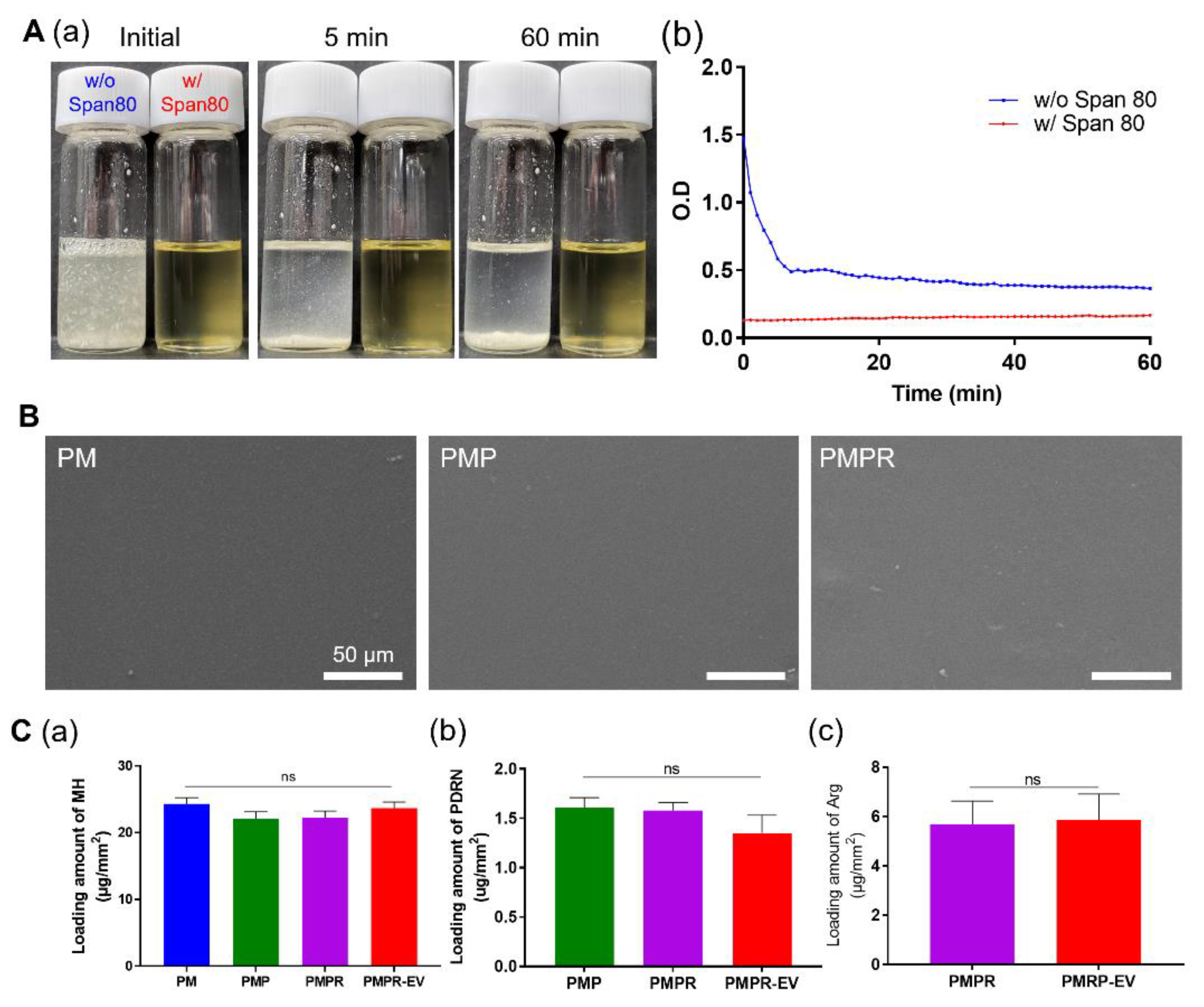
Abstract
:1. Introduction
2. Materials and Methods
2.1. Materials
2.2. Preparation and Characterization of the PLLA Composites
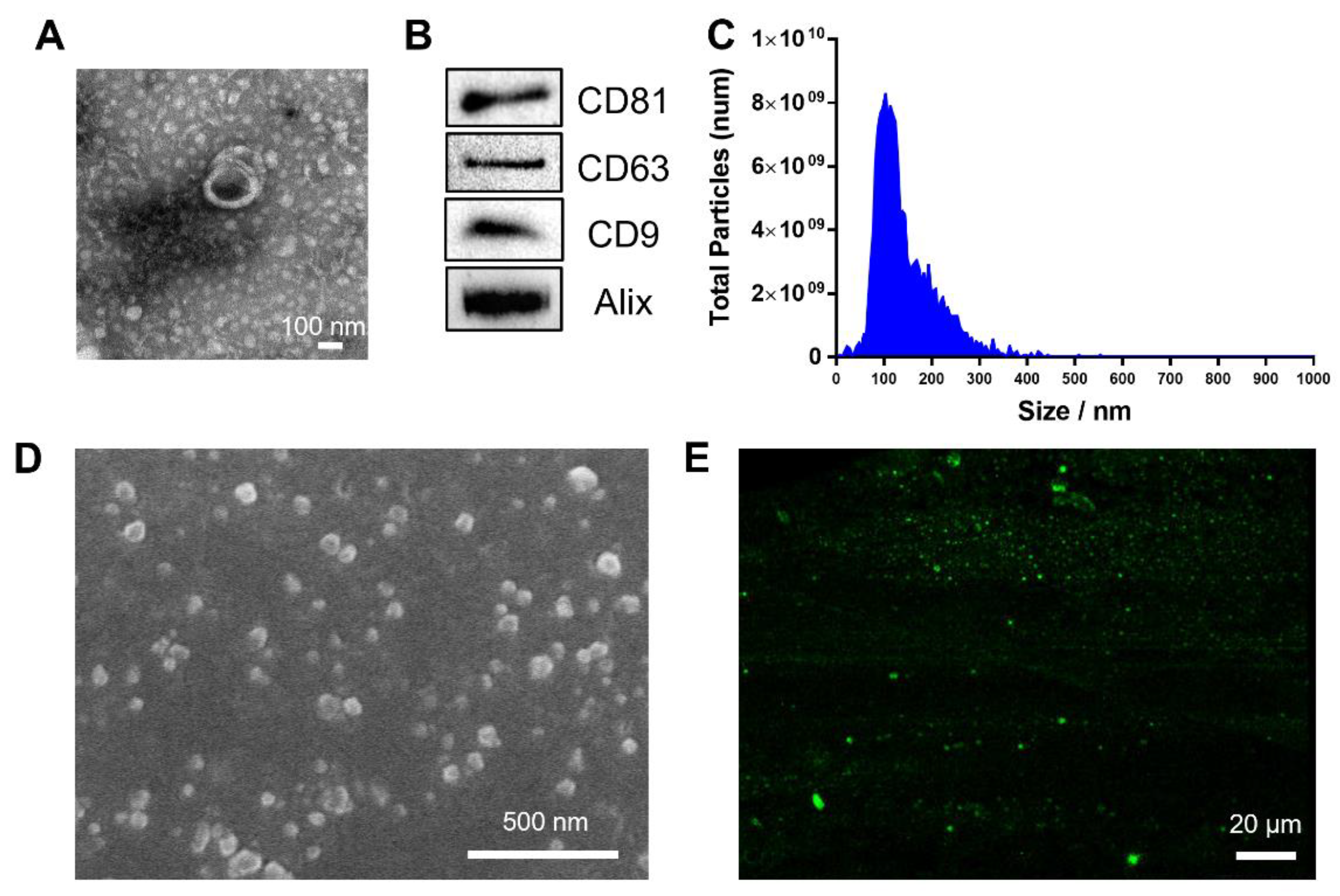
2.3. MSC-Derived EV Isolation
2.4. Characterization of MSC-Derived EVs
2.5. Dispersion Stability Test
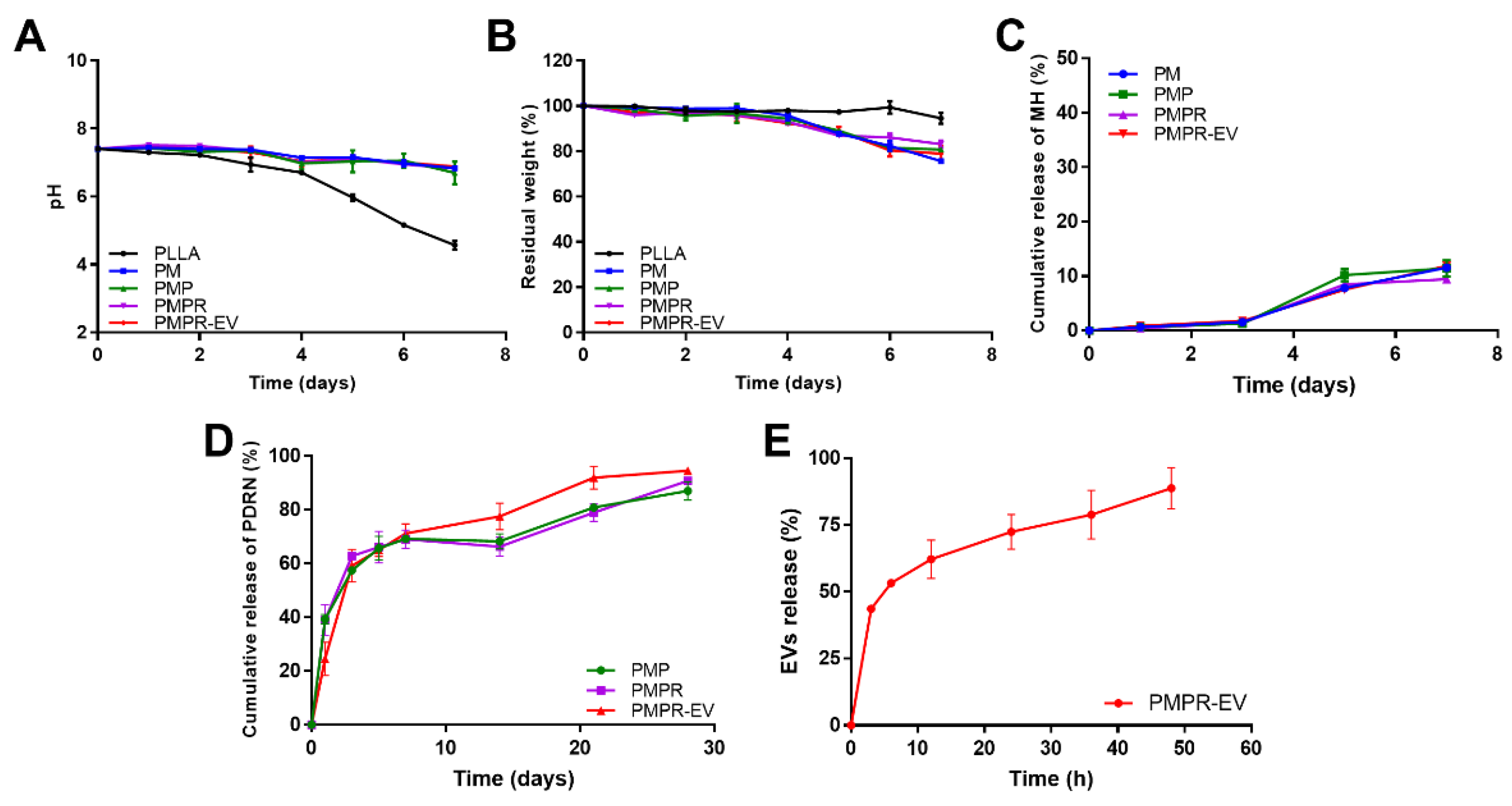
2.6. Degradation and Release Behavior
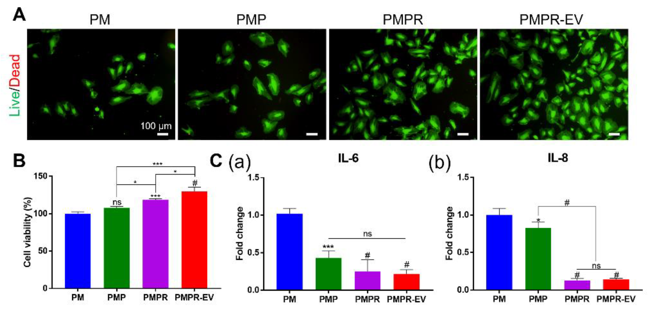
2.7. Cell Culture and Cell Viability Assay
2.8. RNA Extraction and Quantitative Real-Time PCR (qRT-PCR)
2.9. DAF-FM Analysis
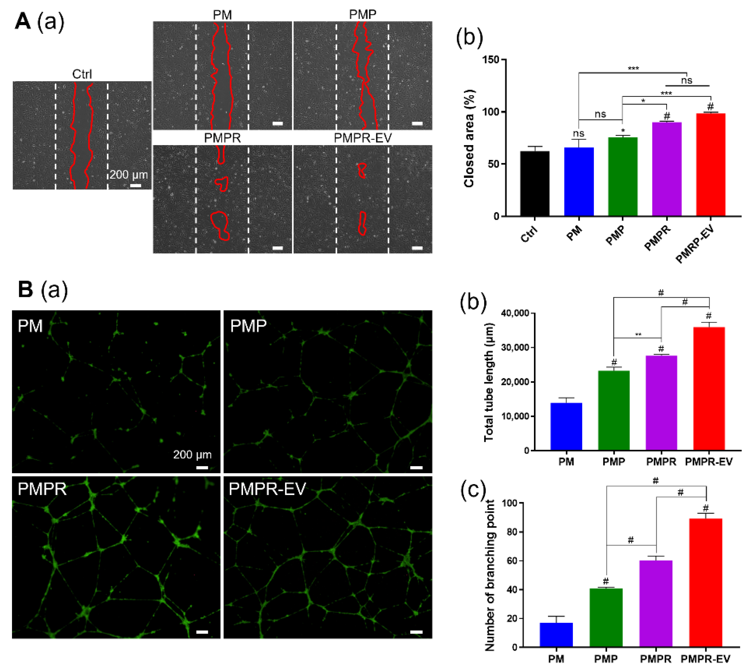
2.10. Wound-Healing Assay/Migration Assay
2.11. Tube Formation Assay
2.12. Statistical Analysis
3. Results and Discussion
3.1. Preparation and Characterization of The PLLA Composites
3.2. Degradation and Release Behavior
3.3. Biocompatibility of The PLLA Composites
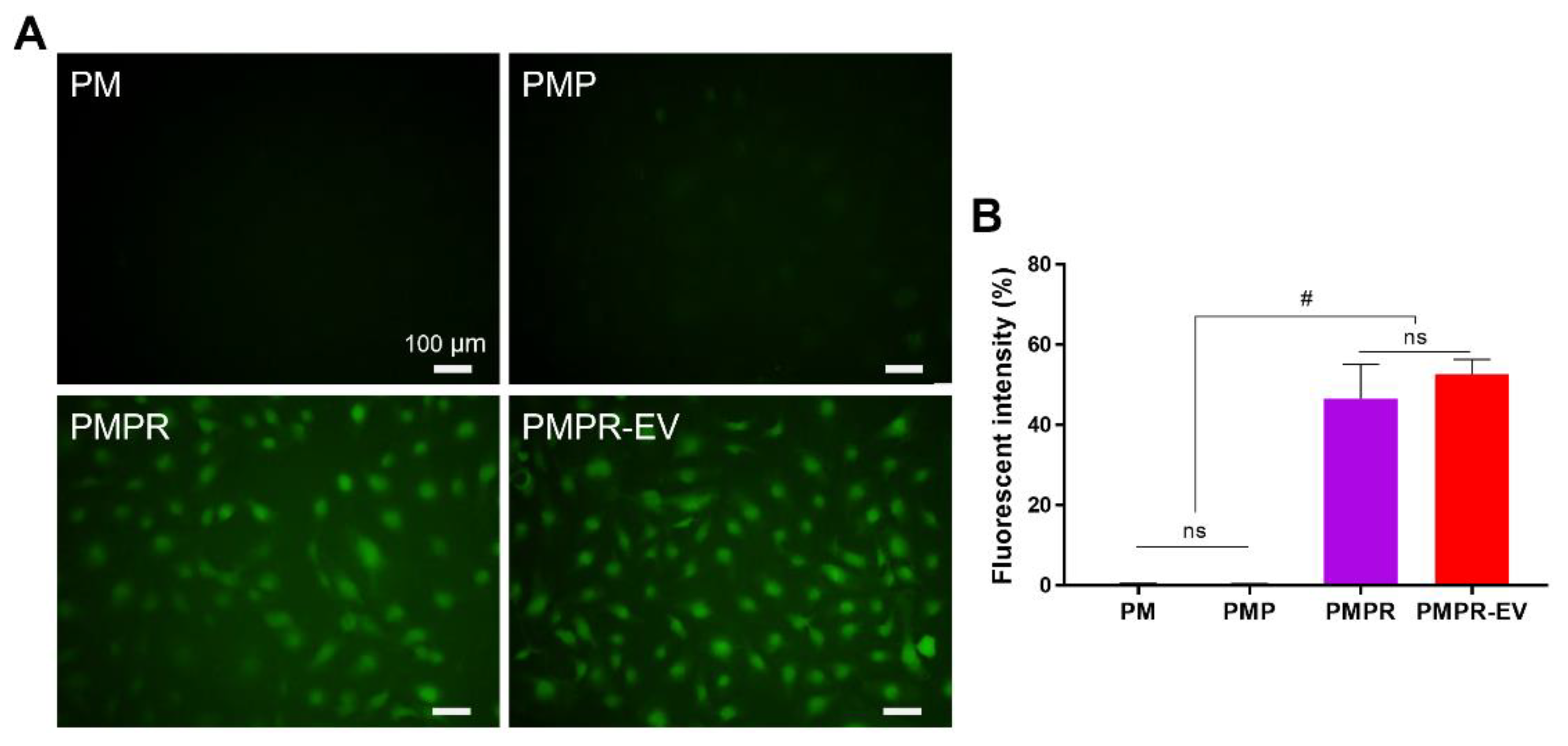
3.4. Confirmation of NO Releasing Ability and Effects of NO
3.5. Various Effects of the Composites on HUVECs
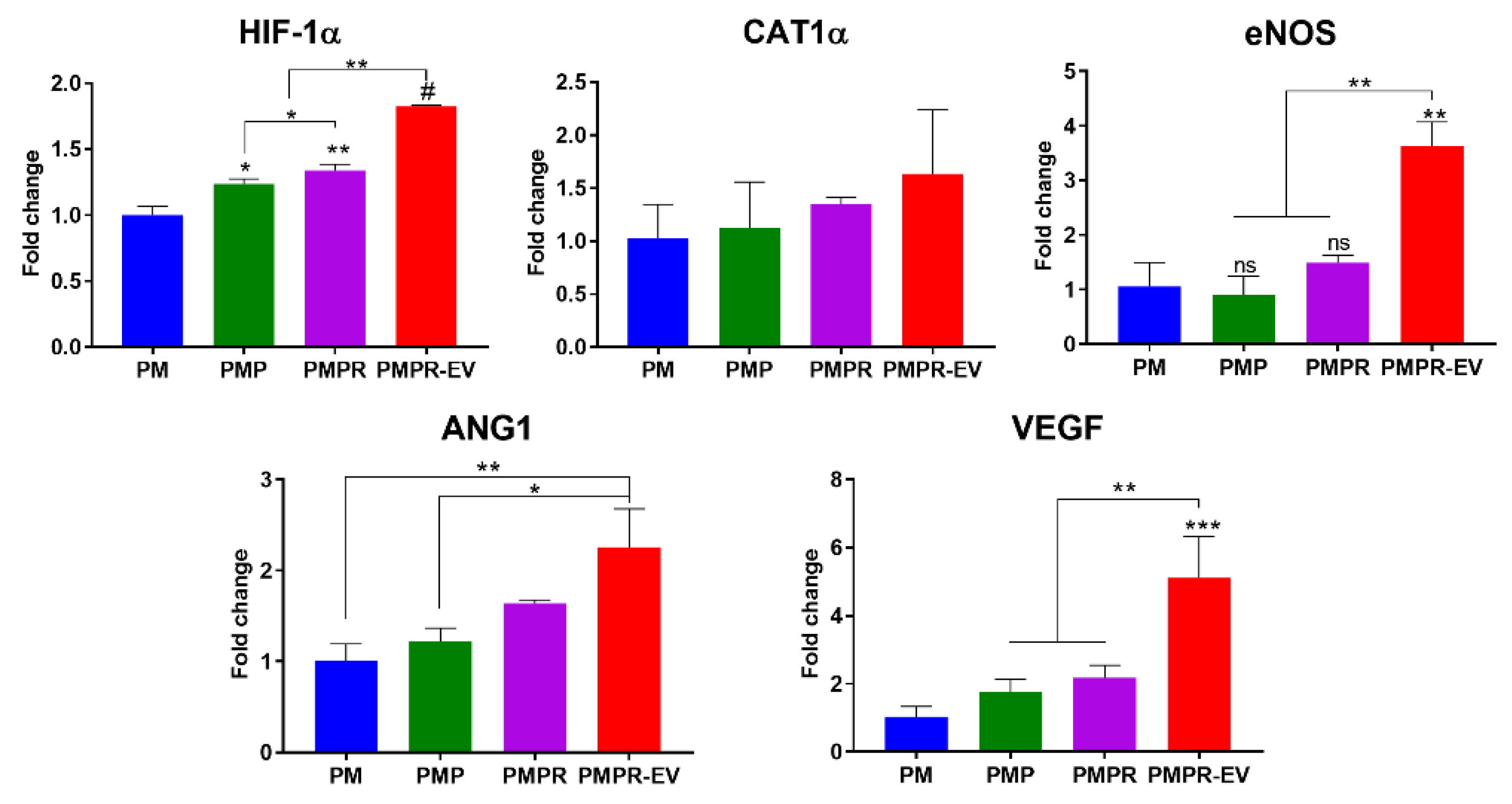
3.6. Genetic Assessment to Verify the Angiogenic Effect
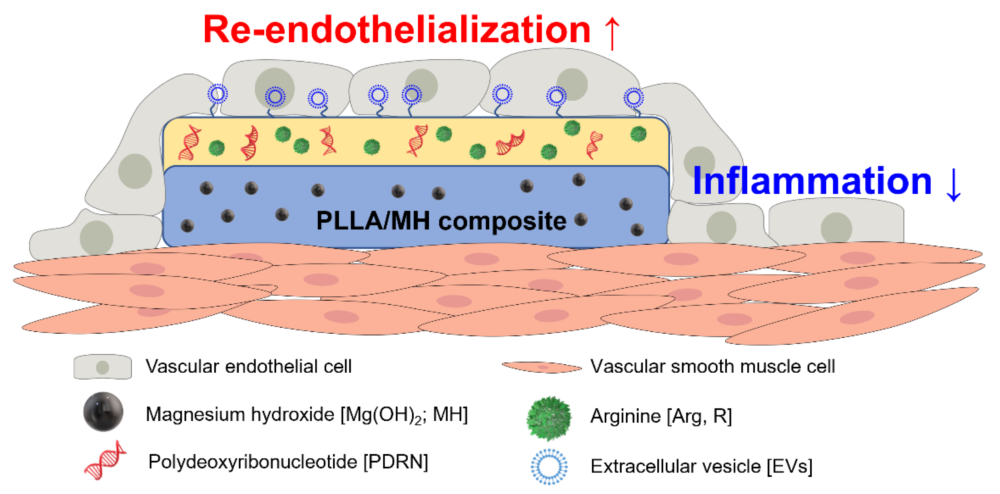
4. Conclusions
Supplementary Materials
Author Contributions
Funding
Institutional Review Board Statement
Informed Consent Statement
Data Availability Statement
Conflicts of Interest
References
- Gunatillake, P.; Mayadunne, R.; Adhikari, R. Recent developments in biodegradable synthetic polymers. Biotechnol. Annu. Rev. 2006, 12, 301–347. [Google Scholar] [PubMed]
- Silvestre-Roig, C.; Braster, Q.; Ortega-Gomez, A.; Soehnlein, O. Neutrophils as regulators of cardiovascular inflammation. Nat. Rev. Cardiol. 2020, 17, 327–340. [Google Scholar] [CrossRef] [PubMed]
- Zhang, W.; Zhang, K.; Li, G.; Yan, S.; Cui, L.; Yin, J. Effects of large dimensional deformation of a porous structure on stem cell fate activated by poly(l-glutamic acid)-based shape memory scaffolds. Biomater. Sci. 2018, 6, 2738–2749. [Google Scholar] [CrossRef] [PubMed]
- Li, L.; Gao, Y.; Liu, Z.; Dong, C.; Wang, W.; Wu, K.; Gu, S.; Zhou, Y. GDF11 alleviates neointimal hyperplasia in a rat model of artery injury by regulating endothelial NLRP3 inflammasome activation and rapid re-endothelialization. J. Transl. Med. 2022, 20, 28. [Google Scholar] [CrossRef] [PubMed]
- Zhang, B.; Yao, R.; Li, L.; Wang, Y.; Luo, R.; Yang, L.; Wang, Y. Green Tea Polyphenol Induced Mg2+-rich Multilayer Conversion Coating: Toward Enhanced Corrosion Resistance and Promoted in Situ Endothelialization of AZ31 for Potential Cardiovascular Applications. ACS Appl. Mater. Interfaces 2019, 11, 41165–41177. [Google Scholar] [CrossRef]
- Kersani, D.; Mougin, J.; Lopez, M.; Degoutin, S.; Tabary, N.; Cazaux, F.; Janus, L.; Maton, M.; Chai, F.; Sobocinski, J.; et al. Stent coating by electrospinning with chitosan/poly-cyclodextrin based nanofibers loaded with simvastatin for restenosis prevention. Eur. J. Pharm. Biopharm. 2020, 150, 156–167. [Google Scholar] [CrossRef] [PubMed]
- Abraham, M.-K.; Jost, E.; Hohmann, J.D.; Searle, A.K.; Bongcaron, V.; Song, Y.; Wendel, H.P.; Peter, K.; Krajewski, S.; Wang, X. A Recombinant Fusion Construct between Human Serum Albumin and NTPDase CD39 Allows Anti-Inflammatory and Anti-Thrombotic Coating of Medical Devices. Pharmaceutics 2021, 13, 1504. [Google Scholar] [CrossRef]
- Bedair, T.M.; Lee, C.K.; Kim, D.-S.; Baek, S.-W.; Bedair, H.M.; Joshi, H.P.; Choi, U.Y.; Park, K.-H.; Park, W.; Han, I.; et al. Magnesium hydroxide-incorporated PLGA composite attenuates inflammation and promotes BMP2-induced bone formation in spinal fusion. J. Tissue Eng. 2020, 11, 2041731420967591. [Google Scholar] [CrossRef]
- Ko, K.-W.; Choi, B.; Kang, E.Y.; Shin, S.-W.; Baek, S.-W.; Han, D.K. The antagonistic effect of magnesium hydroxide particles on vascular endothelial activation induced by acidic PLGA degradation products. Biomater. Sci. 2021, 9, 892–907. [Google Scholar] [CrossRef]
- Omidi, M.; Mansouri, V.; Mohammadi Amirabad, L.; Tayebi, L. Impact of Lipid/Magnesium Hydroxide Hybrid Nanoparticles on the Stability of Vascular Endothelial Growth Factor-Loaded PLGA Microspheres. ACS Appl. Mater. Interfaces 2021, 13, 24370–24384. [Google Scholar] [CrossRef]
- Baek, S.-W.; Kim, J.H.; Song, D.H.; Kim, D.-S.; Park, C.G.; Han, D.K. Enhanced Mechanical Properties and Anti–Inflammation of Poly(L–Lactic Acid) by Stereocomplexes of PLLA/PDLA and Surface–Modified Magnesium Hydroxide Nanoparticles. Polymers 2022, 14, 3790. [Google Scholar] [PubMed]
- Baek, S.-W.; Song, D.H.; Lee, H.I.; Kim, D.-S.; Heo, Y.; Kim, J.H.; Park, C.G.; Han, D.K. Poly(L-Lactic Acid) Composite with Surface-Modified Magnesium Hydroxide Nanoparticles by Biodegradable Oligomer for Augmented Mechanical and Biological Properties. Materials 2021, 14, 5869. [Google Scholar] [CrossRef] [PubMed]
- Kang, E.Y.; Park, S.-B.; Choi, B.; Baek, S.-W.; Ko, K.-W.; Rhim, W.-K.; Park, W.; Kim, I.-H.; Han, D.K. Enhanced mechanical and biological characteristics of PLLA composites through surface grafting of oligolactide on magnesium hydroxide nanoparticles. Biomater. Sci. 2020, 8, 2018–2030. [Google Scholar] [CrossRef] [PubMed]
- Baek, A.; Kim, Y.; Lee, J.W.; Lee, S.C.; Cho, S.R. Effect of polydeoxyribonucleotide on angiogenesis and wound healing in an in vitro model of osteoarthritis. Cell Transpl. 2018, 27, 1623–1633. [Google Scholar] [CrossRef] [PubMed] [Green Version]
- Kim, D.-S.; Lee, J.-K.; Jung, J.-W.; Baek, S.-W.; Kim, J.H.; Heo, Y.; Kim, T.-H.; Han, D.K. Promotion of bone regeneration using bioinspired PLGA/MH/ECM scaffold combined with bioactive PDRN. Materials 2021, 14, 4149. [Google Scholar] [CrossRef]
- Bogdan, C. Nitric oxide and the immune response. Nat. Immunol. 2001, 2, 907–916. [Google Scholar] [CrossRef]
- Calabrese, V.; Mancuso, C.; Calvani, M.; Rizzarelli, E.; Butterfield, D.A.; Giuffrida Stella, A.M. Nitric oxide in the central nervous system: Neuroprotection versus neurotoxicity. Nat. Rev. Neurosci. 2007, 8, 766–775. [Google Scholar] [CrossRef]
- Chang, J.Y.H.; Stamer, W.D.; Bertrand, J.; Read, A.T.; Marando, C.M.; Ethier, C.R.; Overby, D.R. Role of nitric oxide in murine conventional outflow physiology. Am. J. Physiol.-Cell Physiol. 2015, 309, C205–C214. [Google Scholar] [CrossRef] [Green Version]
- Lundberg, J.O.; Gladwin, M.T.; Weitzberg, E. Strategies to increase nitric oxide signalling in cardiovascular disease. Nat. Rev. Drug Discov. 2015, 14, 623–641. [Google Scholar] [CrossRef]
- Yang, T.; Fruergaard, A.S.; Winther, A.K.; Zelikin, A.N.; Chandrawati, R. Zinc oxide particles catalytically generate nitric oxide from endogenous and exogenous prodrugs. Small 2020, 16, 1906744. [Google Scholar] [CrossRef]
- Han, C.; Yu, Q.; Jiang, J.; Zhang, X.; Wang, F.; Jiang, M.; Yu, R.; Deng, T.; Yu, C. Bioenzyme-responsive l-arginine-based carbon dots: The replenishment of nitric oxide for nonpharmaceutical therapy. Biomater. Sci. 2021, 9, 7432–7443. [Google Scholar] [CrossRef] [PubMed]
- Jobgen, W.S.; Fried, S.K.; Fu, W.J.; Meininger, C.J.; Wu, G. Regulatory role for the arginine–nitric oxide pathway in metabolism of energy substrates. J. Nutr. Biochem. 2006, 17, 571–588. [Google Scholar] [CrossRef] [PubMed]
- Liu, A.; Wang, Q.; Zhao, Z.; Wu, R.; Wang, M.; Li, J.; Sun, K.; Sun, Z.; Lv, Z.; Xu, J.; et al. Nitric oxide nanomotor driving exosomes-loaded microneedles for achilles tendinopathy healing. ACS Nano 2021, 15, 13339–13350. [Google Scholar] [CrossRef]
- Li, X.J.; Ren, Z.J.; Tang, J.H.; Yu, Q. Exosomal MicroRNA MiR-1246 promotes cell proliferation, invasion and drug resistance by targeting CCNG2 in breast cancer. Cell. Physiol. Biochem. 2017, 44, 1741–1748. [Google Scholar] [CrossRef] [PubMed]
- Yang, T.; Zhao, F.; Zhou, L.; Liu, J.; Xu, L.; Dou, Q.; Xu, Z.; Jia, R. Therapeutic potential of adipose-derived mesenchymal stem cell exosomes in tissue-engineered bladders. J. Tissue Eng. 2021, 12, 20417314211001545. [Google Scholar] [CrossRef] [PubMed]
- Yea, J.-H.; Yoon, Y.M.; Lee, J.H.; Yun, C.W.; Lee, S.H. Exosomes isolated from melatonin-stimulated mesenchymal stem cells improve kidney function by regulating inflammation and fibrosis in a chronic kidney disease mouse model. J. Tissue Eng. 2021, 12, 20417314211059624. [Google Scholar] [CrossRef]
- Yang, G.H.; Lee, Y.B.; Kang, D.; Choi, E.; Nam, Y.; Lee, K.H.; You, H.-J.; Kang, H.J.; An, S.H.; Jeon, H. Overcome the barriers of the skin: Exosome therapy. Biomater. Res. 2021, 25, 22. [Google Scholar] [CrossRef]
- Kim, J.Y.; Rhim, W.-K.; Yoo, Y.-I.; Kim, D.-S.; Ko, K.-W.; Heo, Y.; Park, C.G.; Han, D.K. Defined MSC exosome with high yield and purity to improve regenerative activity. J. Tissue Eng. 2021, 12, 20417314211008626. [Google Scholar] [CrossRef] [PubMed]
- Go, Y.Y.; Chae, S.-W.; Song, J.-J. Osteogenic effects of exosomes derived from human chorion membrane extracts. Biomater. Res. 2021, 25, 16. [Google Scholar] [CrossRef] [PubMed]
- Kim, J.Y.; Rhim, W.-K.; Seo, H.J.; Lee, J.Y.; Park, C.G.; Han, D.K. Comparative analysis of MSC-derived exosomes depending on cell culture media for regenerative bioactivity. Tissue Eng. Regen. Med. 2021, 18, 355–367. [Google Scholar] [CrossRef]
- Park, S.-Y.; Kim, D.-S.; Kim, H.-M.; Lee, J.-K.; Hwang, D.-Y.; Kim, T.-H.; You, S.; Han, D.K. Human mesenchymal stem cell-derived extracellular vesicles promote neural differentiation of neural progenitor cells. Int. J. Mol. Sci. 2022, 23, 7047. [Google Scholar] [CrossRef] [PubMed]
- Najib, S.B.M.; Kamaruddin, K.S.N.; Rashid, N.M.; Ibrahim, N.; Sokri, M.N.M.; Zaini, N.; Nordin, N. The Effect of MDEA/AMP and Span-80 in Water-in-Oil (W/O) Emulsion for Carbon Dioxide Absorption. J. Appl. Membr. Sci. Technol. 2022, 26, 17–27. [Google Scholar] [CrossRef]
- Woo, J.; Ko, K.-W.; Cha, S.-G.; Heo, Y.; Han, D.K. Comparison of Surface Functionalization of PLGA Composite to Immobilize Extracellular Vesicles. Polymers 2021, 13, 3643. [Google Scholar] [CrossRef] [PubMed]
- Onuma, Y.; Serruys, P.W. Bioresorbable scaffold. Circulation 2011, 123, 779–797. [Google Scholar] [CrossRef] [PubMed] [Green Version]
- Yamashita, K.; Kikkawa, Y.; Kurokawa, K.; Doi, Y. Enzymatic degradation of poly(l-lactide) film by proteinase K: Quartz crystal microbalance and atomic force microscopy study. Biomacromolecules 2005, 6, 850–857. [Google Scholar] [CrossRef]
- Bartkowiak-Jowsa, M.; Będziński, R.; Kozłowska, A.; Filipiak, J.; Pezowicz, C. Mechanical, rheological, fatigue, and degradation behavior of PLLA, PGLA and PDGLA as materials for vascular implants. Meccanica 2013, 48, 721–731. [Google Scholar] [CrossRef] [Green Version]
- Lee, J.H.; Kim, E.D.; Jun, E.J.; Yoo, H.S.; Lee, J.W. Analysis of trends and prospects regarding stents for human blood vessels. Biomater. Res. 2018, 22, 8. [Google Scholar] [CrossRef] [Green Version]
- Wang, Y.; Zhang, X. Vascular restoration therapy and bioresorbable vascular scaffold. Regen. Biomater. 2014, 1, 49–55. [Google Scholar] [CrossRef]
- Jia, L.; Zhou, X.; Huang, X.; Xu, X.; Jia, Y.; Wu, Y.; Yao, J.; Wu, Y.; Wang, K. Maternal and umbilical cord serum-derived exosomes enhance endothelial cell proliferation and migration. FASEB J. 2018, 32, 4534–4543. [Google Scholar] [CrossRef] [Green Version]
- Namin, S.M.; Nofallah, S.; Joshi, M.S.; Kavallieratos, K.; Tsoukias, N.M. Kinetic analysis of DAF-FM activation by NO: Toward calibration of a NO-sensitive fluorescent dye. Nitric Oxide 2013, 28, 39–46. [Google Scholar] [CrossRef] [PubMed]
- Nasuno, R.; Shino, S.; Yoshikawa, Y.; Yoshioka, N.; Sato, Y.; Kamiya, K.; Takagi, H. Detection system of the intracellular nitric oxide in yeast by HPLC with a fluorescence detector. Anal. Biochem. 2020, 598, 113707. [Google Scholar] [CrossRef] [PubMed]
- Wen, C.; Zhang, J.; Li, Y.; Zheng, W.; Liu, M.; Zhu, Y.; Sui, X.; Zhang, X.; Han, Q.; Lin, Y.; et al. A zwitterionic hydrogel coated titanium surface with high-efficiency endothelial cell selectivity for rapid re-endothelialization. Biomater. Sci. 2020, 8, 5441–5451. [Google Scholar] [CrossRef] [PubMed]
- Gong, M.; Yu, B.; Wang, J.; Wang, Y.; Liu, M.; Paul, C.; Millard, R.W.; Xiao, D.-S.; Ashraf, M.; Xu, M. Mesenchymal stem cells release exosomes that transfer miRNAs to endothelial cells and promote angiogenesis. Oncotarget 2017, 8, 45200–45212. [Google Scholar] [CrossRef] [PubMed] [Green Version]
- Salimian Rizi, B.; Achreja, A.; Nagrath, D. Nitric oxide: The forgotten child of tumor metabolism. Trends Cancer 2017, 3, 659–672. [Google Scholar] [CrossRef]
- Zhang, L.; Jiao, G.; Ren, S.; Zhang, X.; Li, C.; Wu, W.; Wang, H.; Liu, H.; Zhou, H.; Chen, Y. Exosomes from bone marrow mesenchymal stem cells enhance fracture healing through the promotion of osteogenesis and angiogenesis in a rat model of nonunion. Stem Cell Res. Ther. 2020, 11, 38. [Google Scholar] [CrossRef] [PubMed]


Publisher’s Note: MDPI stays neutral with regard to jurisdictional claims in published maps and institutional affiliations. |
© 2022 by the authors. Licensee MDPI, Basel, Switzerland. This article is an open access article distributed under the terms and conditions of the Creative Commons Attribution (CC BY) license (https://creativecommons.org/licenses/by/4.0/).
Share and Cite
Baek, S.-W.; Kim, D.-S.; Song, D.H.; Lee, S.; Lee, J.-K.; Park, S.-Y.; Kim, J.H.; Kim, T.-H.; Park, C.G.; Han, D.K. PLLA Composites Combined with Delivery System of Bioactive Agents for Anti-Inflammation and Re-Endothelialization. Pharmaceutics 2022, 14, 2661. https://doi.org/10.3390/pharmaceutics14122661
Baek S-W, Kim D-S, Song DH, Lee S, Lee J-K, Park S-Y, Kim JH, Kim T-H, Park CG, Han DK. PLLA Composites Combined with Delivery System of Bioactive Agents for Anti-Inflammation and Re-Endothelialization. Pharmaceutics. 2022; 14(12):2661. https://doi.org/10.3390/pharmaceutics14122661
Chicago/Turabian StyleBaek, Seung-Woon, Da-Seul Kim, Duck Hyun Song, Semi Lee, Jun-Kyu Lee, So-Yeon Park, Jun Hyuk Kim, Tae-Hyung Kim, Chun Gwon Park, and Dong Keun Han. 2022. "PLLA Composites Combined with Delivery System of Bioactive Agents for Anti-Inflammation and Re-Endothelialization" Pharmaceutics 14, no. 12: 2661. https://doi.org/10.3390/pharmaceutics14122661