
Tocotrienol in the Treatment of Topical Wounds: Recent Updates

 , , , , , and
Abstract
:1. Introduction
2. Topical Wounds
3. Tocotrienol: A Form of Vitamin E with a Superior Role
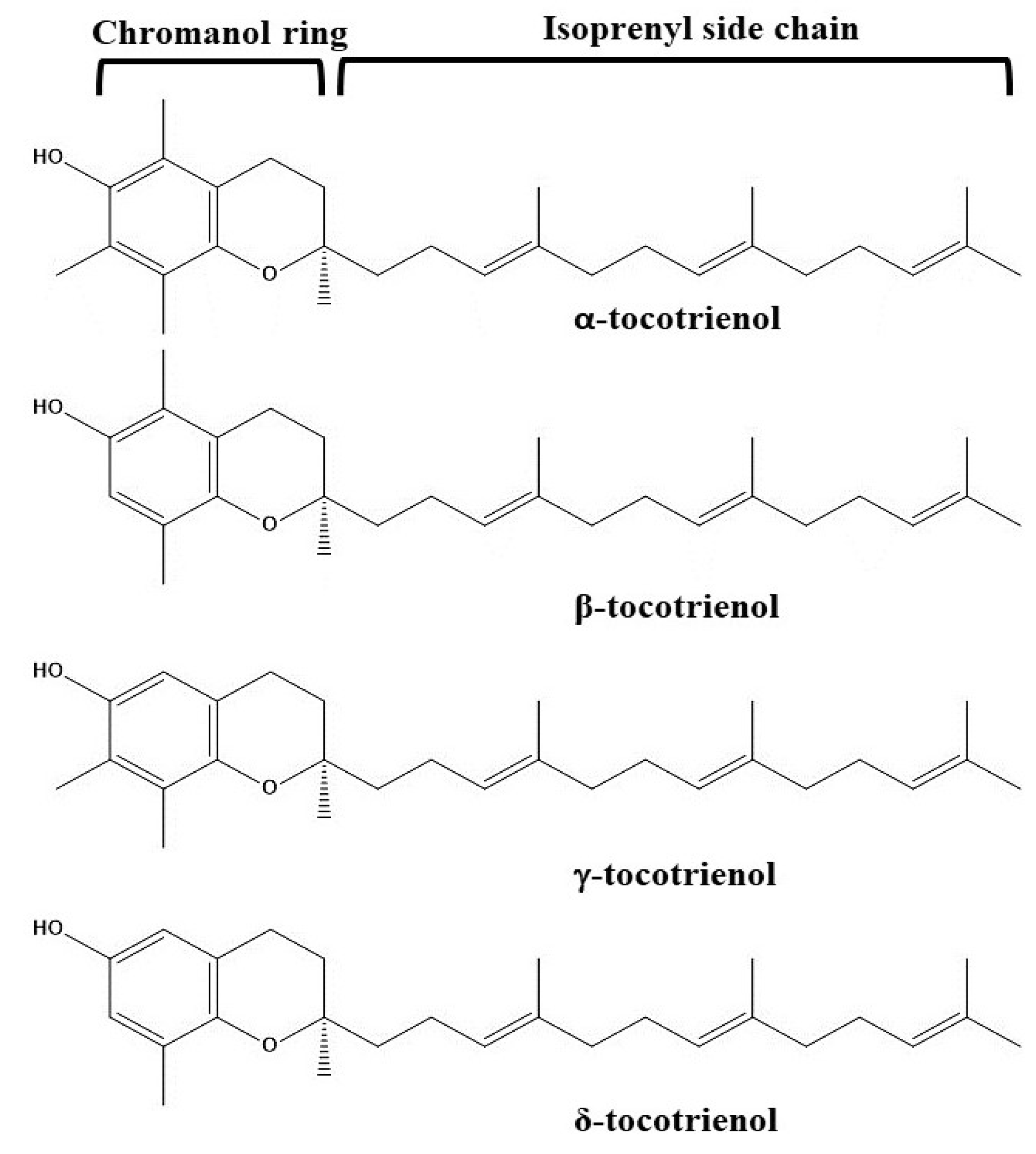
Types of Tocotrienols
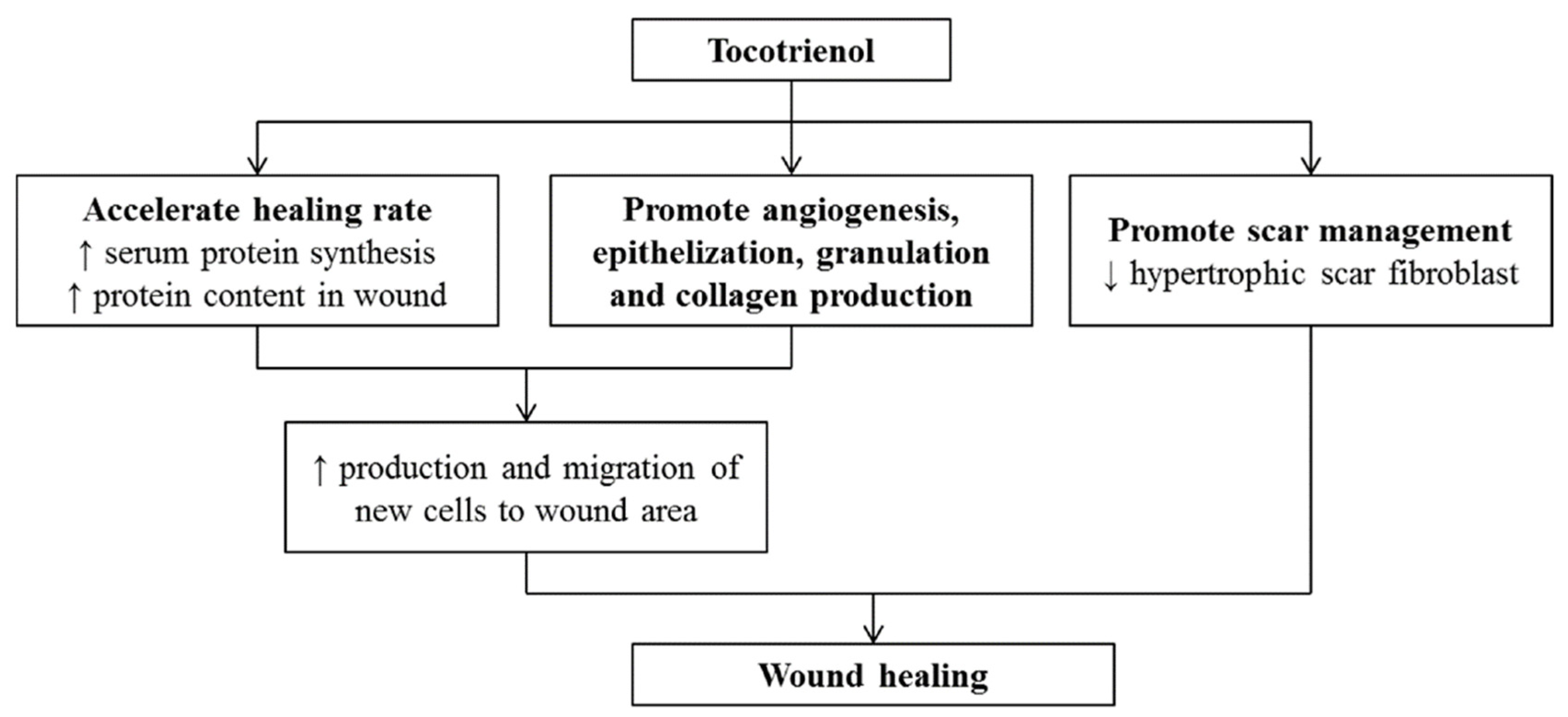
4. Mechanisms of Tocotrienols in Wound Healing
5. Application of Tocotrienols in the Treatment of Wounds: Recent Update
6. Challenges of Tocotrienol Application
7. Conclusions and Future Trend
Author Contributions
Funding
Institutional Review Board Statement
Informed Consent Statement
Data Availability Statement
Acknowledgments
Conflicts of Interest
References
- Santos, A.M.N.; Moreira, A.P.D.; Carvalho, C.W.P.; Luchese, R.; Ribeiro, E.; McGuinness, G.B.; Mendes, M.F.; Oliveira, R.N. Physically Cross-Linked Gels of PVA with Natural Polymers as Matrices for Manuka Honey Release in Wound-Care Applications. Materials 2019, 12, 559. [Google Scholar] [CrossRef] [PubMed] [Green Version]
- Akrawi, S.H.; Gorain, B.; Nair, A.B.; Choudhury, H.; Pandey, M.; Shah, J.N.; Venugopala, K.N. Development and Optimization of Naringenin-Loaded Chitosan-Coated Nanoemulsion for Topical Therapy in Wound Healing. Pharmaceutics 2020, 12, 893. [Google Scholar] [CrossRef] [PubMed]
- Liu, H.; Wang, C.; Li, C.; Qin, Y.; Wang, Z.; Yang, F.; Li, Z.; Wang, J. A Functional Chitosan-Based Hydrogel as a Wound Dressing and Drug Delivery System in the Treatment of Wound Healing. RSC Adv. 2018, 8, 7533–7549. [Google Scholar] [CrossRef] [Green Version]
- Cho, C.Y.; Lo, J.S. Dressing the Part. Dermatol. Clin. 1998, 16, 25–47. [Google Scholar] [CrossRef]
- Winter, G.D. Formation of the Scab and the Rate of Epithelization of Superficial Wounds in the Skin of the Young Domestic Pig. Nature 1962, 193, 293–294. [Google Scholar] [CrossRef]
- Jeckson, T.A.; Neo, Y.P.; Sisinthy, S.P.; Gorain, B. Delivery of Therapeutics from Layer-by-Layer Electrospun Nanofiber Matrix for Wound Healing: An Update. J. Pharm. Sci. 2021, 110, 635–653. [Google Scholar] [CrossRef] [PubMed]
- Choudhury, H.; Pandey, M.; Hua, C.K.; Mun, C.S.; Jing, J.K.; Kong, L.; Ern, L.Y.; Ashraf, N.A.; Kit, S.W.; Yee, T.S.; et al. An Update on Natural Compounds in the Remedy of Diabetes Mellitus: A Systematic Review. J. Tradit. Complement. Med. 2018, 8, 361–376. [Google Scholar] [CrossRef]
- Gorain, B.; Pandey, M.; Leng, N.H.; Yan, C.W.; Nie, K.W.; Kaur, S.J.; Marshall, V.; Sisinthy, S.P.; Panneerselvam, J.; Molugulu, N.; et al. Advanced Drug Delivery Systems Containing Herbal Components for Wound Healing. Int. J. Pharm. 2022, 617, 121617. [Google Scholar] [CrossRef]
- Sharma, A.; Khanna, S.; Kaur, G.; Singh, I. Medicinal Plants and Their Components for Wound Healing Applications. Futur. J. Pharm. Sci. 2021, 7, 53. [Google Scholar] [CrossRef]
- Salehi, B.; Stojanović-Radić, Z.; Matejić, J.; Sharifi-Rad, M.; Anil Kumar, N.V.; Martins, N.; Sharifi-Rad, J. The Therapeutic Potential of Curcumin: A Review of Clinical Trials. Eur. J. Med. Chem. 2019, 163, 527–545. [Google Scholar] [CrossRef]
- Ryan, J.L.; Heckler, C.E.; Ling, M.; Katz, A.; Williams, J.P.; Pentland, A.P.; Morrow, G.R. Curcumin for Radiation Dermatitis: A Randomized, Double-Blind, Placebo-Controlled Clinical Trial of Thirty Breast Cancer Patients. Radiat. Res. 2013, 180, 34–43. [Google Scholar] [CrossRef] [PubMed] [Green Version]
- Heng, M.C.Y.; Song, M.K.; Harker, J.; Heng, M.K. Drug-Induced Suppression of Phosphorylase Kinase Activity Correlates with Resolution of Psoriasis as Assessed by Clinical, Histological and Immunohistochemical Parameters. Br. J. Dermatol. 2000, 143, 937–949. [Google Scholar] [CrossRef] [PubMed]
- Iwasaki, K.; Kobayashi, S.; Chimura, Y.; Taguchi, M.; Inoue, K.; Cho, S.; Akiba, T.; Arai, H.; Cyong, J.C.; Sasaki, H. A Randomized, Double-Blind, Placebo-Controlled Clinical Trial of the Chinese Herbal Medicine “Ba Wei Di Huang Wan” in the Treatment of Dementia. J. Am. Geriatr. Soc. 2004, 52, 1518–1521. [Google Scholar] [CrossRef] [PubMed]
- Kummar, S.; Sitki Copur, M.; Rose, M.; Wadler, S.; Stephenson, J.; O’Rourke, M.; Brenckman, W.; Tilton, R.; Liu, S.H.; Jiang, Z.; et al. A Phase I Study of the Chinese Herbal Medicine PHY906 as a Modulator of Irinotecan-Based Chemotherapy in Patients with Advanced Colorectal Cancer. Clin. Colorectal Cancer 2011, 10, 85–96. [Google Scholar] [CrossRef]
- Heyer, K.; Herberger, K.; Protz, K.; Glaeske, G.; Augustin, M. Epidemiology of Chronic Wounds in Germany: Analysis of Statutory Health Insurance Data. Wound Repair Regen. 2016, 24, 434–442. [Google Scholar] [CrossRef]
- Guest, J.F.; Ayoub, N.; McIlwraith, T.; Uchegbu, I.; Gerrish, A.; Weidlich, D.; Vowden, K.; Vowden, P. Health Economic Burden That Different Wound Types Impose on the UK’s National Health Service. Int. Wound J. 2017, 14, 322–330. [Google Scholar] [CrossRef]
- Chong, W.T.; Tan, C.P.; Cheah, Y.K.; Lai, O.M. In-Vitro and in-Vivo Evaluations of Tocotrienol-Rich Nanoemulsified System on Skin Wound Healing. PLoS ONE 2022, 17, e0267381. [Google Scholar] [CrossRef]
- Szewczyk, K.; Chojnacka, A.; Górnicka, M. Tocopherols and Tocotrienols—Bioactive Dietary Compounds; What Is Certain, What Is Doubt? Int. J. Mol. Sci. 2021, 22, 6222. [Google Scholar] [CrossRef]
- Sen, C.K.; Gordillo, G.M.; Roy, S.; Kirsner, R.; Lambert, L.; Hunt, T.K.; Gottrup, F.; Gurtner, G.C.; Longaker, M.T. Human Skin Wounds: A Major and Snowballing Threat to Public Health and the Economy. Wound Repair Regen. 2009, 17, 763–771. [Google Scholar] [CrossRef] [Green Version]
- Singer, A.J.; Clark, R.A.F. Cutaneous Wound Healing. N. Engl. J. Med. 1999, 341, 738–746. [Google Scholar] [CrossRef]
- Schreml, S.; Szeimies, R.M.; Prantl, L.; Karrer, S.; Landthaler, M.; Babilas, P. Oxygen in Acute and Chronic Wound Healing. Br. J. Dermatol. 2010, 163, 257–268. [Google Scholar] [CrossRef] [PubMed]
- Singh, M.R.; Saraf, S.; Vyas, A.; Jain, V.; Singh, D. Innovative Approaches in Wound Healing: Trajectory and Advances. Artif. Cells Nanomed. Biotechnol. 2013, 41, 202–212. [Google Scholar] [CrossRef] [PubMed]
- Chhabra, S.; Chhabra, N.; Kaur, A.; Gupta, N. Wound Healing Concepts in Clinical Practice of OMFS. J. Maxillofac. Oral Surg. 2017, 16, 423. [Google Scholar] [CrossRef]
- Sharma, M.; Khajja, B.; Hainendra, J.; Mathur, G. Forensic Interpretation of Injuries/Wounds Found on the Human Body. J. Punjab Acad. Forensic Med. Toxicol. 2011, 11, 105–109. [Google Scholar]
- Jiang, Q. Natural Forms of Vitamin E: Metabolism, Antioxidant, and Anti-Inflammatory Activities and Their Role in Disease Prevention and Therapy. Free Radic. Biol. Med. 2014, 72, 76–90. [Google Scholar] [CrossRef] [Green Version]
- Mahdy, Z.A.; Chin, K.Y.; Nik-Ahmad-zuky, N.L.; Kalok, A.; Rahman, R.A. Tocotrienol in Pre-Eclampsia Prevention: A Mechanistic Analysis in Relation to the Pathophysiological Framework. Cells 2022, 11, 614. [Google Scholar] [CrossRef] [PubMed]
- Niu, Y.; Zhang, Q.; Wang, J.; Li, Y.; Wang, X.; Bao, Y. Vitamin E Synthesis and Response in Plants. Front. Plant Sci. 2022, 13, 994058. [Google Scholar] [CrossRef] [PubMed]
- Chin, K.Y.; Ima-Nirwana, S. The Biological Effects of Tocotrienol on Bone: A Review on Evidence from Rodent Models. Drug Des. Devel. Ther. 2015, 9, 2061. [Google Scholar] [CrossRef] [Green Version]
- Lim, Y.; Traber, M.G. Alpha-Tocopherol Transfer Protein (α-TTP): Insights from Alpha-Tocopherol Transfer Protein Knockout Mice. Nutr. Res. Pract. 2007, 1, 253. [Google Scholar] [CrossRef] [Green Version]
- Lashkari, S.; Krogh Jensen, S.; Bernes, G. Biodiscrimination of α-Tocopherol Stereoisomers in Plasma and Tissues of Lambs Fed Different Proportions of All-Rac-α-Tocopheryl Acetate and RRR-α-Tocopheryl Acetate. J. Anim. Sci. 2019, 97, 1233. [Google Scholar] [CrossRef]
- Jiang, Q.; Christen, S.; Shigenaga, M.K.; Ames, B.N. γ-Tocopherol, the Major Form of Vitamin E in the US Diet, Deserves More Attention. Am. J. Clin. Nutr. 2001, 74, 714–722. [Google Scholar] [CrossRef] [PubMed]
- Sen, C.K.; Khanna, S.; Roy, S. Tocotrienols: Vitamin E beyond Tocopherols. Life Sci. 2006, 78, 2088–2098. [Google Scholar] [CrossRef] [PubMed] [Green Version]
- Zafar, H.; Mirza, I.A.; Hussain, W.; Fayyaz, M. Comparative Efficacy of Tocotrienol and Tocopherol for Their Anti Diabetic Effects. Biomed. J. Sci. Tech. Res. 2021, 38, 30835–30840. [Google Scholar] [CrossRef]
- Wong, S.K.; Chin, K.Y.; Suhaimi, F.H.; Ahmad, F.; Ima-Nirwana, S. The Effects of Palm Tocotrienol on Metabolic Syndrome and Bone Loss in Male Rats Induced by High-Carbohydrate High-Fat Diet. J. Funct. Foods 2018, 44, 246–254. [Google Scholar] [CrossRef]
- Wong, S.K.; Chin, K.Y.; Ima-Nirwana, S. The Effects of Tocotrienol on Bone Peptides in a Rat Model of Osteoporosis Induced by Metabolic Syndrome: The Possible Communication between Bone Cells. Int. J. Environ. Res. Public Health 2019, 16, 3313. [Google Scholar] [CrossRef] [Green Version]
- Pang, K.L.; Chin, K.Y. The Role of Tocotrienol in Protecting against Metabolic Diseases. Molecules 2019, 24, 923. [Google Scholar] [CrossRef] [Green Version]
- Xia, W.; Mo, H. Potential of Tocotrienols in the Prevention and Therapy of Alzheimer’s Disease. J. Nutr. Biochem. 2016, 31, 1–9. [Google Scholar] [CrossRef]
- Nakaso, K.; Tajima, N.; Horikoshi, Y.; Nakasone, M.; Hanaki, T.; Kamizaki, K.; Matsura, T. The Estrogen Receptor β-PI3K/Akt Pathway Mediates the Cytoprotective Effects of Tocotrienol in a Cellular Parkinson’s Disease Model. Biochim. Biophys. Acta-Mol. Basis Dis. 2014, 1842, 1303–1312. [Google Scholar] [CrossRef] [Green Version]
- Park, H.A.; Kubicki, N.; Gnyawali, S.; Chan, Y.C.; Roy, S.; Khanna, S.; Sen, C.K. Natural Vitamin E α-Tocotrienol Protects Against Ischemic Stroke by Induction of Multidrug Resistance-Associated Protein 1. Stroke 2011, 42, 2308–2314. [Google Scholar] [CrossRef] [Green Version]
- Rink, C.; Christoforidis, G.; Khanna, S.; Peterson, L.; Patel, Y.; Khanna, S.; Abduljalil, A.; Irfanoglu, O.; MacHiraju, R.; Bergdall, V.K.; et al. Tocotrienol Vitamin e Protects against Preclinical Canine Ischemic Stroke by Inducing Arteriogenesis. J. Cereb. Blood Flow Metab. 2011, 31, 2218–2230. [Google Scholar] [CrossRef] [Green Version]
- Visioli, F.; Balestrieri, M.L.; Shaikh, S.A.; Varatharajan, R.; Muthuraman, A. Palm Oil Derived Tocotrienol-Rich Fraction Attenuates Vascular Dementia in Type 2 Diabetic Rats. Int. J. Mol. Sci. 2022, 23, 13531. [Google Scholar] [CrossRef]
- Tiwari, V.; Kuhad, A.; Chopra, K. Suppression of Neuro-Inflammatory Signaling Cascade by Tocotrienol Can Prevent Chronic Alcohol-Induced Cognitive Dysfunction in Rats. Behav. Brain Res. 2009, 203, 296–303. [Google Scholar] [CrossRef] [PubMed]
- Kumari, M.; Ramdas, P.; Radhakrishnan, A.K.; Haleagrahara, N.; Kutty, M.K. Tocotrienols Ameliorate Neurodegeneration and Motor Deficits in the 6-OHDA-Induced Rat Model of Parkinsonism: Behavioural and Immunohistochemistry Analysis. Nutrients 2021, 13, 1583. [Google Scholar] [CrossRef] [PubMed]
- Nakaso, K.; Horikoshi, Y.; Takahashi, T.; Hanaki, T.; Nakasone, M.; Kitagawa, Y.; Koike, T.; Matsura, T. Estrogen Receptor-Mediated Effect of δ-Tocotrienol Prevents Neurotoxicity and Motor Deficit in the MPTP Mouse Model of Parkinson’s Disease. Neurosci. Lett. 2016, 610, 117–122. [Google Scholar] [CrossRef]
- Sadikan, M.Z.; Nasir, N.A.A.; Agarwal, R.; Ismail, N.M. Protective Effect of Palm Oil-Derived Tocotrienol-Rich Fraction Against Retinal Neurodegenerative Changes in Rats with Streptozotocin-Induced Diabetic Retinopathy. Biomolecules 2020, 10, 556. [Google Scholar] [CrossRef] [Green Version]
- Abdul Nasir, N.A.; Agarwal, R.; Vasudevan, S.; Tripathy, M.; Alyautdin, R.; Mohd Ismail, N. Effects of Topically Applied Tocotrienol on Cataractogenesis and Lens Redox Status in Galactosemic Rats. Mol. Vis. 2014, 20, 835. [Google Scholar]
- Teo, C.W.L.; Tay, S.H.Y.; Tey, H.L.; Ung, Y.W.; Yap, W.N. Vitamin E in Atopic Dermatitis: From Preclinical to Clinical Studies. Dermatology 2021, 237, 553–564. [Google Scholar] [CrossRef]
- Chang, P.N.; Yap, W.N.; Wing Lee, D.T.; Ling, M.T.; Wong, Y.C.; Yap, Y.L. Evidence of γ-Tocotrienol as an Apoptosis-Inducing, Invasion-Suppressing, and Chemotherapy Drug-Sensitizing Agent in Human Melanoma Cells. Nutr. Cancer 2009, 61, 357–366. [Google Scholar] [CrossRef]
- Fernandes, N.V.; Guntipalli, P.K.; Mo, H. D-δ-Tocotrienol-Mediated Cell Cycle Arrest and Apoptosis in Human Melanoma Cells. Anticancer Res. 2010, 30, 4937–4944. [Google Scholar]
- Ji, X.; Wang, Z.; Geamanu, A.; Sarkar, F.H.; Gupta, S.V. Inhibition of Cell Growth and Induction of Apoptosis in Non-Small Cell Lung Cancer Cells by Delta-Tocotrienol Is Associated with Notch-1 down-Regulation. J. Cell. Biochem. 2011, 112, 2773–2783. [Google Scholar] [CrossRef]
- Peh, H.Y.; Tan, W.S.D.; Chan, T.K.K.; Pow, C.W.; Foster, P.S.; Wong, W.S.F. Vitamin E Isoform γ-Tocotrienol Protects against Emphysema in Cigarette Smoke-Induced COPD. Free Radic. Biol. Med. 2017, 110, 332–344. [Google Scholar] [CrossRef] [PubMed]
- Ji, X.; Yao, H.; Meister, M.; Gardenhire, D.S.; Mo, H. Tocotrienols: Dietary Supplements for Chronic Obstructive Pulmonary Disease. Antioxidants 2021, 10, 883. [Google Scholar] [CrossRef] [PubMed]
- Norsidah, K.Z.; Asmadi, A.Y.; Azizi, A.; Faizah, O.; Kamisah, Y. Palm Tocotrienol-Rich Fraction Reduced Plasma Homocysteine and Heart Oxidative Stress in Rats Fed with a High-Methionine Diet. J. Physiol. Biochem. 2013, 69, 441–449. [Google Scholar] [CrossRef]
- Ramli, F.F.; Ali, A.; Ibrahim, N.I. Protective Effects of Tocotrienols in Cerebral and Myocardial Ischemia-Reperfusion Injury: A Systematic Review. Appl. Sci. 2021, 11, 7994. [Google Scholar] [CrossRef]
- Ramanathan, N.; Tan, E.; Loh, L.J.; Soh, B.S.; Yap, W.N. Tocotrienol Is a Cardioprotective Agent against Ageing-Associated Cardiovascular Disease and Its Associated Morbidities. Nutr. Metab. 2018, 15, 6. [Google Scholar] [CrossRef] [Green Version]
- Nur Azlina, M.F.; Nafeeza, M.I. Tocotrienol and Alpha-Tocopherol Reduce Corticosterone and Noradrenalin Levels in Rats Exposed to Restraint Stress. Pharmazie 2008, 63, 890–892. [Google Scholar] [CrossRef] [PubMed]
- Nur Azlina, M.F.; Kamisah, Y.; Chua, K.H.; Qodriyah, H.M.S. Tocotrienol Attenuates Stress-Induced Gastric Lesions via Activation of Prostaglandin and Upregulation of COX-1 MRNA. Evid.-Based Complement. Altern. Med. 2013, 2013, 804796. [Google Scholar] [CrossRef] [Green Version]
- Manu, K.A.; Shanmugam, M.K.; Ramachandran, L.; Li, F.; Fong, C.W.; Kumar, A.P.; Tan, P.; Sethi, G. First Evidence That γ-Tocotrienol Inhibits the Growth of Human Gastric Cancer and Chemosensitizes It to Capecitabine in a Xenograft Mouse Model through the Modulation of NF-ΚB Pathway. Clin. Cancer Res. 2012, 18, 2220–2229. [Google Scholar] [CrossRef] [Green Version]
- Magosso, E.; Ansari, M.A.; Gopalan, Y.; Shuaib, I.L.; Wong, J.W.; Khan, N.A.K.; Abu Bakar, M.R.; Ng, B.H.; Yuen, K.H. Tocotrienols for Normalisation of Hepatic Echogenic Response in Nonalcoholic Fatty Liver: A Randomised Placebo-Controlled Clinical Trial. Nutr. J. 2013, 12, 166. [Google Scholar] [CrossRef] [Green Version]
- Saw, T.Y.; Malik, N.A.; Lim, K.P.; Teo, C.W.L.; Wong, E.S.M.; Kong, S.C.; Fong, C.W.; Petkov, J.; Yap, W.N. Oral Supplementation of Tocotrienol-Rich Fraction Alleviates Severity of Ulcerative Colitis in Mice. J. Nutr. Sci. Vitaminol. 2019, 65, 318–327. [Google Scholar] [CrossRef] [Green Version]
- Luna, J.; Masamunt, M.C.; Rickmann, M.; Mora, R.; España, C.; Delgado, S.; Llach, J.; Vaquero, E.; Sans, M. Tocotrienols Have Potent Antifibrogenic Effects in Human Intestinal Fibroblasts. Inflamm. Bowel Dis. 2011, 17, 732–741. [Google Scholar] [CrossRef] [PubMed]
- Yusof, K.M.; Makpol, S.; Jamal, R.; Harun, R.; Mokhtar, N.; Ngah, W.Z.W. γ-Tocotrienol and 6-Gingerol in Combination Synergistically Induce Cytotoxicity and Apoptosis in HT-29 and SW837 Human Colorectal Cancer Cells. Molecules 2015, 20, 10280–10297. [Google Scholar] [CrossRef] [PubMed] [Green Version]
- Zhang, J.S.; Li, D.M.; Ma, Y.; He, N.; Gu, Q.; Wang, F.S.; Jiang, S.Q.; Chen, B.Q.; Liu, J.R. γ-Tocotrienol Induces Paraptosis-Like Cell Death in Human Colon Carcinoma SW620 Cells. PLoS ONE 2013, 8, e57779. [Google Scholar] [CrossRef] [PubMed]
- González, A.M.; Garcia, T.; Samper, E.; Rickmann, M.; Vaquero, E.C.; Molero, X. Assessment of the Protective Effects of Oral Tocotrienols in Arginine Chronic-like Pancreatitis. Am. J. Physiol. 2011, 301, 846–855. [Google Scholar] [CrossRef] [PubMed]
- Shin-Kang, S.; Ramsauer, V.P.; Lightner, J.; Chakraborty, K.; Stone, W.; Campbell, S.; Reddy, S.A.G.; Krishnan, K. Tocotrienols Inhibit AKT and ERK Activation and Suppress Pancreatic Cancer Cell Proliferation by Suppressing the ErbB2 Pathway. Free Radic. Biol. Med. 2011, 51, 1164–1174. [Google Scholar] [CrossRef]
- Kamsani, Y.S.; Rajikin, M.H.; Mohamed Nor Khan, N.A.; Abdul Satar, N.; Chatterjee, A. Nicotine-Induced Cessation of Embryonic Development Is Reversed by γ-Tocotrienol in Mice. Med. Sci. Monit. Basic Res. 2013, 19, 92. [Google Scholar] [CrossRef] [Green Version]
- Hamirah, N.K.; Kamsani, Y.S.; Mohamed Nor Khan, N.A.; Ab Rahim, S.; Rajikin, M.H. Effects of Nicotine and Tocotrienol-Rich Fraction Supplementation on Cytoskeletal Structures of Murine Pre-Implantation Embryos. Med. Sci. Monit. Basic Res. 2017, 23, 379. [Google Scholar] [CrossRef] [Green Version]
- Thomsen, C.B.; Andersen, R.F.; Steffensen, K.D.; Adimi, P.; Jakobsen, A. Delta Tocotrienol in Recurrent Ovarian Cancer. A Phase II Trial. Pharmacol. Res. 2019, 141, 392–396. [Google Scholar] [CrossRef]
- Comitato, R.; Guantario, B.; Leoni, G.; Nesaretnam, K.; Ronci, M.B.; Canali, R.; Virgili, F. Tocotrienols Induce Endoplasmic Reticulum Stress and Apoptosis in Cervical Cancer Cells. Genes Nutr. 2016, 11, 32. [Google Scholar] [CrossRef] [Green Version]
- Nowak, G.; Megyesi, J. γ-Tocotrienol Protects against Mitochondrial Dysfunction, Energy Deficits, Morphological Damage, and Decreases in Renal Functions after Renal Ischemia. Int. J. Mol. Sci. 2021, 22, 12674. [Google Scholar] [CrossRef]
- Rashid Khan, M.; Ahsan, H.; Siddiqui, S.; Siddiqui, W.A. Tocotrienols Have a Nephroprotective Action against Lipid-Induced Chronic Renal Dysfunction in Rats. Ren. Fail. 2014, 37, 136–143. [Google Scholar] [CrossRef] [PubMed]
- Kuhad, A.; Chopra, K. Attenuation of Diabetic Nephropathy by Tocotrienol: Involvement of NFkB Signaling Pathway. Life Sci. 2009, 84, 296–301. [Google Scholar] [CrossRef] [PubMed]
- Zhao, L.; Fang, X.; Marshall, M.R.; Chung, S.; Pan, M.H. Regulation of Obesity and Metabolic Complications by Gamma and Delta Tocotrienols. Molecules 2016, 21, 344. [Google Scholar] [CrossRef] [PubMed]
- Trujillo, M.; Kharbanda, A.; Corley, C.; Simmons, P.; Allen, A.R. Tocotrienols as an Anti-Breast Cancer Agent. Antioxidants 2021, 10, 1383. [Google Scholar] [CrossRef]
- Kani, K.; Momota, Y.; Harada, M.; Yamamura, Y.; Aota, K.; Yamanoi, T.; Takano, H.; Motegi, K.; Azuma, M. γ-Tocotrienol Enhances the Chemosensitivity of Human Oral Cancer Cells to Docetaxel through the Downregulation of the Expression of NF-ΚB-Regulated Anti-Apoptotic Gene Products. Int. J. Oncol. 2013, 42, 75–82. [Google Scholar] [CrossRef] [Green Version]
- Niki, E.; Abe, K. Vitamin E: Structure, Properties and Functions. In Vitamin E: Chemistry and Nutritional Benefits; Royal Society of Chemistry: London, UK, 2019; Volume 1, pp. 1–11. ISBN 9781782628309. [Google Scholar]
- Yang, C.; Zhao, Y.; Im, S.; Nakatsu, C.; Jones-Hall, Y.; Jiang, Q. Vitamin E Delta-Tocotrienol and Metabolite 13′-Carboxychromanol Inhibit Colitis-Associated Colon Tumorigenesis and Modulate Gut Microbiota in Mice. J. Nutr. Biochem. 2021, 89, 108567. [Google Scholar] [CrossRef]
- Müller, L.; Theile, K.; Böhm, V. In Vitro Antioxidant Activity of Tocopherols and Tocotrienols and Comparison of Vitamin E Concentration and Lipophilic Antioxidant Capacity in Human Plasma. Mol. Nutr. Food Res. 2010, 54, 731–742. [Google Scholar] [CrossRef]
- Yang, S.; Yang, J.; Zhao, H.; Deng, R.; Fan, H.; Zhang, J.; Yang, Z.; Zeng, H.; Kuang, B.; Shao, L. The Protective Effects of γ-Tocotrienol on Muscle Stem Cells through Inhibiting Reactive Oxidative Stress Production. Front. Cell Dev. Biol. 2022, 10, 588. [Google Scholar] [CrossRef]
- Birringer, M.; Lington, D.; Vertuani, S.; Manfredini, S.; Scharlau, D.; Glei, M.; Ristow, M. Proapoptotic Effects of Long-Chain Vitamin E Metabolites in HepG2 Cells Are Mediated by Oxidative Stress. Free Radic. Biol. Med. 2010, 49, 1315–1322. [Google Scholar] [CrossRef]
- Yang, Z.; Lee, M.J.; Zhao, Y.; Yang, C.S. Metabolism of Tocotrienols in Animals and Synergistic Inhibitory Actions of Tocotrienols with Atorvastatin in Cancer Cells. Genes Nutr. 2012, 7, 18. [Google Scholar] [CrossRef] [Green Version]
- Jiang, Q.; Yin, X.; Lil, M.A.; Danielson, M.L.; Freiser, H.; Huang, J. Long-Chain Carboxychromanols, Metabolites of Vitamin E, Are Potent Inhibitors of Cyclooxygenases. Proc. Natl. Acad. Sci. USA 2008, 105, 20464–20469. [Google Scholar] [CrossRef] [PubMed] [Green Version]
- Birringer, M.; Pfluger, P.; Kluth, D.; Landes, N.; Brigelius-Flohé, R. Identities and Differences in the Metabolism of Tocotrienols and Tocopherols in HepG2 Cells. J. Nutr. 2002, 132, 3113–3118. [Google Scholar] [CrossRef] [PubMed] [Green Version]
- Liu, K.Y.; Jiang, Q. Tocopherols and Tocotrienols Are Bioavailable in Rats and Primarily Excreted in Feces as the Intact Forms and 13′-Carboxychromanol Metabolites. J. Nutr. 2020, 150, 222–230. [Google Scholar] [CrossRef] [PubMed]
- Terashima, K.; Shimamura, T.; Tanabayashi, M.; Aqil, M.; Akinniyi, J.A.; Niwa, M. Constituents of the Seeds of Garcinia Kola: Two New Antioxidants, Garcinoic Acid and Garcinal. Heterocycles 1997, 45, 1559–1566. [Google Scholar] [CrossRef]
- Zaulkffali, A.S.; Razip, N.N.M.; Alwi, S.S.S.; Jalil, A.A.; Mutalib, M.S.A.; Gopalsamy, B.; Chang, S.K.; Zainal, Z.; Ibrahim, N.N.; Zakaria, Z.A.; et al. Vitamins D and E Stimulate the PI3K-AKT Signalling Pathway in Insulin-Resistant SK-N-SH Neuronal Cells. Nutrients 2019, 11, 2525. [Google Scholar] [CrossRef] [Green Version]
- Zainal, Z.; Rahim, A.A.; Radhakrishnan, A.K.; Chang, S.K.; Khaza’ai, H. Investigation of the Curative Effects of Palm Vitamin E Tocotrienols on Autoimmune Arthritis Disease in Vivo. Sci. Rep. 2019, 9, 16793. [Google Scholar] [CrossRef] [Green Version]
- Kanchi, M.M.; Shanmugam, M.K.; Rane, G.; Sethi, G.; Kumar, A.P. Tocotrienols: The Unsaturated Sidekick Shifting New Paradigms in Vitamin E Therapeutics. Drug Discov. Today 2017, 22, 1765–1781. [Google Scholar] [CrossRef]
- Ekeuku, S.O.; Mohd Ramli, E.S.; Abdullah Sani, N.; Abd Ghafar, N.; Soelaiman, I.N.; Chin, K.Y. Tocotrienol as a Protecting Agent against Glucocorticoid-Induced Osteoporosis: A Mini Review of Potential Mechanisms. Molecules 2022, 27, 5862. [Google Scholar] [CrossRef]
- Zainal, Z.; Rahim, A.A.; Khaza’ai, H.; Chang, S.K. Effects of Palm Oil Tocotrienol-Rich Fraction (TRF) and Carotenes in Ovalbumin (OVA)-Challenged Asthmatic Brown Norway Rats. Int. J. Mol. Sci. 2019, 20, 1764. [Google Scholar] [CrossRef] [Green Version]
- Zingg, J.M. Vitamin E: A Role in Signal Transduction. Annu. Rev. Nutr. 2015, 35, 135–173. [Google Scholar] [CrossRef]
- Thiele, J.J.; Hsieh, S.N.; Ekanayake-Mudiyanselage, S. Vitamin E: Critical Review of Its Current Use in Cosmetic and Clinical Dermatology. Dermatol. Surg. 2005, 31, 805–813. [Google Scholar] [CrossRef] [PubMed]
- Guo, H.F.; Hamid, R.A.; Ali, R.M.; Chang, S.K.; Rahman, M.H.; Zainal, Z.; Khaza’ai, H. Healing Properties of Epidermal Growth Factor and Tocotrienol-Rich Fraction in Deep Partial-Thickness Experimental Burn Wounds. Antioxidants 2020, 9, 130. [Google Scholar] [CrossRef] [PubMed] [Green Version]
- Zampieri, N.; Zuin, V.; Burro, R.; Ottolenghi, A.; Camoglio, F.S. A Prospective Study in Children: Pre- and Post-Surgery Use of Vitamin E in Surgical Incisions. J. Plast. Reconstr. Aesthet. Surg. 2010, 63, 1474–1478. [Google Scholar] [CrossRef] [PubMed]
- Adib, Y.; Bensussan, A.; Michel, L. Cutaneous Wound Healing: A Review about Innate Immune Response and Current Therapeutic Applications. Mediators Inflamm. 2022, 2022, 5344085. [Google Scholar] [CrossRef]
- Rodrigues, M.; Kosaric, N.; Bonham, C.A.; Gurtner, G.C. Wound Healing: A Cellular Perspective. Physiol. Rev. 2019, 99, 665–706. [Google Scholar] [CrossRef] [PubMed]
- Spampinato, S.F.; Caruso, G.I.; De Pasquale, R.; Sortino, M.A.; Merlo, S. The Treatment of Impaired Wound Healing in Diabetes: Looking among Old Drugs. Pharmaceuticals 2020, 13, 60. [Google Scholar] [CrossRef] [PubMed] [Green Version]
- Monika, P.; Chandraprabha, M.N.; Rangarajan, A.; Waiker, P.V.; Chidambara Murthy, K.N. Challenges in Healing Wound: Role of Complementary and Alternative Medicine. Front. Nutr. 2021, 8, 791899. [Google Scholar] [CrossRef] [PubMed]
- Hobson, R. Vitamin E and Wound Healing: An Evidence-Based Review. Int. Wound J. 2016, 13, 331–335. [Google Scholar] [CrossRef]
- Xu, C.; Bentinger, M.; Savu, O.; Moshfegh, A.; Sunkari, V.; Dallner, G.; Swiezewska, E.; Catrina, S.B.; Brismar, K.; Tekle, M. Mono-Epoxy-Tocotrienol-α Enhances Wound Healing in Diabetic Mice and Stimulates in Vitro Angiogenesis and Cell Migration. J. Diabetes Complicat. 2017, 31, 4–12. [Google Scholar] [CrossRef]
- Rousselle, P.; Braye, F.; Dayan, G. Re-Epithelialization of Adult Skin Wounds: Cellular Mechanisms and Therapeutic Strategies. Adv. Drug Deliv. Rev. 2019, 146, 344–365. [Google Scholar] [CrossRef]
- Lin, T.S.; Abd Latiff, A.; Abd Hamid, N.A.; Wan Ngah, W.Z.B.; Mazlan, M. Evaluation of Topical Tocopherol Cream on Cutaneous Wound Healing in Streptozotocin-Induced Diabetic Rats. Evid. Based Complement. Alternat. Med. 2012, 2012, 491027. [Google Scholar] [CrossRef] [PubMed] [Green Version]
- Dallner, G.; Bentinger, M.; Hussain, S.; Sinha, I.; Yang, J.; Schwank-Xu, C.; Zheng, X.; Swiezewska, E.; Brismar, K.; Valladolid-Acebes, I.; et al. Dehydro-Tocotrienol-β Counteracts Oxidative-Stress-Induced Diabetes Complications in Db/Db Mice. Antioxidants 2021, 10, 1070. [Google Scholar] [CrossRef] [PubMed]
- Tanaydin, V.; Conings, J.; Malyar, M.; Van Der Hulst, R.; Van Der Lei, B. The Role of Topical Vitamin E in Scar Management: A Systematic Review. Aesthetic Surg. J. 2016, 36, 959–965. [Google Scholar] [CrossRef] [PubMed]
- Khoo, T.L.; Halim, A.S.; Zakaria, Z.; Mat Saad, A.Z.; Wu, L.Y.; Lau, H.Y. A Prospective, Randomised, Double-Blinded Trial to Study the Efficacy of Topical Tocotrienol in the Prevention of Hypertrophic Scars. J. Plast. Reconstr. Aesthetic Surg. 2011, 64, e137–e145. [Google Scholar] [CrossRef] [PubMed]
- Wong, S.K.; Kamisah, Y.; Mohamed, N.; Muhammad, N.; Masbah, N.; Fahami, N.A.M.; Mohamed, I.N.; Shuid, A.N.; Saad, Q.M.; Abdullah, A.; et al. Potential Role of Tocotrienols on Non-Communicable Diseases: A Review of Current Evidence. Nutrients 2020, 12, 259. [Google Scholar] [CrossRef] [PubMed] [Green Version]
- Yeo, E.; Yew Chieng, C.J.; Choudhury, H.; Pandey, M.; Gorain, B. Tocotrienols-Rich Naringenin Nanoemulgel for the Management of Diabetic Wound: Fabrication, Characterization and Comparative in Vitro Evaluations. Curr. Res. Pharmacol. Drug Discov. 2021, 2, 100019. [Google Scholar] [CrossRef]
- Hasan, Z.A.A.; Idris, Z.; Gani, S.S.A.; Basri, M. In Vitro Safety Evaluation of Palm Tocotrienol-Rich Fraction Nanoemulsion for Topical Application. J. Palm Oil Res. 2018, 30, 150–162. [Google Scholar] [CrossRef]
- Hoff, J.; Karl, B.; Gerstmeier, J.; Beekmann, U.; Schmölz, L.; Börner, F.; Kralisch, D.; Bauer, M.; Werz, O.; Fischer, D.; et al. Controlled Release of the α-Tocopherol-Derived Metabolite α-13′-Carboxychromanol from Bacterial Nanocellulose Wound Cover Improves Wound Healing. Nanomaterials 2021, 11, 1939. [Google Scholar] [CrossRef]
- Elsy, B.; Khan, A.A.; Maheshwari, V. Therapeutic Potential of D-δ-Tocotrienol Rich Fraction on Excisional Skin Wounds in Diabetic Rats. Nasza Dermatol. Online 2017, 8, 376–384. [Google Scholar] [CrossRef]
- Elsy, B.; Khan, A. Effect of Vitamin E Isoforms on the Primary Intention Skin Wound Healing of Diabetic Rats. Our Dermatol. Online 2017, 8, 369–375. [Google Scholar] [CrossRef]
- Zaini, A.; Khaza’ai, H.; Ali, R.M.; Mutalib, S.A.; Baharuddin, A. Topical Treatment of Tocotrienol-Rich Fraction (TRF) on Deep Partial-Thickness Burn Wounds in Rats. J. Dermatol. Clin. Res. 2016, 4, 1063. [Google Scholar]
- Guo, H.F.; Mohd Ali, R.; Abd Hamid, R.; Chang, S.K.; Rahman, M.H.; Zainal, Z.; Khaza’ai, H. Epidermal Growth Factor and Tocotrienol-Rich Fraction Cream Formulation Accelerates Burn Healing Process Based on Its Gene Expression Pattern in Deep Partial-Thickness Burn Wound Model. Int. J. Low. Extrem. Wounds 2020, 21, 1534734620971066. [Google Scholar] [CrossRef]
- Pierpaoli, E.; Orlando, F.; Cirioni, O.; Simonetti, O.; Giacometti, A.; Provinciali, M. Supplementation with Tocotrienols from Bixa Orellana Improves the in Vivo Efficacy of Daptomycin against Methicillin-Resistant Staphylococcus Aureus in a Mouse Model of Infected Wound. Phytomedicine 2017, 36, 50–53. [Google Scholar] [CrossRef]
- Harun, M.S.; Wong, T.W.; Fong, C.W. Advancing Skin Delivery of α-Tocopherol and γ-Tocotrienol for Dermatitis Treatment via Nanotechnology and Microwave Technology. Int. J. Pharm. 2021, 593, 120099. [Google Scholar] [CrossRef] [PubMed]
- Sen, C.K. Efficacy of Tocotrienol a Natural Vitamin E in Biopsy Wound. Available online: clinicaltrials.gov (accessed on 11 October 2022).
- Aggarwal, B.B.; Sundaram, C.; Prasad, S.; Kannappan, R. Tocotrienols, the Vitamin E of the 21st Century: Its Potential against Cancer and Other Chronic Diseases. Biochem. Pharmacol. 2010, 80, 1613–1631. [Google Scholar] [CrossRef] [PubMed] [Green Version]
- Simon, L.C.; Stout, R.W.; Sabliov, C. Bioavailability of Orally Delivered Alpha-Tocopherol by Poly (Lactic-Co-Glycolic) Acid (PLGA) Nanoparticles and Chitosan Covered PLGA Nanoparticles in F344 Rats. Nanobiomedicine 2016, 3, 8. [Google Scholar] [CrossRef] [PubMed]
- Cichewicz, A.; Pacleb, C.; Connors, A.; Hass, M.A.; Lopes, L.B. Cutaneous Delivery of α-Tocopherol and Lipoic Acid Using Microemulsions: Influence of Composition and Charge. J. Pharm. Pharmacol. 2013, 65, 817–826. [Google Scholar] [CrossRef] [PubMed] [Green Version]
- Alayoubi, A.; Satyanarayanajois, S.D.; Sylvester, P.W.; Nazzal, S. Molecular Modelling and Multisimplex Optimization of Tocotrienol-Rich Self Emulsified Drug Delivery Systems. Int. J. Pharm. 2012, 426, 153–161. [Google Scholar] [CrossRef]
- Carter, P.; Narasimhan, B.; Wang, Q. Biocompatible Nanoparticles and Vesicular Systems in Transdermal Drug Delivery for Various Skin Diseases. Int. J. Pharm. 2019, 555, 49–62. [Google Scholar] [CrossRef]
- Nair, A.B.; Al-Dhubiab, B.E.; Shah, J.; Gorain, B.; Jacob, S.; Attimarad, M.; Sreeharsha, N.; Venugopala, K.N.; Morsy, M.A. Constant Voltage Iontophoresis Technique to Deliver Terbinafine via Transungual Delivery System: Formulation Optimization Using Box-Behnken Design and in Vitro Evaluation. Pharmaceutics 2021, 13, 1692. [Google Scholar] [CrossRef]
- Park, D.; Park, H.; Seo, J.; Lee, S. Sonophoresis in Transdermal Drug Deliverys. Ultrasonics 2014, 54, 56–65. [Google Scholar] [CrossRef] [PubMed]
- Wong, T.W. Electrical, Magnetic, Photomechanical and Cavitational Waves to Overcome Skin Barrier for Transdermal Drug Delivery. J. Control. Release 2014, 193, 257–269. [Google Scholar] [CrossRef] [PubMed]
- Choudhury, H.; Gorain, B.; Pandey, M.; Chatterjee, L.A.; Sengupta, P.; Das, A.; Molugulu, N.; Kesharwani, P. Recent Update on Nanoemulgel as Topical Drug Delivery System. J. Pharm. Sci. 2017, 106, 1736–1751. [Google Scholar] [CrossRef] [PubMed]
- Lane, M.E. Skin Penetration Enhancers. Int. J. Pharm. 2013, 447, 12–21. [Google Scholar] [CrossRef] [PubMed]
- Nair, R.S.; Billa, N.; Leong, C.O.; Morris, A.P. An Evaluation of Tocotrienol Ethosomes for Transdermal Delivery Using Strat-M® Membrane and Excised Human Skin. Pharm. Dev. Technol. 2020, 26, 243–251. [Google Scholar] [CrossRef]
- Gorain, B.; Al-Dhubiab, B.E.; Nair, A.; Kesharwani, P.; Pandey, M.; Choudhury, H. Multivesicular Liposome: A Lipid-Based Drug Delivery System for Efficient Drug Delivery. Curr. Pharm. Des. 2021, 27, 4404–4415. [Google Scholar] [CrossRef]
- Peralta, M.F.; Guzmán, M.L.; Pérez, A.P.; Apezteguia, G.A.; Fórmica, M.L.; Romero, E.L.; Olivera, M.E.; Carrer, D.C. Liposomes Can Both Enhance or Reduce Drugs Penetration through the Skin. Sci. Rep. 2018, 8, 13253. [Google Scholar] [CrossRef] [Green Version]
- Touti, R.; Noun, M.; Guimberteau, F.; Lecomte, S.; Faure, C. What Is the Fate of Multi-Lamellar Liposomes of Controlled Size, Charge and Elasticity in Artificial and Animal Skin? Eur. J. Pharm. Biopharm. 2020, 151, 18–31. [Google Scholar] [CrossRef]
- Bragagni, M.; Mennini, N.; Maestrelli, F.; Cirri, M.; Mura, P. Comparative Study of Liposomes, Transfersomes and Ethosomes as Carriers for Improving Topical Delivery of Celecoxib. Drug Deliv. 2012, 19, 354–361. [Google Scholar] [CrossRef]
- Chakraborty, T.; Gupta, S.; Nair, A.; Chauhan, S.; Saini, V. Wound Healing Potential of Insulin-Loaded Nanoemulsion with Aloe Vera Gel in Diabetic Rats. J. Drug Deliv. Sci. Technol. 2021, 64, 102601. [Google Scholar] [CrossRef]
- Morsy, M.A.; Abdel-Latif, R.G.; Nair, A.B.; Venugopala, K.N.; Ahmed, A.F.; Elsewedy, H.S.; Shehata, T.M. Preparation and Evaluation of Atorvastatin-Loaded Nanoemulgel on Wound-Healing Efficacy. Pharmaceutics 2019, 11, 609. [Google Scholar] [CrossRef] [PubMed] [Green Version]
- Pham, J.; Nayel, A.; Hoang, C.; Elbayoumi, T. Enhanced Effectiveness of Tocotrienol-Based Nano-Emulsified System for Topical Delivery against Skin Carcinomas. Drug Deliv. 2016, 23, 1514–1524. [Google Scholar] [CrossRef] [Green Version]
- Jacob, S.; Nair, A.B.; Shah, J.; Sreeharsha, N.; Gupta, S.; Shinu, P. Emerging Role of Hydrogels in Drug Delivery Systems, Tissue Engineering and Wound Management. Pharmaceutics 2021, 13, 357. [Google Scholar] [CrossRef]
- Wong, Y.L.; Pandey, M.; Choudhury, H.; Lim, W.M.; Bhattamisra, S.K.; Gorain, B. Development of In-Situ Spray for Local Delivery of Antibacterial Drug for Hidradenitis Suppurativa: Investigation of Alternative Formulation. Polymers 2021, 13, 2770. [Google Scholar] [CrossRef] [PubMed]
- Pandey, M.; Choudhury, H.; Abdul-Aziz, A.; Bhattamisra, S.K.; Gorain, B.; Carine, T.; Toong, T.W.; Yi, N.J.; Yi, L.W. Promising Drug Delivery Approaches to Treat Microbial Infections in the Vagina: A Recent Update. Polymers 2020, 13, 26. [Google Scholar] [CrossRef] [PubMed]


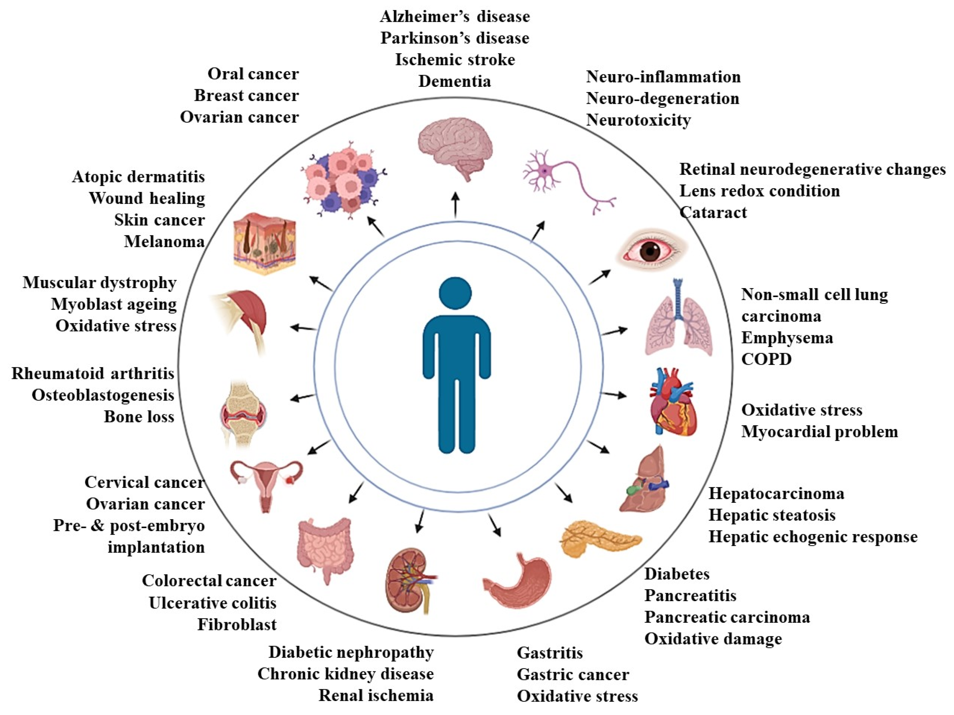
| System Involved | Disease State | Possible Results | Source |
|---|---|---|---|
| Central nervous system | Alzheimer’s disease |
| [37] |
| Parkinson’s disease |
| [38] | |
| Ischemic stroke |
| [39,40] | |
| Dementia |
| [41] | |
| Neuro-inflammation |
| [42] | |
| Neuro-degeneration |
| [43] | |
| Neurotoxicity |
| [44] | |
| Sensory organ: Eye | Retinal neurodegenerative changes |
| [45] |
| Lens redox condition |
| [46] | |
| Cataract |
| [46] | |
| Sensory organ: Skin | Atopic dermatitis |
| [47] |
| Skin cancer |
| [48,49] | |
| Respiratory system | Non-small lung carcinoma |
| [50] |
| Emphysema |
| [51] | |
| Chronic obstructive pulmonary disease (COPD) |
| [51,52] | |
| Cardiac system | Oxidative stress |
| [53] |
| Myocardial ischemia |
| [54] | |
| Cardioprotective |
| [55] | |
| Digestive system | Gastritis |
| [56,57] |
| Gastric cancer |
| [58] | |
| Hepatic echogenic response |
| [59] | |
| Ulcerative colitis |
| [60] | |
| Fibroblast |
| [61] | |
| Colorectal cancer |
| [62,63] | |
| Pancreatitis |
| [64] | |
| Pancreatic carcinoma |
| [65] | |
| Hepatocarcinoma |
| [66] | |
| Reproductive system | Embryo implantation |
| [66,67] |
| Ovarian cancer |
| [68] | |
| Cervical cancer |
| [69] | |
| Renal system | Renal ischemia |
| [70] |
| Chronic kidney disease |
| [71] | |
| Diabetic nephropathy |
| [72] | |
| Metabolic disorders and other cancers | Diabetes |
| [36] |
| Obesity |
| [36,73] | |
| Breast cancer |
| [74] | |
| Oral cancer |
| [75] |
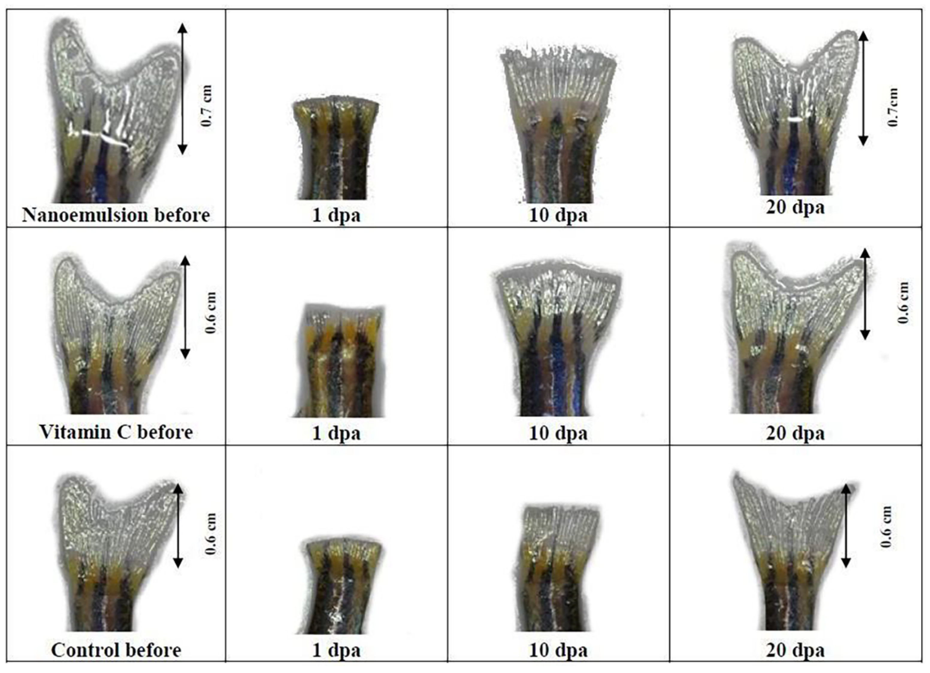
| API/Formulation | Type of Wound Treated | Observations of the Research | Source |
|---|---|---|---|
| Tocotrienols/ nanoemulsion | Topical wound |
| [17] |
| Tocotrienols-rich fraction/ nanoemulsion | Ocular and dermal tissue |
| [108] |
| Mono-epoxy-tocotrienol-α | Diabetic wound |
| [100] |
| Dehydro-tocotrienol-β | Diabetic wound |
| [103] |
| Long-chain metabolites of vitamin E, garcinoic acid | Diabetic wound |
| [109] |
| d-δ-tocotrienol-rich fraction | Diabetic wound |
| [110] |
| d-δ-tocotrienol-rich fraction and α-tocopherol | Diabetic wound |
| [111] |
| Tocotrienols-rich fraction | Burn wound |
| [112] |
| Tocotrienols and epidermal growth factor | Burn wound |
| [93] |
| Tocotrienols and epidermal growth factor | Burn wound |
| [113] |
| Daptomycin with Tocotrienols | Open wound |
| [114] |
Publisher’s Note: MDPI stays neutral with regard to jurisdictional claims in published maps and institutional affiliations. |
© 2022 by the authors. Licensee MDPI, Basel, Switzerland. This article is an open access article distributed under the terms and conditions of the Creative Commons Attribution (CC BY) license (https://creativecommons.org/licenses/by/4.0/).
Share and Cite
Nair, A.B.; Gorain, B.; Pandey, M.; Jacob, S.; Shinu, P.; Aldhubiab, B.; Almuqbil, R.M.; Elsewedy, H.S.; Morsy, M.A. Tocotrienol in the Treatment of Topical Wounds: Recent Updates. Pharmaceutics 2022, 14, 2479. https://doi.org/10.3390/pharmaceutics14112479
Nair AB, Gorain B, Pandey M, Jacob S, Shinu P, Aldhubiab B, Almuqbil RM, Elsewedy HS, Morsy MA. Tocotrienol in the Treatment of Topical Wounds: Recent Updates. Pharmaceutics. 2022; 14(11):2479. https://doi.org/10.3390/pharmaceutics14112479
Chicago/Turabian StyleNair, Anroop B., Bapi Gorain, Manisha Pandey, Shery Jacob, Pottathil Shinu, Bandar Aldhubiab, Rashed M. Almuqbil, Heba S. Elsewedy, and Mohamed A. Morsy. 2022. "Tocotrienol in the Treatment of Topical Wounds: Recent Updates" Pharmaceutics 14, no. 11: 2479. https://doi.org/10.3390/pharmaceutics14112479