

Films, Gels and Electrospun Fibers from Serum Albumin Globular Protein for Medical Device Coating, Biomolecule Delivery and Regenerative Engineering

Abstract
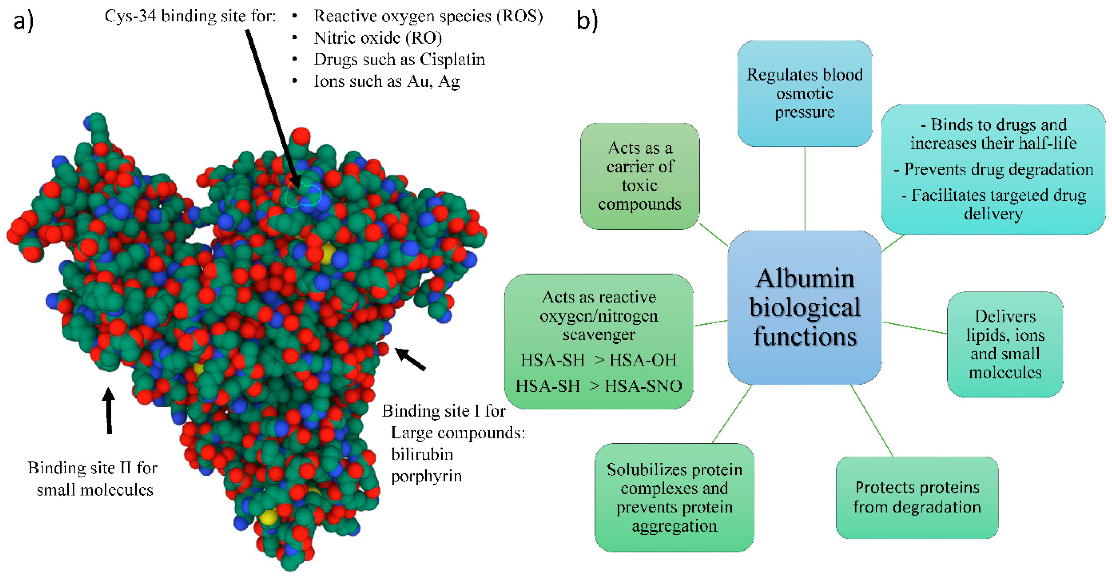
:1. Introduction
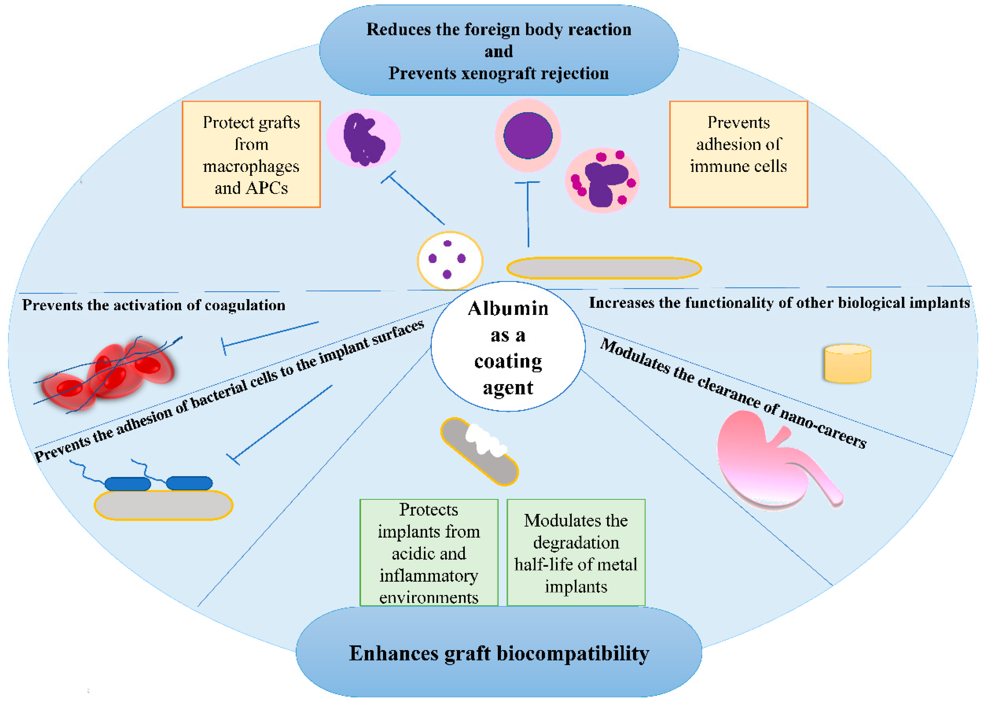
2. Albumin-Coated Implant Materials
3. Albumin-Derived Gels
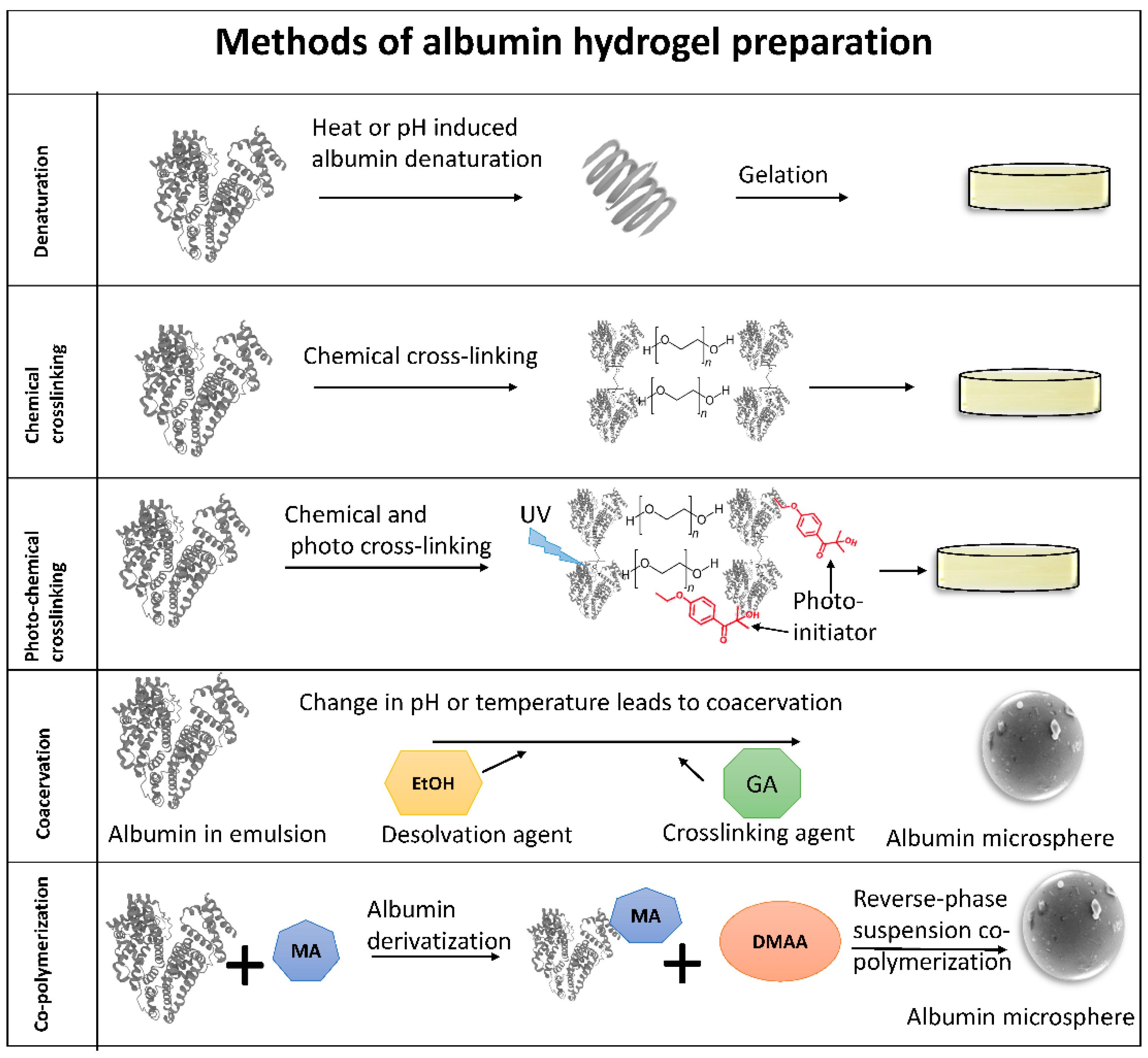
3.1. Gelation Process and Mechanism
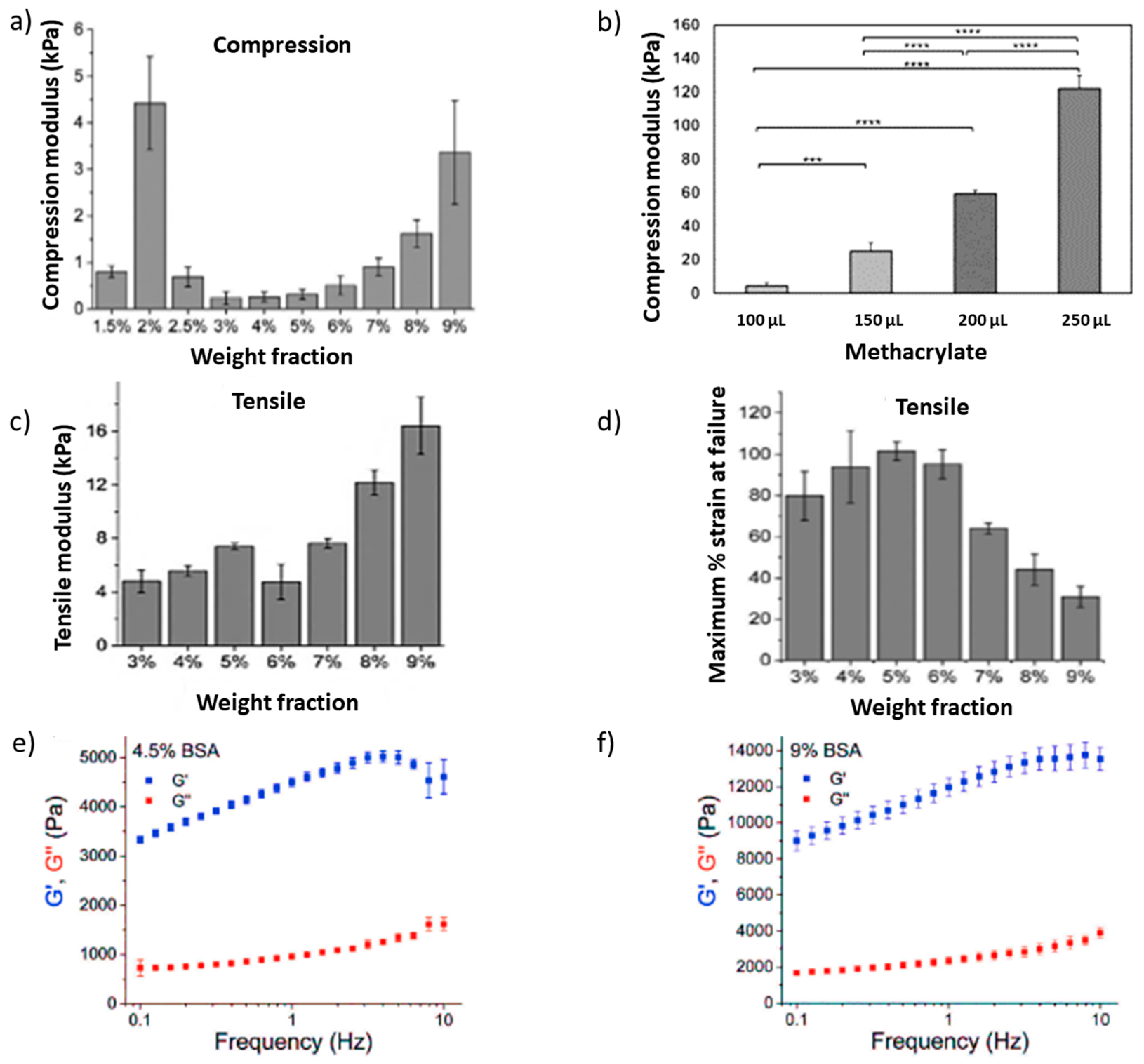
3.2. Mechanical Properties of Albumin Gels
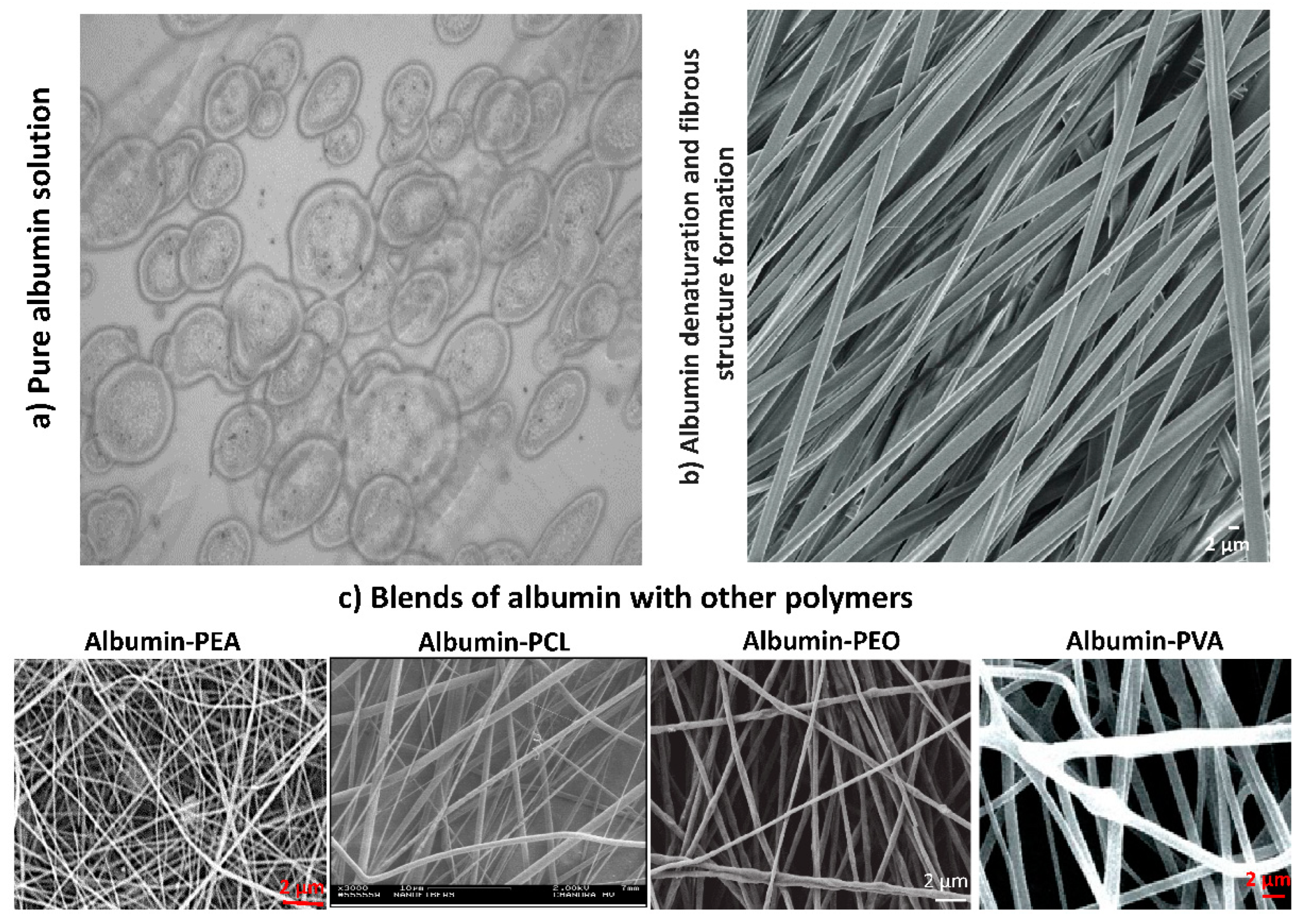
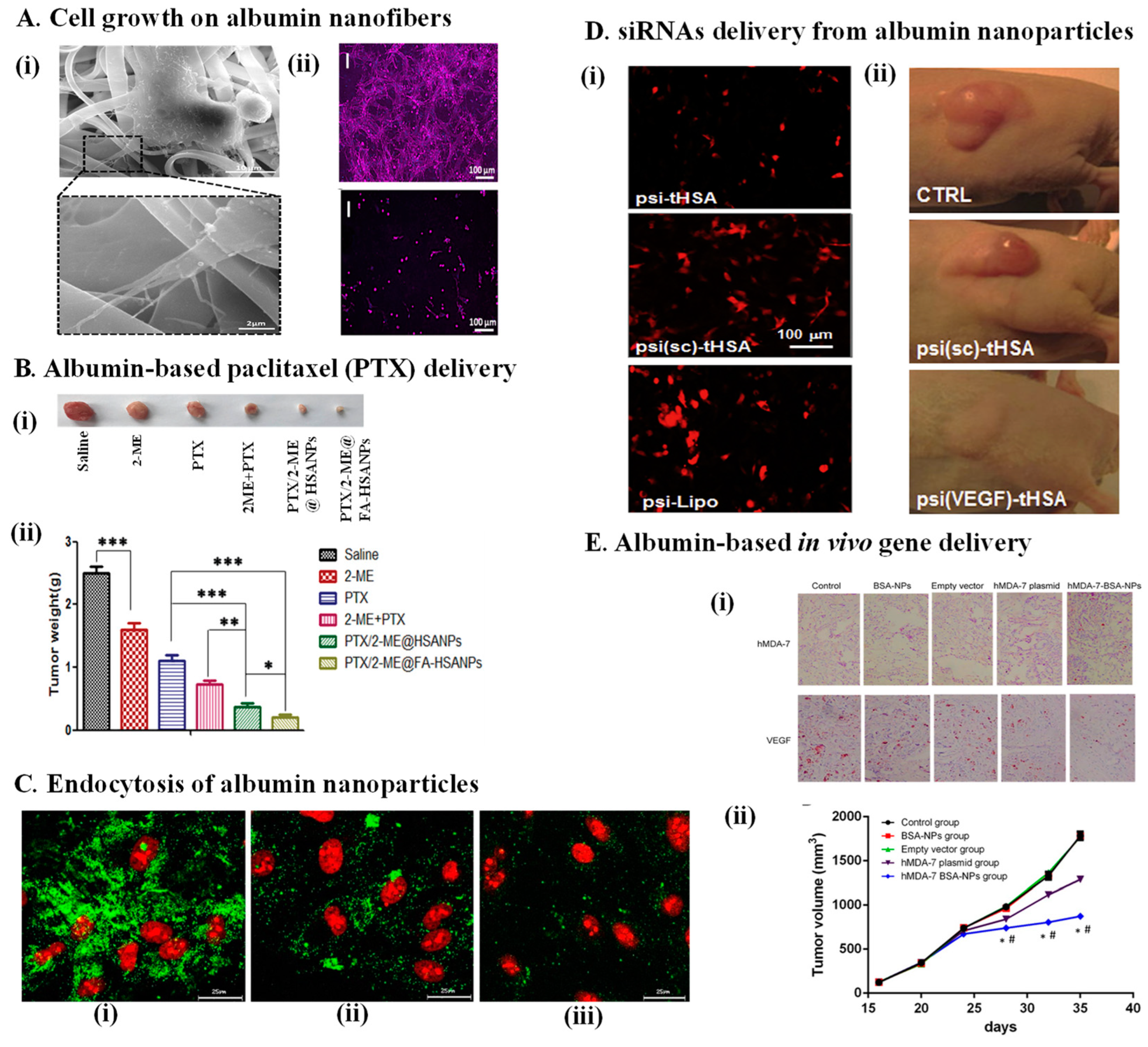
4. Electrospun Albumin Fibers

5. Albumin in Biomolecule Delivery
6. Albumin in Cell Delivery and Tissue Engineering
7. Albumin-Mediated Gene Delivery

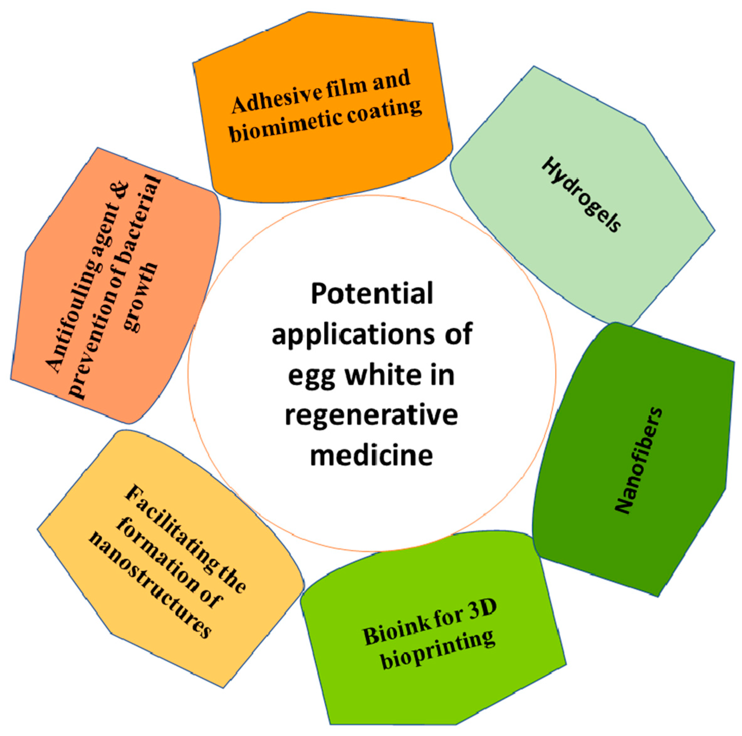
8. Egg White as a Rich Source of Albumin
9. Conclusions and Future Perspectives
Funding
Institutional Review Board Statement
Informed Consent Statement
Data Availability Statement
Conflicts of Interest
References
- Quinlan, G.J.; Martin, G.S.; Evans, T.W. Albumin: Biochemical properties and therapeutic potential. Hepatology 2005, 41, 1211–1219. [Google Scholar] [CrossRef] [PubMed]
- He, X.M.; Carter, D.C. Atomic structure and chemistry of human serum albumin. Nature 1992, 358, 209–215. [Google Scholar] [CrossRef] [PubMed] [Green Version]
- Merlot, A.M.; Kalinowski, D.S.; Richardson, D.R. Unraveling the mysteries of serum albumin-more than just a serum protein. Front. Physiol. 2014, 5, 299. [Google Scholar] [CrossRef] [PubMed] [Green Version]
- Finn, T.E.; Nunez, A.C.; Sunde, M.; Easterbrook-Smith, S.B. Serum Albumin Prevents Protein Aggregation and Amyloid Formation and Retains Chaperone-like Activity in the Presence of Physiological Ligands. J. Biol. Chem. 2012, 287, 21530–21540. [Google Scholar] [CrossRef] [Green Version]
- Li, P.-S.; Lee, I.-L.; Yu, W.-L.; Sun, J.-S.; Jane, W.-N.; Shen, H.-H. A novel albumin-Based tissue scaffold for autogenic tissue engineering applications. Sci. Rep. 2014, 4, 05600. [Google Scholar] [CrossRef] [Green Version]
- Taguchi, K.; Okamoto, Y.; Matsumoto, K.; Otagiri, M.; Chuang, V. When Albumin Meets Liposomes: A Feasible Drug Carrier for Biomedical Applications. Pharmaceuticals 2021, 14, 296. [Google Scholar] [CrossRef]
- Ong, J.; Zhao, J.; Justin, A.W.; Markaki, A.E. Albumin-based hydrogels for regenerative engineering and cell transplantation. Biotechnol. Bioeng. 2019, 116, 3457–3468. [Google Scholar] [CrossRef]
- Pella, O.K.; Hornyák, I.; Horváthy, D.; Fodor, E.; Nehrer, S.; Lacza, Z. Albumin as a Biomaterial and Therapeutic Agent in Regenerative Medicine. Int. J. Mol. Sci. 2022, 23, 10557. [Google Scholar] [CrossRef]
- Yamazoe, H.; Tanabe, T. Drug-Carrying Albumin Film for Blood-Contacting Biomaterials. J. Biomater. Sci. Polym. Ed. 2010, 21, 647–657. [Google Scholar] [CrossRef]
- Kottke-Marchant, K.; Anderson, J.M.; Umemura, Y.; Marchant, R. Effect of albumin coating on the in vitro blood compatibility of Dacron®® arterial prostheses. Biomaterials 1989, 10, 147–155. [Google Scholar] [CrossRef]
- Marois, Y.; Chakfé, N.; Guidoin, R.; Duhamel, R.C.; Roy, R.; Marois, M.; King, M.W.; Douville, Y. An albumin-Coated polyester arterial graft: In vivo assessment of biocompatibility and healing characteristics. Biomaterials 1996, 17, 3–14. [Google Scholar] [CrossRef]
- Reynolds, E.C.; Wong, A. Effect of adsorbed protein on hydroxyapatite zeta potential and Streptococcus mutans adherence. Infect Immun. 1983, 39, 1285–1290. [Google Scholar] [CrossRef] [PubMed] [Green Version]
- Martín, M.L.; Pfaffen, V.; Valenti, L.E.; Giacomelli, C.E. Albumin biofunctionalization to minimize the Staphylococcus aureus adhesion on solid substrates. Colloids Surf. B. Biointerfaces 2018, 167, 156–164. [Google Scholar] [CrossRef] [PubMed] [Green Version]
- Vincent, M.P.; Bobbala, S.; Karabin, N.B.; Frey, M.; Liu, Y.; Navidzadeh, J.O.; Stack, T.; Scott, E.A. Surface chemistry-Mediated modulation of adsorbed albumin folding state specifies nano-Carrier clearance by distinct macrophage subsets. Nat. Commun. 2021, 12, 648. [Google Scholar] [CrossRef]
- Tao, C.; Zhu, W.; Iqbal, J.; Xu, C.; Wang, D.-A. Stabilized albumin coatings on engineered xenografts for attenuation of acute immune and inflammatory responses. J. Mater. Chem. B. 2020, 8, 6080–6091. [Google Scholar] [CrossRef]
- Oriňakováa, R.; Gorejová, R.; Králová, Z.O.; Oriňak, A.; Shepa, I.; Hovancová, J.; Kovalčíková, A.; Bujňáková, Z.L.; Király, N.; Kaňuchová, M.; et al. Influence of albumin interaction on corrosion resistance of sintered iron biomaterials with polyethyleneimine coating. Appl. Surf. Sci. 2020, 509, 145379. [Google Scholar] [CrossRef]
- Höhn, S.; Braem, A.; Neirinck, B.; Virtanen, S. Albumin coatings by alternating current electrophoretic deposition for improving corrosion resistance and bioactivity of titanium implants. Mater. Sci. Eng. C 2017, 73, 798–807. [Google Scholar] [CrossRef]
- An, Y.H.; Stuart, G.W.; McDowell, S.J.; McDaniel, S.E.; Kang, Q.; Friedman, R.J. Prevention of bacterial adherence to implant surfaces with a cross-linked albumin coating in vitro. J. Orthop. Res. 1996, 14, 846–849. [Google Scholar] [CrossRef]
- Cometta, S.; Bock, N.; Suresh, S.; Dargaville, T.R.; Hutmacher, D.W. Antibacterial Albumin-Tannic Acid Coatings for Scaffold-Guided Breast Reconstruction. Front. Bioeng. Biotechnol. 2021, 9, 638577. [Google Scholar] [CrossRef]
- Katarivas, L.G.; Ong, J.; Birch, M.A.; Justin, A.W.; Markaki, A.E. Albumin-Enriched Fibrin Hydrogel Embedded in Active Ferromagnetic Networks Improves Osteoblast Differentiation and Vascular Self-Organisation. Polymer. 2019, 11, 1743. [Google Scholar] [CrossRef]
- Ungor, D.; Juhász, Á.; Varga, N.; Csapó, E. Evaluation of noble metal nanostructure-serum albumin interactions in 2D and 3D systems: Thermodynamics and possible mechanisms. Adv. Colloid Interface Sci. 2022, 301, 102616. [Google Scholar] [CrossRef] [PubMed]
- Mariam, J.; Sivakami, S.; Dongre, P.M. Albumin corona on nanoparticles–A strategic approach in drug delivery. Drug Deliv. 2016, 23, 2668–2676. [Google Scholar] [CrossRef] [PubMed] [Green Version]
- Singh, A.V.; Bandgar, B.M.; Kasture, M.; Prasad, B.L.V.; Sastry, M. Synthesis of gold, silver and their alloy nanoparticles using bovine serum albumin as foaming and stabilizing agent. J. Mater. Chem. 2005, 15, 5115–5121. [Google Scholar] [CrossRef]
- Murawala, P.; Phadnis, S.; Bhonde, R.; Prasad, B. In situ synthesis of water dispersible bovine serum albumin capped gold and silver nanoparticles and their cytocompatibility studies. Colloids Surf. B Biointerfaces 2009, 73, 224–228. [Google Scholar] [CrossRef] [PubMed]
- Morales-Sánchez, E.; Guajardo-Pacheco, J.; Noriega-Treviño, M.; Quintero-González, C.; Compeán-Jasso, M.; López-Salinas, F.; González-Hernández, J.; Ruiz, F. Synthesis of Silver Nanoparticles Using Albumin as a Reducing Agent. Mater. Sci. Appl. 2011, 2, 578–581. [Google Scholar] [CrossRef] [Green Version]
- Kathiravan, A.; Renganathan, R.; Anandan, S. Interaction of colloidal AgTiO2 nanoparticles with bovine serum albumin. Polyhedron 2009, 28, 157–161. [Google Scholar] [CrossRef]
- Ahmed, E.M. Hydrogel: Preparation, characterization, and applications: A review. J. Adv. Res. 2015, 6, 105–121. [Google Scholar] [CrossRef] [Green Version]
- Kopeček, J.; Yang, J. Hydrogels as smart biomaterials. Polym. Int. 2007, 56, 1078–1098. [Google Scholar] [CrossRef]
- Rohanizadeh, R.; Kokabi, N. Heat denatured/aggregated albumin-based biomaterial: Effects of preparation parameters on biodegradability and mechanical properties. J. Mater. Sci. Mater. Med. 2009, 20, 2413–2418. [Google Scholar] [CrossRef]
- Amdursky, N.; Mazo, M.M.; Thomas, M.R.; Humphrey, E.J.; Puetzer, J.L.; St-Pierre, J.-P.; Skaalure, S.C.; Richardson, R.M.; Terracciano, C.M.; Stevens, M.M. Elastic serum-albumin based hydrogels: Mechanism of formation and application in cardiac tissue engineering. J. Mater. Chem. B 2018, 6, 5604–5612. [Google Scholar] [CrossRef]
- Baler, K.; Michael, R.; Szleifer, I.; Ameer, G.A. Albumin Hydrogels Formed by Electrostatically Triggered Self-Assembly and Their Drug Delivery Capability. Biomacromolecules 2014, 15, 3625–3633. [Google Scholar] [CrossRef] [PubMed] [Green Version]
- Arabi, S.H.; Aghelnejad, B.; Schwieger, C.; Meister, A.; Kerth, A.; Hinderberger, D. Serum albumin hydrogels in broad pH and temperature ranges: Characterization of their self-assembled structures and nanoscopic and macroscopic properties. Biomater. Sci. 2018, 6, 478–492. [Google Scholar] [CrossRef] [PubMed] [Green Version]
- Raja, S.T.; Thiruselvi, T.; Mandal, A.B.; Gnanamani, A. pH and redox sensitive albumin hydrogel: A self-Derived biomaterial. Sci. Rep. 2015, 5, 15977. [Google Scholar] [CrossRef] [PubMed] [Green Version]
- Overby, R.J.; Feldman, D.S. Influence of Poly(Ethylene Glycol) End Groups on Poly(Ethylene Glycol)-Albumin System Properties as a Potential Degradable Tissue Scaffold. J. Funct. Biomater. 2018, 10, 1. [Google Scholar] [CrossRef] [PubMed] [Green Version]
- Oss-Ronen, L.; Seliktar, D. Photopolymerizable Hydrogels Made from Polymer-Conjugated Albumin for Affinity-Based Drug Delivery. Adv. Eng. Mater. 2010, 12, B45–B52. [Google Scholar] [CrossRef]
- Yamazoe, H.; Uemura, T.; Tanabe, T. Facile Cell Patterning on an Albumin-Coated Surface. Langmuir 2008, 24, 8402–8404. [Google Scholar] [CrossRef]
- Gallego, L.; Junquera, L.; García, E.; García, V.; Álvarez-Viejo, M.; Costilla, S.; Fresno, M.F.; Meana, A. Repair of Rat Mandibular Bone Defects by Alveolar Osteoblasts in a Novel Plasma-Derived Albumin Scaffold. Tissue Eng. Part A 2010, 16, 1179–1187. [Google Scholar] [CrossRef] [Green Version]
- Yoon, D.; Kang, B.-J.; Kim, Y.; Lee, S.H.; Rhew, D.; Kim, W.H.; Kweon, O.-K. Effect of serum-derived albumin scaffold and canine adipose tissue-derived mesenchymal stem cells on osteogenesis in canine segmental bone defect model. J. Veter. Sci. 2015, 16, 397–404. [Google Scholar] [CrossRef] [Green Version]
- Gallego, L.; Junquera, L.; Meana, A.; Álvarez-Viejo, M.; Fresno, M. Ectopic Bone Formation from Mandibular Osteoblasts Cultured in a Novel Human Serum-derived Albumin Scaffold. J. Biomater. Appl. 2010, 25, 367–381. [Google Scholar] [CrossRef]
- Zhang, Y.; Pham, H.M.; Munguia-Lopez, J.G.; Kinsella, J.M.; Tran, S.D. The Optimization of a Novel Hydrogel-Egg White-Alginate for 2.5D Tissue Engineering of Salivary Spheroid-Like Structure. Molecules 2020, 25, 5751. [Google Scholar] [CrossRef]
- Lantigua, D.; Nguyen, M.A.; Wu, X.; Suvarnapathaki, S.; Kwon, S.; Gavin, W.; Camci-Unal, G. Synthesis and characterization of photocrosslinkable albumin-based hydrogels for biomedical applications. Soft Matter 2020, 16, 9242–9252. [Google Scholar] [CrossRef] [PubMed]
- Rusu, A.G.; Chiriac, A.P.; Nita, L.E.; Mititelu-Tartau, L.; Tudorachi, N.; Ghilan, A.; Rusu, D. Multifunctional BSA Scaffolds Prepared with a Novel Combination of UV-Crosslinking Systems. Macromol. Chem. Phys. 2019, 220, 1900378. [Google Scholar] [CrossRef]
- Merodio, M.; Arnedo, A.; Renedo, M.; Irache, J.M. Ganciclovir-loaded albumin nanoparticles: Characterization and in vitro release properties. Eur. J. Pharm. Sci. 2001, 12, 251–259. [Google Scholar] [CrossRef]
- Sahin, S.; Selek, H.; Ponchel, G.; Ercan, M.T.; Sargon, M.; Hincal, A.; Kas, H. Preparation, characterization and in vivo distribution of terbutaline sulfate loaded albumin microspheres. J. Control. Release 2002, 82, 345–358. [Google Scholar] [CrossRef]
- Iemma, F.; Spizzirri, U.G.; Muzzalupo, R.; Puoci, F.; Trombino, S.; Picci, N. Spherical hydrophilic microparticles obtained by the radical copolymerisation of functionalised bovine serum albumin. Colloid Polym. Sci. 2004, 283, 250–256. [Google Scholar] [CrossRef]
- Sebak, S.; Mirzaei, M.; Malhotra, M.; Kulamarva, A.; Prakash, S. Human serum albumin nanoparticles as an efficient noscapine drug delivery system for potential use in breast cancer: Preparation and in vitro analysis. Int. J. Nanomed. 2010, 5, 525–532. [Google Scholar]
- Li, Q.; Chen, F.; Liu, Y.; Yu, S.; Gai, X.; Ye, M.; Yang, X.; Pan, W. A novel albumin wrapped nanosuspension of meloxicam to improve inflammation-targeting effects. Int. J. Nanomed. 2018, 13, 4711–4725. [Google Scholar] [CrossRef] [Green Version]
- Iemma, F.; Spizzirri, U.G.; Puoci, F.; Muzzalupo, R.; Trombino, S.; Picci, N. Radical Cross-Linked Albumin Microspheres as Potential Drug Delivery Systems: Preparation and In Vitro Studies. Drug Deliv. 2005, 12, 179–184. [Google Scholar] [CrossRef]
- Zu, Y.-G.; Li, Q.; Liu, C.; Zhao, X.; Wang, Y.; Zhang, B.; Zhao, D.; Zhao, Q.; Su, L.; Gao, Y.; et al. Preparation, characterization and targeting of micronized 10-hydroxycamptothecin-loaded folate-conjugated human serum albumin nanoparticles to cancer cells. Int. J. Nanomed. 2011, 6, 397–405. [Google Scholar] [CrossRef] [Green Version]
- Kang, Y.; Kim, H.; Shin, W.-S.; Woo, G.; Moon, T. Effect of Disulfide Bond Reduction on Bovine Serum Albumin-Stabilized Emulsion Gel Formed by Microbial Transglutaminase. J. Food Sci. 2006, 68, 2215–2220. [Google Scholar] [CrossRef]
- Oyen, M.L. Mechanical characterisation of hydrogel materials. Int. Mater. Rev. 2014, 59, 44–59. [Google Scholar] [CrossRef]
- Ahearne, M.; Yang, Y.; Liu, K.-K. Mechanical Characterisation of Hydrogels for Tissue Engineering Applications. In Topics in Tissue Engineering; Ashammakhi, N., Reis, R., Chiellini, F., Eds.; 2008; Chapter 4; Volume 4, Available online: https://www.oulu.fi/spareparts/ebook_topics_in_t_e_vol4/index.html (accessed on 10 September 2022).
- Yan, C.; Pochan, D.J. Rheological properties of peptide-based hydrogels for biomedical and other applications. Chem. Soc. Rev. 2010, 39, 3528–3540. [Google Scholar] [CrossRef] [PubMed] [Green Version]
- Lu, S.; Zhu, L.; Wang, Q.; Liu, Z.; Tang, C.; Sun, H.; Yang, J.; Qin, G.; Sun, G.; Chen, Q. High-Strength Albumin Hydrogels With Hybrid Cross-Linking. Front. Chem. 2020, 8, 106. [Google Scholar] [CrossRef] [PubMed]
- Xue, J.; Wu, T.; Dai, Y.; Xia, Y. Electrospinning and Electrospun Nanofibers: Methods, Materials, and Applications. Chem. Rev. 2019, 119, 5298–5415. [Google Scholar] [CrossRef]
- Williams, G.R.; Raimi-Abraham, B.T.; Luo, C.J. Electrospinning fundamentals. In Nanofibres in Drug Delivery; UCL Press: London, UK, 2018. [Google Scholar]
- Dror, Y.; Ziv, T.; Makarov, V.; Wolf, H.; Admon, A.; Zussman, E. Nanofibers Made of Globular Proteins. Biomacromolecules 2008, 9, 2749–2754. [Google Scholar] [CrossRef]
- Fleischer, S.; Shapira, A.; Regev, O.; Nseir, N.; Zussman, E.; Dvir, T. Albumin fiber scaffolds for engineering functional cardiac tissues. Biotechnol. Bioeng. 2014, 111, 1246–1257. [Google Scholar] [CrossRef]
- Nseir, N.; Regev, O.; Kaully, T.; Blumenthal, J.; Levenberg, S.; Zussman, E. Biodegradable Scaffold Fabricated of Electrospun Albumin Fibers: Mechanical and Biological Characterization. Tissue Eng. Part C Methods 2013, 19, 257–264. [Google Scholar] [CrossRef]
- Raic, A.; Friedrich, F.; Kratzer, D.; Bieback, K.; Lahann, J.; Lee-Thedieck, C. Potential of electrospun cationic BSA fibers to guide osteogenic MSC differentiation via surface charge and fibrous topography. Sci. Rep. 2019, 9, 20003–20015. [Google Scholar] [CrossRef] [Green Version]
- Martín-Alfonso, J.; Cuadri, A.; Greiner, A. The combined effect of formulation and pH on properties of polyethylene oxide composite fiber containing egg albumen protein. Int. J. Biol. Macromol. 2018, 112, 996–1004. [Google Scholar] [CrossRef]
- Kowalczyk, T.; Nowicka, A.; Elbaum, D.; Kowalewski, T.A. Electrospinning of Bovine Serum Albumin. Optimization and the Use for Production of Biosensors. Biomacromolecules 2008, 9, 2087–2090. [Google Scholar] [CrossRef]
- Fehér, B.; Lyngsø, J.; Bartók, B.; Mihály, J.; Varga, Z.; Mészáros, R.; Pedersen, J.S.; Bóta, A.; Varga, I. Effect of pH on the conformation of bovine serume albumin—Gold bioconjugates. J. Mol. Liq. 2020, 309, 113065. [Google Scholar] [CrossRef]
- Raghuwanshi, V.S.; Yu, B.; Browne, C.; Garnier, G. Reversible pH Responsive Bovine Serum Albumin Hydrogel Sponge Nanolayer. Front. Bioeng. Biotechnol. 2020, 8, 573. [Google Scholar] [CrossRef] [PubMed]
- Noszczyk, B.H.; Kowalczyk, T.; Łyżniak, M.; Zembrzycki, K.; Mikulowski, G.; Wysocki, J.; Kawiak, J.; Pojda, Z. Biocompatibility of electrospun human albumin: A pilot study. Biofabrication 2015, 7, 015011. [Google Scholar] [CrossRef] [PubMed]
- Yamazoe, H.; Okuyama, T.; Suzuki, H.; Fukuda, J. Fabrication of patterned cell co-cultures on albumin-based substrate: Applications for microfluidic devices. Acta Biomater. 2010, 6, 526–533. [Google Scholar] [CrossRef] [PubMed]
- Zahedi, P.; Fallah-Darrehchi, M. Electrospun egg albumin-PVA nanofibers containing tetracycline hydrochloride: Morphological, drug release, antibacterial, thermal and mechanical properties. Fibers Polym. 2015, 16, 2184–2192. [Google Scholar] [CrossRef]
- Chernonosova, V.S.; Kvon, R.I.; Stepanova, A.O.; Larichev, Y.V.; Karpenko, A.A.; Chelobanov, B.P.; Kiseleva, E.V.; Laktionov, P.P. Human serum albumin in electrospun PCL fibers: Structure, release, and exposure on fiber surface. Polym. Adv. Technol. 2017, 28, 819–827. [Google Scholar] [CrossRef]
- Valmikinathan, C.M.; Defroda, S.; Yu, X. Polycaprolactone and Bovine Serum Albumin Based Nanofibers for Controlled Release of Nerve Growth Factor. Biomacromolecules 2009, 10, 1084–1089. [Google Scholar] [CrossRef]
- Homaeigohar, S.; Monavari, M.; Koenen, B.; Boccaccini, A.R. Biomimetic biohybrid nanofibers containing bovine serum albumin as a bioactive moiety for wound dressing. Mater. Sci. Eng. C Mater. Biol. Appl. 2021, 123, 111965. [Google Scholar] [CrossRef]
- Pavlova, E.; Nikishin, I.; Bogdanova, A.; Klinov, D.; Bagrov, D. The miscibility and spatial distribution of the components in electrospun polymer–protein mats. RSC Adv. 2020, 10, 4672–4680. [Google Scholar] [CrossRef] [Green Version]
- Zhang, Y.Z.; Wang, X.; Feng, Y.; Li, J.; Lim, C.T.; Ramakrishna, S. Coaxial electrospinning of (fluorescein isothiocyanate-conjugated bovine serum albumin)-encapsulated poly(epsilon-caprolactone) nanofibers for sustained release. Biomacromolecules 2006, 7, 1049–1057. [Google Scholar] [CrossRef]
- Li, X.; Su, Y.; Liu, S.; Tan, L.; Mo, X.; Ramakrishna, S. Encapsulation of proteins in poly(l-lactide-co-caprolactone) fibers by emulsion electrospinning. Colloids Surfaces B Biointerfaces 2010, 75, 418–424. [Google Scholar] [CrossRef] [PubMed]
- Scimeca, M.; Bischetti, S.; Lamsira, H.K.; Bonfiglio, R.; Bonanno, E. Energy Dispersive X-ray (EDX) microanalysis: A powerful tool in biomedical research and diagnosis. Eur. J. Histochem. 2018, 62, 2841. [Google Scholar] [CrossRef] [PubMed]
- Dennis, M.S.; Zhang, M.; Meng, Y.G.; Kadkhodayan, M.; Kirchhofer, D.; Combs, D.; Damico, L.A. Albumin Binding as a General Strategy for Improving the Pharmacokinetics of Proteins. J. Biol. Chem. 2002, 277, 35035–35043. [Google Scholar] [CrossRef] [Green Version]
- Chemmanur, A.T.; Wu, G.Y. Drug evaluation: Albuferon-Alpha-An antiviral interferon-Alpha/albumin fusion protein. Curr. Opin. Investig. Drugs 2006, 7, 750–758. [Google Scholar]
- Desai, N.; Trieu, V.; Yao, Z.; Louie, L.; Ci, S.; Yang, A.; Tao, C.; De, T.; Beals, B.; Dykes, D.; et al. Increased antitumor activity, intratumor paclitaxel concentrations, and endothelial cell transport of cremophor-free, albumin-bound paclitaxel, ABI-007, compared with cremophor-based paclitaxel. Clin. Cancer Res. 2006, 12, 1317–1324. [Google Scholar] [CrossRef] [Green Version]
- Stehle, G.; Sinn, H.; Wunder, A.; Schrenk, H.H.; Stewart, J.M.; Hartung, G.; Maier-Borst, W.; Heene, D.L. Plasma protein (albumin) catabolism by the tumor itself—Implications for tumor metabolism and the genesis of cachexia. Crit. Rev. Oncol. 1997, 26, 77–100. [Google Scholar] [CrossRef]
- Schnitzer, J.E.; Oh, P. Antibodies to SPARC inhibit albumin binding to SPARC, gp60, and microvascular endothelium. Am. J. Physiol. Circ. Physiol. 1992, 263 Pt 2, H1872–H1879. [Google Scholar] [CrossRef]
- Wunder, A.; Müller-Ladner, U.; Stelzer, E.H.K.; Funk, J.; Neumann, E.; Stehle, G.; Pap, T.; Sinn, H.; Gay, S.; Fiehn, C. Albumin-Based Drug Delivery as Novel Therapeutic Approach for Rheumatoid Arthritis. J. Immunol. 2003, 170, 4793–4801. [Google Scholar] [CrossRef] [Green Version]
- Uddin, S.; Melnyk, N.; Foster, D.A. Albumin promotes the progression of fibroblasts through late G1 into S-phase in the absence of growth factors. Cell Cycle 2020, 19, 2158–2167. [Google Scholar] [CrossRef]
- Khadka, D.B.; Haynie, D.T. Protein- and peptide-based electrospun nanofibers in medical biomaterials. Nanomed. Nanotechnol. Biol. Med. 2012, 8, 1242–1262. [Google Scholar] [CrossRef]
- Iemma, F.; Spizzirri, U.G.; Puoci, F.; Muzzalupo, R.; Trombino, S.; Cassano, R.; Leta, S.; Picci, N. pH-Sensitive hydrogels based on bovine serum albumin for oral drug delivery. Int. J. Pharm. 2006, 312, 151–157. [Google Scholar] [CrossRef] [PubMed]
- Tarhini, M.; Pizzoccaro, A.; Benlyamani, I.; Rebaud, C.; Greige-Gerges, H.; Fessi, H.; Elaissari, A.; Bentaher, A. Human serum albumin nanoparticles as nanovector carriers for proteins: Application to the antibacterial proteins “neutrophil elastase” and “secretory leukocyte protease inhibitor”. Int. J. Pharm. 2020, 579, 119150. [Google Scholar] [CrossRef]
- Liu, X.; Zhao, T.; Xu, Y.; Huo, P.; Xu, X.; Zhang, Z.; Tian, Q.; Zhang, N. Co-administration of paclitaxel and 2-methoxyestradiol using folate-conjugated human serum albumin nanoparticles for improving drug resistance and antitumor efficacy. Pharm. Dev. Technol. 2021, 26, 1–10. [Google Scholar] [CrossRef] [PubMed]
- Hasanpoor, Z.; Mostafaie, A.; Nikokar, I.; Hassan, Z.M. Curcumin-human serum albumin nanoparticles decorated with PDL1 binding peptide for targeting PDL1-expressing breast cancer cells. Int. J. Biol. Macromol. 2020, 159, 137–153. [Google Scholar] [CrossRef] [PubMed]
- Miele, E.; Spinelli, G.P.; Miele, E.; Tomao, F.; Tomao, S. Albumin-bound formulation of paclitaxel (Abraxane ABI-007) in the treatment of breast cancer. Int. J. Nanomed. 2009, 4, 99–105. [Google Scholar]
- Ferrero-Gutierrez, A.; Menendez-Menendez, Y.; Alvarez-Viejo, M.; Meana, A.; Otero, J. New serum-derived albumin scaffold seeded with adipose-derived stem cells and olfactory ensheathing cells used to treat spinal cord injured rats. Histol. Histopathol. 2013, 28, 89–100. [Google Scholar]
- Prasopdee, T.; Sinthuvanich, C.; Chollakup, R.; Uttayarat, P.; Smitthipong, W. The albumin/starch scaffold and its biocompatibility with living cells. Mater. Today Commun. 2021, 27, 102164. [Google Scholar] [CrossRef]
- Hsu, C.-C.; Serio, A.; Amdursky, N.; Besnard, C.; Stevens, M.M. Fabrication of Hemin-Doped Serum Albumin-Based Fibrous Scaffolds for Neural Tissue Engineering Applications. ACS Appl. Mater. Interfaces 2018, 10, 5305–5317. [Google Scholar] [CrossRef]
- Filippi, M.; Born, G.; Chaaban, M.; Scherberich, A. Natural Polymeric Scaffolds in Bone Regeneration. Front. Bioeng. Biotechnol. 2020, 8, 474. [Google Scholar] [CrossRef]
- Mo, Y.; Barnett, M.E.; Takemoto, D.; Davidson, H.; Kompella, U.B. Human serum albumin nanoparticles for efficient delivery of Cu, Zn superoxide dismutase gene. Mol. Vis. 2007, 13, 746–757. [Google Scholar]
- Wartlick, H.; Spänkuch-Schmitt, B.; Strebhardt, K.; Kreuter, J.; Langer, K. Tumour cell delivery of antisense oligonuclceotides by human serum albumin nanoparticles. J. Control. Release 2004, 96, 483–495. [Google Scholar] [CrossRef] [PubMed]
- Lu, W.; Sun, Q.; Wan, J.; She, Z.; Jiang, X.-G. Cationic Albumin–Conjugated Pegylated Nanoparticles Allow Gene Delivery into Brain Tumors via Intravenous Administration. Cancer Res. 2006, 66, 11878–11887. [Google Scholar] [CrossRef] [PubMed] [Green Version]
- Guan, G.; Song, B.; Zhang, J.; Chen, K.; Hu, H.; Wang, M. An Effective Cationic Human Serum Albumin-Based Gene-Delivery Carrier Containing the Nuclear Localization Signal. Pharmaceutics 2019, 11, 608. [Google Scholar] [CrossRef] [PubMed] [Green Version]
- Zhu, Q.; Pan, X.; Sun, Y.; Wang, Z.; Liu, F.; Li, A.; Zhao, Z.; Wang, Y.; Li, K.; Mi, L. Biological nanoparticles carrying the Hmda-7 gene are effective in inhibiting pancreatic cancer in vitro and in vivo. PLoS ONE 2017, 12, e0185507. [Google Scholar] [CrossRef] [Green Version]
- Rhaese, S.; von Briesen, H.; Rübsamen-Waigmann, H.; Kreuter, J.; Langer, K. Human serum albumin–polyethylenimine nanoparticles for gene delivery. J. Control. Release 2003, 92, 199–208. [Google Scholar] [CrossRef]
- Li, H.; Liu, Y.; Chen, L.; Liu, Q.; Qi, S.; Cheng, X.; Lee, Y.B.; Ahn, C.-H.; Kim, D.J.; Lee, R.J. Folate receptor-targeted lipid-albumin nanoparticles (F-LAN) for therapeutic delivery of an Akt1 antisense oligonucleotide. J. Drug Target. 2018, 26, 466–473. [Google Scholar] [CrossRef]
- Langiu, M.; Dadparvar, M.; Kreuter, J.; Ruonala, M.O. Human Serum Albumin-Based Nanoparticle-Mediated In Vitro Gene Delivery. PLoS ONE 2014, 9, e107603. [Google Scholar] [CrossRef] [Green Version]
- 101 Look, J.; Wilhelm, N.; Von Briesen, H.; Noske, N.; Günther, C.; Langer, K.; Gorjup, E. Ligand-Modified Human Serum Albumin Nanoparticles for Enhanced Gene Delivery. Mol. Pharm. 2015, 12, 3202–3213. [Google Scholar] [CrossRef]
- Son, S.; Song, S.; Lee, S.J.; Min, S.; Kim, S.A.; Yhee, J.Y.; Huh, M.S.; Kwon, L.C.; Jeong, S.Y.; Byun, Y.; et al. Self-Crosslinked human serum albumin nano-carriers for systemic delivery of polymerized siRNA to tumors. Biomater. 2013, 34, 9475–9485. [Google Scholar] [CrossRef]
- Choi, J.H.; Hwang, H.J.; Shin, S.W.; Choi, J.W.; Um, S.H.; Oh, B.K. A novel albumin nanocomplex containing both small interfering RNA and gold nanorods for synergetic anticancer therapy. Nanoscale 2015, 7, 9229–9237. [Google Scholar] [CrossRef]
- Jin, Q.; Qiao, C.; Li, J.; Li, J.; Xiao, X. An engineered serum albumin-binding AAV9 capsid achieves improved liver transduction after intravenous delivery in mice. Gene Ther. 2020, 27, 237–244. [Google Scholar] [CrossRef] [PubMed]
- Mann, K. The chicken egg white proteome. Proteomics 2007, 7, 3558–3568. [Google Scholar] [CrossRef] [PubMed]
- Mann, K.; Mann, M. In-depth analysis of the chicken egg white proteome using an LTQ Orbitrap Velos. Proteome Sci. 2011, 9, 7. [Google Scholar] [CrossRef] [Green Version]
- Dong, X.; Zhang, Y. An insight on egg white: From most common functional food to biomaterial application. J. Biomed. Mater. Res. Part B Appl. Biomater. 2021, 109, 1045–1058. [Google Scholar] [CrossRef] [PubMed]
- Jalili-Firoozinezhad, S.; Filippi, M.; Mohabatpour, F.; Letourneur, D.; Scherberich, A. Chicken egg white: Hatching of a new old biomaterial. Mater. Today 2020, 40, 193–214. [Google Scholar] [CrossRef]
- Jerez, A.; Partal, P.; Martínez, I.; Gallegos, C.; Guerrero, A. Egg white-based bioplastics developed by thermomechanical processing. J. Food Eng. 2007, 82, 608–617. [Google Scholar] [CrossRef]
- Chang, J.-W.; Wang, C.-G.; Huang, C.-Y.; Tsai, T.-D.; Guo, T.-F.; Wen, T.-C. Chicken Albumen Dielectrics in Organic Field-Effect Transistors. Adv. Mater. 2011, 23, 4077–4081. [Google Scholar] [CrossRef]
- Delkash, Y.; Gouin, M.; Rimbeault, T.; Mohabatpour, F.; Papagerakis, P.; Maw, S.; Chen, X. Bioprinting and In Vitro Characterization of an Eggwhite-Based Cell-Laden Patch for Endothelialized Tissue Engineering Applications. J. Funct. Biomater. 2021, 12, 45. [Google Scholar] [CrossRef]
- Chang, Q.; Darabi, M.A.; Liu, Y.; He, Y.; Zhong, W.; Mequanin, K.; Li, B.; Lu, F.; Xing, M.M.Q. Hydrogels from natural egg white with extraordinary stretchability, direct-writing 3D printability and self-healing for fabrication of electronic sensors and actuators. J. Mater. Chem. A 2019, 7, 24626–24640. [Google Scholar] [CrossRef]
- Somaratne, G.; Nau, F.; Ferrua, M.J.; Singh, J.; Ye, A.; Dupont, D.; Singh, R.P.; Floury, J. Characterization of egg white gel microstructure and its relationship with pepsin diffusivity. Food Hydrocoll. 2020, 98, 105258. [Google Scholar] [CrossRef]
- Offengenden, M.; Wu, J. Egg white ovomucin gels: Structured fluids with weak polyelectrolyte properties. RSC Adv. 2013, 3, 910–917. [Google Scholar] [CrossRef]
- Nojima, T.; Iyoda, T. Egg white-based strong hydrogel via ordered protein condensation. NPG Asia Mater. 2018, 10, e460. [Google Scholar] [CrossRef]
- Rathna, G.; Li, J.; Gunasekaran, S. Functionally-Modified egg white albumen hydrogels. Polym. Int. 2004, 53, 1994–2000. [Google Scholar] [CrossRef]
- Jahani-Javanmardi, A.; Sirousazar, M.; Shaabani, Y.; Kheiri, F. Egg white/poly (vinyl alcohol)/MMT nanocomposite hydrogels for wound dressing. J. Biomater. Sci. Polym. Ed. 2016, 27, 1262–1276. [Google Scholar] [CrossRef]
- Rafati, Z.; Sirousazar, M.; Hassan, Z.M.; Kheiri, F. Honey-Loaded Egg White/Poly(vinyl alcohol)/Clay Bionanocomposite Hydrogel Wound Dressings: In Vitro and In Vivo Evaluations. J. Polym. Environ. 2020, 28, 32–46. [Google Scholar] [CrossRef]
- Rathna, G.V.N.; Jog, J.P.; Gaikwad, A.B. Development of non-woven nanofibers of egg albumen-poly (vinyl alcohol) blends: Influence of solution properties on morphology of nanofibers. Polym. J. 2011, 43, 654–661. [Google Scholar] [CrossRef] [Green Version]
- Huang, K.; Hou, J.; Gu, Z.; Wu, J. Egg-White-/Eggshell-Based Biomimetic Hybrid Hydrogels for Bone Regeneration. ACS Biomater. Sci. Eng. 2019, 5, 5384–5391. [Google Scholar] [CrossRef]
- Carpena, N.; Abueva, C.D.; Padalhin, A.R.; Lee, B. Evaluation of egg white ovomucin-based porous scaffold as an implantable biomaterial for tissue engineering. J. Biomed. Mater. Res. Part B Appl. Biomater. 2017, 105, 2107–2117. [Google Scholar] [CrossRef]
- Rajaraman, S.; Subbiahdoss, G.; Patchirajan, P. Effect of Hen Egg White on Microbial Adhesion and Biofilm Growth of Biomaterial Associated Infection Causing Pathogens. Int. J. Bio-Sci. Bio-Technol. 2014, 6, 99–106. [Google Scholar] [CrossRef] [Green Version]
- Sun, X.; Huang, J.; Zeng, H.; Wu, J. Protein-Resistant Property of Egg White Ovomucin under Different pHs and Ionic Strengths. J. Agric. Food Chem. 2018, 66, 11034–11042. [Google Scholar] [CrossRef]
- Rauf, M.A.; Zubair, S.; Ateeq, H.; Dabeer, K.; Pachauri, S.; Ajmal, M.; Owais, M. Synergistic Effect of Diallyl Sulfide With Zinc Oxide Nanorods: A Novel and Effective Approach for Treatment of Acute Dermatitis in Model Animals. Front. Microbiol. 2018, 9, 586. [Google Scholar] [CrossRef] [PubMed] [Green Version]
- Huang, J.; You, X.; Xin, P.; Gu, Z.; Chen, C.; Wu, J. Egg white as a natural and safe biomaterial for enhanced cancer therapy. Chin. Chem. 2021, 32, 1737–1742. [Google Scholar] [CrossRef]
- Gadomska, M.; Musiał, K.; Sionkowska, A. New materials based on hyaluronic acid and egg albumin mixture. Eng. Biomater. 2021, 160, 15–21. [Google Scholar]


| Delivery System | Loaded Drug | Type of Study | Key Findings | Refs |
|---|---|---|---|---|
| Albumin nanoparticles | Interferon alpha (Albuferon®) | Clinical trial | Significant antiviral activity with mild adverse effects similar to interferon alphas alone | [76] |
| Albumin nanoparticles | Paclitaxel (Abraxane®) | Clinical trial (FDA approved) | Reduced the toxicity of Paclitaxel | [87] |
| Albumin- methotrexate conjugate | Methotrexate | Preclinical | Enhanced delivery of drug to the arthritic joints More effective suppression of arthritis | [80] |
| Albumin/PVA nanofibers | Tetracycline hydrochloride | Preclinical | Controlled release of drug | [67] |
| Albumin/PCL nanofibers | Nerve growth factor | Preclinical | Prolong controlled release of drug | [69] |
| PEG-conjugated albumin hydrogels | Warfarin and Naproxen | Preclinical | Controlled release of small molecules | [35] |
| pH-sensitive albumin hydrogel microspheres | β-Propranolol | Preclinical | Efficient release of drugs in the pH similar to oral cavity | [83] |
| Albumin nanoparticles loaded with noscapine | Noscapine | Preclinical | Enhanced effectiveness on breast cancer cells | [46] |
| Albumin microspheres | Terbutaline sulfate | Preclinical | lung-specific delivery of medicine | [44] |
| Albumin nanoparticles | Meloxicam | Preclinical | Targeted delivery to inflamed tissues. Sustained release and increased half-life and bioavailability of drugs | [47] |
| Albumin nanoparticles | Ganciclovir | Preclinical | Sustained release of drugs | [43] |
| Folic acid conjugated albumin nanoparticles | 10-Hydroxycamptothecin and paclitaxel/2-methoxyestradiol | Preclinical | Prolonged release, increased uptake by cancerous cells and targeted inhibition of tumor growth | [49,85] |
Publisher’s Note: MDPI stays neutral with regard to jurisdictional claims in published maps and institutional affiliations. |
© 2022 by the authors. Licensee MDPI, Basel, Switzerland. This article is an open access article distributed under the terms and conditions of the Creative Commons Attribution (CC BY) license (https://creativecommons.org/licenses/by/4.0/).
Share and Cite
Mahdipour, E.; Mequanint, K. Films, Gels and Electrospun Fibers from Serum Albumin Globular Protein for Medical Device Coating, Biomolecule Delivery and Regenerative Engineering. Pharmaceutics 2022, 14, 2306. https://doi.org/10.3390/pharmaceutics14112306
Mahdipour E, Mequanint K. Films, Gels and Electrospun Fibers from Serum Albumin Globular Protein for Medical Device Coating, Biomolecule Delivery and Regenerative Engineering. Pharmaceutics. 2022; 14(11):2306. https://doi.org/10.3390/pharmaceutics14112306
Chicago/Turabian StyleMahdipour, Elahe, and Kibret Mequanint. 2022. "Films, Gels and Electrospun Fibers from Serum Albumin Globular Protein for Medical Device Coating, Biomolecule Delivery and Regenerative Engineering" Pharmaceutics 14, no. 11: 2306. https://doi.org/10.3390/pharmaceutics14112306