


Multifunctional Polydopamine-Based Nanoparticles for Dual-Mode Imaging Guided Targeted Therapy of Lupus Nephritis
 , and
, and 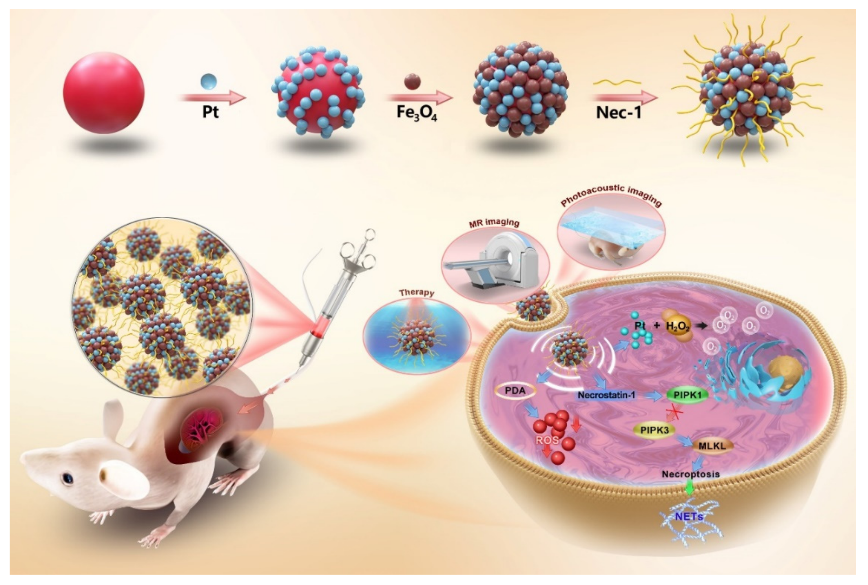
Abstract
:1. Introduction
2. Experimental Section
2.1. Materials
2.2. Preparation of Polydopamine (PDA) Nanoparticles
2.3. Preparation of PDA@Pt Nanoparticles
2.4. Preparation of PDA@Pt-Fe3O4 Nanoparticles
2.5. General Characterization
2.6. Nec-1 Loading and Release
2.7. Evaluating the Formation of NETs
2.8. Animal Model Establishment
2.9. MR Imaging and Photoacoustic Imaging of the Nec-1/PDA@Pt-Fe3O4
2.10. Therapeutic Effect of Nec-1/mPDA@Pt-Fe3O4 on LN
2.11. Biosafety Assessment of Nec-1/mPDA@Pt-Fe3O4
2.12. Statistical Analysis
3. Results and Discussion
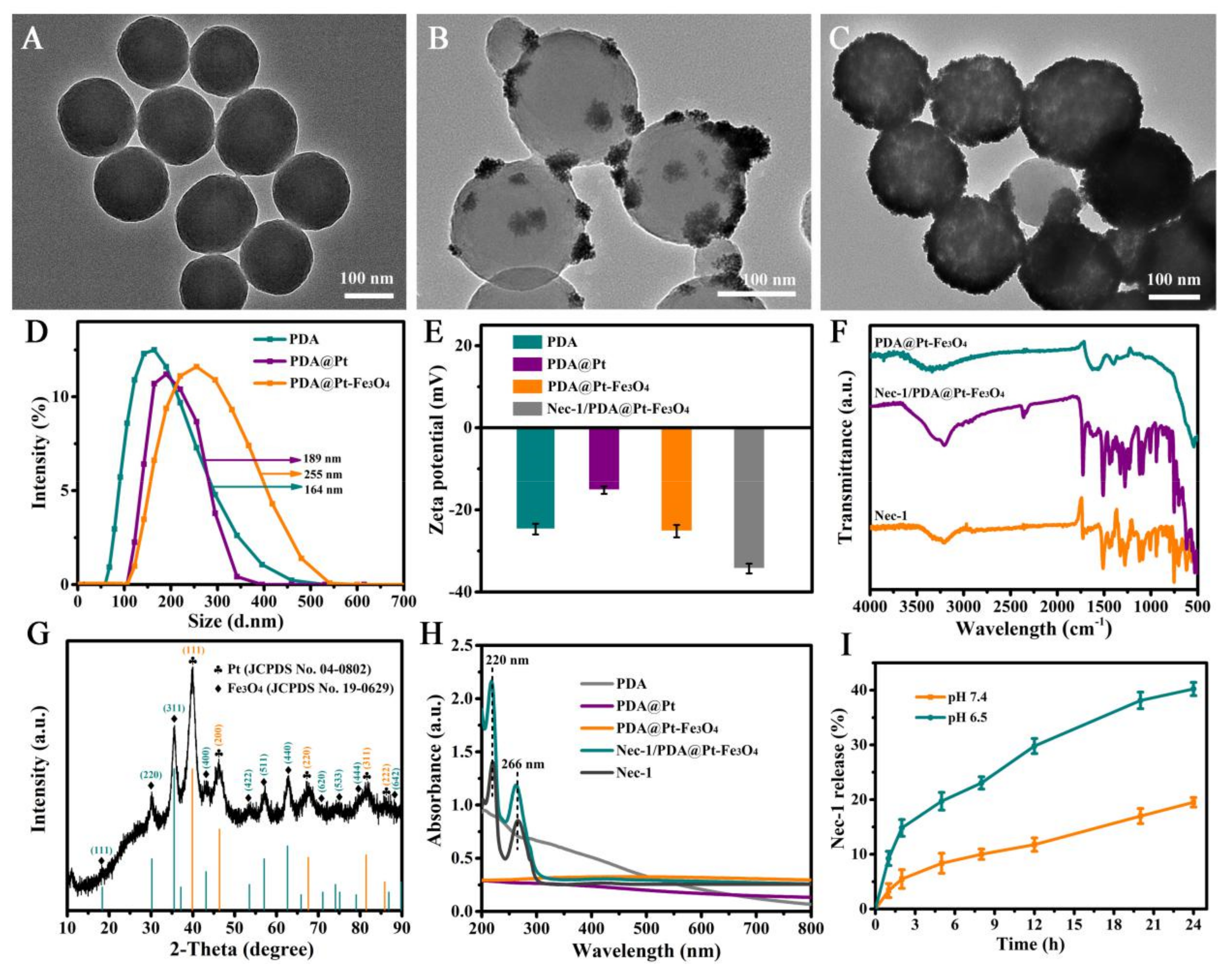
3.1. Preparation and Characterization of Nec-1/PDA@Pt-Fe3O4
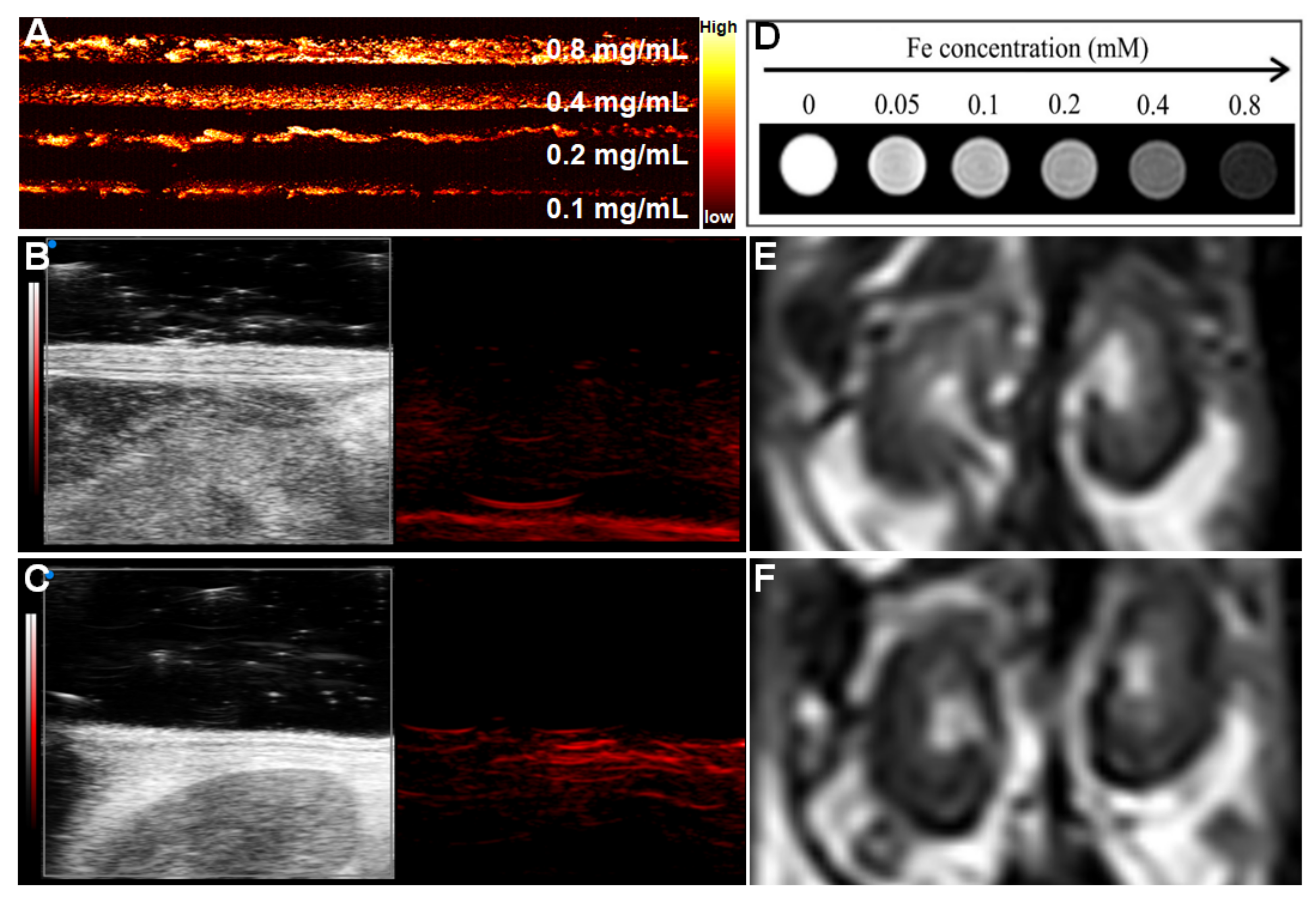
3.2. PA and MR Dual-Mode Image Tracking
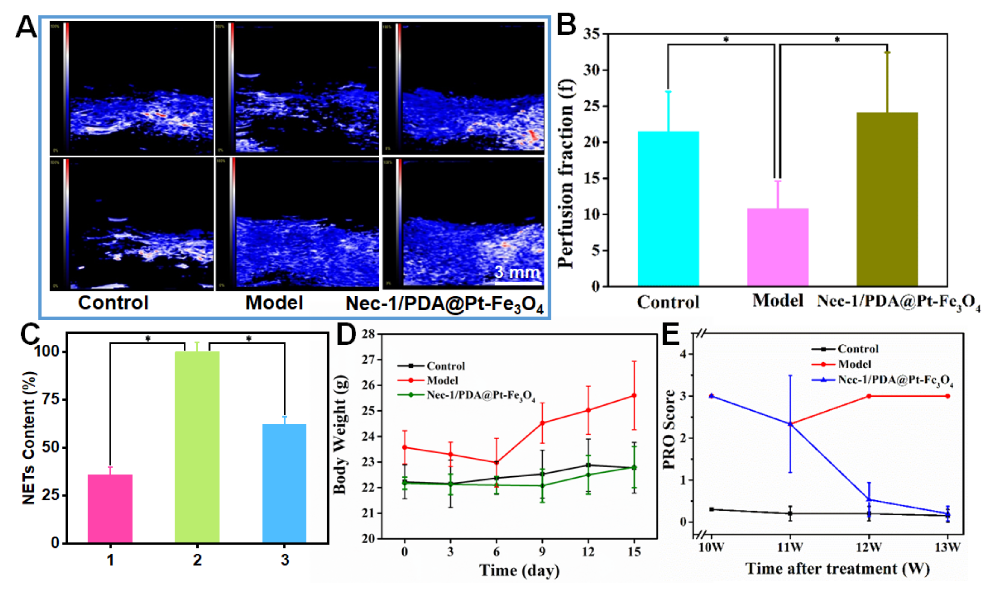
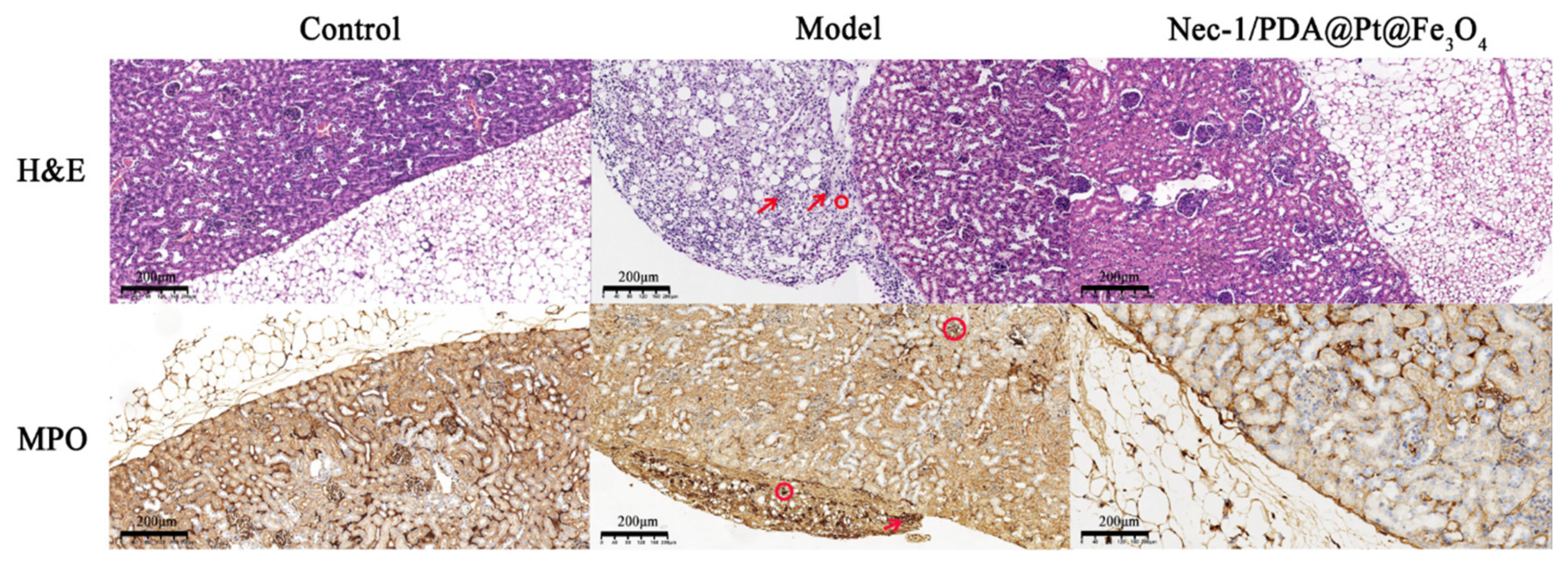
3.3. Therapeutic Effect of Nec-1/PDA@Pt-Fe3O4 on LN
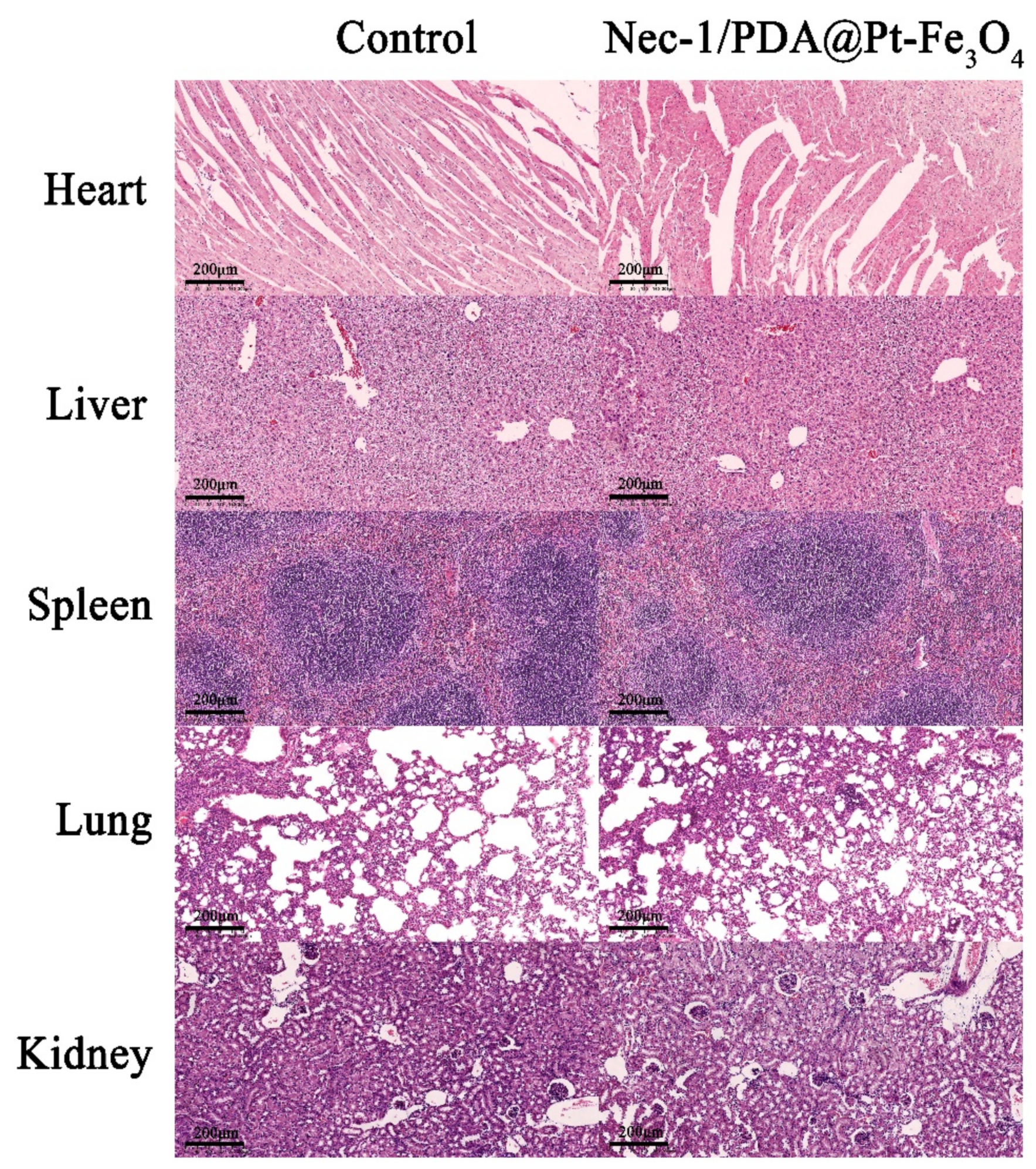
3.4. Biosafety Assessment of Nec-1/PDA@Pt-Fe3O4
4. Conclusions
Author Contributions
Funding
Institutional Review Board Statement
Informed Consent Statement
Data Availability Statement
Conflicts of Interest
Abbreviations
References
- Anders, H.-J.; Saxena, R.; Zhao, M.-H.; Parodis, I.; Salmon, J.E.; Mohan, C. Lupus nephritis. Nat. Rev. Dis. Primers 2020, 6, 1–8. [Google Scholar] [CrossRef] [PubMed]
- Maroz, N.; Segal, M.S. Lupus Nephritis and End-stage Kidney Disease. Am. J. Med. Sci. 2013, 346, 319–323. [Google Scholar] [CrossRef] [PubMed]
- Moroni, G.; Ponticelli, C. The multifaceted aspects of refractory lupus nephritis. Expert Rev. Clin. Immunol. 2015, 11, 281–288. [Google Scholar] [CrossRef]
- Anders, H.-J.; Hiepe, F. Treatment Options for Refractory Lupus Nephritis. Clin. J. Am. Soc. Nephrol. 2019, 14, 653–655. [Google Scholar] [CrossRef] [PubMed]
- Jin, L.; Liu, P.; Yin, M.; Zhang, M.; Kuang, Y.; Zhu, W. RIPK1: A rising star in inflammatory and neoplastic skin diseases. J. Dermatol. Sci. 2020, 99, 146–151. [Google Scholar] [CrossRef] [PubMed]
- Mifflin, L.; Ofengeim, D.; Yuan, J. Receptor-interacting protein kinase 1 (RIPK1) as a therapeutic target. Nat. Rev. Drug Discov. 2020, 19, 553–571. [Google Scholar] [CrossRef]
- Pieterse, E.; Rother, N.; Garsen, M.; Hofstra, J.M.; Satchell, S.C.; Hoffmann, M.; Loeven, M.A.; Knaapen, H.K.; van der Heijden, O.W.H.; Berden, J.H.M.; et al. Neutrophil Extracellular Traps Drive Endothelial-to-Mesenchymal Transition. Arterioscler. Thromb. Vasc. Biol. 2017, 37, 1371–1379. [Google Scholar] [CrossRef]
- Kurata, Y.; Tanaka, T.; Nangaku, M. The role of hypoxia in the pathogenesis of lupus nephritis. Kidney Int. 2020, 98, 821–823. [Google Scholar] [CrossRef]
- Wang, M.; Ishikawa, T.; Lai, Y.; Nallapothula, D.; Singh, R.R. Diverse Roles of NETosis in the Pathogenesis of Lupus. Front. Immunol. 2022, 13, 895216. [Google Scholar] [CrossRef]
- Clarke, J. NETs directly injure cartilage in RA. Nat. Rev. Rheumatol. 2020, 16, 410. [Google Scholar] [CrossRef]
- Frangou, E.; Vassilopoulos, D.; Boletis, J.; Boumpas, D.T. An emerging role of neutrophils and NETosis in chronic inflammation and fibrosis in systemic lupus erythematosus (SLE) and ANCA-associated vasculitides (AAV): Implications for the pathogenesis and treatment. Autoimmun. Rev. 2019, 18, 751–760. [Google Scholar] [CrossRef]
- Han, X.A.; Jie, H.Y.; Wang, J.H.; Zhang, X.M.; Wang, J.; Yu, C.X.; Zhang, J.L.; He, J.; Chen, J.Q.; Lai, K.F.; et al. Necrostatin-1 Ameliorates Neutrophilic Inflammation in Asthma by Suppressing MLKL Phosphorylation to Inhibiting NETs Release. Front. Immunol. 2020, 11, 666. [Google Scholar] [CrossRef] [PubMed]
- McHugh, J. Targeted delivery of immunosuppressant in SLE. Nat. Rev. Rheumatol. 2020, 16, 410. [Google Scholar] [CrossRef] [PubMed]
- Siddique, S.; Chow, J.C.L. Recent Advances in Functionalized Nanoparticles in Cancer Theranostics. Nanomaterials 2022, 12, 2826. [Google Scholar] [CrossRef] [PubMed]
- Siddique, S.; Chow, J.C.L. Application of Nanomaterials in Biomedical Imaging and Cancer Therapy. Nanomaterials 2020, 10, 1700. [Google Scholar] [CrossRef] [PubMed]
- Zhang, J.; Chen, C.; Fu, H.; Yu, J.; Sun, Y.; Huang, H.; Tang, Y.; Shen, N.; Duan, Y. MicroRNA-125a-Loaded Polymeric Nanoparticles Alleviate Systemic Lupus Erythematosus by Restoring Effector/Regulatory T Cells Balance. ACS Nano 2020, 14, 4414–4429. [Google Scholar] [CrossRef]
- Wang, Z.; Zou, Y.; Li, Y.; Cheng, Y. Metal-Containing Polydopamine Nanomaterials: Catalysis, Energy, and Theranostics. Small 2020, 16, 1907042. [Google Scholar] [CrossRef]
- Ryu, J.H.; Messersmith, P.B.; Lee, H. Polydopamine Surface Chemistry: A Decade of Discovery. ACS Appl. Mater. Interfaces 2018, 10, 7523–7540. [Google Scholar] [CrossRef]
- Zhao, H.; Zeng, Z.; Liu, L.; Chen, J.; Zhou, H.; Huang, L.; Huang, J.; Hu, X.; Xu, Y.; Chen, Z.; et al. Polydopamine nanoparticles for the treatment of acute inflammation-induced injury. Nanoscale 2018, 10, 6981–6991. [Google Scholar] [CrossRef]
- Ding, F.; Gao, X.; Huang, X.; Ge, H.; Xie, M.; Qian, J.; Song, J.; Li, Y.; Zhu, X.; Zhang, C. Polydopamine-coated nucleic acid nanogel for siRNA-mediated low-temperature photothermal therapy. Biomaterials 2020, 245, 119976. [Google Scholar] [CrossRef]
- Chen, Y.; Wang, Y.; Chen, Z.; Cai, J.; Li, K.; Huang, H.; Song, F.; Gao, M.; Yang, Y.; Zheng, L.; et al. NIR-driven polydopamine based nanoenzymes as ROS scavengers to suppress osteoarthritis progression. Mater. Today Nano 2022, 19, 100240. [Google Scholar] [CrossRef]
- Ma, W.; Zhang, X.; Liu, Y.; Fan, L.; Gan, J.; Liu, W.; Zhao, Y.; Sun, L. Polydopamine Decorated Microneedles with Fe-MSC-Derived Nanovesicles Encapsulation for Wound Healing. Adv. Sci. 2022, 9, 2103317. [Google Scholar] [CrossRef] [PubMed]
- Xie, L.; Pang, X.; Yan, X.; Dai, Q.; Lin, H.; Ye, J.; Cheng, Y.; Zhao, Q.; Ma, X.; Zhang, X.; et al. Photoacoustic Imaging-Trackable Magnetic Microswimmers for Pathogenic Bacterial Infection Treatment. ACS Nano 2020, 14, 2880–2893. [Google Scholar] [CrossRef] [PubMed]
- Xu, Y.; Wang, K.; Zhao, S.; Xiong, Q.; Liu, G.; Li, Y.; Fang, Q.; Gong, X.; Xuan, S. Rough surface NiFe2O4@Au/Polydopamine with a magnetic field enhanced photothermal antibacterial effect. Chem. Eng. J. 2022, 437, 135282. [Google Scholar] [CrossRef]
- Zhang, H.; Deng, L.; Liu, H.; Mai, S.; Cheng, Z.; Shi, G.; Zeng, H.; Wu, Z. Enhanced fluorescence/magnetic resonance dual imaging and gene therapy of liver cancer using cationized amylose nanoprobe. Mater. Today Bio 2022, 13, 100220. [Google Scholar] [CrossRef]
- Segers, F.M.E.; Ruder, A.V.; Westra, M.M.; Lammers, T.; Dadfar, S.M.; Roemhild, K.; Lam, T.S. Magnetic resonance imaging contrast-enhancement with superparamagnetic iron oxide nanoparticles amplifies macrophage foam cell apoptosis in human and murine atherosclerosis. Cardiovasc. Res. 2022, 1–14. [Google Scholar] [CrossRef]
- Sargsyan, S.A.; Serkova, N.J.; Renner, B.; Hasebroock, K.M.; Larsen, B.; Stoldt, C.; McFann, K.; Pickering, M.C.; Thurman, J.M. Detection of glomerular complement C3 fragments by magnetic resonance imaging in murine lupus nephritis. Kidney Int. 2012, 81, 152–159. [Google Scholar] [CrossRef]
- Liang, S.; Liu, B.; Xiao, X.; Yuan, M.; Yang, L.; Ma, P.A.; Cheng, Z.; Lin, J. A Robust Narrow Bandgap Vanadium Tetrasulfide Sonosensitizer Optimized by Charge Separation Engineering for Enhanced Sonodynamic Cancer Therapy. Adv. Mater. 2021, 33, 2101467. [Google Scholar] [CrossRef]
- Zhang, L.; Li, M.; Zhou, Q.; Dang, M.; Tang, Y.; Wang, S.; Fu, J.; Teng, Z.; Lu, G. Computed tomography and photoacoustic imaging guided photodynamic therapy against breast cancer based on mesoporous platinum with insitu oxygen generation ability. Acta Pharm. Sin. B 2020, 10, 1719–1729. [Google Scholar] [CrossRef]
- Wang, T.; Liu, Y.; Zhou, J.; Lin, L.; Jia, C.; Wang, J.; Yu, L.; Wang, Y.; Yan, Z. Controllable hydrogen release for gas-assisted chemotherapy and ultrasonic imaging of drug-resistant tumors. Chem. Eng. J. 2021, 421, 129917. [Google Scholar] [CrossRef]
- Bian, S.W.; Liu, S.; Chang, L. Synthesis of magnetically recyclable Fe3O4@polydopamine–Pt composites and their application in hydrogenation reactions. J. Mater. Sci. 2016, 51, 3643–3649. [Google Scholar] [CrossRef]
- Wang, Y.; Liu, X.; Deng, G.; Wang, Q.; Zhang, L.; Wang, Q.; Lu, J. Multifunctional PS@CS@Au-Fe3O4-FA nanocomposites for CT, MR and fluorescence imaging guided targeted-photothermal therapy of cancer cells. J. Mater. Chem. B 2017, 5, 4221–4232. [Google Scholar] [CrossRef] [PubMed]
- Dema, B.; Lamri, Y.; Pellefigues, C.; Pacreau, E.; Saidoune, F.; Bidault, C.; Karasuyama, H.; Sacré, K.; Daugaus, E.; Charles, N. Basophils contribute to pristane-induced Lupus-like nephritis model. Sci. Rep. 2017, 7, 7969. [Google Scholar] [CrossRef] [PubMed]
- Pravda, J. Hydrogen peroxide and disease: Towards a unified system of pathogenesis and therapeutics. Mol. Med. 2020, 26, 41. [Google Scholar] [CrossRef] [PubMed]
- Gutiérrez de la Rosa, S.Y.; Muñiz Diaz, R.; Villalobos Gutiérrez, P.T.; Patakfalvi, R.; Gutiérrez Coronado, Ó. Functionalized Platinum Nanoparticles with Biomedical Applications. Int. J. Mol. Sci. 2022, 23, 9404. [Google Scholar] [CrossRef] [PubMed]
- Jie, H.; He, Y.; Huang, X.; Zhou, Q.; Han, Y.; Li, X.; Bai, Y.; Sun, E. Necrostatin-1 enhances the resolution of inflammation by specifically inducing neutrophil apoptosis. Oncotarget 2016, 7, 19367–19381. [Google Scholar] [CrossRef]
- Hu, B.F.; Gong, Q.; Chen, S.Q.; Yue, L.; Ma, W.X.; Wang, F.; Feng, X.-W.; Wang, J.-N.; Li, C.; Liu, M.-M.; et al. Protective effect of inhibiting necroptosis on gentamicin-induced nephrotoxicity. FASEB J. 2022, 36, e22487. [Google Scholar] [CrossRef]


Publisher’s Note: MDPI stays neutral with regard to jurisdictional claims in published maps and institutional affiliations. |
© 2022 by the authors. Licensee MDPI, Basel, Switzerland. This article is an open access article distributed under the terms and conditions of the Creative Commons Attribution (CC BY) license (https://creativecommons.org/licenses/by/4.0/).
Share and Cite
Li, M.; Wang, Y.; Han, X.; Liu, Y.; Ma, M.; Zhang, L. Multifunctional Polydopamine-Based Nanoparticles for Dual-Mode Imaging Guided Targeted Therapy of Lupus Nephritis. Pharmaceutics 2022, 14, 1988. https://doi.org/10.3390/pharmaceutics14101988
Li M, Wang Y, Han X, Liu Y, Ma M, Zhang L. Multifunctional Polydopamine-Based Nanoparticles for Dual-Mode Imaging Guided Targeted Therapy of Lupus Nephritis. Pharmaceutics. 2022; 14(10):1988. https://doi.org/10.3390/pharmaceutics14101988
Chicago/Turabian StyleLi, Mifang, Yeying Wang, Xinai Han, Yibiao Liu, Mingliang Ma, and Lingyan Zhang. 2022. "Multifunctional Polydopamine-Based Nanoparticles for Dual-Mode Imaging Guided Targeted Therapy of Lupus Nephritis" Pharmaceutics 14, no. 10: 1988. https://doi.org/10.3390/pharmaceutics14101988