
Silica-Based Stimuli-Responsive Systems for Antitumor Drug Delivery and Controlled Release


Abstract
:1. Introduction
2. Stimuli-Responsive Systems Based in Endogenous Activity
2.1. pH-Responsive Systems

| Stimulus | Drug Loading | Release System | Release Mechanism | Ref. |
|---|---|---|---|---|
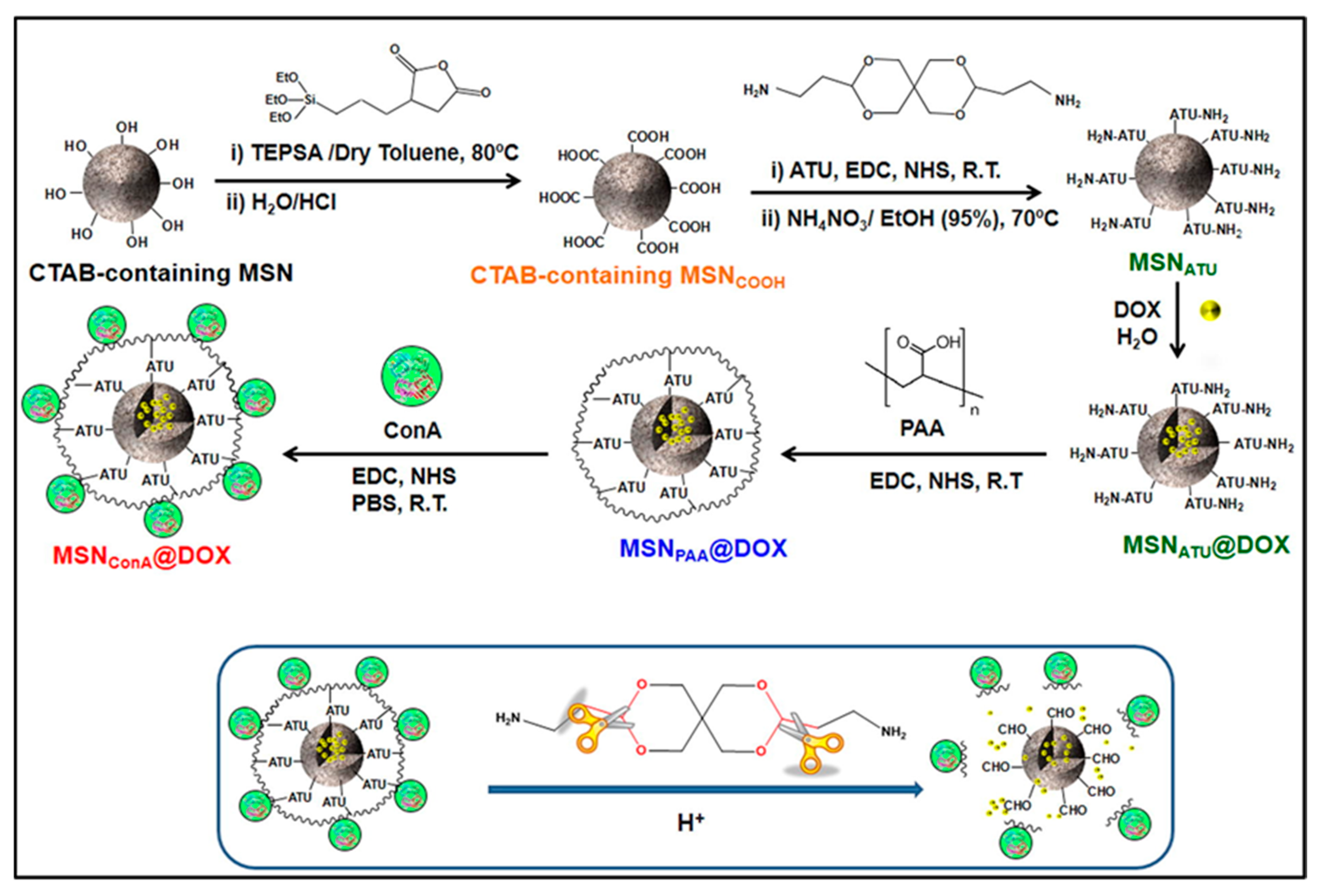
| pH | Doxorubicin | MSNs grafted with the pH sensitive linker ATU and coated with the acid degradable polymer PAA | Acid-cleavable acetal (ATU) linker | [19] |
| Doxorubicin and pheophorbide a | Hollow MSNs decorated with chitosan as a capping layer and GPTMS as crosslinking and attaching agent | At acidic pH, the CS/GPTMS layer swells, leaving the pores free. | [20] | |
| Doxorubicin | MSNs conjugated with supramolecular switches forming by hydrazone bond, azobenzene and α-cyclodextrin | Hydrolyzation of acid-sensitive hydrazine bonds | [21] | |
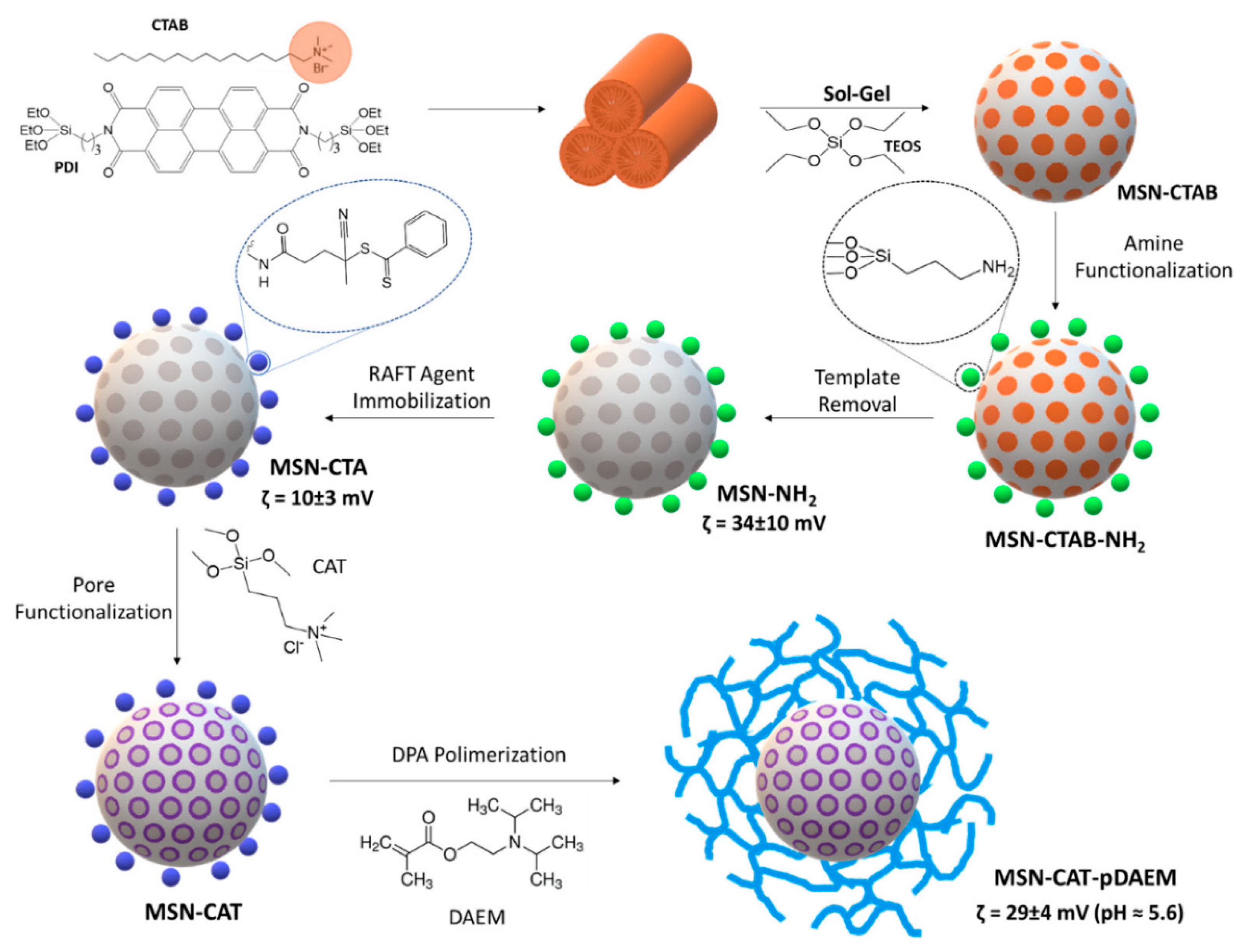
| Sulforhodamine B | MSNs with functionalized pore walls and grafted with a pH-responsive cross-linked polymer pDAEM | Protonation/deprotonation of tertiary amines of polymer | [22] | |
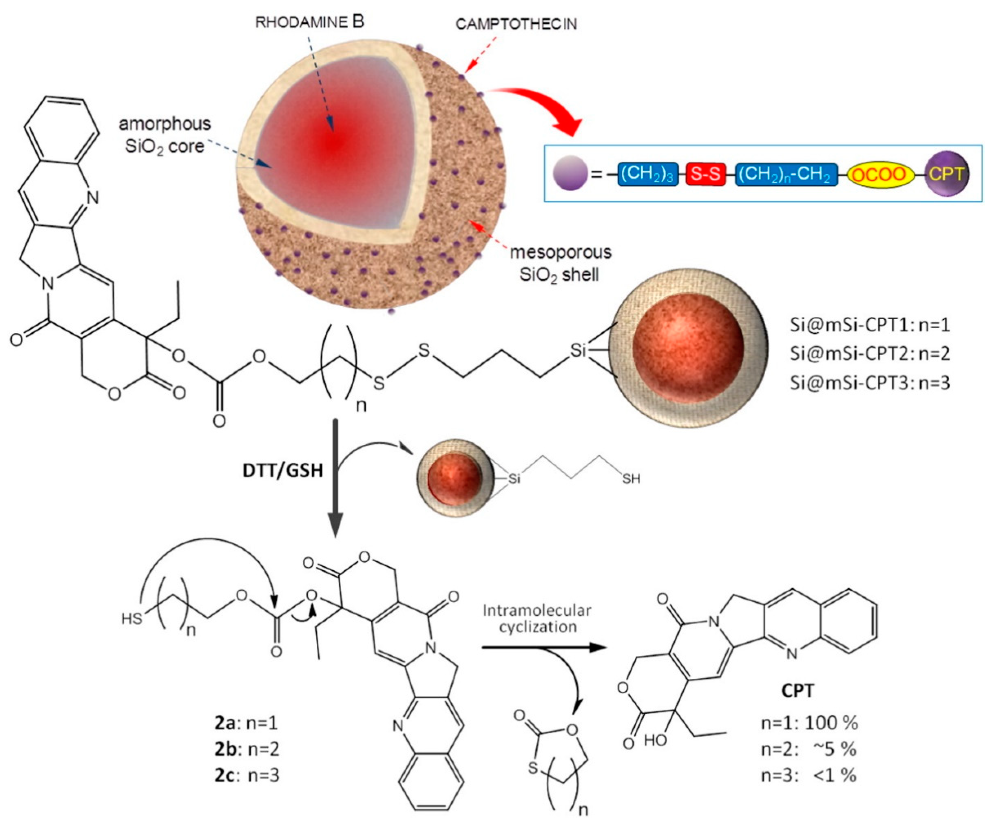
| Redox | Camptothecin (CPT) | Silica hybrid nanoparticles conjugated with pyridine-2-yldisulfanyl)alkyl carbonate derivatives of CPT | Disulfide reduction, intra-molecular cyclization, and dissociation of nanoparticles | [23,24] |
| Pyrene | Spherical PLGA nanoparticles containing hydrophobic molecules covered by a thin layer of a redox-responsive amorphous organosilica shell | Disulfide bridge reduction and pore opening | [25] | |
| Hydroxycamptothecin (HCPT) | Disulfide-doped organosilica-micellar hybrid nanoparticles | Two stage rocket-mimetic redox responsive mechanism. First, detachment of disulfide-bond of PEG and second, degradation of disulfide-doped silsesquioxane framework | [26] | |
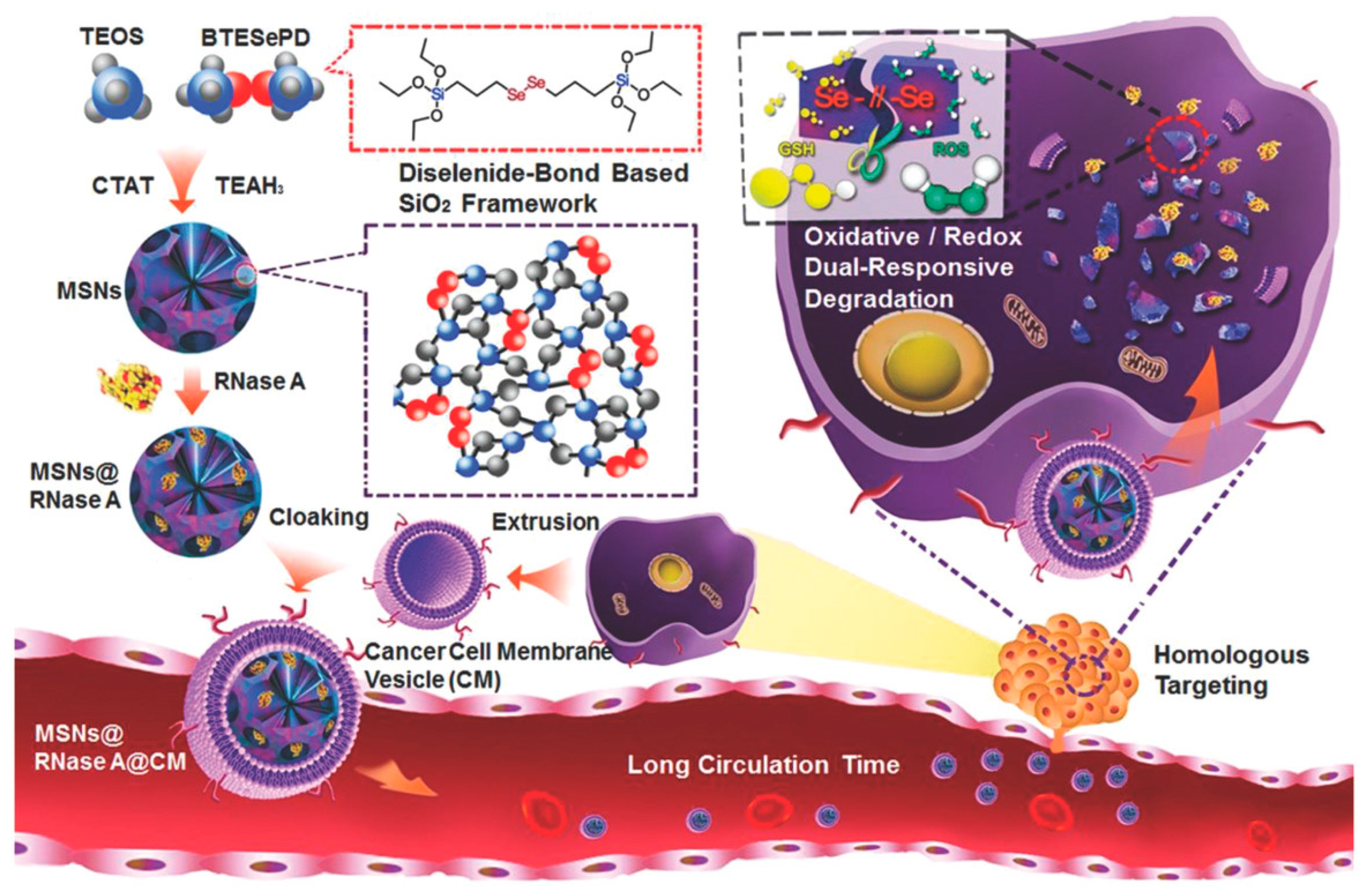
| Ribonuclease A (RNase A) | Diselenide-bridged mesoporous SNPs | Degradation of diselenide bridge in oxidative and reduction conditions | [27] | |
| Enzyme | Doxorubicin | Hollow MSNs grafted with chitosan as a gatekeeper by an azo linkage | Degradation of azo bonds | [28] |
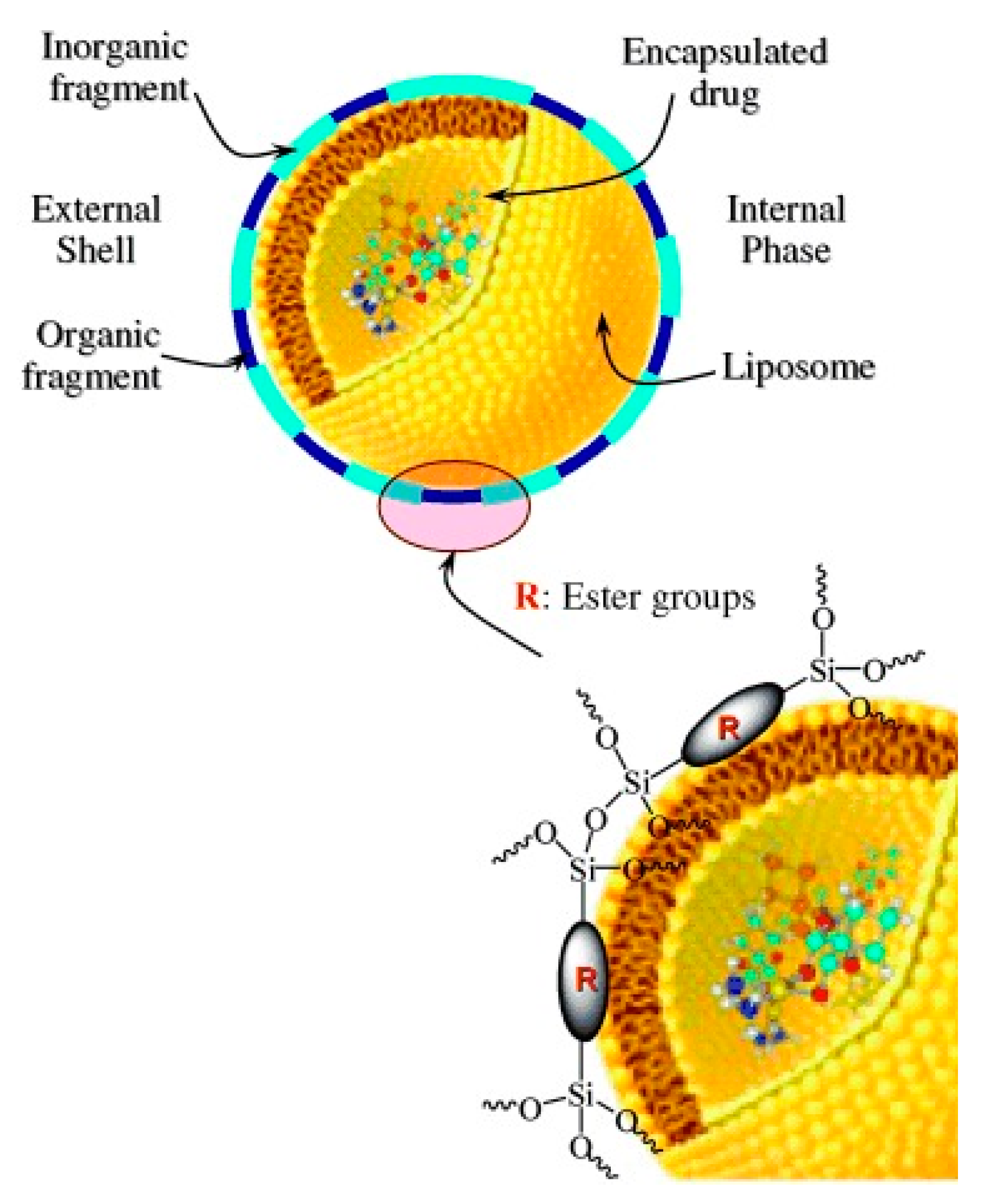
| Doxorubicin | Hybrid nanospheres composed of an organic core (liposome) and an inorganic shell formed by ester fragments bonded covalently to silica units | Ester bond hydrolysis | [29] | |
| Camptothecin | Amorphous SNPs decorated with CPT | Ester bond hydrolysis | [30] | |
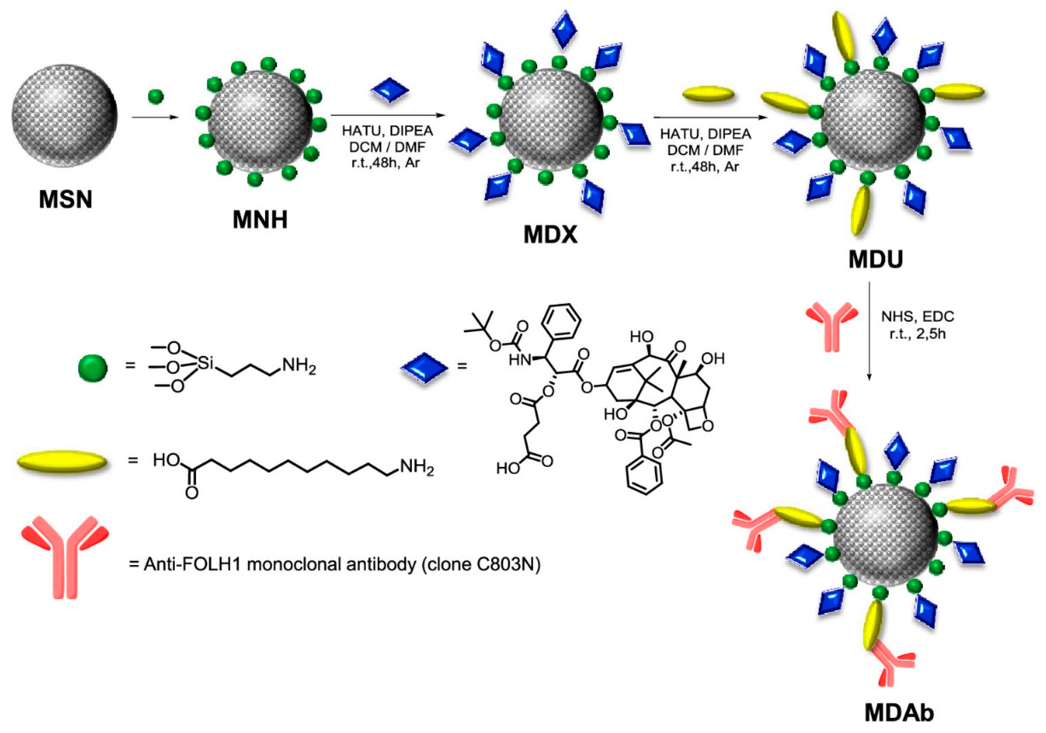
| Docetaxel (DTX) | MSNs conjugated with DTX and a PSMA antibody | Ester bond hydrolysis | [31] | |
| Temperature | Doxorubicin hydrochloride | Magnetic MSNs coated with polymer poly(N-isopropylacrylamide-co-acrylamide) as a gate-keeper | Conformational change in thermoresponsive polymer P(NIPAM-co-MAA) | [32] |
| Rhodamine 6G | Solid core mesoporous shells and nonporous solid corer SNPs grafted with poly(N-isopropylacrylami-de) brushes | Conformational change in thermoresponsive polymer PNIPAM | [33] | |
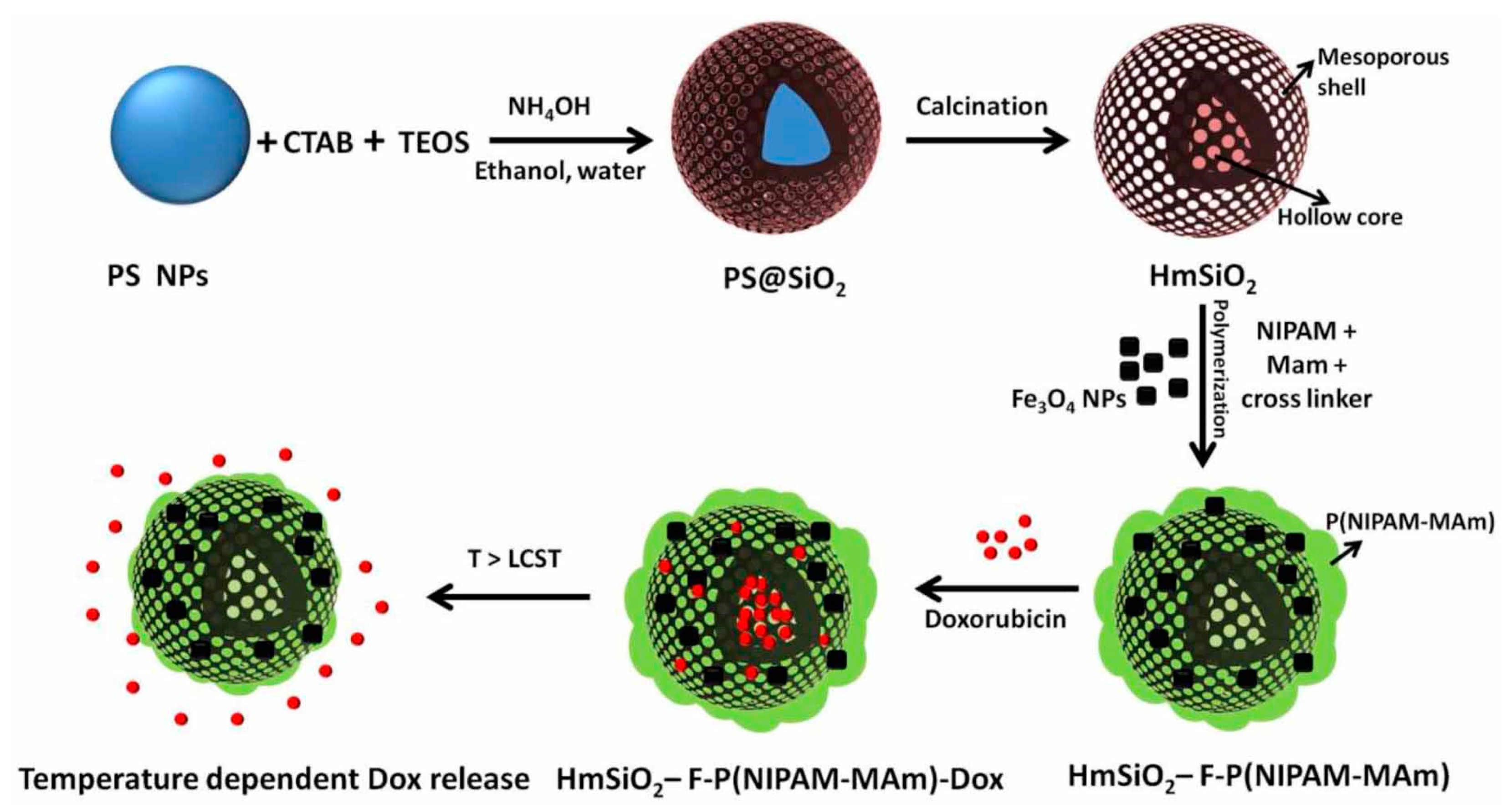
| Doxorubicin | Hollow MSNs coated with poly(N-isopropylacrylamide) modified with metha acrylamide (Mam) and with Fe3O4 nanoparticles embedded in the polymer shell | Conformational change in thermoresponsive polymer P(NIPAM-Mam) | [34] |
2.2. Redox-Responsive Systems
2.3. Enzyme-Responsive Systems
2.4. Thermo-Responsive Systems
3. Stimuli-Responsive Systems Based in Exogenous Activity
3.1. Magnetic-Responsive Systems
| Stimulus | Drug Loading | Release System | Release Mechanism | Ref. |
|---|---|---|---|---|
| Magnetic | Camptothecin | MSNs capped with monodispersed Fe3O4 nanoparticles through chemical bond | Chemical bond cleavage | [48] |
| Doxorubicin | Monodispersed manganese and cobalt doped iron oxide nanoparticles with a silica shell conjugated with the 4,4′-azobis(4-cyanovaleric acid) as a gate-keeper | Cleavage of the gatekeeper | [49] | |
| Light | Fluorescein disodium and Camptothecin | MSNs modified with an optimal molar ratio of spiropyran and perfluorodecyltriethoxysilane | Conformational conversion of spiropyran | [50] |
| Camptothecin | Light-activated mesostructured silica (LAMSs) nanoparticles functionalized with azobenzene moieties | Trans-cis photoisomerization of azobenzene | [51] | |
| Camptothecin | Nanoimpellers functionalized with azobenzene moieties and a two-photon fluorophore F | Trans-cis photoisomerization of azobenzene | [52] | |
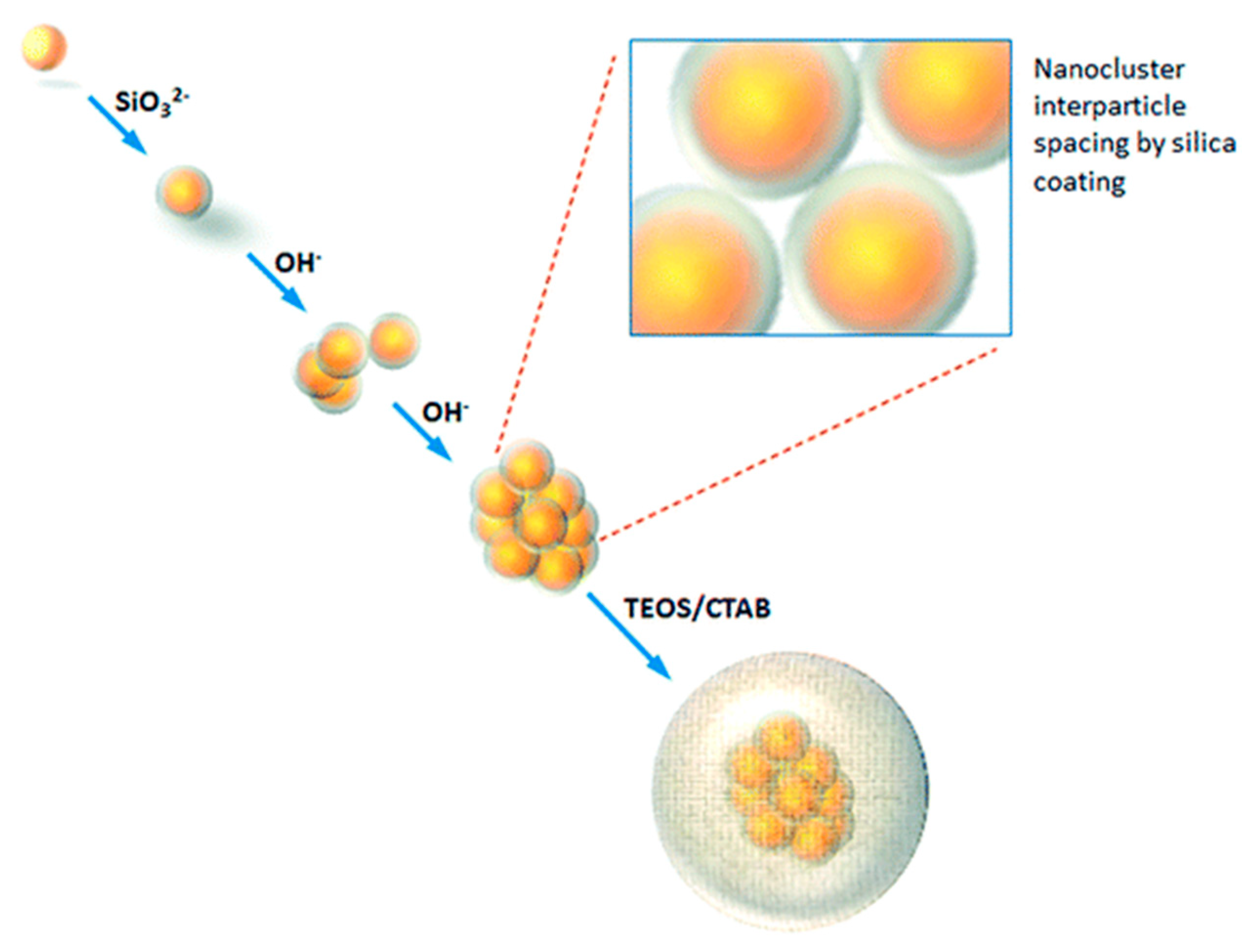
| Camptothecin | Gold nanoclusters with a homogeneous thin monolayer of amorphous silica (Au@SiO2) | Diffusion (promoted by local hyperthermia) | [53] | |
| Ultrasound | Topotecan hydrochloride | MSNs functionalized with poly(ethylene glycol) and 4,4′-azobis(4-cyanovaleric acid) | Cleavage of the azo moiety of the thermosensitive linker | [54] |
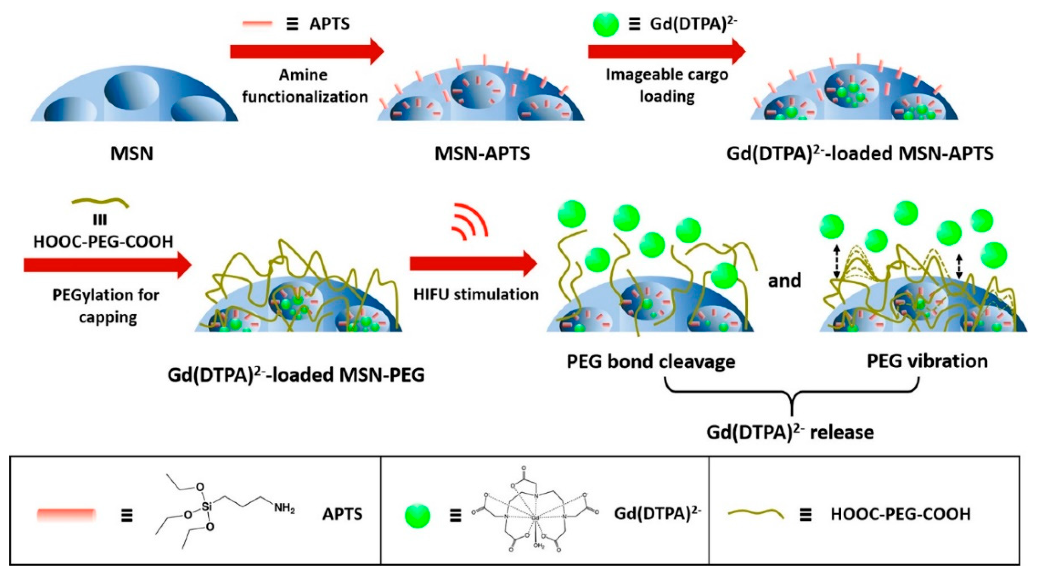
| Gadopentetate dimeglumine Gd(DTPA)2− | MSNs with pores capped with poly(ethylene glycol) | Poly(ethylene glycol) bond cleavage | [55] |
3.2. Light-Responsive Systems
3.3. Ultrasound-Responsive Systems
4. Targeting Molecules
5. Clinical Testing
| Material | Clinical Trial | Patients | Status | Action | Active Agent | Pathology | Via | Outcome | Ref. |
|---|---|---|---|---|---|---|---|---|---|
| Lipoceramic (silica@lipid) | Clinical Study | 16 | Completed | Bioavailability study | Ibuprofen | --- | Oral | Improved PK | [88] |
| ACTRN 12618001929291 | 12 | Completed | Bioavailability study | Simvastatin | --- | Improved PK | [89] | ||
| MSN | Clinical Study | 12 | Completed | Bioavailability study | Fenofibrate | --- | Oral | Improved PK | [90] |
| Au@SiO2 and Au/Fe3O4@SiO2 (core–shell) | NCT01270139 | 180 | Completed | Photothermal therapy | Gold nanoparticles | Atherosclerosis | IV | Reduced coronary atherosclerosis | [92] |
| NCT01436123 | 62 | Terminated | Photothermal therapy | Gold nanoparticles | Atherosclerosis | IV | Reduced risk of atherosclerosis | [92] | |
| Aurolase (SiO2@Au) | NCT00848042 | 11 | Completed | Photothermal therapy | Gold nanoshells | Head and neck cancer | IV | Tumor ablation | [91] |
| AuroShell (SiO2@Au) | NCT02680535 | 45 | Completed | Photothermal therapy | Gold nanoshells | Neoplasms of the prostate | IV | Pending b | [91] |
| NCT04240639 | 60 | Recruiting | Photothermal therapy | Gold nanoshells | Neoplasms of the prostate | IV | Pending b | [91] | |
| Cornell dots (ultra small SNPs) | NCT03465618 | 10 | Recruiting | PET Imaging, Fluorescent Imaging | 89Zr, Cy5.5 | Malignant brain tumors | IV | Pending | [93] |
| NCT02106598 | 86 | Recruiting | Fluorescent Imaging | Cy5.5 | Melanoma | IV | Pending | [93] | |
| NCT01266096 | 10 | Active, not recruiting | PET Imaging | 124I | Melanoma and malignant brain tumors | IV | Pending | [93] | |
| NCT04167969 | 10 | Recruiting | PET Imaging, Fluorescent Imaging | 64Cu, Cy5.5 | Prostate cancer | IV | Pending | [93] |
6. Conclusions and Future Direction
- (a)
- Small particle diameter: a very small particle diameter (e.g., <10 nm) can favor particle elimination by renal filtration. This has been conducted in the case of Cornell dots [93], with no significant side effects due to a short plasma half-life (<9 h). This is an interesting property for clinical imaging agents, but is not recommendable for drug delivery systems, as the smaller particles may extravasate before reaching the target cells, reducing the therapeutic response, and leading to severe undesired effects.
- (b)
- High drug loading: the higher the drug content in the nanomedicine, the lower the silica accumulation in tissue. This allows the administered dose to be reduced, and the therapeutic window to be enlarged. Mesoporous materials with well-developed internal geometric structures and high external surface areas for the incorporation of organic groups are probably the best choice for this purpose.
- (c)
- Targeting: as already shown in this review, the incorporation of targeting molecules in the nanoparticles favors tumor accumulation, thus allowing the dose to be reduced.
- (d)
- Organic–silica nanomaterials: hybrid nanomaterials containing silica and organic moieties gathered in a single system basically limit the amount of silica administered. In this review we have presented some potential nanomedicines based on an innocuous and biodegradable organic core (e.g., liposomes, polystyrene, etc.), and a silicate shell containing chemical doors able to be opened by specific stimuli. In these conjugates, the silica content can be reduced as far as 95% with regards the equivalent solid silica nanoparticles.
- (e)
- Replacing silica by structured organic materials. In the last decade, many groups have focused their research on novel materials for bioimaging and drug delivery, as coordination polymers [94,95], and covalent organic frameworks (COFs) [96]. These nanomaterials present well-defined topologies and high surface area, facilitating the incorporation of large quantities of active principles and other functional molecules. Furthermore, they are mostly organic (100% in case of COFs), and can be fully degraded inside the cells releasing their building components, which are later on eliminated by the renal route. In this way, toxicity issues should be no longer an obstacle for the development of novel nanomedicines able to perform efficient and selective chemotherapies, fully free of side effects.
Funding
Conflicts of Interest
References
- Cun, D.; Zhang, C.; Bera, H.; Yang, M. Particle Engineering Principles and Technologies for Pharmaceutical Biologics. Adv. Drug Deliv. Rev. 2021, 174, 140–167. [Google Scholar] [CrossRef]
- Ahmadi, S.; Rabiee, N.; Bagherzadeh, M.; Elmi, F.; Fatahi, Y.; Farjadian, F.; Baheiraei, N.; Nasseri, B.; Rabiee, M.; Dastjerd, N.T.; et al. Stimulus-Responsive Sequential Release Systems for Drug and Gene Delivery. Nano Today 2020, 34, 100914. [Google Scholar] [CrossRef]
- Dasgupta, A.; Biancacci, I.; Kiessling, F.; Lammers, T. Imaging-Assisted Anticancer Nanotherapy. Theranostics 2020, 10, 956–967. [Google Scholar] [CrossRef] [PubMed]
- Vallet-Regí, M.; Balas, F.; Arcos, D. Mesoporous Materials for Drug Delivery. Angew. Chem.-Int. Ed. 2007, 46, 7548–7558. [Google Scholar] [CrossRef] [PubMed]
- Rosenholm, J.M.; Lindén, M. Towards Establishing Structure-Activity Relationships for Mesoporous Silica in Drug Delivery Applications. J. Control. Release 2008, 128, 157–164. [Google Scholar] [CrossRef] [PubMed]
- Tang, S.; Huang, X.; Chen, X.; Zheng, N. Hollow Mesoporous Zirconia Nanocapsules for Drug Delivery. Adv. Funct. Mater. 2010, 20, 2442–2447. [Google Scholar] [CrossRef]
- Popova, M.; Koseva, N.; Trendafilova, I.; Lazarova, H.; Mitova, V.; Mihály, J.; Momekova, D.; Konstantinov, S.; Koleva, I.Z.; Petkov, P.S.; et al. Design of PEG-Modified Magnetic Nanoporous Silica Based Miltefosine Delivery System: Experimental and Theoretical Approaches. Microporous Mesoporous Mater. 2021, 310, 110664. [Google Scholar] [CrossRef]
- Yang, P.; Gai, S.; Lin, J. Functionalized Mesoporous Silica Materials for Controlled Drug Delivery. Chem. Soc. Rev. 2012, 41, 3679–3698. [Google Scholar] [CrossRef]
- Liu, M.; Du, H.; Zhang, W.; Zhai, G. Internal Stimuli-Responsive Nanocarriers for Drug Delivery: Design Strategies and Applications. Mater. Sci. Eng. C 2017, 71, 1267–1280. [Google Scholar] [CrossRef]
- Karimi, M.; Ghasemi, A.; Zangabad, P.S.; Rahighi, R.; Masoud, S.; Basri, M.; Mirshekari, H.; Amiri, M.; Pishabad, Z.S.; Aslani, A.; et al. Smart Micro/Nanoparticles in Stimulus-Responsive Drug/Gene Delivery Systems. Chem. Soc. Rev. 2016, 45, 1457–1501. [Google Scholar] [CrossRef] [Green Version]
- Mohapatra, A.; Uthaman, S.; Park, I.K. External and Internal Stimuli-Responsive Metallic Nanotherapeutics for Enhanced Anticancer Therapy. Front. Mol. Biosci. 2021, 7, 437. [Google Scholar] [CrossRef]
- Farokhzad, O.C.; Cheng, J.; Teply, B.A.; Sherifi, I.; Jon, S.; Kantoff, P.W.; Richie, J.P.; Langer, R. Targeted Nanoparticle-Aptamer Bioconjugates for Cancer Chemotherapy in Vivo. Proc. Natl. Acad. Sci. USA. 2006, 103, 6315–6320. [Google Scholar] [CrossRef] [Green Version]
- Wang, R.T.; Zhi, X.Y.; Yao, S.Y.; Zhang, Y. LFC131 Peptide-Conjugated Polymeric Nanoparticles for the Effective Delivery of Docetaxel in CXCR4 Overexpressed Lung Cancer Cells. Colloids Surf. B Biointerfaces 2015, 133, 43–50. [Google Scholar] [CrossRef]
- Scott, A.M.; Wolchok, J.D.; Old, L.J. Antibody Therapy of Cancer. Nat. Rev. Cancer 2012, 12, 278–287. [Google Scholar] [CrossRef]
- Arruebo, M.; Valladares, M.; González-Fernández, Á. Antibody-Conjugated Nanoparticles for Biomedical Applications. J. Nanomater. 2009, 2009, 439389. [Google Scholar] [CrossRef] [Green Version]
- Chen, X.; Soeriyadi, A.H.; Lu, X.; Sagnella, S.M.; Kavallaris, M.; Gooding, J.J. Dual Bioresponsive Mesoporous Silica Nanocarrier as an “AND” Logic Gate for Targeted Drug Delivery Cancer Cells. Adv. Funct. Mater. 2014, 24, 6999–7006. [Google Scholar] [CrossRef]
- Torchilin, V.P. Responsive Drug and Gene Delivery Systems; The Royal Society of Chemistry: London, UK, 2018; ISBN 9781788013536. [Google Scholar]
- Zhuo, S.; Zhang, F.; Yu, J.; Zhang, X.; Yang, G.; Liu, X. PH-Sensitive Biomaterials for Drug Delivery. Molecules 2020, 25, 5649. [Google Scholar] [CrossRef] [PubMed]
- Martínez-Carmona, M.; Lozano, D.; Colilla, M.; Vallet-Regí, M. Lectin-Conjugated PH-Responsive Mesoporous Silica Nanoparticles for Targeted Bone Cancer Treatment. Acta Biomater. 2018, 65, 393–404. [Google Scholar] [CrossRef] [PubMed]
- Yan, T.; He, J.; Liu, R.; Liu, Z.; Cheng, J. Chitosan Capped PH-Responsive Hollow Mesoporous Silica Nanoparticles for Targeted Chemo-Photo Combination Therapy. Carbohydr. Polym. 2020, 231, 115706. [Google Scholar] [CrossRef] [PubMed]
- Wang, M.; Wang, T.; Wang, D.; Jiang, W.; Fu, J. Acid and Light Stimuli-Responsive Mesoporous Silica Nanoparticles for Controlled Release. J. Mater. Sci. 2019, 54, 6199–6211. [Google Scholar] [CrossRef]
- Gonçalves, J.L.M.; Lopes, A.B.C.; Baleizão, C.; Farinha, J.P.S. Mesoporous Silica Nanoparticles Modified inside and out for on:Off Ph-Modulated Cargo Release. Pharmaceutics 2021, 13, 716. [Google Scholar] [CrossRef]
- Muniesa, C.; Vicente, V.; Quesada, M.; Sáez-Atiénzar, S.; Blesa, J.R.; Abasolo, I.; Fernández, Y.; Botella, P. Glutathione-Sensitive Nanoplatform for Monitored Intracellular Delivery and Controlled Release of Camptothecin. RSC Adv. 2013, 3, 15121–15131. [Google Scholar] [CrossRef]
- Botella, P.; Muniesa, C.; Vicente, V.; Cabrera-García, A. Effect of Drug Precursor in Cell Uptake and Cytotoxicity of Redox-Responsive Camptothecin Nanomedicines. Mater. Sci. Eng. C 2016, 58, 692–699. [Google Scholar] [CrossRef]
- Quesada, M.; Muniesa, C.; Botella, P. Hybrid PLGA-Organosilica Nanoparticles with Redox-Sensitive Molecular Gates. Chem. Mater. 2013, 25, 2597–2602. [Google Scholar] [CrossRef]
- Jia, X.; He, J.; Shen, L.; Chen, J.; Wei, Z.; Qin, X.; Niu, D.; Li, Y.; Shi, J. Gradient Redox-Responsive and Two-Stage Rocket-Mimetic Drug Delivery System for Improved Tumor Accumulation and Safe Chemotherapy. Nano Lett. 2019, 19, 8690–8700. [Google Scholar] [CrossRef] [PubMed]
- Shao, D.; Li, M.; Wang, Z.; Zheng, X.; Lao, Y.H.; Chang, Z.; Zhang, F.; Lu, M.; Yue, J.; Hu, H.; et al. Bioinspired Diselenide-Bridged Mesoporous Silica Nanoparticles for Dual-Responsive Protein Delivery. Adv. Mater. 2018, 30, 1801198. [Google Scholar] [CrossRef] [PubMed]
- Cai, D.; Han, C.; Liu, C.; Ma, X.; Qian, J.; Zhou, J.; Li, Y.; Sun, Y.; Zhang, C.; Zhu, W. Chitosan-Capped Enzyme-Responsive Hollow Mesoporous Silica Nanoplatforms for Colon-Specific Drug Delivery. Nanoscale Res. Lett. 2020, 15, 123. [Google Scholar] [CrossRef]
- Corma, A.; Díaz, U.; Arrica, M.; Fernández, E.; Ortega, Í. Organic-Inorganic Nanospheres with Responsive Molecular Gates for Drug Storage and Release. Angew. Chem.-Int. Ed. 2009, 48, 6247–6250. [Google Scholar] [CrossRef]
- Botella, P.; Abasolo, I.; Fernández, Y.; Muniesa, C.; Miranda, S.; Quesada, M.; Ruiz, J.; Schwartz, S.; Corma, A. Surface-Modified Silica Nanoparticles for Tumor-Targeted Delivery of Camptothecin and Its Biological Evaluation. J. Control. Release 2011, 156, 246–257. [Google Scholar] [CrossRef]
- Rivero-Buceta, E.; Vidaurre-Agut, C.; Vera-Donoso, C.D.; Benlloch, J.M.; Moreno-Manzano, V.; Botella, P. PSMA-Targeted Mesoporous Silica Nanoparticles for Selective Intracellular Delivery of Docetaxel in Prostate Cancer Cells. ACS Omega 2019, 4, 1281–1291. [Google Scholar] [CrossRef] [Green Version]
- Tian, Z.; Yu, X.; Ruan, Z.; Zhu, M.; Zhu, Y.; Hanagata, N. Magnetic Mesoporous Silica Nanoparticles Coated with Thermo-Responsive Copolymer for Potential Chemo- and Magnetic Hyperthermia Therapy. Microporous Mesoporous Mater. 2018, 256, 1–9. [Google Scholar] [CrossRef]
- Baliś, A.; Wolski, K.; Zapotoczny, S. Thermoresponsive Polymer Gating System on Mesoporous Shells of Silica Particles Serving as Smart Nanocontainers. Polymers 2020, 12, 888. [Google Scholar] [CrossRef] [PubMed]
- Asghar, K.; Qasim, M.; Dharmapuri, G.; Das, D. Thermoresponsive Polymer Gated and Superparamagnetic Nanoparticle Embedded Hollow Mesoporous Silica Nanoparticles as Smart Multifunctional Nanocarrier for Targeted and Controlled Delivery of Doxorubicin. Nanotechnology 2020, 31, 455604. [Google Scholar] [CrossRef]
- Guo, X.; Cheng, Y.; Zhao, X.; Luo, Y.; Chen, J.; Yuan, W.E. Advances in Redox-Responsive Drug Delivery Systems of Tumor Microenvironment. J. Nanobiotechnol. 2018, 16, 74. [Google Scholar] [CrossRef] [Green Version]
- Mollazadeh, S.; Mackiewicz, M.; Yazdimamaghani, M. Recent Advances in the Redox-Responsive Drug Delivery Nanoplatforms: A Chemical Structure and Physical Property Perspective. Mater. Sci. Eng. C 2021, 118, 111536. [Google Scholar] [CrossRef]
- Castillo, R.R.; Lozano, D.; González, B.; Manzano, M.; Izquierdo-Barba, I.; Vallet-Regí, M. Advances in Mesoporous Silica Nanoparticles for Targeted Stimuli-Responsive Drug Delivery: An Update. Expert Opin. Drug Deliv. 2019, 16, 415–439. [Google Scholar] [CrossRef]
- Zhou, R.; Zhu, S.; Gong, L.; Fu, Y.; Gu, Z.; Zhao, Y. Recent Advances of Stimuli-Responsive Systems Based on Transition Metal Dichalcogenides for Smart Cancer Therapy. J. Mater. Chem. B 2019, 7, 2588–2607. [Google Scholar] [CrossRef]
- Hu, Q.; Katti, P.S.; Gu, Z. Enzyme-Responsive Nanomaterials for Controlled Drug Delivery. Nanoscale 2014, 6, 12273–12286. [Google Scholar] [CrossRef] [PubMed]
- Minko, T.; Paranjpe, P.V.; Qiu, B.; Lalloo, A.; Won, R.; Stein, S.; Sinko, P.J. Enhancing the Anticancer Efficacy of Camptothecin Using Biotinylated Poly(Ethyleneglycol) Conjugates in Sensitive and Multidrug-Resistant Human Ovarian Carcinoma Cells. Cancer Chemother. Pharmacol. 2002, 50, 143–150. [Google Scholar] [CrossRef]
- Pham, S.H.; Choi, Y.; Choi, J. Stimuli-Responsive Nanomaterials for Application in Antitumor Therapy and Drug Delivery. Pharmaceutics 2020, 12, 630. [Google Scholar] [CrossRef] [PubMed]
- Kim, Y.J.; Matsunaga, Y.T. Thermo-Responsive Polymers and Their Application as Smart Biomaterials. J. Mater. Chem. B 2017, 5, 4307–4321. [Google Scholar] [CrossRef]
- Sponchioni, M.; Capasso Palmiero, U.; Moscatelli, D. Thermo-Responsive Polymers: Applications of Smart Materials in Drug Delivery and Tissue Engineering. Mater. Sci. Eng. C 2019, 102, 589–605. [Google Scholar] [CrossRef]
- Raza, A.; Rasheed, T.; Nabeel, F.; Hayat, U.; Bilal, M.; Iqbal, H.M.N. Endogenous and Exogenous Stimuli-Responsive Drug Delivery Systems for Programmed Site-Specific Release. Molecules 2019, 24, 1117. [Google Scholar] [CrossRef] [Green Version]
- Jia, R.; Teng, L.; Gao, L.; Su, T.; Fu, L.; Qiu, Z.; Bi, Y. Advances in Multiple Stimuli-Responsive Drug-Delivery Systems for Cancer Therapy. Int. J. Nanomed. 2021, 16, 1525–1551. [Google Scholar] [CrossRef]
- Mura, S.; Nicolas, J.; Couvreur, P. Stimuli-Responsive Nanocarriers for Drug Delivery. Nat. Mater. 2013, 12, 991–1003. [Google Scholar] [CrossRef]
- Zhu, N.; Ji, H.; Yu, P.; Niu, J.; Farooq, M.U.; Akram, M.W.; Udego, I.O.; Li, H.; Niu, X. Surface Modification of Magnetic Iron Oxide Nanoparticles. Nanomaterials 2018, 8, 810. [Google Scholar] [CrossRef] [Green Version]
- Chen, P.J.; Hu, S.H.; Hsiao, C.S.; Chen, Y.Y.; Liu, D.M.; Chen, S.Y. Multifunctional Magnetically Removable Nanogated Lids of Fe 3O4-Capped Mesoporous Silica Nanoparticles for Intracellular Controlled Release and MR Imaging. J. Mater. Chem. 2011, 21, 2535–2543. [Google Scholar] [CrossRef]
- Chen, W.; Cheng, C.A.; Zink, J.I. Spatial, Temporal, and Dose Control of Drug Delivery Using Noninvasive Magnetic Stimulation. ACS Nano 2019, 13, 1292–1308. [Google Scholar] [CrossRef]
- Chen, L.; Wang, W.; Su, B.; Wen, Y.; Li, C.; Zhou, Y.; Li, M.; Shi, X.; Du, H.; Song, Y.; et al. A Light-Responsive Release Platform by Controlling the Wetting Behavior of Hydrophobic Surface. ACS Nano 2014, 8, 744–751. [Google Scholar] [CrossRef] [PubMed]
- Lu, J.; Choi, E.; Tamanoi, F.; Zink, J.I. Light-Activated Nanoimpeller-Controlled Drug Release in Cancer Cells. Small 2008, 4, 421–426. [Google Scholar] [CrossRef] [PubMed] [Green Version]
- Croissant, J.; Maynadier, M.; Gallud, A.; Peindy N’Dongo, H.; Nyalosaso, J.L.; Derrien, G.; Charnay, C.; Durand, J.O.; Raehm, L.; Serein-Spirau, F.; et al. Two-Photon-Triggered Drug Delivery in Cancer Cells Using Nanoimpellers. Angew. Chem.-Int. Ed. 2013, 52, 13813–13817. [Google Scholar] [CrossRef]
- Botella, P.; Ortega, Í.; Quesada, M.; Madrigal, R.F.; Muniesa, C.; Fimia, A.; Fernández, E.; Corma, A. Multifunctional Hybrid Materials for Combined Photo and Chemotherapy of Cancer. Dalt. Trans. 2012, 41, 9286–9296. [Google Scholar] [CrossRef]
- Paris, J.L.; Manzano, M.; Cabañas, M.V.; Vallet-Regí, M. Mesoporous Silica Nanoparticles Engineered for Ultrasound-Induced Uptake by Cancer Cells. Nanoscale 2018, 10, 6402–6408. [Google Scholar] [CrossRef] [Green Version]
- Cheng, C.A.; Chen, W.; Zhang, L.; Wu, H.H.; Zink, J.I. A Responsive Mesoporous Silica Nanoparticle Platform for Magnetic Resonance Imaging-Guided High-Intensity Focused Ultrasound-Stimulated Cargo Delivery with Controllable Location, Time, and Dose. J. Am. Chem. Soc. 2019, 141, 17670–17684. [Google Scholar] [CrossRef] [PubMed]
- Jochum, F.D.; Theato, P. Temperature- and Light-Responsive Smart Polymer Materials. Chem. Soc. Rev. 2013, 42, 7468–7483. [Google Scholar] [CrossRef] [PubMed]
- Mena-Giraldo, P.; Pérez-Buitrago, S.; Londoño-Berrío, M.; Ortiz-Trujillo, I.C.; Hoyos-Palacio, L.M.; Orozco, J. Photosensitive Nanocarriers for Specific Delivery of Cargo into Cells. Sci. Rep. 2020, 10, 2110. [Google Scholar] [CrossRef] [Green Version]
- Linsley, C.S.; Wu, B.M. Recent Advances in Light-Responsive on-Demand Drug-Delivery Systems. Ther. Deliv. 2017, 8, 89–107. [Google Scholar] [CrossRef] [PubMed] [Green Version]
- Zhao, T.; Chen, L.; Li, Q.; Li, X. Near-Infrared Light Triggered Drug Release from Mesoporous Silica Nanoparticles. J. Mater. Chem. B 2018, 6, 7112–7121. [Google Scholar] [CrossRef]
- Botella, P.; Corma, A.; Navarro, M.T.; Quesada, M. Design of Optically Active Nanoclusters of Gold Particles with Mesostructured Silica Coating. J. Mater. Chem. 2009, 19, 3168–3175. [Google Scholar] [CrossRef]
- Chandan, R.; Mehta, S.; Banerjee, R. Ultrasound-Responsive Carriers for Therapeutic Applications. ACS Biomater. Sci. Eng. 2020, 6, 4731–4747. [Google Scholar] [CrossRef]
- Gu, W.; Meng, F.; Haag, R.; Zhong, Z. Actively Targeted Nanomedicines for Precision Cancer Therapy: Concept, Construction, Challenges and Clinical Translation. J. Control. Release 2021, 329, 676–695. [Google Scholar] [CrossRef] [PubMed]
- Nichols, J.W.; Bae, Y.H. EPR: Evidence and Fallacy. J. Control. Release 2014, 190, 451–464. [Google Scholar] [CrossRef] [PubMed]
- Baeza, A.; Colilla, M.; Vallet-Regí, M. Advances in Mesoporous Silica Nanoparticles for Targeted Stimuli-Responsive Drug Delivery. Expert Opin. Drug Deliv. 2015, 12, 319–337. [Google Scholar] [CrossRef]
- Meng, H.; Xue, M.; Xia, T.; Zhao, Y.L.; Tamanoi, F.; Stoddart, J.F.; Zink, J.I.; Nel, A.E. Autonomous in Vitro Anticancer Drug Release from Mesoporous Silica Nanoparticles by PH-Sensitive Nanovalves. J. Am. Chem. Soc. 2010, 132, 12690–12697. [Google Scholar] [CrossRef] [Green Version]
- Porta, F.; Lamers, G.E.M.; Morrhayim, J.; Chatzopoulou, A.; Schaaf, M.; den Dulk, H.; Backendorf, C.; Zink, J.I.; Kros, A. Folic Acid-Modified Mesoporous Silica Nanoparticles for Cellular and Nuclear Targeted Drug Delivery. Adv. Healthc. Mater. 2013, 2, 281–286. [Google Scholar] [CrossRef]
- Kim, K.; Choi, H.; Choi, E.S.; Park, M.H.; Ryu, J.H. Hyaluronic Acid-Coated Nanomedicine for Targeted Cancer Therapy. Pharmaceutics 2019, 11, 301. [Google Scholar] [CrossRef] [PubMed] [Green Version]
- Yu, M.; Jambhrunkar, S.; Thorn, P.; Chen, J.; Gu, W.; Yu, C. Hyaluronic Acid Modified Mesoporous Silica Nanoparticles for Targeted Drug Delivery to CD44-Overexpressing Cancer Cells. Nanoscale 2013, 5, 178–183. [Google Scholar] [CrossRef]
- Yao, V.J.; D’Angelo, S.; Butler, K.S.; Theron, C.; Smith, T.L.; Marchiò, S.; Gelovani, J.G.; Sidman, R.L.; Dobroff, A.S.; Brinker, C.J.; et al. Ligand-Targeted Theranostic Nanomedicines against Cancer. J. Control. Release 2016, 240, 267–286. [Google Scholar] [CrossRef] [PubMed] [Green Version]
- Sun, M.; Wang, T.; Li, L.; Li, X.; Zhai, Y.; Zhang, J.; Li, W. The Application of Inorganic Nanoparticles in Molecular Targeted Cancer Therapy: EGFR Targeting. Front. Pharmacol. 2021, 12, 1–15. [Google Scholar] [CrossRef]
- Sundarraj, S.; Thangam, R.; Sujitha, M.V.; Vimala, K.; Kannan, S. Ligand-Conjugated Mesoporous Silica Nanorattles Based on Enzyme Targeted Prodrug Delivery System for Effective Lung Cancer Therapy. Toxicol. Appl. Pharmacol. 2014, 275, 232–243. [Google Scholar] [CrossRef] [PubMed]
- She, X.; Chen, L.; Velleman, L.; Li, C.; He, C.; Denman, J.; Wang, T.; Shigdar, S.; Duan, W.; Kong, L. The Control of Epidermal Growth Factor Grafted on Mesoporous Silica Nanoparticles for Targeted Delivery. J. Mater. Chem. B 2015, 3, 6094–6104. [Google Scholar] [CrossRef]
- Landgraf, M.; Lahr, C.A.; Kaur, I.; Shafiee, A.; Sanchez-Herrero, A.; Janowicz, P.W.; Ravichandran, A.; Howard, C.B.; Cifuentes-Rius, A.; McGovern, J.A.; et al. Targeted Camptothecin Delivery via Silicon Nanoparticles Reduces Breast Cancer Metastasis. Biomaterials 2020, 240, 119791. [Google Scholar] [CrossRef]
- He, F.; Wen, N.; Xiao, D.; Yan, J.; Xiong, H.; Cai, S.; Liu, Z.; Liu, Y. Aptamer-Based Targeted Drug Delivery Systems: Current Potential and Challenges. Curr. Med. Chem. 2018, 27, 2189–2219. [Google Scholar] [CrossRef]
- Wang, K.; Yao, H.; Meng, Y.; Wang, Y.; Yan, X.; Huang, R. Specific Aptamer-Conjugated Mesoporous Silica-Carbon Nanoparticles for HER2-Targeted Chemo-Photothermal Combined Therapy. Acta Biomater. 2015, 16, 196–205. [Google Scholar] [CrossRef]
- Li, Y.; Duo, Y.; Bao, S.; He, L.; Ling, K.; Luo, J.; Zhang, Y.; Huang, H.; Zhang, H.; Yu, X. EpCAM Aptamer-Functionalized Polydopamine-Coated Mesoporous Silica Nanoparticles Loaded with DM1 for Targeted Therapy in Colorectal Cancer. Int. J. Nanomed. 2017, 12, 6239–6257. [Google Scholar] [CrossRef] [PubMed] [Green Version]
- von Baeckmann, C.; Kählig, H.; Lindén, M.; Kleitz, F. On the Importance of the Linking Chemistry for the PEGylation of Mesoporous Silica Nanoparticles. J. Colloid Interface Sci. 2021, 589, 453–461. [Google Scholar] [CrossRef] [PubMed]
- Dreau, D.; Moore, L.J.; Alvarez-Berrios, M.P.; Tarannum, M.; Mukherjee, P.; Vivero-Escoto, J.L. Mucin-1-Antibody-Conjugated Mesoporous Silica Nanoparticles for Selective Breast Cancer Detection in a Mucin-1 Transgenic Murine Mouse Model. J. Biomed. Nanotechnol. 2016, 12, 12172–12184. [Google Scholar] [CrossRef] [PubMed]
- Goel, S.; Chen, F.; Luan, S.; Valdovinos, H.F.; Shi, S.; Graves, S.A.; Ai, F.; Barnhart, T.E.; Theuer, C.P.; Cai, W. Engineering Intrinsically Zirconium-89 Radiolabeled Self-Destructing Mesoporous Silica Nanostructures for In Vivo Biodistribution and Tumor Targeting Studies. Adv. Sci. 2016, 3, 1600122. [Google Scholar] [CrossRef] [PubMed] [Green Version]
- Von Hoff, D.D.; Mita, M.M.; Ramanathan, R.K.; Weiss, G.J.; Mita, A.C.; Lorusso, P.M.; Burris, H.A.; Hart, L.L.; Low, S.C.; Parsons, D.M.; et al. Phase I Study of PSMA-Targeted Docetaxel-Containing Nanoparticle BIND-014 in Patients with Advanced Solid Tumors. Clin. Cancer Res. 2016, 22, 3157–3163. [Google Scholar] [CrossRef] [PubMed] [Green Version]
- Bharali, D.J.; Sudha, T.; Cui, H.; Mian, B.M.; Mousa, S.A. Anti-CD24 Nano-Targeted Delivery of Docetaxel for the Treatment of Prostate Cancer. Nanomed. Nanotechnol. Biol. Med. 2017, 13, 263–273. [Google Scholar] [CrossRef]
- Mangadlao, J.D.; Wang, X.; McCleese, C.; Escamilla, M.; Ramamurthy, G.; Wang, Z.; Govande, M.; Basilion, J.P.; Burda, C. Prostate-Specific Membrane Antigen Targeted Gold Nanoparticles for Theranostics of Prostate Cancer. ACS Nano 2018, 12, 3714–3725. [Google Scholar] [CrossRef] [PubMed]
- Jiang, W.; He, X.; Fang, H.; Zhou, X.; Ran, H.; Guo, D. Novel Gadopentetic Acid-Doped Silica Nanoparticles Conjugated with YPSMA-1 Targeting Prostate Cancer for MR Imaging: An in Vitro Study. Biochem. Biophys. Res. Commun. 2018, 499, 202–208. [Google Scholar] [CrossRef]
- Taari, K.; Isotalo, T.; Malmsten, L.-. åke; Lennerna, H. Pharmacokinetics of an Injectable Modi Fi Ed-Release 2-Hydroxy Fl Utamide Formulation in the Human Prostate Gland Using a Semiphysiologically Based Biopharmaceutical Model. Mol. Pharm. 2014, 11, 3097–3111. [Google Scholar] [CrossRef]
- Mohammadpour, R.; Cheney, D.L.; Grunberger, J.W.; Yazdimamaghani, M.; Jedrzkiewicz, J.; Isaacson, K.J.; Dobrovolskaia, M.A.; Ghandehari, H. One-Year Chronic Toxicity Evaluation of Single Dose Intravenously Administered Silica Nanoparticles in Mice and Their Ex Vivo Human Hemocompatibility. J. Control. Release 2020, 324, 471–481. [Google Scholar] [CrossRef] [PubMed]
- Botella, P.; Rivero-Buceta, E. Safe Approaches for Camptothecin Delivery: Structural Analogues and Nanomedicines. J. Control. Release 2017, 247, 28–54. [Google Scholar] [CrossRef]
- Janjua, T.I.; Cao, Y.; Yu, C.; Popat, A. Clinical Translation of Silica Nanoparticles. Nat. Rev. Mater. 2021. [Google Scholar] [CrossRef]
- Tan, A.; Eskandar, N.G.; Rao, S.; Prestidge, C.A. First in Man Bioavailability and Tolerability Studies of a Silica-Lipid Hybrid (Lipoceramic) Formulation: A Phase I Study with Ibuprofen. Drug Deliv. Transl. Res. 2014, 4, 212–221. [Google Scholar] [CrossRef]
- Meola, T.R.; Abuhelwa, A.Y.; Joyce, P.; Clifton, P.; Prestidge, C.A. A Safety, Tolerability, and Pharmacokinetic Study of a Novel Simvastatin Silica-Lipid Hybrid Formulation in Healthy Male Participants. Drug Deliv. Transl. Res. 2021, 11, 1261–1272. [Google Scholar] [CrossRef]
- Bukara, K.; Schueller, L.; Rosier, J.; Martens, M.A.; Daems, T.; Verheyden, L.; Eelen, S.; Van Speybroeck, M.; Libanati, C.; Martens, J.A.; et al. Ordered Mesoporous Silica to Enhance the Bioavailability of Poorly Water-Soluble Drugs: Proof of Concept in Man. Eur. J. Pharm. Biopharm. 2016, 108, 220–225. [Google Scholar] [CrossRef] [PubMed]
- Rastinehad, A.R.; Anastos, H.; Wajswol, E.; Winoker, J.S.; Sfakianos, J.P.; Doppalapudi, S.K.; Carrick, M.R.; Knauer, C.J.; Taouli, B.; Lewis, S.C.; et al. Gold Nanoshell-Localized Photothermal Ablation of Prostate Tumors in a Clinical Pilot Device Study. Proc. Natl. Acad. Sci. USA 2019, 116, 18590–18596. [Google Scholar] [CrossRef] [Green Version]
- Kharlamov, A.N.; Tyurnina, A.E.; Veselova, V.S.; Kovtun, O.P.; Shur, V.Y.; Gabinsky, J.L. Silica-Gold Nanoparticles for Atheroprotective Management of Plaques: Results of the NANOM-FIM Trial. Nanoscale 2015, 7, 8003–8015. [Google Scholar] [CrossRef] [PubMed]
- Zanoni, D.K.; Stambuk, H.E.; Madajewski, B.; Montero, P.H.; Matsuura, D.; Busam, K.J.; Ma, K.; Turker, M.Z.; Sequeira, S.; Gonen, M.; et al. Use of Ultrasmall Core-Shell Fluorescent Silica Nanoparticles for Image-Guided Sentinel Lymph Node Biopsy in Head and Neck Melanoma: A Nonrandomized Clinical Trial. JAMA Netw. Open 2021, 4, e211936. [Google Scholar] [CrossRef] [PubMed]
- García-García, P.; Moreno, J.M.; Díaz, U.; Bruix, M.; Corma, A. Organic-Inorganic Supramolecular Solid Catalyst Boosts Organic Reactions in Water. Nat. Commun. 2016, 7, 10835. [Google Scholar] [CrossRef] [PubMed] [Green Version]
- Suárez-García, S.; Solórzano, R.; Novio, F.; Alibés, R.; Busqué, F.; Ruiz-Molina, D. Coordination Polymers Nanoparticles for Bioimaging. Coord. Chem. Rev. 2021, 432, 213716. [Google Scholar] [CrossRef]
- Oliveira, A.D.S.; Rivero-Buceta, E.M.; Vidaurre-Agut, C.; Misturini, A.; Moreno, V.; Jordá, J.L.; Sastre, G.; Pergher, S.B.C.; Botella, P. Sequential Pore Wall Functionalization in Covalent Organic Frameworks and Application to Stable Camptothecin Delivery Systems. Mater. Sci. Eng. C 2020, 117, 111263. [Google Scholar] [CrossRef]


Publisher’s Note: MDPI stays neutral with regard to jurisdictional claims in published maps and institutional affiliations. |
© 2022 by the authors. Licensee MDPI, Basel, Switzerland. This article is an open access article distributed under the terms and conditions of the Creative Commons Attribution (CC BY) license (https://creativecommons.org/licenses/by/4.0/).
Share and Cite
Corma, A.; Botella, P.; Rivero-Buceta, E. Silica-Based Stimuli-Responsive Systems for Antitumor Drug Delivery and Controlled Release. Pharmaceutics 2022, 14, 110. https://doi.org/10.3390/pharmaceutics14010110
Corma A, Botella P, Rivero-Buceta E. Silica-Based Stimuli-Responsive Systems for Antitumor Drug Delivery and Controlled Release. Pharmaceutics. 2022; 14(1):110. https://doi.org/10.3390/pharmaceutics14010110
Chicago/Turabian StyleCorma, Avelino, Pablo Botella, and Eva Rivero-Buceta. 2022. "Silica-Based Stimuli-Responsive Systems for Antitumor Drug Delivery and Controlled Release" Pharmaceutics 14, no. 1: 110. https://doi.org/10.3390/pharmaceutics14010110