Next Article in Journal
Polishing the Therapy of Onychomycosis Induced by Candida spp.: Amphotericin B–Loaded Nail Lacquer
Next Article in Special Issue
Co-Encapsulation of Fisetin and Cisplatin into Liposomes for Glioma Therapy: From Formulation to Cell Evaluation
Previous Article in Journal
Assessment of Metabolic Interaction between Repaglinide and Quercetin via Mixed Inhibition in the Liver: In Vitro and In Vivo
Previous Article in Special Issue
Encapsulation of ε-Viniferin into Multi-Lamellar Liposomes: Development of a Rapid, Easy and Cost-Efficient Separation Method to Determine the Encapsulation Efficiency
 
 
Retraction published on 30 January 2024, see Pharmaceutics 2024, 16(2), 195.
Article

RETRACTED: Lipidic Nano-Sized Emulsomes Potentiates the Cytotoxic and Apoptotic Effects of Raloxifene Hydrochloride in MCF-7 Human Breast Cancer Cells: Factorial Analysis and In Vitro Anti-Tumor Activity Assessment

by
Hibah M. Aldawsari
1,2,
Osama A. A. Ahmed
1,3,
Nabil A. Alhakamy
1,2,3,
Thikryat Neamatallah
4,
Usama A. Fahmy
1,3,* and
Shaimaa M. Badr-Eldin
1,5
1
Department of Pharmaceutics, Faculty of Pharmacy, King Abdulaziz University, Jeddah 21589, Saudi Arabia
2
Center of Excellence for Drug Research and Pharmaceutical Industries, King Abdulaziz University, Jeddah 21589, Saudi Arabia
3
Mohamed Saeed Tamer Chair for Pharmaceutical Industries, King Abdulaziz University, Jeddah 21589, Saudi Arabia
4
Department of Pharmacology and Toxicology, Faculty of Pharmacy, King Abdulaziz University, Jeddah 21589, Saudi Arabia
5
Department of Pharmaceutics and Industrial Pharmacy, Faculty of Pharmacy, Cairo University, Cairo 11562, Egypt
*
Author to whom correspondence should be addressed.
Pharmaceutics 2021, 13(6), 783; https://doi.org/10.3390/pharmaceutics13060783
Submission received: 16 April 2021 / Revised: 17 May 2021 / Accepted: 21 May 2021 / Published: 24 May 2021 / Retracted: 30 January 2024
(This article belongs to the Special Issue Liposomal Drug Delivery Systems)

Abstract

:
Raloxifene hydrochloride (RLX), an antiosteoporotic agent, has been utilized for guarding against breast cancer and recently, for the disease management owing to its estrogen antagonist activity. Nevertheless, RLX exhibits poor bioavailability that could be attributed to reduced water solubility and first pass metabolism. To overcome these challenges, this study aimed at formulating and optimizing RLX emulsomes (RLX-EMLs) to enhance the drug antitumor activity. A 4131 factorial design was employed for assessing the effect of lipoid: solid lipid ratio and solid lipid type on the emulsomes characteristics. The anticancer potential of the optimized formulation and apoptotic parameters were assessed. Vesicle size, entrapment, and release efficiency were significantly influenced by both variables, while zeta potential was influenced by lipoid: solid lipid at p < 0.05. The optimal formulation exhibited vesicle size of 236 ± 8.6 nm, zeta potential of −18.6 ± 0.7 mV, drug entrapment of 98.9 ± 4.9%, and release efficiency of 42.7 ± 1.8%. MTT assay showed concentration-dependent inhibition of MCF-7 cells viability. In addition, cells treated with RLX-EMLs showed significant arrest at G2/M phase associated with significant increase in apoptotic and necrotic cells. The enhanced cytotoxic and anti-proliferative effect of RLX-EMLs relative to raw drug was authenticated through increased Bax/Bcl-2 ratio, caspase-9 activation and depletion of mitochondrial membrane potential.

1. Introduction

Breast cancer is the most spread cancer form among females. Estrogen receptor positive (ER+) breast cancer accounts for the greatest percentage of all breast cancer cases [1,2]. Selective estrogen-receptor modulators (SERMs), such as raloxifene are the most common agents that are applied for the treatment of such type of cancer. Raloxifene hydrochloride (RLX) is the first US-FDA approved drug from the selective estrogen-receptor modulators class as a preventive and treatment agent for osteoporosis in postmenopausal women. Thereafter, RLX was approved by FDA also for attenuation of breast cancerous tumors risk, and more recently, studies have directed to investigate the possibility of utilizing the drug for breast cancer treatment [3,4,5]. This indication has been suggested on the basis of the drug’s estrogen antagonist activity. RLX has been reported to stimulate gene promoters regulated by ERβ subtype that guards against tumor formation in response to estrogen; therefore, it exhibits anti-tumor activity in breast cancer cells [6]. Nevertheless, RLX suffers from poor aqueous solubility and exposure to dramatic pre-systemic metabolism that lead to diminished bioavailability that does not surpass 2% [7,8].
During the last decades, nano-sized formulations have been explored as an effective approach to deliver biologically active drugs to malignant tumors [9]. Nano-systems are regarded as a promising surrogate for conventional treatment methods because of their ability to accumulate in the tumor through improved permeation and retention within the cancerous cells. The ability of these systems to invade the tumor exhibited an inverse relationship with their sizes [10].
Emulsomes are nanoemulsions of particles comprising solid fat cores stabilized by phospholipid (PL) envelope at the aqueous interface forming a bilayer. Such systems combines the advantages of both nanoemulsions and liposomes [11]. A featured property of emulsomes is the lipidic core that is present in a liquid crystalline or solid phase rather than being fluid as in emulsions. This distinguishable property enables the encapsulation of higher amounts of lipophilic drugs with extended release. The phospholipid lipid bilayer increases the solubility and bioavailability of lipophilic drugs. Moreover, the site specificity of emulsomes, due to their nano-size that enhances drug targeting action, is considered an additional advantage in case of cancer therapy [12,13]. Owing to the aforementioned characteristics, emulsomes could be considered a promising surfactant-free economical alternative to current lipid-based formulations that could safely and effectively minimize the drugs’ dosing frequency [14].
Emulsomes have been recently explored for enhancing the anti-tumor activity of lipophilic therapeutic moieties [15]. Varshosaz et al. [16] have designed a formulation for targeted delivery to hepatocellular carcinoma cells utilizing PEGylated trimethyl chitosan coated EMLs conjugated with octreotide. The prepared optimized EMLs formula showed a particle size of 127 nm, with 95% sorafenib loading efficiency, and drug release efficiency of 62% within 52 h. The optimized targeted EMLs formula showed more cytotoxicity activity when compared with free sorafenib and non-targeted EMs. Bolat et al. [17] have investigated EMLs nano-formulation loaded with curcumin and piperine for antiproliferative activity against colon cancer cells (HCT116). Their findings have revealed that the proposed combinational treatment improved the anticancer activity of the curcumin and piperine. Ucisik et al. [18] have investigated EMLs surface modification utilizing S-layer proteins. Their findings revealed that EMLs surface modification with a crystalline S-layer lattice protected cells from oxidative stress and membrane damage. Researches conducted in our laboratory have explored the utilization of EMLs in enhancing the cytotoxic and pro-apoptotic effects of statins, Piceatannol and febuxostat [19,20,21,22].
Thus, the current study aimed at formulating and optimizing RLX emulsomes (RLX-EMLs) to provide controlled release and enhanced anti-cancer activity of the drug. The optimized emulsomal formulation with minimized vesicle size and release efficiency, in addition to maximized zeta potential absolute value and drug entrapment was further characterized for in vitro anti-tumor activity in human breast cancer cells.

2. Materials and Methods

2.1. Materials

Raloxifene hydrochloride (RLX) was from Qingdao Sigma Chemical Co., Ltd., Qingdao, China. Cholesterol, Tripalmitin (TP), Tristearin (TS), and MTT (3-(4,5-Dimethylthiazol-2-yl)-2,5-Diphenyltetrazolium Bromide) cell viability kit were purchased from Sigma-Aldrich Inc. (St. Louis, MI, USA). Lipoid® 90 H was kindly gifted by Lipoid GmbH (Ludwigshafen, Germany). Compritol® ATO 888 was a gift sample from Gattefosse. MCF-7 cells were from American Type Culture Collection (Manassas, VA, USA). Dulbecco’s modified Eagle’s medium (DMEM), fetal bovine serum (FBS), penicillin-streptomycin, and trypsin/EDTA were from Thermo Fisher Scientific (Waltham, MA, USA). Propidium Iodide Flow Cytometry Kit was from Abcam (ab139418, Cambridge, UK).

2.2. Formulation of RLX-EMLs

RLX-EMLs were prepared as per the reported method of Paliwal et al. [23]. Specified quantities of RLX (60 mg), Lipoid® 90 H, cholesterol (5 mg), and tripalmitin were solubilized in 10 mL chloroform/methanol mixture (2:1, v/v). Evaporation was executed under reduced pressure using Rotavapor at 40 °C. The deposited films were dried in a vacuum oven for 24 h to achieve total elimination of the solvent. The residual films were then hydrated with 10 mL phosphate buffer with gentle agitation for 2 h at ambient temperature. The formed dispersion was subjected to ultrasonication (40% amplitude, 750 W, 20 kHz) for 1 min in two cycles with a time gap of three minutes between each cycle to obtain emulsomes [12,20].

2.3. Characterization of RLX-EMLs

2.3.1. Vesicle Size and Zeta Potential

Light scattering and electrophoretic techniques were applied to determine the average vesicle size (z-average) and zeta potential, respectively, using Nano ZSP (Malvern Panalytical, Malvern, UK) at 25 ± 1 °C. Mixed mode measurement combined with phase analysis light scattering (M3-PALS) was used to measure electrophoretic mobility and determine zeta potential mean and distribution during the same measurement. Emulsomes were diluted 100 folds appropriately before measurement using phosphate buffer pH 7.4 [24]. Results for each parameter were computed as a mean of six measurements.

2.3.2. Entrapment Efficiency (EE%)

EE% of RLX-EMLs was determined by indirect method. To separate the unentrapped drug, 1 mL of RLX loaded emulsomal dispersion (equivalent to 0.1 mg RLX) was ultracentrifuged at 100,000 rpm for 1 h at 4 °C (OptimaTM MAX-XP; Beckman Coulter Inc., Indianapolis, IN, USA). The residue was washed twice with phosphate buffer and re-centrifuged again for 1 h [25]. The combined supernatant was analyzed using a previously reported HPLC method [26]. All determinations were performed in triplicate. The EE% was determined using Equation (1).
EE % = D t D u D t × 100
where Dt and Du represent the amount of total drug and the amount of unentrapped drug, respectively.

2.3.3. In Vitro Release

Glass basket dialysis technique was adopted to study the release of RLX from the prepared emulsomes [27]. Samples of emulsomal dispersions equivalent to 10 mg of drug were placed inside glass cylinder tube. Each tube was tightly covered with previously soaked dialysis membrane (MWCO 12,000–14,000 Da) at one end, while the other end was attached to the USP dissolution apparatus I shaft instead of basket. The shaft was then lowered to the dissolution vessel containing PBS (pH 7.4) with Tween 80 (0.1% v/v) as a release medium. The temperature was kept constant at 37 ± 0.5 °C and at 50 rpm. Aliquots were withdrawn at predetermined intervals for 6 h that were replaced with fresh medium. The amount of RLX released was assayed using the aforementioned HPLC method. The experiment was performed thrice for each formulation. The mean release efficiency after 6 h was then computed as in vitro release evaluation parameter. Decreased release efficiency was considered as indication for the ability of the emulsomal formulations to control the drug release [28,29].

2.4. Experimental Design (4131 Full Factorial Design)

The parameters effect was examined using a 4131 full factorial design. Two formulation parameters namely; Lipoid® 90 H: solid lipid ratio (PL:SL:X1, w/w) and solid lipid type (X2), were selected as independent variables. X1 was studied at 4 levels, while X2 was studied at three levels. The levels of each variable are compiled in Table 1. The design yielded 12 runs, Table 2. The impact of the independent variables on the responses including vesicle size (VS:Y1, nm), zeta potential (ZP:Y2, mV), entrapment efficiency (EE:Y3, %), and release efficiency after 6 h (RE:Y4, %) were analyzed using Design-Expert software (version 12; Stat-Ease, Inc., Minneapolis, MN, USA, 2019). Best fitting polynomial model for the response was chosen according to the maximum adjusted and predicted determination coefficients (R2) and lowest predicted residual sum of squares (PRESS). The software was employed to analyze experimental results to point out the main effects of the variables under investigation; the significance of each variable was assessed via performing Analysis of variance (ANOVA).

2.5. Optimization of RLX-EMLs

The optimal emulsomal formulation to be subjected for further investigations was selected based on the desirability function that integrated the responses into a single variable in order to anticipate the optimum levels of studied variables [30,31]. Achieving minimized VS and RE associated with maximized EE% and absolute value of ZP were set as goals for the optimum formulation as shown in Table 1.

2.6. In Vitro Anti-Tumor Activity of Optimized RLX-EMLs in Human Breast Cancer Cells

2.6.1. Cell Culture

Human breast cancer cell line (MCF-7) cell line was obtained from the American Type Culture Collection (ATCC; www.atcc.org, accessed on 4 April 2021). The cells were cultured in DMEM supplemented with 10% FBS, penicillin (1% v/v), and streptomycin (1% v/v) at 37 °C in humidified 5% CO2 incubator.

2.6.2. MTT Assay

The effects of RLX-EMLs, RLX, and blank EMLs on MCF-7 cells viability were evaluated using MTT assay as previously described [32,33]. The cells were plated at a density of 5 × 10³ cells per well in 96-well plate, and kept in the incubator overnight prior to exposition to the treatments with RLX-EMLs and RLX at different concentrations: 100, 25, 6.25, 1.56, and 0.39 µg/mL for 48 h. RLX was dissolved in 0.1% DMSO and this vehicle concentration showed negative cytotoxic effect on MCF-7 cells. Appropriate control wells from untreated cells were prepared at the same time. After the incubation time, treatment was replaced with 10 µL of MTT solution (5 mg/mL) for 4 h. After that, media containing MTT was removed from each well, and 200 µL/well of 100% DMSO were added to dissolve the formazan crystals. The colorimetric signals were assessed at 570 nm using microplate reader (Bio Tek, Santa Clara, CA, USA; Winooski, VT, USA). Results were expressed as percentage viability relative to the control. All experiments were performed in triplicate of wells per concentration and repeated at least in three independent times. Dose response curves were plotted, and the half-maximal inhibitory concentrations (IC50) were obtained using the commercial software Prism (GraphPad Software, Inc., La Jolla, CA, USA).

2.6.3. Cell Cycle Analysis

The effects of RLX-EMLs, RLX, and blank EMLs on MCF-7 cell cycle distributions were determined by flow cytometry as previously described [34,35]. The cells were plated at a density of 3 × 10⁵ cells per well in 6-well plate and further incubated with RLX-EMLs, RLX, or blank EMLs, for 48 h. Afterward, cells were washed, collected by trypsinisation and centrifuged at 10,000× g for 15 min. The cells were then fixed with 70% ethanol and incubated overnight at −20 °C. Fixed cells were later centrifuged at 10,000× g for 15 min and stained with PI (10 μg/mL) and RNase treatment. Cell DNA contents were measured using a flow cytometer (FACScalibur, BD Bioscience, San Jose, CA, USA) and a minimum of 20,000 events were acquired for each treatment. The data was analyzed using the CellQuest Software (Becton-Dickinson, Franklin Lakes, NJ, USA).

2.6.4. Apoptosis Assay

The effects of RLX-EMLs, RLX, and blank EMLs on the extent of apoptosis in MCF-7 were examined-using an annexin V-FTCI/PI apoptosis kit as previously described [36]. The cells were plated at a density of 1 × 10⁶ in 6-well plate and further incubated with RLX, RLX-EMLs or blank EMLs for 48 h. The cells were then collected by trypsinization and centrifuged at 10,000× g for 5 min. After that, the supernatant was discarded, and the cells were washed in PBS and incubated with annexin V-FITC/PI dyes in the dark for 30 min on ice. The stained cells were analyzed by FACScalibur (BD Bioscience, San Jose, CA, USA) and a minimum of 20,000 events were acquired for each treatment.

2.6.5. Evaluation of Bax and Bcl-2 Expressions

The effects of RLX-EMLs, RLX, and blank EMLs on the expression of Bax and Bcl-2 proteins were investigated in MCF-7 cells using Human Bax ELISA Kit (EIA-4487, DRG) and Human Zymed® Bcl-2 ELISA Kit (99-0042, Invitrogen, Thermo Fisher Scientific, Waltham, MA, USA). The cells were plated at a density of 5 × 10⁴ in 24-well plate were left over night and further treated with RLX-EMLs, RLX, or blank EMLs for 48 h. Cell lysate was measured for human active Bax and Bcl2 content according to the manufacturers’ protocol for each kit. Microplate reader (Bio Tek, Santa Clara, CA, USA) was used to assess the absorbance at 450 nm.

2.6.6. Caspase-3 Activation Assay

The effects of RLX-EMLs, RLX, and blank EMLs on the activation of caspase3 activation were assessed using kit for colorimetric assay (556485, BD Biosciences, San Jose, CA, USA) as instructed by the provided protocol. Briefly, 5 × 10⁴ cells were cultured in 96-well plates and treated with RLX-EMLs, RLX, and blank EMLs 48 h. Cells were then washed, lysed and subjected to centrifugation at 10,000× g for 1 min. The amount of released p nitroaniline (pNA) was measured in the supernatants at 405 nm using microplate reader (Bio Tek, Santa Clara, CA, USA; Winooski, VT, USA).

2.6.7. Mitochondrial Membrane Potential (MMP)

The effects of RLX-EMLs, RLX, and blank EMLs on MMP was estimated by MitoProbe™ TMRM Kit (Thermo Fisher Scientific, Waltham, MA, USA) as instructed. Briefly, 1 × 10⁵ cells were seeded in black clear-bottom 96-well and treated with RLX-EMLs, RLX, and blank EMLs for 48 h. Cells were stained with 20 µM tetramethylrhodamine, methyl ester (TMRM) and incubated for 30 min at 37 °C. A bright signal is produced upon accumulation of TMRM dye in active mitochondria of cells with intact membrane potentials. During apoptosis, there is loss of MMP and the signal disappears. The cells were loaded with live-cell imaging buffer after washing with PBS and then analyzed by using flow cytometry (FACSCalibur, BD Bioscience, USA). For detection: TMRM is excited by the 488 nm laser and should be detected in the ap-propriate filter channel for peak emission at 575 nm.

3. Results and Discussion

3.1. Factorial Design Analysis

Owing to their ability to penetrate and accumulate within solid tumors, nano-sized formulations have gained increased attention for enhancing the delivery of anticancer drugs. However, it is necessary to control the characteristics of the nano-formulation, such as size and surface charge because these properties control their biological fate, and accordingly their therapeutic potential [37,38]. Identifying the factors that could influence the features of the nano-formulation and obtaining the optimized composition using a conventional one-at-a-time screening method is not economic and time-consuming. Therefore, this study was directed towards the use of design of experiments. Design of experiments is a beneficial statistical tool for planning research that aims at determining the influence of formulation or process variables on the investigated critical parameters of the formulation (response) with reduced number of experimental runs [39,40]. Factorial design is one of the experimental design approaches that can address more than one inquiry in one study and provide insight into the relation between the variables while reducing the required sample size or runs. Generally, an n-factor study reduces the required runs by a factor of n [41]. Factorial design was therefore successfully utilized to optimize nano-formulations in the last years [28,39,42,43]. Accordingly, in this study full factorial design was applied for the optimization of RLX_EMLs. In previous studies, the significant influence of the relative amounts of phospholipid and solid lipid in addition to solid lipid type on the emulsomes characteristics has been reported. Elzafrany et al. [12] reported significant increase in size and drug entrapment of oxcarbazepine emulsomes with increasing phospholipid to solid lipid ratio. Significant direct relation between PL content and either vesicle size or drug entrapment has also been reported in previous researches conducted in our laboratory [19,20,21]. Vyas et al. [44] reported the significant effect of SL type on vesicle size of zidovudine emulsomes. Accordingly, both factors were selected as independent variables in the applied factorial design. Selection of the variables’ levels was performed according to preliminary experiments (data not shown) to figure out the probable ranges that can be examined to yield a reproducible promising results for predicting the optimized formulation. For all measured responses, the predicted R2 values reasonably coincide with the adjusted R2, Table 3. Adequate precision with a ratio >4 (a desirable value) for all the responses assured the applicability of model to navigate the experimental design space [3,31].

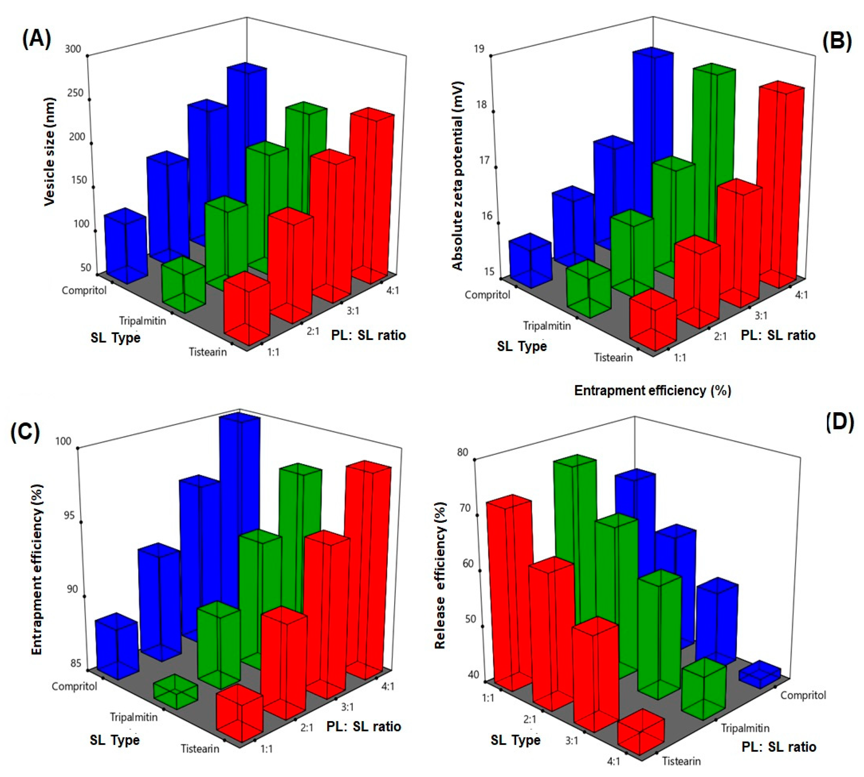
3.1.1. Variables Influence on Vesicle Size (Y1)

The size of vesicles is an important parameter that affect stability, encapsulation efficiency, release, biodistribution, as well as cellular uptake and permeation of bioactive molecules [45,46]. The prepared emulsomes showed vesicles size ranging from 97.2 ± 1.8 to 247.1 ± 9.8 nm, Table 2. The influence of the PL:SL (X1), and SL type (X2) on the vesicle size (Y1) of RLX emulsomes is graphically illustrated as 3D-surface plots represented in Figure 1A. ANOVA revealed that both variables had significant effect on vesicle size. The effect of X1 was more pronounced on the vesicle size as proven by its lower p-value (<0.0001) compared to that of X2 (0.0003).
It was evident that increasing PL:SL ratio led to marked increase in the vesicle size. This finding could be linked to the formation of multiple bilayers with PL amount increase that leads to increase in the vesicle size [12]. Vyas et al. [44] revealed similar findings by reporting increase in zidovudine liver targeted emulsomes vesicle size with increasing molar ratio of phosphatidylcholine with respect to solid lipid. Moreover, Elzafrany et al. reported oxcarbazepine emulsomal size increase with increasing relative phospholipid content [12]. Regarding SL type, the vesicle size was increasing in order of TP < TS < Compritol. TP based emulsomes showed markedly smaller vesicle size compared to that of TS and Compritol. This could be probably due to the lower chain length of TP. Similar results indicated that tristearin-based emulsomes possessed larger size than trilaurin-based emulsomes [44].

3.1.2. Variables Influence on Zeta Potential (Y2)

Zeta potential absolute value is indication for stability of the vesicular systems. All emulsomes formulations exhibited negative charge as demonstrated in Table 2. Elzafrany et al. have also reported negative emulsomal surface charge that was attributed to the location of negatively charged PL in the outer layer. The influence of the PL:SL (X1) and SL type (X2) on the zeta potential (Y2) of RLX-EMLs is graphically illustrated as 3D-surface plot represented in Figure 1B. ANOVA results showed that PL: SL ratio had significant effect on ZP (p = 0.0001). As illustrated, increasing PL:SL ratio led to increasing the absolute values of zeta potential. This could be ascribed to the relative increase of the negative anionic soya phosphatidylcholine in the outer layers [12,15]. Similar findings by Tefas et al. [47] who reported increased negative values of zeta potential of anti-proliferative doxorubicin and curcumin liposomal formulation with increasing PL content. Nevertheless, no significant difference in zeta potential was noted among different solid lipids. Similarly, Nayak et al. [48] observed no different in zeta potential among solid lipid types for curcuminoids-loaded lipid nanoparticles. This finding could be explained by presence of the solid lipid in the core of emulsomes. Accordingly, they offer no contribution to the net surface charge.

3.1.3. Variables Influence on Entrapment Efficiency (Y3)

Encapsulation of drug in the lipid vesicular system as emulsomes could improve its biological availability, as well as control its release [49]. The mean percentages of entrapment efficiency of prepared emulsomes are listed in Table 2. The influence of the PL:SL (X1) and SL type (X2) on the entrapment efficiency (Y3) of RLX-EMLs is graphically illustrated as 3D-surface plots represented in Figure 1C. Statistical analysis using ANOVA showed that both PL:SL and SL type significantly affected the entrapment efficiency of prepared emulsomes (p < 0.0001 and p = 0.0100, respectively), however the effect of PL: SL ratio was more significant than the solid lipid type as evidenced by its lower p-value. The increase in entrapment at higher PL:SL ratios possibly related to the formation of multiple bilayers with PL amount increase resulting in increased drug entrapment within these bilayers [46]. Similar results were obtained by Elzafray et al. [12] who reported increase in the emulsomes drug entrapment with increasing PL content at different total lipid amounts.

3.1.4. Variables Influence on Drug Release (Y4)

All the formulations showed slow drug release within first two hours, and no burst release was observed. This is possibly related to the entrapment of lipophilic drug inside the hydrophobic part of emulsomes bilayer, with consequent retardation of drug release. RLX was released gradually from the prepared emulsomes over the course of 6 h (data not shown). This finding agrees with previously reported results [50]. Release efficiency after 6 h (RE) from prepared RLX-EMLs were computed as a parameter for evaluating drug release from the prepared emulsomes, Table 2. Statistical analysis revealed that the significant factor affected drug release was PL:SL ratio followed by SL type (p < 0.0001 and 0.0028, respectively). The influence of the PL:SL (X1) and SL type (X2) on RE (Y4) of RLX-EMLs is graphically illustrated as 3D-surface plot represented in Figure 1D.
The significant decrease in the percentage of drug release after 6 h associated with PL:SL ratio increase could be attributed to the increased entrapment of the drug within the multiple phospholipid bilayers formed at higher PL concentrations. Regarding the solid lipid type, it was evident that TP showed the highest release compared to other solid lipids. This could be related to the influence of SL type on vesicle size. TP based emulsomes that showed the lowest vesicle size exhibited the highest release among the used solid lipids and vice versa. It was previously reported that as particles size decrease, the attributed surface area: volume ratio increases indicating more drug could be closer to the particle surface and this could leads to enhanced release [51].

3.2. Selection of the Optimized RLX-EMLs

Desirability function was utilized to select the optimized RLX-EMLs using Design-Expert software. The optimization was based on minimizing the vesicle size and release efficiency after 6 h, and maximizing absolute magnitude of zeta potential and entrapment efficiency. The compositions of optimized formulation were PL:SL (4:1) and SL type (TS). The optimized formulation fulfilled the requirement with desirability of 0.718. EML-12, whose composition includes the combination of the optimized variables, showed vesicle size of 236 ± 8.6 nm, zeta potential of −18.6 ± 0.7 mV, EE of 98.9% ± 4.9%, and RE of 42.7% ± 1.8%. Hence, EML-12 was chosen for further investigations.

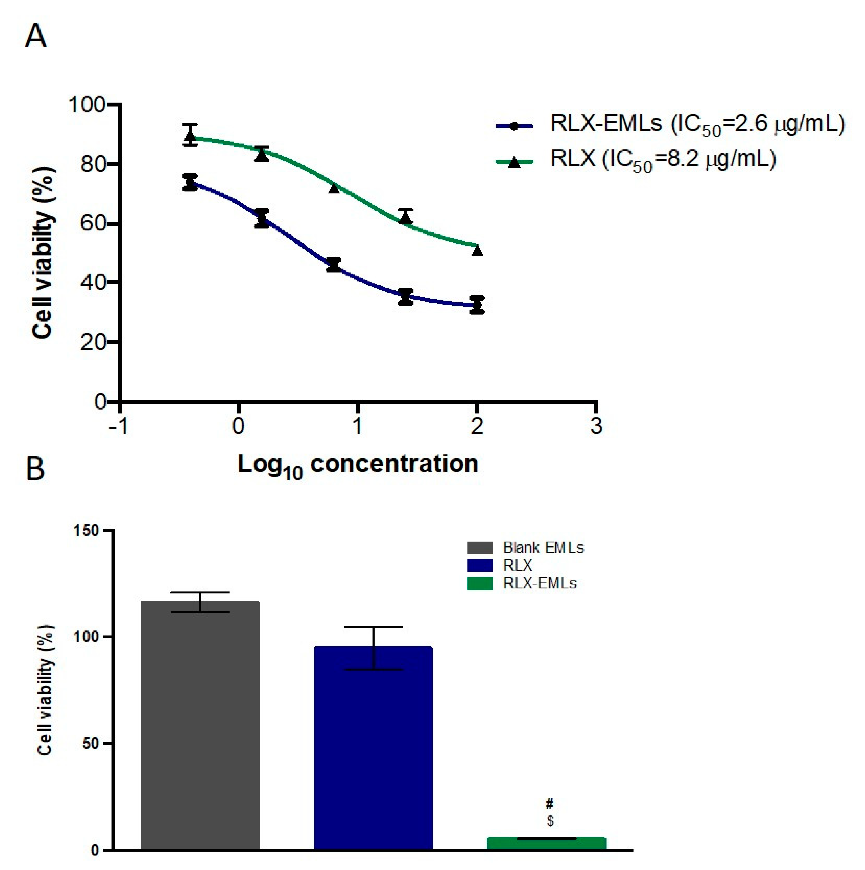
3.3. In-Vitro Antitumor Activity of Optimized RLX-EMLs

3.3.1. MTT Assay

Cytotoxicity of RLX-EMLs, RLX, and blank EMLs in MCF-7 cells was evaluated by MTT test. MCF-7 cells percent growth inhibition at different doses were compared to that of untreated cells. After 48 h, blank EMLs did not show significant alteration on the viability of the cell line. Both RLX and RLX-EMLs significantly decreased cell viability in comparison with the untreated cells over a concentration range of (100–0.39 μg/mL) (p < 0.05). Increasing in the concentration of RLX and RLX-EMLs causes a gradual decrease in cell viability. The IC50 value was calculated as 8.2 μg/mL and 2.6 μg/mL for RLX and RLX-EMLs-treated cells consequently, Figure 2A. Earlier studies reported an antiproliferative effect of RLX on MCF-7 cells for up to 100 μg/mL in concentration. For instance, pure RLX and RLX-loaded mixed micelles using Pluronic F68 and Gelucire 44/14 exhibited IC50 values of 94.71 and 22.5 μg/mL, respectively on MCF-7 cells [8]. Additionally, Kim, D et al. reported the use of about 5 μg/mL of RLX to kill 50% of MCF-7 cells within 48 h [52]. However, significant improvement in the antiproliferative activity on MCF-7 cell line was observed by the newly formulated RLX-EMLs over these previous studies. Based on the IC50 values, 0.1 μM of RLX—a dose that does not affect cell viability—and an equivalent concentration of RLX-EMLs, were selected for the rest of the experiments in order to confirm the enhancement for antineoplastic effect of RLX. At this low dose, RLX-EMLs had a significant cytotoxic effect on MCF-7 cells when compared to the control and RLX (p < 0.0001) Figure 2B. These results confirm the correlation between the advanced design of RLX-EMLs along with its release profile and its enhanced antiproliferative effect on MCF-7 cells.

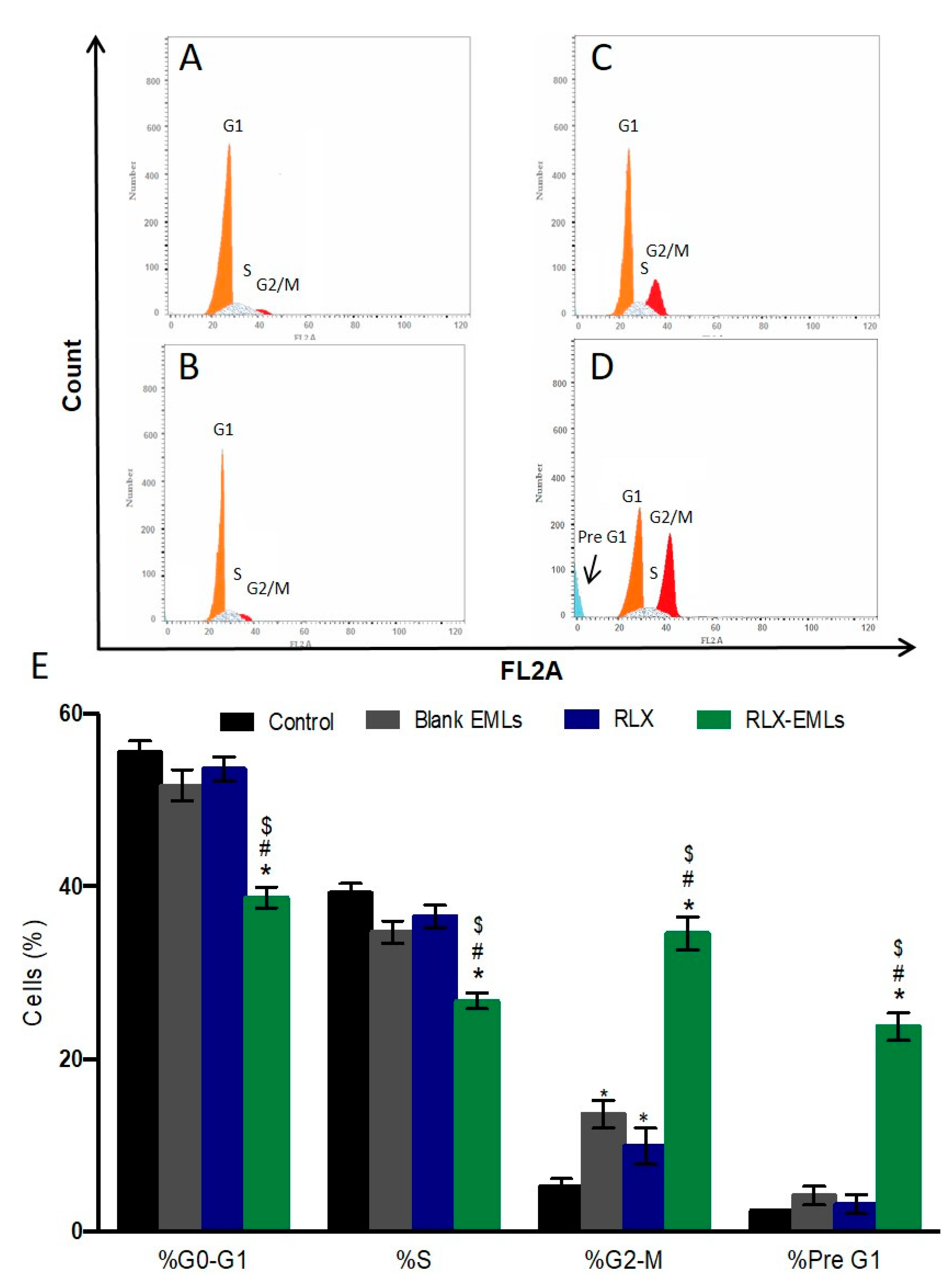
3.3.2. Cell Cycle Analysis

To further confirm the antiproliferative efficacy of RLX-EMLs against MCF-7 cells, the progression of the cell cycle was assessed by flow cytometric analysis of propidium iodide staining. Blank EMLs did not induce changes in the cell cycle of MCF-7, Figure 3B,E. Although RLX significantly arrested G2/M (p < 0.05) when compared to the control cells, it had little effect upon other cell cycle stages, Figure 3A,C,E. Similarly, It has been shown that RLX (10 µM) did not influence cell cycle distribution in several breast cancer cell lines such as BCap37, Bats-72, and Bads-200 cells [52,53]. RLX at (15 µM) arrested the cell cycle, promoted the accumulation in the G1 phase and decrease of the percentage of cells in the S and G2/M phases [54]. However, the newly formulated RLX-EMLs significantly induced of apoptotic pre G1 peak and G2/M arrest at a concentration of 0.1 μM in MCF-7 cells, Figure 3D,E. In addition, RLX-EMLs caused a significant damage to DNA at S phase when compared to the control, RLX and blank EMLs. This RLX-EMLs-induced cell arrest describes the mechanism of its cytotoxicity in MCF-7 cells. The apoptotic effect RLX-EMLs will be further examined through annexin V/PI assay as well as the expression of BAX, BCL2, and caspase-3.

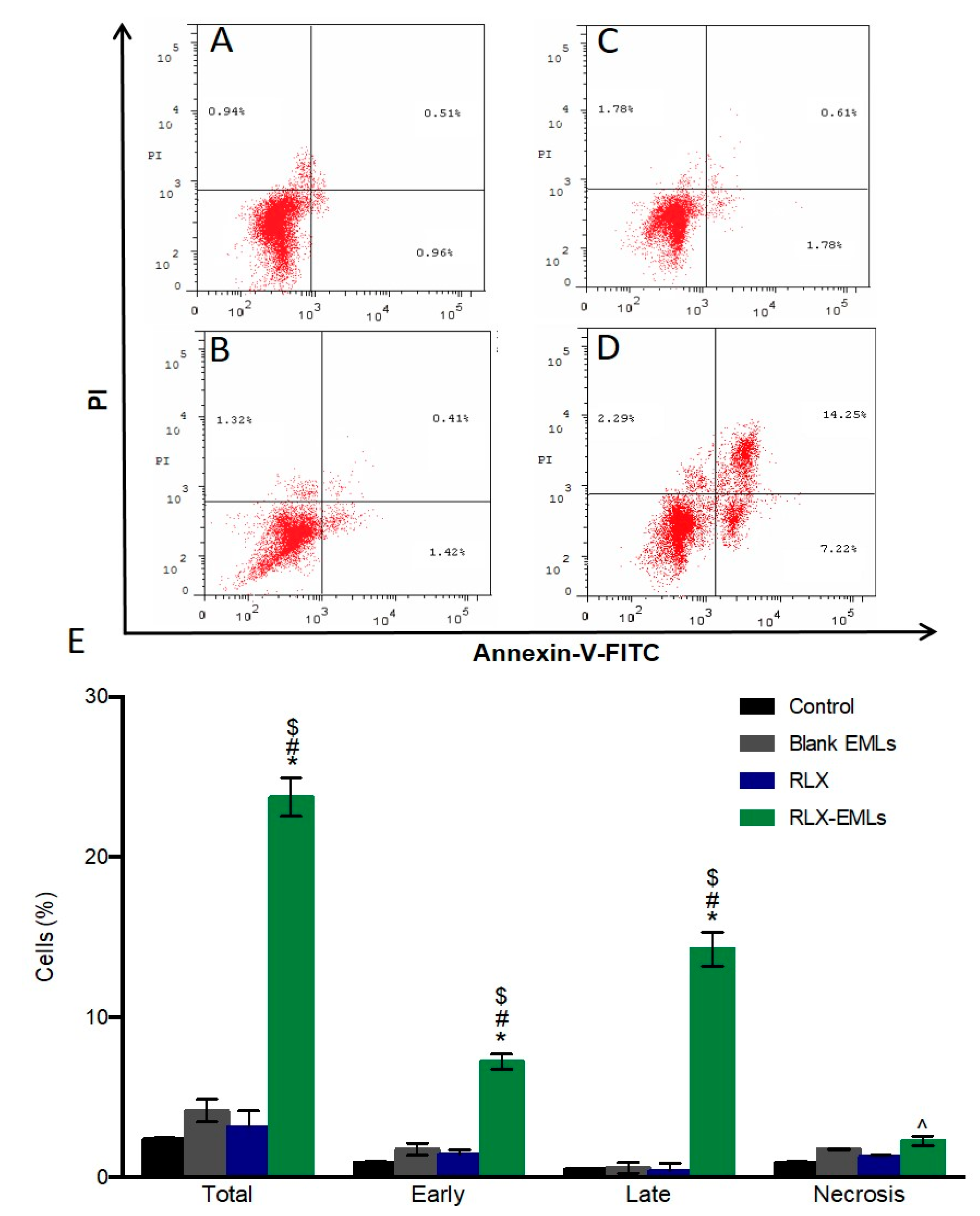
3.3.3. Apoptosis Assay

Annexin V/PI staining was utilized to demonstrate MCF-7 cells apoptosis exposed to RLX-EMLs, RLX, and blank EMLs for 48 h. Figure 4B,C,E shows neither RLX nor blank RLX display any significant apoptosis in MCF-7 cells. Compared to control, Figure 4A. RLX-EMLs-treated cells underwent apoptosis after 48 h with early apoptotic cell populations of 7.99%, late apoptotic cell population of 14.25 and total apoptotic population of 23.76%. Necrotic cells were also significantly increased after RLX-EMLs treatment compared to RLX, and blank EMLs groups (p < 0.05), Figure 4D,E. In agreement with our data, RLX at a concentration of 10 µM failed to induce this level of apoptosis in MCF-7 and several breast cancer cell lines such as BCap37, Bats-72, and Bads-200 cells [52,53]. This weak apoptotic effect was further confirmed through examining the cleavage of caspase-9 protein. The significant changes in MCF-7 population by RLX-EMLs indicates that the cell becomes apoptotic as result of RLX-EMLs-induction of antitumor activity. The RLX-EMLs possible apoptotic mode of action will be further investigated though its effect on Bax, BCL-2, Bax/BCL2 ratio, and caspase-9.

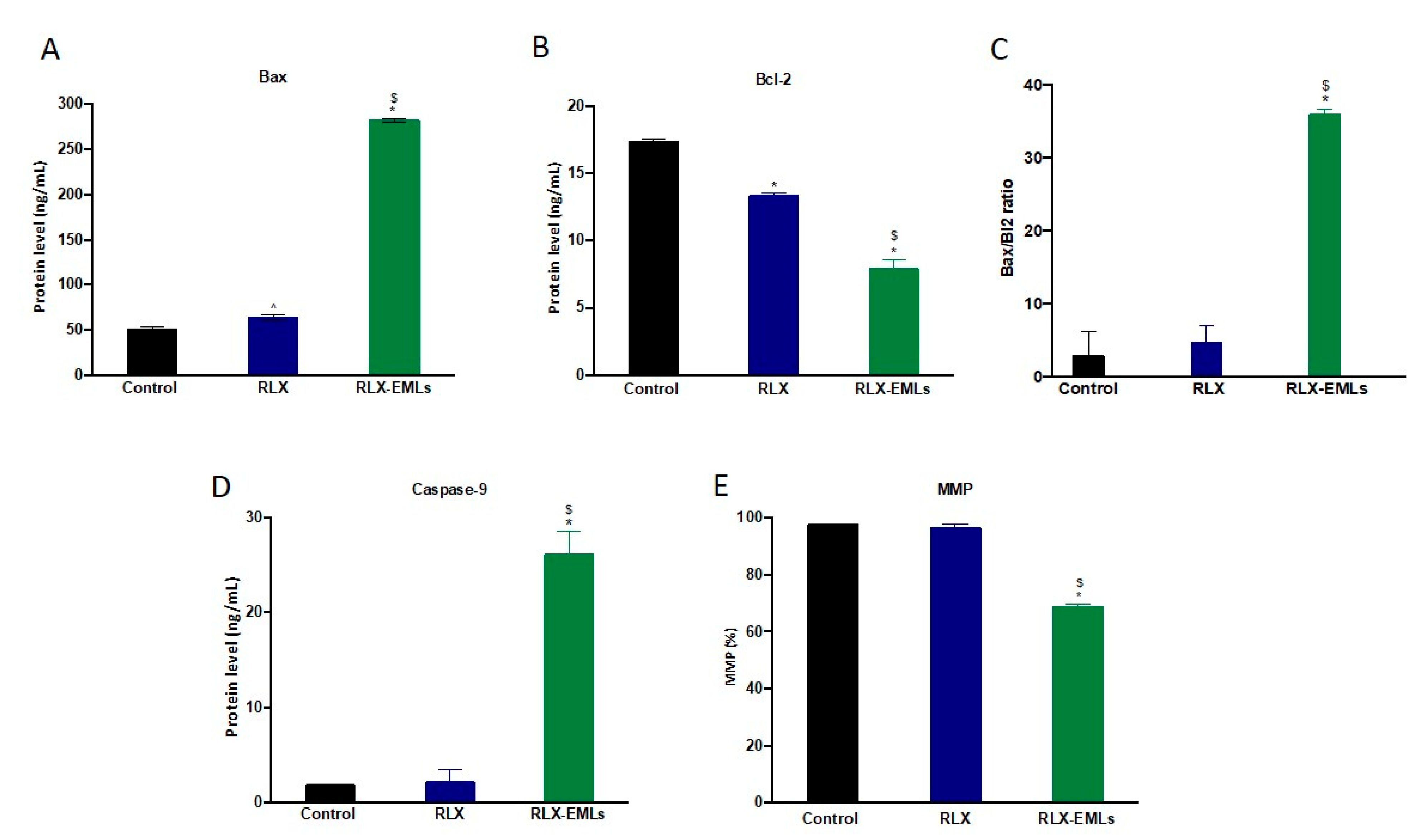
3.3.4. Determination of Bax and Bcl2 Expressions, Caspase-9 Activation, and MMP

The intrinsic apoptotic pathway involve intracellular signals that alter mitochondrial membrane permeability causing loss of MMP [55,56]. Bcl-2 family proteins significantly regulate these apoptotic events thought releasing of cytochrome c thus; they are assessed to determine if the cell commits to apoptosis [55,57]. The anti-apoptotic Bcl-2 protein inhibits cytochrome C release from the mitochondria and caspase-9 activation. In contrast, pro-apoptotic Bax protein expression enhances the release of cytochrome c and reduces MMP causing rapid cell death [58]. Here in, the expression of Bax was increased significantly under the effect of RLX-EMLs, while the expression Bcl-2 consequently decreased when compared to the control and RLX, Figure 5A,B. As a result, RLX-EMLs showed significant elevation in Bax/Bcl-2 ratio, which supports its ability to induce apoptosis in MCF-7 cells Figure 5C. In addition, apoptosis was confirmed by assessing caspase-9 production level. RLX-EMLs significantly enhanced the activity of caspase-9 compared to the control and RLX, Figure 5D. Neither RLX nor blank EMLs significantly affected MCF-7, Bax, Bcl-2, and caspase-9. Furthermore, RLX-EMLs-treated MCF-7 showed a significant reduction in MMP compared to the control and RLX, Figure 5E. These data suggest that RLX-EMLs-potent apoptotic effect is mediated by the induction of the intrinsic mitochondrial apoptosis pathway.
Based on these findings, it is obvious that the proposed optimized RLX-EMLs exhibited a novel formula for the improved delivery of RLX (a poorly water-soluble drug). The optimized formula showed reduced size and controlled RLX release that correlated with the significant improvement in the antiproliferative activity and enhanced induction of apoptosis in MCF-7 cells at a much lower concentration (0.1 µM) of RLX compared to previous studies. The improved RLX anticancer activity proved the successfulness of the utilized factorial experimental design for formulating novel promising emulsomal RLX formulation with desired properties that could be encouragingly implemented for further clinical applications.

4. Conclusions

In the current study, a full 4131 factorial design was employed effectively for formulation and optimization of RLX-EMLs with minimized vesicle size and controlled release, associated with maximized drug entrapment and absolute zeta potential value. The assessed factors, namely, lipoid: solid lipid ratio and solid lipid type exhibited significant effect on vesicle size, drug entrapment, and release efficiency, while zeta potential was only affected by lipoid: solid lipid ratio. The examined formulation improved RLX cytotoxic effect, reduced the viability of MCF-7 cells at a very low concentration of 0.1 µM through inhibiting S cell cycle phase and arresting G2/M phase. The formulation further induced the intrinsic apoptosis pathway of the MCF-7 cells through increasing the ratio of Bax/Bcl-2, activating caspase-9 and reducing MMP. The ability of EMLs in enhancing delivery and cytotoxic activity of RLX revealed their promising potential as nano-carrier for anticancer therapy. EMLs as a nano-formulation could offer a synergistic combination with synthetic drugs to provide coordinated release profile and improved efficacy. These characteristics could lead to promising clinical applications in the field of cancer therapy.

Author Contributions

Conceptualization, O.A.A.A., U.A.F.; methodology, U.A.F.; software, S.M.B.-E.; validation, H.M.A. and N.A.A.; formal analysis, H.M.A. and U.A.F.; investigation, O.A.A.A. and U.A.F.; resources, H.M.A. and U.A.F.; data curation, O.A.A.A. and T.N.; writing—original draft preparation, T.N. and S.M.B.-E.; writing—review and editing, T.N. and S.M.B.-E.; visualization, O.A.A.A.; supervision, H.M.A.; project administration, O.A.A.A. and N.A.A.; funding acquisition, S.M.B.-E. All authors have read and agreed to the published version of the manuscript.

Funding

This work was funded by the Deanship of Scientific Research (DSR) at King Abdulaziz University, Jeddah, under grant no. (RG-4–166–41).

Institutional Review Board Statement

Not applicable.

Informed Consent Statement

Not applicable.

Data Availability Statement

Data are contained in the article.

Acknowledgments

This project was funded by the Deanship of Scientific Research (DSR) at King Abdulaziz University, Jeddah, under grant no. (RG-4–166–41). The authors, therefore, acknowledge with thanks the DSR for technical and financial support.

Conflicts of Interest

The authors declare no conflict of interest.

References

  1. Harbeck, N.; Penault-Llorca, F.; Cortes, J.; Gnant, M.; Houssami, N.; Poortmans, P.; Ruddy, K.; Tsang, J.; Cardoso, F. Breast cancer. Nat. Rev. Dis. Prim. 2019, 5, 1–31. [Google Scholar] [CrossRef] [PubMed]
  2. Dai, X.; Xiang, L.; Li, T.; Bai, Z. Cancer hallmarks, biomarkers and breast cancer molecular subtypes. J. Cancer 2016, 7, 1281–1294. [Google Scholar] [CrossRef] [PubMed]
  3. Ahmed, O.A.A.; Badr-Eldin, S.M. In situ misemgel as a multifunctional dual-absorption platform for nasal delivery of raloxifene hydrochloride: Formulation, characterization, and in vivo performance. Int. J. Nanomed. 2018, 13, 6325–6335. [Google Scholar] [CrossRef]
  4. Murthy, A.; Rao Ravi, P.; Kathuria, H.; Malekar, S. Oral Bioavailability Enhancement of Raloxifene with Nanostructured Lipid Carriers. Nanomaterials 2020, 10, 1085. [Google Scholar] [CrossRef]
  5. Waters, E.A.; McNeel, T.S.; Stevens, W.M.; Freedman, A.N. Use of tamoxifen and raloxifene for breast cancer chemoprevention in 2010. Breast Cancer Res. Treat. 2012, 134, 875–880. [Google Scholar] [CrossRef]
  6. Levy, N.; Tatomer, D.; Herber, C.B.; Zhao, X.; Tang, H.; Sargeant, T.; Ball, L.J.; Summers, J.; Speed, T.P.; Leitman, D.C. Differential regulation of native estrogen receptor-regulatory elements by estradiol, tamoxifen, and raloxifene. Mol. Endocrinol. 2008, 22, 287–303. [Google Scholar] [CrossRef] [PubMed]
  7. Wang, Z.; Li, Y. Raloxifene/SBE-β-CD inclusion complexes formulated into nanoparticles with chitosan to overcome the absorption barrier for bioavailability enhancement. Pharmaceutics 2018, 10, 76. [Google Scholar] [CrossRef]
  8. Kanade, R.; Boche, M.; Pokharkar, V. Self-Assembling Raloxifene Loaded Mixed Micelles: Formulation Optimization, In Vitro Cytotoxicity and In Vivo Pharmacokinetics. AAPS PharmSciTech 2018, 19, 1105–1115. [Google Scholar] [CrossRef] [PubMed]
  9. Patra, J.K.; Das, G.; Fraceto, L.F.; Campos, E.V.R.; Rodriguez-Torres, M.D.P.; Acosta-Torres, L.S.; Diaz-Torres, L.A.; Grillo, R.; Swamy, M.K.; Sharma, S.; et al. Nano based drug delivery systems: Recent developments and future prospects 10 Technology 1007 Nanotechnology 03 Chemical Sciences 0306 Physical Chemistry (incl. Structural) 03 Chemical Sciences 0303 Macromolecular and Materials Chemistry 11 Medical and Health Sciences 1115 Pharmacology and Pharmaceutical Sciences 09 Engineering 0903 Biomedical Engineering Prof Ueli Aebi, Prof Peter Gehr. J. Nanobiotechnol. 2018, 16, 71. [Google Scholar]
  10. Su, Y.L.; Hu, S.H. Functional nanoparticles for tumor penetration of therapeutics. Pharmaceutics 2018, 10, 193. [Google Scholar] [CrossRef]
  11. Pal, A.; Gupta, S.; Jaiswal, A.; Dube, A.; Vyas, S.P. Development and evaluation of tripalmitin emulsomes for the treatment of experimental visceral leishmaniasis. J. Liposome Res. 2012, 22, 62–71. [Google Scholar] [CrossRef]
  12. El-Zaafarany, G.M.; Soliman, M.E.; Mansour, S.; Awad, G.A.S. Identifying lipidic emulsomes for improved oxcarbazepine brain targeting: In vitro and rat in vivo studies. Int. J. Pharm. 2016, 503, 127–140. [Google Scholar] [CrossRef] [PubMed]
  13. Gupta, S.; Vyas, S.P. Development and characterization of amphotericin B bearing emulsomes for passive and active macrophage targeting. J. Drug Target. 2007, 15, 206–217. [Google Scholar] [CrossRef]
  14. Kumar, R.; Seth, N.; Hari kumar, S.L. Emulsomes: An Emerging Vesicular Drug Delivery System. J. Drug Deliv. Ther. 2013, 3, 133. [Google Scholar] [CrossRef]
  15. Zhou, X.; Chen, Z. Preparation and performance evaluation of emulsomes as a drug delivery system for silybin. Arch. Pharm. Res. 2015, 38, 2193–2200. [Google Scholar] [CrossRef] [PubMed]
  16. Varshosaz, J.; Raghami, F.; Rostami, M.; Jahanian, A. PEGylated trimethylchitosan emulsomes conjugated to octreotide for targeted delivery of sorafenib to hepatocellular carcinoma cells of HepG2. J. Liposome Res. 2019, 29, 383–398. [Google Scholar] [CrossRef] [PubMed]
  17. Bolat, Z.B.; Islek, Z.; Demir, B.N.; Yilmaz, E.N.; Sahin, F.; Ucisik, M.H. Curcumin- and Piperine-Loaded Emulsomes as Combinational Treatment Approach Enhance the Anticancer Activity of Curcumin on HCT116 Colorectal Cancer Model. Front. Bioeng. Biotechnol. 2020, 8, 50. [Google Scholar] [CrossRef] [PubMed]
  18. Ucisik, M.H.; Sleytr, U.B.; Schuster, B. Emulsomes Meet S-layer Proteins: An Emerging Targeted Drug Delivery System. Curr. Pharm. Biotechnol. 2015, 16, 392–405. [Google Scholar] [CrossRef]
  19. Awan, Z.A.; Fahmy, U.A.; Badr-eldin, S.M.; Ibrahim, T.S.; Asfour, H.Z.; Al-rabia, M.W.; Alfarsi, A.; Alhakamy, N.A.; Abdulaal, W.H.; Al Sadoun, H.; et al. The enhanced cytotoxic and pro-apoptotic effects of optimized simvastatin-loaded emulsomes on MCF-7 breast cancer cells. Pharmaceutics 2020, 12, 597. [Google Scholar] [CrossRef]
  20. Alhakamy, N.A.; Badr-Eldin, S.M.; Ahmed, O.A.A.; Asfour, H.Z.; Aldawsari, H.M.; Algandaby, M.M.; Eid, B.G.; Abdel-Naim, A.B.; Awan, Z.A.; Alghaith, A.F.; et al. Piceatannol-loaded emulsomes exhibit enhanced cytostatic and apoptotic activities in colon cancer cells. Antioxidants 2020, 9, 419. [Google Scholar] [CrossRef]
  21. Fahmy, U.A.; Aldawsari, H.M.; Badr-Eldin, S.M.; Ahmed, O.A.A.; Alhakamy, N.A.; Alsulimani, H.H.; Caraci, F.; Caruso, G. The Encapsulation of Febuxostat into Emulsomes Strongly Enhances the Cytotoxic Potential of the Drug on HCT 116 Colon Cancer Cells. Pharmaceutics 2020, 12, 956. [Google Scholar] [CrossRef] [PubMed]
  22. Ahmed, O.A.A.; Fahmy, U.A.; Badr-Eldin, S.M.; Aldawsari, H.M.; Awan, Z.A.; Asfour, H.Z.; Kammoun, A.K.; Caruso, G.; Caraci, F.; Alfarsi, A.; et al. Application of nanopharmaceutics for flibanserin brain delivery augmentation via the nasal route. Nanomaterials 2020, 10, 1270. [Google Scholar] [CrossRef] [PubMed]
  23. Paliwal, R.; Paliwal, S.R.; Mishra, N.; Mehta, A.; Vyas, S.P. Engineered chylomicron mimicking carrier emulsome for lymph targeted oral delivery of methotrexate. Int. J. Pharm. 2009, 380, 181–188. [Google Scholar] [CrossRef] [PubMed]
  24. Alhakamy, N.A.; Ahmed, O.A.A.; Fahmy, U.A.; Md, S. Development and in vitro evaluation of 2-methoxyestradiol loaded polymeric micelles for enhancing anticancer activities in prostate cancer. Polymers 2021, 13, 884. [Google Scholar] [CrossRef]
  25. Narayan, R.; Singh, M.; Ranjan, O.P.; Nayak, Y.; Garg, S.; Shavi, G.V.; Nayak, U.Y. Development of risperidone liposomes for brain targeting through intranasal route. Life Sci. 2016, 163, 38–45. [Google Scholar] [CrossRef] [PubMed]
  26. Trontelj, J.; Vovk, T.; Bogataj, M.; Mrhar, A. HPLC analysis of raloxifene hydrochloride and its application to drug quality control studies. Pharmacol. Res. 2005, 52, 334–339. [Google Scholar] [CrossRef]
  27. Abdel-Mottaleb, M.M.A.; Lamprecht, A. Standardized in vitro drug release test for colloidal drug carriers using modified USP dissolution apparatus i. Drug Dev. Ind. Pharm. 2011, 37, 178–184. [Google Scholar] [CrossRef]
  28. Darwesh, B.; Aldawsari, H.M.; Badr-Eldin, S.M. Optimized chitosan/anion polyelectrolyte complex based inserts for vaginal delivery of fluconazole: In vitro/in vivo evaluation. Pharmaceutics 2018, 10, 227. [Google Scholar] [CrossRef]
  29. Ibrahim, H.K.; Fahmy, R.H. Localized rosuvastatin via implantable bioerodible sponge and its potential role in augmenting bone healing and regeneration. Drug Deliv. 2016, 23, 3181–3192. [Google Scholar] [CrossRef]
  30. Aldawsari, H.M.; Badr-Eldin, S.M. Enhanced pharmacokinetic performance of dapoxetine hydrochloride via the formulation of instantly-dissolving buccal films with acidic pH modifier and hydrophilic cyclodextrin: Factorial analysis, in vitro and in vivo assessment. J. Adv. Res. 2020, 24, 281–290. [Google Scholar] [CrossRef]
  31. Al-Mahallawi, A.M.; Abdelbary, A.A.; Aburahma, M.H. Investigating the potential of employing bilosomes as a novel vesicular carrier for transdermal delivery of tenoxicam. Int. J. Pharm. 2015, 485, 329–340. [Google Scholar] [CrossRef] [PubMed]
  32. Hamidreza Kheiri, M.; Alimohammadi, N.; Danafar, H. Preparation of biocompatible copolymeric micelles as a carrier of atorvastatin and rosuvastatin for potential anticancer activity study. Pharm. Dev. Technol. 2019, 24, 303–313. [Google Scholar] [CrossRef] [PubMed]
  33. Franco, M.S.; Roque, M.C.; Oliveira, M.C. Short and long-term effects of the exposure of breast cancer cell lines to different ratios of free or co-encapsulated liposomal paclitaxel and doxorubicin. Pharmaceutics 2019, 11, 178. [Google Scholar] [CrossRef] [PubMed]
  34. Hasanzadeh, D.; Mahdavi, M.; Dehghan, G.; Charoudeh, H.N. Farnesiferol C induces cell cycle arrest and apoptosis mediated by oxidative stress in MCF-7 cell line. Toxicol. Rep. 2017, 4, 420–426. [Google Scholar] [CrossRef]
  35. Foo, J.B.; Ng, L.S.; Lim, J.H.; Tan, P.X.; Lor, Y.Z.; Loo, J.S.E.; Low, M.L.; Chan, L.C.; Beh, C.Y.; Leong, S.W.; et al. Induction of cell cycle arrest and apoptosis by copper complex Cu (SBCM) 2 towards oestrogen-receptor positive MCF-7 breast cancer cells. RSC Adv. 2019, 9, 18359–18370. [Google Scholar] [CrossRef]
  36. Ji, Y.; Yu, M.; Qi, Z.; Cui, D.; Xin, G.; Wang, B.; Jia, W.; Chang, L. Study on apoptosis effect of human breast cancer cell MCF-7 induced by lycorine hydrochloride via death receptor pathway. Saudi Pharm. J. 2017, 25, 633–637. [Google Scholar] [CrossRef]
  37. Hoshyar, N.; Gray, S.; Han, H.; Bao, G. The effect of nanoparticle size on in vivo pharmacokinetics and cellular interaction. Nanomedicine 2016, 11, 673–692. [Google Scholar] [CrossRef]
  38. Blanco, E.; Shen, H.; Ferrari, M. Principles of nanoparticle design for overcoming biological barriers to drug delivery. Nat. Biotechnol. 2015, 33, 941–951. [Google Scholar] [CrossRef] [PubMed]
  39. Ramalho, M.J.; Loureiro, J.A.; Coelho, M.A.N.; Pereira, M.C. Factorial design as a tool for the optimization of plga nanoparticles for the co-delivery of temozolomide and o6-benzylguanine. Pharmaceutics 2019, 11, 401. [Google Scholar] [CrossRef]
  40. Araújo, J.; Gonzalez-Mira, E.; Egea, M.A.; Garcia, M.L.; Souto, E.B. Optimization and physicochemical characterization of a triamcinolone acetonide-loaded NLC for ocular antiangiogenic applications. Int. J. Pharm. 2010, 393, 167–175. [Google Scholar] [CrossRef]
  41. Riffenburgh, R. Statistics in Medicine; Elsevier Inc.: Amsterdam, The Netherlands, 2012; ISBN 9780123848642. [Google Scholar]
  42. Ahmed, O.A.A.; Badr-Eldin, S.M. Biodegradable self-assembled nanoparticles of PEG-PLGA amphiphilic diblock copolymer as a promising stealth system for augmented vinpocetine brain delivery. Int. J. Pharm. 2020, 588. [Google Scholar] [CrossRef] [PubMed]
  43. Jain, A.; Jain, S.K. Formulation and optimization of temozolomide nanoparticles by 3 factor 2 level factorial design. Biomatter 2013, 3. [Google Scholar] [CrossRef] [PubMed]
  44. Vyas, S.P.; Subhedar, R.; Jain, S. Development and characterization of emulsomes for sustained and targeted delivery of an antiviral agent to liver. J. Pharm. Pharmacol. 2006, 58, 321–326. [Google Scholar] [CrossRef] [PubMed]
  45. Danaei, M.; Dehghankhold, M.; Ataei, S.; Hasanzadeh Davarani, F.; Javanmard, R.; Dokhani, A.; Khorasani, S.; Mozafari, M.R. Impact of particle size and polydispersity index on the clinical applications of lipidic nanocarrier systems. Pharmaceutics 2018, 10, 57. [Google Scholar] [CrossRef] [PubMed]
  46. Ahmed, T.A.; Badr-Eldin, S.M.; Ahmed, O.A.A.; Aldawsari, H. Intranasal optimized solid lipid nanoparticles loaded in situ gel for enhancing trans-mucosal delivery of simvastatin. J. Drug Deliv. Sci. Technol. 2018, 48, 499–508. [Google Scholar] [CrossRef]
  47. Tefas, L.R.; Sylvester, B.; Sesarman, A.; Licarete, E.; Banciu, M. Development of antiproliferative long-circulating liposomes co-encapsulating doxorubicin and curcumin, through the use of a quality-by-design approach. Drug Des. Devel. Ther. 2017, 11, 1605–1621. [Google Scholar] [CrossRef]
  48. Nayak, A.P.; Tiyaboonchai, W.; Patankar, S.; Madhusudhan, B.; Souto, E.B. Curcuminoids-loaded lipid nanoparticles: Novel approach towards malaria treatment. Colloids Surf. B Biointerfaces 2010, 81, 263–273. [Google Scholar] [CrossRef]
  49. Gim, S.; Ong, M.; Ming, L.C.; Lee, K.S.; Yuen, K.H. Influence of the Encapsulation Efficiency and Size of Liposome on the Oral Bioavailability of Griseofulvin-Loaded Liposomes. J. Pharm. 2016, 8, 25. [Google Scholar] [CrossRef]
  50. Tiwari, G.; Tiwari, R.; Wal, P.; Wal, A. Development and Optimization of Liposomes Containing 5 Fluorouracil and Tretinoin for Skin Warts: 32 Experimental Design. J. Pharm. 2019, 44, 17–26. [Google Scholar]
  51. Rizvi, S.A.A.; Saleh, A.M. Applications of nanoparticle systems in drug delivery technology. Saudi Pharm. J. 2018, 26, 64–70. [Google Scholar] [CrossRef]
  52. Kim, D.E.; Kim, Y.; Cho, D.H.; Jeong, S.Y.; Kim, S.B.; Suh, N.; Lee, J.S.; Choi, E.K.; Koh, J.Y.; Hwang, J.J.; et al. Raloxifene induces autophagy-dependent cell death in breast cancer cells via the activation of amp-activated protein kinase. Mol. Cells 2015, 38, 138–144. [Google Scholar] [CrossRef]
  53. Xu, L.; Lei, J.; Jiang, D.; Zhou, L.; Wang, S.; Fan, W. Reversal effects of Raloxifene on paclitaxel resistance in 2 MDR breast cancer cells. Cancer Biol. Ther. 2015, 16, 1794–1801. [Google Scholar] [CrossRef] [PubMed]
  54. Taurin, S.; Nimick, M.; Larsen, L.; Rosengren, R.J. A novel curcumin derivative increases the cytotoxicity of raloxifene in estrogen receptor-negative breast cancer cell lines. Int. J. Oncol. 2016, 48, 385–398. [Google Scholar] [CrossRef]
  55. Elmore, S. Apoptosis: A Review of Programmed Cell Death. Toxicol. Pathol. 2007, 35, 495–516. [Google Scholar] [CrossRef] [PubMed]
  56. Saelens, X.; Festjens, N.; Vande Walle, L.; Van Gurp, M.; Van Loo, G.; Vandenabeele, P. Toxic proteins released from mitochondria in cell death. Oncogene 2004, 23, 2861–2874. [Google Scholar] [CrossRef]
  57. Cory, S.; Adams, J.M. The BCL2 family: Regulators of the cellular life-or-death switch. Nat. Rev. Cancer 2002, 2, 647–656. [Google Scholar] [CrossRef] [PubMed]
  58. Liu, F.T.; Newland, A.C.; Jia, L. Bax conformational change is a crucial step for PUMA-mediated apoptosis in human leukemia. Biochem. Biophys. Res. Commun. 2003, 310, 956–962. [Google Scholar] [CrossRef]
Figure 1. Response 3D plot for PL:SL weight ratio effect; X1 and SL type; X2 on (A) vesicle size; Y1, (B) zeta potential; Y2, (C) entrapment efficiency; Y3, and (D) release efficiency after 6 h; Y4 of RLX-EMLs. Abbreviations: RLX-EMLs, raloxifene hydrochloride emulsomes; PL:SL, lipoid: solid lipid weight ratio; SL type: solid lipid type.
Pharmaceutics 13 00783 g001
Figure 2. Effect of RLX-EMLs or RLX on MCF-7 cell viability using MTT assay. The cells were treated with RLX-EMLs or RLX for 48 h. (A) dose-response curve with IC50 values; (B) comparison of cell viability results using 0.1 µM of RLX. $ p < 0.0001 considered significantly different from RLX, # p < 0.0001 considered significantly different from blank EMLs.
Pharmaceutics 13 00783 g002
Figure 3. Effect of RLX-EMLs or RLX on MCF-7 cell cycle changes of MCF-7 cells using flow cytometry analysis. The cells were treated with RLX-EMLs or RLX for 48 h. (A) control, (B) blank EMLs, (C) RLX, and (D) RLX-EMLs. (E) graphical presentation of each phase. All data are presented as the mean ± SE of three independent experiments. * p < 0.0001 considered significantly different from the control. $ p < 0.0001 considered significantly different from RLX, # p < 0.0001 considered significantly different from blank EMLs.
Pharmaceutics 13 00783 g003
Figure 4. Effect of RLX-EMLs or RLX on MCF-7 apoptosis profile using annexin V/PI staining. (A) control, (B) blank EMLs, (C) RLX, and (D) RLX-EMLs. (E) graphical presentation of each phase (early apoptotic, late apoptotic, total apoptotic, and dead cells). All data are expressed as the mean ±SE of three independent experiments. * p < 0.05 considered significantly different from the control. $ p < 0.0001 considered significantly different from RLX, # p < 0.0001 considered significantly different from blank EMLs. ^ p < 0.05 considered significantly different from the control, RLX, or blank EMLs.
Pharmaceutics 13 00783 g004
Figure 5. Effect of RLX-EMLs or RLX on Bcl-2 proteins, caspase-9 activation and MMP. (A,B) protein levels for BAX and Bcl-2 consequently. (C) Bax/Bcl-2 ratio. (D) activation level of caspase-9. (E) percentage of MMP. All data are expressed as the mean ± SE of three independent experiments. * p < 0.0001 considered significantly different from the control. $ p < 0.0001 considered significantly different from RLX.
Pharmaceutics 13 00783 g005
Table 1. Variables’ levels and responses’ desirability constraints applied in a 4131 factorial design.
Independent VariablesLevels
X1: Lipoid: solid lipid weight ratio1:12:13:14:1
X2: Solid lipid typeTristearinTripalmitinCompritol
ResponsesDesirability constraints
Y1: Vesicle size (nm)Minimize
Y2: Zeta potential (mV)Maximize
Y3: Entrapment efficiency (%)Maximize
Y4: Release efficiency after 6 h (%)Minimize
RLX-EMLs, raloxifene hydrochloride emulsomes.
Table 2. Variables and responses for RLX-EMLs experimental runs prepared based on a 4131 factorial design.
Trial NumberRun OrderVariables LevelsMean Responses ± SD
X1:PL:SLX2:SL TypeY1:VS (nm)Y2:ZP (mV)Y3:EE (%)Y4:RE (%)
1EML-61:1Tristearin106.0 ± 3.8−15.6 ± 0.387.2 ± 3.372.7 ± 3.1
2EML-72:1Tristearin160.9 ± 5.2−16.3 ± 0.691.2 ± 3.964.7 ± 3.7
3EML-23:1Tristearin206.2 ± 5.6−17.1 ± 0.495.3 ± 4.858.2 ± 2.9
4EML-124:1Tristearin236.2 ± 8.6−18.6 ± 0.798.9 ± 4.942.7 ± 1.8
5EML-41:1Tripalmitin97.2 ± 1.8−15.4 ± 0.685.5 ± 2.875.9 ± 3.2
6EML-92:1Tripalmitin137.0 ± 5.2−16.7 ± 0.690.4 ± 3.866.9 ± 2.9
7EML-53:1Tripalmitin192.2 ± 5.1−17.1 ± 0.293.1 ± 2.760.9 ± 2.4
8EML-104:1Tripalmitin223.2 ± 8.7−17.9 ± 0.898.2 ± 4.649.3 ± 2.3
9EML-11:1Compritol119.1 ± 3.3−16.1 ± 0.689.2 ± 3.171.2 ± 3.3
10EML-32:1Compritol174.9 ± 4.2−15.9 ± 0.391.9 ± 3.963.2 ± 2.7
11EML-113:1Compritol215.3± 9.3−16.8 ± 0.596.8 ± 4.153.1 ± 2.1
12EML-84:1Compritol247.1 ± 9.8−18.9 ± 0.799.1 ± 4.141.6 ± 1.9
RLX-EMLs, raloxifene hydrochloride emulsomes; PL:SL, lipoid: solid lipid weight ratio; SL type: solid lipid type; VS, vesicle size; ZP, zeta potential; EE, entrapment efficiency; RE, release efficiency after 6 h; SD, standard deviation.
Table 3. Output data analysis of the 4131 factorial design used for the formulation of RLX-EMLs.
Responsep-ValueR2Adjusted R2Predicted R2Adequate PrecisionSignificant Factors
Y1:VS (nm)<0.00010.99650.99370.986252.94X1, X2
Y2:ZP (mV)0.00310.92010.85340.78029.57X1
Y3:EE (%)<0.00010.98590.97410.943426.42X1, X2
Y4:RE (%)<0.00010.99170.98470.966734.92X1, X2
RLX-EMLs, raloxifene hydrochloride emulsomes; PL:SL, lipoid: solid lipid weight ratio; SL type: solid lipid type; VS, vesicle size; ZP, zeta potential; EE, entrapment efficiency; RE, release efficiency after 6 h.
Publisher’s Note: MDPI stays neutral with regard to jurisdictional claims in published maps and institutional affiliations.

