
Polymer Particles Bearing Recombinant LEL CD81 as Trapping Systems for Hepatitis C Virus

 ,
,  , and
, and
Abstract
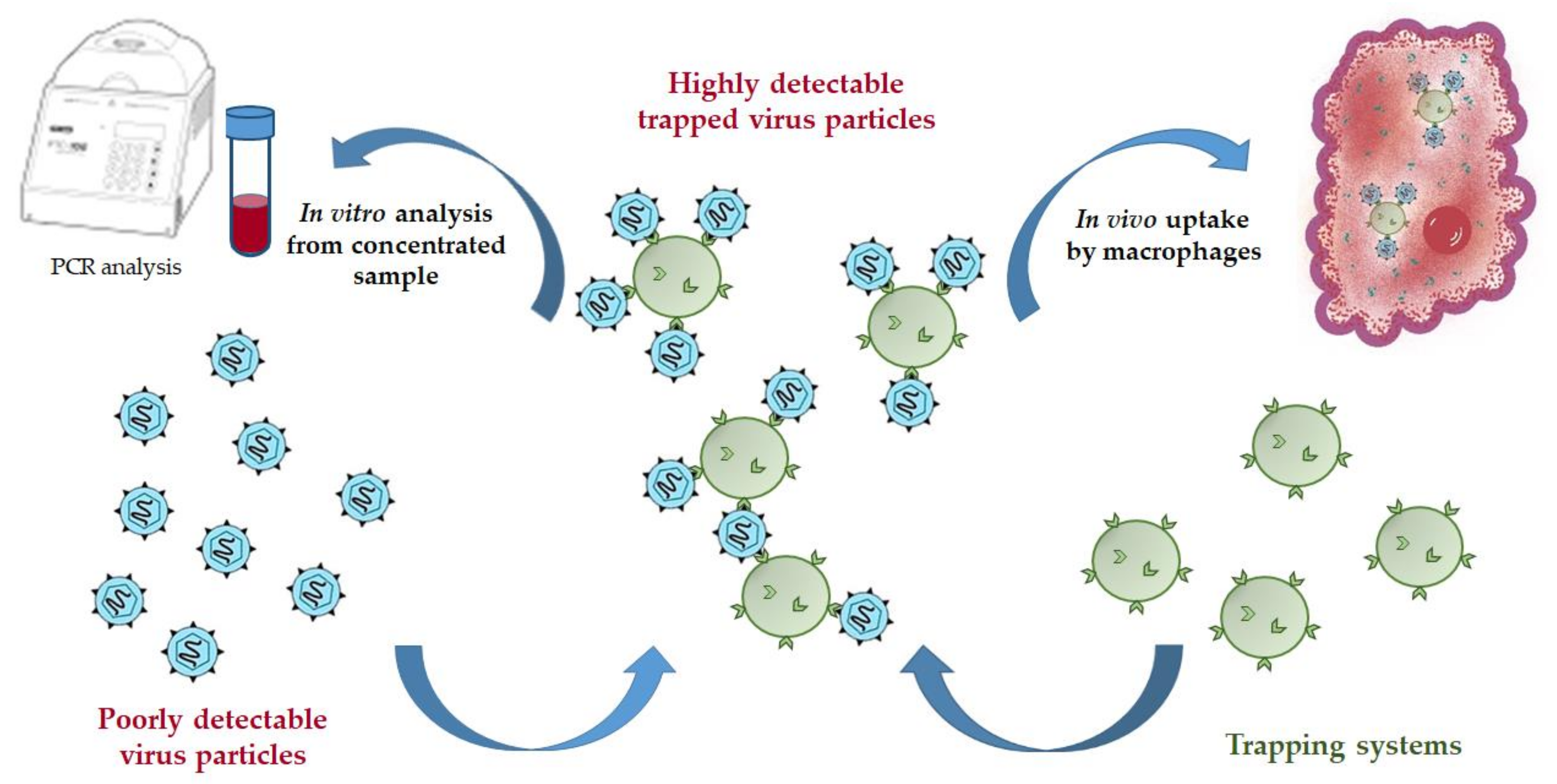
:1. Introduction
2. Materials and Methods
2.1. Materials
2.2. Synthesis of LEL CD81-SA Fusion Protein
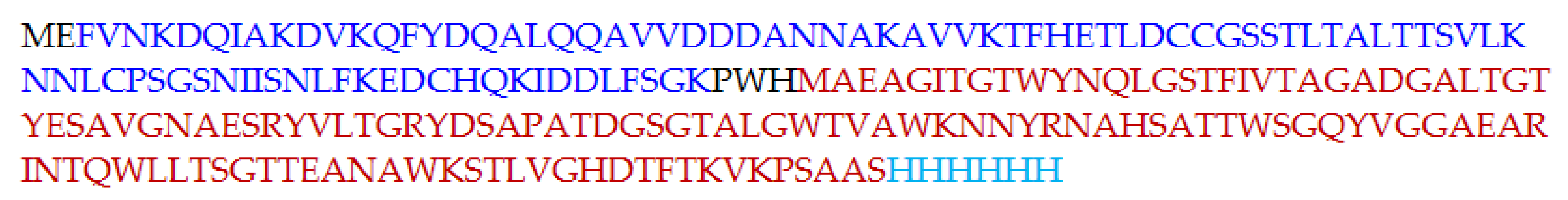
2.2.1. Production of LEL CD81-SA in E. coli
2.2.2. Separation and Purification of LEL CD81-SA
2.3. Synthesis of Chimeric E2 core-GFP Protein in E. coli
2.4. Synthesis and Characterization of poly(d,l-lactic acid) and poly(ethylene glycol)-b-poly(d,l-lactic acid) (PEG-b-PLA)
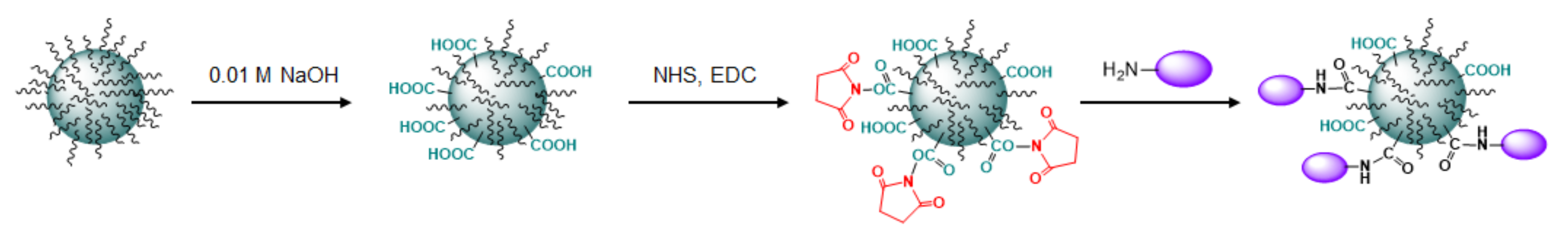
2.5. Preparation of PLA Microparticles and PEG-b-PLA Nanoparticles
2.6. Preparation of HC Micro- and Nanotraps and HC VMPs
2.7. Interaction of E2–GFP with Trapping Systems
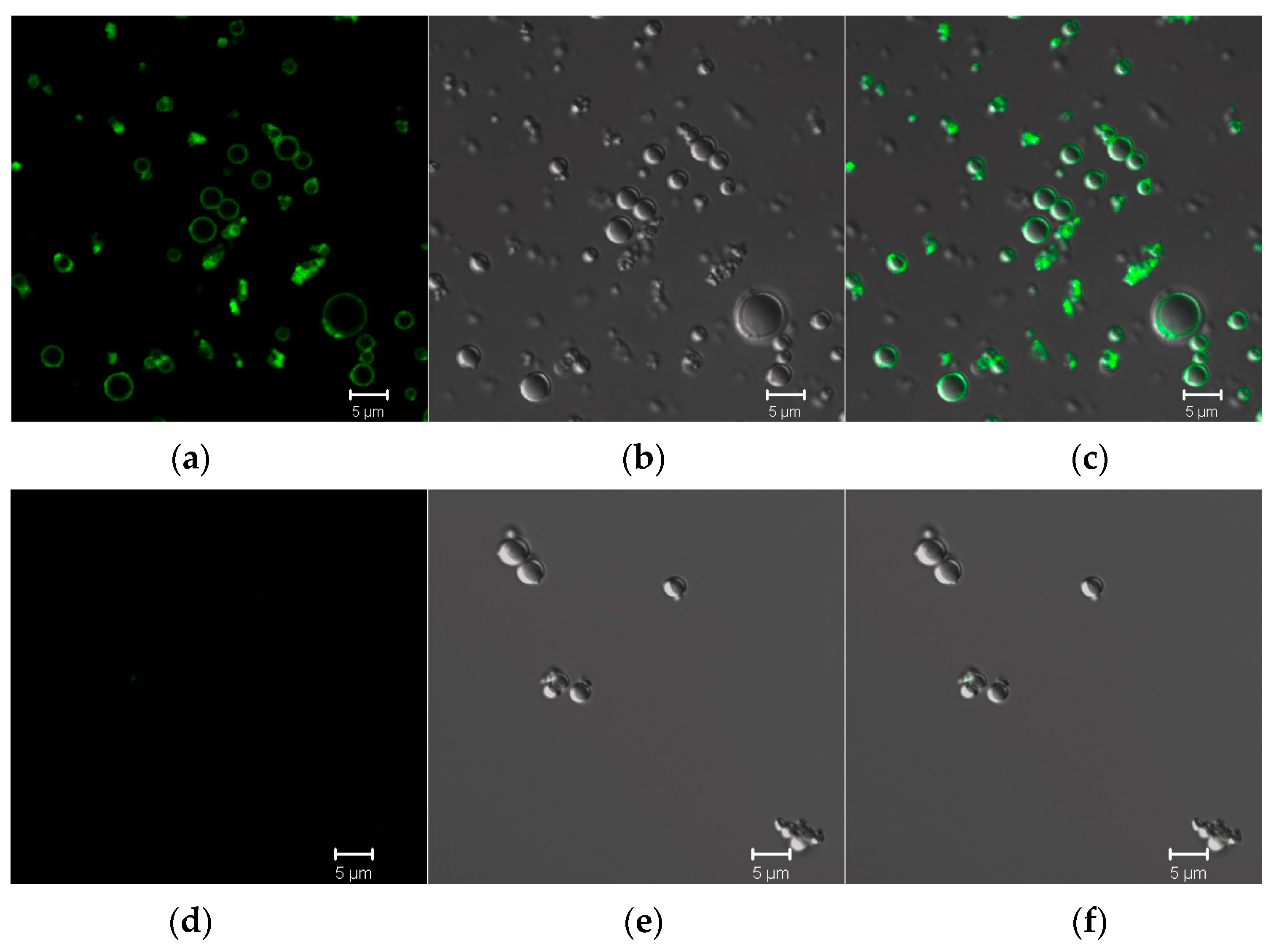
2.7.1. Interaction of E2-GFP with Microtraps at Static Conditions
2.7.2. Interaction of E2-GFP with Microtraps at Dynamic Conditions
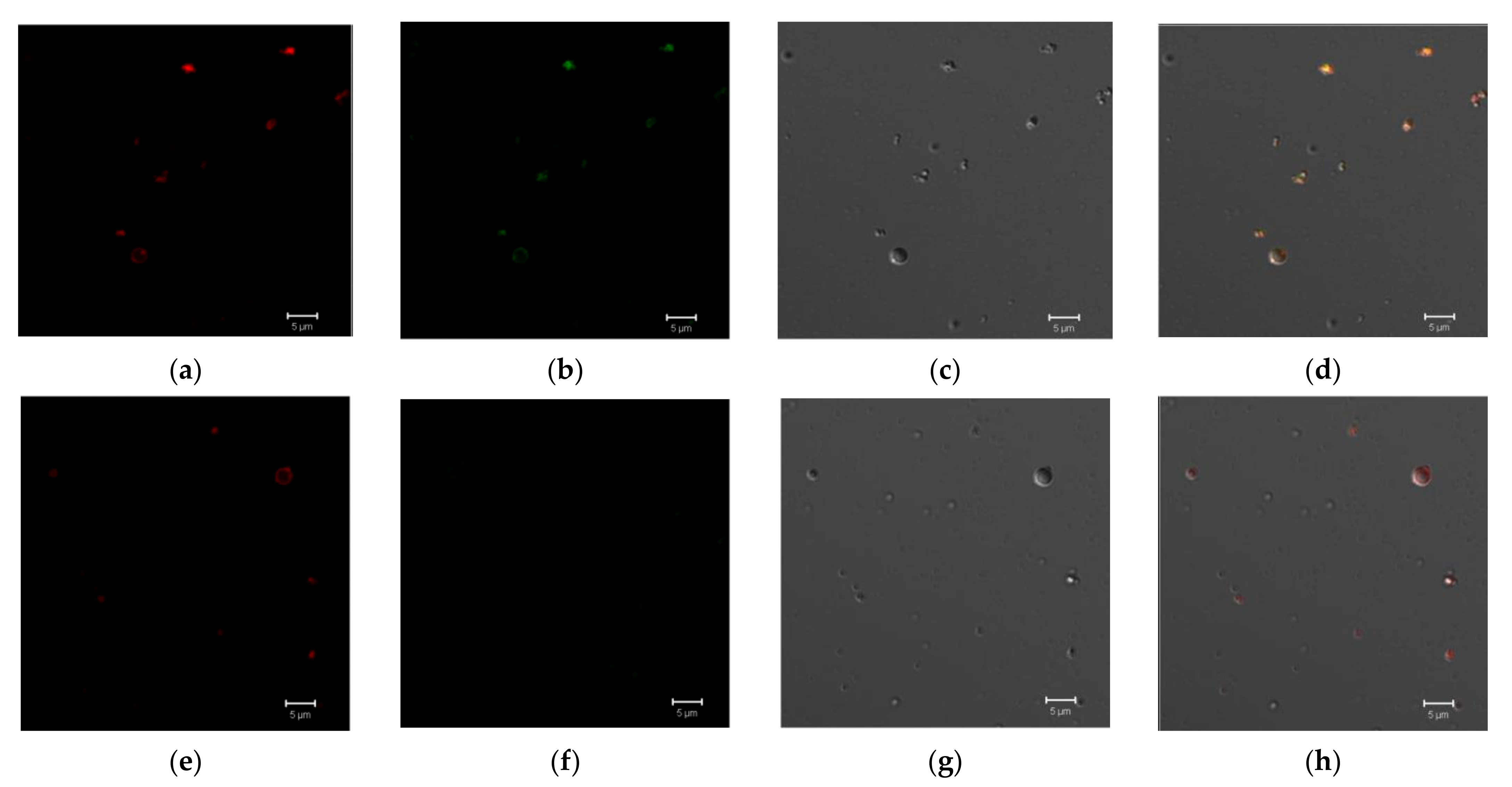
2.8. Interaction of HC VMPs with Nanotraps
2.9. Theoretical Calculations
3. Results and Discussion
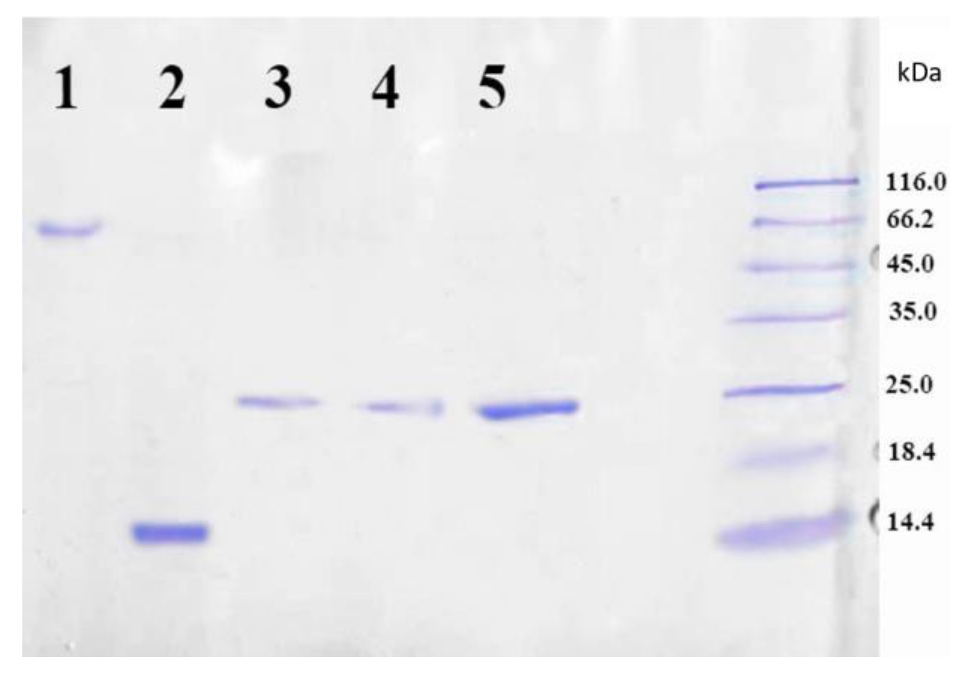
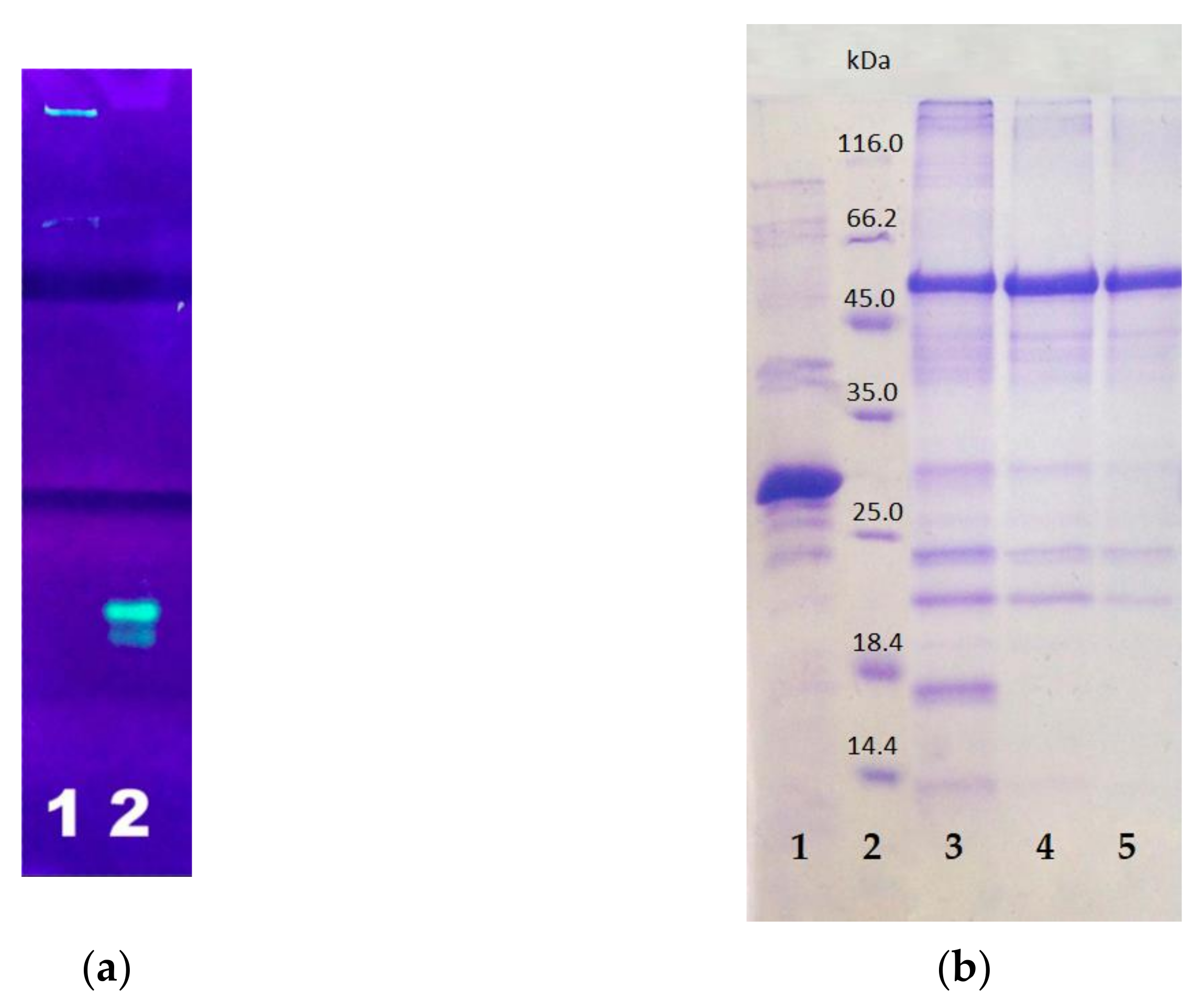
3.1. Production of the LEL CD81-SA Fusion Protein
3.2. Production of the E2-GFP Fusion Protein
3.3. Study of the Interaction between CD81-SA and E2-GFP Proteins
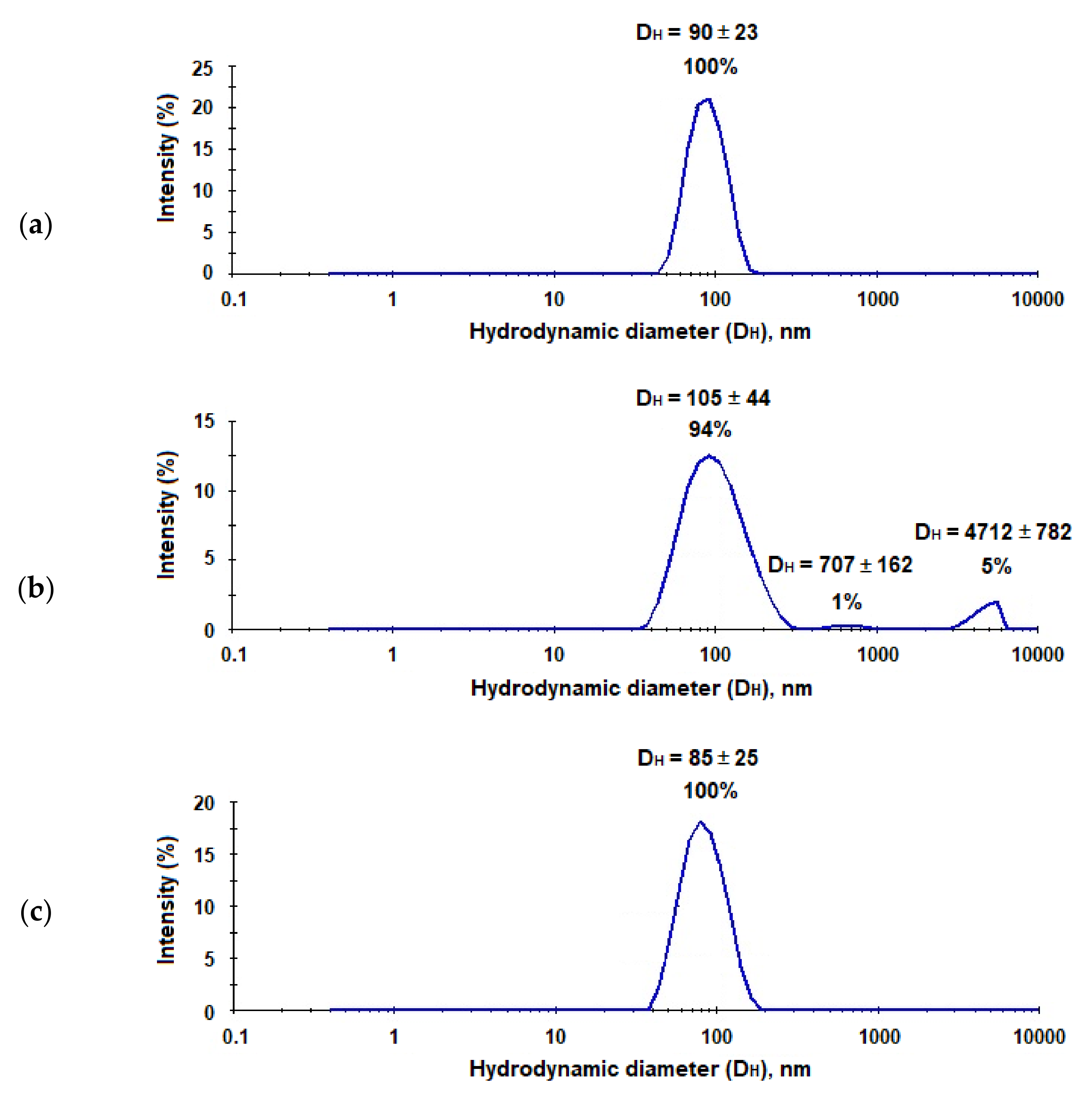
3.4. Preparation of HC Micro- and Nanotraps and HC VMPs
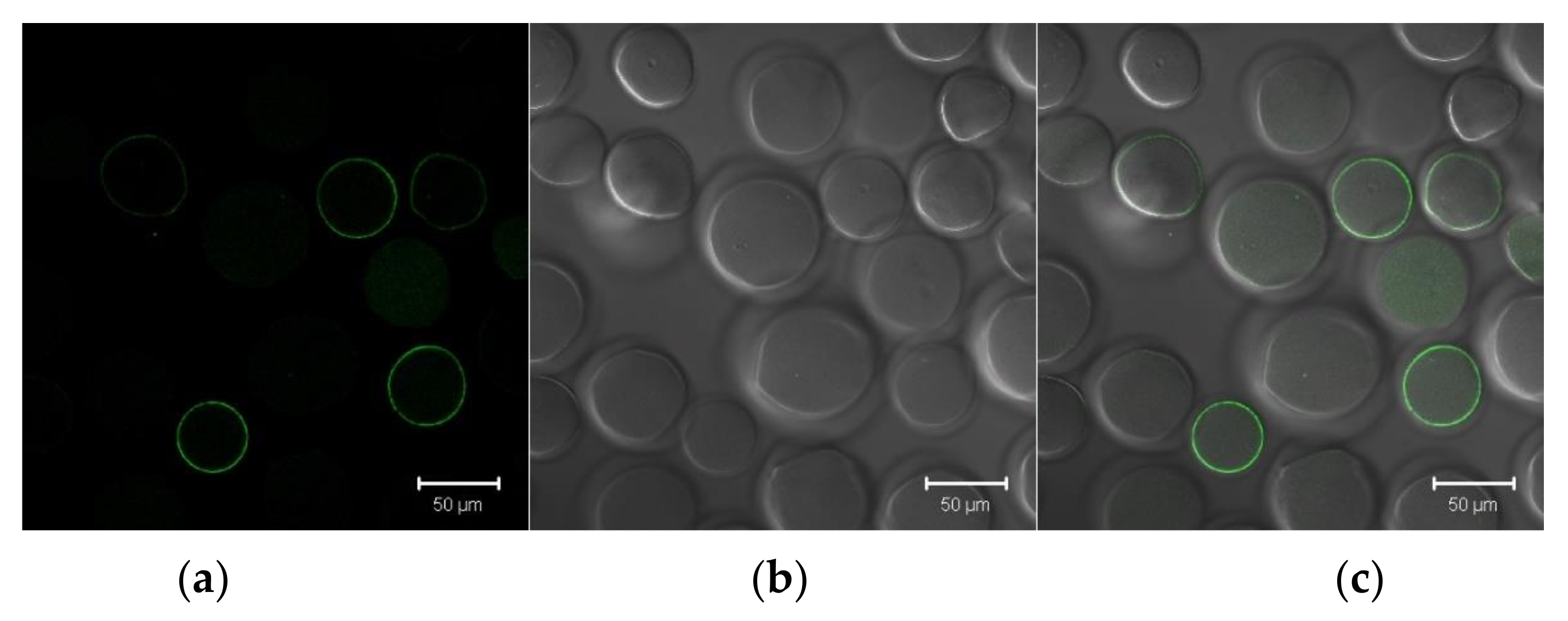
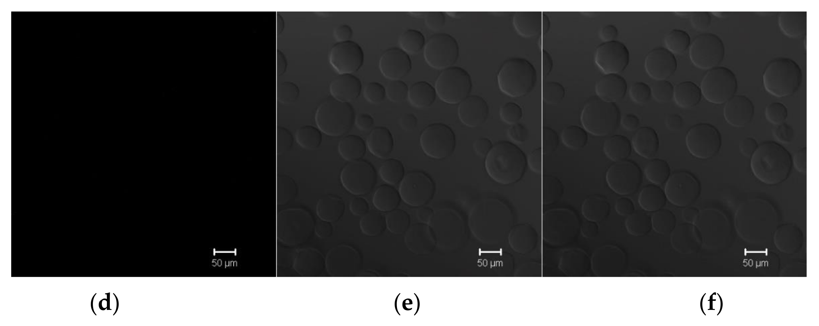
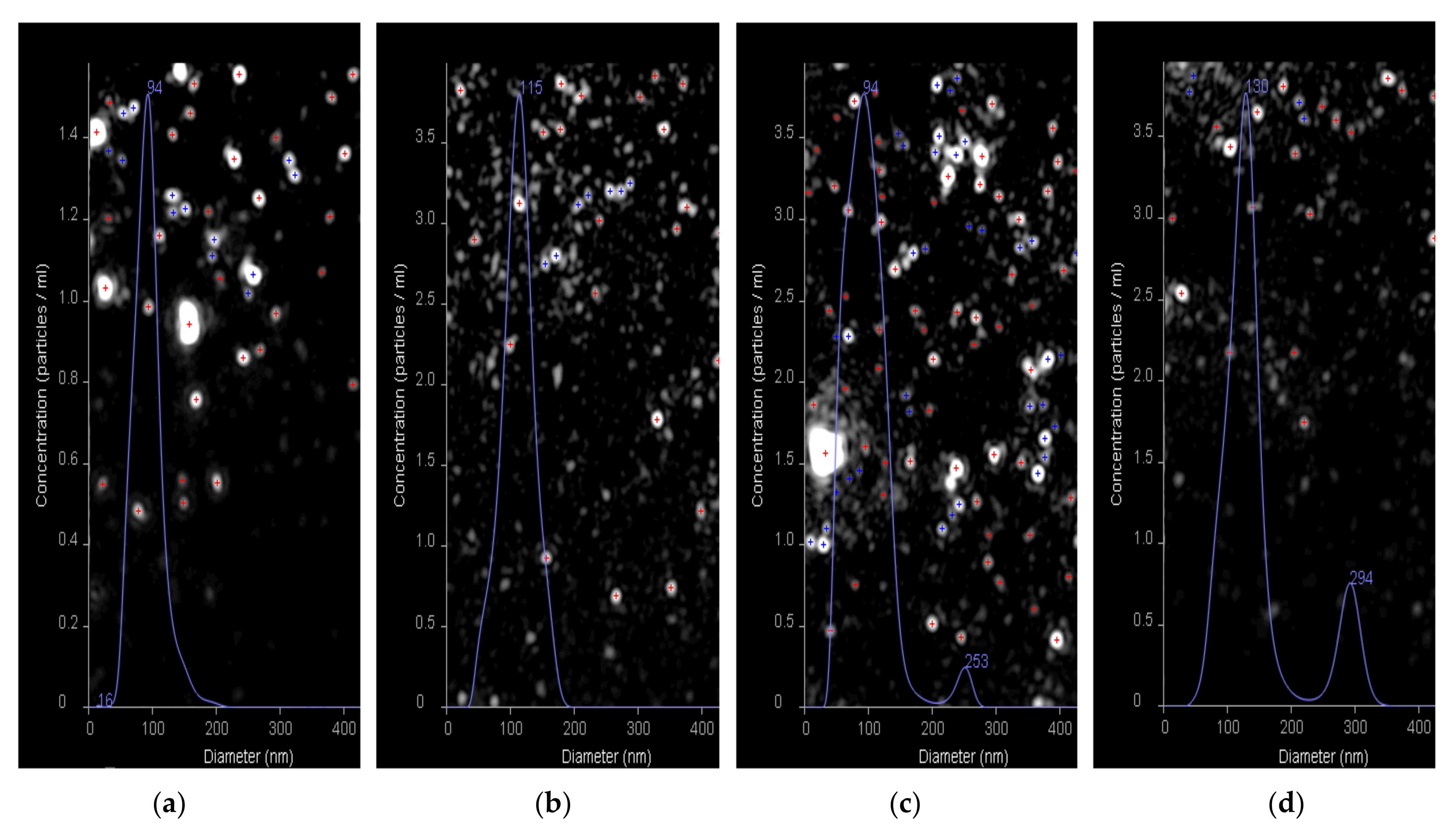
3.5. Study of the Interaction between HC Trapping Systems and E2-GFP/HC VMPs
4. Conclusions
Supplementary Materials
Author Contributions
Funding
Institutional Review Board Statement
Informed Consent Statement
Acknowledgments
Conflicts of Interest
References
- Masavuli, M.G.; Wijesundara, D.K.; Torresi, J.; Gowans, E.J.; Grubor-Bauk, B. Preclinical development and production of virus-like particles as vaccine candidates for hepatitis C. Front. Microbiol. 2017, 8, 2413. [Google Scholar] [CrossRef]
- Carlsen, T.H.R.; Scheel, T.K.H.; Ramirez, S.; Foung, S.K.H.; Bukh, J. Characterization of Hepatitis C Virus Recombinants with Chimeric E1/E2 Envelope Proteins and Identification of Single Amino Acids in the E2 Stem Region Important for Entry. J. Virol. 2013, 87, 1385–1399. [Google Scholar] [CrossRef] [Green Version]
- Freedman, H.; Logan, M.R.; Law, J.L.M.; Houghton, M. Structure and Function of the Hepatitis C Virus Envelope Glycoproteins E1 and E2: Antiviral and Vaccine Targets. ACS Infect. Dis. 2016, 2, 749–762. [Google Scholar] [CrossRef] [PubMed]
- Lujan Cuestas, M. Therapy of Chronic Hepatitis C in the Era of Nanotechnology: Drug Delivery Systems and Liver Targeting. Mini-Rev. Med. Chem. 2017, 17, 295–304. [Google Scholar] [CrossRef] [PubMed]
- Elberry, M.H.; Darwish, N.H.E.; Mousa, S.A. Hepatitis C virus management: Potential impact of nanotechnology. Virol. J. 2017, 14, 88. [Google Scholar] [CrossRef] [Green Version]
- Chakravarty, M.; Vora, A. Nanotechnology-based antiviral therapeutics. Drug Deliv. Transl. Res. 2021, 11, 748–787. [Google Scholar] [CrossRef]
- Shafagati, N.; Patanarut, A.; Luchini, A.; Lundberg, L.; Bailey, C.; Petricoin, E.; Liotta, L.; Narayanan, A.; Lepene, B.; Kehn-Hall, K. The use of Nanotrap particles for biodefense and emerging infectious disease diagnostics. Pathog. Dis. 2014, 71, 164–176. [Google Scholar] [CrossRef] [Green Version]
- Jaworski, E.; Saifuddin, M.; Sampey, G.; Shafagati, N.; Van Duyne, R.; Iordanskiy, S.; Kehn-Hall, K.; Liotta, L.; Petricoin, E.; Young, M.; et al. The Use of Nanotrap Particles Technology in Capturing HIV-1 Virions and Viral Proteins from Infected Cells. PLoS ONE 2014, 9, e96778. [Google Scholar] [CrossRef] [Green Version]
- Shafagati, N.; Lundberg, L.; Baer, A.; Patanarut, A.; Fite, K.; Lepene, B.; Kehn-Hall, K. The Use of Nanotrap Particles in the Enhanced Detection of Rift Valley Fever Virus Nucleoprotein. PLoS ONE 2015, 10, e0128215. [Google Scholar] [CrossRef]
- Akhrymuk, I.; Lin, S.-C.; Sun, M.; Patnaik, A.; Lehman, C.; Altamura, L.; Minogue, T.; Lepene, B.; van Hoek, M.L.; Kehn-Hall, K. Magnetic Nanotrap Particles Preserve the Stability of Venezuelan Equine Encephalitis Virus in Blood for Laboratory Detection. Front. Vet. Sci. 2020, 6, 509. [Google Scholar] [CrossRef] [Green Version]
- Shafagati, N.; Fite, K.; Patanarut, A.; Baer, A.; Pinkham, C.; An, S.; Foote, B.; Lepene, B.; Kehn-Hall, K. Enhanced detection of respiratory pathogens with nanotrap particles. Virulence 2016, 7, 756–769. [Google Scholar] [CrossRef] [PubMed] [Green Version]
- Anderson, M.R.; Pleet, M.L.; Enose-Akahata, Y.; Erickson, J.; Monaco, M.C.; Akpamagbo, Y.; Velluci, A.; Tanaka, Y.; Azodi, S.; Lepene, B.; et al. Viral antigens detectable in CSF exosomes from patients with retrovirus associated neurologic disease: Functional role of exosomes. Clin. Transl. Med. 2018, 7, 24. [Google Scholar] [CrossRef] [PubMed] [Green Version]
- Lin, S.-C.; Carey, B.D.; Callahan, V.; Lee, J.-H.; Bracci, N.; Patnaik, A.; Smith, A.K.; Narayanan, A.; Lepene, B.; Kehn-Hall, K. Use of Nanotrap particles for the capture and enrichment of Zika, chikungunya and dengue viruses in urine. PLoS ONE 2020, 15, e0227058. [Google Scholar] [CrossRef] [PubMed]
- Cheng, H.; Chen, J.; Cai, Z.; Du, L.; Hou, J.; Qiao, X.; Zheng, Q. Development of GEM-PA-nanotrap for purification of foot-and-mouth disease virus. Vaccine 2019, 37, 3205–3213. [Google Scholar] [CrossRef]
- Büyüktiryaki, S.; Uzun, L.; Denizli, A.; Say, R.; Ersöz, A. Simultaneous depletion of albumin and immunoglobulin G by using twin affinity magnetic nanotraps. Sep. Sci. Technol. 2016, 51, 2080–2089. [Google Scholar] [CrossRef]
- Guryanov, I.; Cipriani, S.; Fiorucci, S.; Zashikhina, N.; Marchianò, S.; Scarpelli, P.; Korzhikov-Vlakh, V.; Popova, E.; Korzhikova-Vlakh, E.; Biondi, B.; et al. Nanotraps with biomimetic surface as decoys for chemokines. Nanomed. Nanotechnol. Biol. Med. 2017, 13, 2575–2585. [Google Scholar] [CrossRef] [PubMed]
- Petracca, R.; Falugi, F.; Galli, G.; Norais, N.; Rosa, D.; Campagnoli, S.; Burgio, V.; Di Stasio, E.; Giardina, B.; Houghton, M.; et al. Structure-Function Analysis of Hepatitis C Virus Envelope-CD81 Binding. J. Virol. 2000, 74, 4824–4830. [Google Scholar] [CrossRef] [Green Version]
- Miao, Z.; Xie, Z.; Miao, J.; Ran, J.; Feng, Y.; Xia, X. Regulated Entry of Hepatitis C Virus into Hepatocytes. Viruses 2017, 9, 100. [Google Scholar] [CrossRef] [Green Version]
- Sharma, G.; Raheja, H.; Das, S. Hepatitis C virus: Enslavement of host factors. IUBMB Life 2018, 70, 41–49. [Google Scholar] [CrossRef] [PubMed] [Green Version]
- Bartosch, B.; Vitelli, A.; Granier, C.; Goujon, C.; Dubuisson, J.; Pascale, S.; Scarselli, E.; Cortese, R.; Nicosia, A.; Cosset, F.L. Cell Entry of Hepatitis C Virus Requires a Set of Co-receptors that Include the CD81 Tetraspanin and the SR-B1 Scavenger Receptor. J. Biol. Chem. 2003, 278, 41624–41630. [Google Scholar] [CrossRef] [Green Version]
- Kong, L.; Giang, E.; Nieusma, T.; Kadam, R.U.; Cogburn, K.E.; Hua, Y.; Dai, X.; Stanfield, R.L.; Burton, D.R.; Ward, A.B.; et al. Hepatitis C virus E2 envelope glycoprotein core structure. Sci. 80 2013, 342, 1090–1094. [Google Scholar] [CrossRef] [Green Version]
- Khan, A.G.; Whidby, J.; Miller, M.T.; Scarborough, H.; Zatorski, A.V.; Cygan, A.; Price, A.A.; Yost, S.A.; Bohannon, C.D.; Jacob, J.; et al. Structure of the core ectodomain of the hepatitis C virus envelope glycoprotein 2. Nature 2014, 509, 381–384. [Google Scholar] [CrossRef] [Green Version]
- Dubuisson, J.; Cosset, F.L. Virology and cell biology of the hepatitis C virus life cycle—An update. J. Hepatol. 2014, 61, S3–S13. [Google Scholar] [CrossRef] [Green Version]
- Brunelle, J.L.; Green, R. One-dimensional SDS-polyacrylamide gel electrophoresis (1D SDS-PAGE). In Methods in Enzymology; Academic Press: London, UK, 2014; Volume 541, pp. 151–159. [Google Scholar]
- SDS-PAGE Protocol. Available online: http://microbiology.ucdavis.edu/privalsky/sites/default/files/SDS-PAGE_0.pdf (accessed on 24 March 2021).
- Stepanenko, O.V.; Stepanenko, O.V.; Kuznetsova, I.M.; Verkhusha, V.V.; Turoverov, K.K. Structural perturbation of superfolder GFP in the presence of guanidine thiocyanate. Spectroscopy 2012, 27, 381–386. [Google Scholar] [CrossRef]
- Stepanenko, O.V.; Stepanenko, O.V.; Kuznetsova, I.M.; Shcherbakova, D.M.; Verkhusha, V.V.; Turoverov, K.K. Distinct Effects of Guanidine Thiocyanate on the Structure of Superfolder GFP. PLoS ONE 2012, 7, e48809. [Google Scholar] [CrossRef] [PubMed] [Green Version]
- Korzhikov, V.; Averianov, I.; Litvinchuk, E.; Tennikova, T.B. Polyester-based microparticles of different hydrophobicity: The patterns of lipophilic drug entrapment and release. J. Microencapsul. 2016, 33, 199–208. [Google Scholar] [CrossRef] [PubMed]
- Kritchenkov, I.S.; Zhukovsky, D.D.; Mohamed, A.; Mohamed, A.; Korzhikov-Vlakh, V.A.; Tennikova, T.B.; Lavrentieva, A.; Scheper, T.; Pavlovskiy, V.V.; Porsev, V.V.; et al. Functionalized Pt(II) and Ir(III) NIR Emitters and Their Covalent Conjugates with Polymer-Based Nanocarriers. Bioconjug. Chem. 2020, 31, 1327–1343. [Google Scholar] [CrossRef]
- Shawky, H.; Maghraby, A.S.; Solliman, M.E.D.; El-Mokadem, M.T.; Sherif, M.M.; Arafa, A.; Bahgat, M.M. Expression, immunogenicity and diagnostic value of envelope proteins from an Egyptian hepatitis C virus isolate. Arch. Virol. 2015, 160, 945–958. [Google Scholar] [CrossRef] [PubMed]
- Higginbottom, A.; Quinn, E.R.; Kuo, C.-C.; Flint, M.; Wilson, L.H.; Bianchi, E.; Nicosia, A.; Monk, P.N.; McKeating, J.A.; Levy, S. Identification of Amino Acid Residues in CD81 Critical for Interaction with Hepatitis C Virus Envelope Glycoprotein E2. J. Virol. 2000, 74, 3642–3649. [Google Scholar] [CrossRef] [Green Version]
- Yurkova, M.S.; Patel, A.H.; Fedorov, A.N. Characterisation of bacterially expressed structural protein E2 of hepatitis C virus. Protein Expr. Purif. 2004, 37, 119–125. [Google Scholar] [CrossRef]
- Solovyov, K.V.; Polyakov, D.S.; Grudinina, N.A.; Egorov, V.V.; Morozova, I.V.; Aleynikova, T.D.; Shavlovsky, M.M. Expression in E. coli and purification of the fibrillogenic fusion proteins ttr-sfgfp and β2M-sfGFP. Prep. Biochem. Biotechnol. 2011, 41, 337–349. [Google Scholar] [CrossRef] [PubMed]
- Soundrarajan, N.; Cho, H.S.; Ahn, B.; Choi, M.; Thong, L.M.; Choi, H.; Cha, S.Y.; Kim, J.H.; Park, C.K.; Seo, K.; et al. Green fluorescent protein as a scaffold for high efficiency production of functional bacteriotoxic proteins in Escherichia coli. Sci. Rep. 2016, 6, 1–10. [Google Scholar] [CrossRef] [PubMed] [Green Version]
- Lee, B.K.; Yun, Y.; Park, K. PLA micro- and nano-particles. Adv. Drug Deliv. Rev. 2016, 107, 176–191. [Google Scholar] [CrossRef] [Green Version]
- Prabha, S.; Arya, G.; Chandra, R. Effect of size on biological properties of nanoparticles employed in gene delivery. Nanomed. Biotechnol. 2016, 44, 83–91. [Google Scholar] [CrossRef] [PubMed]
- Iudin, D.; Zashikhina, N.; Demyanova, E.; Korzhikov-Vlakh, V.; Shcherbakova, E.; Boroznjak, R.; Tarasenko, I.; Zakharova, N.; Lavrentieva, A.; Skorik, Y.; et al. Polypeptide self-assembled nanoparticles as delivery systems for polymyxins B and E. Pharmaceutics 2020, 12, 868. [Google Scholar] [CrossRef]
- Suk, J.S.; Xu, Q.; Kim, N.; Hanes, J.; Ensign, L.M. PEGylation as a strategy for improving nanoparticle-based drug and gene delivery. Adv. Drug Deliv. Rev. 2016, 99, 28–51. [Google Scholar] [CrossRef] [Green Version]
- Fang, C.; Shi, B.; Pei, Y.Y.; Hong, M.H.; Wu, J.; Chen, H.Z. In vivo tumor targeting of tumor necrosis factor-α-loaded stealth nanoparticles: Effect of MePEG molecular weight and particle size. Eur. J. Pharm. Sci. 2006, 27, 27–36. [Google Scholar] [CrossRef]
- Nicolete, R.; Santos, D.F.D.; Faccioli, L.H. The uptake of PLGA micro or nanoparticles by macrophages provokes distinct in vitro inflammatory response. Int. Immunopharmacol. 2011, 11, 1557–1563. [Google Scholar] [CrossRef] [Green Version]
- Mayer, G.; Vogel, V.; Weyermann, J.; Lochmann, D.; Van Den Broek, J.A.; Tziatzios, C.; Haase, W.; Wouters, D.; Schubert, U.S.; Zimmer, A.; et al. Oligonucleotide-protamine-albumin nanoparticles: Protamine sulfate causes drastic size reduction. J. Control. Release 2005, 106, 181–187. [Google Scholar] [CrossRef]
- Churilov, L.; Korzhikov-Vlakh, V.; Sinitsyna, E.; Polyakov, D.; Darashkevich, O.; Poida, M.; Platonova, G.; Vinogradova, T.; Utekhin, V.; Zabolotnykh, N.; et al. Enhanced delivery of 4-thioureidoiminomethylpyridinium perchlorate in tuberculosis models with igg functionalized poly(Lactic acid)-based particles. Pharmaceutics 2019, 11, 2. [Google Scholar] [CrossRef] [Green Version]
- De Jong, W.H.; Hagens, W.I.; Krystek, P.; Burger, M.C.; Sips, A.J.A.M.; Geertsma, R.E. Particle size-dependent organ distribution of gold nanoparticles after intravenous administration. Biomaterials 2008, 29, 1912–1919. [Google Scholar] [CrossRef] [PubMed]
- Koide, H.; Asai, T.; Hatanaka, K.; Urakami, T.; Ishii, T.; Kenjo, E.; Nishihara, M.; Yokoyama, M.; Ishida, T.; Kiwada, H.; et al. Particle size-dependent triggering of accelerated blood clearance phenomenon. Int. J. Pharm. 2008, 362, 197–200. [Google Scholar] [CrossRef] [PubMed]


| System | Polymer Particles | Protein for Immobilization | Characteristics of Neat Polymer Particles (in PBS) | Characteristics of Modified Polymer Particles (in PBS) | |||||
|---|---|---|---|---|---|---|---|---|---|
| DH, nm | PDI | ζ-Potential | DH, nm | PDI | ζ-Potential | Amount of Bound Protein, µg/mg of Particles | |||
| Microtraps | PLA | LEL CD81-SA-Cy3 | 800 | 0.39 | −34 | 850 | 0.26 | −68 | 3 |
| 1640 | 0.37 | −38 | 1860 | 0.28 | −48 | 2 | |||
| Negative control | PLA | SBTI-Cy3 | 800 | 0.39 | −34 | 860 | 0.23 | −75 | 2.5 |
| Nanotraps | PEG-b-PLA | LEL CD81-SA-Cy3 | 90 | 0.05 | −24 | 105 | 0.29 | −43 | 15 |
| HC VMPs | PEG-b-PLA | E2-GFP | 90 | 0.05 | −24 | 85 | 0.19 | −35 | 15 |
Publisher’s Note: MDPI stays neutral with regard to jurisdictional claims in published maps and institutional affiliations. |
© 2021 by the authors. Licensee MDPI, Basel, Switzerland. This article is an open access article distributed under the terms and conditions of the Creative Commons Attribution (CC BY) license (https://creativecommons.org/licenses/by/4.0/).
Share and Cite
Polyakov, D.; Sinitsyna, E.; Grudinina, N.; Antipchik, M.; Sakhabeev, R.; Korzhikov-Vlakh, V.; Shavlovsky, M.; Korzhikova-Vlakh, E.; Tennikova, T. Polymer Particles Bearing Recombinant LEL CD81 as Trapping Systems for Hepatitis C Virus. Pharmaceutics 2021, 13, 672. https://doi.org/10.3390/pharmaceutics13050672
Polyakov D, Sinitsyna E, Grudinina N, Antipchik M, Sakhabeev R, Korzhikov-Vlakh V, Shavlovsky M, Korzhikova-Vlakh E, Tennikova T. Polymer Particles Bearing Recombinant LEL CD81 as Trapping Systems for Hepatitis C Virus. Pharmaceutics. 2021; 13(5):672. https://doi.org/10.3390/pharmaceutics13050672
Chicago/Turabian StylePolyakov, Dmitry, Ekaterina Sinitsyna, Natalia Grudinina, Mariia Antipchik, Rodion Sakhabeev, Viktor Korzhikov-Vlakh, Mikhail Shavlovsky, Evgenia Korzhikova-Vlakh, and Tatiana Tennikova. 2021. "Polymer Particles Bearing Recombinant LEL CD81 as Trapping Systems for Hepatitis C Virus" Pharmaceutics 13, no. 5: 672. https://doi.org/10.3390/pharmaceutics13050672