
Hybrid System for Local Drug Delivery and Magnetic Hyperthermia Based on SPIONs Loaded with Doxorubicin and Epirubicin

 , , , , and
Abstract
:1. Introduction
2. Materials and Methods
2.1. Chemicals
2.2. Synthesis of Holmium-Doped SPIONs
2.3. Modification of SPIONs with Citric Acid
2.4. Modification of Nanoparticles with Drug
2.5. Interaction with Biological Membranes
2.6. In Vitro Cytotoxicity Evaluation
2.7. Flow Cytometry Analysis of Cell Apoptosis
2.8. Statistical Analysis
2.9. Techniques
3. Results and Discussion
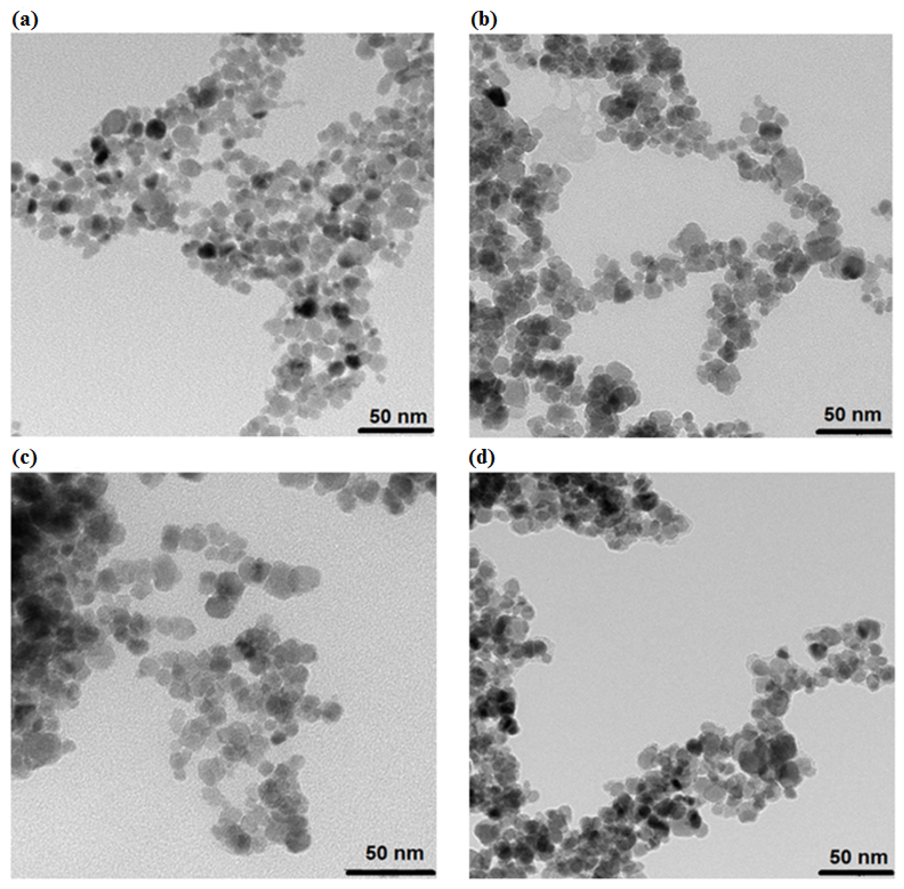
3.1. Morphology Studies
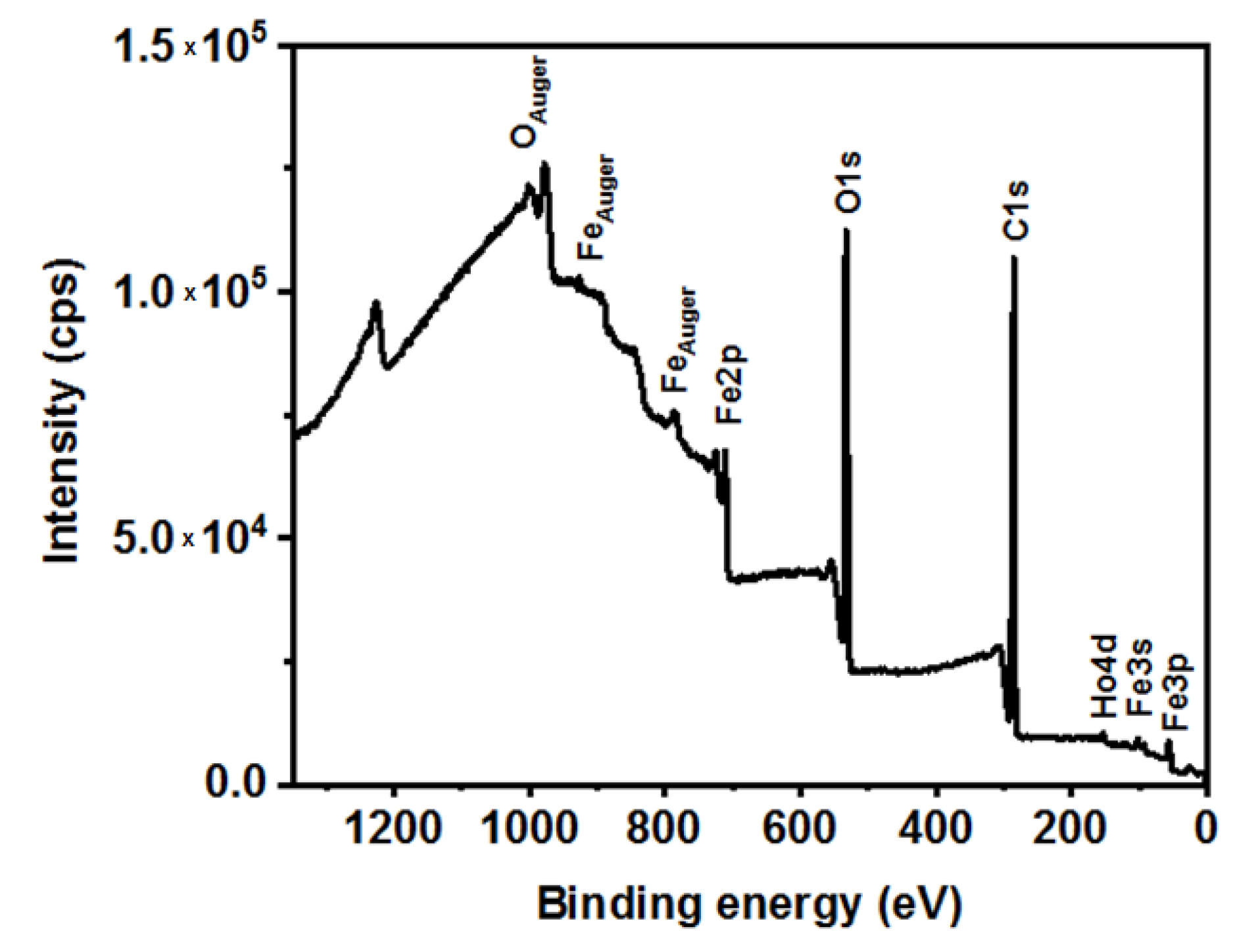
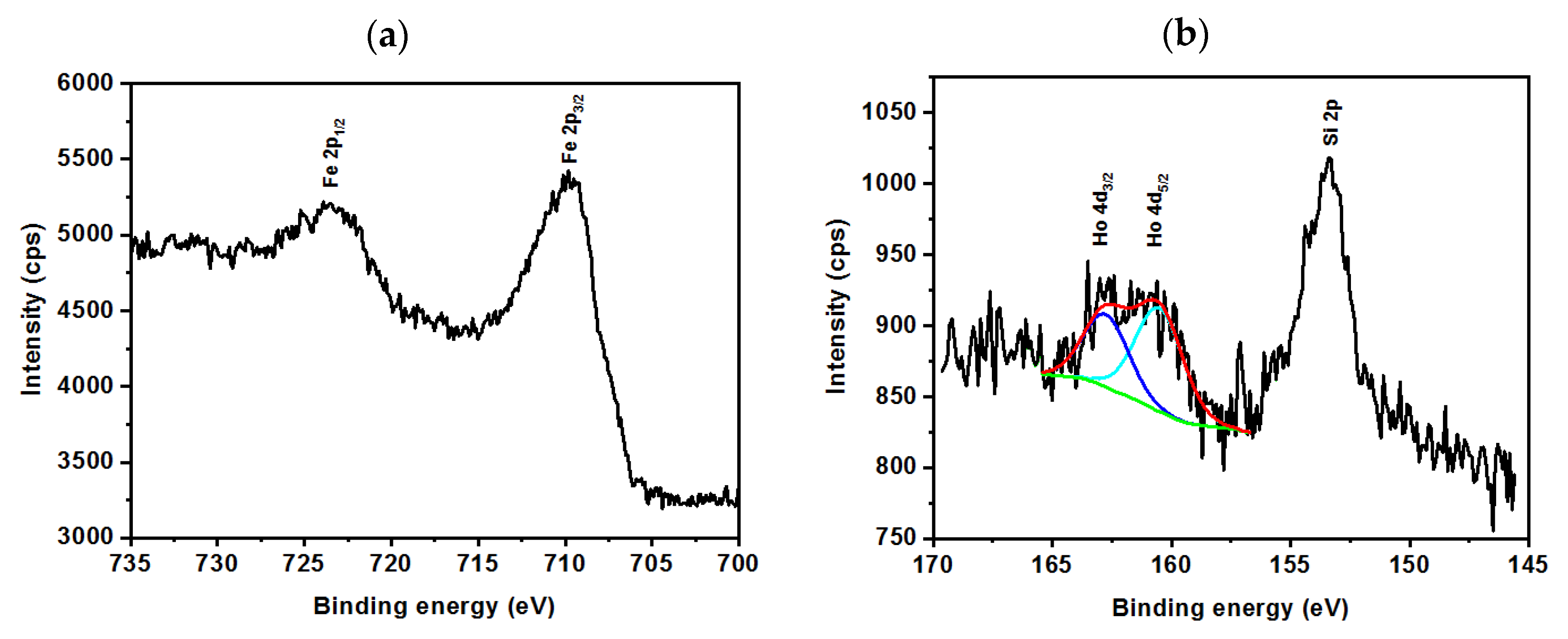
3.2. XPS Analysis
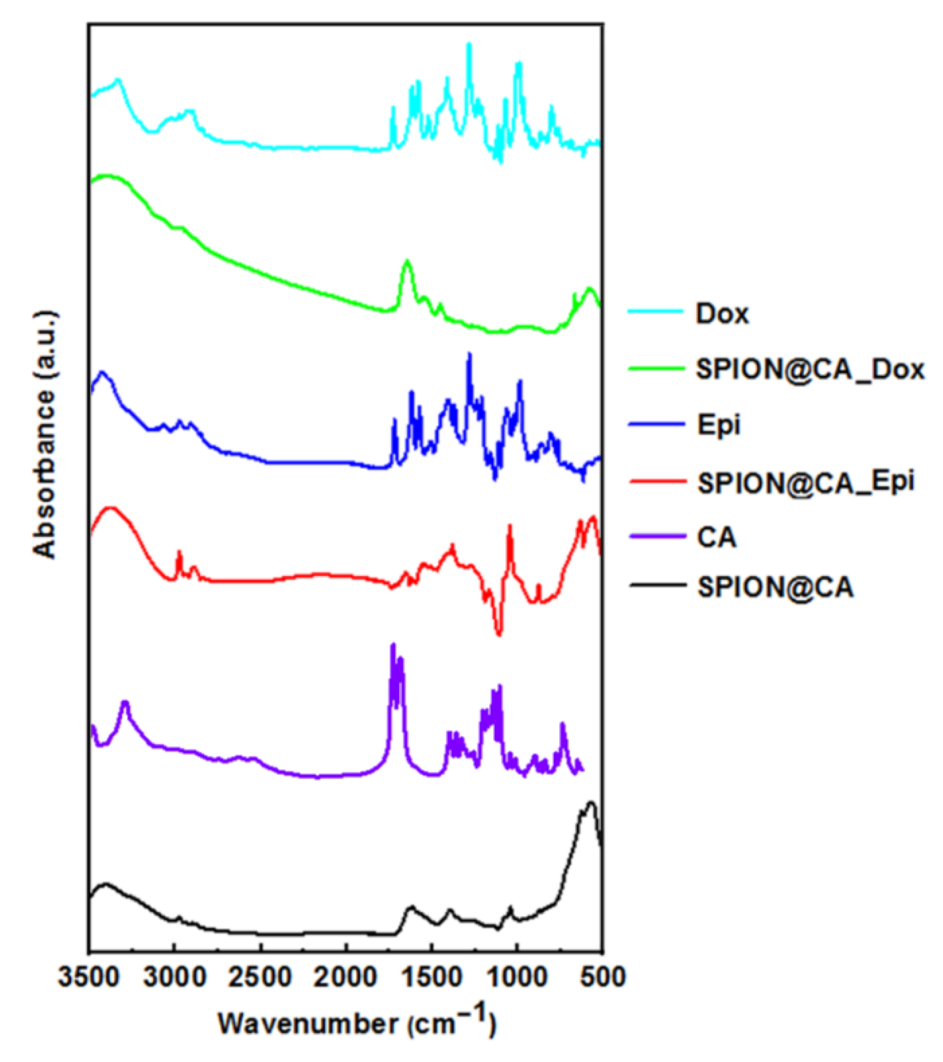
3.3. FT-IR Studies
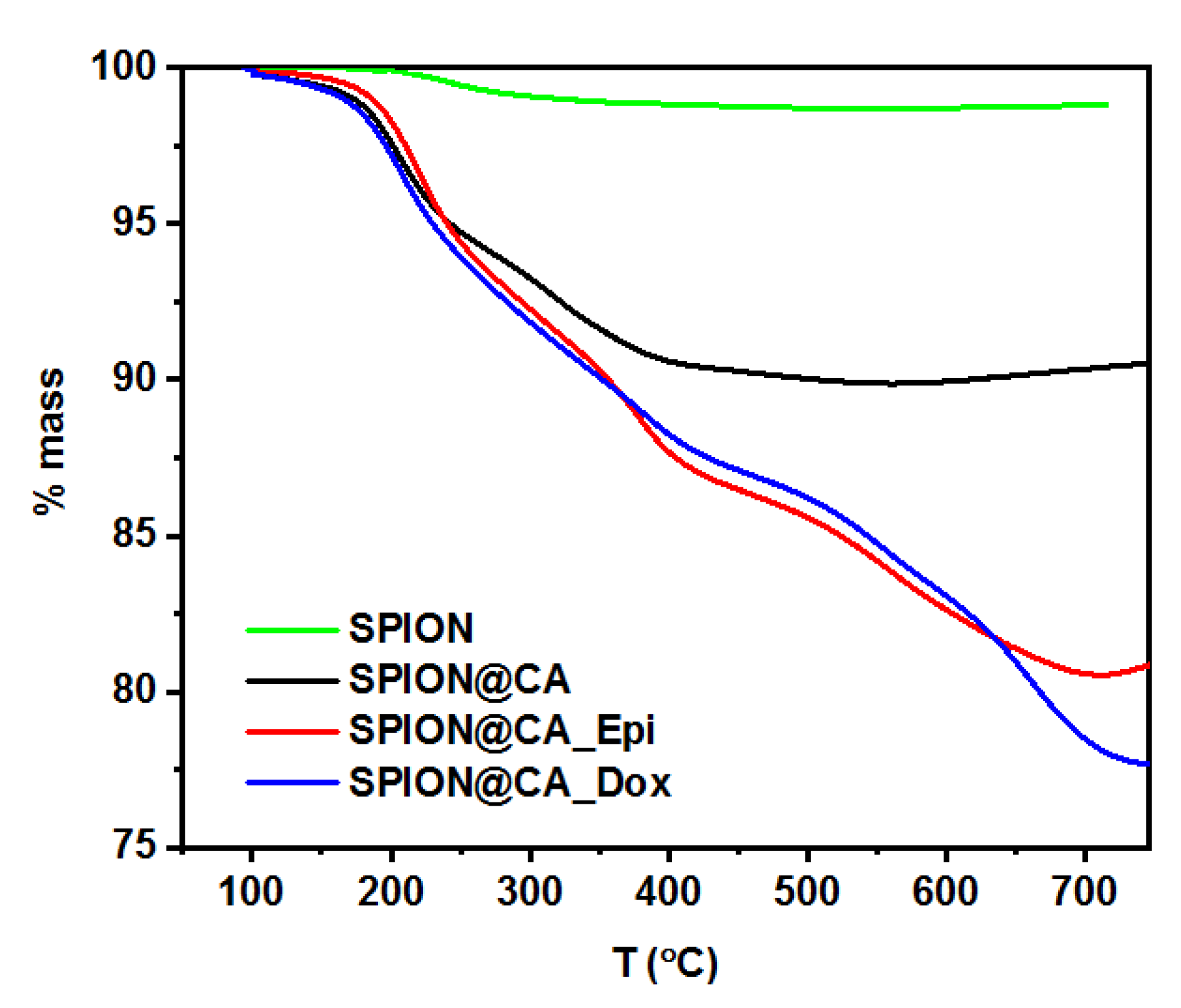
3.4. Drug Content in Hybrid
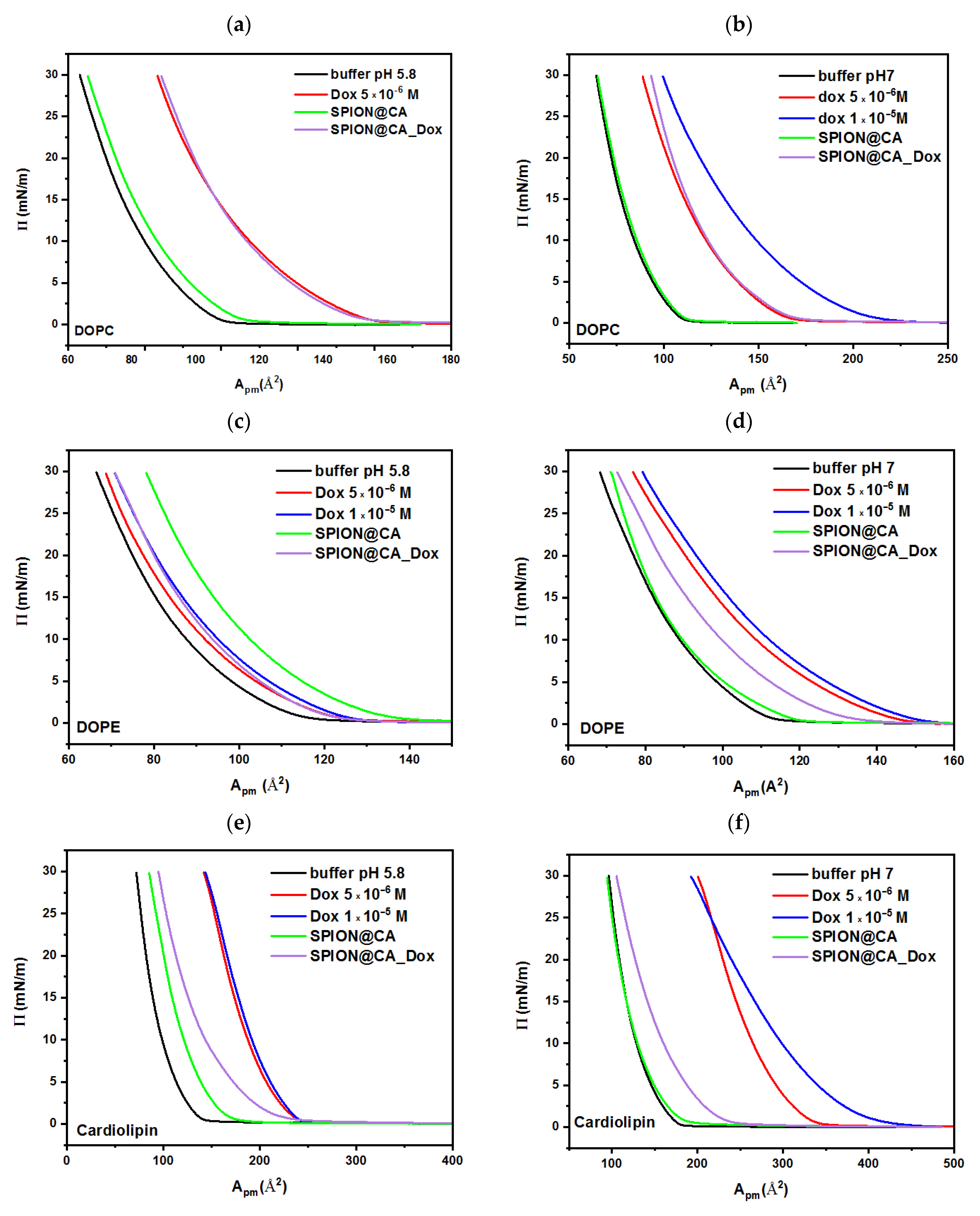
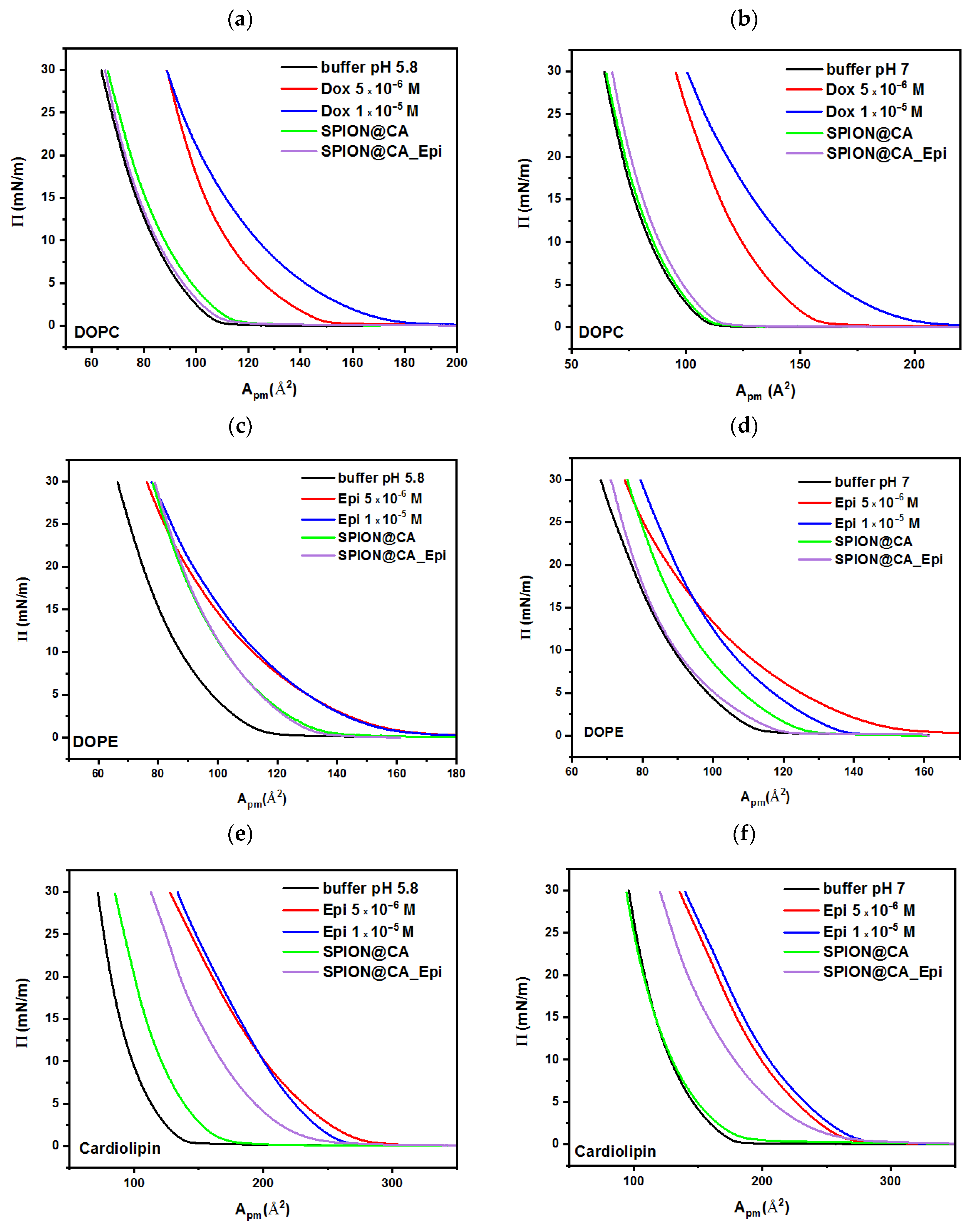
3.5. Investigation of Interactions of Hybrid with Biological Membranes
3.5.1. SPION@CA
3.5.2. Doxorubicin
3.5.3. Epirubicin
3.5.4. SPION@CA_Dox and SPION@CA_Epi
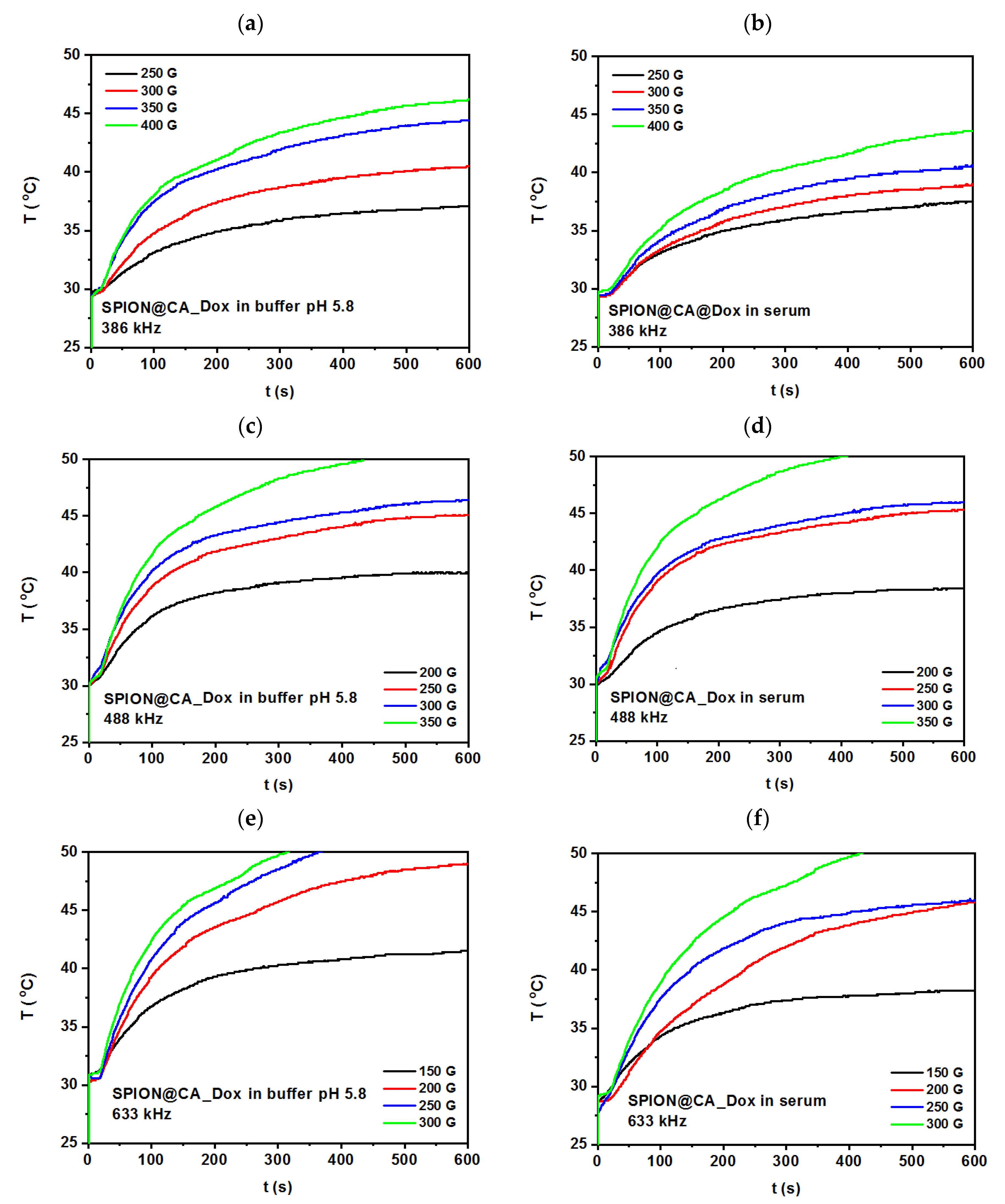
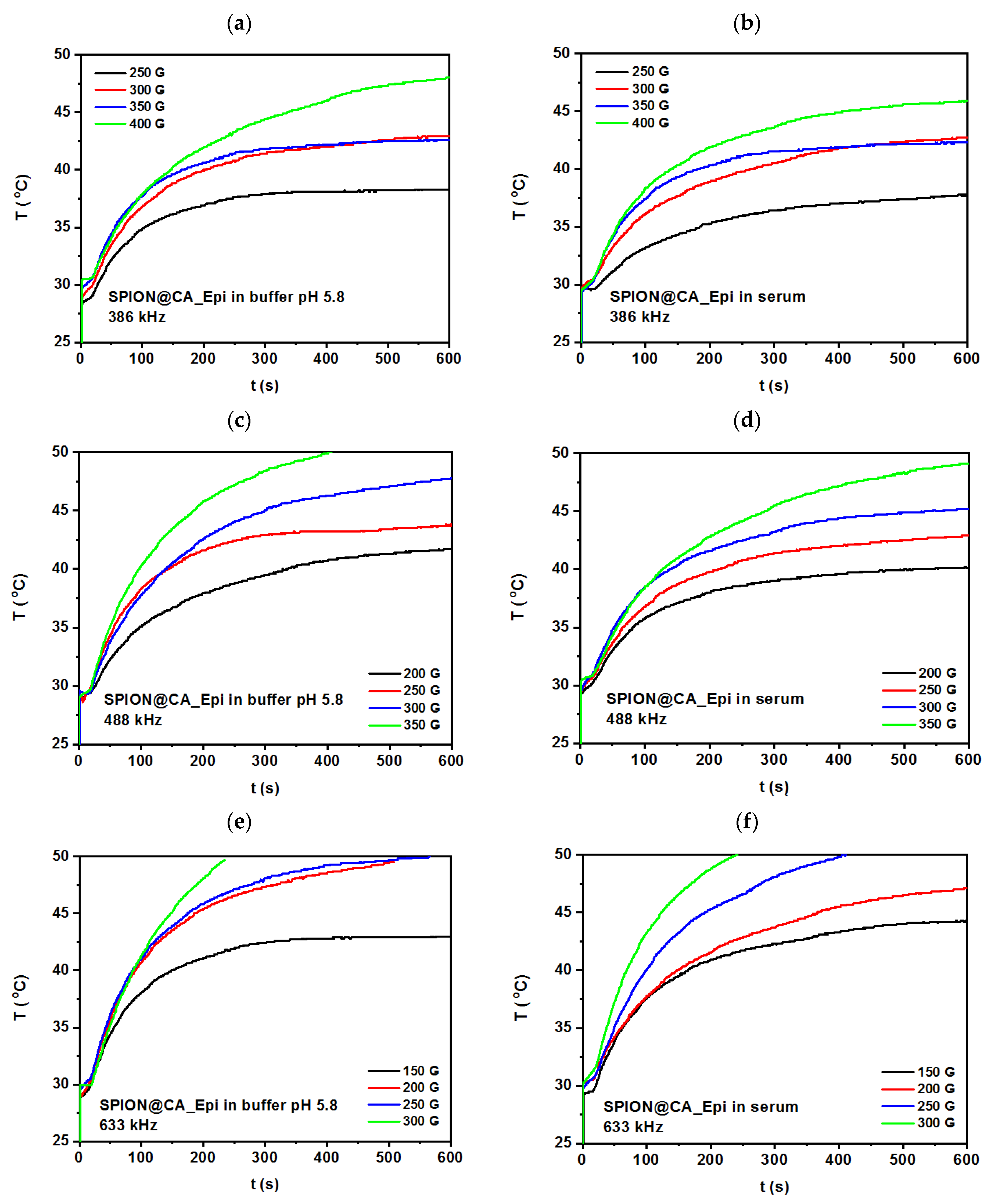
3.6. Magnetic Hyperthermia Studies
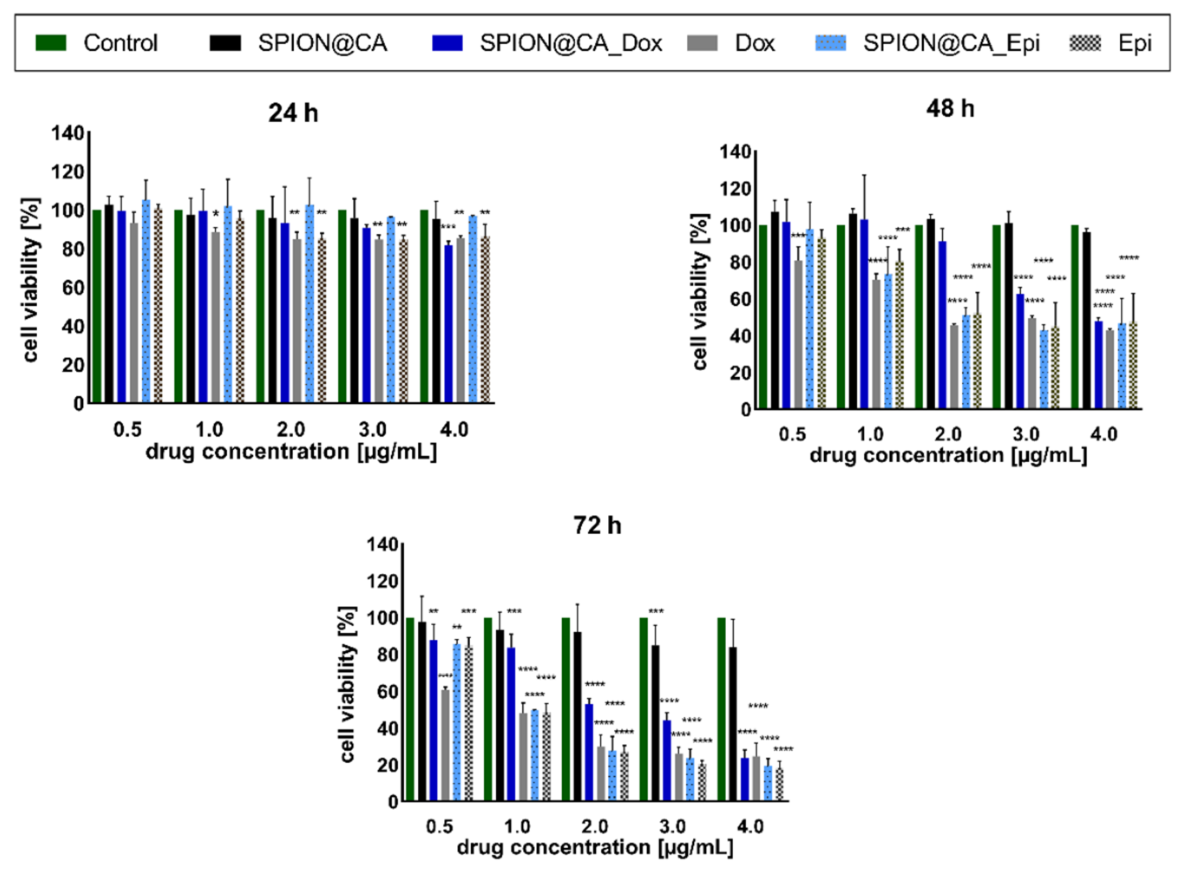
3.7. Cytotoxicity Results
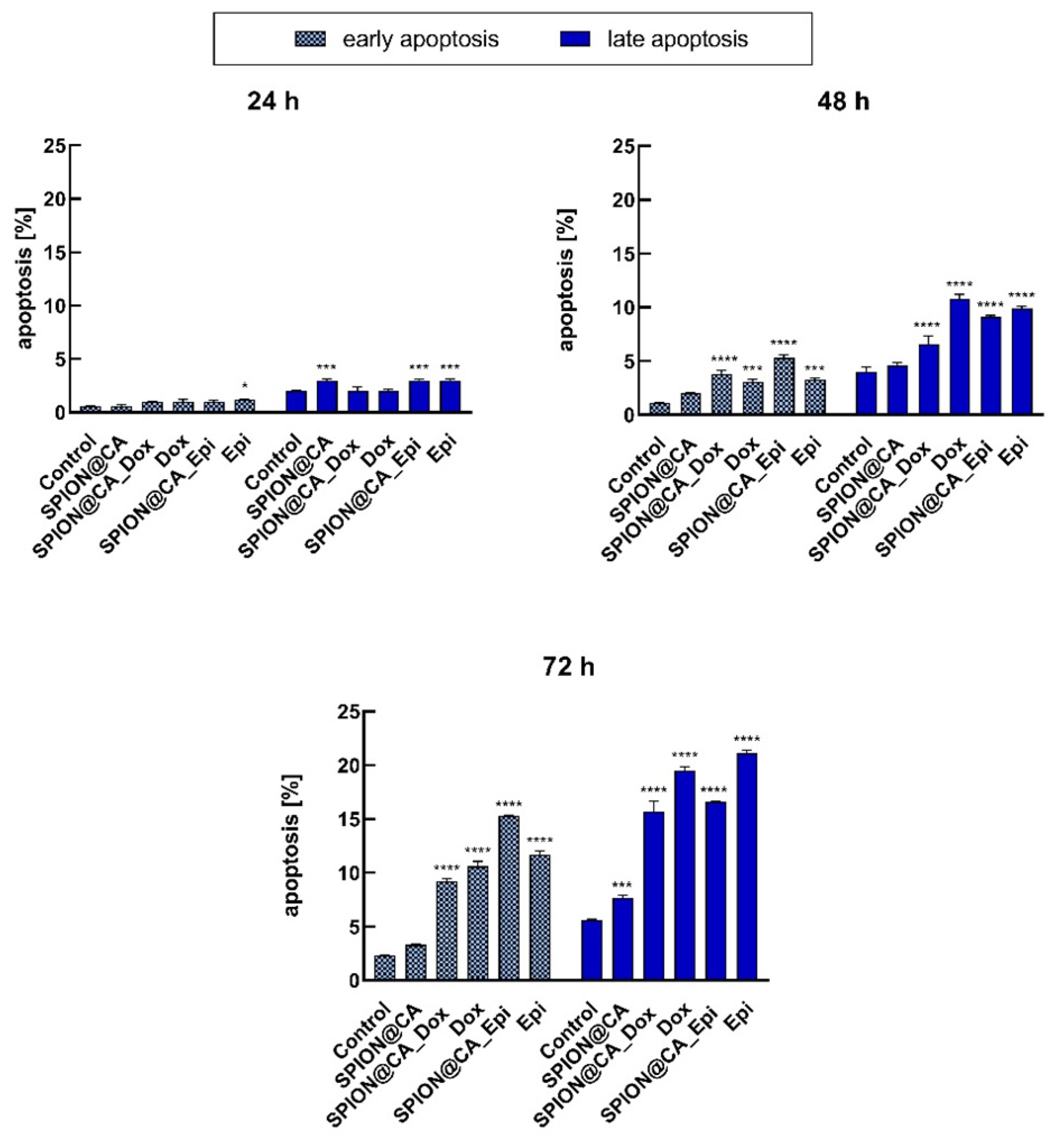
3.8. Apoptosis
4. Discussion
5. Conclusions
Supplementary Materials
Author Contributions
Funding
Institutional Review Board Statement
Informed Consent Statement
Data Availability Statement
Acknowledgments
Conflicts of Interest
References
- Wu, W.; Wu, Z.; Yu, T.; Jiang, C.; Kim, W.-S. Recent Progress on Magnetic Iron Oxide Nanoparticles: Synthesis, Surface Functional Strategies and Biomedical Applications. Sci. Technol. Adv. Mater. 2015, 16, 023501. [Google Scholar] [CrossRef] [PubMed]
- Dadfar, S.M.; Roemhild, K.; Drude, N.I.; von Stillfried, S.; Knüchel, R.; Kiessling, F.; Lammers, T. Iron Oxide Nanoparticles: Diagnostic, Therapeutic and Theranostic Applications. Adv. Drug Deliv. Rev. 2019, 138, 302–325. [Google Scholar] [CrossRef] [PubMed]
- Ohki, A.; Saito, S.; Fukuchi, K. Magnetic Resonance Imaging of Umbilical Cord Stem Cells Labeled with Superparamagnetic Iron Oxide Nanoparticles: Effects of Labelling and Transplantation Parameters. Sci. Rep. 2020, 10, 13684. [Google Scholar] [CrossRef]
- Freund, B.; Tromsdorf, U.I.; Bruns, O.T.; Heine, M.; Giemsa, A.; Bartelt, A.; Salmen, S.C.; Raabe, N.; Heeren, J.; Ittrich, H.; et al. A Simple and Widely Applicable Method to 59Fe-Radiolabel Monodisperse Superparamagnetic Iron Oxide Nanoparticles for in Vivo Quantification Studies. ACS Nano 2012, 6, 7318–7325. [Google Scholar] [CrossRef] [PubMed]
- Hervault, A.; Thanh, N.T.K. Magnetic Nanoparticle-Based Therapeutic Agents for Thermo-Chemotherapy Treatment of Cancer. Nanoscale 2014, 6, 11553–11573. [Google Scholar] [CrossRef] [Green Version]
- Reczyńska, K.; Marszałek, M.; Zarzycki, A.; Reczyński, W.; Kornaus, K.; Pamuła, E.; Chrzanowski, W. Superparamagnetic Iron Oxide Nanoparticles Modified with Silica Layers as Potential Agents for Lung Cancer Treatment. Nanomaterials 2020, 10, 1076. [Google Scholar] [CrossRef]
- Marekova, D.; Turnovcova, K.; Sursal, T.H.; Gandhi, C.D.; Jendelova, P.; Jhanwar-Uniyal, M. Potential for Treatment of Glioblastoma: New Aspects of Superparamagnetic Iron Oxide Nanoparticles. Anticancer Res. 2020, 40, 5989–5994. [Google Scholar] [CrossRef]
- Musielak, M.; Piotrowski, I.; Suchorska, W.M. Superparamagnetic Iron Oxide Nanoparticles (SPIONs) as a Multifunctional Tool in Various Cancer Therapies. Rep. Pract. Oncol. Radiother. 2019, 24, 307–314. [Google Scholar] [CrossRef]
- Zhang, Q.; Liu, J.; Yuan, K.; Zhang, Z.; Zhang, X.; Fang, X. A Multi-Controlled Drug Delivery System Based on Magnetic Mesoporous Fe3O4 Nanopaticles and a Phase Change Material for Cancer Thermo-Chemotherapy. Nanotechnology 2017, 28, 405101. [Google Scholar] [CrossRef]
- Kim, J.; Piao, Y.; Hyeon, T. Multifunctional Nanostructured Materials for Multimodal Imaging, and Simultaneous Imaging and Therapy. Chem. Soc. Rev. 2009, 38, 372–390. [Google Scholar] [CrossRef]
- Chen, F.H.; Gao, Q.; Ni, J.Z. The Grafting and Release Behavior of Doxorubincin from Fe3O4@SiO2 Core-Shell Structure Nanoparticles via an Acid Cleaving Amide Bond: The Potential for Magnetic Targeting Drug Delivery. Nanotechnology 2008, 19, 165103. [Google Scholar] [CrossRef]
- Mieloch, A.A.; Żurawek, M.; Giersig, M.; Rozwadowska, N.; Rybka, J.D. Bioevaluation of Superparamagnetic Iron Oxide Nanoparticles (SPIONs) Functionalized with Dihexadecyl Phosphate (DHP). Sci. Rep. 2020, 10, 2725. [Google Scholar] [CrossRef] [PubMed] [Green Version]
- Lee, N.; Hyeon, T. Designed Synthesis of Uniformly Sized Iron Oxide Nanoparticles for Efficient Magnetic Resonance Imaging Contrast Agents. Chem. Soc. Rev. 2012, 41, 2575–2589. [Google Scholar] [CrossRef] [PubMed]
- Ma, X.; Gong, A.; Chen, B.; Zheng, J.; Chen, T.; Shen, Z.; Wu, A. Exploring a New SPION-Based MRI Contrast Agent with Excellent Water-Dispersibility, High Specificity to Cancer Cells and Strong MR Imaging Efficacy. Colloids Surf. B Biointerfaces 2015, 126, 44–49. [Google Scholar] [CrossRef] [PubMed]
- Cędrowska, E.; Pruszynski, M.; Gawęda, W.; Żuk, M.; Krysiński, P.; Bruchertseifer, F.; Morgenstern, A.; Karageorgou, M.-A.; Bouziotis, P.; Bilewicz, A. Trastuzumab Conjugated Superparamagnetic Iron Oxide Nanoparticles Labeled with 225Ac as a Perspective Tool for Combined α-Radioimmunotherapy and Magnetic Hyperthermia of HER2-Positive Breast Cancer. Molecules 2020, 25, 1025. [Google Scholar] [CrossRef] [PubMed] [Green Version]
- Salimi, M.; Sarkar, S.; Hashemi, M.; Saber, R. Treatment of Breast Cancer-Bearing BALB/c Mice with Magnetic Hyperthermia Using Dendrimer Functionalized Iron-Oxide Nanoparticles. Nanomaterials 2020, 10, 2310. [Google Scholar] [CrossRef] [PubMed]
- Mondal, S.; Manivasagan, P.; Bharathiraja, S.; Santha Moorthy, M.; Nguyen, V.T.; Kim, H.H.; Nam, S.Y.; Lee, K.D.; Oh, J. Hydroxyapatite Coated Iron Oxide Nanoparticles: A Promising Nanomaterial for Magnetic Hyperthermia Cancer Treatment. Nanomaterials 2017, 7, 426. [Google Scholar] [CrossRef] [Green Version]
- Kumar, C.S.; Leuschner, C.; Urbina, M.; Ozkaya, T.; Hormes, J. Glutaric Acid as a Spacer Facilitates Improved Intracellular Uptake of LHRH–SPION into Human Breast Cancer Cells. Int. J. Nanomed. 2007, 2, 175–179. [Google Scholar]
- Mekseriwattana, W.; Srisuk, S.; Kriangsaksri, R.; Niamsiri, N.; Prapainop, K. The Impact of Serum Proteins and Surface Chemistry on Magnetic Nanoparticle Colloidal Stability and Cellular Uptake in Breast Cancer Cells. AAPS PharmSciTech 2019, 20, 55. [Google Scholar] [CrossRef] [PubMed]
- Khosroshahi, M.; Tajabadi, M. Multifunctional Nanoplatform for Targeted Laser-Induced Hyperthermia and Microscopy of Breast Cancer Cells Using SPION-Based Gold and Folic Acid Conjugated Nanodendrimers: An in Vitro Assay. J. Nanomed. Nanotechnol. 2017, 8, 1–11. [Google Scholar] [CrossRef] [Green Version]
- Chee, H.L.; Gan, C.R.R.; Ng, M.; Low, L.; Fernig, D.G.; Bhakoo, K.K.; Paramelle, D. Biocompatible Peptide-Coated Ultrasmall Superparamagnetic Iron Oxide Nanoparticles for In Vivo Contrast-Enhanced Magnetic Resonance Imaging. ACS Nano 2018, 12, 6480–6491. [Google Scholar] [CrossRef] [PubMed]
- Chelluri, L.K.; Mohanram, Y.; Jain, R.; Mallarpu, C.S.; Ponnana, M.; Kumar, D.; Krishna Venuganti, V.V.; Kancherla, R.; Papineni, R.V.L.; Towner, R.; et al. Effect of Engineered Superparamagnetic Iron Oxide Nanoparticles in Targeted Cardiac Precursor Cell Delivery by MRI. Biochem. Biophys. Res. Commun. 2021, 541, 15–21. [Google Scholar] [CrossRef] [PubMed]
- Moghadam, S.M.M.; Alibolandi, M.; Babaei, M.; Mosafer, J.; Saljooghi, A.S.; Ramezani, M. Fabrication of Deferasirox-Decorated Aptamer-Targeted Superparamagnetic Iron Oxide Nanoparticles (SPION) as a Therapeutic and Magnetic Resonance Imaging Agent in Cancer Therapy. J. Biol. Inorg. Chem. 2020. [Google Scholar] [CrossRef] [PubMed]
- Gawęda, W.; Osial, M.; Żuk, M.; Pękała, M.; Bilewicz, A.; Krysinski, P. Lanthanide-Doped SPIONs Bioconjugation with Trastuzumab for Potential Multimodal Anticancer Activity and Magnetic Hyperthermia. Nanomaterials 2020, 10, 288. [Google Scholar] [CrossRef] [Green Version]
- Mu, K.; Zhang, S.; Ai, T.; Jiang, J.; Yao, Y.; Jiang, L.; Zhou, Q.; Xiang, H.; Zhu, Y.; Yang, X.; et al. Monoclonal Antibody–Conjugated Superparamagnetic Iron Oxide Nanoparticles for Imaging of Epidermal Growth Factor Receptor–Targeted Cells and Gliomas. Mol. Imaging 2015, 14. [Google Scholar] [CrossRef]
- de Souza Albernaz, M.; Toma, S.H.; Clanton, J.; Araki, K.; Santos-Oliveira, R. Decorated Superparamagnetic Iron Oxide Nanoparticles with Monoclonal Antibody and Diethylene-Triamine-Pentaacetic Acid Labeled with Thechnetium-99m and Galium-68 for Breast Cancer Imaging. Pharm. Res. 2018, 35, 24. [Google Scholar] [CrossRef]
- Putri Fauzia, R.; Denkova, A.G.; Djanashvili, K. Potential of MRI in Radiotherapy Mediated by Small Conjugates and Nanosystems. Inorganics 2019, 7, 59. [Google Scholar] [CrossRef] [Green Version]
- Klaassen, N.J.M.; Arntz, M.J.; Gil Arranja, A.; Roosen, J.; Nijsen, J.F.W. The Various Therapeutic Applications of the Medical Isotope Holmium-166: A Narrative Review. EJNMMI Radiopharm. Chem. 2019, 4, 19. [Google Scholar] [CrossRef] [Green Version]
- Rękorajska, A.; Cichowicz, G.; Cyranski, M.K.; Pękała, M.; Krysinski, P. Synthesis and Characterization of Gd3+- and Tb3+-Doped Iron Oxide Nanoparticles for Possible Endoradiotherapy and Hyperthermia. J. Magn. Magn. Mater. 2019, 479, 50–58. [Google Scholar] [CrossRef]
- Nosrati, S.; Shanehsazzadeh, S.; Yousefnia, H.; Gholami, A.; Grüttner, C.; Jalilian, A.R.; Hosseini, R.H.; Lahooti, A. Biodistribution Evaluation of 166Ho–DTPA–SPION in Normal Rats. J. Radioanal. Nucl. Chem. 2016, 307, 1559–1566. [Google Scholar] [CrossRef]
- Chakraborty, S.; Vimalnath, K.V.; Sharma, K.S.; Rajeswari, A.; Sarma, H.D.; Ningthoujam, R.S.; Vatsa, R.; Dash, A. Synthesis and Biological Evaluation of Holmium-166 Agglomerated Iron Oxide Nanoparticles for Treatment of Arthritis of Knee Joints. J. Nucl. Med. 2016, 57, 1105. [Google Scholar]
- Tang, Y.; Flesch, R.C.C.; Jin, T.; Gao, Y.; He, M. Effect of Nanoparticle Shape on Therapeutic Temperature Distribution during Magnetic Hyperthermia. J. Phys. Appl. Phys. 2021, 54, 165401. [Google Scholar] [CrossRef]
- Tang, Y.; Jin, T.; Flesch, R.C.C.; Gao, Y.; He, M. Effect of Nanofluid Distribution on Therapeutic Effect Considering Transient Bio-Tissue Temperature during Magnetic Hyperthermia. J. Magn. Magn. Mater. 2021, 517, 167391. [Google Scholar] [CrossRef]
- Maier-Hauff, K.; Ulrich, F.; Nestler, D.; Niehoff, H.; Wust, P.; Thiesen, B.; Orawa, H.; Budach, V.; Jordan, A. Efficacy and Safety of Intratumoral Thermotherapy Using Magnetic Iron-Oxide Nanoparticles Combined with External Beam Radiotherapy on Patients with Recurrent Glioblastoma Multiforme. J. Neurooncol. 2011, 103, 317–324. [Google Scholar] [CrossRef] [PubMed] [Green Version]
- Peviani, M.; Spano, G.; Pagani, A.; Brugnara, G.; Covino, C.; Galli, R.; Biffi, A.; Politi, L.S. Lipophilic Dye-Compatible Brain Clearing Technique Allowing Correlative Magnetic Resonance/High-Resolution Fluorescence Imaging in Rat Models of Glioblastoma. Sci. Rep. 2020, 10, 17974. [Google Scholar] [CrossRef] [PubMed]
- Jermy, B.R.; Alomari, M.; Ravinayagam, V.; Almofty, S.A.; Akhtar, S.; Borgio, J.F.; AbdulAzeez, S. SPIONs/3D SiSBA-16 Based Multifunctional Nanoformulation for Target Specific Cisplatin Release in Colon and Cervical Cancer Cell Lines. Sci. Rep. 2019, 9, 14523. [Google Scholar] [CrossRef] [PubMed] [Green Version]
- Ebrahimpour, S.; Shahidi, S.B.; Abbasi, M.; Tavakoli, Z.; Esmaeili, A. Quercetin-Conjugated Superparamagnetic Iron Oxide Nanoparticles (QCSPIONs) Increases Nrf2 Expression via MiR-27a Mediation to Prevent Memory Dysfunction in Diabetic Rats. Sci. Rep. 2020, 10, 15957. [Google Scholar] [CrossRef]
- Alapan, Y.; Yasa, O.; Schauer, O.; Giltinan, J.; Tabak, A.F.; Sourjik, V.; Sitti, M. Soft Erythrocyte-Based Bacterial Microswimmers for Cargo Delivery. Sci. Robot. 2018, 3. [Google Scholar] [CrossRef] [Green Version]
- Arias, L.S.; Pessan, J.P.; Vieira, A.P.M.; de Lima, T.M.T.; Delbem, A.C.B.; Monteiro, D.R. Iron Oxide Nanoparticles for Biomedical Applications: A Perspective on Synthesis, Drugs, Antimicrobial Activity, and Toxicity. Antibiotics 2018, 7, 46. [Google Scholar] [CrossRef] [Green Version]
- Yen, S.K.; Padmanabhan, P.; Selvan, S.T. Multifunctional Iron Oxide Nanoparticles for Diagnostics, Therapy and Macromolecule Delivery. Theranostics 2013, 3, 986–1003. [Google Scholar] [CrossRef] [PubMed] [Green Version]
- Cricchio, V.; Best, M.; Reverchon, E.; Maffulli, N.; Phillips, G.; Santin, M.; Porta, G.D. Novel Superparamagnetic Microdevices Based on Magnetized PLGA/PLA Microparticles Obtained by Supercritical Fluid Emulsion and Coating by Carboxybetaine-Functionalized Chitosan Allowing the Tuneable Release of Therapeutics. J. Pharm. Sci. 2017, 106, 2097–2105. [Google Scholar] [CrossRef]
- Munnier, E.; Cohen-Jonathan, S.; Hervé, K.; Linassier, C.; Soucé, M.; Dubois, P.; Chourpa, I. Doxorubicin Delivered to MCF-7 Cancer Cells by Superparamagnetic Iron Oxide Nanoparticles: Effects on Subcellular Distribution and Cytotoxicity. J. Nanoparticle Res. 2011, 13, 959–971. [Google Scholar] [CrossRef]
- Siminzar, P.; Omidi, Y.; Golchin, A.; Aghanejad, A.; Barar, J. Targeted Delivery of Doxorubicin by Magnetic Mesoporous Silica Nanoparticles Armed with Mucin-1 Aptamer. J. Drug Target. 2020, 28, 92–101. [Google Scholar] [CrossRef]
- Basuki, J.S.; Duong, H.T.T.; Macmillan, A.; Erlich, R.B.; Esser, L.; Akerfeldt, M.C.; Whan, R.M.; Kavallaris, M.; Boyer, C.; Davis, T.P. Using Fluorescence Lifetime Imaging Microscopy to Monitor Theranostic Nanoparticle Uptake and Intracellular Doxorubicin Release. ACS Nano 2013, 7, 10175–10189. [Google Scholar] [CrossRef] [PubMed]
- Buhl, A.; Christensen, T.; Christensen, I.; Nelausen, K.; Balslev, E.; Knoop, A.; Brix, E.; Svensson, E.; Glavicic, V.; Luczak, A.; et al. Predicting Efficacy of Epirubicin by a Multigene Assay in Advanced Breast Cancer within a Danish Breast Cancer Cooperative Group (DBCG) Cohort: A Retrospective-Prospective Blinded Study. Breast Cancer Res. Treat. 2018, 172. [Google Scholar] [CrossRef] [PubMed] [Green Version]
- Ansari, L.; Jaafari, M.R.; Bastami, T.R.; Malaekeh-Nikouei, B. Improved Anticancer Efficacy of Epirubicin by Magnetic Mesoporous Silica Nanoparticles: In Vitro and in Vivo Studies. Artif. Cells Nanomed. Biotechnol. 2018, 46, 594–606. [Google Scholar] [CrossRef] [PubMed] [Green Version]
- Avilés, A.; Arévila, N.; Maqueo, J.C.D.; Nambo, M.J. Late Cardiac Toxicity of Doxorubicin, Epirubicin, and Mitoxantrone Therapy for Hodgkin’s Disease in Adults. Leuk. Lymphoma 1993, 11, 275–279. [Google Scholar] [CrossRef] [PubMed]
- Hoang Thi, T.T.; Nguyen Tran, D.-H.; Bach, L.G.; Vu-Quang, H.; Nguyen, D.C.; Park, K.D.; Nguyen, D.H. Functional Magnetic Core-Shell System-Based Iron Oxide Nanoparticle Coated with Biocompatible Copolymer for Anticancer Drug Delivery. Pharmaceutics 2019, 11, 120. [Google Scholar] [CrossRef] [PubMed] [Green Version]
- Jalalian, S.H.; Taghdisi, S.M.; Shahidi Hamedani, N.; Kalat, S.A.M.; Lavaee, P.; Zandkarimi, M.; Ghows, N.; Jaafari, M.R.; Naghibi, S.; Danesh, N.M.; et al. Epirubicin Loaded Super Paramagnetic Iron Oxide Nanoparticle-Aptamer Bioconjugate for Combined Colon Cancer Therapy and Imaging in Vivo. Eur. J. Pharm. Sci. Off. J. Eur. Fed. Pharm. Sci. 2013, 50, 191–197. [Google Scholar] [CrossRef] [PubMed]
- Rao, Y.; Chen, W.; Liang, X.; Huang, Y.; Miao, J.; Liu, L.; Lou, Y.; Zhang, X.; Wang, B.; Tang, R.; et al. Epirubicin-Loaded Superparamagnetic Iron-Oxide Nanoparticles for Transdermal Delivery: Cancer Therapy by Circumventing the Skin Barrier. Small Weinh. Bergstr. Ger. 2015, 11, 239–247. [Google Scholar] [CrossRef]
- Cazares-Cortes, E.; Espinosa, A.; Guigner, J.-M.; Michel, A.; Griffete, N.; Wilhelm, C.; Ménager, C. Doxorubicin Intracellular Remote Release from Biocompatible Oligo(Ethylene Glycol) Methyl Ether Methacrylate-Based Magnetic Nanogels Triggered by Magnetic Hyperthermia. ACS Appl. Mater. Interfaces 2017, 9, 25775–25788. [Google Scholar] [CrossRef] [Green Version]
- Cazares-Cortes, E.; Nerantzaki, M.; Fresnais, J.; Wilhelm, C.; Griffete, N.; Ménager, C. Magnetic Nanoparticles Create Hot Spots in Polymer Matrix for Controlled Drug Release. Nanomaterials 2018, 8, 850. [Google Scholar] [CrossRef] [Green Version]
- Giustini, A.J.; Petryk, A.A.; Cassim, S.M.; Tate, J.A.; Baker, I.; Hoopes, P.J. Magnetic Nanoparticle Hyperthermia in Cancer Treatment. Nano Life 2010, 1. [Google Scholar] [CrossRef]
- Osial, M.; Rybicka, P.; Pękała, M.; Cichowicz, G.; Cyrański, M.K.; Krysiński, P. Easy Synthesis and Characterization of Holmium-Doped SPIONs. Nanomaterials 2018, 8, 430. [Google Scholar] [CrossRef] [Green Version]
- Wilson, D.; Langell, M.A. XPS Analysis of Oleylamine/Oleic Acid Capped Fe3O4 Nanoparticles as a Function of Temperature. Appl. Surf. Sci. 2014, 303, 6–13. [Google Scholar] [CrossRef]
- Yamashita, T.; Hayes, P. Analysis of XPS Spectra of Fe2+ and Fe3+ Ions in Oxide Materials. Appl. Surf. Sci. 2008, 254, 2441–2449. [Google Scholar] [CrossRef]
- Fan, Z.; Wu, T.; Xu, X. Synthesis of Reduced Grapheme Oxide as A Platform for Loading β-NaYF 4:Ho 3+ @TiO2 Based on An Advanced Visible Light-Driven Photocatalyst. Sci. Rep. 2017, 7, 13833. [Google Scholar] [CrossRef] [PubMed] [Green Version]
- Noqta, O.A.; Sodipo, B.K.; Aziz, A.A. One-Pot Synthesis of Highly Magnetic and Stable Citrate Coated Superparamagnetic Iron Oxide Nanoparticles by Modified Coprecipitation Method. Funct. Compos. Struct. 2020, 2, 045005. [Google Scholar] [CrossRef]
- Stein, R.; Friedrich, B.; Mühlberger, M.; Cebulla, N.; Schreiber, E.; Tietze, R.; Cicha, I.; Alexiou, C.; Dutz, S.; Boccaccini, A.R.; et al. Synthesis and Characterization of Citrate-Stabilized Gold-Coated Superparamagnetic Iron Oxide Nanoparticles for Biomedical Applications. Molecules 2020, 25, 4425. [Google Scholar] [CrossRef]
- Majewski, P.; Krysinski, P. Synthesis, Surface modifications, and size-sorting of mixed nickel-zinc ferrite colloidal magnetic nanoparticles. Chem. Eur. J. 2008, 14, 7961–7989. [Google Scholar] [CrossRef]
- Wulandari, P.; Nagahiro, T.; Fukada, N.; Kimura, Y.; Niwano, M.; Tamada, K. Characterization of Citrates on Gold and Silver Nanoparticles. J. Colloid Interface Sci. 2015, 438, 244–248. [Google Scholar] [CrossRef] [PubMed]
- Ding, J.; Chen, G.; Chen, G.; Guo, M. One-Pot Synthesis of Epirubicin-Capped Silver Nanoparticles and Their Anticancer Activity against Hep G2 Cells. Pharmaceutics 2019, 11, 123. [Google Scholar] [CrossRef] [PubMed] [Green Version]
- Wu, S.; Zhao, X.; Li, Y.; Du, Q.; Sun, J.; Wang, Y.; Wang, X.; Xia, Y.; Wang, Z.; Xia, L. Adsorption Properties of Doxorubicin Hydrochloride onto Graphene Oxide: Equilibrium, Kinetic and Thermodynamic Studies. Materials 2013, 6, 2026–2042. [Google Scholar] [CrossRef] [PubMed] [Green Version]
- Nieciecka, D.; Kijewska, K.; Baumler, S.M.; Puszko, A.K.; Misicka, A.; Krysinski, P. Interactions of Mitoxantrone-Modified Superparamagnetic Iron Oxide Nanoparticles with Biomimetic Membranes and Cells. J. Mater. Sci. Eng. B 2020, 10, 34. [Google Scholar] [CrossRef]
- Piehler, S.; Dähring, H.; Grandke, J.; Göring, J.; Couleaud, P.; Aires, A.; Cortajarena, A.L.; Courty, J.; Latorre, A.; Somoza, Á.; et al. Iron Oxide Nanoparticles as Carriers for DOX and Magnetic Hyperthermia after Intratumoral Application into Breast Cancer in Mice: Impact and Future Perspectives. Nanomaterials 2020, 10, 1016. [Google Scholar] [CrossRef]
- Nieciecka, D.; Królikowska, A.; Kijewska, K.; Blanchard, G.; Krysiński, P. Hydrophilic Iron Oxide Nanoparticles Probe the Organization of Biomimetic Layers: Electrochemical and Spectroscopic Evidence. Electrochim. Acta 2016, 209. [Google Scholar] [CrossRef]
- Zhang, H.; Jiang, H.; Sun, F.; Wang, H.; Zhao, J.; Chen, B.; Wang, X. Rapid Diagnosis of Multidrug Resistance in Cancer by Electrochemical Sensor Based on Carbon Nanotubes-Drug Supramolecular Nanocomposites. Biosens. Bioelectron. 2011, 26, 3361–3366. [Google Scholar] [CrossRef]
- Nieciecka, D.; Królikowska, A.; Krysinski, P. Probing the Interactions of Mitoxantrone with Biomimetic Membranes with Electrochemical and Spectroscopic Techniques. Electrochim. Acta 2015, 165, 430–442. [Google Scholar] [CrossRef]
- Obaidat, I.M.; Narayanaswamy, V.; Alaabed, S.; Sambasivam, S.; Muralee Gopi, C.V.V. Principles of Magnetic Hyperthermia: A Focus on Using Multifunctional Hybrid Magnetic Nanoparticles. Magnetochemistry 2019, 5, 67. [Google Scholar] [CrossRef] [Green Version]
- Eslami, P.; Rossi, F.; Fedeli, S. Hybrid Nanogels: Stealth and Biocompatible Structures for Drug Delivery Applications. Pharmaceutics 2019, 11, 71. [Google Scholar] [CrossRef] [Green Version]
- Oh, J.K.; Drumright, R.; Siegwart, D.J.; Matyjaszewski, K. The Development of Microgels/Nanogels for Drug Delivery Applications. Prog. Polym. Sci. 2008, 33, 448–477. [Google Scholar] [CrossRef]
- Tay, Z.W.; Chandrasekharan, P.; Chiu-Lam, A.; Hensley, D.W.; Dhavalikar, R.; Zhou, X.Y.; Yu, E.Y.; Goodwill, P.W.; Zheng, B.; Rinaldi, C.; et al. Magnetic Particle Imaging-Guided Heating in Vivo Using Gradient Fields for Arbitrary Localization of Magnetic Hyperthermia Therapy. ACS Nano 2018, 12, 3699–3713. [Google Scholar] [CrossRef] [PubMed]
- Niculaes, D.; Lak, A.; Anyfantis, G.C.; Marras, S.; Laslett, O.; Avugadda, S.K.; Cassani, M.; Serantes, D.; Hovorka, O.; Chantrell, R.; et al. Asymmetric Assembling of Iron Oxide Nanocubes for Improving Magnetic Hyperthermia Performance. ACS Nano 2017, 11, 12121–12133. [Google Scholar] [CrossRef] [PubMed]
- Liu, H.; Xie, Y.; Zhang, Y.; Cai, Y.; Li, B.; Mao, H.; Yu, R. CA4-Loaded Doxorubicin Prodrug Coating Fe3O4 Nanoparticles for Tumor Combination Therapy. RSC Adv. 2016, 6, 113933–113939. [Google Scholar] [CrossRef]


| SPION@CA_Dox | |||
|---|---|---|---|
| PBS pH 5.8 | 250 G | 488 kHz | 258.9 W·g−1 |
| Serum | 250 G | 488 kHz | 224.7 W·g−1 |
| PBS pH 5.8 | 300 G | 488 kHz | 322.1 W·g−1 |
| Serum | 300 G | 488 kHz | 306.8 W·g−1 |
| SPION@CA_Epi | |||
| PBS pH 5.8 | 250 G | 488 kHz | 283.5 W·g−1 |
| Serum | 250 G | 488 kHz | 256.4 W·g−1 |
| PBS pH 5.8 | 300 G | 488 kHz | 369.7 W·g−1 |
| Serum | 300 G | 488 kHz | 337.3 W·g−1 |
Publisher’s Note: MDPI stays neutral with regard to jurisdictional claims in published maps and institutional affiliations. |
© 2021 by the authors. Licensee MDPI, Basel, Switzerland. This article is an open access article distributed under the terms and conditions of the Creative Commons Attribution (CC BY) license (https://creativecommons.org/licenses/by/4.0/).
Share and Cite
Nieciecka, D.; Celej, J.; Żuk, M.; Majkowska-Pilip, A.; Żelechowska-Matysiak, K.; Lis, A.; Osial, M. Hybrid System for Local Drug Delivery and Magnetic Hyperthermia Based on SPIONs Loaded with Doxorubicin and Epirubicin. Pharmaceutics 2021, 13, 480. https://doi.org/10.3390/pharmaceutics13040480
Nieciecka D, Celej J, Żuk M, Majkowska-Pilip A, Żelechowska-Matysiak K, Lis A, Osial M. Hybrid System for Local Drug Delivery and Magnetic Hyperthermia Based on SPIONs Loaded with Doxorubicin and Epirubicin. Pharmaceutics. 2021; 13(4):480. https://doi.org/10.3390/pharmaceutics13040480
Chicago/Turabian StyleNieciecka, Dorota, Joanna Celej, Michał Żuk, Agnieszka Majkowska-Pilip, Kinga Żelechowska-Matysiak, Antoni Lis, and Magdalena Osial. 2021. "Hybrid System for Local Drug Delivery and Magnetic Hyperthermia Based on SPIONs Loaded with Doxorubicin and Epirubicin" Pharmaceutics 13, no. 4: 480. https://doi.org/10.3390/pharmaceutics13040480