
Single- versus Dual-Targeted Nanoparticles with Folic Acid and Biotin for Anticancer Drug Delivery

 , , , , and
Abstract
:1. Introduction
2. Active Targeting of Nanoparticles
3. Folic Acid—Targeted Nanoparticles
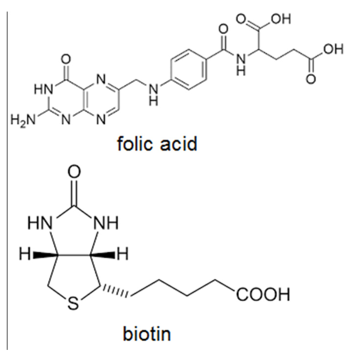
3.1. Folic Acid as a Targeting Ligand
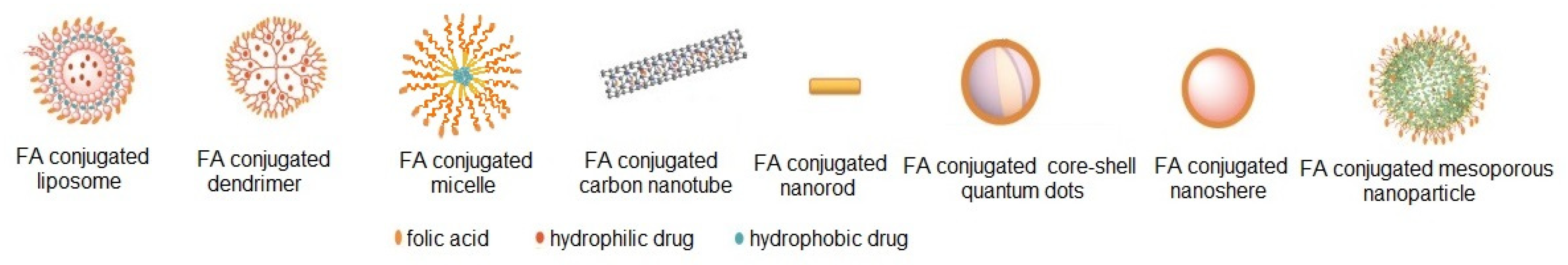
3.2. Examples of Folic Acid-Targeted Nanoparticles
3.2.1. Drug Delivery Application
Single-Drug Delivery
Dual-Drug Delivery
3.2.2. Gene Delivery
3.2.3. FA-Targeted NPs for Thermo-, Photo-, Radiotherapy and Diagnostic or Theranostic Application
3.3. Clinical Trials and Patents
3.4. Summary
4. Biotin—Targeted Nanoparticles
4.1. Biotin as a Targeting Ligand
4.2. Biotin-Targeted Nanoparticles
4.2.1. Drug-Delivery Application
Biotin-Targeted NPs for Chemo-Photodynamic Combination Therapy
4.2.2. Gene Delivery
4.3. Summary
5. Dual-Targeted Nanoparticles
5.1. Tumour Heterogeneity
5.2. Dual-Targeting
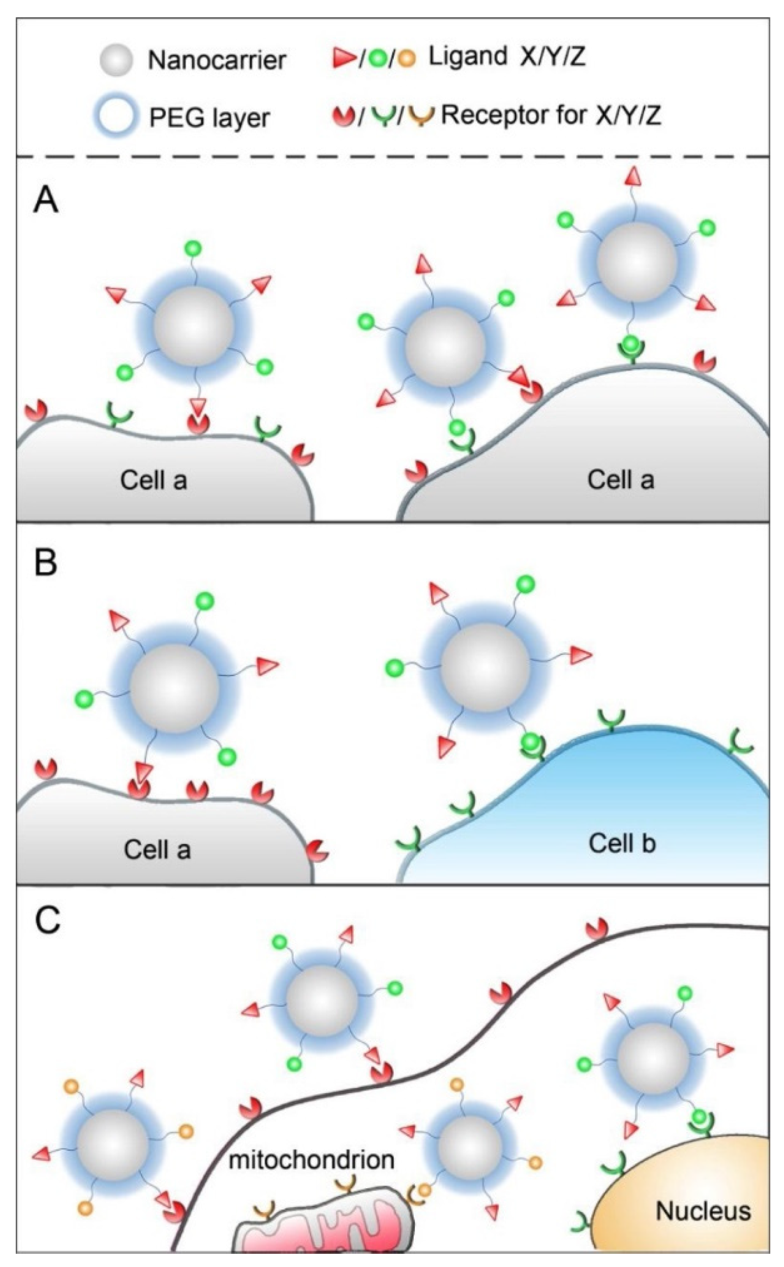
5.2.1. Dual-Molecular Targeting
5.2.2. Dual-Targeting with Folic Acid
5.2.3. Dual-Targeting with Biotin
5.2.4. Dual-Targeting with Combination of Folic Acid and Biotin
NPs Targeted with Folic Acid and Biotin
5.3. Summary
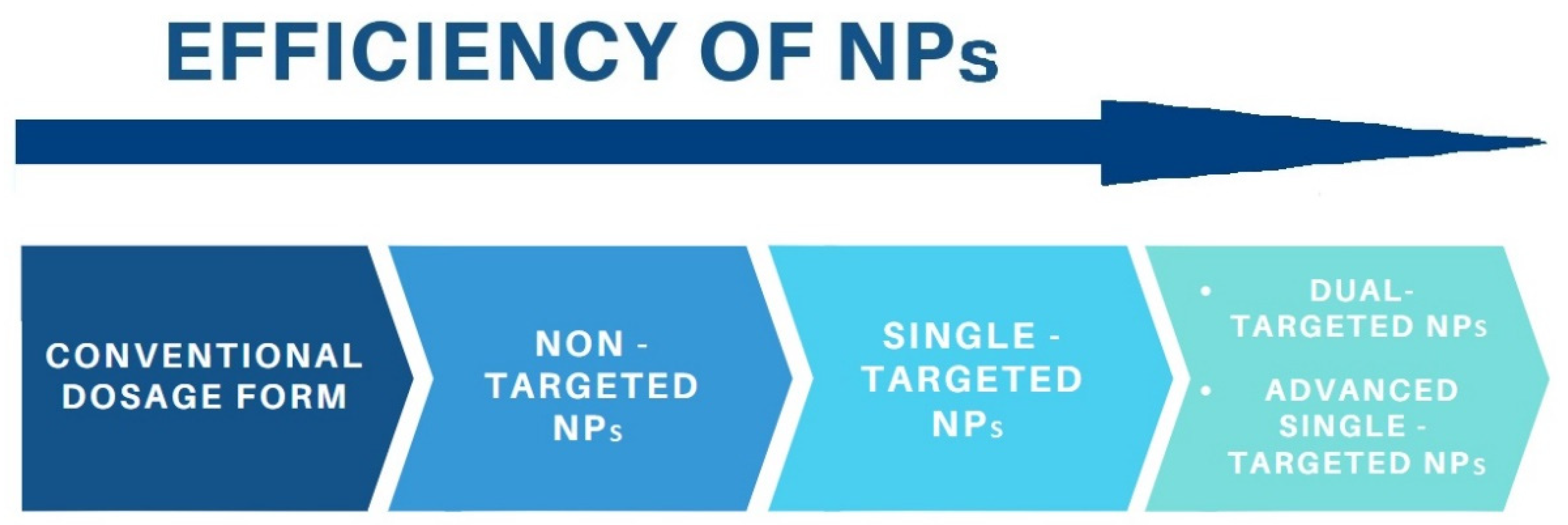
6. Single-Targeted versus Dual-Targeted Nanoparticles
7. Conclusions and Future Perspectives
Author Contributions
Funding
Institutional Review Board Statement
Informed Consent Statement
Data Availability Statement
Conflicts of Interest
References
- Wild, C.P.; Weiderpass, E.; Stewart, B.W. World Cancer Report: Cancer Research for Cancer Prevention; Wild, C.P., Weiderpass, E., Stewart, B.W., Eds.; International Agency for Research on Cancer: Lyon, France, 2020.
- Mantovani, A. Inflaming metastasis. Nature 2009, 457, 36–37. [Google Scholar] [CrossRef]
- Golombek, S.K.; May, J.-N.; Theek, B.; Appold, L.; Drude, N.; Kiessling, F.; Lammers, T. Tumor targeting via EPR: Strategies to enhance patient responses. Adv. Drug Deliv. Rev. 2018, 130, 17–38. [Google Scholar] [CrossRef]
- Wolinsky, J.B.; Colson, Y.L.; Grinstaff, M.W. Local drug delivery strategies for cancer treatment: Gels, nanoparticles, polymeric films, rods, and wafers. J. Control Release 2012, 159, 14–26. [Google Scholar] [CrossRef] [Green Version]
- Swetha, K.L.; Roy, A. Tumor heterogeneity and nanoparticle-mediated tumor targeting: The importance of delivery system personalization. Drug Deliv. Transl. Res. 2018, 8, 1508–1526. [Google Scholar] [CrossRef] [PubMed]
- Zhu, Y.; Feijen, J.; Zhong, Z. Dual-targeted nanomedicines for enhanced tumor treatment. Nano Today 2018, 18, 65–85. [Google Scholar] [CrossRef]
- Hare, J.I.; Lammers, T.; Ashford, M.B.; Puri, S.; Storm, G.; Barry, S.T. Challenges and strategies in anti-cancer nanomedicine development: An industry perspective. Adv. Drug Deliv. Rev. 2017, 108, 25–38. [Google Scholar] [CrossRef] [PubMed] [Green Version]
- Kumar, P.; Huo, P.; Liu, B. Formulation Strategies for Folate-Targeted Liposomes and Their Biomedical Applications. Pharmaceutics 2019, 11, 381. [Google Scholar] [CrossRef] [PubMed] [Green Version]
- Arias, J.L.; Clares, B.; Morales, M.E.; Gallardo, V.; Ruiz, M.A. Lipid-based drug delivery systems for cancer treatment. Curr. Drug Targets 2011, 12, 1151–1165. [Google Scholar] [CrossRef]
- Clares, B.; Biedma-Ortiz, R.A.; Sáez-Fernández, E.; Prados, J.C.; Melguizo, C.; Cabeza, L.; Ortiz, R.; Arias, J.L. Nano-engineering of 5-fluorouracil-loaded magnetoliposomes for combined hyperthermia and chemotherapy against colon cancer. Eur. J. Pharm. Biopharm. 2013, 85, 329–338. [Google Scholar] [CrossRef]
- Lorente, C.; Cabeza, L.; Clares, B.; Ortiz, R.; Halbaut, L.; Delgado, Á.V.; Perazzoli, G.; Prados, J.; Arias, J.L.; Melguizo, C. Formulation and in vitro evaluation of magnetoliposomes as a potential nanotool in colorectal cancer therapy. Colloids Surf. B Biointerfaces 2018, 171, 553–565. [Google Scholar] [CrossRef]
- Lin, L.; Fan, Y.; Gao, F.; Jin, L.; Li, D.; Sun, W.; Li, F.; Qin, P.; Shi, Q.; Shi, X.; et al. UTMD-Promoted Co-Delivery of Gemcitabine and miR-21 Inhibitor by Dendrimer-Entrapped Gold Nanoparticles for Pancreatic Cancer Therapy. Theranostics 2018, 8, 1923–1939. [Google Scholar] [CrossRef] [PubMed]
- Farran, B.; Montenegro, R.C.; Kasa, P.; Pavitra, E.; Huh, Y.S.; Han, Y.K.; Kamal, M.A.; Nagaraju, G.P.; Rama Raju, G.S. Folate-conjugated nanovehicles: Strategies for cancer therapy. Mater. Sci. Eng. C Mater. Biol. Appl. 2020, 107, 110341. [Google Scholar] [CrossRef]
- Abedi-Gaballu, F.; Dehghan, G.; Ghaffari, M.; Yekta, R.; Abbaspour-Ravasjani, S.; Baradaran, B.; Ezzati Nazhad Dolatabadi, J.; Hamblin, M.R. PAMAM dendrimers as efficient drug and gene delivery nanosystems for cancer therapy. Appl. Mater. Today 2018, 12, 177–190. [Google Scholar] [CrossRef]
- Li, Y.; Yu, A.; Li, L.; Zhai, G. The development of stimuli-responsive polymeric micelles for effective delivery of chemotherapeutic agents. J. Drug Target 2018, 26, 753–765. [Google Scholar] [CrossRef]
- van Eerden, R.A.G.; Mathijssen, R.H.J.; Koolen, S.L.W. Recent Clinical Developments of Nanomediated Drug Delivery Systems of Taxanes for the Treatment of Cancer. Int. J. Nanomed. 2020, 15, 8151–8166. [Google Scholar] [CrossRef]
- Nishiyama, N.; Kataoka, K. Current state, achievements, and future prospects of polymeric micelles as nanocarriers for drug and gene delivery. Pharm. Ther. 2006, 112, 630–648. [Google Scholar] [CrossRef] [PubMed]
- Hao, W.; Wang, T.; Liu, D.; Shang, Y.; Zhang, J.; Xu, S.; Liu, H. Folate-conjugated pH-controllable fluorescent nanomicelles acting as tumor targetable drug carriers. Microchim. Acta 2017, 184. [Google Scholar] [CrossRef]
- Di, Y.; Li, T.; Zhu, Z.; Chen, F.; Jia, L.; Liu, W.; Gai, X.; Wang, Y.; Pan, W.; Yang, X. pH-sensitive and folic acid-targeted MPEG-PHIS/FA-PEG-VE mixed micelles for the delivery of PTX-VE and their antitumor activity. Int. J. Nanomed. 2017, 12, 5863–5877. [Google Scholar] [CrossRef] [Green Version]
- Li, Y.; Zhang, H.; Zhai, G.X. Intelligent polymeric micelles: Development and application as drug delivery for docetaxel. J. Drug Target 2017, 25, 285–295. [Google Scholar] [CrossRef]
- Danhier, F. To exploit the tumor microenvironment: Since the EPR effect fails in the clinic, what is the future of nanomedicine? J. Control Release 2016, 244, 108–121. [Google Scholar] [CrossRef]
- Youn, Y.S.; Bae, Y.H. Perspectives on the past, present, and future of cancer nanomedicine. Adv. Drug Deliv. Rev. 2018, 130, 3–11. [Google Scholar] [CrossRef]
- Seidi, K.; Neubauer, H.A.; Moriggl, R.; Jahanban-Esfahlan, R.; Javaheri, T. Tumor target amplification: Implications for nano drug delivery systems. J. Control. Release 2018, 275, 142–161. [Google Scholar] [CrossRef]
- van der Meel, R.; Vehmeijer, L.J.C.; Kok, R.J.; Storm, G.; van Gaal, E.V.B. Ligand-targeted particulate nanomedicines undergoing clinical evaluation: Current status. Adv. Drug Deliv. Rev. 2013, 65, 1284–1298. [Google Scholar] [CrossRef]
- Tang, Y.; Soroush, F.; Tong, Z.; Kiani, M.F.; Wang, B. Targeted multidrug delivery system to overcome chemoresistance in breast cancer. Int. J. Nanomed. 2017, 12, 671–681. [Google Scholar] [CrossRef] [Green Version]
- Kunjachan, S.; Rychlik, B.; Storm, G.; Kiessling, F.; Lammers, T. Multidrug resistance: Physiological principles and nanomedical solutions. Adv. Drug Deliv. Rev. 2013, 65, 1852–1865. [Google Scholar] [CrossRef] [Green Version]
- Zhong, Y.A.; Meng, F.H.; Deng, C.; Zhong, Z.Y. Ligand-Directed Active Tumor-Targeting Polymeric Nanoparticles for Cancer Chemotherapy. Biomacromolecules 2014, 15, 1955–1969. [Google Scholar] [CrossRef]
- Liu, Y.; Wang, H.; Tang, M.; Cao, W.; Zhang, Z.; Li, X. Hierarchically targetable fiber rods decorated with dual targeting ligands and detachable zwitterionic coronas. Acta Biomater. 2020, 110, 231–241. [Google Scholar] [CrossRef]
- Platet, N.; Cathiard, A.M.; Gleizes, M.; Garcia, M. Estrogens and their receptors in breast cancer progression: A dual role in cancer proliferation and invasion. Crit. Rev. Oncol. Hematol. 2004, 51, 55–67. [Google Scholar] [CrossRef] [Green Version]
- Chen, C.; Ke, J.; Zhou, X.E.; Yi, W.; Brunzelle, J.S.; Li, J.; Yong, E.L.; Xu, H.E.; Melcher, K. Structural basis for molecular recognition of folic acid by folate receptors. Nature 2013, 500, 486–489. [Google Scholar] [CrossRef] [PubMed] [Green Version]
- Ross, J.F.; Chaudhuri, P.K.; Ratnam, M. Differential regulation of folate receptor isoforms in normal and malignant tissues in vivo and in established cell lines. Physiologic and clinical implications. Cancer 1994, 73, 2432–2443. [Google Scholar] [CrossRef]
- Weitman, S.D.; Lark, R.H.; Coney, L.R.; Fort, D.W.; Frasca, V.; Zurawski, V.R., Jr.; Kamen, B.A. Distribution of the folate receptor GP38 in normal and malignant cell lines and tissues. Cancer Res. 1992, 52, 3396–3401. [Google Scholar]
- Lu, Y.; Low, P.S. Folate-mediated delivery of macromolecular anticancer therapeutic agents. Adv. Drug Deliv. Rev. 2012, 64, 342–352. [Google Scholar] [CrossRef]
- Gruner, B.A.; Weitman, S.D. The folate receptor as a potential therapeutic anticancer target. Investig. New Drugs 1998, 16, 205–219. [Google Scholar] [CrossRef] [PubMed]
- Xiang, G.; Wu, J.; Lu, Y.; Liu, Z.; Lee, R.J. Synthesis and evaluation of a novel ligand for folate-mediated targeting liposomes. Int. J. Pharm. 2008, 356, 29–36. [Google Scholar] [CrossRef] [Green Version]
- Saul, J.M.; Annapragada, A.; Natarajan, J.V.; Bellamkonda, R.V. Controlled targeting of liposomal doxorubicin via the folate receptor in vitro. J. Control Release 2003, 92, 49–67. [Google Scholar] [CrossRef]
- Norton, N.; Youssef, B.; Hillman, D.W.; Nassar, A.; Geiger, X.J.; Necela, B.M.; Liu, H.; Ruddy, K.J.; Polley, M.-Y.C.; Ingle, J.N.; et al. Folate receptor alpha expression associates with improved disease-free survival in triple negative breast cancer patients. NPJ Breast Cancer 2020, 6, 4. [Google Scholar] [CrossRef]
- Crozier, J.A.; Necela, B.M.; Thompson, E.A.; Geiger, X.; Moreno-Aspitia, A.; McCullough, A.E.; Pockaj, B.A.; Cunliffe, H.; Sun, Z.; Kalari, K.R.; et al. Increased expression of folate receptor-α (FRA) in triple-negative breast cancer: A potential therapeutic target. J. Clin. Oncol. 2013, 31, 1037. [Google Scholar] [CrossRef]
- Ginter, P.S.; McIntire, P.J.; Cui, X.; Irshaid, L.; Liu, Y.; Chen, Z.; Shin, S.J. Folate Receptor Alpha Expression Is Associated With Increased Risk of Recurrence in Triple-negative Breast Cancer. Clin. Breast Cancer 2017, 17, 544–549. [Google Scholar] [CrossRef]
- Nagai, T.; Kyo, A.; Hasui, K.; Takao, S.; Matsuyama, T. Efficacy of an immunotoxin to folate receptor beta in the intra-articular treatment of antigen-induced arthritis. Arthritis Res. Ther. 2012, 14, R106. [Google Scholar] [CrossRef] [Green Version]
- Fernandez, M.; Javaid, F.; Chudasama, V. Advances in Targeting the Folate Receptor in the Treatment/Imaging of Cancers. Chem. Sci. 2017, 9. [Google Scholar] [CrossRef] [Green Version]
- Fasehee, H.; Dinarvand, R.; Ghavamzadeh, A.; Esfandyari-Manesh, M.; Moradian, H.; Faghihi, S.; Ghaffari, S.H. Delivery of disulfiram into breast cancer cells using folate-receptor-targeted PLGA-PEG nanoparticles: In vitro and in vivo investigations. J. Nanobiotechnol. 2016, 14, 32. [Google Scholar] [CrossRef] [PubMed] [Green Version]
- Paulmurugan, R.; Bhethanabotla, R.; Mishra, K.; Devulapally, R.; Foygel, K.; Sekar, T.V.; Ananta, J.S.; Massoud, T.F.; Joy, A. Folate Receptor-Targeted Polymeric Micellar Nanocarriers for Delivery of Orlistat as a Repurposed Drug against Triple-Negative Breast Cancer. Mol. Cancer Ther. 2016, 15, 221–231. [Google Scholar] [CrossRef] [Green Version]
- Tang, Q.; Chen, D. Study of the therapeutic effect of 188Re labeled folate targeting albumin nanoparticle coupled with cis-diamminedichloroplatinum cisplatin on human ovarian cancer. Biomed. Mater. Eng. 2014, 24, 711–722. [Google Scholar] [CrossRef]
- Wang, Y.; Li, P.; Chen, L.; Gao, W.; Zeng, F.; Kong, L.X. Targeted delivery of 5-fluorouracil to HT-29 cells using high efficient folic acid-conjugated nanoparticles. Drug Deliv. 2015, 22, 191–198. [Google Scholar] [CrossRef]
- Sajjad, M.; Khan, M.I.; Naveed, S.; Ijaz, S.; Qureshi, O.S.; Raza, S.A.; Shahnaz, G.; Sohail, M.F. Folate-Functionalized Thiomeric Nanoparticles for Enhanced Docetaxel Cytotoxicity and Improved Oral Bioavailability. AAPS Pharmscitech. 2019, 20, 81. [Google Scholar] [CrossRef]
- Thapa, R.K.; Choi, J.Y.; Gupta, B.; Ramasamy, T.; Poudel, B.K.; Ku, S.K.; Youn, Y.S.; Choi, H.G.; Yong, C.S.; Kim, J.O. Liquid crystalline nanoparticles encapsulating cisplatin and docetaxel combination for targeted therapy of breast cancer. Biomater. Sci. 2016, 4, 1340–1350. [Google Scholar] [CrossRef] [PubMed]
- He, Z.; Huang, J.; Xu, Y.; Zhang, X.; Teng, Y.; Huang, C.; Wu, Y.; Zhang, X.; Zhang, H.; Sun, W. Co-delivery of cisplatin and paclitaxel by folic acid conjugated amphiphilic PEG-PLGA copolymer nanoparticles for the treatment of non-small lung cancer. Oncotarget 2015, 6, 42150–42168. [Google Scholar] [CrossRef] [Green Version]
- He, Z.; Shi, Z.; Sun, W.; Ma, J.; Xia, J.; Zhang, X.; Chen, W.; Huang, J. Hemocompatibility of folic-acid-conjugated amphiphilic PEG-PLGA copolymer nanoparticles for co-delivery of cisplatin and paclitaxel: Treatment effects for non-small-cell lung cancer. Tumour Biol. 2016, 37, 7809–7821. [Google Scholar] [CrossRef] [PubMed]
- Hallaj, S.; Heydarzadeh Asl, S.; Alian, F.; Farshid, S.; Eshaghi, F.S.; Namdar, A.; Atyabi, F.; Masjedi, A.; Hallaj, T.; Ghorbani, A.; et al. Inhibition of CD73 using folate targeted nanoparticles carrying anti-CD73 siRNA potentiates anticancer efficacy of Dinaciclib. Life Sci. 2020, 259, 118150. [Google Scholar] [CrossRef]
- Li, J.M.; Zhang, W.; Su, H.; Wang, Y.Y.; Tan, C.P.; Ji, L.N.; Mao, Z.W. Reversal of multidrug resistance in MCF-7/Adr cells by codelivery of doxorubicin and BCL2 siRNA using a folic acid-conjugated polyethylenimine hydroxypropyl-β-cyclodextrin nanocarrier. Int. J. Nanomed. 2015, 10, 3147–3162. [Google Scholar] [CrossRef] [Green Version]
- Qian, J.; Xu, M.; Suo, A.; Xu, W.; Liu, T.; Liu, X.; Yao, Y.; Wang, H. Folate-decorated hydrophilic three-arm star-block terpolymer as a novel nanovehicle for targeted co-delivery of doxorubicin and Bcl-2 siRNA in breast cancer therapy. Acta Biomater. 2015, 15, 102–116. [Google Scholar] [CrossRef] [PubMed]
- Maiyo, F.; Singh, M. Polymerized Selenium Nanoparticles for Folate-Receptor-Targeted Delivery of Anti-Luc-siRNA: Potential for Gene Silencing. Biomedicines 2020, 8, 76. [Google Scholar] [CrossRef] [Green Version]
- Agabeigi, R.; Rasta, S.H.; Rahmati-Yamchi, M.; Salehi, R.; Alizadeh, E. Novel Chemo-Photothermal Therapy in Breast Cancer Using Methotrexate-Loaded Folic Acid Conjugated Au@SiO(2) Nanoparticles. Nanoscale Res. Lett. 2020, 15, 62. [Google Scholar] [CrossRef] [PubMed] [Green Version]
- Li, H.; Liu, C.; Zeng, Y.-P.; Hao, Y.-H.; Huang, J.-W.; Yang, Z.-Y.; Li, R. Nanoceria-Mediated Drug Delivery for Targeted Photodynamic Therapy on Drug-Resistant Breast Cancer. ACS Appl. Mater. Interfaces 2016, 8, 31510–31523. [Google Scholar] [CrossRef]
- Shen, Y.; Ma, Z.; Chen, F.; Dong, Q.; Hu, Q.; Bai, L.; Chen, J. Effective photothermal chemotherapy with docetaxel-loaded gold nanospheres in advanced prostate cancer. J. Drug Target 2015, 23, 568–576. [Google Scholar] [CrossRef]
- Keating, J.J.; Runge, J.J.; Singhal, S.; Nims, S.; Venegas, O.; Durham, A.C.; Swain, G.; Nie, S.; Low, P.S.; Holt, D.E. Intraoperative near-infrared fluorescence imaging targeting folate receptors identifies lung cancer in a large-animal model. Cancer 2017, 123, 1051–1060. [Google Scholar] [CrossRef] [PubMed] [Green Version]
- Liu, Q.; Xu, S.; Niu, C.; Li, M.; He, D.; Lu, Z.; Ma, L.; Na, N.; Huang, F.; Jiang, H.; et al. Distinguish cancer cells based on targeting turn-on fluorescence imaging by folate functionalized green emitting carbon dots. Biosens. Bioelectron. 2015, 64, 119–125. [Google Scholar] [CrossRef]
- Apurba, K.; Pal, K.; Sharma, V.; Sahoo, D.; Kapuria, N. Large Stokes-shifted NIR-emission from Nanospace-induced Aggregation of Perylenemonoimide Doped Polymer Nanoparticle: Imaging of Folate Receptor Expression. Chem. Commun. 2017. [Google Scholar] [CrossRef]
- Qian, J.; Quan, F.; Zhao, F.; Wu, C.; Wang, Z.; Zhou, L. Aconitic acid derived carbon dots: Conjugated interaction for the detection of folic acid and fluorescence targeted imaging of folate receptor overexpressed cancer cells. Sens. Actuators B Chem. 2018, 262, 444–451. [Google Scholar] [CrossRef]
- Chen, Q.; Meng, X.; McQuade, P.; Rubins, D.; Lin, S.A.; Zeng, Z.; Haley, H.; Miller, P.; González Trotter, D.; Low, P.S. Folate-PEG-NOTA-Al(18)F: A New Folate Based Radiotracer for PET Imaging of Folate Receptor-Positive Tumors. Mol. Pharm. 2017, 14, 4353–4361. [Google Scholar] [CrossRef]
- Song, Y.; Zhou, B.; Du, X.; Wang, Y.; Zhang, J.; Ai, Y.; Xia, Z.; Zhao, G. Folic acid (FA)-conjugated mesoporous silica nanoparticles combined with MRP-1 siRNA improves the suppressive effects of myricetin on non-small cell lung cancer (NSCLC). Biomed. Pharm. 2020, 125, 109561. [Google Scholar] [CrossRef]
- Gunduz, U.; Keskin, T.; Tansık, G.; Mutlu, P.; Yalcin, S.; Unsoy, G.; Yakar, A.; Khodadust, R.; Gunduz, G. Idarubicin-loaded folic acid conjugated magnetic nanoparticles as a targetable drug delivery system for breast cancer. Biomed. Pharm. 2014, 68, 729–736. [Google Scholar] [CrossRef]
- Ghorbani, M.; Hamishehkar, H.; Arsalani, N.; Entezami, A.A. Surface decoration of magnetic nanoparticles with folate-conjugated poly(N-isopropylacrylamide-co-itaconic acid): A facial synthesis of dual-responsive nanocarrier for targeted delivery of doxorubicin. Int. J. Polym. Mater. Polym. Biomater. 2016, 65, 683–694. [Google Scholar] [CrossRef]
- Ding, Y.; Yin, H.; Chen, R.; Bai, R.; Chen, C.; Hao, X.; Shen, S.; Sun, K.; Liu, F. Carboxymethyl chitosan based nanocomposites containing chemically bonded quantum dots and magnetic nanoparticles. Appl. Surf. Sci. 2018, 433, 188–196. [Google Scholar] [CrossRef]
- Ngernyuang, N.; Seubwai, W.; Daduang, S.; Boonsiri, P.; Limpaiboon, T.; Daduang, J. Targeted delivery of 5-fluorouracil to cholangiocarcinoma cells using folic acid as a targeting agent. Mater. Sci. Eng. C Mater. Biol. Appl. 2016, 60, 411–415. [Google Scholar] [CrossRef]
- Handali, S.; Moghimipour, E.; Rezaei, M.; Ramezani, Z.; Kouchak, M.; Amini, M.; Angali, K.A.; Saremy, S.; Dorkoosh, F.A. A novel 5-Fluorouracil targeted delivery to colon cancer using folic acid conjugated liposomes. Biomed. Pharm. 2018, 108, 1259–1273. [Google Scholar] [CrossRef] [PubMed]
- Xiong, S.; Yu, B.; Wu, J.; Li, H.; Lee, R.J. Preparation, therapeutic efficacy and intratumoral localization of targeted daunorubicin liposomes conjugating folate-PEG-CHEMS. Biomed. Pharm. 2011, 65, 2–8. [Google Scholar] [CrossRef] [PubMed]
- Foroud, N.; Ardjmand, M.; Heidarinasab, A.; Akbarzadeh, A. Delivery of cisplatin by folic acid-targeted liposomal nanoparticles into liver cancer cell line. Int. J. Polym. Mater. Polym. Biomater. 2018, 67, 865–872. [Google Scholar] [CrossRef]
- Chiani, M.; Norouzian, D.; Shokrgozar, M.A.; Azadmanesh, K.; Najmafshar, A.; Mehrabi, M.R.; Akbarzadeh, A. Folic acid conjugated nanoliposomes as promising carriers for targeted delivery of bleomycin. Artif. Cells Nanomed. Biotechnol. 2018, 46, 757–763. [Google Scholar] [CrossRef]
- Wang, W.-Y.; Cao, Y.-X.; Zhou, X.; Wei, B. Delivery of folic acid-modified liposomal curcumin for targeted cervical carcinoma therapy. Drug Des. Dev. Ther. 2019, 13, 2205–2213. [Google Scholar] [CrossRef] [Green Version]
- Zhang, Z.; Yao, J. Preparation of irinotecan-loaded folate-targeted liposome for tumor targeting delivery and its antitumor activity. AAPS Pharmscitech. 2012, 13, 802–810. [Google Scholar] [CrossRef] [PubMed] [Green Version]
- Golshan, M.; Salami-Kalajahi, M.; Mirshekarpour, M.; Roghani-Mamaqani, H.; Mohammadi, M. Synthesis and characterization of poly(propylene imine)-dendrimer-grafted gold nanoparticles as nanocarriers of doxorubicin. Colloids Surf. B Biointerfaces 2017, 155, 257–265. [Google Scholar] [CrossRef] [PubMed] [Green Version]
- Kaur, A.; Jain, K.; Mehra, N.K.; Jain, N.K. Development and characterization of surface engineered PPI dendrimers for targeted drug delivery. Artif. Cells Nanomed. Biotechnol. 2017, 45, 414–425. [Google Scholar] [CrossRef] [Green Version]
- Zhang, M.; Zhu, J.; Zheng, Y.; Guo, R.; Wang, S.; Mignani, S.; Caminade, A.M.; Majoral, J.P.; Shi, X. Doxorubicin-Conjugated PAMAM Dendrimers for pH-Responsive Drug Release and Folic Acid-Targeted Cancer Therapy. Pharmaceutics 2018, 10, 162. [Google Scholar] [CrossRef] [Green Version]
- Pourjavadi, A.; Tehrani, Z.M.; Moghanaki, A.A. Folate-Conjugated pH-Responsive Nanocarrier Designed for Active Tumor Targeting and Controlled Release of Gemcitabine. Pharm. Res. 2016, 33, 417–432. [Google Scholar] [CrossRef]
- Zhang, G.; Huang, L.; Wu, J.; Liu, Y.; Zhang, Z.; Guan, Q. Doxorubicin-loaded folate-mediated pH-responsive micelle based on Bletilla striata polysaccharide: Release mechanism, cellular uptake mechanism, distribution, pharmacokinetics, and antitumor effects. Int. J. Biol. Macromol. 2020, 164, 566–577. [Google Scholar] [CrossRef]
- Varshosaz, J.; Taymouri, S.; Hassanzadeh, F.; Javanmard, S.H.; Rostami, M. Folated synperonic-cholesteryl hemisuccinate polymeric micelles for the targeted delivery of docetaxel in melanoma. Biomed. Res. Int. 2015, 2015, 746093. [Google Scholar] [CrossRef]
- Wei, L.; Lu, B.; Cui, L.; Peng, X.; Wu, J.; Li, D.; Liu, Z.; Guo, X. Folate-conjugated pH-responsive nanocarrier designed for active tumor targeting and controlled release of doxorubicin. Front. Mater. Sci. 2017, 11, 328–343. [Google Scholar] [CrossRef]
- Wu, W.; Zheng, Y.; Wang, R.; Huang, W.; Liu, L.; Hu, X.; Liu, S.; Yue, J.; Tong, T.; Jing, X. Antitumor activity of folate-targeted, paclitaxel-loaded polymeric micelles on a human esophageal EC9706 cancer cell line. Int. J. Nanomed. 2012, 7, 3487–3502. [Google Scholar] [CrossRef] [Green Version]
- Jelonek, K.; Kasperczyk, J.; Li, S.; Nguyen, T.H.N.; Orchel, A.; Chodurek, E.; Paduszynski, P.; Jaworska-Kik, M.; Chrobak, E.; Bebenek, E.; et al. Bioresorbable filomicelles for targeted delivery of betulin derivative—In vitro study. Int. J. Pharm. 2019, 557, 43–52. [Google Scholar] [CrossRef]
- Sargazi, A.; Kuhestani, K.; Nahoki, T.N.; Majd, M.H. Specific targeting of folate receptor by methotrexate conjugated modified magnetic nanoparticles: Enzymatic release and cytotoxic study. Int. J. Pharm. Sci. Res. 2015, 6, 5047. [Google Scholar]
- Rajpoot, K.; Jain, S.K. Irinotecan hydrochloride trihydrate loaded folic acid-tailored solid lipid nanoparticles for targeting colorectal cancer: Development, characterization, and in vitro cytotoxicity study using HT-29 cells. J. Microencapsul. 2019, 36, 659–676. [Google Scholar] [CrossRef]
- Qiu, L.; Dong, C.; Kan, X. Lymphoma-targeted treatment using a folic acid-decorated vincristine-loaded drug delivery system. Drug Des. Dev. Ther. 2018, 12, 863–872. [Google Scholar] [CrossRef] [Green Version]
- Huang, M.; Pu, Y.; Peng, Y.; Fu, Q.; Guo, L.; Wu, Y.; Zheng, Y. Biotin and glucose dual-targeting, ligand-modified liposomes promote breast tumor-specific drug delivery. Bioorg. Med. Chem. Lett. 2020, 30, 127151. [Google Scholar] [CrossRef]
- Ding, N.; Lu, Y.; Lee, R.J.; Yang, C.; Huang, L.; Liu, J.; Xiang, G. Folate receptor-targeted fluorescent paramagnetic bimodal liposomes for tumor imaging. Int. J. Nanomed. 2011, 6, 2513–2520. [Google Scholar] [CrossRef] [PubMed] [Green Version]
- Low, P.S.; Henne, W.A.; Doorneweerd, D.D. Discovery and development of folic-acid-based receptor targeting for imaging and therapy of cancer and inflammatory diseases. Acc. Chem. Res. 2008, 41, 120–129. [Google Scholar] [CrossRef]
- Patel, N.R.; Piroyan, A.; Ganta, S.; Morse, A.B.; Candiloro, K.M.; Solon, A.L.; Nack, A.H.; Galati, C.A.; Bora, C.; Maglaty, M.A.; et al. In Vitro and In Vivo evaluation of a novel folate-targeted theranostic nanoemulsion of docetaxel for imaging and improved anticancer activity against ovarian cancers. Cancer Biol. Ther. 2018, 19, 554–564. [Google Scholar] [CrossRef] [Green Version]
- Yang, Y.; Li, Y.; Chen, K.; Zhang, L.; Qiao, S.; Tan, G.; Chen, F.; Pan, W. Dual Receptor-Targeted and Redox-Sensitive Polymeric Micelles Self-Assembled from a Folic Acid-Hyaluronic Acid-SS-Vitamin E Succinate Polymer for Precise Cancer Therapy. Int. J. Nanomed. 2020, 15, 2885–2902. [Google Scholar] [CrossRef] [Green Version]
- Rajpoot, K.; Jain, S.K. Colorectal cancer-targeted delivery of oxaliplatin via folic acid-grafted solid lipid nanoparticles: Preparation, optimization, and in vitro evaluation. Artif. Cells Nanomed. Biotechnol. 2018, 46, 1236–1247. [Google Scholar] [CrossRef] [Green Version]
- Rajpoot, K.; Jain, S.K. Oral delivery of pH-responsive alginate microbeads incorporating folic acid-grafted solid lipid nanoparticles exhibits enhanced targeting effect against colorectal cancer: A dual-targeted approach. Int. J. Biol. Macromol. 2020, 151, 830–844. [Google Scholar] [CrossRef]
- Peres-Filho, M.J.; Dos Santos, A.P.; Nascimento, T.L.; de Ávila, R.I.; Ferreira, F.S.; Valadares, M.C.; Lima, E.M. Antiproliferative Activity and VEGF Expression Reduction in MCF7 and PC-3 Cancer Cells by Paclitaxel and Imatinib Co-encapsulation in Folate-Targeted Liposomes. AAPS Pharmscitech. 2018, 19, 201–212. [Google Scholar] [CrossRef]
- Gazzano, E.; Rolando, B.; Chegaev, K.; Salaroglio, I.C.; Kopecka, J.; Pedrini, I.; Saponara, S.; Sorge, M.; Buondonno, I.; Stella, B.; et al. Folate-targeted liposomal nitrooxy-doxorubicin: An effective tool against P-glycoprotein-positive and folate receptor-positive tumors. J. Control Release 2018, 270, 37–52. [Google Scholar] [CrossRef] [Green Version]
- Yang, T.; Li, B.; Qi, S.; Liu, Y.; Gai, Y.; Ye, P.; Yang, G.; Zhang, W.; Zhang, P.; He, X.; et al. Co-delivery of doxorubicin and Bmi1 siRNA by folate receptor targeted liposomes exhibits enhanced anti-tumor effects in vitro and in vivo. Theranostics 2014, 4, 1096–1111. [Google Scholar] [CrossRef] [Green Version]
- Li, W.; Yan, R.; Liu, Y.; He, C.; Zhang, X.; Lu, Y.; Khan, M.W.; Xu, C.; Yang, T.; Xiang, G. Co-delivery of Bmi1 small interfering RNA with ursolic acid by folate receptor-targeted cationic liposomes enhances anti-tumor activity of ursolic acid in vitro and in vivo. Drug Deliv. 2019, 26, 794–802. [Google Scholar] [CrossRef] [Green Version]
- Yang, T.; Chen, Y.; Zhao, P.; Xue, H.; You, J.; Li, B.; Liu, Y.; He, C.; Zhang, X.; Fan, L.; et al. Enhancing the therapeutic effect via elimination of hepatocellular carcinoma stem cells using Bmi1 siRNA delivered by cationic cisplatin nanocapsules. Nanomedicine 2018, 14, 2009–2021. [Google Scholar] [CrossRef]
- Mbatha, L.S.; Maiyo, F.C.; Singh, M. Dendrimer functionalized folate-targeted gold nanoparticles for luciferase gene silencing in vitro: A proof of principle study. Acta Pharm. 2019, 69, 49–61. [Google Scholar] [CrossRef] [PubMed] [Green Version]
- Xu, L.; Yeudall, W.A.; Yang, H. Folic acid-decorated polyamidoamine dendrimer exhibits high tumor uptake and sustained highly localized retention in solid tumors: Its utility for local siRNA delivery. Acta Biomater. 2017, 57, 251–261. [Google Scholar] [CrossRef]
- Dong, H.; Yang, G.-X.; Zhang, X.; Meng, X.-B.; Sheng, J.-L.; Sun, X.-J.; Feng, Y.-J.; Zhang, F.-M. Folic Acid-functionalized Zr-based Metal-Organic Frameworks as Drug Carriers for Active Tumor-Targeted. Chem. Eur. J. 2018, 24. [Google Scholar] [CrossRef]
- Chen, C.; Sun, J.; Chen, S.; Liu, Y.; Zhu, S.; Wang, Z.; Chang, S. A multifunctional-targeted nanoagent for dual-mode image-guided therapeutic effects on ovarian cancer cells. Int. J. Nanomed. 2019, 14, 753–769. [Google Scholar] [CrossRef] [PubMed] [Green Version]
- Lin, J.; Hu, W.; Gao, F.; Qin, J.; Peng, C.; Lu, X. Folic acid-modified diatrizoic acid-linked dendrimer-entrapped gold nanoparticles enable targeted CT imaging of human cervical cancer. J. Cancer 2018, 9, 564–577. [Google Scholar] [CrossRef] [PubMed] [Green Version]
- Luong, D.; Sau, S.; Kesharwani, P.; Iyer, A.K. Polyvalent Folate-Dendrimer-Coated Iron Oxide Theranostic Nanoparticles for Simultaneous Magnetic Resonance Imaging and Precise Cancer Cell Targeting. Biomacromolecules 2017, 18, 1197–1209. [Google Scholar] [CrossRef]
- Song, M.; Guo, Z.; Gao, M.; Shi, C.; Xu, D.; You, L.; Wu, X.; Su, X.; Zhuang, R.; Pan, W.; et al. Synthesis and preliminary evaluation of a 99mTc-labeled folate-PAMAM dendrimer for FR imaging. Chem. Biol. Drug Des. 2017, 89, 755–761. [Google Scholar] [CrossRef]
- Mendoza-Nava, H.; Ferro-Flores, G.; Ramírez, F.M.; Ocampo-García, B.; Santos-Cuevas, C.; Azorín-Vega, E.; Jiménez-Mancilla, N.; Luna-Gutiérrez, M.; Isaac-Olivé, K. Fluorescent, Plasmonic, and Radiotherapeutic Properties of the (177)Lu-Dendrimer-AuNP-Folate-Bombesin Nanoprobe Located Inside Cancer Cells. Mol. Imaging 2017, 16, 1536012117704768. [Google Scholar] [CrossRef] [Green Version]
- Narmani, A.; Arani, M.A.A.; Mohammadnejad, J.; Vaziri, A.Z.; Solymani, S.; Yavari, K.; Talebi, F.; Darzi, S.J. Breast Tumor Targeting with PAMAM-PEG-5FU-(99m)Tc As a New Therapeutic Nanocomplex: In In-vitro and In-vivo studies. Biomed. Microdevices 2020, 22, 31. [Google Scholar] [CrossRef]
- Narmani, A.; Yavari, K.; Mohammadnejad, J. Imaging, biodistribution and in vitro study of smart (99m)Tc-PAMAM G4 dendrimer as novel nano-complex. Colloids Surf. B Biointerfaces 2017, 159, 232–240. [Google Scholar] [CrossRef]
- Scodeller, P.; Asciutto, E.K. Targeting Tumors Using Peptides. Molecules 2020, 25, 808. [Google Scholar] [CrossRef] [Green Version]
- Deneka, A.Y.; Boumber, Y.; Beck, T.; Golemis, E.A. Tumor-Targeted Drug Conjugates as an Emerging Novel Therapeutic Approach in Small Cell Lung Cancer (SCLC). Cancers 2019, 11, 1297. [Google Scholar] [CrossRef] [Green Version]
- Randall, L.M.; Wenham, R.M.; Low, P.S.; Dowdy, S.C.; Tanyi, J.L. A phase II, multicenter, open-label trial of OTL38 injection for the intra-operative imaging of folate receptor-alpha positive ovarian cancer. Gynecol. Oncol. 2019, 155, 63–68. [Google Scholar] [CrossRef] [PubMed]
- Zempleni, J.; Wijeratne, S.S.K.; Hassan, Y.I. Biotin. Biofactors 2009, 35, 36–46. [Google Scholar] [CrossRef] [Green Version]
- Aqil, A.; Qiu, H.; Greisch, J.-F.; Jérôme, R.; De Pauw, E.; Jérôme, C. Coating of gold nanoparticles by thermosensitive poly(N-isopropylacrylamide) end-capped by biotin. Polymer 2008, 49, 1145–1153. [Google Scholar] [CrossRef]
- Doerflinger, A.; Quang, N.N.; Gravel, E.; Pinna, G.; Vandamme, M.; Ducongé, F.; Doris, E. Biotin-functionalized targeted polydiacetylene micelles. Chem. Commun. 2018, 54, 3613–3616. [Google Scholar] [CrossRef]
- Vadlapudi, A.D.; Vadlapatla, R.K.; Mitra, A.K. Sodium dependent multivitamin transporter (SMVT): A potential target for drug delivery. Curr. Drug Targets 2012, 13, 994–1003. [Google Scholar] [CrossRef] [PubMed]
- Ren, W.X.; Han, J.; Uhm, S.; Jang, Y.J.; Kang, C.; Kim, J.-H.; Kim, J.S. Recent development of biotin conjugation in biological imaging, sensing, and target delivery. Chem. Commun. 2015, 51, 10403–10418. [Google Scholar] [CrossRef]
- Kim, S.Y.; Cho, S.H.; Lee, Y.M.; Chu, L.-Y. Biotin-conjugated block copolymeric nanoparticles as tumor-targeted drug delivery systems. Macromol. Res. 2007, 15, 646–655. [Google Scholar] [CrossRef]
- Yang, W.; Cheng, Y.; Xu, T.; Wang, X.; Wen, L.-p. Targeting cancer cells with biotin–dendrimer conjugates. Eur. J. Med. Chem. 2009, 44, 862–868. [Google Scholar] [CrossRef] [PubMed]
- Bildstein, L.; Dubernet, C.; Couvreur, P. Prodrug-based intracellular delivery of anticancer agents. Adv. Drug Deliv. Rev. 2011, 63, 3–23. [Google Scholar] [CrossRef]
- Bolla, P.K.; Gote, V.; Singh, M.; Patel, M.; Clark, B.A.; Renukuntla, J. Lutein-Loaded, Biotin-Decorated Polymeric Nanoparticles Enhance Lutein Uptake in Retinal Cells. Pharmaceutics 2020, 12, 798. [Google Scholar] [CrossRef]
- Russell-Jones, G.; McTavish, K.; McEwan, J.; Rice, J.; Nowotnik, D. Vitamin-mediated targeting as a potential mechanism to increase drug uptake by tumours. J. Inorg. Biochem. 2004, 98, 1625–1633. [Google Scholar] [CrossRef]
- Dai, Y.; Xing, H.; Song, F.; Yang, Y.; Qiu, Z.; Lu, X.; Liu, Q.; Ren, S.; Chen, X.; Li, N. Biotin-Conjugated Multilayer Poly [D,L-lactide-co-glycolide]-Lecithin-Polyethylene Glycol Nanoparticles for Targeted Delivery of Doxorubicin. J. Pharm. Sci. 2016, 105, 2949–2958. [Google Scholar] [CrossRef] [Green Version]
- Purushothaman, B.; Choi, J.; Park, S.; Lee, J.; Samson, A.A.S.; Hong, S.; Song, J.M. Biotin-conjugated PEGylated porphyrin self-assembled nanoparticles co-targeting mitochondria and lysosomes for advanced chemo-photodynamic combination therapy. J. Mater. Chem. B 2019, 7, 65–79. [Google Scholar] [CrossRef]
- Lv, L.; Liu, C.; Chen, C.; Yu, X.; Chen, G.; Shi, Y.; Qin, F.; Ou, J.; Qiu, K.; Li, G. Quercetin and doxorubicin co-encapsulated biotin receptor-targeting nanoparticles for minimizing drug resistance in breast cancer. Oncotarget 2016, 7, 32184–32199. [Google Scholar] [CrossRef] [PubMed]
- Wang, Y.; van Steenbergen, M.J.; Beztsinna, N.; Shi, Y.; Lammers, T.; van Nostrum, C.F.; Hennink, W.E. Biotin-decorated all-HPMA polymeric micelles for paclitaxel delivery. J. Control. Release 2020, 328, 970–984. [Google Scholar] [CrossRef]
- Rompicharla, S.V.K.; Kumari, P.; Bhatt, H.; Ghosh, B.; Biswas, S. Biotin functionalized PEGylated poly(amidoamine) dendrimer conjugate for active targeting of paclitaxel in cancer. Int. J. Pharm. 2019, 557, 329–341. [Google Scholar] [CrossRef]
- Hanurry, E.Y.; Mekonnen, T.W.; Andrgie, A.T.; Darge, H.F.; Birhan, Y.S.; Hsu, W.-H.; Chou, H.-Y.; Cheng, C.-C.; Lai, J.-Y.; Tsai, H.-C. Biotin-Decorated PAMAM G4.5 Dendrimer Nanoparticles to Enhance the Delivery, Anti-Proliferative, and Apoptotic Effects of Chemotherapeutic Drug in Cancer Cells. Pharmaceutics 2020, 12, 443. [Google Scholar] [CrossRef]
- Nosrati, H.; Barzegari, P.; Danafar, H.; Kheiri Manjili, H. Biotin-functionalized copolymeric PEG-PCL micelles for in vivo tumour-targeted delivery of artemisinin. Artif. Cells Nanomed. Biotechnol. 2019, 47, 104–114. [Google Scholar] [CrossRef] [Green Version]
- Parashar, P.; Tripathi, C.B.; Arya, M.; Kanoujia, J.; Singh, M.; Yadav, A.; Kumar, A.; Guleria, A.; Saraf, S.A. Biotinylated naringenin intensified anticancer effect of gefitinib in urethane-induced lung cancer in rats: Favourable modulation of apoptotic regulators and serum metabolomics. Artif. Cells Nanomed. Biotechnol. 2018, 46, S598–S610. [Google Scholar] [CrossRef] [Green Version]
- Liu, X.; Chu, H.; Cui, N.; Wang, T.; Dong, S.; Cui, S.; Dai, Y.; Wang, D. In vitro and in vivo evaluation of biotin-mediated PEGylated nanostructured lipid as carrier of disulfiram coupled with copper ion. J. Drug Deliv. Sci. Technol. 2019, 51, 651–661. [Google Scholar] [CrossRef]
- Jin, Y.; Wu, Z.; Wu, C.; Zi, Y.; Chu, X.; Liu, J.; Zhang, W. Size-adaptable and ligand (biotin)-sheddable nanocarriers equipped with avidin scavenging technology for deep tumor penetration and reduced toxicity. J. Control. Release Off. J. Control. Release Soc. 2020, 320, 142–158. [Google Scholar] [CrossRef]
- Cheng, M.; Zhu, W.; Li, Q.; Dai, D.; Hou, Y. Anti-cancer efficacy of biotinylated chitosan nanoparticles in liver cancer. Oncotarget 2017, 8, 59068–59085. [Google Scholar] [CrossRef] [Green Version]
- Brahmachari, S.; Ghosh, M.; Dutta, S.; Das, P.K. Biotinylated amphiphile-single walled carbon nanotube conjugate for target-specific delivery to cancer cells. J. Mater. Chem. B 2014, 2, 1160–1173. [Google Scholar] [CrossRef]
- Gupta, N.; Bhagat, S.; Singh, M.; Jangid, A.K.; Bansal, V.; Singh, S.; Pooja, D.; Kulhari, H. Site-specific delivery of a natural chemotherapeutic agent to human lung cancer cells using biotinylated 2D rGO nanocarriers. Mater. Sci. Eng. C 2020, 112, 110884. [Google Scholar] [CrossRef]
- Pramanik, A.K.; Siddikuzzaman; Palanimuthu, D.; Somasundaram, K.; Samuelson, A.G. Biotin Decorated Gold Nanoparticles for Targeted Delivery of a Smart-Linked Anticancer Active Copper Complex: In Vitro and In Vivo Studies. Bioconjug. Chem. 2016, 27, 2874–2885. [Google Scholar] [CrossRef]
- D’Andrea, G. Quercetin: A flavonol with multifaceted therapeutic applications? Fitoterapia 2015, 106, 256–271. [Google Scholar] [CrossRef]
- Jauhari, S.; Singh, S.; Dash, A.K. Chapter 7—Paclitaxel. In Profiles of Drug Substances, Excipients and Related Methodology; Brittain, H.G., Ed.; Academic Press: Cambridge, MA, USA, 2009; Volume 34, pp. 299–344. [Google Scholar]
- Toschi, L.; Finocchiaro, G.; Bartolini, S.; Gioia, V.; Cappuzzo, F. Role of gemcitabine in cancer therapy. Future Oncol. 2005, 1, 7–17. [Google Scholar] [CrossRef] [PubMed]
- Zhang, W.; Li, C.; Jin, Y.; Liu, X.; Wang, Z.; Shaw, J.P.; Baguley, B.C.; Wu, Z.; Liu, J. Multiseed liposomal drug delivery system using micelle gradient as driving force to improve amphiphilic drug retention and its anti-tumor efficacy. Drug Deliv. 2018, 25, 611–622. [Google Scholar] [CrossRef] [PubMed]
- Jain, A.; Cheng, K. The principles and applications of avidin-based nanoparticles in drug delivery and diagnosis. J. Control. Release 2017, 245, 27–40. [Google Scholar] [CrossRef] [Green Version]
- Lu, R.; Zhou, L.; Yue, Q.; Liu, Q.; Cai, X.; Xiao, W.; Hai, L.; Guo, L.; Wu, Y. Liposomes modified with double-branched biotin: A novel and effective way to promote breast cancer targeting. Bioorg. Med. Chem. 2019, 27, 3115–3127. [Google Scholar] [CrossRef] [PubMed]
- Eck, W.; Craig, G.; Sigdel, A.; Ritter, G.; Old, L.J.; Tang, L.; Brennan, M.F.; Allen, P.J.; Mason, M.D. PEGylated gold nanoparticles conjugated to monoclonal F19 antibodies as targeted labeling agents for human pancreatic carcinoma tissue. ACS Nano 2008, 2, 2263–2272. [Google Scholar] [CrossRef]
- Sun, T.; Zhang, Y.S.; Pang, B.; Hyun, D.C.; Yang, M.; Xia, Y. Engineered nanoparticles for drug delivery in cancer therapy. Angew. Chem. (Int. Ed. Engl.) 2014, 53, 12320–12364. [Google Scholar] [CrossRef] [PubMed]
- Simões, D.; Miguel, S.P.; Ribeiro, M.P.; Coutinho, P.; Mendonça, A.G.; Correia, I.J. Recent advances on antimicrobial wound dressing: A review. Eur. J. Pharm. Biopharm. 2018, 127, 130–141. [Google Scholar] [CrossRef]
- Sivashankari, P.R.; Prabaharan, M. Prospects of chitosan-based scaffolds for growth factor release in tissue engineering. Int. J. Biol. Macromol. 2016, 93, 1382–1389. [Google Scholar] [CrossRef]
- Li, J.; Cai, C.; Li, J.; Li, J.; Li, J.; Sun, T.; Wang, L.; Wu, H.; Yu, G. Chitosan-Based Nanomaterials for Drug Delivery. Molecules 2018, 23, 2661. [Google Scholar] [CrossRef] [Green Version]
- Chuan, D.; Jin, T.; Fan, R.; Zhou, L.; Guo, G. Chitosan for gene delivery: Methods for improvement and applications. Adv. Colloid Interface Sci. 2019, 268, 25–38. [Google Scholar] [CrossRef]
- Melo, F.D.S.E.; Vermeulen, L.; Fessler, E.; Medema, J.P. Cancer heterogeneity—A multifaceted view. EMBO Rep. 2013, 14, 686–695. [Google Scholar] [CrossRef] [Green Version]
- Urnauer, S.; Schmohl, K.A.; Tutter, M.; Schug, C.; Schwenk, N.; Morys, S.; Ziegler, S.; Bartenstein, P.; Clevert, D.-A.; Wagner, E.; et al. Dual-targeted NIS polyplexes—a theranostic strategy toward tumors with heterogeneous receptor expression. Gene Ther. 2019, 26, 93–108. [Google Scholar] [CrossRef] [PubMed] [Green Version]
- Marusyk, A.; Almendro, V.; Polyak, K. Intra-tumour heterogeneity: A looking glass for cancer? Nat. Rev. Cancer 2012, 12, 323–334. [Google Scholar] [CrossRef]
- Marusyk, A.; Polyak, K. Tumor heterogeneity: Causes and consequences. Biochim. Biophys. Acta (BBA) Rev. Cancer 2010, 1805, 105–117. [Google Scholar] [CrossRef] [PubMed] [Green Version]
- Morgillo, F.; Lee, H.-Y. Resistance to epidermal growth factor receptor-targeted therapy. Drug Resist. Updates 2005, 8, 298–310. [Google Scholar] [CrossRef] [PubMed]
- Schmidt, F.; Efferth, T. Tumor Heterogeneity, Single-Cell Sequencing, and Drug Resistance. Pharmaceuticals 2016, 9, 33. [Google Scholar] [CrossRef]
- Supernat, A.; Łapińska-Szumczyk, S.; Majewska, H.; Gulczyński, J.; Biernat, W.; Wydra, D.; Żaczek, A.J. Tumor Heterogeneity at Protein Level as an Independent Prognostic Factor in Endometrial Cancer. Transl. Oncol. 2014, 7, 613–619. [Google Scholar] [CrossRef] [Green Version]
- Li, J.; Jiang, E.; Wang, X.; Shangguan, A.J.; Zhang, L.; Yu, Z. Dormant Cells: The Original Cause of Tumor Recurrence and Metastasis. Cell Biochem. Biophys. 2015, 72, 317–320. [Google Scholar] [CrossRef]
- Bar-Zeev, M.; Livney, Y.D.; Assaraf, Y.G. Targeted nanomedicine for cancer therapeutics: Towards precision medicine overcoming drug resistance. Drug Resist. Updates 2017, 31, 15–30. [Google Scholar] [CrossRef]
- Liu, Y.; Sun, J.; Lian, H.; Cao, W.; Wang, Y.; He, Z. Folate and CD44 Receptors Dual-Targeting Hydrophobized Hyaluronic Acid Paclitaxel-Loaded Polymeric Micelles for Overcoming Multidrug Resistance and Improving Tumor Distribution. J. Pharm. Sci. 2014, 103, 1538–1547. [Google Scholar] [CrossRef] [PubMed]
- Liu, G.-X.; Fang, G.-Q.; Xu, W. Dual targeting biomimetic liposomes for paclitaxel/DNA combination cancer treatment. Int. J. Mol. Sci. 2014, 15, 15287–15303. [Google Scholar] [CrossRef] [Green Version]
- Gao, J.-Q.; Lv, Q.; Li, L.-M.; Tang, X.-J.; Li, F.-Z.; Hu, Y.-L.; Han, M. Glioma targeting and blood–brain barrier penetration by dual-targeting doxorubincin liposomes. Biomaterials 2013, 34, 5628–5639. [Google Scholar] [CrossRef]
- Saul, J.M.; Annapragada, A.V.; Bellamkonda, R.V. A dual-ligand approach for enhancing targeting selectivity of therapeutic nanocarriers. J. Control Release 2006, 114, 277–287. [Google Scholar] [CrossRef]
- Zhu, Y.; Cheng, L.; Cheng, L.; Huang, F.; Hu, Q.; Li, L.; Tian, C.; Wei, L.; Chen, D. Folate and TAT peptide co-modified liposomes exhibit receptor-dependent highly efficient intracellular transport of payload in vitro and in vivo. Pharm. Res. 2014, 31, 3289–3303. [Google Scholar] [CrossRef] [PubMed]
- Yang, Y.; Zhao, Z.; Xie, C.; Zhao, Y. Dual-targeting liposome modified by glutamic hexapeptide and folic acid for bone metastatic breast cancer. Chem. Phys. Lipids 2020, 228, 104882. [Google Scholar] [CrossRef]
- Ai, J.; Ga, L.; Wang, Y. A dual-targeting AS1411-folic acid fluorescent nanocomposite for cancer cell and drug delivery. Anal. Methods 2018, 10, 1949–1951. [Google Scholar] [CrossRef]
- Niu, J.; Wang, L.; Yuan, M.; Zhang, J.; Chen, H.; Zhang, Y. Dual-targeting nanocarrier based on glucose and folic acid functionalized pluronic P105 polymeric micelles for enhanced brain distribution. J. Drug Deliv. Sci. Technol. 2020, 57, 101343. [Google Scholar] [CrossRef]
- Liu, Y.; Zong, Y.; Yang, Z.; Luo, M.; Li, G.; Yingsa, W.; Cao, Y.; Xiao, M.; Kong, T.; He, J.; et al. Dual-Targeted Controlled Delivery Based on Folic Acid Modified Pectin-Based Nanoparticles for Combination Therapy of Liver Cancer. ACS Sustain. Chem. Eng. 2019, 7, 3614–3623. [Google Scholar] [CrossRef]
- Samani, R.K.; Tavakoli, M.B.; Maghsoudinia, F.; Motaghi, H.; Hejazi, S.H.; Mehrgardi, M.A. Trastuzumab and folic acid functionalized gold nanoclusters as a dual-targeted radiosensitizer for megavoltage radiation therapy of human breast cancer. Eur. J. Pharm. Sci. 2020, 153, 105487. [Google Scholar] [CrossRef]
- Kumar, A.; Lale, S.V.; Aji Alex, M.R.; Choudhary, V.; Koul, V. Folic acid and trastuzumab conjugated redox responsive random multiblock copolymeric nanocarriers for breast cancer therapy: In-vitro and in-vivo studies. Colloids Surf. B Biointerfaces 2017, 149, 369–378. [Google Scholar] [CrossRef]
- Zhao, Y.; He, D.S.; Ma, L.F.; Guo, L. Synthesis and Preliminary Evaluation of Novel Bone-targeting NSAIDs Prodrugs based on Glutamic Acid Oligopeptides. Lett. Drug Des. Discov. 2015, 12, 585–590. [Google Scholar] [CrossRef]
- Rosenberg, J.E.; Bambury, R.M.; Van Allen, E.M.; Drabkin, H.A.; Lara, P.N.; Harzstark, A.L.; Wagle, N.; Figlin, R.A.; Smith, G.W.; Garraway, L.A.; et al. A phase II trial of AS1411 (a novel nucleolin-targeted DNA aptamer) in metastatic renal cell carcinoma. Investig. New Drugs 2014, 32, 178–187. [Google Scholar] [CrossRef]
- Xiong, J.; Han, S.; Ding, S.; He, J.; Zhang, H. Antibody-nanoparticle conjugate constructed with trastuzumab and nanoparticle albumin-bound paclitaxel for targeted therapy of human epidermal growth factor receptor 2-positive gastric cancer. Oncol. Rep. 2018, 39, 1396–1404. [Google Scholar] [CrossRef]
- Zhou, Z.; Badkas, A.; Stevenson, M.; Lee, J.-Y.; Leung, Y.-K. Herceptin conjugated PLGA-PHis-PEG pH sensitive nanoparticles for targeted and controlled drug delivery. Int. J. Pharm. 2015, 487, 81–90. [Google Scholar] [CrossRef]
- Zhang, Q.; Li, F.; Zhuo, R.-X.; Zhang, X.-Z.; Cheng, S.-X. Self-assembled complexes with dual-targeting properties for gene delivery. J. Mater. Chem. 2011, 21, 4636–4643. [Google Scholar] [CrossRef]
- Cheng, M.; Ma, D.; Zhi, K.; Liu, B.; Zhu, W. Synthesis of Biotin-Modified Galactosylated Chitosan Nanoparticles and Their Characteristics in Vitro and in Vivo. Cell. Physiol. Biochem. Int. J. Exp. Cell. Physiol. Biochem. Pharmacol. 2018, 50, 569–584. [Google Scholar] [CrossRef]
- Ohmori, H.; Luo, Y.; Kuniyasu, H. Non-histone nuclear factor HMGB1 as a therapeutic target in colorectal cancer. Expert Opin. Ther. Targets 2011, 15, 183–193. [Google Scholar] [CrossRef]
- Ojima, I.; Zuniga, E.S.; Berger, W.T.; Seitz, J.D. Tumor-targeting drug delivery of new-generation taxoids. Future Med. Chem. 2012, 4, 33–50. [Google Scholar] [CrossRef] [PubMed] [Green Version]
- Feng, D.; Song, Y.; Shi, W.; Li, X.; Ma, H. Distinguishing Folate-Receptor-Positive Cells from Folate-Receptor-Negative Cells Using a Fluorescence Off–On Nanoprobe. Anal. Chem. 2013, 85, 6530–6535. [Google Scholar] [CrossRef]
- Subedi, D.; Ashley, A.K.; Chavez, M.V.; Smirnov, S.N. Mixed Silane Monolayers Reveal the Disparity of Biotin and Folate in Targeting Cancer Cells. ACS Appl. Nano Mater. 2020, 3, 5372–5380. [Google Scholar] [CrossRef]
- Marshalek, J.P.; Sheeran, P.S.; Ingram, P.; Dayton, P.A.; Witte, R.S.; Matsunaga, T.O. Intracellular delivery and ultrasonic activation of folate receptor-targeted phase-change contrast agents in breast cancer cells in vitro. J. Control. Release Off. J. Control. Release Soc. 2016, 243, 69–77. [Google Scholar] [CrossRef] [Green Version]
- Shin, W.S.; Han, J.; Kumar, R.; Lee, G.G.; Sessler, J.L.; Kim, J.-H.; Kim, J.S. Programmed activation of cancer cell apoptosis: A tumor-targeted phototherapeutic topoisomerase I inhibitor. Sci. Rep. 2016, 6, 29018. [Google Scholar] [CrossRef] [Green Version]
- Vineberg, J.G.; Zuniga, E.S.; Kamath, A.; Chen, Y.J.; Seitz, J.D.; Ojima, I. Design, synthesis, and biological evaluations of tumor-targeting dual-warhead conjugates for a taxoid-camptothecin combination chemotherapy. J. Med. Chem. 2014, 57, 5777–5791. [Google Scholar] [CrossRef]
- Song, D.-G.; Ye, Q.; Poussin, M.; Chacon, J.A.; Figini, M.; Powell, D.J., Jr. Effective adoptive immunotherapy of triple-negative breast cancer by folate receptor-alpha redirected CAR T cells is influenced by surface antigen expression level. J. Hematol. Oncol. 2016, 9, 56. [Google Scholar] [CrossRef] [Green Version]
- Hu, D.; Sheng, Z.; Fang, S.; Wang, Y.; Gao, D.; Zhang, P.; Gong, P.; Ma, Y.; Cai, L. Folate receptor-targeting gold nanoclusters as fluorescence enzyme mimetic nanoprobes for tumor molecular colocalization diagnosis. Theranostics 2014, 4, 142–153. [Google Scholar] [CrossRef] [Green Version]
- Su, J.; Chen, F.; Cryns, V.L.; Messersmith, P.B. Catechol Polymers for pH-Responsive, Targeted Drug Delivery to Cancer Cells. J. Am. Chem. Soc. 2011, 133, 11850–11853. [Google Scholar] [CrossRef]
- Chung, K.N.; Saikawa, Y.; Paik, T.H.; Dixon, K.H.; Mulligan, T.; Cowan, K.H.; Elwood, P.C. Stable transfectants of human MCF-7 breast cancer cells with increased levels of the human folate receptor exhibit an increased sensitivity to antifolates. J. Clin. Investig. 1993, 91, 1289–1294. [Google Scholar] [CrossRef] [PubMed] [Green Version]
- Fam, K.T.; Collot, M.; Klymchenko, A.S. Probing biotin receptors in cancer cells with rationally designed fluorogenic squaraine dimers. Chem. Sci. 2020, 11, 8240–8248. [Google Scholar] [CrossRef]
- Siwowska, K.; Schmid, R.M.; Cohrs, S.; Schibli, R.; Muller, C. Folate Receptor-Positive Gynecological Cancer Cells: In Vitro and In Vivo Characterization. Pharmaceuticals 2017, 10, 72. [Google Scholar] [CrossRef]
- Tripodo, G.; Mandracchia, D.; Collina, S.; Rui, M.; Rossi, M. New Perspectives in Cancer Therapy: The Biotin-Antitumor Molecule Conjugates. Cancer Prev. Ther. 2014, S1, 4. [Google Scholar] [CrossRef] [Green Version]
- Meng, X.; Sun, P.; Xu, H.; Wang, Z. Folic acid-functionalized magnetic nanoprobes via a PAMAM dendrimer/SA-biotin mediated cascade-amplifying system for the efficient enrichment of circulating tumor cells. Biomater. Sci. 2020, 8, 6395–6403. [Google Scholar] [CrossRef]
- Sambi, M.; DeCarlo, A.; Malardier-Jugroot, C.; Szewczuk, M.R. Next-Generation Multimodality of Nanomedicine Therapy: Size and Structure Dependence of Folic Acid Conjugated Copolymers Actively Target Cancer Cells in Disabling Cell Division and Inducing Apoptosis. Cancers 2019, 11, 1698. [Google Scholar] [CrossRef] [Green Version]
- Jung, D.; Maiti, S.; Lee, J.H.; Lee, J.H.; Kim, J.S. Rational design of biotin–disulfide–coumarin conjugates: A cancer targeted thiol probe and bioimaging. Chem. Commun. 2014, 50, 3044–3047. [Google Scholar] [CrossRef]
- Li, L.P.; He, S.J.; Yu, L.Z.; Elshazly, E.H.; Wang, H.; Chen, K.M.; Zhang, S.; Ke, L.X.; Gong, R.M. Codelivery of DOX and siRNA by folate-biotin-quaternized starch nanoparticles for promoting synergistic suppression of human lung cancer cells. Drug Deliv. 2019, 26, 499–508. [Google Scholar] [CrossRef]
- Cai, L.; Michelakos, T.; Ferrone, C.R.; Zhang, L.; Deshpande, V.; Shen, Q.; DeLeo, A.; Yamada, T.; Zhang, G.; Ferrone, S.; et al. Expression status of folate receptor alpha is a predictor of survival in pancreatic ductal adenocarcinoma. Oncotarget 2017, 8, 37646–37656. [Google Scholar] [CrossRef]
- Chen, S.; Zhao, X.; Chen, J.; Chen, J.; Kuznetsova, L.; Wong, S.S.; Ojima, I. Mechanism-Based Tumor-Targeting Drug Delivery System. Validation of Efficient Vitamin Receptor-Mediated Endocytosis and Drug Release. Bioconjugate Chem. 2010, 21, 979–987. [Google Scholar] [CrossRef] [Green Version]
- Bhuniya, S.; Maiti, S.; Kim, E.J.; Lee, H.; Sessler, J.L.; Hong, K.S.; Kim, J.S. An activatable theranostic for targeted cancer therapy and imaging. Angew. Chem. (Int. Ed. Engl.) 2014, 53, 4469–4474. [Google Scholar] [CrossRef]
- Yoshida, T.; Oide, N.; Sakamoto, T.; Yotsumoto, S.; Negishi, Y.; Tsuchiya, S.; Aramaki, Y. Induction of cancer cell-specific apoptosis by folate-labeled cationic liposomes. J. Control Release 2006, 111, 325–332. [Google Scholar] [CrossRef]
- Russell-Jones, G.; McTavish, K.; McEwan, J. Preliminary studies on the selective accumulation of vitamin-targeted polymers within tumors. J. Drug Target. 2011, 19, 133–139. [Google Scholar] [CrossRef]
- Daglioglu, C. Environmentally Responsive Dual-Targeting Nanoparticles: Improving Drug Accumulation in Cancer Cells as a Way of Preventing Anticancer Drug Efflux. J. Pharm. Sci. 2018, 107, 934–941. [Google Scholar] [CrossRef]
- Ilyas, S.; Ullah, N.K.; Ilyas, M.; Wennhold, K.; Iqbal, M.; Schlößer, H.A.; Hussain, M.S.; Mathur, S. Mediating the Fate of Cancer Cell Uptake: Dual-Targeted Magnetic Nanovectors with Biotin and Folate Surface Ligands. ACS Biomater. Sci. Eng. 2020, 6, 6138–6147. [Google Scholar] [CrossRef] [PubMed]
- Jelonek, K.; Zajdel, A.; Wilczok, A.; Latocha, M.; Musiał-Kulik, M.; Foryś, A.; Kasperczyk, J. Dual-targeted biodegradable micelles for anticancer drug delivery. Mater. Lett. 2019, 241, 187–189. [Google Scholar] [CrossRef]
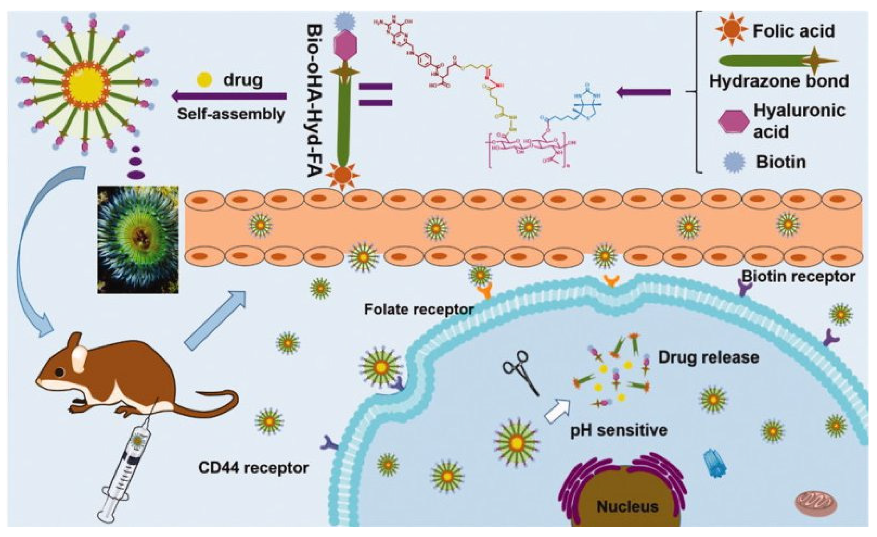
- Liu, M.; Wang, B.; Guo, C.; Hou, X.; Cheng, Z.; Chen, D. Novel multifunctional triple folic acid, biotin and CD44 targeting pH-sensitive nano-actiniaes for breast cancer combinational therapy. Drug Deliv. 2019, 26, 1002–1016. [Google Scholar] [CrossRef] [Green Version]
- Chrobak, E.; Bębenek, E.; Kadela-Tomanek, M.; Latocha, M.; Jelsch, C.; Wenger, E.; Boryczka, S. Betulin Phosphonates; Synthesis, Structure, and Cytotoxic Activity. Molecules 2016, 21, 1123. [Google Scholar] [CrossRef] [Green Version]
- Tomeh, M.A.; Hadianamrei, R.; Zhao, X. A Review of Curcumin and Its Derivatives as Anticancer Agents. Int. J. Mol. Sci. 2019, 20, 1033. [Google Scholar] [CrossRef] [Green Version]
- Wang, M.; Huang, M.; Wang, J.; Ye, M.; Deng, Y.; Li, H.; Qian, W.; Zhu, B.; Zhang, Y.; Gong, R. Facile One-Pot Synthesis of Self-Assembled Folate-Biotin-Pullulan Nanoparticles for Targeted Intracellular Anticancer Drug Delivery. J. Nanomater. 2016, 2016, 5752921. [Google Scholar] [CrossRef]


| DDS | Material | Drug | Tumour | Status | Ref. |
|---|---|---|---|---|---|
| Liposomes | FA-PEG-DSPE Cholesterol DPPC | 5-FU | Colon | In vitro—T-29 cells, Caco-2 human colorectal adenocarcinoma cell line, CT26 mouse colon carcinoma cell line, HeLa cells, MCF-7 cells | [67] |
| In vivo—mice | |||||
| Liposomes | HSPC/Chol/mPEG 2000-DSPE/folate-PEG-CHEMS | Dnr | Leukaemia | In vitro—L1210JF murine lymphocytic leukaemia cells In vivo—mice | [68] |
| Liposomes | FA-PEG2000 | Cis | Liver | In vitro—PLC/PRF/5 Alexander hepatoma cells | [69] |
| Phosphatidylcholine, Cholesterol | |||||
| Liposomes | FA-PEG-DSPE | Blm | Unspecified | In vitro—HeLa cells, MCF-7 cells | [70] |
| cholesterol | |||||
| Liposomes | FA-PEG-DSPE | Cur | Unspecified | In vitro—HeLa cells | [71] |
| Cholesterol | |||||
| SPC | In vivo—mice | ||||
| Liposomes | FA-PEG-DSPE | Iht | Solid | In vivo—mice | [72] |
| Dendrimers | AU-FA-PPI | Dox | In vitro | [73] | |
| Dendrimers | FA-PPI | Mtx | Breast | In vitro—MCF-7 cells | [74] |
| In vivo—rats | |||||
| Dendrimers | FA-PAMAM | Dox | Unspecified | In vitro—KB cells | [75] |
| Dendrimers | FA-PAMAM + | Gem | Unspecified | In vitro—A431 epidermoid carcinoma cell line | [76] |
| Magnetic mesoporous silica coated graphene oxide | |||||
| Micelles | FA-BSP-SA | Dox | Unspecified | In vitro—4T1 cells, HepG2 cells | [77] |
| In vivo–rats and mice | |||||
| Micelles | MPEG-PHIS | Ptx | Breast | In vitro—MCF-7 cells | [19] |
| FA-PEG-VE | |||||
| In vivo—mice | |||||
| Micelles | FA-PF127-CHOL | Dtx | Melanoma | In vitro—B16-F10 cells, HepG2 cells, L929 mouse fibroblasts cells | [78] |
| In vivo—mice | |||||
| Micelles | FA-PCL-b-P(HEMA-co-DMAEMA | Dox | Unspecified | In vitro—HeLa cells | [79] |
| Micelles | FA-MPEG-b-P(LA-co-DHP); | Ptx | oesophageal | In vitro—EC9706 human oesophageal cancer cell line, MCF-7 cells | [80] |
| MPEG-b-P(LA-co-MCC) | |||||
| In vivo—mice | |||||
| Micelles | PLA-Jeff-FA/PLA3000PEG2000 | Bet | Unspecified | In vitro–HeLa cells | [81] |
| Nanoparticles | FA–PEG–MNP | Idr | Breast | In vitro—MCF-7 cells | [63] |
| Nanoparticles | Fe3O4-DPA-PEG- | Mtx | Unspecified | In vitro—MCF-7 cells, A549 cells | [82] |
| Nanoparticles | PLGA-1,3-diaminopropane-FA | 5-FU | Colon | In vitro—HT-29 cells | [45] |
| Nanoparticles | FA-TCS | Dtx | Breast | In vitro—MDA-MBB-231 breast cancer cell line | [46] |
| Ex vivo—rats | |||||
| In vivo—rabbits | |||||
| Solid lipid nanoparticles | DSPE-FA | Iht | Colon | In vitro—HT-29 cells | [83] |
| Lipid-polymer hybrid nanoparticles | PGLA, PEG, SA, FA | Vcr | Lymphoma | In vitro–Raji human Burkitt’s lymphoma cell line, Raji/Vcr cells, A20 mouse reticulum sarcoma cells, HUVEC human umbilical vein endothelial cells | [84] |
| In vivo mice |
| Number | Study Title | Cancer | Drug | Phase | Status |
|---|---|---|---|---|---|
| NCT00308269 | Study of Vintafolide (MK-8109, EC145) for the Treatment of Recurrent or Refractory Solid Tumors (MK-8109-006, EC-FV-01) | Unspecified | EC145 | I | Completed in 2007 |
| NCT00852189 | Study of EC0489 for the Treatment of Refractory or Metastatic Tumors | Unspecified | EC0489, EC20 | I | Completed in 2012 |
| NCT00441870 | Study of EC0225 for the Treatment of Refractory or Metastatic Tumors | Unspecified | EC0225, EC20 | I | Completed in 2012 |
| NCT01689727 | Safety and Efficacy of FolateScan (Technetium Tc 99m EC20) in Subjects With Pituitary Tumors | Pituitary Tumors | EC20 | II | Completed in 2012 |
| NCT01689636 | Safety and Biodistribution of Technetium Tc 99m EC20 in Normal Volunteers and Ovarian Cancer Patients | Ovarian Cancer | EC20 | I | Completed in 2012 |
| Healthy Volunteers | |||||
| NCT01686256 | Safety and Efficacy of FolateScan (Technetium Tc 99m EC20) in Women With Suspected Ovarian or Endometrial Cancer | Ovarian Cancer | EC20 | I | Completed in 2012 |
| Endometrial Cancer | |||||
| NCT01689662 | Safety and Efficacy of FolateScan (Technetium Tc 99m EC20) in Subjects With Suspected Metastatic Renal Cell Carcinoma | Metastatic Renal Cell Carcinoma | EC20 | II | Completed in 2012 |
| NCT01689714 | Safety and Efficacy of Folatescan (Technetium TC 99M EC20) in Patients With Suspected Ovarian Carcinoma or Recurrent Endometrial Carcinoma | Ovarian Carcinoma | EC20 | II | Completed in 2012 |
| Recurrent Endometrial Carcinoma | |||||
| NCT00485563 | A Phase II Study of EC17 (Folate-hapten Conjugate) in Patients With Progressive Metastatic Renal Cell Carcinoma | Renal Cell Carcinoma | EC17 | II | Terminated in 2012 |
| Biological: EC90 (KLH-FITC) | |||||
| Biological: GPI-0100; Interleukin-2 Interferon-alpha | |||||
| NCT01002924 | Extension Study of EC145 (Vintafolide) for Subjects Enrolled in a Previous Study With EC145 (MK-8109-010) | Solid Tumors | EC145 | II | Completed in 2013 |
| NCT02049281 | A Study of Vintafolide (MK-8109) in Participants With Advanced Solid Tumor (MK-8109-011) | Solid Tumor | EC145 | I | Terminated in 2014 |
| NCT01953536 | Safety and Efficacy Study of Vintafolide and Vintafolide Plus Paclitaxel Compared to Paclitaxel Alone in Participants With Triple Negative Breast Cancer (TNBC) (MK-8109-004) | Breast Neoplasms | EC145; Paclitaxel 80 mg/m2; Etarfolatide; | II | Withdrawn in 2014 |
| Folic acid; Premedication for Paclitaxel | |||||
| NCT01577654 | Phase 2 Study of EC145 Alone Versus EC145+Docetaxel Versus Docetaxel Alone in Participants With FR(++) 2nd Line Non Small Cell Lung Cancer | Non Small Cell Lung Cancer | EC145, EC145 + Docetaxel; Docetaxel, EC20 | II | Completed in 2015 |
| NCT01170650 | Study for Women With Platinum Resistant Ovarian Cancer Evaluating EC145 in Combination With Doxil® | Ovarian Cancer | EC145; Pegylated Liposomal Doxorubicin (PLD/Doxil®/Caelyx®) | III | Suspended in 2015 |
| placebo; EC20 | |||||
| NCT00507741 | Study of Vintafolide (MK-8109, EC145) in Participants With Advanced Ovarian and Endometrial Cancers (MK-8109-007, EC-FV-02) | Ovarian Cancer | EC145, Etarfolatide | II | Completed in 2015 |
| Endometrial Cancer | |||||
| NCT00511485 | Study of Vintafolide (MK-8109, EC145) in Participants With Progressive Adenocarcinoma of the Lung (MK-8109-008, EC-FV-03) | Adenocarcinoma of the Lung | EC145, Etarfolatide | II | Completed in 2015 |
| NCT00722592 | Platinum Resistant Ovarian Cancer Evaluation of Doxil and Vintafolide (MK-8109, EC145) Combination Therapy (8109-009, EC-FV-04) | Ovarian Cancer | EC145; pegylated liposomal doxorubicin (PLD); EC20 | II | Completed in 2015 |
| NCT01688791 | A Study of MK-8109 (Vintafolide) Given Alone or With Chemotherapy in Participants With Advanced Cancers (MK-8109-001) | Advanced Cancer | EC145, Carboplatin, Paclitaxel | I | Terminated in 2015 |
| NCT01778920 | Pilot and Feasibility Study of the Imaging Potential of EC17: Intraoperative Folate-fluorescein Conjugate (EC17) Lung Cancer (CA) | Lung and Pleural Malignancies Neoplasms Nodules Adenocarcinoma | EC17 | I | Completed in 2016 |
| NCT02000778 | EC17 for Intraoperative Imaging in Occult Ovarian Cancer | Ovarian Cancer | EC17 | I | Completed in 2018 |
| NCT01994369 | Intraoperative Imagery of Breast Cancer With Folate-FITC (EC17) | Resectable Breast Cancer | EC17 | I | Completed in 2018 |
| NCT02629549 | Intraoperative Imaging of Pituitary Adenomas by OTL | Neoplasms Pituitary Neoplasms | OTL38 | I | Terminated in 2018 |
| NCT02317705 | Phase 2 Study of OTL38 for Intra-operative Imaging of Folate Receptor-alpha Positive Ovarian Cancer | Ovarian Cancer | OTL38 | II | Completed in 2019 |
| NCT01999738 | Folic Acid-Tubulysin Conjugate EC1456 In Patients With Advanced Solid Tumors | Solid Tumors | EC1456, EC20 | I | Completed in 2019 |
| Non Small Cell Lung Carcinoma | |||||
| NCT03011320 | An Exploratory Study of the Folic Acid-tubulysin Conjugate EC1456 in Ovarian Cancer Subjects Undergoing Surgery | Ovary Cancer | EC1456, Etarfolatide | I | Completed in 2019 |
| NCT03180307 | OTL38 for Intra-operative Imaging of Folate Receptor Positive Ovarian Cancer | Ovarian Cancer | OTL38 | III | Completed in 2020 |
| NCT02872701 | OTL38 Injection for Intraoperative Imaging of Folate Receptor Positive Lung Nodules | Lung Neoplasms | OTL38 | II | Completed in 2020 |
| Lung Cancer | |||||
| NCT02602119 | Intraoperative Imaging of Pulmonary Nodules by OTL38 | Neoplasms | OTL38 | I | Recruiting |
| NCT04241315 | ELUCIDATE: Enabling Lung Cancer Identification Using Folate Receptor Targeting | Lung Neoplasms | OTL38 | III | Recruiting |
| Lung Cancer |
| Number | Patent Title | Type of DDS |
|---|---|---|
| CA2487564A1 | Folic acid-chitosan-DNA nanoparticles | NPs |
| US2010040694A1 | Low-molecular weight, water-soluble chitosan nanoparticle for gene delivery with folic acid conjugated thereto as target ligand and preparation method therefor | NPs |
| CN102824306A | Folic acid modified chitosan coated plasmid nanoparticles and preparation method thereof | NPs |
| CN102961759A | Targeting gene transferring method of folic acid-functionalized PAMAM (polyamidoamine dendrimers) wrapped by gold nanoparticles | dendrimers |
| CN103223178A | Preparation method of folic acid modified multifunctional targeted contrast agent magnetic iron oxide/gold nanoparticles | NPs |
| CN103143041A | Preparation method of targeted MRI (magnetic resonance imaging) contrast medium based on folic acid modified iron oxide nanoparticles | NPs |
| CN103251595A | Technology for preparing folic acid-glucan-camptothecin composite nanoparticles through supercritical CO2 anti-solvent method | NPs |
| CN103083682A | Folic acid modified chitosan quaternary ammonium salt-taxol polymer medicine, as well as preparation method and application thereof | NPs |
| CN104087555A | Folic acid targeting magnetic color-developing nanoparticles and preparation method thereof | NPs |
| CN103933584A | Preparation method of folic acid-modified ultra-superparamagnetic iron oxide (USPIO) nanoparticles | NPs |
| CN103908978A | Folic acid-nano-TiO2 composite photocatalyst and its preparation method and use | NPs |
| CN103961705A | Preparation method and application of folic acid modified hollow copper sulfide/polydopamine compound | NPs |
| IN680DE2013A | Folic acid funcjonalized liquid crystalline nanoparticles for improved tumour delivery of anti-cancer agents | NPs |
| CN104436193A | Preparation method of folic acid coupled gold nano-rod/polypyrrole/ferroferric oxide multifunctional composite nano diagnosis and treatment agent | NPs |
| CN105396146A | Preparation method of folic-acid-modified gold nanoparticles | NPs |
| CN105381474A | Preparation method of folic acid modified ferriferrous oxide/gold star-shaped nanoparticles | NPs |
| CN105327368A | Method for preparing fluorescent silicon dioxide coated and folic acid marked gold nanoparticles | NPs |
| CN106110331A | Folic acid molecule targeted magnetic nano-drug and preparation method thereof | NPs |
| CN105920601A | Folic acid coupled targeted ferriferrous oxide/mesoporous silica/copper sulfide nano-composite particle as well as preparation method and application thereof | NPs |
| CN106511453A | Preparation method of nanoparticles modified by pecan kernel tannin folic acid | NPs |
| CN106267248A | Lipid ultrasound micro-bubble carrying folic acid modified mesoporous silicon dioxide nanoparticles and preparation method thereof | NPs |
| CN107970453A | Dual-targeting delivery method of pectin nanoparticles modified by folic acid | NPs |
| CN110652592A | Preparation method and application of folic acid-targeted dual-drug-loaded nanoparticles | NPs |
| CN108578427A | Folic acid modified gold nanoparticle, preparation method thereof and applications of folic acid modified gold nanoparticle in preparation of radiosensitization therapy medicines | NPs |
| CN108546682A | Mixed photonic crystal composite material based on folic acid modification and application | NPs |
| CN110123787A | Nanoparticles having paclitaxel coated with N-succinyl chitosan modified by folic acid and small molecular polypeptide and preparation method thereof | NPs |
| CN110501208A | Folic acid functionalized and streptavidin modified magnetic nanoparticles and preparation method and application thereof | NPs |
| CN111195239A | Preparation method of folic acid targeted silymarin solid lipid nanoparticles | NPs |
| CN111249254A | Preparation method and application of folic acid coupling albumin nanoparticles loaded with baicalin | NPs |
| CN111265482A | Glycyrrhetinic acid and/or folic acid ligand modified cantharidin solid lipid nanoparticle and preparation method thereof | NPs |
| US2020271655A1 | Folic acid functionalized copper sulfide nanoparticles for the detection of ovarian cancer cells in flow | NPs |
| CN111035624A | Folic acid modified ABT-737 loaded mesoporous silica nanoparticles and preparation method thereof | NPs |
| CN110812494A | Folic acid-modified and block copolymer-wrapped gold nanoparticles and preparation method and application thereof | NPs |
| DDS | Material | Drug | Tumour | Status | Ref. |
|---|---|---|---|---|---|
| Nanoparticles | PEG/PCL | Ptx | unspecified | In vitro—MCF-7 cells, HeLa 229 human uterine cervix adenocarcinoma cell line | [115] |
| Nanoparticles | Poly(D,L-lactide-co- | Dox | unspecified | In vitro—HepG2 cells, Heps murine hepatocarcinoma cell line | [120] |
| glycolide)-Lecithin- | |||||
| Polyethylene Glycol | In vivo—mice | ||||
| Nanoparticles | TPP–PEG–biotin | Dox | breast | In vitro—MCF-7 cells, HMEC normal human primary mammary epithelial cell line | [121] |
| meso- | |||||
| tetraphenylporphyrin (TPP)–PEG–biotin | |||||
| Nanoparticles | poly(ethylene glycol)-b- | Que and Dox | breast | In vitro—MCF-7 cells, MCF-7/ADR multidrug- resistant human breast cancer cell line In vivo—mice | [122] |
| poly(ε-caprolactone) | |||||
| Micelles | poly(N-2-hydroxypropyl methacrylamide)-block- | Ptx | unspecified | In vitro—A549 cells, HEC293 human embryonic kidney cell line | [123] |
| poly(N-2-benzoyloxypropyl methacrylamide) (p(HPMAm)-b-p(HPMAm-Bz)) | |||||
| Dendrimers | poly(amidoamine) (PAMAM) | Ptx | unspecified | In vitro—A549 cells, | [124] |
| Dendrimers | poly(amido)amine- diethylenetriamine | Gem | unspecified | In vitro–HeLa cells, | [125] |
| HaCaT cells | |||||
| Micelles | PEG-PCL | Art | breast and others | In vitro—MCF-7 cells, HFF2 normal human | [126] |
| foreskin fibroblast cell line, 4T1 cells | |||||
| In vivo—mice | |||||
| Nanoparticles | biotin-PEG-PCL | Gnb and Nar | lung | In vitro–A549 cells | [127] |
| In vivo—rats | |||||
| Nanostructured lipid carriers | polyethylene glycol 2000- distearyl phosphatidyl ethanolamine | Ds | breast | In vitro—4T1 cells, L1210 murine lymphocytic leukaemia cell line | [128] |
| In vivo—mice | |||||
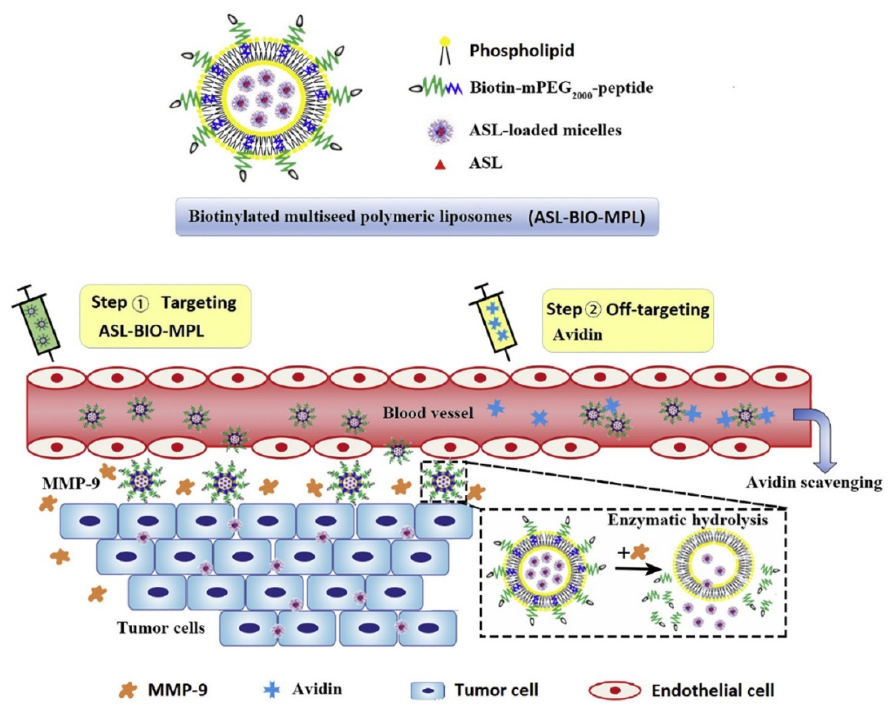
| ‘Multi-seed’ polymeric liposomes | Biotin-mPEG2000- | Asl | unspecified | In vitro—4T1 cells | [129] |
| polypeptide | In vivo—mice | ||||
| Nanoparticles | chitosan | Plasmid DNA | liver | In vitro—SMMC- | [130] |
| 7721 human hepatocellular carcinoma cell line, LO2 normal human hepatic cell line, SW480 human | |||||
| colon cancer cell line, H22 cells | |||||
| In vivo—mice | |||||
| Nanotubes | Carbon nanotubes modified with | Dox | unspecified | In vitro–HeLa cells, HepG2 cells, CHO Chinese hamster ovary cell line, HEK-293 | [131] |
| human embryonic kidney cell line | |||||
| Nanocarriers | poly-(ethylene glycol) bis-(amine) | GA | lung and others | In vitro—A549 cells | [132] |
| (BPBA)-coated reduced graphene oxide | |||||
| Nanoparticles | Biotin Decorated Gold Nanoparticles stabilized by amine terminated lipoic acid-polyethylene glycol (PEG) | copper(II) complex | unspecified | In vitro—HeLa cells, | [133] |
| HaCaT cells | |||||
| In vivo—mice |
| DDS | Ligand | Material | Drug | Tumour | Status | Ref. |
|---|---|---|---|---|---|---|
| Micelles | FA + HA | octadecyl | Ptx | breast | In vitro—MCF-7 and MCF-7/ADR cells | [155] |
| In vivo—rats | ||||||
| Liposomes | FA + HA | polyethylenimine | Ptx + DNA | melanoma; hepatoma | In vitro—murine melanoma cell line B16 and HepG2 cells | [156] |
| Liposomes | FA + Tf | DSPE-PEG2000 | Dox | glioblastoma | In vitro—C6 glioma cells and bEnd3 cells | [157] |
| In vivo—rats | ||||||
| Liposomes | FA + mAb225 | DSPE-PEG3350 | Dox | unspecified | In vitro—KB cells | [158] |
| Liposomes | FA + TAT peptide | DSPE-PEG2000 | Dox | unspecified | In vitro—KB cells | [159] |
| In vivo—mice | ||||||
| Liposomes | FA + glutamic hexapeptide | SPC + Cholesterol | Ptx | bone | In vitro—MDA-MB-231 cells and MCF10A cells | [160] |
| In vivo—rats; mice | ||||||
| Gold Nanocomposites | FA + AS1411 | AuNPs | Dox | unspecified | In vitro—Hela cells | [161] |
| Micelles | FA + glucose | pluronic P105 | Dox | glioma | In vivo—rats | [162] |
| Nanoparticles | FA + galactose | polyethylene glycol-dihydroartemisinin/hydroxycamptothecin | DHA | liver | In vitro—H22 cells and HepG2 cells | [163] |
| In vivo—mice | ||||||
| Gold Nanoclusters | FA + trastuzumab | AuNCs | - | breast | In vitro—SK-BR3 human breast cancer and normal mouse fibroblast (L929) | [164] |
| DDS | Ligand | Material | Drug | Tumour | Status | Ref. |
|---|---|---|---|---|---|---|
| Liposomes | Bio + Glu | modified cholesterol | Ptx | breast | In vitro—4T1 cells and MCF-7 cells | [85] |
| In vivo—mice | ||||||
| Dendrimers | Bio + HMGB1 | PAMAM | DNA | unspecified | In vitro—HeLa cells | [170] |
| Nanoparticles | Bio + Gal | chitosan | 5-FU | liver | In vitro—SMMC-7721 cells and LO2 cells | [171] |
| In vivo—mice |
| Name of Cell Line | Origin | Folate Receptor * | Biotin Receptor * | Ref. |
|---|---|---|---|---|
| MCF7 | human breast cancer | + | + (+++) | [114,174,175,176,177,178,179,180] |
| RasV12 | human breast cancer | no data | + (++) | [114,181] |
| BT-20 | human breast cancer | no data | + (++) | [114,178] |
| LCC6-WT | human breast cancer | no data | + (++) | [114,178] |
| LCC6-MDR | human breast cancer | no data | + (+) | [114,178] |
| MDA-MB 231 | human breast cancer | + | + (++) | [114,176,177,178,181] |
| SkBr3 | human breast cancer | + | + (++) | [114,178,179,180] |
| MMT06056 | human breast cancer | no data | + (+++) | [114,182] |
| T47D | human breast cancer | + | [179] | |
| 4T1 | mouse mammary carcinoma | + | + (+++) | [182,183] |
| JC | mouse mammary carcinoma | + | + (+++) | [114,119] |
| KB | human papilloma | + (+++) | + | [184,185,186] |
| HeLa | human cervical cancer | + (+++) | + (+++) | [114,154,175,177,186] |
| KB-V1 | human cervical cancer | + (+++) | no data | [186] |
| OVCAR-3 | human ovarian cancer | + (+++) | + (++) | [114,119,187] |
| OV2008 | human ovarian cancer | + (+++) | + (++) | [182] |
| IGROV-1 | human ovarian cancer | + (+++) | no data | [186] |
| SKOV-3 | human ovarian cancer | + (+++) | no data | [179,180,186,188] |
| SKOV-3.ip | human ovarian cancer | + (+++) | no data | [186] |
| A1847 | human ovarian cancer | + | no data | [179] |
| ID8 | mouse ovarian cancer | + (+++) | + (++) | [178,182] |
| PC-3 | human prostate cancer | + (+++) | + | [176,186] |
| Du145 | human prostate cancer | +/− | + | [177,189] |
| PC3 | human prostate cancer | no data | + | [177] |
| A549 | human lung carcinoma | + | + (+++) | [114,177,190,191] |
| M109 | mouse lung carcinoma | + | + (+++) | [182] |
| Colo-26 | mouse colorectal | +/− | + (+++) | [114,182] |
| adenocarcinoma | ||||
| HepG2 | human hepatic carcinoma | +/− | + (+++) | [114,177,180] |
| Huh7 | human liver cancer | no data | + | [177] |
| Hep3B | human liver cancer | − | + | [177] |
| NCI-N87 | human gastric cancer | no data | + | [94,177] |
| AGS | human gastric cancer | no data | + | [177] |
| Panc-1 | human pancreatic cancer | − | + | [177,182,192] |
| RENCA | mouse renal | + (+) | + (+++) | [182] |
| adenocarcinoma | ||||
| RD0995 | mouse renal cancer | + (+) | + (+++) | [182] |
| P815 | mouse mastocytoma | +/− | + (+++) | [182] |
| BW5147 | mouse lymphoma | +/− | +/− | [168] |
| L1210FR | mouse leukaemia | + | + | [173,178] |
| L1219FR | mouse lymphocytic | ++ | + (+++) | [114,160,178,182,193] |
| leukaemia |
| Name of Cell Line | Origin | Folate Receptor | Biotin Receptor | Ref. |
|---|---|---|---|---|
| WI38 | human normal lung fibroblasts | - | - | [114,178,190,193,194,195] |
| LL-2 | mouse Lewis lung carcinoma | - | - | [114,182,196] |
| HEK-293 | human embryonic kidney | no data | - | [114,131,185] |
| NIH3T3 | mouse embryonic fibroblast | no data | - | [114,194] |
| HCT-116 | human Colon cancer | - | - | [114,182,196] |
| L1210 | mouse lymphocytic | - | - | [114,178,193] |
| leukaemia | ||||
| BW5147 | mouse lymphoma T-cell | no data | - | [114,182,196] |
| B16-F10 | mouse melanoma | - | - | [114,195] |
| B16 | mouse melanoma | - | - | [114,182,196] |
| DDS | Ligand | Material | Drug | Tumour | Status | Ref. |
|---|---|---|---|---|---|---|
| Nanoparticles | FA + BIO | Silica-coated Fe3O4 | Dox | unspecified | In vitro—A549 (human epithelial lung carcinoma) and BEAS-2B (immortalized human lung epithelial) cell lines | [197] |
| Nanoparticles | FA + BIO | Fe3O4 | - | unspecified | In vitro—E-G7 and human HeLa cells | [198] |
| Micelles | FA + BIO | PLA-PEG | Ptx | unspecified | In vitro—OVCAR3 (ovarian cancer cells) | [199] |
| Nanomicelles | FA + BIO CD44 | oHA | Ica + Cur | breast | In vitro—MCF-7 cells and BCSCs cells | [200] |
| In vivo—mice | ||||||
| Nanorods | FA + BIO | PELA or Styrene-maleic anhydride copolymer | Dox | unspecified | In vitro—4T1 cells and RAW 246.7 macrophages | [28] |
| In vivo—mice |
Publisher’s Note: MDPI stays neutral with regard to jurisdictional claims in published maps and institutional affiliations. |
© 2021 by the authors. Licensee MDPI, Basel, Switzerland. This article is an open access article distributed under the terms and conditions of the Creative Commons Attribution (CC BY) license (http://creativecommons.org/licenses/by/4.0/).
Share and Cite
Jurczyk, M.; Jelonek, K.; Musiał-Kulik, M.; Beberok, A.; Wrześniok, D.; Kasperczyk, J. Single- versus Dual-Targeted Nanoparticles with Folic Acid and Biotin for Anticancer Drug Delivery. Pharmaceutics 2021, 13, 326. https://doi.org/10.3390/pharmaceutics13030326
Jurczyk M, Jelonek K, Musiał-Kulik M, Beberok A, Wrześniok D, Kasperczyk J. Single- versus Dual-Targeted Nanoparticles with Folic Acid and Biotin for Anticancer Drug Delivery. Pharmaceutics. 2021; 13(3):326. https://doi.org/10.3390/pharmaceutics13030326
Chicago/Turabian StyleJurczyk, Magdalena, Katarzyna Jelonek, Monika Musiał-Kulik, Artur Beberok, Dorota Wrześniok, and Janusz Kasperczyk. 2021. "Single- versus Dual-Targeted Nanoparticles with Folic Acid and Biotin for Anticancer Drug Delivery" Pharmaceutics 13, no. 3: 326. https://doi.org/10.3390/pharmaceutics13030326