Review

Strategies for Vaccination: Conventional Vaccine Approaches Versus New-Generation Strategies in Combination with Adjuvants

by
Abdellatif Bouazzaoui
1,2,*,
Ahmed A. H. Abdellatif
3,4,
Faisal A. Al-Allaf
1,2,5,
Neda M. Bogari
1,
Saied Al-Dehlawi
6 and
Sameer H. Qari
7
1
Department of Medical Genetics, Faculty of Medicine, Umm Al-Qura University, P.O. Box 715, Makkah 21955, Saudi Arabia
2
Science and Technology Unit, Umm Al Qura University, P.O. Box 715, Makkah 21955, Saudi Arabia
3
Department of Pharmaceutics, College of Pharmacy, Qassim University, Qassim 51452, Saudi Arabia
4
Department of Pharmaceutics and Industrial Pharmacy, Faculty of Pharmacy, Al-Azhar University, Assiut 71524, Egypt
5
Department of Laboratory and Blood Bank, Molecular Diagnostics Unit, King Abdullah Medical City, Makkah 21955, Saudi Arabia
6
Regional Laboratory, Makkah 25321, Saudi Arabia
7
Biology Department, Aljumum University College, Umm Al-Qura University, Makkah 21955, Saudi Arabia
*
Author to whom correspondence should be addressed.
Pharmaceutics 2021, 13(2), 140; https://doi.org/10.3390/pharmaceutics13020140
Submission received: 27 November 2020 / Revised: 12 January 2021 / Accepted: 19 January 2021 / Published: 22 January 2021
(This article belongs to the Special Issue Discovery and Evaluation of Novel Adjuvants for Vaccine Formulations)

Abstract

:
The current COVID-19 pandemic, caused by severe acute respiratory syndrome-coronavirus 2 (SARS-CoV-2), has raised significant economic, social, and psychological concerns. The rapid spread of the virus, coupled with the absence of vaccines and antiviral treatments for SARS-CoV-2, has galvanized a major global endeavor to develop effective vaccines. Within a matter of just a few months of the initial outbreak, research teams worldwide, adopting a range of different strategies, embarked on a quest to develop effective vaccine that could be effectively used to suppress this virulent pathogen. In this review, we describe conventional approaches to vaccine development, including strategies employing proteins, peptides, and attenuated or inactivated pathogens in combination with adjuvants (including genetic adjuvants). We also present details of the novel strategies that were adopted by different research groups to successfully transfer recombinantly expressed antigens while using viral vectors (adenoviral and retroviral) and non-viral delivery systems, and how recently developed methods have been applied in order to produce vaccines that are based on mRNA, self-amplifying RNA (saRNA), and trans-amplifying RNA (taRNA). Moreover, we discuss the methods that are being used to enhance mRNA stability and protein production, the advantages and disadvantages of different methods, and the challenges that are encountered during the development of effective vaccines.

1. Introduction

The outbreak of the COVID-19 pandemic [1,2,3,4], which was caused by severe acute respiratory syndrome-coronavirus 2 (SARS-CoV-2), has triggered a global race to develop effective vaccines. Approximately 150 different research groups are currently involved, and more than 100 clinical trials have been initiated since the outbreak was first reported [5]. They all have the singular goal of developing and producing an antiviral vaccine that is effective in individuals of all age groups with all conditions, and, thereby, control the course of the pandemic. Nevertheless, the development of a vaccine is a laborious process, the mass production, distribution, and administration of which present extraordinary challenges, particularly in developing countries. Accordingly, the strategy that has been employed for vaccine production needs not only to take into consideration the effect of the vaccine on the immune system and its efficacy against the virus, but also the procedures for mass production, distribution, storage, and mass vaccination [5]. To this end, the participating research groups are employing a diverse range of different formulations, techniques, and strategies to produce effective vaccines against SARS-CoV-2. In this regard, there are four main methods of vaccine development, namely, employing pathogens (inactivated or with low virulence) for the production of vaccines; recombinant protein vaccines; vector-based vaccines that include DNA vectors or viral vectors; and, the latest technology using RNA molecules for vaccination. Among these, the more innovatory next-generation vaccines only use a part of the virus protein structure and, thus, can be expected to have a superior safety profile. However, these novel vaccines tend to have low immunogenicity and they often fail to induce a sufficient immune response. Consequently, we also describe the use different adjuvants, which can be employed in order to enhance immunogenicity and establish an enduring immune memory.

2. Traditional Vaccines

Historically, the first vaccines were based on pathogens with reduced virulence. Among the pioneers in this field, scientists, such as Plett and Jenner, used the cowpox or horsepox [6,7,8] virus in order to generate vaccines against smallpox. These types of vaccine with relatively low virulence have several advantages, notably only causing mild infection with symptoms that are similar to those of the target pathogen, and the body subsequently develops a strong immune response, with immunity potentially persisting for years. However, such traditional vaccines have one particularly vital drawback, namely, a high infection risk due to the potential for pathogens with low virulence to become more virulent [9]. The second method of traditional vaccination involves the administration of inactivated vaccines, which are safer than the first type. However, the use of such vaccines necessitates multiple injections in order to achieve strong and long-lasting immunity. Although live attenuated or inactivated vaccines can be more readily and rapidly developed than other vaccine types [10], these traditional vaccines tend to have a poor safety record, and a defect in the production process can potentially be a source of disease outbreaks. Indeed, such an incident occurred in the year 1955, when the administration of a defective polio vaccine caused 10 deaths, paralysis in 200 recipients, and a total of 40,000 cases of polio infection [11]. Accordingly, the development of alternative vaccines with better safety profiles is a priority.

3. Next-Generation Vaccines

In 1990, Wolf et al. demonstrated that mice injected with plasmids harboring a cloned protein subsequently showed an expression of the transgenic protein cloned in the plasmid DNA [12]. These observations provided an impetus for the development of a new strategy of vaccination, and marked the advent of an era of next-generation vaccines. The initial strategy adopted for these novel vaccines was a DNA-based technique, which was subsequently followed by the development of viral vectors, including adeno-associated virus (AAV), lentiviral, or adenoviral vectors for vaccination, and, more recently, by RNA-based vaccines. The salient point of this research is that it demonstrates that only a portion of the viral protein structure is sufficient for promoting immunity against a given pathogen. Consequently, these innovatory vaccines tend to only include a specific viral antigen, instead of employing the entire pathogen, thereby resulting in a better safety profile [13]. However, the design of such vaccines requires a more in-depth understanding of viral structures and the interaction between viral proteins and host cell receptors, and, accordingly, these next-generation vaccines tend to require a lengthy phase of preliminary studies before development can commence.

3.1. Recombinant Protein Vaccines

Recombinant protein vaccines are based on the use of recombinant viral structural proteins to induce an immune response. In this respect, the SARS-CoV-2 genome comprises four structural proteins, namely, membrane, envelope, nucleocapsid, and spike proteins. Among these, the spike protein is of particular importance, given that it interacts with angiotensin-converting enzyme 2 (ACE2) receptors that are localized on the surface of host cells, thereby facilitating endocytosis [14]. Consequently, most vaccination strategies for the SARS-CoV-2 virus have focused on this protein, owing to its importance in the virus lifecycle. However, vaccination with whole spike protein has been shown to promote liver damage in treated animals [15] and, thus, the use of only a part of this protein, such as the receptor-binding domain (RBD), which interacts with the ACE2 receptor protein, is considered to be the best alternative with respect to producing a safer vaccine [16]. Initial research in this regard suggests that immunization with recombinant protein or only the RBD results in the production of neutralizing antibodies [16,17,18]. Observations indicated that the protein is processed by dendritic cells, followed by the presentation of the antigen to naïve B and T cells, resulting in their activation and subsequent immunity development. However, the use of this strategy for immunization has a notably important drawback, namely, that, owing to the use of only a small part of the protein for immunization, specific immune reactions induced by the vaccine confer only partial protection [18,19,20]. Moreover, these immune reactions tend not to be particularly strong [21]. Consequently, vaccination with recombinant proteins necessitates the use of substrates, referred to as adjuvants, to boost the immune response. The use of such adjuvants enhances antigen presentation in antigen-presenting cells (APCs), thereby enhancing vaccine efficacy and resulting in long-term protection.

3.2. Plasmid DNA Vaccines

Wolff et al. demonstrated that intramuscular injection of nucleic acids resulted in the in vivo expression of a protein encoded by plasmid DNA [12], and it was later shown that vaccination with plasmid DNA can induce a strong immune response, as mentioned previously [22,23,24]. Collectively, the findings of these studies have provided evidence of the potential of plasmid DNA to produce immunization on injection. Subsequently, researchers began to examine the utility of DNA vaccines for the treatment of cancer, infections, and autoimmune diseases, including allergies [25]. However, the early-stage clinical studies in humans tended to be unsuccessful, owing to the poor transfection efficacy and low immunogenicity. Nevertheless, DNA vaccines do offer certain advantages [25]. First, the use of plasmid DNA for vaccination is safer than certain traditional vaccines, in that it avoids the administration of a live virus. Second, plasmid DNAs tend to be more stable than proteins, viruses, or mRNAs, and they can be freeze-dried and maintained in long-term storage. Third, the production of these vaccines is more straightforward and cost-effective. In recent years, improved transfection methods, such as electroporation based on the use of electric pulses to perforate the cell membrane, have been developed in order to enhance plasmid transfer into cells. The use of adjuvants to boost the immune reaction has been further advance in the development of DNA vaccines, which has increased the suitability of DNA vaccines as an ideal type of vaccine for mass administration. In this context, the company Inovio performed one of the earliest vaccination studies targeting the MERS coronavirus in order to develop a new DNA vaccine for COVID-19 [26]. Immunization with the synthetic DNA-based vaccine (INO-4800) targeting the SARS-CoV-2 spike protein resulted in the strong expression of this protein, and it promoted antigen-specific T cell responses and the production of antibodies, which were able to bind to ACE receptors and neutralize SARS-CoV-2 infection [26]. Previously, Inovio had also developed similar DNA vaccines against the Ebola [27], SARS [28], MERS [29,30], and Zika [31] viruses. Other previous studies have similarly used DNA-based vaccines to generate immunity against Toxoplasma gondii in mice [24], and also to produce a T-cell-dependent antibody response to glutamic acid decarboxylase [23].

3.3. Viral Vector Vaccines

Although the use viral vectors for therapeutic purposes commenced in the late 1990s, the application of these vectors for disease treatment was primarily overshadowed by the death of Jesse Gelsinger, who was administered an adenoviral vector [32], as well as the development of leukemia in children with severe combined immunodeficiency (SCID) treated with retroviral vectors [33,34]. However, in recent years, significant progress in the development of viral vector vaccines has yielded encouraging results with respect to dendritic cells, and an increasing number of studies have begun to focus on the use of different viral vectors, including RNA (retroviral and lentiviral), adenoviral, and Adeno-associated virus (AAV) vectors [35,36,37,38]. Immunization based on viral vector vaccines entails cloning the immunogenicity-causing antigen in a pseudovirus, which lacks the ability to propagate and transfer in dendritic cells, thereby producing stronger immune stimulation than recombinant proteins [39].

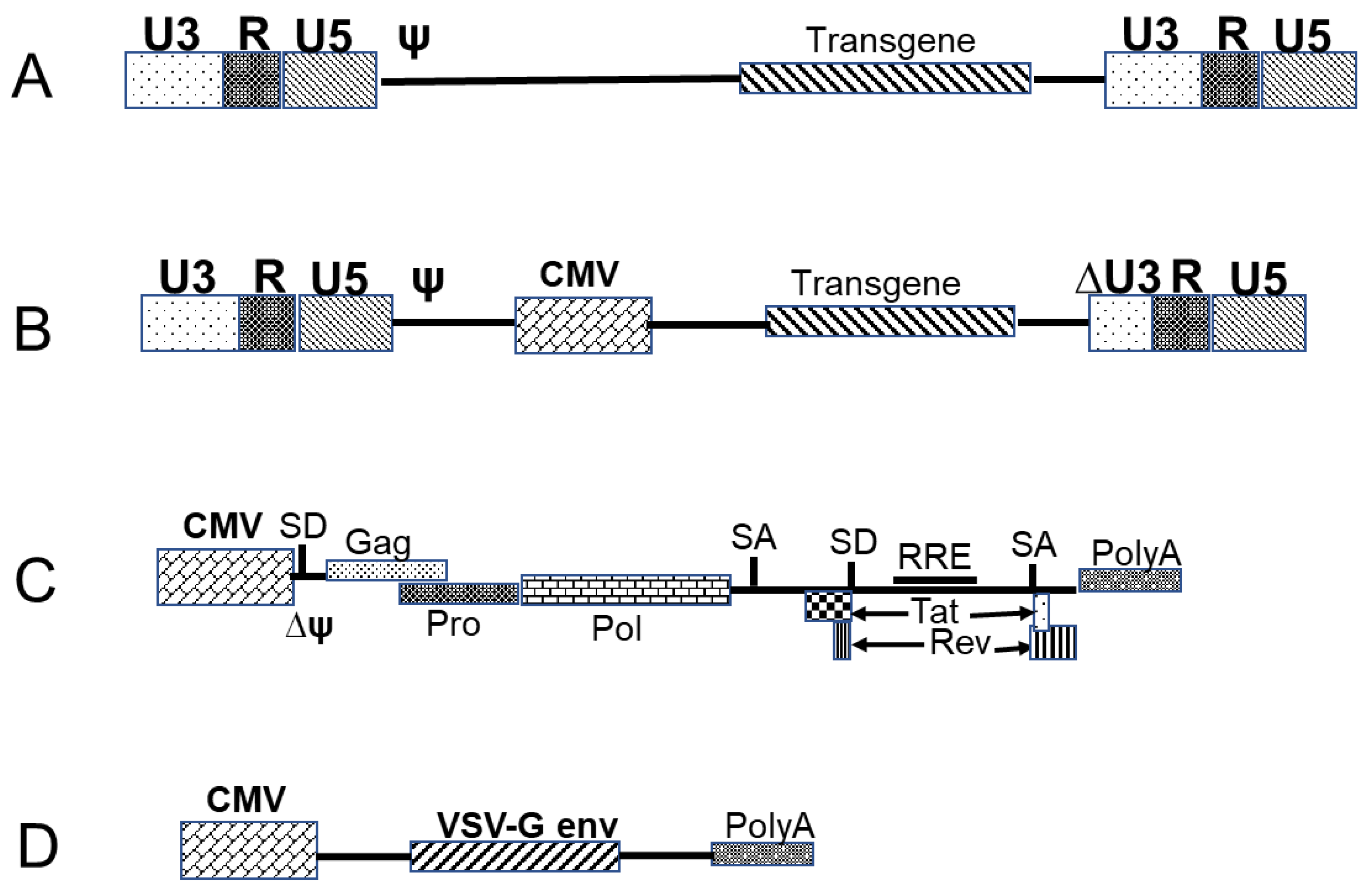
3.3.1. Retrovirus- and Lentivirus-Based Vectors

Retroviruses have a single-stranded RNA genome that encodes all of the proteins that are required for replication, including structural proteins [40]. Of the studied retroviruses, Moloney murine leukemia virus (MMLV)-based vectors are amongst the most efficient engineered vectors with a high transduction efficiency in dividing cells, and they are characterized by good integration and high expression of transgenes [41]. Retroviral expression vector can carry genes of interest of up to 8 kb in size and a diverse range of envelope proteins can be used for packaging. These envelope proteins can be modified to recognize receptors that are only found on mouse and rat cells (ecotropic), or be amphoteric, thereby facilitating the targeting of a broad range of receptors on mammalian cells. Moreover, retroviral vectors can be pseudotyped with envelope proteins that are derived from other virus strains, such as vesicular stomatitis virus G-protein (VSV-G), which exhibits broad-spectrum tropism and facilitates the infection of non-mammalian cells [42]. Therapy using retroviral vectors has been demonstrated to be an efficient treatment for different disorders, including X-linked SCID (SCID-X1), chronic granulomatous disease (CGD), and adenosine deaminase-deficient SCID [43,44,45,46]. However, critical complications, such as leukemias in patients with SCID-X1 and myelodysplastic-like syndromes in patients with CGD [47], highlighted the limitations of the first generation of retroviral vectors. Subsequently, modified vectors with modest architectural changes and a more favorable profile than the first-generation retroviral vectors were developed [48,49,50,51,52,53]. These vectors are characterized by the deletion of the long terminal repeat (LTR) promoter region, which results in the self-inactivation (SIN) of the vector and good biosafety (Figure 1). A further type of retrovirus, lentiviruses, can infect non-dividing cells, such as dendritic cells and macrophages [54,55]. The packaging capacity of lentiviral vectors is similar to that of murine leukemia virus vectors, and they can be pseudotyped with different envelopes, including VSV-G [56]. Furthermore, lentiviral-based vectors exhibit strong and prolonged expression, owing to random chromosomal integration. However, this genomic integration has been associated with the development of leukemia in patients with SCID [33,34], which initially represented a considerable drawback for the use of retroviral vectors. Nevertheless, this disadvantage has recently been overcome by the development of new vectors that are characterized by targeted integration [57]. The development of non-integrating lentiviral vector (NILVs) [58], which have the capacity to express transgenes transiently in dividing cells or episomes in non-dividing cells and can also be introduced into different cell types, has been a further interesting advancement in the use of lentiviral vectors. In this regard, the Shenzhen Geno-immune Medical Institute has recently developed two lentiviral vector-based vaccines (Covid-19/aAPC and LV-SMENP-DC) for the treatment of COVID-19 infections [5]. These vectors have been constructed in order to harbor multiple viral genes as antigens, including conserved, structural, and protease protein domains, and the vaccines are currently undergoing phase 1 (Covid-19/aAPC) and phase 2 (LV-SMENP-DC) trials.

3.3.2. Adenovirus-Based Vectors

Adenoviruses, which belong to the family Adenoviridae, are non-enveloped, double-stranded DNA viruses, approximately 90 nm in diameter. First discovered in 1953, they are known to infect humans and a range bird, reptile, fish, amphibian, and non-human primate species [59,60,61]. In humans, more than 100 types of adenovirus have been identified, some of which are implicated in respiratory infections, conjunctivitis, or gastroenteritis [59,62]. The linear double-stranded DNA of the virus measures between 25 and 48 kb and it includes non-coding inverted terminal repeats (ITRs) at both ends and genes encoding approximately 35 proteins (Figure 2A) that are expressed in two different phases, i.e., early genes, including E1A, E1B, and E2–E4, and five late genes (L1–L5). The early genes play important roles in gene regulation in the host cell and in the initiation of virus replication, whereas the late genes encode structural proteins that are essential for capsid assembly [62]. Of the early genes, the E3 gene is not essential for adenovirus replication, whereas, E1, E2, and E4, are necessary. In the first-generation adenovirus-based vectors, which had a packaging capacity of approximately 8 kb, the E1 and E3 genes were deleted (Figure 2B), whereas, in the second-generation vectors, with 14-kb packaging capacity, the E2 and E4 genes were deleted (Figure 2C), and, in the case of high-capacity adenovirus-based vectors, all of the genes were deleted, leaving only the cis-acting sequences necessary for viral DNA replication and packaging, which enabled the packaging of transgenes of up to 35 kb in size. The genes that are required for replication can be stably expressed in packaging cells or cloned into helper plasmids and co-transfected with the transgene plasmid [63]. In previous studies, the most commonly used adenoviral vector has been based on human adenovirus serotype 5 (AdHu5), which has been found to be efficient in inducing immune responses in preclinical and clinical studies, as well as in gene therapy applications [64,65,66,67]. Recently, an adenovirus-based vector has been used in order to develop a new COVID-19 vaccine [5]. The use of adenovirus-based vectors has several advantages, including strong transgene expression and immune responses via an induction of innate immunity, allowing for large-scale production and purification, and ensuring safe human application. Furthermore, they can be delivered via mucosal or systemic routes [68,69,70]. Collectively, these advantages have contributed to establishing adenovirus-based vectors as among the most successful strategies in medical research.

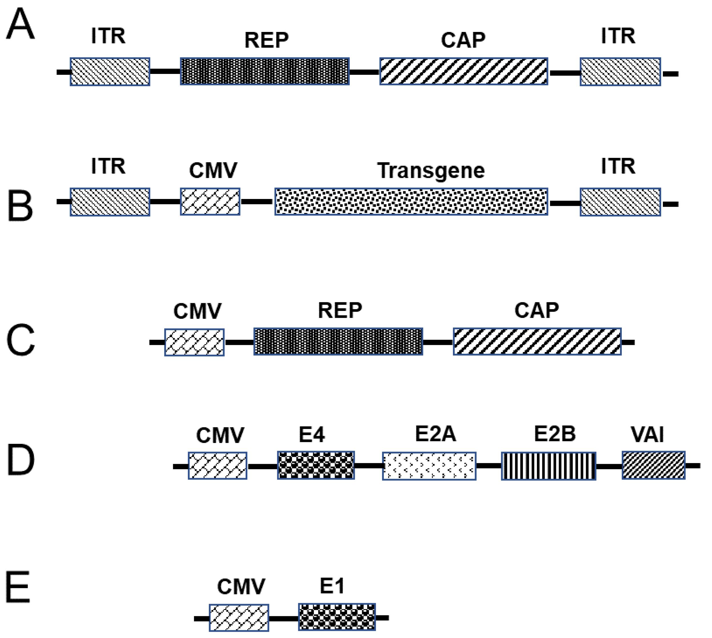
3.3.3. Adeno-Associated Virus Vectors as a Platform for Vaccination

Adeno-associated viruses (AAVs) are small non-enveloped viruses that belong to the genus Dependovirus within the family Parvoviridae [71,72,73]. The first AAV was discovered in 1965 [72] and, during the subsequent 20 years of research, several important aspects of AAV were characterized, including the genome structure [74,75,76,77], infection latency [78,79,80,81], replication/transcription [82,83,84], virion assembly [85], genetic characteristics [86,87], and sequence of the entire genome [88]. The virion of the AAV is a single-stranded particle of approximately 4.7 kb in length that can either be a sense or anti-sense strand [89] encapsulated within a 25-nm capsid [71]. The AAV genome includes an ITR at either end, which serve as origins of replication and packaging signals. With respect to replication, the genome contains a single replication (rep) gene encoding four proteins (Rep4, Rep52, Rep68, and Rep78), a capsid (cap) gene encoding three subunits via differential splicing and translation variants, and an assembly activating protein (AAP) that is localized within the capsid sequence that promotes virion assembly [90,91]. In response to an interaction between the capsid and a cell receptor, the AAV particles undergo a series of pH-dependent structural modifications within endosomes [92] and subsequently enter the nucleus by way of interacting with the nuclear pore complex following endosomal escape [93,94,95], during which the single-stranded DNA genome is released. A second strand is synthesized from the self-primed ITR at the 3′-end [96,97], followed by strand annealing via base pairing. The double-stranded genome thus generated undergoes circularization via intra- or intermolecular genome recombination at the ITRs [98], which stabilizes the recombinant AAV (rAAV) genome as episomal DNA, leading to persistent gene expression in post-mitotic cells. Although AAVs lack the capacity to autonomously replicate, they can infect both non-dividing and dividing cells in different hosts, including humans and non-human primates [71]. However, in order to reproduce in cells, these viruses require the mediation of a helper virus, such as herpes simplex virus (HSVs) or an adenovirus [99]. Although AAV infection is common in humans, these viruses are not known to cause any disease [100,101,102], and, consequently, AAVs are considered ideal vectors for gene transfer and vaccination [103,104]. Nevertheless, the packaging capacity of these viruses is limited and, thus, to maximize loading, the entire genome, with the exception of the ITRs, must be removed (Figure 3), leading to low cytotoxicity and immunogenicity when AAVs are delivered in vivo. Since its introduction in the 1980s, rAAVs have become the gold standard for gene transfer [105]. In terms of packaging, the transgene is cloned into a plasmid between the ITRs and transfected into cells along with helper plasmids (Figure 3), namely, one for the rep and cap genes and another, including adenovirus helper genes [106,107]. This strategy facilitates relatively straightforward packaging of the gene of interest [108,109]. Different studies have confirmed the efficacy and safety of rAAVs with respect to delivering genes in target cells during several preclinical and clinical trials for the treatment of genetic diseases, including hemophilia, spinal muscular atrophy, inherited retinal disease, and lipoprotein lipase deficiency [110,111,112], and they have been licensed for treatment [113,114]. Given the therapeutic promise of these vectors, recent studies have focused on developing an AAV-type COVID-19 vaccine [5].

3.4. RNA-Based Vaccines and Nanoparticle (NP) Formulations

RNA-based vaccines are the most recent development in the quest to produce safe and efficacious means of vaccination. One of the major factors that has hitherto prohibited the use of RNA for vaccination is its low stability. Furthermore, RNA only enables transient expression and it is negatively charged and, consequently, the use of additional substrates is necessary for facilitating the entry of RNA into cells. However, recently, different strategies have been developed to enhance mRNA stability and the delivery of RNA into cells, which have contributed to making RNA-based strategies among the most efficient methods of vaccination.

3.4.1. RNA-Based Vaccines

mRNA-based vaccines have already been used in the treatment of different diseases [115,116,117]. However, being negatively charged, the efficiency of mRNA transfection tends to be very low, thus necessitating the use of other substrates in order to facilitate the delivery of mRNA into cells, among which lipid nanoparticles (LNPs) are some of the most widely used transport vehicles [118,119]. Although the mechanisms whereby mRNAs are delivered by LNPs are incompletely understood, it has been established that mRNA-LNP complexes are taken up by endocytosis after interaction with the cell membrane [120], and are thereafter routed to the endosomes. As a consequence of a change in pH, the residual amines of the LNPs subsequently disrupt the endosome membrane, leading to the endosomal escape of mRNA into the cytoplasm, which, in turn, enables the transient expression of mRNA in order to produce a particular protein [121]. Given that entry into the cell nucleus is not a prerequisite for mRNA vaccine antigen expression, the expression is only transient and, accordingly, the risk of integration into the host DNA is negligible, which is one of the salient advantages of these vaccines. However, this transient expression of mRNA is typically low, due to rapid degradation by cytoplasmically localized endonucleases. In an effort overcome these limitations, several recent studies have investigated strategies to increase mRNA stability, thereby facilitating high protein production. These strategies include the use of nanoparticle (NP) delivery methods [122] and modified nucleosides [123], which have been reported to confer enhanced mRNA stability and improved bioavailability for the production of larger quantities of antigens. Interestingly, in this regard, Beissert et al. recently identified a new generation of RNA molecule with vaccine potential, referred to as self-amplifying RNA (saRNA) [124]. saRNAs are based on the genome of alphaviruses, in which the RNA replication genes remain intact, whereas the structural genes are deleted. The delivery of saRNAs can be achieved while using plasmid DNA, the transcription of which is dependent on prior entry into the nucleus. In addition, the RNA can be transcribed in vitro and transported to the cytoplasm while using viral vectors or non-viral NPs (Figure 4), thereby facilitating the expression of antigens in both non-dividing and dividing cells, as well as promoting a more extensive and stronger immune response than mRNA [125]. A further development in this context is a strategy that is based on the delivery of two constructs in trans-amplifying RNA (taRNA), one of which carries the transgene of interest and the other harbors a replicase gene [124]. In response to immunization with nanogram doses of the antigen, the authors noted high antibody production in the treated mice [124]. This approach tends to have a better safety profile than normal saRNA vaccines, which could be attributed to the use of two different RNA constructs that further reduces the possibility of engineered viral particles being transferred into host cells. The first COVID-19 vaccine to enter clinical trials was an RNA-based vaccine and it has been anticipated to make a considerable contribution in the fight against the current pandemic [126]. In addition, the application of as little as 50 ng of taRNA in mice was found to result in the production of antibodies against influenza hemagglutinin antigen [124]. Furthermore, saRNA viral vectors have been used for the expression of tumor and viral antigens, which results in strong cellular and humoral immune responses, and such vaccines have yielded promising results in clinical trials that were conducted to assess efficacy against the Ebola virus. RNA-based vaccines provide a potentially rapid and straightforward platform for vaccination and, depending on the sequence of the gene of interest, these vaccines can be produced within a few weeks, with clinical trials being initiated within a couple of months. In addition, the possibility of using different NP strategies to facilitate RNA transfer within cells offer considerable scope for broadening the therapeutic approaches. Collectively, these factors contribute to making RNA-based vaccines potentially the most promising strategy available for meeting the urgent demand for an effective COVID-19 vaccine.

3.4.2. NP Formulation for RNA Delivery

For more than two decades, researchers have been attempting to address the considerable challenges related to the therapeutic application of RNAs, notably intracellular distribution, stability, and stimulation of an immune response. In this regard, the priority is to develop procedures for the effective and nontoxic delivery of curatively applicable RNAs, the lack thereof has, to date, limited their use in humans. Efficient distribution mechanisms are urgently required, and in this respect, several nanomaterials have recently been engineered to deliver RNA, protect mRNA against extracellular degradation, and promote endosomal leakage subsequent to cellular uptake. NP networks have considerably broadened the scope of RNA-based therapies and, thereby, provided a basis for prospective applications in protein substitute therapy, therapeutic vaccinations, cancer immunotherapy, and gene editing [122]. NPs, including liposomes and polymeric, dendrimer, and metal NPs, such as silver, gold, and quantum dots, can be used as carriers to conjugate or encapsulate siRNA for gene silencing [127], and polyethylenimine has been widely used for the delivery of siRNA in vitro and in vivo via electrostatic interactions [128,129].
The concept of using exogenous RNA for protein expression dates back to 1978, when Dimitriadis demonstrated that rabbit globin mRNA entrapped in liposomes could be incorporated into lymphocytes [130]. However, as mentioned previously, RNA has yet to be used as a therapeutic agent, owing to limitations, such as unsatisfactory stability and cell penetration and high development costs [122]. Nevertheless, circumstances have gradually improved over the past few years, as our knowledge regarding nucleic acid chemistry has increased and the manufacturing costs for RNAs have declined [131]. As transport vehicles, NPs protect RNA against degradation by RNases, enhance cellular absorption, and promote endosomal escape, with the subsequent cytosolic expression of functional proteins [122].
As an example of the efficacy of NP-mediated RNA delivery, Pardi et al. engendered mRNA-LNPs, which were injected into mice at doses of between 0.005 and 0.250 mg/kg via six different routes, and accordingly detected high levels of protein translation that could be measured by in vivo imaging [132]. mRNA that was administered via subcutaneous, intramuscular, and intradermal injection was found to be translated locally at the site of injection for up to 10 days, and high levels of protein production could be obtained in the lungs following intratracheal administration of mRNA. Further, intraperitoneal and intramuscular administration was found to contribute to the systematic transfer of mRNA-LNPs into the liver for 1–4 days. Thus, these results indicate that LNPs can be transported by passive targeting [132]. The potential efficacy of RNA therapy has been investigated with respect to various genetic disorders, and lipid-derived nanomaterials are considered to be among the most promising biomaterials for effective RNA delivery [133]. A longstanding challenge has been to develop safe and effective delivery mechanisms for therapeutic biomacromolecules [134] and, in this regard, Sedic et al. [135] published a safety review of LNPs that were loaded with human erythropoietin mRNA delivered to both Sprague–Dawley rats and cynomolgus monkeys, and defined their pharmacology, pharmacokinetics, and safety profiles. The authors found that. following the intravenous application of mRNA-LNPs to rats and monkeys, plasma concentrations reached their highest levels at 6 h after administration.
The administered RNA-NP preparations undergo complex or multistage cellular absorption (endocytosis) via multiple mechanisms, including clathrin-dependent and clathrin-associated pathways, whereby they become enclosed in membrane-bound organelles, referred to as endosomes [136], from which they must subsequently be released into the cytosol, via endosome–lysosome formation, in order to enable mRNA translation. Lysosomes regulate mTOR signaling, as well as cell proliferation and mRNA translation. By activating mTOR, the formation of endosome–lysosome complexes can either enhance or suppress the mRNA delivery pathway [137]. Although the mechanisms that are associated with endosomal escape have yet to sufficiently determined [138], it has been proposed that the release of nucleic acids from endosomes is spatiotemporally limited. Given the comparatively low levels of intact introduced mRNA, it is believed that most undergo lysosome-mediated degradation [139]. NPs are assumed to counter the influence of mild to moderate acidosis by increasing endosome osmotic pressure via endo-lysosomal maturation. A further hypothesis maintains that cationic NPs associate with anionic lipids on the endosomal membrane in order to produce a hexagonal phase. [137]. Additionally, membrane transporters can facilitate the efflux of nucleic acids from endosomes, an example of which is the transmembrane cholesterol transporter that is found on late endosomes/lysosomes that mediates the efflux of siRNA to the extracellular milieu [140]. Moreover, it has been established that antisense oligonucleotides can interact with cellular proteins to facilitate transport to the cell membrane [141]. However, despite recent advances in our understanding of endosomal escape routes, this is an area warranting further in-depth study [138].
The use of liposomes to transfer genes has multiple applications in a number biomedical fields. These vesicles have been shown to contribute to stabilizing therapeutic drugs and genes, resolving cell and tissue absorption barriers, and enhancing the bio-distribution of compounds to in vivo target sites [142]. In this regard, Pisal et al. described the use of lipid-based transport vehicles that are characterized by diverse molecular architectures, including liposomes, solid-lipid NPs, oily suspensions, submicron lipid emulsions, lipid implants, lipid microbubbles, inverse lipid micelles, cochlear liposomes, lipid microtubules, and lipid microcylinders [143], whereas other studies have demonstrated the successful delivery of mRNA and translation of DNA in different cells while using such vehicles [144,145].
Polymeric NPs are a further class of nanostructure distribution networks, of which several types have been synthesized and characterized, including polyamines, polypeptides, and triblock polymers. Polyethyleneimine (PEI) is a further example, which is used as a cationic polymer for the delivery of nucleic acids. PEI polymers comprise linear or branched chains that can readily attract and carry nucleic acids and have a proton sponge effect that can facilitate endosomal escape [122]. PEI dendrimers can also be employed for the delivery of mRNA. Moreover, mRNA delivered via polymeric NPs has been demonstrated to promote the development of good immunity against influenza H1N1, Ebola, Toxoplasma gondii, and Zika [146].
Protamine is an FDA-approved arginine-rich protein that is used as an insulin transport system, and several studies have shown that lipid/polymer protamine/RNA complexes can be used in order to enhance mRNA stability and tumor aggregation. While sing different portions of a cationic lipid (1,2-dioleoyl-3-trimethylammonium-propane) or the cationic biopolymer protamine as models, it has been demonstrated that NPs comprising a mixture of lipidic and polymeric materials can function as carriers for mRNA transfection. The results indicated that both hybrid structures incorporating lipid and polymer facilitated substantially higher stable transfection than lipid/mRNA and polymer/mRNA particles alone [147]. Researchers [148] have also developed protamine-RNA LPRs (loaded liposomes) targeting herpes simplex virus 1-thymidine kinase. These LPRs have be shown to inhibit tumor development in a human lung cancer xenograft rat model. Subsequently, poly(π-caprolactone) was used in order to develop protamine/RNA complexes as pH-sensitive NPs, and these core-shell NPs as pH-dependent formulations were observed to produce RNA in three cell lines [149].
Metal-based NPs, such as nano-gold and nano-silver, and nano-metal oxides (zinc oxide, titanium dioxide, iron oxide, and quantum dots) can be used for biological and medical applications [150]. Gold and silver NPs are considered to be particularly important and applicable tools in nanotechnology. Gold NPs (AuNPs), polymer-lipid hybrid NPs, and peptide complexes can be used to deliver mRNA, whereas thiolated AgNPs, loaded or coated with a short DNA oligonucleotide, have been demonstrated to undergo complementary binding with unique RNA sequences [151]. AuNPs are considered to be an appropriate platform for the delivery of nucleic acids and, in this regard, it has been observed that AuNP/RNA complexes not only cause cell apoptosis in vitro, but also inhibit xenograft tumor development in mice following subcutaneous injection [152]. AuNPs can be synthesized as uniform materials with low size dispersity and they are readily functionalized via modification with different multifunctional monolayers, moieties, and targeting agents. Moreover, their toxicity and biodistribution in vivo can be controlled by optimizing particle size and surface functionality, and it has been demonstrated that these NPs can be readily engulfed by reticule endothelial cells [153,154,155]. As an example of the therapeutic potential of AuNPs, Yeom et al. [152] injected AuNPs that were coated with an mRNA encoding Bcl-2-associated X (BAX) protein, a pro-apoptotic factor, into mice xenograft tumors and observed the subsequent release of mRNA and production BAX protein, resulting in the inhibition of tumor growth.

3.5. Vaccine Adjuvants

Vaccines have been extensively established as powerful tools in combating diverse diseases. Traditional vaccines, including the use of inactivated pathogens or pathogens with reduced virulence, are characterized by the induction of strong immunogenicity, low production costs, and relatively straightforward preparation processes. However, generally, they tend to have poor safety profiles [11], which has led to the emergence of alternative next-generation vaccines, including recombinant protein vaccines, DNA-, virus-, and RNA-based vaccines with better safety profiles. However, these novel vaccines, particularly those employing RNA, plasmids, and recombinant proteins, are typically characterized by low immunogenicity [21]. Consequently, there is an urgent need to develop adjuvants that can be used in order to enhance the immune reaction and increase vaccine efficacy. Adjuvants enhance antigen presentation in antigen-presenting cells (APCs), thereby improving immunogenicity and ensuring long-term protection. As long ago as 1930, aluminum adjuvants were first used in clinical trials and they are still used in approximately 80% of those vaccines delivered in adjuvants [156]. Aluminum adjuvants can stimulate the immune system via different pathways, and they have been shown to bind to and alter the membrane structure of dendritic cells [157]. Moreover, they may either induce apoptosis or stimulate NLRP3 inflammasomes in order to produce threat signals, thereby initiating an immune reaction [158,159]. However, as the use of aluminum adjuvants can be associated with the induction of weak cellular immunity and they are ineffective against intracellular viral infection [160], a new type of adjuvant containing monophosphoryl lipid A and aluminum hydroxide has been developed for vaccines for hepatitis B and papillomaviruses [161]. Similarly, a combination of aluminum and CpG has been used against malaria [162], and nano-aluminum adjuvants [163] have also been employed. Furthermore, Jiang et al. developed PEG-coated nano-aluminum particles that could enter lymph nodes and showed synergistic effects with CpG [164]. Recently, different companies have developed emulsion adjuvants, being classified as oil-in-water emulsion adjuvants, including AF03, MF59, AS02, and AS03 [165,166,167], or water-in-oil emulsions, including Montanide ISA51 and ISA720 [168,169]. These emulsion adjuvants can be used to induce high humoral immunity via different interactions. For example, in the case of MF59, this effect is attributable to the induction of threat signal release from muscle cells at the injection site. Furthermore, the effect was found to be associated with apoptosis-related speck-like proteins (ASC) containing a caspase recruitment domain, and the activation of the MyD88 gene [170]. More recently, Xia et al. coated a core comprising a mixture of squalene and all-trans retinoic acid with a shell of poly(lactic-co-glycolic acid), which was found to enhance the expression of CCR9 on the surface of dendritic cells, resulting in antigen uptake, homing of these cells in the lymph nodes, and, consequently, the induction of strong mucosal immunity [171].
AS01, which is used as an adjuvant with vaccines for herpes zoster and malaria, is an adjuvant system of particular interest. This preparation is based on liposomes that are derived from cholesterol in combination with dioleoylphosphatidyl-choline and two immunostimulants, namely, QS21 (purified saponin) and MPL (a derivative of lipopolysaccharide), which have a synergistic effect [172,173]. Although QS21 is potentially toxic, cholesterol reduces this toxicity, thereby improving the safety of the adjuvant. After administration, QS21 translocates to the lymph nodes, wherein it accumulates and stimulates caspase-1, which is followed by the production of high-mobility group protein B1 and activation of the TLR4-MyD88-related pathway [174]. A further adjuvant derived from AS01 is AS015, which, combined with CpG oligodeoxynucleotide 7909, has been used in conjunction with a vaccine for melanoma [175,176], and it can also enhance anti-cancer activity [177,178]. Other researchers have used [poly(lactic-co-glycolic acid)] or natural chitosan, which have good safety and biocompatibility profiles, to protect antigens and enhance antigen uptake by APCs [171,179]. Chitosan adjuvants comprise particles of differing forms, sizes, pH values, and surface charges. In the case of acid-soluble chitosan adjuvants, following uptake by APCs, the particles are solubilized in lysosomes, thereby promoting changes in lysosome pH and conformation and, consequently, the release and expression of the antigen. Subsequent to degradation, APCs present the antigen to naïve T cells, which are accordingly activated [179].
Protein adjuvants are the final types of adjuvant described in this review, which include heat shock protein (HSP), GM-CSF, flagellin, and cytokine (e.g., IL2)-based preparations. Protein adjuvants are delivered and expressed as a single protein in combination with the antigen and they are characterized by a good safety profile. Moreover, the findings of previous studies have indicated that co-delivery of the antigen with these adjuvants can significantly strengthen the immune reaction [180,181,182].

4. Conclusions

The use of vaccines can be traced back to the 18th century, when diseases, such as smallpox, were successfully treated while using pathogens with reduced virulence. Since that time, vaccination strategies have undergone a continual evolution and a number of different vaccine types have been used to treat diseases that are caused by a diverse range of pathogens, as well as in combatting cancer. In addition to the more traditional methods of vaccination, there is an ongoing emergence of new-generation technologies, including viral vector-based techniques and RNA-based vaccines. Progress in the development of each of these novel vaccine types has had to contend with multiple challenges, not only with respect to the underlying scientific concepts, but also in terms of the logistics of mass production, distribution, storage, and mass vaccination. During the development of vaccines for the treatment of Covid-19, the efficacy of all strategies developed thus far has been assessed. On the basis of present evidence, it can be concluded that the RNA-based vaccines are probably superior with respect the timescale of development; however, the associated costs tend to be higher than those of other strategies, due to the necessary specifications of production, distribution, and storage.

Author Contributions

Resources, A.B., A.A.H.A., F.A.A.-A., N.M.B., S.A.-D., and S.H.Q.; writing—original draft preparation, A.B. and A.A.H.A.; writing—review and editing, A.B., A.A.H.A., F.A.A.-A., N.M.B., S.A.-D., and S.H.Q.; funding acquisition, A.B. All authors have read and agreed to the published version of the manuscript.

Funding

This work was supported by a grant from the Dean of Scientific Research, at Umm Al-Qura University of the Kingdom of Saudi Arabia, to Abdellatif Bouazzaoui (Grant Code: 20-MED-4-13-0015).

Data Availability Statement

No new data were created or analyzed in this study. Data sharing is not applicable to this article.

Acknowledgments

The authors would like to thank Muhammad Al-Soufi (Dean of Scientific Research) and the staff of the Science and Technology Unit and Deanship of Scientific Research at Umm Al-Qura University for their continuous support.

Conflicts of Interest

The authors declare no conflict of interest.

References

  1. Guan, W.J.; Ni, Z.Y.; Hu, Y.; Liang, W.H.; Ou, C.Q.; He, J.X.; Liu, L.; Shan, H.; Lei, C.L.; Hui, D.S.C.; et al. Clinical Characteristics of Coronavirus Disease 2019 in China. N. Engl. J. Med. 2020, 382, 1708–1720. [Google Scholar] [CrossRef] [PubMed]
  2. Lai, C.C.; Shih, T.P.; Ko, W.C.; Tang, H.J.; Hsueh, P.R. Severe acute respiratory syndrome coronavirus 2 (SARS-CoV-2) and coronavirus disease-2019 (COVID-19): The epidemic and the challenges. Int. J. Antimicrob. Agents 2020, 55, 105924. [Google Scholar] [CrossRef] [PubMed]
  3. Zhu, N.; Zhang, D.; Wang, W.; Li, X.; Yang, B.; Song, J.; Zhao, X.; Huang, B.; Shi, W.; Lu, R.; et al. A Novel Coronavirus from Patients with Pneumonia in China, 2019. N. Engl. J. Med. 2020, 382, 727–733. [Google Scholar] [CrossRef] [PubMed]
  4. Wu, F.; Zhao, S.; Yu, B.; Chen, Y.-M.; Wang, W.; Song, Z.-G.; Hu, Y.; Tao, Z.-W.; Tian, J.-H.; Pei, Y.-Y. A new coronavirus associated with human respiratory disease in China. Nature 2020, 579, 265–269. [Google Scholar] [CrossRef] [PubMed] [Green Version]
  5. Wang, J.; Peng, Y.; Xu, H.; Cui, Z.; Williams, R.O., 3rd. The COVID-19 Vaccine Race: Challenges and Opportunities in Vaccine Formulation. AAPS PharmSciTech 2020, 21, 225. [Google Scholar] [CrossRef] [PubMed]
  6. Plett, P.C. Peter Plett and other discoverers of cowpox vaccination before Edward Jenner. Sudhoffs Arch 2006, 90, 219. [Google Scholar]
  7. Jenner, E. Jenner, on the Vaccine Inoculation. Med. Phys. J. 1800, 3, 502–503. [Google Scholar]
  8. Esparza, J.; Schrick, L.; Damaso, C.R.; Nitsche, A. Equination (inoculation of horsepox): An early alternative to vaccination (inoculation of cowpox) and the potential role of horsepox virus in the origin of the smallpox vaccine. Vaccine 2017, 35, 7222–7230. [Google Scholar] [CrossRef]
  9. Zepp, F. Principles of vaccine design-Lessons from nature. Vaccine 2010, 28 (Suppl 3), C14–C24. [Google Scholar] [CrossRef]
  10. Murphy, K.; Weaver, C.; Janeway, C. Janeway’s Immunobiology; Garland science, Taylor & Francis Group LLC: New York, NY, USA, 2017. [Google Scholar]
  11. Offit, P.A. The Cutter Incident, 50 Years Later. N. Engl. J. Med. 2005, 352, 1411–1412. [Google Scholar] [CrossRef]
  12. Wolff, J.A.; Malone, R.W.; Williams, P.; Chong, W.; Acsadi, G.; Jani, A.; Felgner, P.L. Direct gene transfer into mouse muscle in vivo. Science 1990, 247, 1465–1468. [Google Scholar] [CrossRef] [PubMed]
  13. Vartak, A.; Sucheck, S.J. Recent Advances in Subunit Vaccine Carriers. Vaccines 2016, 4, 12. [Google Scholar] [CrossRef] [PubMed] [Green Version]
  14. Lan, J.; Ge, J.; Yu, J.; Shan, S.; Zhou, H.; Fan, S.; Zhang, Q.; Shi, X.; Wang, Q.; Zhang, L.; et al. Structure of the SARS-CoV-2 spike receptor-binding domain bound to the ACE2 receptor. Nature 2020, 581, 215–220. [Google Scholar] [CrossRef] [PubMed] [Green Version]
  15. Weingartl, H.; Czub, M.; Czub, S.; Neufeld, J.; Marszal, P.; Gren, J.; Smith, G.; Jones, S.; Proulx, R.; Deschambault, Y.; et al. Immunization with Modified Vaccinia Virus Ankara-Based Recombinant Vaccine against Severe Acute Respiratory Syndrome Is Associated with Enhanced Hepatitis in Ferrets. J. Virol. 2004, 78, 12672–12676. [Google Scholar] [CrossRef] [Green Version]
  16. He, Y.; Zhou, Y.; Liu, S.; Kou, Z.; Li, W.; Farzan, M.; Jiang, S. Receptor-binding domain of SARS-CoV spike protein induces highly potent neutralizing antibodies: Implication for developing subunit vaccine. Biochem. Biophys. Res. Commun. 2004, 324, 773–781. [Google Scholar] [CrossRef]
  17. Du, L.; Zhao, G.; He, Y.; Guo, Y.; Zheng, B.J.; Jiang, S.; Zhou, Y. Receptor-binding domain of SARS-CoV spike protein induces long-term protective immunity in an animal model. Vaccine 2007, 25, 2832–2838. [Google Scholar] [CrossRef]
  18. Iyer, S.S.; Gangadhara, S.; Victor, B.; Shen, X.; Chen, X.; Nabi, R.; Kasturi, S.P.; Sabula, M.J.; Labranche, C.C.; Reddy, P.B.; et al. Virus-Like Particles Displaying Trimeric Simian Immunodeficiency Virus (SIV) Envelope gp160 Enhance the Breadth of DNA/Modified Vaccinia Virus Ankara SIV Vaccine-Induced Antibody Responses in Rhesus Macaques. J. Virol. 2016, 90, 8842–8854. [Google Scholar] [CrossRef] [Green Version]
  19. Fang, M.; Cheng, H.; Dai, Z.; Bu, Z.; Sigal, L.J. Immunization with a single extracellular enveloped virus protein produced in bacteria provides partial protection from a lethal orthopoxvirus infection in a natural host. Virology 2006, 345, 231–243. [Google Scholar] [CrossRef] [Green Version]
  20. Galmiche, M.C.; Goenaga, J.; Wittek, R.; Rindisbacher, L. Neutralizing and protective antibodies directed against vaccinia virus envelope antigens. Virology 1999, 254, 71–80. [Google Scholar] [CrossRef] [Green Version]
  21. McKee, A.S.; MacLeod, M.K.; Kappler, J.W.; Marrack, P. Immune mechanisms of protection: Can adjuvants rise to the challenge? BMC Biol. 2010, 8, 37. [Google Scholar] [CrossRef] [Green Version]
  22. Li, Y.P.; Kang, H.N.; Babiuk, L.A.; Liu, Q. Elicitation of strong immune responses by a DNA vaccine expressing a secreted form of hepatitis C virus envelope protein E2 in murine and porcine animal models. World J. Gastroenterol. 2006, 12, 7126–7135. [Google Scholar] [CrossRef] [PubMed]
  23. Wiest-Ladenburger, U.; Fortnagel, A.; Richter, W.; Reimann, J.; Boehm, B.O. DNA vaccination with glutamic acid decarboxylase (GAD) generates a strong humoral immune response in BALB/c, C57BL/6, and in diabetes-prone NOD mice. Horm. Metab. Res. 1998, 30, 605–609. [Google Scholar] [CrossRef]
  24. Gao, Q.; Zhang, N.Z.; Zhang, F.K.; Wang, M.; Hu, L.Y.; Zhu, X.Q. Immune response and protective effect against chronic Toxoplasma gondii infection induced by vaccination with a DNA vaccine encoding profilin. BMC Infect. Dis. 2018, 18, 117. [Google Scholar] [CrossRef] [PubMed] [Green Version]
  25. Hobernik, D.; Bros, M. DNA Vaccines-How Far From Clinical Use? Int. J. Mol. Sci. 2018, 19, 3605. [Google Scholar] [CrossRef] [PubMed] [Green Version]
  26. Smith, T.R.F.; Patel, A.; Ramos, S.; Elwood, D.; Zhu, X.; Yan, J.; Gary, E.N.; Walker, S.N.; Schultheis, K.; Purwar, M.; et al. Immunogenicity of a DNA vaccine candidate for COVID-19. Nat. Commun. 2020, 11, 2601. [Google Scholar] [CrossRef] [PubMed]
  27. Tebas, P.; Kraynyak, K.A.; Patel, A.; Maslow, J.N.; Morrow, M.P.; Sylvester, A.J.; Knoblock, D.; Gillespie, E.; Amante, D.; Racine, T. Intradermal SynCon® Ebola GP DNA vaccine is temperature stable and safely demonstrates cellular and humoral immunogenicity advantages in healthy volunteers. J. Infect. Dis. 2019, 220, 400–410. [Google Scholar] [CrossRef]
  28. Yang, Z.-y.; Kong, W.-p.; Huang, Y.; Roberts, A.; Murphy, B.R.; Subbarao, K.; Nabel, G.J. A DNA vaccine induces SARS coronavirus neutralization and protective immunity in mice. Nature 2004, 428, 561–564. [Google Scholar] [CrossRef] [Green Version]
  29. Modjarrad, K.; Roberts, C.C.; Mills, K.T.; Castellano, A.R.; Paolino, K.; Muthumani, K.; Reuschel, E.L.; Robb, M.L.; Racine, T.; Oh, M.-d. Safety and immunogenicity of an anti-Middle East respiratory syndrome coronavirus DNA vaccine: A phase 1, open-label, single-arm, dose-escalation trial. Lancet Infect. Dis. 2019, 19, 1013–1022. [Google Scholar] [CrossRef] [Green Version]
  30. Muthumani, K.; Falzarano, D.; Reuschel, E.L.; Tingey, C.; Flingai, S.; Villarreal, D.O.; Wise, M.; Patel, A.; Izmirly, A.; Aljuaid, A. A synthetic consensus anti–spike protein DNA vaccine induces protective immunity against Middle East respiratory syndrome coronavirus in nonhuman primates. Sci. Transl. Med. 2015, 7, 301ra132. [Google Scholar] [CrossRef] [Green Version]
  31. Tebas, P.; Roberts, C.C.; Muthumani, K.; Reuschel, E.L.; Kudchodkar, S.B.; Zaidi, F.I.; White, S.; Khan, A.S.; Racine, T.; Choi, H.; et al. Safety and Immunogenicity of an Anti-Zika Virus DNA Vaccine—Preliminary Report. N. Engl. J. Med. 2017. [Google Scholar] [CrossRef]
  32. Raper, S.E.; Chirmule, N.; Lee, F.S.; Wivel, N.A.; Bagg, A.; Gao, G.P.; Wilson, J.M.; Batshaw, M.L. Fatal systemic inflammatory response syndrome in a ornithine transcarbamylase deficient patient following adenoviral gene transfer. Mol. Genet. Metab. 2003, 80, 148–158. [Google Scholar] [CrossRef] [PubMed]
  33. McCormack, M.P.; Rabbitts, T.H. Activation of the T-cell oncogene LMO2 after gene therapy for X-linked severe combined immunodeficiency. N. Engl. J. Med. 2004, 350, 913–922. [Google Scholar] [CrossRef] [PubMed]
  34. Hacein-Bey-Abina, S.; Garrigue, A.; Wang, G.P.; Soulier, J.; Lim, A.; Morillon, E.; Clappier, E.; Caccavelli, L.; Delabesse, E.; Beldjord, K.; et al. Insertional oncogenesis in 4 patients after retrovirus-mediated gene therapy of SCID-X1. J. Clin. Investig. 2008, 118, 3132–3142. [Google Scholar] [CrossRef] [PubMed]
  35. Wang, F.; Wang, Z.; Tian, H.; Qi, M.; Zhai, Z.; Li, S.; Li, R.; Zhang, H.; Wang, W.; Fu, S.; et al. Biodistribution and safety assessment of bladder cancer specific recombinant oncolytic adenovirus in subcutaneous xenografts tumor model in nude mice. Curr. Gene Ther. 2012, 12, 67–76. [Google Scholar] [CrossRef] [PubMed] [Green Version]
  36. Samulski, R.J.; Muzyczka, N. AAV-Mediated Gene Therapy for Research and Therapeutic Purposes. Annu. Rev. Virol. 2014, 1, 427–451. [Google Scholar] [CrossRef] [PubMed]
  37. Epstein, A.L.; Marconi, P.; Argnani, R.; Manservigi, R. HSV-1-derived recombinant and amplicon vectors for gene transfer and gene therapy. Curr. Gene Ther. 2005, 5, 445–458. [Google Scholar] [CrossRef]
  38. Ady, J.W.; Johnsen, C.; Mojica, K.; Heffner, J.; Love, D.; Pugalenthi, A.; Belin, L.J.; Chen, N.G.; Yu, Y.A.; Szalay, A.A.; et al. Oncolytic gene therapy with recombinant vaccinia strain GLV-2b372 efficiently kills hepatocellular carcinoma. Surgery 2015, 158, 331–338. [Google Scholar] [CrossRef] [Green Version]
  39. Cohn, L.; Delamarre, L. Dendritic cell-targeted vaccines. Front. Immunol. 2014, 5, 255. [Google Scholar] [CrossRef]
  40. Schambach, A.; Morgan, M. Retroviral Vectors for Cancer Gene Therapy. Recent Results Cancer Res. 2016, 209, 17–35. [Google Scholar] [CrossRef]
  41. Cone, R.D.; Mulligan, R.C. High-efficiency gene transfer into mammalian cells: Generation of helper-free recombinant retrovirus with broad mammalian host range. Proc. Natl. Acad. Sci. USA 1984, 81, 6349–6353. [Google Scholar] [CrossRef] [Green Version]
  42. Yee, J.K.; Friedmann, T.; Burns, J.C. Generation of high-titer pseudotyped retroviral vectors with very broad host range. Methods Cell Biol. 1994, 43, Pt A. 99–112. [Google Scholar] [CrossRef]
  43. Hacein-Bey-Abina, S.; Le Deist, F.; Carlier, F.; Bouneaud, C.; Hue, C.; De Villartay, J.-P.; Thrasher, A.J.; Wulffraat, N.; Sorensen, R.; Dupuis-Girod, S. Sustained correction of X-linked severe combined immunodeficiency by ex vivo gene therapy. N. Engl. J. Med. 2002, 346, 1185–1193. [Google Scholar] [CrossRef] [PubMed]
  44. Aiuti, A.; Slavin, S.; Aker, M.; Ficara, F.; Deola, S.; Mortellaro, A.; Morecki, S.; Andolfi, G.; Tabucchi, A.; Carlucci, F. Correction of ADA-SCID by stem cell gene therapy combined with nonmyeloablative conditioning. Science 2002, 296, 2410–2413. [Google Scholar] [CrossRef] [PubMed] [Green Version]
  45. Aiuti, A.; Cattaneo, F.; Galimberti, S.; Benninghoff, U.; Cassani, B.; Callegaro, L.; Scaramuzza, S.; Andolfi, G.; Mirolo, M.; Brigida, I. Gene therapy for immunodeficiency due to adenosine deaminase deficiency. N. Engl. J. Med. 2009, 360, 447–458. [Google Scholar] [CrossRef] [PubMed] [Green Version]
  46. Ott, M.G.; Schmidt, M.; Schwarzwaelder, K.; Stein, S.; Siler, U.; Koehl, U.; Glimm, H.; Kühlcke, K.; Schilz, A.; Kunkel, H. Correction of X-linked chronic granulomatous disease by gene therapy, augmented by insertional activation of MDS1-EVI1, PRDM16 or SETBP1. Nat. Med. 2006, 12, 401–409. [Google Scholar] [CrossRef] [PubMed]
  47. Hacein-Bey-Abina, S.; Von Kalle, C.; Schmidt, M.; Le Deist, F.; Wulffraat, N.; McIntyre, E.; Radford, I.; Villeval, J.-L.; Fraser, C.C.; Cavazzana-Calvo, M. A serious adverse event after successful gene therapy for X-linked severe combined immunodeficiency. N. Engl. J. Med. 2003, 348, 255–256. [Google Scholar] [CrossRef] [PubMed] [Green Version]
  48. Zychlinski, D.; Schambach, A.; Modlich, U.; Maetzig, T.; Meyer, J.; Grassman, E.; Mishra, A.; Baum, C. Physiological promoters reduce the genotoxic risk of integrating gene vectors. Mol. Ther. 2008, 16, 718–725. [Google Scholar] [CrossRef]
  49. Montini, E.; Cesana, D.; Schmidt, M.; Sanvito, F.; Ponzoni, M.; Bartholomae, C.; Sergi, L.S.; Benedicenti, F.; Ambrosi, A.; Di Serio, C. Hematopoietic stem cell gene transfer in a tumor-prone mouse model uncovers low genotoxicity of lentiviral vector integration. Nat. Biotechnol. 2006, 24, 687–696. [Google Scholar] [CrossRef]
  50. Montini, E.; Cesana, D.; Schmidt, M.; Sanvito, F.; Bartholomae, C.C.; Ranzani, M.; Benedicenti, F.; Sergi, L.S.; Ambrosi, A.; Ponzoni, M. The genotoxic potential of retroviral vectors is strongly modulated by vector design and integration site selection in a mouse model of HSC gene therapy. J. Clin. Investig. 2009, 119, 964–975. [Google Scholar] [CrossRef]
  51. Modlich, U.; Bohne, J.; Schmidt, M.; von Kalle, C.; Knöss, S.; Schambach, A.; Baum, C. Cell-culture assays reveal the importance of retroviral vector design for insertional genotoxicity. Blood 2006, 108, 2545–2553. [Google Scholar] [CrossRef] [Green Version]
  52. Bouazzaoui, A.; Kreutz, M.; Eisert, V.; Dinauer, N.; Heinzelmann, A.; Hallenberger, S.; Strayle, J.; Walker, R.; Rubsamen-Waigmann, H.; Andreesen, R.; et al. Stimulated trans-acting factor of 50 kDa (Staf50) inhibits HIV-1 replication in human monocyte-derived macrophages. Virology 2006, 356, 79–94. [Google Scholar] [CrossRef] [PubMed]
  53. Al-Allaf, F.A.; Abduljaleel, Z.; Taher, M.M.; Abdellatif, A.A.H.; Athar, M.; Bogari, N.M.; Al-Ahdal, M.N.; Al-Mohanna, F.; Al-Hassnan, Z.N.; Alzabeedi, K.H.Y.; et al. Molecular Dynamics Simulation Reveals Exposed Residues in the Ligand-Binding Domain of the Low-Density Lipoprotein Receptor that Interacts with Vesicular Stomatitis Virus-G Envelope. Viruses 2019, 11, 1063. [Google Scholar] [CrossRef] [PubMed] [Green Version]
  54. Vigna, E.; Naldini, L. Lentiviral vectors: Excellent tools for experimental gene transfer and promising candidates for gene therapy. J. Gene Med. 2000, 2, 308–316. [Google Scholar] [CrossRef]
  55. Kay, M.A.; Glorioso, J.C.; Naldini, L. Viral vectors for gene therapy: The art of turning infectious agents into vehicles of therapeutics. Nat. Med. 2001, 7, 33–40. [Google Scholar] [CrossRef] [PubMed]
  56. Lévy, C.; Verhoeyen, E.; Cosset, F.L. Surface engineering of lentiviral vectors for gene transfer into gene therapy target cells. Curr. Opin. Pharmacol. 2015, 24, 79–85. [Google Scholar] [CrossRef] [PubMed]
  57. Loew, R.; Meyer, Y.; Kuehlcke, K.; Gama-Norton, L.; Wirth, D.; Hauser, H.; Stein, S.; Grez, M.; Thornhill, S.; Thrasher, A.; et al. A new PG13-based packaging cell line for stable production of clinical-grade self-inactivating γ-retroviral vectors using targeted integration. Gene Ther. 2010, 17, 272–280. [Google Scholar] [CrossRef]
  58. Luis, A. The Old and the New: Prospects for Non-Integrating Lentiviral Vector Technology. Viruses 2020, 12, 1103. [Google Scholar] [CrossRef]
  59. Sayedahmed, E.E.; Elkashif, A.; Alhashimi, M.; Sambhara, S.; Mittal, S.K. Adenoviral Vector-Based Vaccine Platforms for Developing the Next Generation of Influenza Vaccines. Vaccines 2020, 8, 574. [Google Scholar] [CrossRef]
  60. Cheng, C.; Wang, L.; Ko, S.Y.; Kong, W.P.; Schmidt, S.D.; Gall, J.G.D.; Colloca, S.; Seder, R.A.; Mascola, J.R.; Nabel, G.J. Combination recombinant simian or chimpanzee adenoviral vectors for vaccine development. Vaccine 2015, 33, 7344–7351. [Google Scholar] [CrossRef] [Green Version]
  61. Zhang, C.; Chi, Y.; Zhou, D. Development of Novel Vaccines Against Infectious Diseases Based on Chimpanzee Adenoviral Vector. Methods Mol. Biol. 2017, 1581, 3–13. [Google Scholar] [CrossRef]
  62. Liu, L. Fields Virology, 6th Edition. Clin. Infect. Dis. 2014, 59, 613. [Google Scholar] [CrossRef]
  63. Kovesdi, I.; Hedley, S.J. Adenoviral producer cells. Viruses 2010, 2, 1681–1703. [Google Scholar] [CrossRef] [PubMed]
  64. Wold, W.S.; Toth, K. Adenovirus vectors for gene therapy, vaccination and cancer gene therapy. Curr. Gene Ther. 2013, 13, 421–433. [Google Scholar] [CrossRef]
  65. Alonso-Padilla, J.; Papp, T.; Kaján, G.L.; Benkő, M.; Havenga, M.; Lemckert, A.; Harrach, B.; Baker, A.H. Development of Novel Adenoviral Vectors to Overcome Challenges Observed With HAdV-5-based Constructs. Mol. Ther. 2016, 24, 6–16. [Google Scholar] [CrossRef] [PubMed] [Green Version]
  66. Abbink, P.; Lemckert, A.A.; Ewald, B.A.; Lynch, D.M.; Denholtz, M.; Smits, S.; Holterman, L.; Damen, I.; Vogels, R.; Thorner, A.R.; et al. Comparative seroprevalence and immunogenicity of six rare serotype recombinant adenovirus vaccine vectors from subgroups B and D. J. Virol. 2007, 81, 4654–4663. [Google Scholar] [CrossRef] [PubMed] [Green Version]
  67. Ledgerwood, J.E.; Costner, P.; Desai, N.; Holman, L.; Enama, M.E.; Yamshchikov, G.; Mulangu, S.; Hu, Z.; Andrews, C.A.; Sheets, R.A.; et al. A replication defective recombinant Ad5 vaccine expressing Ebola virus GP is safe and immunogenic in healthy adults. Vaccine 2010, 29, 304–313. [Google Scholar] [CrossRef]
  68. Appledorn, D.M.; Patial, S.; McBride, A.; Godbehere, S.; Van Rooijen, N.; Parameswaran, N.; Amalfitano, A. Adenovirus Vector-Induced Innate Inflammatory Mediators, MAPK Signaling, As Well As Adaptive Immune Responses Are Dependent upon Both TLR2 and TLR9 In Vivo. J. Immunol. 2008, 181, 2134–2144. [Google Scholar] [CrossRef]
  69. Fejer, G.; Freudenberg, M.; Greber, U.F.; Gyory, I. Adenovirus-triggered innate signalling pathways. Eur. J. Microbiol. Immunol. (Bp) 2011, 1, 279–288. [Google Scholar] [CrossRef] [Green Version]
  70. Zhu, J.; Huang, X.; Yang, Y. Innate Immune Response to Adenoviral Vectors Is Mediated by both Toll-Like Receptor-Dependent and -Independent Pathways. J. Virol. 2007, 81, 3170–3180. [Google Scholar] [CrossRef] [Green Version]
  71. Balakrishnan, B.; Jayandharan, G.R. Basic biology of adeno-associated virus (AAV) vectors used in gene therapy. Curr. Gene Ther. 2014, 14, 86–100. [Google Scholar] [CrossRef]
  72. Atchison, R.W.; Casto, B.C.; Hammon, W.M. Adenovirus-Associated Defective Virus Particles. Science 1965, 149, 754–756. [Google Scholar] [CrossRef] [PubMed]
  73. Hoggan, M.D.; Blacklow, N.R.; Rowe, W.P. Studies of small DNA viruses found in various adenovirus preparations: Physical, biological, and immunological characteristics. Proc. Natl. Acad. Sci. USA 1966, 55, 1467–1474. [Google Scholar] [CrossRef] [PubMed] [Green Version]
  74. Crawford, L.V.; Follett, E.A.; Burdon, M.G.; McGeoch, D.J. The DNA of a minute virus of mice. J. Gen. Virol. 1969, 4, 37–46. [Google Scholar] [CrossRef] [PubMed]
  75. Rose, J.A.; Berns, K.I.; Hoggan, M.D.; Koczot, F.J. Evidence for a single-stranded adenovirus-associated virus genome: Formation of a DNA density hybrid on release of viral DNA. Proc. Natl. Acad. Sci. USA 1969, 64, 863–869. [Google Scholar] [CrossRef] [PubMed] [Green Version]
  76. Carter, B.J.; Khoury, G.; Denhardt, D.T. Physical map and strand polarity of specific fragments of adenovirus-associated virus DNA produced by endonuclease R-EcoRI. J. Virol. 1975, 16, 559–568. [Google Scholar] [CrossRef] [Green Version]
  77. Lusby, E.; Fife, K.H.; Berns, K.I. Nucleotide sequence of the inverted terminal repetition in adeno-associated virus DNA. J. Virol. 1980, 34, 402–409. [Google Scholar] [CrossRef] [Green Version]
  78. Berns, K.I.; Pinkerton, T.C.; Thomas, G.F.; Hoggan, M.D. Detection of adeno-associated virus (AAV)-specific nucleotide sequences in DNA isolated from latently infected Detroit 6 cells. Virology 1975, 68, 556–560. [Google Scholar] [CrossRef]
  79. Cheung, A.K.; Hoggan, M.D.; Hauswirth, W.W.; Berns, K.I. Integration of the adeno-associated virus genome into cellular DNA in latently infected human Detroit 6 cells. J. Virol. 1980, 33, 739–748. [Google Scholar] [CrossRef] [Green Version]
  80. Kotin, R.M.; Berns, K.I. Organization of adeno-associated virus DNA in latently infected Detroit 6 cells. Virology 1989, 170, 460–467. [Google Scholar] [CrossRef]
  81. Kotin, R.M.; Siniscalco, M.; Samulski, R.J.; Zhu, X.D.; Hunter, L.; Laughlin, C.A.; McLaughlin, S.; Muzyczka, N.; Rocchi, M.; Berns, K.I. Site-specific integration by adeno-associated virus. Proc. Natl. Acad. Sci. USA 1990, 87, 2211–2215. [Google Scholar] [CrossRef] [Green Version]
  82. Carter, B.J.; Khoury, G.; Rose, J.A. Adenovirus-associated virus multiplication. IX. Extent of transcription of the viral genome in vivo. J. Virol. 1972, 10, 1118–1125. [Google Scholar] [CrossRef] [PubMed] [Green Version]
  83. Hauswirth, W.W.; Berns, K.I. Origin and termination of adeno-associated virus DNA replication. Virology 1977, 78, 488–499. [Google Scholar] [CrossRef]
  84. Marcus, C.J.; Laughlin, C.A.; Carter, B.J. Adeno-associated virus RNA transcription in vivo. Eur. J. Biochem. 1981, 121, 147–154. [Google Scholar] [CrossRef] [PubMed]
  85. Myers, M.W.; Carter, B.J. Assembly of adeno-associated virus. Virology 1980, 102, 71–82. [Google Scholar] [CrossRef]
  86. Samulski, R.J.; Berns, K.I.; Tan, M.; Muzyczka, N. Cloning of adeno-associated virus into pBR322: Rescue of intact virus from the recombinant plasmid in human cells. Proc. Natl. Acad. Sci. USA 1982, 79, 2077–2081. [Google Scholar] [CrossRef] [PubMed] [Green Version]
  87. Laughlin, C.A.; Tratschin, J.D.; Coon, H.; Carter, B.J. Cloning of infectious adeno-associated virus genomes in bacterial plasmids. Gene 1983, 23, 65–73. [Google Scholar] [CrossRef]
  88. Srivastava, A.; Lusby, E.W.; Berns, K.I. Nucleotide sequence and organization of the adeno-associated virus 2 genome. J. Virol. 1983, 45, 555–564. [Google Scholar] [CrossRef] [Green Version]
  89. Fields, B.N.; Knipe, D.M.; Howley, P.M.; Griffin, D.E. Fields’ Virology; Wolters Kluwer Health/Lippincott Williams & Wilkins: Philadelphia, PA, USA, 2007. [Google Scholar]
  90. Sonntag, F.; Schmidt, K.; Kleinschmidt, J.A. A viral assembly factor promotes AAV2 capsid formation in the nucleolus. Proc. Natl. Acad. Sci. USA 2010, 107, 10220–10225. [Google Scholar] [CrossRef] [Green Version]
  91. Sonntag, F.; Köther, K.; Schmidt, K.; Weghofer, M.; Raupp, C.; Nieto, K.; Kuck, A.; Gerlach, B.; Böttcher, B.; Müller, O.J.; et al. The assembly-activating protein promotes capsid assembly of different adeno-associated virus serotypes. J. Virol. 2011, 85, 12686–12697. [Google Scholar] [CrossRef] [Green Version]
  92. Sonntag, F.; Bleker, S.; Leuchs, B.; Fischer, R.; Kleinschmidt, J.A. Adeno-associated virus type 2 capsids with externalized VP1/VP2 trafficking domains are generated prior to passage through the cytoplasm and are maintained until uncoating occurs in the nucleus. J. Virol. 2006, 80, 11040–11054. [Google Scholar] [CrossRef] [Green Version]
  93. Xiao, W.; Warrington, K.H., Jr.; Hearing, P.; Hughes, J.; Muzyczka, N. Adenovirus-facilitated nuclear translocation of adeno-associated virus type 2. J. Virol. 2002, 76, 11505–11517. [Google Scholar] [CrossRef] [PubMed] [Green Version]
  94. Nicolson, S.C.; Samulski, R.J. Recombinant adeno-associated virus utilizes host cell nuclear import machinery to enter the nucleus. J. Virol. 2014, 88, 4132–4144. [Google Scholar] [CrossRef] [PubMed] [Green Version]
  95. Kelich, J.M.; Ma, J.; Dong, B.; Wang, Q.; Chin, M.; Magura, C.M.; Xiao, W.; Yang, W. Super-resolution imaging of nuclear import of adeno-associated virus in live cells. Mol. Ther. Methods Clin. Dev. 2015, 2, 15047. [Google Scholar] [CrossRef] [PubMed]
  96. Zhou, X.; Zeng, X.; Fan, Z.; Li, C.; McCown, T.; Samulski, R.J.; Xiao, X. Adeno-associated virus of a single-polarity DNA genome is capable of transduction in vivo. Mol. Ther. 2008, 16, 494–499. [Google Scholar] [CrossRef] [PubMed]
  97. Zhong, L.; Zhou, X.; Li, Y.; Qing, K.; Xiao, X.; Samulski, R.J.; Srivastava, A. Single-polarity recombinant adeno-associated virus 2 vector-mediated transgene expression in vitro and in vivo: Mechanism of transduction. Mol. Ther. 2008, 16, 290–295. [Google Scholar] [CrossRef]
  98. Duan, D.; Yan, Z.; Yue, Y.; Engelhardt, J.F. Structural analysis of adeno-associated virus transduction circular intermediates. Virology 1999, 261, 8–14. [Google Scholar] [CrossRef] [Green Version]
  99. Marie-Claude, G.; Anna, S. Helper Functions Required for Wild Type and Recombinant Adeno- Associated Virus Growth. Curr. Gene Ther. 2005, 5, 265–271. [Google Scholar] [CrossRef]
  100. Calcedo, R.; Morizono, H.; Wang, L.; McCarter, R.; He, J.; Jones, D.; Batshaw, M.L.; Wilson, J.M. Adeno-associated virus antibody profiles in newborns, children, and adolescents. Clin. Vaccine Immunol. 2011, 18, 1586–1588. [Google Scholar] [CrossRef] [Green Version]
  101. Erles, K.; Sebökovà, P.; Schlehofer, J.R. Update on the prevalence of serum antibodies (IgG and IgM) to adeno-associated virus (AAV). J. Med. Virol. 1999, 59, 406–411. [Google Scholar] [CrossRef]
  102. Li, C.; Narkbunnam, N.; Samulski, R.J.; Asokan, A.; Hu, G.; Jacobson, L.J.; Manco-Johnson, M.J.; Monahan, P.E. Neutralizing antibodies against adeno-associated virus examined prospectively in pediatric patients with hemophilia. Gene Ther. 2012, 19, 288–294. [Google Scholar] [CrossRef] [Green Version]
  103. Nakai, H.; Yant, S.R.; Storm, T.A.; Fuess, S.; Meuse, L.; Kay, M.A. Extrachromosomal recombinant adeno-associated virus vector genomes are primarily responsible for stable liver transduction in vivo. J. Virol. 2001, 75, 6969–6976. [Google Scholar] [CrossRef] [PubMed] [Green Version]
  104. Nieto, K.; Salvetti, A. AAV Vectors Vaccines Against Infectious Diseases. Front. Immunol. 2014, 5, 5. [Google Scholar] [CrossRef] [PubMed]
  105. Weitzman, M.D.; Linden, R.M. Adeno-associated virus biology. Methods Mol. Biol. 2011, 807, 1–23. [Google Scholar] [CrossRef] [PubMed]
  106. Grieger, J.C.; Samulski, R.J. Adeno-associated virus vectorology, manufacturing, and clinical applications. Methods Enzymol. 2012, 507, 229–254. [Google Scholar] [CrossRef] [PubMed]
  107. Wright, J.F. Manufacturing and characterizing AAV-based vectors for use in clinical studies. Gene Ther. 2008, 15, 840–848. [Google Scholar] [CrossRef]
  108. Liu, Y.L.; Wagner, K.; Robinson, N.; Sabatino, D.; Margaritis, P.; Xiao, W.; Herzog, R.W. Optimized production of high-titer recombinant adeno-associated virus in roller bottles. Biotechniques 2003, 34, 184–189. [Google Scholar] [CrossRef] [Green Version]
  109. Robert, M.A.; Chahal, P.S.; Audy, A.; Kamen, A.; Gilbert, R.; Gaillet, B. Manufacturing of recombinant adeno-associated viruses using mammalian expression platforms. Biotechnol. J. 2017, 12, 1600193. [Google Scholar] [CrossRef] [PubMed]
  110. Nathwani, A.C.; Tuddenham, E.G.; Rangarajan, S.; Rosales, C.; McIntosh, J.; Linch, D.C.; Chowdary, P.; Riddell, A.; Pie, A.J.; Harrington, C.; et al. Adenovirus-associated virus vector-mediated gene transfer in hemophilia B. N. Engl. J. Med. 2011, 365, 2357–2365. [Google Scholar] [CrossRef]
  111. Gaudet, D.; Méthot, J.; Déry, S.; Brisson, D.; Essiembre, C.; Tremblay, G.; Tremblay, K.; de Wal, J.; Twisk, J.; van den Bulk, N.; et al. Efficacy and long-term safety of alipogene tiparvovec (AAV1-LPLS447X) gene therapy for lipoprotein lipase deficiency: An open-label trial. Gene Ther. 2013, 20, 361–369. [Google Scholar] [CrossRef] [Green Version]
  112. Mingozzi, F.; High, K.A. Therapeutic in vivo gene transfer for genetic disease using AAV: Progress and challenges. Nat. Rev. Genet. 2011, 12, 341–355. [Google Scholar] [CrossRef]
  113. Büning, H. Gene therapy enters the pharma market: The short story of a long journey. EMBO Mol. Med. 2013, 5, 1–3. [Google Scholar] [CrossRef]
  114. Hoy, S.M. Onasemnogene Abeparvovec: First Global Approval. Drugs 2019, 79, 1255–1262. [Google Scholar] [CrossRef] [PubMed]
  115. Lin, Y.X.; Wang, Y.; Blake, S.; Yu, M.; Mei, L.; Wang, H.; Shi, J. RNA Nanotechnology-Mediated Cancer Immunotherapy. Theranostics 2020, 10, 281–299. [Google Scholar] [CrossRef]
  116. Schlake, T.; Thess, A.; Fotin-Mleczek, M.; Kallen, K.J. Developing mRNA-vaccine technologies. RNA Biol. 2012, 9, 1319–1330. [Google Scholar] [CrossRef] [PubMed] [Green Version]
  117. Kramps, T.; Probst, J. Messenger RNA-based vaccines: Progress, challenges, applications. Wiley Interdiscip Rev. RNA 2013, 4, 737–749. [Google Scholar] [CrossRef] [PubMed]
  118. Kowalski, P.S.; Rudra, A.; Miao, L.; Anderson, D.G. Delivering the Messenger: Advances in Technologies for Therapeutic mRNA Delivery. Mol. Ther. 2019, 27, 710–728. [Google Scholar] [CrossRef] [Green Version]
  119. Kauffman, K.J.; Dorkin, J.R.; Yang, J.H.; Heartlein, M.W.; DeRosa, F.; Mir, F.F.; Fenton, O.S.; Anderson, D.G. Optimization of Lipid Nanoparticle Formulations for mRNA Delivery in Vivo with Fractional Factorial and Definitive Screening Designs. Nano Lett. 2015, 15, 7300–7306. [Google Scholar] [CrossRef]
  120. Yanez Arteta, M.; Kjellman, T.; Bartesaghi, S.; Wallin, S.; Wu, X.; Kvist, A.J.; Dabkowska, A.; Székely, N.; Radulescu, A.; Bergenholtz, J.; et al. Successful reprogramming of cellular protein production through mRNA delivered by functionalized lipid nanoparticles. Proc. Natl. Acad. Sci. USA 2018, 115, E3351–E3360. [Google Scholar] [CrossRef] [Green Version]
  121. Martens, T.F.; Remaut, K.; Demeester, J.; De Smedt, S.C.; Braeckmans, K. Intracellular delivery of nanomaterials: How to catch endosomal escape in the act. Nano Today 2014, 9, 344–364. [Google Scholar] [CrossRef] [Green Version]
  122. Li, B.; Zhang, X.; Dong, Y. Nanoscale platforms for messenger RNA delivery. Wiley Interdiscip Rev. Nanomed. Nanobiotechnol. 2019, 11, e1530. [Google Scholar] [CrossRef]
  123. Boo, S.H.; Kim, Y.K. The emerging role of RNA modifications in the regulation of mRNA stability. Exp. Mol. Med. 2020, 52, 400–408. [Google Scholar] [CrossRef] [PubMed] [Green Version]
  124. Beissert, T.; Perkovic, M.; Vogel, A.; Erbar, S.; Walzer, K.C.; Hempel, T.; Brill, S.; Haefner, E.; Becker, R.; Türeci, Ö. A trans-amplifying RNA vaccine strategy for induction of potent protective immunity. Mol. Ther. 2020, 28, 119–128. [Google Scholar] [CrossRef] [PubMed]
  125. Vogel, A.B.; Lambert, L.; Kinnear, E.; Busse, D.; Erbar, S.; Reuter, K.C.; Wicke, L.; Perkovic, M.; Beissert, T.; Haas, H.; et al. Self-Amplifying RNA Vaccines Give Equivalent Protection against Influenza to mRNA Vaccines but at Much Lower Doses. Mol. Ther. 2018, 26, 446–455. [Google Scholar] [CrossRef] [PubMed] [Green Version]
  126. Fuller, D.H.; Berglund, P. Amplifying RNA Vaccine Development. N. Engl. J. Med. 2020, 382, 2469–2471. [Google Scholar] [CrossRef] [PubMed]
  127. Mu, X.; Lu, H.; Fan, L.; Yan, S.; Hu, K. Efficient Delivery of Therapeutic siRNA with Nanoparticles Induces Apoptosis in Prostate Cancer Cells. J. Nanomater. 2018, 2018, 1–10. [Google Scholar] [CrossRef]
  128. Li, X.; Chen, Y.; Wang, M.; Ma, Y.; Xia, W.; Gu, H. A mesoporous silica nanoparticle--PEI--fusogenic peptide system for siRNA delivery in cancer therapy. Biomaterials 2013, 34, 1391–1401. [Google Scholar] [CrossRef]
  129. Gunther, M.; Lipka, J.; Malek, A.; Gutsch, D.; Kreyling, W.; Aigner, A. Polyethylenimines for RNAi-mediated gene targeting in vivo and siRNA delivery to the lung. Eur. J. Pharm. Biopharm. 2011, 77, 438–449. [Google Scholar] [CrossRef]
  130. Dimitriadis, G.J. Translation of rabbit globin mRNA introduced by liposomes into mouse lymphocytes. Nature 1978, 274, 923–924. [Google Scholar] [CrossRef]
  131. Grabbe, S.; Haas, H.; Diken, M.; Kranz, L.M.; Langguth, P.; Sahin, U. Translating nanoparticulate-personalized cancer vaccines into clinical applications: Case study with RNA-lipoplexes for the treatment of melanoma. Nanomedicine (Lond.) 2016, 11, 2723–2734. [Google Scholar] [CrossRef]
  132. Pardi, N.; Tuyishime, S.; Muramatsu, H.; Kariko, K.; Mui, B.L.; Tam, Y.K.; Madden, T.D.; Hope, M.J.; Weissman, D. Expression kinetics of nucleoside-modified mRNA delivered in lipid nanoparticles to mice by various routes. J. Control. Release 2015, 217, 345–351. [Google Scholar] [CrossRef] [Green Version]
  133. Zhang, X.; Zhao, W.; Nguyen, G.N.; Zhang, C.; Zeng, C.; Yan, J.; Du, S.; Hou, X.; Li, W.; Jiang, J.; et al. Functionalized lipid-like nanoparticles for in vivo mRNA delivery and base editing. Sci. Adv. 2020, 6, eabc2315. [Google Scholar] [CrossRef] [PubMed]
  134. Li, Y.; Jarvis, R.; Zhu, K.; Glass, Z.; Ogurlu, R.; Gao, P.; Li, P.; Chen, J.; Yu, Y.; Yang, Y.; et al. Protein and mRNA Delivery Enabled by Cholesteryl-Based Biodegradable Lipidoid Nanoparticles. Angew. Chem. Int. Ed. Engl. 2020. [Google Scholar] [CrossRef]
  135. Sedic, M.; Senn, J.J.; Lynn, A.; Laska, M.; Smith, M.; Platz, S.J.; Bolen, J.; Hoge, S.; Bulychev, A.; Jacquinet, E.; et al. Safety Evaluation of Lipid Nanoparticle-Formulated Modified mRNA in the Sprague-Dawley Rat and Cynomolgus Monkey. Vet. Pathol. 2018, 55, 341–354. [Google Scholar] [CrossRef] [PubMed]
  136. Sahay, G.; Alakhova, D.Y.; Kabanov, A.V. Endocytosis of nanomedicines. J. Control. Release 2010, 145, 182–195. [Google Scholar] [CrossRef] [Green Version]
  137. Patel, S.; Ashwanikumar, N.; Robinson, E.; DuRoss, A.; Sun, C.; Murphy-Benenato, K.E.; Mihai, C.; Almarsson, O.; Sahay, G. Boosting Intracellular Delivery of Lipid Nanoparticle-Encapsulated mRNA. Nano Lett. 2017, 17, 5711–5718. [Google Scholar] [CrossRef]
  138. Dowdy, S.F. Overcoming cellular barriers for RNA therapeutics. Nat. Biotechnol. 2017, 35, 222–229. [Google Scholar] [CrossRef]
  139. Gilleron, J.; Querbes, W.; Zeigerer, A.; Borodovsky, A.; Marsico, G.; Schubert, U.; Manygoats, K.; Seifert, S.; Andree, C.; Stoter, M.; et al. Image-based analysis of lipid nanoparticle-mediated siRNA delivery, intracellular trafficking and endosomal escape. Nat. Biotechnol. 2013, 31, 638–646. [Google Scholar] [CrossRef]
  140. Sahay, G.; Querbes, W.; Alabi, C.; Eltoukhy, A.; Sarkar, S.; Zurenko, C.; Karagiannis, E.; Love, K.; Chen, D.; Zoncu, R.; et al. Efficiency of siRNA delivery by lipid nanoparticles is limited by endocytic recycling. Nat. Biotechnol. 2013, 31, 653–658. [Google Scholar] [CrossRef] [Green Version]
  141. Crooke, S.T.; Wang, S.; Vickers, T.A.; Shen, W.; Liang, X.H. Cellular uptake and trafficking of antisense oligonucleotides. Nat. Biotechnol. 2017, 35, 230–237. [Google Scholar] [CrossRef]
  142. Sercombe, L.; Veerati, T.; Moheimani, F.; Wu, S.Y.; Sood, A.K.; Hua, S. Advances and Challenges of Liposome Assisted Drug Delivery. Front. Pharmacol. 2015, 6, 286. [Google Scholar] [CrossRef] [Green Version]
  143. Pisal, D.S.; Kosloski, M.P.; Balu-Iyer, S.V. Delivery of therapeutic proteins. J. Pharm. Sci. 2010, 99, 2557–2575. [Google Scholar] [CrossRef] [PubMed] [Green Version]
  144. Wang, H.X.; Li, M.; Lee, C.M.; Chakraborty, S.; Kim, H.W.; Bao, G.; Leong, K.W. CRISPR/Cas9-Based Genome Editing for Disease Modeling and Therapy: Challenges and Opportunities for Nonviral Delivery. Chem. Rev. 2017, 117, 9874–9906. [Google Scholar] [CrossRef] [PubMed]
  145. Martinon, F.; Krishnan, S.; Lenzen, G.; Magne, R.; Gomard, E.; Guillet, J.G.; Levy, J.P.; Meulien, P. Induction of virus-specific cytotoxic T lymphocytes in vivo by liposome-entrapped mRNA. Eur. J. Immunol. 1993, 23, 1719–1722. [Google Scholar] [CrossRef] [PubMed]
  146. Chahal, J.S.; Fang, T.; Woodham, A.W.; Khan, O.F.; Ling, J.; Anderson, D.G.; Ploegh, H.L. An RNA nanoparticle vaccine against Zika virus elicits antibody and CD8+ T cell responses in a mouse model. Sci. Rep. 2017, 7, 252. [Google Scholar] [CrossRef]
  147. Siewert, C.D.; Haas, H.; Cornet, V.; Nogueira, S.S.; Nawroth, T.; Uebbing, L.; Ziller, A.; Al-Gousous, J.; Radulescu, A.; Schroer, M.A.; et al. Hybrid Biopolymer and Lipid Nanoparticles with Improved Transfection Efficacy for mRNA. Cells 2020, 9, 2034. [Google Scholar] [CrossRef]
  148. Wang, Y.; Su, H.H.; Yang, Y.; Hu, Y.; Zhang, L.; Blancafort, P.; Huang, L. Systemic delivery of modified mRNA encoding herpes simplex virus 1 thymidine kinase for targeted cancer gene therapy. Mol. Ther. 2013, 21, 358–367. [Google Scholar] [CrossRef]
  149. Palama, I.E.; Cortese, B.; D’Amone, S.; Gigli, G. mRNA delivery using non-viral PCL nanoparticles. Biomater. Sci. 2015, 3, 144–151. [Google Scholar] [CrossRef]
  150. Luo, Y.H.; Chang, L.W.; Lin, P. Metal-Based Nanoparticles and the Immune System: Activation, Inflammation, and Potential Applications. Biomed Res. Int. 2015, 2015, 143720. [Google Scholar] [CrossRef]
  151. Chan, K.P.; Chao, S.H.; Kah, J.C.Y. Universal mRNA Translation Enhancement with Gold Nanoparticles Conjugated to Oligonucleotides with a Poly(T) Sequence. ACS Appl. Mater. Interfaces 2018, 10, 5203–5212. [Google Scholar] [CrossRef]
  152. Yeom, J.H.; Ryou, S.M.; Won, M.; Park, M.; Bae, J.; Lee, K. Inhibition of Xenograft tumor growth by gold nanoparticle-DNA oligonucleotide conjugates-assisted delivery of BAX mRNA. PLoS ONE 2013, 8, e75369. [Google Scholar] [CrossRef] [Green Version]
  153. Gomez-Aguado, I.; Rodriguez-Castejon, J.; Vicente-Pascual, M.; Rodriguez-Gascon, A.; Solinis, M.A.; Del Pozo-Rodriguez, A. Nanomedicines to Deliver mRNA: State of the Art and Future Perspectives. Nanomaterials 2020, 10, 364. [Google Scholar] [CrossRef] [PubMed] [Green Version]
  154. Abdellatif, A.A.H.; Tawfeek, H.M. Development and evaluation of fluorescent gold nanoparticles. Drug Dev. Ind. Pharm. 2018, 44, 1679–1684. [Google Scholar] [CrossRef] [PubMed]
  155. Abdellatif, A.A.; Zayed, G.; El-Bakry, A.; Zaky, A.; Saleem, I.Y.; Tawfeek, H.M. Novel gold nanoparticles coated with somatostatin as a potential delivery system for targeting somatostatin receptors. Drug Dev. Ind. Pharm. 2016, 42, 1782–1791. [Google Scholar] [CrossRef] [PubMed]
  156. Fox, C.B. Vaccine Adjuvants; Springer: Berlin/Heidelberg, Germany, 2017. [Google Scholar]
  157. Flach, T.L.; Ng, G.; Hari, A.; Desrosiers, M.D.; Zhang, P.; Ward, S.M.; Seamone, M.E.; Vilaysane, A.; Mucsi, A.D.; Fong, Y. Alum interaction with dendritic cell membrane lipids is essential for its adjuvanticity. Nat. Med. 2011, 17, 479. [Google Scholar] [CrossRef]
  158. Quandt, D.; Rothe, K.; Baerwald, C.; Rossol, M. GPRC6A mediates Alum-induced Nlrp3 inflammasome activation but limits Th2 type antibody responses. Sci. Rep. 2015, 5, 16719. [Google Scholar] [CrossRef] [Green Version]
  159. Kool, M.; Soullié, T.; van Nimwegen, M.; Willart, M.A.; Muskens, F.; Jung, S.; Hoogsteden, H.C.; Hammad, H.; Lambrecht, B.N. Alum adjuvant boosts adaptive immunity by inducing uric acid and activating inflammatory dendritic cells. J. Exp. Med. 2008, 205, 869–882. [Google Scholar] [CrossRef] [Green Version]
  160. Igietseme, J.U.; Eko, F.O.; He, Q.; Black, C.M. Antibody regulation of Tcell immunity: Implications for vaccine strategies against intracellular pathogens. Expert Rev. Vaccines 2004, 3, 23–34. [Google Scholar] [CrossRef]
  161. Didierlaurent, A.M.; Morel, S.; Lockman, L.; Giannini, S.L.; Bisteau, M.; Carlsen, H.; Kielland, A.; Vosters, O.; Vanderheyde, N.; Schiavetti, F.; et al. AS04, an aluminum salt- and TLR4 agonist-based adjuvant system, induces a transient localized innate immune response leading to enhanced adaptive immunity. J. Immunol. 2009, 183, 6186–6197. [Google Scholar] [CrossRef] [Green Version]
  162. Ellis, R.D.; Mullen, G.E.; Pierce, M.; Martin, L.B.; Miura, K.; Fay, M.P.; Long, C.A.; Shaffer, D.; Saul, A.; Miller, L.H. A Phase 1 study of the blood-stage malaria vaccine candidate AMA1-C1/Alhydrogel® with CPG 7909, using two different formulations and dosing intervals. Vaccine 2009, 27, 4104–4109. [Google Scholar] [CrossRef] [Green Version]
  163. Li, X.; Aldayel, A.M.; Cui, Z. Aluminum hydroxide nanoparticles show a stronger vaccine adjuvant activity than traditional aluminum hydroxide microparticles. J. Control. Release 2014, 173, 148–157. [Google Scholar] [CrossRef] [Green Version]
  164. Jiang, H.; Wang, Q.; Li, L.; Zeng, Q.; Li, H.; Gong, T.; Zhang, Z.; Sun, X. Turning the old adjuvant from gel to nanoparticles to amplify CD8+ T cell responses. Adv. Sci. 2018, 5, 1700426. [Google Scholar] [CrossRef] [PubMed]
  165. O’Hagan, D.T.; Ott, G.S.; Nest, G.V.; Rappuoli, R.; Giudice, G.D. The history of MF59(®) adjuvant: A phoenix that arose from the ashes. Expert Rev. Vaccines 2013, 12, 13–30. [Google Scholar] [CrossRef] [PubMed]
  166. Caillet, C.; Piras, F.; Bernard, M.-C.; de Montfort, A.; Boudet, F.; Vogel, F.R.; Hoffenbach, A.; Moste, C.; Kusters, I. AF03-adjuvanted and non-adjuvanted pandemic influenza A (H1N1) 2009 vaccines induce strong antibody responses in seasonal influenza vaccine-primed and unprimed mice. Vaccine 2010, 28, 3076–3079. [Google Scholar] [CrossRef] [PubMed]
  167. Fox, C.B.; Huynh, C.; O’Hara, M.K.; Onu, A. Technology transfer of oil-in-water emulsion adjuvant manufacturing for pandemic influenza vaccine production in Romania. Vaccine 2013, 31, 1633–1640. [Google Scholar] [CrossRef] [Green Version]
  168. Aucouturier, J.; Dupuis, L.; Deville, S.; Ascarateil, S.; Ganne, V. Montanide ISA 720 and 51: A new generation of water in oil emulsions as adjuvants for human vaccines. Expert Rev. Vaccines 2002, 1, 111–118. [Google Scholar] [CrossRef] [PubMed]
  169. Ascarateil, S.; Puget, A.; Gaucheron, J.; Koziol, M.-E. Sustained release of actives with Montanide™ ISA 51 VG and Montanide™ ISA 720 VG, two adjuvants dedicated to human therapeutic vaccines. J. Immunol. Ther. Cancer 2015, 3, P429. [Google Scholar] [CrossRef] [Green Version]
  170. Ellebedy, A.H.; Lupfer, C.; Ghoneim, H.E.; DeBeauchamp, J.; Kanneganti, T.-D.; Webby, R.J. Inflammasome-independent role of the apoptosis-associated speck-like protein containing CARD (ASC) in the adjuvant effect of MF59. Proc. Natl. Acad. Sci. USA 2011, 108, 2927–2932. [Google Scholar] [CrossRef] [Green Version]
  171. Xia, Y.; Wu, J.; Du, Y.; Miao, C.; Su, Z.; Ma, G. Bridging Systemic Immunity with Gastrointestinal Immune Responses via Oil-in-Polymer Capsules. Adv. Mater. 2018, 30, 1801067. [Google Scholar] [CrossRef]
  172. Qureshi, N.; Mascagni, P.; Ribi, E.; Takayama, K. Monophosphoryl lipid A obtained from lipopolysaccharides of Salmonella minnesota R595. Purification of the dimethyl derivative by high performance liquid chromatography and complete structural determination. J. Biol. Chem. 1985, 260, 5271–5278. [Google Scholar] [CrossRef]
  173. Kensil, C.R.; Patel, U.; Lennick, M.; Marciani, D. Separation and characterization of saponins with adjuvant activity from Quillaja saponaria Molina cortex. J. Immunol. 1991, 146, 431–437. [Google Scholar]
  174. Detienne, S.; Welsby, I.; Collignon, C.; Wouters, S.; Coccia, M.; Delhaye, S.; Van Maele, L.; Thomas, S.; Swertvaegher, M.; Detavernier, A.; et al. Central Role of CD169+ Lymph Node Resident Macrophages in the Adjuvanticity of the QS-21 Component of AS01. Sci. Rep. 2016, 6, 39475. [Google Scholar] [CrossRef] [PubMed]
  175. Vansteenkiste, J.F.; Cho, B.C.; Vanakesa, T.; De Pas, T.; Zielinski, M.; Kim, M.S.; Jassem, J.; Yoshimura, M.; Dahabreh, J.; Nakayama, H. Efficacy of the MAGE-A3 cancer immunotherapeutic as adjuvant therapy in patients with resected MAGE-A3-positive non-small-cell lung cancer (MAGRIT): A randomised, double-blind, placebo-controlled, phase 3 trial. Lancet Oncol. 2016, 17, 822–835. [Google Scholar] [CrossRef]
  176. Dreno, B.; Thompson, J.F.; Smithers, B.M.; Santinami, M.; Jouary, T.; Gutzmer, R.; Levchenko, E.; Rutkowski, P.; Grob, J.-J.; Korovin, S. MAGE-A3 immunotherapeutic as adjuvant therapy for patients with resected, MAGE-A3-positive, stage III melanoma (DERMA): A double-blind, randomised, placebo-controlled, phase 3 trial. Lancet Oncol. 2018, 19, 916–929. [Google Scholar] [CrossRef] [Green Version]
  177. Jahrsdörfer, B.; Weiner, G.J. CpG oligodeoxynucleotides as immunotherapy in cancer. Update Cancer Ther. 2008, 3, 27–32. [Google Scholar] [CrossRef] [PubMed] [Green Version]
  178. Shirota, H.; Klinman, D. CpG Oligodeoxynucleotides as adjuvants for clinical use. In Immunopotentiators in Modern Vaccines; Elsevier: Amsterdam, The Netherlands, 2017; pp. 163–198. [Google Scholar]
  179. Wang, Z.-B.; Shan, P.; Li, S.-Z.; Zhou, Y.; Deng, X.; Li, J.-L.; Zhang, Y.; Gao, J.-S.; Xu, J. The mechanism of action of acid-soluble chitosan as an adjuvant in the formulation of nasally administered vaccine against HBV. RSC Adv. 2016, 6, 96785–96797. [Google Scholar] [CrossRef]
  180. Zhang, H.-X.; Qiu, Y.-Y.; Zhao, Y.-H.; Liu, X.-T.; Liu, M.; Yu, A.-L. Immunogenicity of oral vaccination with Lactococcus lactis derived vaccine candidate antigen (UreB) of Helicobacter pylori fused with the human interleukin 2 as adjuvant. Mol. Cell. Probes 2014, 28, 25–30. [Google Scholar] [CrossRef] [PubMed]
  181. Krupka, M.; Zachova, K.; Cahlikova, R.; Vrbkova, J.; Novak, Z.; Sebela, M.; Weigl, E.; Raska, M. Endotoxin-minimized HIV-1 p24 fused to murine hsp70 activates dendritic cells, facilitates endocytosis and p24-specific Th1 response in mice. Immunol. Lett. 2015, 166, 36–44. [Google Scholar] [CrossRef]
  182. Taylor, D.N.; Treanor, J.J.; Sheldon, E.A.; Johnson, C.; Umlauf, S.; Song, L.; Kavita, U.; Liu, G.; Tussey, L.; Ozer, K. Development of VAX128, a recombinant hemagglutinin (HA) influenza-flagellin fusion vaccine with improved safety and immune response. Vaccine 2012, 30, 5761–5769. [Google Scholar] [CrossRef]
Figure 1. Retrovirus-based vectors. (A) Representation of a retrovirus-based vector, and (B) a SIN lentiviral-based vector. The long terminal repeat (LTR) is divided into three regions (U3, R and U5). The packaging of the viral RNA takes place via interaction between the packaging signal ψ and the viral proteins. The bonding of Rev to the rev response element (RRE) enables the transport of un-spliced or once-spliced RNA from the nucleus to the cytoplasm. (C) A helper plasmid with viral protein gag-pro-pol under the expression of the cauliflower mosaic virus (CMV) promoter. (D) A helper plasmid for the env protein.
Pharmaceutics 13 00140 g001
Figure 2. Adenovirus-based vectors. (A) A map of the human adenovirus type 5 (HAd5) genome. It consists of early genes (E1–E4) that suppress cell responses against the virus, and are responsible for the replication and regulation of viral transcription. The late genes (L1–L5) encode the structural proteins of the virus. (B) A first-generation adenovirus-based vector (8-kb packaging capacity) in which the E1 and E3 genes have been deleted. (C) A second-generation vector in which the E2 and E4 genes have been deleted to increase packaging capacity (14-kb packaging capacity). For packaging, the plasmid harboring the transgene is transfected with a helper plasmid for the expression of viral genes E1, E2, and E4.
Pharmaceutics 13 00140 g002
Figure 3. Adeno-associated-based vaccine. (A) The wild-type adeno-associated virus (AAV) genome can be modified by replacing the gene for replication (REP) and structural genes (CAP) with the transgene of interest. (B) A transgene containing promoter and regulatory elements is cloned between the two inverted terminal repeats (ITRs) to generate a recombinant AAV (rAAV) genome. For the production of rAAV particles, the construct carrying the transgene should be co-transfected in permissive cells with plasmids that contain REP and CAP genes (C), and adenovirus helper genes (D,E).
Pharmaceutics 13 00140 g003
Figure 4. RNA based vaccines. Plasmid DNA carrying replicase genes (for the replication of RNA) and/or the transgene (which encodes the gene of interest) can be transcribed in vitro using a T7 promoter transcription system to generate replicons or positive sense RNAs (Pos sense RNA). (A) The replicon RNA encodes the replicase machinery and the transgene are delivered into the cell using lipofectamine or similar synthetic formulations. Within the cytoplasm, the replicon RNA self-replicates and produce transgene mRNA from the subgenomic promoter, which is translated to protein. (B) The replicon RNA encoding the replicase machinery and the transgene are delivered “in trans”. Within the cytoplasm, the replicon RNA self-replicates and produces transgene mRNA using a subgenomic promoter, which is subsequently translated to protein.
Pharmaceutics 13 00140 g004
Publisher’s Note: MDPI stays neutral with regard to jurisdictional claims in published maps and institutional affiliations.

