
Smart Nanotherapeutics and Lung Cancer
by
 , , ,
, , ,
Mohammad Doroudian
1,2,† ,
,
Mohammad H. Azhdari
2,† ,
,
Nima Goodarzi
2,†,
David O’Sullivan
1 and
Seamas C. Donnelly
1,3,* 1
School of Medicine, Trinity Biomedical Sciences Institute, Trinity College, Dublin 2, Ireland
2
Department of Cell and Molecular Sciences, Faculty of Biological Sciences, Kharazmi University, Tehran 15719-14911, Iran
3
Department of Clinical Medicine, Trinity Centre for Health Sciences, Tallaght University Hospital, Tallaght, Dublin 24, Ireland
*
Author to whom correspondence should be addressed.
†
These authors contributed equally.
Pharmaceutics 2021, 13(11), 1972; https://doi.org/10.3390/pharmaceutics13111972
Submission received: 9 October 2021
/
Revised: 12 November 2021
/
Accepted: 17 November 2021
/
Published: 20 November 2021
(This article belongs to the Special Issue Nanomaterials for Smart Therapeutic Treatments)
Abstract
:Lung cancer is a significant health problem worldwide. Unfortunately, current therapeutic strategies lack a sufficient level of specificity and can harm adjacent healthy cells. Consequently, to address the clinical need, novel approaches to improve treatment efficiency with minimal side effects are required. Nanotechnology can substantially contribute to the generation of differentiated products and improve patient outcomes. Evidence from previous research suggests that nanotechnology-based drug delivery systems could provide a promising platform for the targeted delivery of traditional chemotherapeutic drugs and novel small molecule therapeutic agents to treat lung cancer cells more effectively. This has also been found to improve the therapeutic index and reduce the required drug dose. Nanodrug delivery systems also provide precise control over drug release, resulting in reduced toxic side effects, controlled biodistribution, and accelerated effects or responses. This review highlights the most advanced and novel nanotechnology-based strategies, including targeted nanodrug delivery systems, stimuli-responsive nanoparticles, and bio-nanocarriers, which have recently been employed in preclinical and clinical investigations to overcome the current challenges in lung cancer treatments.
1. Introduction
Lung cancer creates a significant global disease burden and is the most common cause (accounting for 18%) of cancer-related deaths worldwide [1]. Moreover, lung cancer is the most frequent second primary malignancy, which is when a new cancer is identified in a person that is unrelated to an earlier cancer diagnosis [2]. There are two main types of lung cancer: small cell lung cancer and nonsmall cell lung cancer (NSCLC). Nearly 85% of patients with lung cancer suffer from NSCLC; this type is categorized into three subtypes: (i) adenocarcinoma, (ii) squamous cell carcinoma, and (iii) large cell carcinoma. The main cause of lung cancer is tobacco smoking which is responsible for around 80% of cases in the United States and other countries with high smoker ratios. Genetics, pollution, environmental radon, second-hand smoke, and asbestos are other factors that may cause lung cancer. Treatment options depend on many factors (e.g., cancer stage and the side effect of treatment) and mainly include surgery, chemotherapy, radiotherapy, immunotherapy, and targeted therapy [3,4]. Therapeutic interventions with anticancer drugs have seen large scale developments in their use in recent years, yet problems and challenges in the treatment of lung cancer still exist. While 10-year survival rates for numerous cancers have increased significantly in the last 30 years (e.g., prostate cancer from 25–84%), similar improvements have not been seen with lung cancer (5-year survival below 20%), particularly in metastatic disease [3,5]. An impressive body of evidence supports the concept that novel approaches are required to improve the efficiency and specificity of current lung cancer treatments [6,7,8].
Nanotechnology-based therapy has been shown to be a promising strategy by which cancer cells can be treated without harming healthy cells [6]. This has consequently become a fast-growing field with numerous medical and therapeutic applications. As one of the widely investigated applications for nanotechnology, nano-based drug delivery systems offer promise for the treatment of lung cancer and various diseases [9,10]. Nanocarriers have become promising tools in cancer therapy due to their intrinsic ability to overcome the current challenges associated with traditional chemotherapy anticancer drugs, including poor specificity, high systemic toxicity, and low water solubility [11,12]. Various types of nanomaterials such as liposomes, solid lipid nanoparticles (SLNs), polymers, dendrimers, and metallic nanoparticles have been employed to increase the delivery of anticancer therapeutics to tumor sites without affecting healthy tissues (Figure 1) [13,14].
Doxil® was the first nanoparticle-based drug delivery approved by the FDA in 1995, and it encapsulates doxorubicin (DOX) chemotherapeutic drugs in liposomal nanoparticles to improve the drug’s prolonged circulation time and toxicity profile [15]. A decade later, in 2005, the FDA approved another nano-based anticancer drug, nanoparticle albumin-bound paclitaxel (NAB-PTX), also known as Abraxane. Abraxane provides numerous advantages including better overall efficiency, a reduced hypersensitivity reaction, an increase in life years gained (LYG) and quality-adjusted life years gained (QALYG) when compared to paclitaxel (PTX). As a result of these promising outcomes, the FDA approved Abraxane for the treatment of patients with NSCLC in 2012 [16]. Biomolecules such as proteins, peptides, aptamers, DNA, and RNA can be loaded into nanoparticles (NPs) to increase the therapeutic efficiency [17,18,19,20]. Despite these enormous advances, one of the most significant challenges in cancer therapy is the failure to develop drugs with tumor specificity [21]. This obstacle in cancer treatment can be addressed by using smart nanodrug delivery systems. This novel approach improves the therapeutic index, reduces the required drug dose, and allows for the control of drug release at the desired site [7,22,23].
2. Smart Nanodrug Delivery Systems
Smart nanodrug delivery systems are new methods that could be used to help overcome the current challenges that exist in lung cancer treatments, such as drug resistance and lack of tumor specificity [6]. These promising strategies are categorized into two main groups: (i) targeted nanodrug delivery systems, and (ii) stimuli-responsive nanodrug delivery systems [7]. Targeted nanodrug delivery is a form of drug transfer that specifically delivers therapeutic agents to the desired action site to localize drug interactions with the diseased area. Consequently, this helps to evade adverse drugs effects, such as damage to healthy cells [6]. Stimuli-responsive nanodrug delivery systems represent a precisely controlled release profile for therapeutic agents [24]. They have been designed to release their cargo into specific tissues and, when confronted with unique stimulating factors, to achieve efficient drug delivery [25]. These stimulations can be divided into “endogenous” (e.g., pH, redox agents, and enzymes) and “exogenous” (e.g., light, ultrasound, and magnetic field) categories [26,27] (Figure 2).
Targeting Strategy
To enhance the therapeutic effects of NPs, they should have the capacity to selectively deliver their cargo to tumor sites. The two main targeting approaches used in targeted nanodrug delivery systems are: (i) active targeting and (ii) passive targeting. In passive targeting, NPs accumulate in the tumor sites rather than in healthy tissues due to the enhanced permeability and retention (EPR) effect. Active targeting generally depends on targeting ligands attached to the surface of the NPs to bind to specific receptors or molecules on tumor cells [10,28].
The irregular formation of blood vessels and lack of normal vascular basement membrane structures in tumor sites causes an influx of NPs into these areas. This phenomenon is known as the EPR effect and it helps to facilitate nanoparticle accumulation in tumors, which is known as “passive targeting”. This process is based on nanoparticle physiochemical properties and intrinsic tumor features and leads to drug accumulation at the tumor site [25,29]. The properties of NPs, such as size, shape, and surface characteristics, affect the drug delivery efficiency through the EPR effect. Nanoparticles with sizes between 40 to 400 nm are favorable to remain longer time in circulation. Also, NPs with this range of size have more accumulation in tumor sites and have the appropriate size to bypass uptake by the liver and spleen and escape from renal clearance. For instance, Magnetic NPs such as iron oxide NPs are commonly used as nanodrug delivery systems [30,31], and modifying their size could change their biodistribution and accumulation in the liver and spleen. Designing NPs larger than 200 nm increases the risk of their degradation by spleen and kidney, whereas NPs smaller than 30 nm are more likely to return into the vessel from tumor tissue. Therefore, to avoid the removal of NPs from circulation, it is crucial to develop NPs with suitable sizes (between about 30 to 200 nm) [32]. The surface charge of the NPs is another factor that determines its durability in blood circulation and reduces their removal over the reticuloendothelial system (RES) [33,34]. In one study, PTX and DOX were loaded into nanostructured lipid carriers (PTX-DOX-NLC) and examined using multidrug-resistant NSCLC. PTX-DOX-NLC showed more anticancer activity than the combination of single drugs with or without NLCs, indicating that passive targeting has a high tumor-targeting capacity [35]. The lymphatic system plays an important role in the spread of cancer. Thus, targeting this system can decrease cancer cell spread and increase antitumor activity [36]. To address this issue, docetaxel (DTX)-loaded polyglutamic acid-polyethylene glycol (PGA-PEG) nanocapsules were developed and examined in a metastasizing orthotopic lung cancer model. The small size of the NPs (approximately 100 nm) lead to a significant accumulation in the lymphatic nodules, which provided a greater anticancer effect when compared to free DTX and successfully eliminated metastatic cells in the lymph nodes [37].
Active targeting increases the cellular uptake of NPs at tumor sites and improves drug delivery efficiency [38]. Targeted drug delivery can be achieved by functionalizing the NP surface to bind specifically to cancer cell receptors. This strategy reduces the required dose of the delivered drug, minimizes side effects, increases the concentration of the therapeutic agent at the desired site, and enhances treatment efficiency (Figure 3) [6,39]. CD44 is an overexpressed receptor on the surface of cancer cells, and it plays several roles in tumor progression [40]. Numerous targeted nanodrug delivery systems have been designed to deliver chemotherapeutic drugs to tumor sites by targeting CD44 and other receptors (Table 1).
Hyaluronic acid (HA) is widely used as a binding ligand when synthesizing CD44-targeted smart nanoparticles [60,61]. In a recent study, a polymeric nanodrug delivery system (HA/Pmet) was developed in which hyaluronic acid was employed to target lung cancer cells. The targeted nanoparticle carrier was also designed for combination therapy with cisplatin and metformin for use in lung cancer treatments. Lewis lung cancer-bearing mice were treated with targeted nanoparticles and this resulted in significantly enhanced tumor accumulation and cell proliferation inhibition, with extended overall survival when compared to free drugs without adverse side effects [44]. Wu et al. investigated another hyaluronic acid-coated polymeric nanoparticle encapsulating DTX as an anticancer drug to improve cellular uptake. This targeted nanodrug carrier showed a more than 4-fold increase in drug concentration in lung cancer cells when compared to the free drug. These effects have resulted in remarkable tumor inhibition and enhanced survival times [42].
Integrins are overexpressed receptors in tumor cells. They are responsible for the crucial properties of cancer cells, from growth to metastasis [62]. To improve the DTX dose-limiting side effects, Zou et al. developed an integrin α3β1 coated artificial vesicle (cNGQ-PS) to precisely target lung cancer cells. DTX was loaded into targeted nanoparticles and examined in a lung cancer animal model. The animal study showed that cNGQ-PS-DTX had an 8-fold higher tolerability and significant drug accumulation in the cancer cells compared to free DTX, resulting in remarkable tumor suppression (Figure 4A) [63]. Another novel targeted treatment strategy to develop practical and highly specific nanodrug carriers for lung cancer cells is to employ peptides as targeting ligands on the surfaces of nanoparticles [64]. One peptide can target multiple receptors or molecules [65]. HRK-19 is a bifunctional peptide that targets integrin, CD13, and N-cadherin receptors. This peptide also contains an apoptosis-inducing motif (AVPIAQK) with antitumor properties. In an in vitro study, HRK-19 and AVPIAQK were used to synthesize a targeted peptide-based nanocarrier carrying DTX anticancer chemotherapeutic agents (Figure 4B–E). The synergy between DTX and AVPIAQK results in high-specificity tumor targeting, which is considerably more powerful at blocking tumor growth and long-term retention in tumors than free DTX [48].
3. Stimuli-Responsive Nanoparticles
3.1. Endogenous Stimuli
3.1.1. PH-Responsive
The pH of the tumor microenvironment is lower than that in healthy parts of the body due to the high glycolytic rate of cancer cells [66]. Numerous studies have taken advantage of this tumor property to develop pH-responsive nanodrug carriers to release the therapeutic agents at the tumor site to treat cancer cells precisely and effectively [67,68,69,70]. A pH-responsive nanoparticle-encapsulating DTX was developed in a study to investigate the drug release profile and anticancer activity. In the acidic pH of the tumor microenvironment, NPs significantly increased their size, resulting in faster and more efficient drug release, and consequently, they had more robust anticancer activity than the nonresponsive NPs and free DTX [71]. It has also been reported that pH-stimuli-responsive nanoparticles can release 80% of their cargo up to 15 h after administration in the acidic tumor microenvironment with no damage to the normal cells [68]. This strategy also provides greater efficiency for lung cancer, which is regularly associated with considerable adverse effects in healthy tissues [69]. Small interfering RNAs (siRNAs) are double-stranded RNA molecules that silence the expression of a specific protein through the RNA interference pathway [72]. siRNAs have emerged as anticancer therapeutics and have been widely used in recent studies [73,74]. However, these oligonucleotides must deal with endosomal entrapment. This issue can be addressed using pH-responsive NPs that are sensitive to the endosomal pH [75]. Specific polo-like kinase (PLK1) has been implicated in cell proliferation and tumor progression. It is consequently an attractive target for lung cancer treatments. PHD/LR is a hybrid polymeric nanodrug delivery system that responds to an acidic pH to deliver anti-PLK1 siRNA. This novel carrier was examined in both in vitro and in vivo human lung cancer models. The results indicated that the nanoparticles effectively released their cargo in the cells due to pH-induced rapid dissociation and there was significantly more drug action when compared with the free siRNA and loaded nanoparticles without pH-responsive properties [76]. pH-responsive NPs have also been employed for the codelivery of multiple drug treatment strategies. This nanoformulation consists of a cationic polyethyleneimine-block-polylactic acid (PEI-PLA), which is responsible for cellular uptake, and a hydrophilic copolymer (PEG-Asp) that helps to improve stability and decrease NP toxicity. When it reaches the acidic intracellular tumor microenvironment, the outer layer (PEG-Asp) detaches from the NPs to expose the cationic PEI-PLA part. Hence, NPs can escape the endosome and deliver their cargo to the cytoplasm to silence survivin expression. In vitro studies showed no toxicity and significant survivin mRNA suppression in the A549 cell line when compared to the non-pH-responsive NPs loaded with drugs. Moreover, the in vivo investigation demonstrated that the codelivery in pH-responsive NPs was an effective antitumor strategy [77]. Some common strategies to obtain the most out of pH-responsive NPs for drug delivery are illustrated in Figure 5.
3.1.2. Enzyme-Responsive
Numerous types of nanomaterials (e.g., polymers and liposomes) have been designed to decompose and release their cargo in the presence of a specific overexpressed enzyme that is found in tumor sites [78]. For example, esterase is an overexpressed enzyme in the tumor microenvironment, and several esterase-responsive NPs have been developed and examined for the treatment of cancer [79]. Tam et al. developed o(L + DLA)10-GEM micelle NPs and evaluated their antitumor efficacy using an A549 human lung cancer xenograft model. The micelle NPs contain prodrugs of gemcitabine and release their cargo in the presence of esterase enzymes at tumor sites. This study demonstrated that the efficacy of o(L + DLA)10-GEM micelles was greater than that of gemcitabine and saline alone. Moreover, the NPs showed higher stability and sustained drug release [80]. Matrix metalloproteinases (MMPs) are overexpressed enzymes that play an essential role in the growth and metastasis of cancer cells [81]. GNP-DTX/Qu/IMA is a gelatin-modified cationic nanostructured lipid carrier (NLC) system produced for the codelivery of three anticancer drugs (DTX, quercetin, and imatinib) into 4T1 tumor cells. Once the nanostructure reaches the tumor area, the gelatin layer of the NPs is degraded by MMP9, leading to the release of imatinib molecules into the gelatin layer and the tumor ECM, respectively. The cationic NLC cores containing two other drugs then enter the cells. After a single administration, this nanoplatform decreased tumor interstitial fluid pressure (IFP) and prevented cancer growth and metastasis in 4T1 tumor-bearing mice [82]. Cathepsin B is another target for enzyme-responsive NPs and has many roles in cancer progression, such as cell growth, cell migration, and angiogenesis [83]. Shim et al. synthesized drug–drug enzyme-responsive nanoparticles (DD-NPs) by conjugating a cathepsin B-cleavable peptide with DOX and SMAC peptide, which facilitates intracellular delivery and has anticancer activity. In cancer cells, overexpressed cathepsin B clefted the nanoformulation and released DOX and SMAC (Figure 6). The synergy of DOX and SMAC reduced cancer growth and metastasis with minimal side effects when compared with traditional chemotherapy [84].
3.1.3. ROS-Responsive
Abnormal concentrations of reactive oxygen species (ROS) is a recognized cancer initiation factor. The ROS concentrations in the tumor microenvironment have been shown to be significantly higher than those in normal cells [85,86]. Consequently, many NPs have been designed using this difference in ROS as a trigger for drug release [87,88]. Curcumin is an anticancer compound, but its anticancer properties are limited by its low solubility in water and instability in the physiological environment. To overcome these issues, curcumin-coordinated ROS-responsive (PHHC) NPs have been utilized to deliver curcumin [89]. In this study, the nanoformulation released curcumin in response to the tumor intracellular ROS after cellular uptake, resulting in remarkable lung cancer cell apoptosis. An in vitro study indicated that PHHC NPs can effectively induce apoptosis in tumor cells in response to ROS, and they showed minimum cytotoxicity in the presence of ROS inhibitors, indicating that PHHC NPs affected tumor cells with high ROS levels, but not normal cells [90].
3.1.4. Redox-Responsive
There are significant differences between the redox potentials found in tumors and those of healthy tissues and between intracellular and extracellular fluids. For instance, the concentration of reductive glutathione (GSH) tripeptides in tumor cells is 100–1000 times higher than that in blood. This difference can be used to help target drug delivery to cancer cells [26]. Several redox-triggered NPs have been reported in combination with chemotherapeutic drugs [91,92,93,94]. In a recent in vitro study, redox-sensitive poly(lactic-co-glycolic acid) (PLGA)-PEG NPs were used to deliver DTX into lung cancer cells via the pulmonary route. The 2D and 3D cell culture models showed that redox-responsive NPs were more effective than non-redox-responsive NPs (Figure 7A) [95]. Lyer et al. developed cisplatin-loaded GSH-sensitive NPs (CGPU) to effectively target lung cancer. GSH-cleavable disulfide bonds led to a GSH dose-dependent release of cisplatin from NPs for up to 5 days. CGPU was cyto-compatible with healthy lung cells and was more effective in preventing tumor growth in mouse models than free drugs [96]. PTX and berberine conjugated with disulfide bonds and formed NPs with GSH-responsive and mitochondria-targeting behavior, providing promising results for the codelivery of anticancer drugs in redox-responsive NPs [97]. Redox-responsive NPs have also been used to deliver different types of RNAs. For example, Kong et al. reported a redox-responsive lipid-polymer hybrid NP that could codeliver p53 mRNA and everolimus (Figure 7B). Employing this nanodrug delivery system led to the restoration of p53 in p53-null Hep3B HCC and H1299 NSCLC. The restoration of p53 results in sensitization to everolimus in these cells and applying both agents was more effective than using p53 mRNA alone. Finally, p53 mRNA hybrid NPs prevented tumor cell growth by inducing cell apoptosis and suppressing the cell cycle in the G1-phase (Figure 8A) [98]. Class III β-tubulin is a cytoskeletal protein associated with taxane resistance [99]. Silencing the βIII-tubulin encoder gene, TUBB3, is considered to be a potential approach to sensitize tumor cells to taxanes. DTX/TUBB3-siRNA is a redox-responsive nanocarrier produced for the codelivery of DTX and TUBB3 siRNA (Figure 7C). Redox-triggered NPs induced more TUBB3 gene silencing and higher cell uptake when compared to non-redox-responsive NPs (Figure 8B). Further examinations demonstrated that DTX/TUBB3-siRNA NPs are suitable for delivery through the pulmonary route [100].
3.2. Exogenous Stimuli
3.2.1. Photoresponsive
Interest in photoresponsive nanodrug delivery systems has increased as they are noninvasive and have controllable temporal and spatial drug release characteristics [101]. This strategy offers numerous advantages for the reduction of adverse drug reactions and to improve controlled biodistribution [24]. Promising outcomes in photoresponsive nanoformulations investigated in preclinical and clinical studies led to the first FDA-approved light-sensitive liposome NPs, known as Visudyne®, in 2000 to treat disorders associated with the eye [102]. Photoresponsive nanotherapeutics act through two main mechanisms: photodynamic therapy (PDT), a local ROS generation in response to radiation with specific wavelengths that can lead to cell apoptosis or necrosis, and photothermal effect (PTT), light-triggered hyperthermia that can lead to liposomes or lipid-based micelles for facilitated drug release, increasing chemotherapeutics cytotoxicity, and also driving the cell to necrosis and apoptosis in higher temperatures [103].
Chlorin e6 (Ce6) is a well-known photosensitizer in photodynamic therapy (PDT) that has a high efficacy and low toxicity in its inactive form. In the presence of light, Ce6 produces ROS, leading to the elimination of cancer cells. In a study, Kumari et al. conjugated Ce6 with a polymeric nanoparticle (mPEG-PLA-Ce6) to increase cellular uptake. The phototoxicity of mPEG-PLA-Ce6 against A549 cells was higher than that of the free drug, and NPs showed higher antiproliferative effects, greater cellular uptake, and prominent cell apoptosis [104].
A number of studies have investigated the use of photosensitive NPs combined with chemotherapeutic agents to achieve both anticancer activities and the controlled release of photosensitizers [105]. FePt-Cys is a photoresponsive nanoparticle designed to have high water solubility, good biocompatibility, uniform distribution, and dispersion. An in vitro study indicated that NPs increased ROS levels and caused apoptosis in tumor cells. Furthermore, FePt-Cys NPs reduced tumor invasion and migration by decreasing MMPs. In addition, the effectiveness of FePt-Cys NPs in combination with cisplatin was measured, and the NPs showed greater efficiency when in synergy with cisplatin and radiation in disturbing angiogenesis and tumor growth in tumor-bearing mice models [106]. In another study, a gemcitabine reduced graphene oxide NP, termed as GEM-rGO, was developed as a chemo photothermal agent. The highest antitumor performance was achieved in human lung carcinoma when near-infrared radiation (NIR) was applied to GEM-rGO NPs [107].
Photoresponsive NPs in combination with therapeutic agents have shown promising results in preventing the lung metastasis of breast cancer [94,108,109]. Luo et al. used a porphyrin−phospholipid (PoP) as a photoresponsive agent and 64Cu as a radiolabelling factor in DOX-loaded liposomes. The NPs released DOX in the presence of 665 nm light emissions, effectively preventing the primary growth of 4T1 cell lines. In addition, other investigations have demonstrated that the accumulation of DOX in the lungs of tumor-bearing mice was higher than that in healthy mice. It is suggested that the expansion of DOX in the lungs is due to the EPR effects. Further adjustment of the NP size and surface characteristics could reduce its accumulation in nonmetastatic lungs [110].
3.2.2. Ultrasound-Responsive
Ultrasound is a safe and noninvasive approach that plays a crucial role in both diagnostic and therapeutic applications. To achieve the selective elimination of tumors, sonodynamic therapy (SDT) has recently been investigated. It has beneficial features such as deep tumor penetration, nonionizing properties, and low costs, making it an ideal approach for designing stimuli-responsive NPs. Employing gold or magnetic NPs with ultrasound showed selective performance against cancer cells [111]. Ultrasound-sensitive NPs have been used as drug delivery systems for various anticancer agents. Zhang et al. encapsulated DOX, Ce6, and perfuoropentane in a PLGA core-shell structure (CPDP NPs) and examined its anticancer activity against the 4T1 cell line. CPDP NPs in the presence of low-intensity focused ultrasound (LIFU) irradiation indicated a potent antimetastatic ability by which lung metastatic nodules were considerably decreased [112] (Figure 9A,B).
Another study developed ultrasound-sensitive chitosan-deoxycholic acid NPs to deliver siRNA into A549 lung cancer cells. The results demonstrated that there was a positive correlation between the energy of ultrasound irradiation and treatment efficacy. When compared to the control group, a 4-fold decrease in cell viability was observed using this nanoformulation, demonstrating the effectiveness of the method for drug delivery and gene silencing [113].
HMME/R837@Lip is a therapeutic NP designed to combine SDT with immunotherapy. Hematoporphyrin monomethyl ether (HMME) was used to produce ROS in the presence of ultrasound irradiation. Imiquimod (R837) has also been used as an immune adjuvant to provoke an immune response after applying SDT. HMME/R837@Lip-augmented SDT combined with anti-PD-L1 blockade inhibited 4T1 tumor growth by 95%, decreased distant tumor growth by 83%, and significantly prevented lung cancer metastasis in tumor-bearing mice. The effects of HMME/R837@Lip + SDT + anti-PD-L1 with the three control groups are shown in Figure 9 [114].
3.3. Multiresponsive Nanodrug Delivery Systems
Multiple combination strategies for smart nanodrug delivery have attracted much attention owing to their numerous advantages, such as improved specific drug delivery capability, increased control over the drug release profile, and synergistic effects. These multiple-responsive nanodrug delivery systems are generated with various types of nanomaterials and are sensitive simultaneously to different physical, chemical, and biological stimuli, providing the capacity for precision medicine [115,116].
In a previous study, PPT/D(DMA)@DOX, a pH and ROS pH-responsive polymeric micelle nanodrug delivery system loaded with DOX, was produced and compared with PPT/D(SA)@dox. The low pH of the tumor microenvironment caused changes in the nanoparticle surface charge from negative to positive, which triggered and promoted nanoparticle cellular uptake. Moreover, the ROS concentration inside the cell triggers TOS, an analogue of vitamin E co-encapsulated in the NPs, enhancing the ROS to activate complete drug release. It has been shown that PPT/D(DMA)@DOX has better antitumor activity both in vitro and in vivo. This novel DOX-loaded pH/ROS-responsive nanocarrier improved the biodistribution profile and tumor accumulation when compared to PPT/D(SA)@DOX [117]. Another study synthesized GSH and H2O2 responsive polymeric-base nanoparticles with a diselenium bond (Se-Se) in the main chain to improve the encapsulation efficiency of cisplatin. After cellular uptake, the Se-Se bonds reacted with GSH and H2O2, resulting in nanoparticle degradation and drug release. The therapeutic impacts of this nanoformulation were evaluated in different types of cancers, including lung cancer. In vitro and in vivo studies have shown that NPs can effectively induce apoptosis, reduce tumor size, and overcome multidrug resistance which are the critical failures of lung cancer chemotherapy [118].
Recently, pH and enzyme dual-responsive NPs have been reported. PM@THL is a pH and enzyme-responsive nanoparticle designed to target breast cancer with lung metastasis. PM@THL is a drug delivery system responsible for the integration of a three-in-one system: (i) HY19991 (a PD-1/PD-L1 interaction inhibitor), (ii) thioridazine (an anticancer stem cell agent), and (iii) PTX. HY19991 and thioridazine release was triggered by MMP-9 in the tumor microenvironment, and the presence of both an acidic pH and MMP-9 results in PTX release. This study suggests that PM@THL NPs can reduce metastatic lung nodules by 97.64% and induce T cell infiltration into tumors [119].
PAHD is a pH and enzyme dual-responsive NP that has been evaluated against breast cancer and lung metastasis. This nanocomplex codelivers DTX and aspirin prodrugs into a cationic polyethyleneimine (PEI)-PEG copolymer shell. Aspirin is a nonsteroidal drug with antimetastatic properties against different types of cancer [120]. The PEG-PEI molecules separate from the NPs because of the tumor acidic microenvironment. Thus, free cationic PEG-PEI facilitates NP cellular uptake. Then, heparanase (an overexpressed enzyme in cancer cells) degrades DTX prodrug to release aspirin and DTX. PAHD caused the infiltration of CD8+ T cells into tumor sites and inhibited the metastasis of 4T1 cells by 98.4% [121].
The development of nanoparticles that are responsive to more than two stimuli has recently been reported. PEG-M-PPMT NPs are pH-, enzyme-, and ROS-responsive NPs that carry sorafenib (an anticancer drug) and chlorine e6, which is a photosensitizer. These NPs lose their PEG coronas in the presence of MMP-2 in the tumor microenvironment, resulting in shrinkage that facilitates NP cellular uptake. Then, the NP degrades in the endosomal acidic pH and tumor cell ROS concentration, leading to sorafenib and chlorine release. Ce6 generates greater ROS by activation through external laser irradiation. As a result of elevated cellular ROS concentrations, tumor cells are driven to apoptosis. In addition, increasing the ROS concentration promotes drug release. In vivo and ex vivo evaluations of PEG-M-PPMT NPs indicate that these NPs can completely eradicate approximately 29% of all tumors without toxicity to healthy organs, including the heart, liver, lung, spleen, and kidney [122]. An overview of some of the recent studies using dual and multiresponsive NPs for lung cancer treatment is shown in Table 2.
4. Targeted Stimuli-Responsive NPs
In recent years, designing NPs with both targeting agents and stimuli-responsive properties is a novel method that has been suggested. NPs modified to be simultaneously targeted and stimuli-responsive demonstrate better cellular uptake and antitumor efficacy than NPs modified for only a single trait.
In a recent study, gold nanostars were coated with bovine serum albumin to increase NP stability and encapsulated IR 780 iodide. The polypepeptide (Ac-GPLGIAGQ) MPP substrate was used to target overexpressed MMP2 in lung cancer cells. IR 780 iodide is an FDA-approved NIR fluorescence dye with PDT and photothermal therapy (PTT) properties in response to laser irradiation, causing ROS generation and local heating, respectively, both of which can lead to tumor cell death. Moreover, the combination of gold nanostars and IR 780 iodide resulted in an NIR/photoacoustic imaging tool. In vitro and in vivo results showed that GNS@BSA/I-MMP2 NPs have much better cellular uptake, antitumor (93% tumor volume decrease) efficacy, and only minor organ damage when compared to the free IR 780 iodide and nontargeted IR 780 encapsulated NPs [131].
PEG-SS-Ce6-MMP2 is another NP targeted to MMP2 and responsive to cellular GSH and laser irradiation that was developed for PDT and NIR imaging simultaneously. The structure of this smart nanocarrier was based on Ce6 conjugation to an MMP2-cleavable polypeptide and coated with cleavable PEG via redox-sensitive disulfide bonds. After cellular uptake through the MMP2-mediated targeting mechanism, the high cellular GSH level causes Ce6 release from the NPs. In response to laser irradiation, Ce6 generates ROS which results in cell apoptosis. Cellular uptake assays using confocal laser scanning microscopy (CLSM) have shown that PEG-SS-Ce6-MMP2 had more potent Ce6 red fluorescence, hence better cellular uptake than PEG-SS-Ce6 (nontargeted PEGylated Ce6) and free Ce6. In addition, PEG-SS-Ce6-MMP2 showed a significant PDT effect after laser irradiation in an in vitro lung cancer model. In the in vivo study, the nanoparticles demonstrated excellent tumor accumulation with minor organ damage and completely suppressed the tumors without recurrence within 30 days in tumor-bearing mice (Figure 10) [132].
Another study combined the effect of MMP2 targeted photosensitive Ce6 with pH-responsive cis-aconitic anhydride-modified DOX (CAD). In this study, gold nanoclusters were decorated with PEG to increase their stability and biocompatibility, they were also coated with MMP2 polypeptides to specifically target the tumor tissue. The nanoparticles were loaded with Ce6 and CAD to combine the matrix and chemotherapy. Cellular uptake evaluation using CLSM indicated that the smart nanodrug delivery system, namely CDGM, enhanced both DOX and Ce6 cellular uptake when compared with the nontargeted nanoparticles and combined free DOX and Ce6. The biodistribution and tumor inhibition assessments of tumor-bearing mice demonstrated that CDGM NPs have excellent tumor accumulation and successfully inhibit tumor growth. No significant body weight loss or organ damage was observed in the 21-day period after injection, indicating that CDGM NPs are relatively safe [133]. These promising results led to another study in which PD-L1 peptides were employed to synthesize targeted stimuli-responsive PEGylated-gold nanoparticles encapsulating Ce6 for PDT and real-time imaging. The GNPs@PEG/Ce6-P showed better cellular uptake and PDT effects in response to laser irradiation when compared to free Ce6 and nontargeted NPs (Figure 11). In vivo evaluation showed effective drug accumulation in the tumor and inhibition of tumor growth with minimal organ damage and body weight loss [134].
A recent study reported a smart hyaluronidase-activated theranostic micelle (HACE) based on HA attached to Ce6, a photosensitizer, that targeted the CD44 HA receptor and was sensitive to laser irradiation and the hyaluronidase enzyme. After CD44 receptor-intermediate endocytosis, hyaluronidase hydrolyzes the nanoparticles’ HA backbone and releases Ce6 into the cytoplasm, for PDT in response to laser irradiation. The in vitro investigation showed that the nanoparticles had a greater cellular uptake and PDT effect than the free Ce6. In vivo studies also indicated that there was increased nanoparticle accumulation in the tumor when compared to the free Ce6. The nanoformulation effectively inhibited tumor growth and decreased tumor volume within 15 days (Figure 12) [135].
DTX/PPN@PPY@HA is another NP that takes advantage of HA as a targeting agent. This NP was based on a fatty acid phase-changing core, photoresponsive polypyrrole outer layer, HA, and DTX combination. In tumor cells, polypyrrole stimulation with laser irradiation causes local hyperthermia, resulting in fatty acid core melting and heat-triggered drug release. This study suggested that NP DTX/PPN@PPY@HA had excellent cellular uptake and remarkable photothermal chemotherapeutic effects, while the blank PPN@PPY@HA showed no cytotoxicity in vitro and no damage to normal tissues in animal studies. In addition, in vivo assessments demonstrated that the intratumoral injection of NPs can completely eradicate the tumor, and its intravenous injection can inhibit tumor growth and decrease tumor mass [136]. The preclinical studies on targeted stimuli-responsive NPs are summarized in Table 3.
5. Clinical Trials
Numerous clinical studies have been conducted to evaluate the efficacy and safety of nanodrug delivery systems, however, to date, the FDA has approved only a few for use with lung cancer patients. Genexol-PM, a polymeric micelle formulation of PTX, and nab-PTX are two nanotechnology-based drugs currently used to treat lung cancer [6]. The majority of nanomedicines that are used in clinical trials are targeted nanocarriers. Targeted nanodrug delivery strategies have been shown to offer practical pharmacokinetics, maximum tumor accumulation with minimum adverse effects, and lower required doses when compared to free anticancer drugs [161]. A summary of some of the clinical studies utilizing nanotherapeutics for the treatment of lung cancer is presented in Table 4.
BIND-014 is a polymeric nanocarrier carrying DTX in hydrophilic polyethylene glycol corona decorated with ligands that target the prostate-specific membrane antigen (PSMA) [162]. Overexpression of PSMA is not limited to prostate cancer and it has been identified in other types of cancer, especially NSCLC [163]. In preclinical studies, BIND-014 was more efficient than solvent-based DTX and nontargeted DTX-loaded NPs in reducing the mean tumor weight and tumor suppression. Recently, two clinical studies, both in the phase II stage, investigated the impact of BIND-014 on patients with lung cancer. In the first study (NCT02283320), BIND-014 was evaluated in patients with KRAS-positive or squamous cell NSCLC, and the other (NCT01792479) examined the efficacy and safety of this nanotherapeutic in patients with advanced NSCLC [164].
TargomiRs are nonliving bacterial minicells loaded with an miR-16-based mimic microRNA (miRNA). Panitumumab, a human monoclonal antibody, selectively binds to overexpressed EGF receptors to efficiently target tumor cells [165,166]. There is only one available phase I trial investigation of TargomiRs, which evaluates the application of these NPs as a second or third line treatment for patients with recurrent malignant pleural mesothelioma and NSCLC (NCT02369198).
SGT-53 is another active targeted NP that delivers the tumor suppressor gene TP53 to tumor cells and restores the function of the p53 protein [167]. p53 is a tumor suppressor protein that is altered in most types of cancer. In this nanocomplex, a single-chain antibody fragment was used to target the transferrin receptor. Phase I and phase IB trials have shown favorable safety profiles [168]. There is a new early phase I trial being conducted to evaluate SGT-53 NPs in pediatric patients with recurrent or progressive CNS malignancies (NCT03554707). A recent study showed that using a combination of anti-PD1 immunotherapy with SGT-53 NPs is a practical approach against NSCLC in mouse models [169]. According to this study, SGT-53 could be potent against lung cancer and should be investigated further in future trials.
6. Future Perspective
Smart nanotechnology-based drug delivery systems provide new hope that we may be able to overcome the current limitations of the anticancer agents used for lung cancer treatment. As we see in the case of Abraxane, the advantages of nanotechnology-based drug delivery were so significant that it ended the monopoly of a traditional chemotherapeutic agent (Taxol). Even though Abraxane gained considerable success, it solely uses passive targeting strategy to reach the tumor site, which, compared to new “smart” NPs, has fewer advantages. Furthermore, this novel strategy could play a key role in precision and personalized medicine in the near future. However, there are still many challenges that need to be addressed in the development of nanotherapeutics with multifunctionalities to improve cancer nanomedicine clinical trials. As NPs become more complicated in terms of structure and formulation, industrial scale-up production becomes more challenging. In addition, it is more difficult to control the physicochemical properties of these smart nanoformulations due to their complexity. Discovering the exact mechanism of smart NPs in the human body seems crucial for the further growth of this field. Differences in endogenous factors (e.g., pH and enzymes) between individuals is another challenge since it could alter the efficiency of smart NPs. This issue became problematic in the case of stimuli-responsive NPs that target one or more endogenous stimuli. In contrast, controlling exogenous stimuli are more straightforward, but optimization is needed to reduce damaging normal tissue and adjust tissue penetration depth. Comprehensive standard protocols for the toxicological profiling of nanoformulations could help to determine the critical physical or chemical properties of NPs which cause their toxicity. Novel nanoformulations and safer strategies for application could result from optimizing the physicochemical properties of NPs while also increasing their therapeutic efficacies. New approaches, such as machine learning and in silico methods, could be employed as a powerful tool in nanomedicine to develop novel nanoformulations and find suitable treatment strategies.
Author Contributions
Writing—review and editing, M.D., M.H.A., N.G., D.O. and S.C.D. All authors have read and agreed to the published version of the manuscript.
Funding
SCD: DOS supported by Science Foundation Ireland (SFI).
Institutional Review Board Statement
Not applicable.
Informed Consent Statement
Not applicable.
Data Availability Statement
Not applicable.
Conflicts of Interest
The authors declare no conflict of interest.
References
- Sung, H.; Ferlay, J.; Siegel, R.L.; Laversanne, M.; Soerjomataram, I.; Jemal, A.; Bray, F. Global Cancer Statistics 2020: GLOBOCAN Estimates of Incidence and Mortality Worldwide for 36 Cancers in 185 Countries. CA Cancer J. Clin. 2021, 71, 209–249. [Google Scholar] [CrossRef] [PubMed]
- Song, C.; Yu, D.; Wang, Y.; Wang, Q.; Guo, Z.; Huang, J.; Li, S.; Hu, W. Dual Primary Cancer Patients with Lung Cancer as a Second Primary Malignancy: A Population-Based Study. Front. Oncol. 2020, 10, 515606. [Google Scholar] [CrossRef] [PubMed]
- Herbst, R.S.; Morgensztern, D.; Boshoff, C. The biology and management of non-small cell lung cancer. Nature 2018, 553, 446–454. [Google Scholar] [CrossRef]
- Li, S.; Xu, S.; Liang, X.; Xue, Y.; Mei, J.; Ma, Y.; Liu, Y.; Liu, Y. Nanotechnology: Breaking the Current Treatment Limits of Lung Cancer. Adv. Health Mater. 2021, 10, 2100078. [Google Scholar] [CrossRef]
- Nicolson, M. ES05.01 Lung Cancer Survival: Progress and Challenges. J. Thorac. Oncol. 2019, 14, S24. [Google Scholar] [CrossRef]
- Doroudian, M.; MacLoughlin, R.; Poynton, F.; Prina-Mello, A.; Donnelly, S.C. Nanotechnology based therapeutics for lung disease. Thorax 2019, 74, 965–976. [Google Scholar] [CrossRef]
- Doroudian, M.; Neill, A.O.; Mac Loughlin, R.; Prina-Mello, A.; Volkov, Y.; Donnelly, S.C. Nanotechnology in pulmonary medicine. Curr. Opin. Pharmacol. 2020, 56, 85–92. [Google Scholar] [CrossRef] [PubMed]
- Sharma, P.; Mehta, M.; Dhanjal, D.S.; Kaur, S.; Gupta, G.; Singh, H.; Thangavelu, L.; Kumar, S.R.; Tambuwala, M.; Bakshi, H.A.; et al. Emerging trends in the novel drug delivery approaches for the treatment of lung cancer. Chem. Interact. 2019, 309, 108720. [Google Scholar] [CrossRef]
- Jain, K.K. Role of Nanobiotechnology in Drug Delivery. Methods Mol. Biol. 2019, 2059, 55–73. [Google Scholar] [CrossRef]
- Mitchell, M.J.; Billingsley, M.M.; Haley, R.M.; Wechsler, M.E.; Peppas, N.A.; Langer, R. Engineering precision nanoparticles for drug delivery. Nat. Rev. Drug Discov. 2020, 20, 101–124. [Google Scholar] [CrossRef]
- Ahmed, N.; Fessi, H.; Elaissari, A. Theranostic applications of nanoparticles in cancer. Drug Discov. Today 2012, 17, 928–934. [Google Scholar] [CrossRef]
- Shi, J.; Kantoff, P.W.; Wooster, R.; Farokhzad, O.C. Cancer nanomedicine: Progress, challenges and opportunities. Nat. Rev. Cancer 2017, 17, 20–37. [Google Scholar] [CrossRef]
- Gu, H.; Liu, Q.; Zhang, J.; Sun, W.; Xie, Q.R.; Xia, W. Delivering hydrophilic and hydrophobic chemotherapeutics simultaneously by magnetic mesoporous silica nanoparticles to inhibit cancer cells. Int. J. Nanomed. 2012, 7, 999–1013. [Google Scholar] [CrossRef] [PubMed] [Green Version]
- Amreddy, N.; Babu, A.; Muralidharan, R.; Panneerselvam, J.; Srivastava, A.; Ahmed, R.; Mehta, M.; Munshi, A.; Ramesh, R. Recent Advances in Nanoparticle-Based Cancer Drug and Gene Delivery. Adv. Cancer Res. 2018, 137, 115–170. [Google Scholar] [CrossRef]
- Chanan-Khan, A.; Szebeni, J.; Savay, S.; Liebes, L.; Rafique, N.M.; Alving, C.R.; Muggia, F.M. Complement activation following first exposure to pegylated liposomal doxorubicin (Doxil®): Possible role in hypersensitivity reactions. Ann. Oncol. 2003, 14, 1430–1437. [Google Scholar] [CrossRef] [PubMed]
- Sofias, A.M.; Dunne, M.; Storm, G.; Allen, C. The battle of “nano” paclitaxel. Adv. Drug Deliv. Rev. 2017, 122, 20–30. [Google Scholar] [CrossRef]
- Panigaj, M.; Johnson, M.B.; Ke, W.; McMillan, J.; Goncharova, E.; Chandler, M.; Afonin, K.A. Aptamers as Modular Components of Therapeutic Nucleic Acid Nanotechnology. ACS Nano 2019, 13, 12301–12321. [Google Scholar] [CrossRef] [PubMed]
- Chen, Q.; Liu, Z. Albumin Carriers for Cancer Theranostics: A Conventional Platform with New Promise. Adv. Mater. 2016, 28, 10557–10566. [Google Scholar] [CrossRef] [PubMed]
- Xin, Y.; Huang, M.; Guo, W.W.; Huang, Q.; Zhang, L.Z.; Jiang, G. Nano-based delivery of RNAi in cancer therapy. Mol. Cancer 2017, 16, 134. [Google Scholar] [CrossRef] [Green Version]
- Dehbidi, M.Y.; Goodarzi, N.; Azhdari, M.H.; Doroudian, M. Mesenchymal stem cells and their derived exosomes to combat Covid. Rev. Med. Virol. 2021, e2281. [Google Scholar] [CrossRef]
- Rosenblum, D.; Joshi, N.; Tao, W.; Karp, J.M.; Peer, D. Progress and challenges towards targeted delivery of cancer therapeutics. Nat. Commun. 2018, 9, 1410. [Google Scholar] [CrossRef] [Green Version]
- Patra, J.K.; Das, G.; Fraceto, L.F.; Campos, E.V.R.; del Pilar Rodriguez-Torres, M.; Acosta-Torres, L.S.; Diaz-Torres, L.A.; Grillo, R.; Swamy, M.K.; Sharma, S.; et al. Nano based drug delivery systems: Recent developments and future prospects. J. Nanobiotechnol. 2018, 16, 71. [Google Scholar] [CrossRef] [PubMed] [Green Version]
- Doroudian, M.; O’Neill, A.; O’Reilly, C.; Tynan, A.; Mawhinney, L.; McElroy, A.; Webster, S.S.; MacLoughlin, R.; Volkov, Y.; Armstrong, M.E.; et al. Aerosolized drug-loaded nanoparticles targeting migration inhibitory factors inhibit Pseudomonas aeruginosa-induced inflammation and biofilm formation. Nanomedicine 2020, 15, 2933–2953. [Google Scholar] [CrossRef] [PubMed]
- Pham, S.H.; Choi, Y.; Choi, J. Stimuli-Responsive Nanomaterials for Application in Antitumor Therapy and Drug Delivery. Pharmaceutics 2020, 12, 630. [Google Scholar] [CrossRef]
- Sun, T.; Zhang, Y.S.; Pang, B.; Hyun, D.C.; Yang, M.; Xia, Y. Engineered nanoparticles for drug delivery in cancer therapy. Angew. Chem. Int. Ed. Engl. 2014, 53, 12320–12364. [Google Scholar] [CrossRef]
- Torchilin, V.P. Multifunctional, stimuli-sensitive nanoparticulate systems for drug delivery. Nat. Rev. Drug Discov. 2014, 13, 813–827. [Google Scholar] [CrossRef] [PubMed] [Green Version]
- Lee, Y.; Thompson, D.H. Stimuli-responsive liposomes for drug delivery. WIREs Nanomed. Nanobiotechnol. 2017, 9, e1450. [Google Scholar] [CrossRef]
- Du, J.-Z.; Lane, L.A.; Nie, S. Stimuli-responsive nanoparticles for targeting the tumor microenvironment. J. Control Release 2015, 219, 205–214. [Google Scholar] [CrossRef] [Green Version]
- Bertrand, N.; Wu, J.; Xu, X.; Kamaly, N.; Farokhzad, O.C. Cancer nanotechnology: The impact of passive and active targeting in the era of modern cancer biology. Adv. Drug Deliv. Rev. 2013, 66, 2–25. [Google Scholar] [CrossRef] [Green Version]
- Farzin, A.; Etesami, S.A.; Quint, J.; Memic, A.; Tamayol, A. Magnetic Nanoparticles in Cancer Therapy and Diagnosis. Adv. Healthc. Mater. 2020, 9, e1901058. [Google Scholar] [CrossRef]
- Dadfar, S.M.; Roemhild, K.; Drude, N.; von Stillfried, S.; Knüchel, R.; Kiessling, F.; Lammers, T. Iron oxide nanoparticles: Diagnostic, therapeutic and theranostic applications. Adv. Drug Deliv. Rev. 2019, 138, 302–325. [Google Scholar] [CrossRef]
- Alphandéry, E. Biodistribution and targeting properties of iron oxide nanoparticles for treatments of cancer and iron anemia disease. Nanotoxicology 2019, 13, 573–596. [Google Scholar] [CrossRef]
- Subhan, M.A.; Yalamarty, S.S.K.; Filipczak, N.; Parveen, F.; Torchilin, V.P. Recent Advances in Tumor Targeting via EPR Effect for Cancer Treatment. J. Pers. Med. 2021, 11, 571. [Google Scholar] [CrossRef] [PubMed]
- Aslan, B.; Ozpolat, B.; Sood, A.K.; Lopez-Berestein, G. Nanotechnology in cancer therapy. J. Drug Target. 2013, 21, 904–913. [Google Scholar] [CrossRef] [PubMed] [Green Version]
- Wang, Y.; Zhang, H.; Hao, J.; Li, B.; Li, M.; Xiuwen, W. Lung cancer combination therapy: Co-delivery of paclitaxel and doxorubicin by nanostructured lipid carriers for synergistic effect. Drug Deliv. 2016, 23, 1398–1403. [Google Scholar] [CrossRef]
- Padera, T.P.; Meijer, E.F.; Munn, L.L. The Lymphatic System in Disease Processes and Cancer Progression. Annu. Rev. Biomed. Eng. 2016, 18, 125–158. [Google Scholar] [CrossRef] [PubMed] [Green Version]
- Borrajo, E.; Abellan-Pose, R.; Soto, A.; Garcia-Fuentes, M.; Csaba, N.; Alonso, M.J.; Vidal, A. Docetaxel-loaded polyglutamic acid-PEG nanocapsules for the treatment of metastatic cancer. J. Control. Release 2016, 238, 263–271. [Google Scholar] [CrossRef]
- Wang, Z.; Qiao, R.; Tang, N.; Lu, Z.; Wang, H.; Zhang, Z.; Xue, X.; Huang, Z.; Zhang, S.; Zhang, G.; et al. Active targeting theranostic iron oxide nanoparticles for MRI and magnetic resonance-guided focused ultrasound ablation of lung cancer. Biomaterials 2017, 127, 25–35. [Google Scholar] [CrossRef] [Green Version]
- Ulbrich, K.; Hola, K.; Šubr, V.; Bakandritsos, A.; Tuček, J.; Zbořil, R. Targeted Drug Delivery with Polymers and Magnetic Nanoparticles: Covalent and Noncovalent Approaches, Release Control, and Clinical Studies. Chem. Rev. 2016, 116, 5338–5431. [Google Scholar] [CrossRef]
- Chen, C.; Zhao, S.; Karnad, A.; Freeman, J.W. The biology and role of CD44 in cancer progression: Therapeutic implications. J. Hematol. Oncol. 2018, 11, 1–23. [Google Scholar] [CrossRef] [Green Version]
- Cadete, A.; Olivera, A.; Besev, M.; Dhal, P.K.; Gonçalves, L.; Almeida, A.; Bastiat, G.; Benoit, J.-P.; De La Fuente, M.; Garcia-Fuentes, M.; et al. Self-assembled hyaluronan nanocapsules for the intracellular delivery of anticancer drugs. Sci. Rep. 2019, 9, 1–11. [Google Scholar] [CrossRef]
- Wu, J.; Deng, C.; Meng, F.; Zhang, J.; Sun, H.; Zhong, Z. Hyaluronic acid coated PLGA nanoparticulate docetaxel effectively targets and suppresses orthotopic human lung cancer. J. Control. Release 2017, 259, 76–82. [Google Scholar] [CrossRef]
- Seo, J.H.; Lee, S.Y.; Hwang, C.R.; Yang, M.; Lee, J.; Lee, S.-H.; Cho, H.-J. Multi-layered cellulose nanocrystal system for CD44 receptor-positive tumor-targeted anticancer drug delivery. Int. J. Biol. Macromol. 2020, 162, 798–809. [Google Scholar] [CrossRef]
- Yang, T.; Yu, S.; Liu, L.; Sun, Y.; Lan, Y.; Ma, X.; Zhu, R.; Li, L.; Hou, Y.; Liu, Y. Dual polymeric prodrug co-assembled nanoparticles with precise ratiometric co-delivery of cisplatin and metformin for lung cancer chemoimmunotherapy. Biomater. Sci. 2020, 8, 5698–5714. [Google Scholar] [CrossRef]
- Shahriari, M.; Taghdisi, S.M.; Abnous, K.; Ramezani, M.; Alibolandi, M. Self-targeted polymersomal co-formulation of doxorubicin, camptothecin and FOXM1 aptamer for efficient treatment of non-small cell lung cancer. J. Control. Release 2021, 335, 369–388. [Google Scholar] [CrossRef]
- Mei, D.; Zhao, L.; Chen, B.; Zhang, X.; Wang, X.; Yu, Z.; Ni, X.; Zhang, Q. alpha-Conotoxin ImI-modified polymeric micelles as potential nanocarriers for targeted docetaxel delivery to alpha7-nAChR overexpressed non-small cell lung cancer. Drug Deliv. 2018, 25, 493–503. [Google Scholar] [CrossRef] [PubMed]
- Zhong, Y.; Cheng, J.; Liu, Y.; Luo, T.; Wang, Y.; Jiang, K.; Mo, F.; Song, J. DNA Nanostructures as Pt(IV) Prodrug Delivery Systems to Combat Chemoresistance. Small 2020, 16. [Google Scholar] [CrossRef]
- Fan, R.; Mei, L.; Gao, X.; Wang, Y.; Xiang, M.; Zheng, Y.; Tong, A.; Zhang, X.; Han, B.; Zhou, L.; et al. Self-Assembled Bifunctional Peptide as Effective Drug Delivery Vector with Powerful Antitumor Activity. Adv. Sci. 2017, 4, 1600285. [Google Scholar] [CrossRef] [PubMed] [Green Version]
- Huang, C.; Liang, J.; Ma, M.; Cheng, Q.; Xu, X.; Zhang, D.; Shi, C.; Shang, N.; Xiao, Z.; Luo, L. Evaluating the Treatment Efficacy of Nano-Drug in a Lung Cancer Model Using Advanced Functional Magnetic Resonance Imaging. Front. Oncol. 2020, 10, 563932. [Google Scholar] [CrossRef] [PubMed]
- Qiu, M.; Sun, H.; Meng, F.; Cheng, R.; Zhang, J.; Deng, C.; Zhong, Z. Lipopepsomes: A novel and robust family of nano-vesicles capable of highly efficient encapsulation and tumor-targeted delivery of doxorubicin hydrochloride in vivo. J. Control. Release 2018, 272, 107–113. [Google Scholar] [CrossRef]
- Xia, Y.; Chen, Y.; Hua, L.; Zhao, M.; Xu, T.; Wang, C.; Li, Y.; Zhu, B. Functionalized selenium nanoparticles for targeted delivery of doxorubicin to improve non-small-cell lung cancer therapy. Int. J. Nanomed. 2018, 13, 6929–6939. [Google Scholar] [CrossRef] [Green Version]
- Song, Z.; Shi, Y.; Han, Q.; Dai, G. Endothelial growth factor receptor-targeted and reactive oxygen species-responsive lung cancer therapy by docetaxel and resveratrol encapsulated lipid-polymer hybrid nanoparticles. Biomed. Pharmacother. 2018, 105, 18–26. [Google Scholar] [CrossRef] [PubMed]
- Viswanadh, M.K.; Jha, A.V.; Adena, S.K.R.; Mehata, A.K.; Priya, V.; Neogi, K.; Poddar, S.; Mahto, S.K.; Muthu, M.S. Formulation and in vivo efficacy study of cetuximab decorated targeted bioadhesive nanomedicine for non-small-cell lung cancer therapy. Nanomedicine 2020, 15, 2345–2367. [Google Scholar] [CrossRef]
- Zhu, X.; Kong, Y.; Liu, Q.; Lu, Y.; Xing, H.; Lu, X.; Yang, Y.; Xu, J.; Li, N.; Zhao, D.; et al. Inhalable dry powder prepared from folic acid-conjugated docetaxel liposomes alters pharmacodynamic and pharmacokinetic properties relevant to lung cancer chemotherapy. Pulm. Pharmacol. Ther. 2019, 55, 50–61. [Google Scholar] [CrossRef] [PubMed]
- Yang, D.; Feng, L.; Dougherty, C.A.; Luker, K.E.; Chen, D.; Cauble, M.A.; Holl, M.M.B.; Luker, G.D.; Ross, B.D.; Liu, Z.; et al. In vivo targeting of metastatic breast cancer via tumor vasculature-specific nano-graphene oxide. Biomaterials 2016, 104, 361–371. [Google Scholar] [CrossRef] [Green Version]
- Gong, Z.; Chen, M.; Ren, Q.; Yue, X.; Dai, Z. Fibronectin-targeted dual-acting micelles for combination therapy of metastatic breast cancer. Signal. Transduct. Target. Ther. 2020, 5, 1–11. [Google Scholar] [CrossRef]
- Abnous, K.; Danesh, N.M.; Ramezani, M.; Charbgoo, F.; Bahreyni, A.; Taghdisi, S.M. Targeted delivery of doxorubicin to cancer cells by a cruciform DNA nanostructure composed of AS1411 and FOXM1 aptamers. Expert Opin. Drug Deliv. 2018, 15, 1045–1052. [Google Scholar] [CrossRef]
- Xu, G.; Chen, Y.; Shan, R.; Wu, X.; Chen, L. Transferrin and tocopheryl-polyethylene glycol-succinate dual ligands decorated, cisplatin loaded nano-sized system for the treatment of lung cancer. Biomed. Pharmacother. 2018, 99, 354–362. [Google Scholar] [CrossRef]
- Pooja, D.; Reddy, T.S.; Kulhari, H.; Kadari, A.; Adams, D.J.; Bansal, V.; Sistla, R. N-acetyl-d-glucosamine-conjugated PAMAM dendrimers as dual receptor-targeting nanocarriers for anticancer drug delivery. Eur. J. Pharm. Biopharm. 2020, 154, 377–386. [Google Scholar] [CrossRef] [PubMed]
- Lee, S.Y.; Kang, M.S.; Jeong, W.Y.; Han, D.-W.; Kim, K.S. Hyaluronic Acid-Based Theranostic Nanomedicines for Targeted Cancer Therapy. Cancers 2020, 12, 940. [Google Scholar] [CrossRef] [Green Version]
- Choi, K.Y.; Han, H.S.; Lee, E.S.; Shin, J.M.; Almquist, B.D.; Lee, D.S.; Park, J.H. Hyaluronic Acid–Based Activatable Nanomaterials for Stimuli-Responsive Imaging and Therapeutics: Beyond CD44-Mediated Drug Delivery. Adv. Mater. 2019, 31, e1803549. [Google Scholar] [CrossRef]
- Hamidi, H.; Ivaska, J. Every step of the way: Integrins in cancer progression and metastasis. Nat. Rev. Cancer 2018, 18, 533–548. [Google Scholar] [CrossRef] [Green Version]
- Zou, Y.; Sun, Y.; Guo, B.; Wei, Y.; Xia, Y.; Huangfu, Z.; Meng, F.; van Hest, J.M.; Yuan, J.; Zhong, Z. alpha3beta1 Integrin-Targeting Polymersomal Docetaxel as an Advanced Nanotherapeutic for Nonsmall Cell Lung Cancer Treatment. ACS Appl. Mater. Interfaces 2020, 12, 14905–14913. [Google Scholar] [CrossRef]
- Kalmouni, M.; Al-Hosani, S.; Magzoub, M. Cancer targeting peptides. Cell. Mol. Life Sci. 2019, 76, 2171–2183. [Google Scholar] [CrossRef]
- Ruoslahti, E. Tumor penetrating peptides for improved drug delivery. Adv. Drug Deliv. Rev. 2016, 110–111, 3–12. [Google Scholar] [CrossRef] [Green Version]
- Boedtkjer, E.; Pedersen, S.F. The Acidic Tumor Microenvironment as a Driver of Cancer. Annu. Rev. Physiol. 2020, 82, 103–126. [Google Scholar] [CrossRef] [Green Version]
- Yilmaz, G.; Guler, E.; Geyik, C.; Demir, B.; Ozkan, M.; Demirkol, D.O.; Ozcelik, S.; Timur, S.; Becer, C.R. pH responsive glycopolymer nanoparticles for targeted delivery of anti-cancer drugs. Mol. Syst. Des. Eng. 2017, 3, 150–158. [Google Scholar] [CrossRef]
- Roshini, A.; Jagadeesanb, S.; Arivazhagan, L.; Cho, Y.; Lim, J.; Doh, Y.; Kim, S.; Na, J.; Choi, K.H. pH-sensitive tangeretin-ZnO quantum dots exert apoptotic and anti-metastatic effects in metastatic lung cancer cell line. Mater. Sci. Eng. C Mater. Biol. Appl. 2018, 92, 477–488. [Google Scholar] [CrossRef] [PubMed]
- Chen, Q.; Luo, L.; Xue, Y.; Han, J.; Liu, Y.; Zhang, Y.; Yin, T.; Wang, L.; Cun, D.; Gou, J.; et al. Cisplatin-loaded polymeric complex micelles with a modulated drug/copolymer ratio for improved in vivo performance. Acta Biomater. 2019, 92, 205–218. [Google Scholar] [CrossRef] [PubMed]
- Feng, L.; Dong, Z.; Tao, D.; Zhang, Y.; Liu, Z. The acidic tumor microenvironment: A target for smart cancer nano-theranostics. Natl. Sci. Rev. 2017, 5, 269–286. [Google Scholar] [CrossRef] [Green Version]
- Yang, Y.; Wang, Z.; Peng, Y.; Ding, J.; Zhou, W. A Smart pH-Sensitive Delivery System for Enhanced Anticancer Efficacy via Paclitaxel Endosomal Escape. Front. Pharmacol. 2019, 10, 10. [Google Scholar] [CrossRef] [PubMed]
- Falato, L.; Gestin, M.; Langel, U. Cell-Penetrating Peptides Delivering siRNAs: An Overview. Methods Mol. Biol. 2021, 2282, 329–352. [Google Scholar] [CrossRef]
- Shi, M.; Zhang, J.; Huang, Z.; Chen, Y.; Pan, S.; Hu, H.; Qiao, M.; Chen, D.-W.; Zhao, X. Stimuli-responsive release and efficient siRNA delivery in non-small cell lung cancer by a poly(l-histidine)-based multifunctional nanoplatform. J. Mater. Chem. B 2020, 8, 1616–1628. [Google Scholar] [CrossRef] [PubMed]
- Shali, H.; Shabani, M.; Pourgholi, F.; Hajivalili, M.; Aghebati-Maleki, L.; Jadidi-Niaragh, F.; Baradaran, B.; Akbari, A.A.M.; Younesi, V.; Yousefi, M. Co-delivery of insulin-like growth factor 1 receptor specific siRNA and doxorubicin using chitosan-based nanoparticles enhanced anticancer efficacy in A549 lung cancer cell line. Artif. Cells Nanomed. Biotechnol. 2017, 46, 293–302. [Google Scholar] [CrossRef]
- Smith, S.A.; Selby, L.I.; Johnston, A.P.R.; Such, G.K. The Endosomal Escape of Nanoparticles: Toward More Efficient Cellular Delivery. Bioconjug. Chem. 2018, 30, 263–272. [Google Scholar] [CrossRef] [PubMed]
- Shi, M.; Zhao, X.; Zhang, J.; Pan, S.; Yang, C.; Wei, Y.; Hu, H.; Qiao, M.; Chen, D.; Zhao, X. pH-responsive hybrid nanoparticle with enhanced dissociation characteristic for siRNA delivery. Int. J. Nanomed. 2018, 13, 6885–6902. [Google Scholar] [CrossRef] [Green Version]
- Jin, M.; Jin, G.; Kang, L.; Chen, L.; Gao, Z.; Huang, W. Smart polymeric nanoparticles with pH-responsive and PEG-detachable properties for co-delivering paclitaxel and survivin siRNA to enhance antitumor outcomes. Int. J. Nanomed. 2018, 13, 2405–2426. [Google Scholar] [CrossRef] [Green Version]
- Mu, J.; Lin, J.; Huang, P.; Chen, X. Development of endogenous enzyme-responsive nanomaterials for theranostics. Chem. Soc. Rev. 2018, 47, 5554–5573. [Google Scholar] [CrossRef]
- Dong, H.; Pang, L.; Cong, H.; Shen, Y.; Yu, B. Application and design of esterase-responsive nanoparticles for cancer therapy. Drug Deliv. 2019, 26, 416–432. [Google Scholar] [CrossRef] [Green Version]
- Tam, Y.T.; Huang, C.; Poellmann, M.; Kwon, G.S. Stereocomplex Prodrugs of Oligo(lactic acid) n-Gemcitabine in Poly(ethylene glycol)- block-poly(d,l-lactic acid) Micelles for Improved Physical Stability and Enhanced Antitumor Efficacy. ACS Nano 2018, 12, 7406–7414. [Google Scholar] [CrossRef]
- Merchant, N.; Nagaraju, G.P.; Rajitha, B.; Lammata, S.; Jella, K.K.; Buchwald, Z.S.; Lakka, S.S.; Ali, A.N. Matrix metalloproteinases: Their functional role in lung cancer. Carcinogenesis 2017, 38, 766–780. [Google Scholar] [CrossRef] [Green Version]
- Gao, X.; Zhang, J.; Huang, Z.; Zuo, T.; Lu, Q.; Wu, G.; Shen, Q. Reducing Interstitial Fluid Pressure and Inhibiting Pulmonary Metastasis of Breast Cancer by Gelatin Modified Cationic Lipid Nanoparticles. ACS Appl. Mater. Interfaces 2017, 9, 29457–29468. [Google Scholar] [CrossRef]
- Mijanović, O.; Branković, A.; Panin, A.N.; Savchuk, S.; Timashev, P.; Ulasov, I.; Lesniak, M.S. Cathepsin B: A sellsword of cancer progression. Cancer Lett. 2019, 449, 207–214. [Google Scholar] [CrossRef] [Green Version]
- Shim, M.K.; Moon, Y.; Yang, S.; Kim, J.; Cho, H.; Lim, S.; Yoon, H.Y.; Seong, J.-K.; Kim, K. Cancer-specific drug-drug nanoparticles of pro-apoptotic and cathepsin B-cleavable peptide-conjugated doxorubicin for drug-resistant cancer therapy. Biomaterials 2020, 261, 120347. [Google Scholar] [CrossRef]
- Prasad, S.; Gupta, S.C.; Tyagi, A.K. Reactive oxygen species (ROS) and cancer: Role of antioxidative nutraceuticals. Cancer Lett. 2016, 387, 95–105. [Google Scholar] [CrossRef] [PubMed]
- Harris, I.; DeNicola, G.M. The Complex Interplay between Antioxidants and ROS in Cancer. Trends Cell Biol. 2020, 30, 440–451. [Google Scholar] [CrossRef]
- Chen, W.; Yu, D.; Sun, S.-Y.; Li, F. Nanoparticles for co-delivery of osimertinib and seltinib to overcome osimertinib-acquired resistance in non-small cell lung cancer. Acta Biomater. 2021, 129, 258–268. [Google Scholar] [CrossRef] [PubMed]
- Xiang, J.; Liu, X.; Zhou, Z.; Zhu, D.; Zhou, Q.; Piao, Y.; Jiang, L.; Tang, J.; Liu, X.; Shen, Y. Reactive Oxygen Species (ROS)-Responsive Charge-Switchable Nanocarriers for Gene Therapy of Metastatic Cancer. ACS Appl. Mater. Interfaces 2018, 10, 43352–43362. [Google Scholar] [CrossRef] [PubMed]
- Tomeh, M.A.; Hadianamrei, R.; Zhao, X. A Review of Curcumin and Its Derivatives as Anticancer Agents. Int. J. Mol. Sci. 2019, 20, 1033. [Google Scholar] [CrossRef] [Green Version]
- Luo, C.-Q.; Xing, L.; Cui, P.-F.; Qiao, J.-B.; He, Y.-J.; Chen, B.-A.; Jin, L.; Jiang, H.-L. Curcumin-coordinated nanoparticles with improved stability for reactive oxygen species-responsive drug delivery in lung cancer therapy. Int. J. Nanomed. 2017, 12, 855–869. [Google Scholar] [CrossRef] [Green Version]
- Xie, X.; Zhan, C.; Wang, J.; Zeng, F.; Wu, S. An Activatable Nano-Prodrug for Treating Tyrosine-Kinase-Inhibitor-Resistant Non-Small Cell Lung Cancer and for Optoacoustic and Fluorescent Imaging. Small 2020, 16, e2003451. [Google Scholar] [CrossRef] [PubMed]
- Tian, J.; Min, Y.; Rodgers, Z.; Au, K.M.; Hagan, C.T.; Zhang, M.; Roche, K.; Yang, F.; Wagner, K.; Wang, A. Co-delivery of paclitaxel and cisplatin with biocompatible PLGA–PEG nanoparticles enhances chemoradiotherapy in non-small cell lung cancer models. J. Mater. Chem. B 2017, 5, 6049–6057. [Google Scholar] [CrossRef]
- Chen, L.; Wang, S.; Liu, Q.; Zhang, Z.; Lin, S.; Zheng, Q.; Cheng, M.; Li, Y.; Cheng, C. Reduction sensitive nanocarriers mPEG-g-gamma-PGA/SSBPEI@siRNA for effective targeted delivery of survivin siRNA against NSCLC. Colloids Surf. B Biointerfaces 2020, 193, 111105. [Google Scholar] [CrossRef] [PubMed]
- Feng, T.; Wan, J.; Li, P.; Ran, H.; Chen, H.; Wang, Z.; Zhang, L. A novel NIR-controlled NO release of sodium nitroprusside-doped Prussian blue nanoparticle for synergistic tumor treatment. Biomaterials 2019, 214, 119213. [Google Scholar] [CrossRef] [PubMed]
- Conte, C.; Mastrotto, F.; Taresco, V.; Tchoryk, A.; Quaglia, F.; Stolnik, S.S.; Alexander, C. Enhanced uptake in 2D- and 3D- lung cancer cell models of redox responsive PEGylated nanoparticles with sensitivity to reducing extra- and intracellular environments. J. Control. Release 2018, 277, 126–141. [Google Scholar] [CrossRef]
- Iyer, R.; Nguyen, T.; Padanilam, D.; Xu, C.; Saha, D.; Nguyen, K.T.; Hong, Y. Glutathione-responsive biodegradable polyurethane nanoparticles for lung cancer treatment. J. Control. Release 2020, 321, 363–371. [Google Scholar] [CrossRef]
- Cheng, Y.; Ji, Y. Mitochondria-targeting nanomedicine self-assembled from GSH-responsive paclitaxel-ss-berberine conjugate for synergetic cancer treatment with enhanced cytotoxicity. J. Control. Release 2020, 318, 38–49. [Google Scholar] [CrossRef]
- Kong, N.; Tao, W.; Ling, X.; Wang, J.; Xiao, Y.; Shi, S.; Ji, X.; Shajii, A.; Gan, S.T.; Kim, N.Y.; et al. Synthetic mRNA nanoparticle-mediated restoration of p53 tumor suppressor sensitizes p53-deficient cancers to mTOR inhibition. Sci. Transl. Med. 2019, 11. [Google Scholar] [CrossRef]
- Karki, R.; Mariani, M.; Andreoli, M.; He, S.; Scambia, G.; Shahabi, S.; Ferlini, C. betaIII-Tubulin: Biomarker of taxane resistance or drug target? Expert Opin. Ther. Targets 2013, 17, 461–472. [Google Scholar] [CrossRef]
- Conte, C.; Monteiro, P.F.; Gurnani, P.; Stolnik, S.; Ungaro, F.; Quaglia, F.; Clarke, P.; Grabowska, A.; Kavallaris, M.; Alexander, C. Multi-component bioresponsive nanoparticles for synchronous delivery of docetaxel and TUBB3 siRNA to lung cancer cells. Nanoscale 2021, 13, 11414–11426. [Google Scholar] [CrossRef]
- Bansal, A.; Zhang, Y. Photocontrolled Nanoparticle Delivery Systems for Biomedical Applications. Acc. Chem. Res. 2014, 47, 3052–3060. [Google Scholar] [CrossRef]
- Chen, W.; Goldys, E.M.; Deng, W. Light-induced liposomes for cancer therapeutics. Prog. Lipid Res. 2020, 79, 101052. [Google Scholar] [CrossRef]
- De Oliveira, S.A.; Borges, R.; Rosa, D.D.S.; de Souza, A.C.S.; Seabra, A.B.; Baino, F.; Marchi, J. Strategies for Cancer Treatment Based on Photonic Nanomedicine. Materials 2021, 14, 1435. [Google Scholar] [CrossRef]
- Kumari, P.; Rompicharla, S.V.K.; Bhatt, H.; Ghosh, B.; Biswas, S. Development of chlorin e6-conjugated poly(ethylene glycol)-poly(d,l-lactide) nanoparticles for photodynamic therapy. Nanomedicine 2019, 14, 819–834. [Google Scholar] [CrossRef] [PubMed]
- Chen, F.; Zhang, H.; Jiang, L.; Wei, W.; Liu, C.; Cang, S. Enhancing the cytotoxic efficacy of combined effect of doxorubicin and Cyclosporin encapsulated photoluminescent graphene dotted mesoporous nanoparticles against lung cancer cell-specific drug targeting for the nursing care of cancer patients. J. Photochem. Photobiol. B Biol. 2019, 198, 111578. [Google Scholar] [CrossRef] [PubMed]
- Sun, Y.; Miao, H.; Ma, S.; Zhang, L.; You, C.; Tang, F.; Yang, C.; Tian, X.; Wang, F.; Luo, Y.; et al. FePt-Cys nanoparticles induce ROS-dependent cell toxicity, and enhance chemo-radiation sensitivity of NSCLC cells in vivo and in vitro. Cancer Lett. 2018, 418, 27–40. [Google Scholar] [CrossRef]
- Wei, X.; Li, P.; Zhou, H.; Hu, X.; Liu, D.; Wu, J.; Wang, Y. Engineering of gemcitabine coated nano-graphene oxide sheets for efficient near-infrared radiation mediated in vivo lung cancer photothermal therapy. J. Photochem. Photobiol. B Biol. 2021, 216, 112125. [Google Scholar] [CrossRef]
- Yang, J.; Su, H.; Sun, W.; Cai, J.; Liu, S.; Chai, Y.; Zhang, C. Dual Chemodrug-Loaded Single-Walled Carbon Nanohorns for Multimodal Imaging-Guided Chemo-Photothermal Therapy of Tumors and Lung Metastases. Theranostics 2018, 8, 1966–1984. [Google Scholar] [CrossRef]
- Wang, S.; Shao, J.; Li, Z.; Ren, Q.; Yu, X.-F.; Liu, S. Black Phosphorus-Based Multimodal Nanoagent: Showing Targeted Combinatory Therapeutics against Cancer Metastasis. Nano Lett. 2019, 19, 5587–5594. [Google Scholar] [CrossRef]
- Luo, D.; Goel, S.; Liu, H.; Carter, K.A.; Jiang, D.; Geng, J.; Kutyreff, C.J.; Engle, J.W.; Huang, W.; Shao, S.; et al. Intrabilayer (64)Cu Labeling of Photoactivatable, Doxorubicin-Loaded Stealth Liposomes. ACS Nano 2017, 11, 12482–12491. [Google Scholar] [CrossRef] [PubMed]
- Kosheleva, O.K.; Lai, T.-C.; Chen, N.G.; Hsiao, M.; Chen, C.-H. Selective killing of cancer cells by nanoparticle-assisted ultrasound. J. Nanobiotechnol. 2016, 14, 46. [Google Scholar] [CrossRef] [PubMed] [Green Version]
- Zhang, Q.; Wang, W.; Shen, H.; Tao, H.; Wu, Y.; Ma, L.; Yang, G.; Chang, R.; Wang, J.; Zhang, H.; et al. Low-Intensity Focused Ultrasound-Augmented Multifunctional Nanoparticles for Integrating Ultrasound Imaging and Synergistic Therapy of Metastatic Breast Cancer. Nanoscale Res. Lett. 2021, 16, 1–15. [Google Scholar] [CrossRef] [PubMed]
- Lee, J.Y.; Crake, C.; Teo, B.; Carugo, D.; de Saint Victor, M.; Seth, A.; Stride, E. Ultrasound-Enhanced siRNA Delivery Using Magnetic Nanoparticle-Loaded Chitosan-Deoxycholic Acid Nanodroplets. Adv. Healthc. Mater. 2017, 6, 1601246. [Google Scholar] [CrossRef] [PubMed] [Green Version]
- Yue, W.; Chen, L.; Yu, L.; Zhou, B.; Yin, H.; Ren, W.; Liu, C.; Guo, L.; Zhang, Y.; Sun, L.; et al. Checkpoint blockade and nanosonosensitizer-augmented noninvasive sonodynamic therapy combination reduces tumour growth and metastases in mice. Nat. Commun. 2019, 10, 1–15. [Google Scholar] [CrossRef] [Green Version]
- Karimi, M.; Ghasemi, A.; Zangabad, P.S.; Rahighi, R.; Basri, S.M.M.; Mirshekari, H.; Amiri, M.; Pishabad, Z.S.; Aslani, A.; Bozorgomid, M.; et al. Smart micro/nanoparticles in stimulus-responsive drug/gene delivery systems. Chem. Soc. Rev. 2016, 45, 1457–1501. [Google Scholar] [CrossRef] [Green Version]
- Cheng, R.; Meng, F.; Deng, C.; Klok, H.-A.; Zhong, Z. Dual and multi-stimuli responsive polymeric nanoparticles for programmed site-specific drug delivery. Biomaterials 2013, 34, 3647–3657. [Google Scholar] [CrossRef]
- Zhang, X.; Zhu, T.; Miao, Y.; Zhou, L.; Zhang, W. Dual-responsive doxorubicin-loaded nanomicelles for enhanced cancer therapy. J. Nanobiotechnol. 2020, 18, 1–17. [Google Scholar] [CrossRef]
- Wei, D.; Yu, Y.; Zhang, X.; Wang, Y.; Chen, H.; Zhao, Y.; Wang, F.; Rong, G.; Wang, W.; Kang, X.; et al. Breaking the Intracellular Redox Balance with Diselenium Nanoparticles for Maximizing Chemotherapy Efficacy on Patient-Derived Xenograft Models. ACS Nano 2020, 14, 16984–16996. [Google Scholar] [CrossRef]
- Lang, T.; Liu, Y.; Zheng, Z.; Ran, W.; Zhai, Y.; Yin, Q.; Zhang, P.; Li, Y. Cocktail Strategy Based on Spatio-Temporally Controlled Nano Device Improves Therapy of Breast Cancer. Adv. Mater. 2019, 31, e1806202. [Google Scholar] [CrossRef]
- Patrignani, P.; Patrono, C. Aspirin and Cancer. J. Am. Coll. Cardiol. 2016, 68, 967–976. [Google Scholar] [CrossRef]
- Liu, Y.; Lang, T.; Zheng, Z.; Cheng, H.; Huang, X.; Wang, G.; Yin, Q.; Li, Y. In Vivo Environment-Adaptive Nanocomplex with Tumor Cell–Specific Cytotoxicity Enhances T Cells Infiltration and Improves Cancer Therapy. Small 2019, 15, e1902822. [Google Scholar] [CrossRef]
- Shu, M.; Tang, J.; Chen, L.; Zeng, Q.; Li, C.; Xiao, S.; Jiang, Z.; Liu, J. Tumor microenvironment triple-responsive nanoparticles enable enhanced tumor penetration and synergetic chemo-photodynamic therapy. Biomaterials 2020, 268, 120574. [Google Scholar] [CrossRef]
- Dai, Z.; Wen, W.; Guo, Z.; Song, X.-Z.; Zheng, K.; Xu, X.; Qi, X.; Tan, Z. SiO2-coated magnetic nano-Fe3O4 photosensitizer for synergistic tumour-targeted chemo-photothermal therapy. Colloids Surf. B Biointerfaces 2020, 195, 111274. [Google Scholar] [CrossRef] [PubMed]
- Yang, Z.; Cheng, R.; Zhao, C.; Sun, N.; Luo, H.; Chen, Y.; Liu, Z.; Li, X.; Liu, J.; Tian, Z. Thermo- and pH-dual responsive polymeric micelles with upper critical solution temperature behavior for photoacoustic imaging-guided synergistic chemo-photothermal therapy against subcutaneous and metastatic breast tumors. Theranostics 2018, 8, 4097–4115. [Google Scholar] [CrossRef]
- He, L.; Xu, J.; Cheng, X.; Sun, M.; Wei, B.; Xiong, N.; Song, J.; Wang, X.; Tang, R. Hybrid micelles based on Pt (IV) polymeric prodrug and TPGS for the enhanced cytotoxicity in drug-resistant lung cancer cells. Colloids Surf. B Biointerfaces 2020, 195, 111256. [Google Scholar] [CrossRef]
- Liang, Y.; Zhang, J.; Tian, B.; Wu, Z.; Svirskis, D.; Han, J. A NAG-Guided Nano-Delivery System for Redox- and pH-Triggered Intracellularly Sequential Drug Release in Cancer Cells. Int. J. Nanomed. 2020, 15, 841–855. [Google Scholar] [CrossRef] [PubMed] [Green Version]
- Lo, Y.-L.; Huang, X.-S.; Chen, H.-Y.; Huang, Y.-C.; Liao, Z.-X.; Wang, L.-F. ROP and ATRP fabricated redox sensitive micelles based on PCL-SS-PMAA diblock copolymers to co-deliver PTX and CDDP for lung cancer therapy. Colloids Surf. B Biointerfaces 2020, 198, 111443. [Google Scholar] [CrossRef] [PubMed]
- Yang, S.; Tang, Z.; Zhang, D.; Deng, M.; Chen, X. pH and redox dual-sensitive polysaccharide nanoparticles for the efficient delivery of doxorubicin. Biomater. Sci. 2017, 5, 2169–2178. [Google Scholar] [CrossRef]
- Shi, H.; Xu, M.; Zhu, J.; Li, Y.; He, Z.; Zhang, Y.; Xu, Q.; Niu, Y. Programmed co-delivery of platinum nanodrugs and gemcitabine by a clustered nanocarrier for precision chemotherapy for NSCLC tumors. J. Mater. Chem. B 2019, 8, 332–342. [Google Scholar] [CrossRef] [PubMed]
- Yue, C.; Yang, Y.; Zhang, C.; Alfranca, G.; Cheng, S.; Ma, L.; Liu, Y.; Zhi, X.; Ni, J.; Jiang, W.; et al. ROS-Responsive Mitochondria-Targeting Blended Nanoparticles: Chemo- and Photodynamic Synergistic Therapy for Lung Cancer with on Demand Drug Release upon Irradiation with a Single Light Source. Theranostics 2016, 6, 2352–2366. [Google Scholar] [CrossRef]
- Xia, F.; Niu, J.; Hong, Y.; Li, C.; Cao, W.; Wang, L.; Hou, W.; Liu, Y.; Cui, D. Matrix metallopeptidase 2 targeted delivery of gold nanostars decorated with IR-780 iodide for dual-modal imaging and enhanced photothermal/photodynamic therapy. Acta Biomater. 2019, 89, 289–299. [Google Scholar] [CrossRef]
- Hou, W.; Xia, F.; Alves, C.S.; Qian, X.; Yang, Y.; Cui, D. MMP2-Targeting and Redox-Responsive PEGylated Chlorin e6 Nanoparticles for Cancer Near-Infrared Imaging and Photodynamic Therapy. ACS Appl. Mater. Interfaces 2016, 8, 1447–1457. [Google Scholar] [CrossRef]
- Xia, F.; Hou, W.; Zhang, C.; Zhi, X.; Cheng, J.; de la Fuente, J.M.; Song, J.; Cu, D. pH-responsive gold nanoclusters-based nanoprobes for lung cancer targeted near-infrared fluorescence imaging and chemo-photodynamic therapy. Acta Biomater. 2018, 68, 308–319. [Google Scholar] [CrossRef]
- Liu, B.; Qiao, G.; Han, Y.; Shen, E.; Tan, G.A.H.; Wang, L.; Pan, S.; Ma, L.; Xiong, W.; Liu, Y.; et al. Targeted theranostics of lung cancer: PD-L1-guided delivery of gold nanoprisms with chlorin e6 for enhanced imaging and photothermal/photodynamic therapy. Acta Biomater. 2020, 117, 361–373. [Google Scholar] [CrossRef]
- Li, W.; Zheng, C.; Pan, Z.; Chen, C.; Hu, D.; Gao, G.; Kang, S.; Cui, H.; Gong, P.; Cai, L. Smart hyaluronidase-actived theranostic micelles for dual-modal imaging guided photodynamic therapy. Biomaterials 2016, 101, 10–19. [Google Scholar] [CrossRef] [PubMed]
- Zhao, T.; Qin, S.; Peng, L.; Li, P.; Feng, T.; Wan, J.; Yuan, P.; Zhang, L. Novel hyaluronic acid-modified temperature-sensitive nanoparticles for synergistic chemo-photothermal therapy. Carbohydr. Polym. 2019, 214, 221–233. [Google Scholar] [CrossRef]
- Cai, X.; Luo, Y.; Zhang, W.; Du, D.; Lin, Y. pH-Sensitive ZnO Quantum Dots-Doxorubicin Nanoparticles for Lung Cancer Targeted Drug Delivery. ACS Appl. Mater. Interfaces 2016, 8, 22442–22450. [Google Scholar] [CrossRef] [PubMed]
- Yin, T.; Wang, Y.; Chu, X.; Fu, Y.; Wang, L.; Zhou, J.; Tang, X.; Liu, J.; Huo, M. Free Adriamycin-Loaded pH/Reduction Dual-Responsive Hyaluronic Acid–Adriamycin Prodrug Micelles for Efficient Cancer Therapy. ACS Appl. Mater. Interfaces 2018, 10, 35693–35704. [Google Scholar] [CrossRef] [PubMed]
- Zhang, M.; Zhang, X.; Cai, S.; Mei, H.; He, Y.; Huang, D.; Shi, W.; Li, S.; Cao, J.; He, B. Photo-induced specific intracellular release EGFR inhibitor from enzyme/ROS-dual sensitive nano-platforms for molecular targeted-photodynamic combinational therapy of non-small cell lung cancer. J. Mater. Chem. B 2020, 8, 7931–7940. [Google Scholar] [CrossRef]
- Lee, R.; Choi, Y.J.; Jeong, M.S.; Park, Y.I.; Motoyama, K.; Kim, M.W.; Kwon, S.; Choi, J.H. Hyaluronic Acid-Decorated Glycol Chitosan Nanoparticles for pH-Sensitive Controlled Release of Doxorubicin and Celecoxib in Nonsmall Cell Lung Cancer. Bioconjug. Chem. 2020, 31, 923–932. [Google Scholar] [CrossRef] [PubMed]
- Seah, G.L.; Yu, J.H.; Koo, B.I.; Lee, D.J.; Nam, Y.S. Cancer-targeted reactive oxygen species-degradable polymer nanoparticles for near infrared light-induced drug release. J. Mater. Chem. B 2018, 6, 7737–7749. [Google Scholar] [CrossRef]
- Gil, M.S.; Thambi, T.; Phan, V.H.G.; Kim, S.H.; Lee, D.S. Injectable hydrogel-incorporated cancer cell-specific cisplatin releasing nanogels for targeted drug delivery. J. Mater. Chem. B 2017, 5, 7140–7152. [Google Scholar] [CrossRef]
- Vaghasiya, K.; Ray, E.; Singh, R.; Jadhav, K.; Sharma, A.; Khan, R.; Katare, O.P.; Verma, R.K. Efficient, enzyme responsive and tumor receptor targeting gelatin nanoparticles decorated with concanavalin-A for site-specific and controlled drug delivery for cancer therapy. Mater. Sci. Eng. C 2021, 123, 112027. [Google Scholar] [CrossRef]
- Wang, J.; Su, G.; Yin, X.; Luo, J.; Gu, R.; Wang, S.; Feng, J.; Chen, B. Non-small cell lung cancer-targeted, redox-sensitive lipid-polymer hybrid nanoparticles for the delivery of a second-generation irreversible epidermal growth factor inhibitor-Afatinib: In vitro and in vivo evaluation. Biomed. Pharmacother. 2019, 120, 109493. [Google Scholar] [CrossRef] [PubMed]
- Jiang, K.; Chi, T.; Li, T.; Zheng, G.; Fan, L.; Liu, Y.; Chen, X.; Chen, S.; Jia, L.; Shao, J. A smart pH-responsive nano-carrier as a drug delivery system for the targeted delivery of ursolic acid: Suppresses cancer growth and metastasis by modulating P53/MMP-9/PTEN/CD44 mediated multiple signaling pathways. Nanoscale 2017, 9, 9428–9439. [Google Scholar] [CrossRef] [PubMed]
- Zhang, W.; Xia, L.; Ren, X.; Cui, M.; Liu, T.; Ling, C.; Xu, Y.; Deng, D.; Zhang, X.; Gu, Y.; et al. The improved targeting of an aspirin prodrug albumin-based nanosystem for visualizing and inhibiting lung metastasis of breast cancer. Biomater. Sci. 2020, 8, 5941–5954. [Google Scholar] [CrossRef]
- Menon, J.U.; Kuriakose, A.; Iyer, R.; Hernandez, E.; Gandee, L.; Zhang, S.; Takahashi, M.; Zhang, Z.; Saha, D.; Nguyen, K.T. Dual-drug containing core-shell nanoparticles for lung cancer therapy. Sci. Rep. 2017, 7, 1–13. [Google Scholar] [CrossRef] [Green Version]
- Tan, S.; Wang, G. Lung cancer targeted therapy: Folate and transferrin dual targeted, glutathione responsive nanocarriers for the delivery of cisplatin. Biomed. Pharmacother. 2018, 102, 55–63. [Google Scholar] [CrossRef]
- Xu, J.; Cheng, X.; Tan, L.; Fu, C.; Ahmed, M.; Tian, J.; Dou, J.; Zhou, Q.; Ren, X.; Wu, Q.; et al. Microwave Responsive Nanoplatform via P-Selectin Mediated Drug Delivery for Treatment of Hepatocellular Carcinoma with Distant Metastasis. Nano Lett. 2019, 19, 2914–2927. [Google Scholar] [CrossRef]
- Gupta, N.; Jangid, A.K.; Singh, M.; Pooja, D.; Kulhari, H.; Kulhari, D.P. Designing Two-Dimensional Nanosheets for Improving Drug Delivery to Fucose-Receptor-Overexpressing Cancer Cells. ChemMedChem 2018, 13, 2644–2652. [Google Scholar] [CrossRef]
- Shen, Y.; Xia, Y.; Yang, E.; Ye, Z.; Ding, Y.; Tu, J.; Zhang, Y.; Xu, P. A polyoxyethylene sorbitan oleate modified hollow gold nanoparticle system to escape macrophage phagocytosis designed for triple combination lung cancer therapy via LDL-R mediated endocytosis. Drug Deliv. 2020, 27, 1342–1359. [Google Scholar] [CrossRef] [PubMed]
- Zhang, Y.; Chang, Y.; Han, L.; Zhang, Y.; Chen, M.; Shu, Y.; Wang, J. Aptamer-anchored di-polymer shell-capped mesoporous carbon as a drug carrier for bi-trigger targeted drug delivery. J. Mater. Chem. B 2017, 5, 6882–6889. [Google Scholar] [CrossRef] [PubMed]
- Zhao, C.; Song, X.; Jin, W.; Wu, F.; Zhang, Q.; Zhang, M.; Zhou, N.; Shen, J. Corrigendum to “Image-guided cancer therapy using aptamer-functionalized cross-linked magnetic-responsive Fe3O4@carbon nanoparticles” [Anal. Chim. Acta 1056 (2019) 108–116]. Anal. Chim. Acta 2019, 1074, 150–151. [Google Scholar] [CrossRef]
- Yang, Y.; Han, Y.; Sun, Q.; Cheng, J.; Yue, C.; Liu, Y.; Song, J.; Jin, W.; Ding, X.; Jesús, M.; et al. Au-siRNA@ aptamer nanocages as a high-efficiency drug and gene delivery system for targeted lung cancer therapy. J. Nanobiotechnol. 2021, 19, 54. [Google Scholar]
- Zou, Y.; Meng, F.; Deng, C.; Zhong, Z. Robust, tumor-homing and redox-sensitive polymersomal doxorubicin: A superior alternative to Doxil and Caelyx? J. Control. Release 2016, 239, 149–158. [Google Scholar] [CrossRef]
- Yuan, Z.-Q.; Chen, W.-L.; You, B.-G.; Liu, Y.; Yang, S.-D.; Li, J.-Z.; Zhu, W.-J.; Zhou, X.-F.; Liu, C.; Zhang, X.-N. Multifunctional nanoparticles co-delivering EZH2 siRNA and etoposide for synergistic therapy of orthotopic non-small-cell lung tumor. J. Control. Release 2017, 268, 198–211. [Google Scholar] [CrossRef] [PubMed]
- Lin, Y.-X.; Wang, Y.; An, H.-W.; Qi, B.; Wang, J.; Wang, L.; Shi, J.; Mei, L.; Wang, H. Peptide-Based Autophagic Gene and Cisplatin Co-delivery Systems Enable Improved Chemotherapy Resistance. Nano Lett. 2019, 19, 2968–2978. [Google Scholar] [CrossRef]
- Wang, G.; Wang, Z.; Li, C.; Duan, G.; Wang, K.; Li, Q.; Tao, T. RGD peptide-modified, paclitaxel prodrug-based, dual-drugs loaded, and redox-sensitive lipid-polymer nanoparticles for the enhanced lung cancer therapy. Biomed. Pharmacother. 2018, 106, 275–284. [Google Scholar] [CrossRef]
- Hong, Y.; Che, S.; Hui, B.; Yang, Y.; Wang, X.; Zhang, X.; Qiang, Y.; Ma, H. Lung cancer therapy using doxorubicin and curcumin combination: Targeted prodrug based, pH sensitive nanomedicine. Biomed. Pharmacother. 2019, 112, 108614. [Google Scholar] [CrossRef]
- Wang, X.; Mao, W.; Wang, Z.; Li, X.; Xiong, Y.; Lu, H.; Wang, X.; Yin, H.; Cao, X.; Xin, H. Enhanced Anti-Brain Metastasis from Non-Small Cell Lung Cancer of Osimertinib and Doxorubicin Co-Delivery Targeted Nanocarrier. Int. J. Nanomed. 2020, 15, 5491–5501. [Google Scholar] [CrossRef]
- He, H.; Liu, L.; Morin, E.E.; Liu, M.; Schwendeman, A. Survey of Clinical Translation of Cancer Nanomedicines—Lessons Learned from Successes and Failures. Acc. Chem. Res. 2019, 52, 2445–2461. [Google Scholar] [CrossRef]
- Autio, K.A.; Dreicer, R.; Anderson, J.; Garcia, J.A.; Alva, A.; Hart, L.L.; Milowsky, M.I.; Posadas, E.M.; Ryan, C.J.; Graf, R.P.; et al. Safety and Efficacy of BIND-014, a Docetaxel Nanoparticle Targeting Prostate-Specific Membrane Antigen for Patients With Metastatic Castration-Resistant Prostate Cancer: A Phase 2 Clinical Trial. JAMA Oncol. 2018, 4, 1344–1351. [Google Scholar] [CrossRef] [Green Version]
- Schmidt, L.H.; Heitkötter, B.; Schulze, A.B.; Schliemann, C.; Steinestel, K.; Trautmann, M.; Marra, A.; Hillejan, L.; Mohr, M.; Evers, G.; et al. Prostate specific membrane antigen (PSMA) expression in non-small cell lung cancer. PLoS ONE 2017, 12, e0186280. [Google Scholar] [CrossRef] [PubMed]
- Cryer, A.M.; Thorley, A.J. Nanotechnology in the diagnosis and treatment of lung cancer. Pharmacol. Ther. 2019, 198, 189–205. [Google Scholar] [CrossRef]
- Viteri, S.; Rosell, R. An innovative mesothelioma treatment based on miR-16 mimic loaded EGFR targeted minicells (TargomiRs). Transl. Lung Cancer Res. 2018, 7, S1–S4. [Google Scholar] [CrossRef] [PubMed] [Green Version]
- Van Zandwijk, N.; Zandwijk, N.; Pavlakis, N.; Kao, S.C.; Linton, A.; Boyer, M.J.; Clarke, S.; Huynh, Y.; Chrzanowska, A.; Fulham, M.J.; et al. Safety and activity of microRNA-loaded minicells in patients with recurrent malignant pleural mesothelioma: A first-in-man, phase 1, open-label, dose-escalation study. Lancet Oncol. 2017, 18, 1386–1396. [Google Scholar] [CrossRef]
- Kim, S.-S.; Harford, J.B.; Moghe, M.; Rait, A.; Chang, E.H. Combination with SGT-53 overcomes tumor resistance to a checkpoint inhibitor. OncoImmunology 2018, 7, e1484982. [Google Scholar] [CrossRef] [Green Version]
- Pirollo, K.F.; Nemunaitis, J.; Leung, P.K.; Nunan, R.; Adams, J.; Chang, E.H. Safety and Efficacy in Advanced Solid Tumors of a Targeted Nanocomplex Carrying the p53 Gene Used in Combination with Docetaxel: A Phase 1b Study. Mol. Ther. 2016, 24, 1697–1706. [Google Scholar] [CrossRef] [Green Version]
- Kim, S.-S.; Harford, J.B.; Moghe, M.; Slaughter, T.; Doherty, C.; Chang, E.H. A tumor-targeting nanomedicine carrying the p53 gene crosses the blood–brain barrier and enhances anti-PD-1 immunotherapy in mouse models of glioblastoma. Int. J. Cancer 2019, 145, 2535–2546. [Google Scholar] [CrossRef] [Green Version]
Figure 1.
Various types of nanoparticles (NPs). Polymeric NPs, with the size of 1 to 10,000 nm, consist of monomers and can contain or entrap drugs or specific contents; solid lipid NPs are solid core spherical particles made from lipids with high structural stability; liposomes are artificial vesicles with at least one lipid layer with the size of 20 to 250 nm; a micelle is a colloidal particle consisting of the aggregation of amphiphilic molecules; ranging from 1.5 to 10 nm, Dendrimers are NPs with radially symmetric branches; in virus-like NPs, capsids of viruses are used as a rigid container for loading therapeutic drugs; protein NPs are composed of a therapeutic agent conjugated into a protein. Abraxane is a well-known example of protein NPs; mesoporous silica NPs are silica particles containing pores with the size of 2 to 50 nm; different metals can be used in the structure of NPs, typically in the core of NP, to improve the efficacy of drug delivery. Metal and metal oxide NPs are sensitive to many stimuli such as magnetic fields and radiation; quantum dots are artificial semiconductor nanocrystals with extensive application. In nanomedicine, quantum dots can be used as a tool for drug delivery.

Figure 2.
Endogenous and exogenous stimuli. One of the strategies in designing smart NPs is to make them sensitive to one or more stimuli. These stimuli could be endogenous (i.e., related to the intrinsic properties of the tumor microenvironment) or exogenous (i.e., stimuli that are applied from outside of the body). Well-known endogenous stimuli are pH (tumor microenvironments have lower pH than healthy tissue), overexpressed enzymes (some enzymes like matrix metalloproteinases are more expressed in tumor cells), reactive oxygen species (ROS), and redox agents (both ROS and redox agents are more concentrated in the tumor microenvironment). NPs can be designed to respond to exogenous stimuli such as light (photoresponsive), ultrasound, magnetic fields, and temperature (thermoresponsive).

Figure 3.
Different molecules that can be utilized for targeting nanoparticles. Different molecules can be employed to generate targeted nanodrug delivery systems for the treatment of lung cancer. These molecules are chosen according to overexpressed receptors or membrane proteins, or even their affinity for the whole tumor cell. Small peptides, saccharides, antibodies, and aptamers are the most common molecules utilized to target nanoparticles.

Figure 4.
DTX containing NPs against lung cancer. (A) Representative bioluminescence imaging of A549 tumor-bearing mice treated with cNGQ-PS-DTX, PS-DTX, free DTX, and PBS. Adapted with permission from Ref. [63]. Copyright 2021 American Chemical Society. (B) Schematic and molecular structure of the HRK-19 peptide. (C) HRK-19 peptides and DTX mixed to form self-assembled NPs, and then intravenously injected into the tumor-bearing mouse. (D) Size distribution graph for DOC/peptide NPs. (E) Transmission electron microscope (TEM) image of DOC/peptide NPs (Dulbecco’s phosphate-buffered saline (DPBS)). The scale bar indicates 100 nm [48].

Figure 5.
Common pH-responsive nanoparticle (NP) strategies. (A) The most common application of pH-responsive NPs is for controlled drug release, which is achieved using pH-simulative NPs that are stable in a physiological pH but degrade in the acidic pH of the tumor cell intracellular environment. (B) DNA and RNA delivery is associated with endosomal entrapment challenges. Utilizing a poly-His module in the nanoformulation can overcome this challenge via the proton sponge effect that results from poly-His protonation in endosomal acidic pH, and increases endosomal membrane permeability, resulting in endosomal escape of the NPs. (C) PEGylation is the most common strategy to increase the biocompatibility and circulation half-life of NPs. However, it can decrease the cellular uptake of NPs. To address this issue, a common approach is the PEGylation of nanoparticles using a pH-sensitive linker that remains stable in physiological pH and degrades in response to the acidic pH of the tumor microenvironment.

Figure 6.
Schematic illustration of DD-NP structure and mechanism of action. (A) Molecular and schematic structure of DD-NPs. (B) DD-NPs accumulate in tumor tissue through the enhanced permeability and retention effect (EPR). Cathepsin B then cleaves the drug–drug interactions to release DOX and SMAC into the cancer cells. Reproduced with permission from Ref. [84].

Figure 7.
Redox-responsive NPs. Various nanoplatforms have been developed to deliver anticancer agents into cancer cells. (A) RR-NP is a polymeric NP designed to carry DTX selectively into tumor cells. (B) Redox-responsive NPs can be used to deliver nucleic acid anticancer agents to tumor tissues. The p53-mRNA NP is a nanocarrier that delivers p53-encoding synthetic messenger RNA (mRNA). P53-null tumor cells that receive p53-encoding genes become sensitive to everolimus (a mammalian target of rapamycin (mTOR) inhibitor). A combination of p53-mRNA NP and everolimus showed efficient antitumor function against NSCLC. (C) PLGA-SS-PEG NP is a spherical nanoformulation with a size of approximately 150 nm. This redox-responsive NP is developed for codelivery of DTX and TUBB3 siRNA. The NP enters the cell by endocytosis, and in the lysosome, GSH induces breakage of the disulfide bonds and disassembly of the NP. Consequently, DTX and TUBB3 siRNA escape from the lysosome and perform their anticancer activity in the cytosol.

Figure 8.
Redox-responsive NPs in combination with anticancer agents. (A) Immunofluorescence microscopy images of A549 tumor cells treated with PLGA-SS-PLGA, PLGA-PEG NPs, and free drugs (βIII-tubulin was stained in green and nuclei were stained with Hoechst 33,258 in blue). The scale bars indicate 10 μm [100]. (B) Metastatic clusters (showed in red dotted ovals) in liver tissue after treatment with p53-mRNA NPs + everolimus, p53-mRNA NPs, everolimus, and EGFP-mRNA NPs. The scale bars represents 100 μm [98].

Figure 9.
The efficiency of different ultrasound-responsive NPs against lung cancer. (A) Confocal laser scanning microscopy images of ROS generation in CPDP NPs + LIFU, Ce6 + LIFU, CPDP NPs, and LIFU treatments (scale bars; 50 μm); and (B) FL intensity analysis of these groups (**** p < 0.0001, n = 3) [112]. (C) Photograph of primary tumors treated with HMME/R837@Lip + US + Anti-PD-L1, HMME/R837@Lip + US, and Anti-PD-L1 and (D) distant tumors in mice treated with these NPs. (E) Photograph of metastatic nodules in mice in different groups and (F) the number of metastatic tumors in the lung counted under the microscope (** p < 0.01). (G) Histological examination of lung metastasis in different groups (hematoxylin and eosin) [114].

Figure 10.
In vivo biodistribution and tumor accumulation of PEG-SS-Ce6-MMP2 NP compared with free Ce6 and PEG-SS-Ce6 NPs. (A) In vivo whole-body fluorescence imaging after intravenous injection of free Ce6, PEG-SS-Ce6, and PEG-SS-Ce6-MMP2 NPs to A549 tumor-bearing mice in a time dependent manner. (B) In vitro fluorescence images of drug accumulation in major organs and tumors of mice, 36 h after the intravenous injection of free Ce6, PEG-SS-Ce6 NPs, or PEG-SS-Ce6-MMP2 NPs. Adapted with permission from Ref. [132]. Copyright 2021 American Chemical Society.

Figure 11.
Impact of PBS, free Ce6, nontargeted NP, and targeted NP on HCC872 cell viability. (A) ROS generation of HCC827 cells after treatment with PBS, free Ce6, nontargeted NP, and targeted NP for 12 h in the absence and presence of laser irradiation. (B) Different NP effects on the viability of HCC827 cells, with and without laser irradiation. (C) Dead/live assay on HCC827 cells with different NPs before and after laser irradiation (green dots and red dots suggest live and dead cells, respectively). Reproduced with permission from Ref. [134].

Figure 12.
HACE NP and free Ce6 cellular uptake and tumor suppression. (A) Confocal microscopy images of free Ce6 and HACE NPs (different size and mass) cellular uptake by A549 cells. Scale bar, 50 μm. (B) Tumor images and (C) tumor volumes for A549 tumor-bearing mice after 15 days of treatment with PBS, free Ce6, and HACE20 (** denotes p < 0.01). Reproduced with permission from Ref. [135].


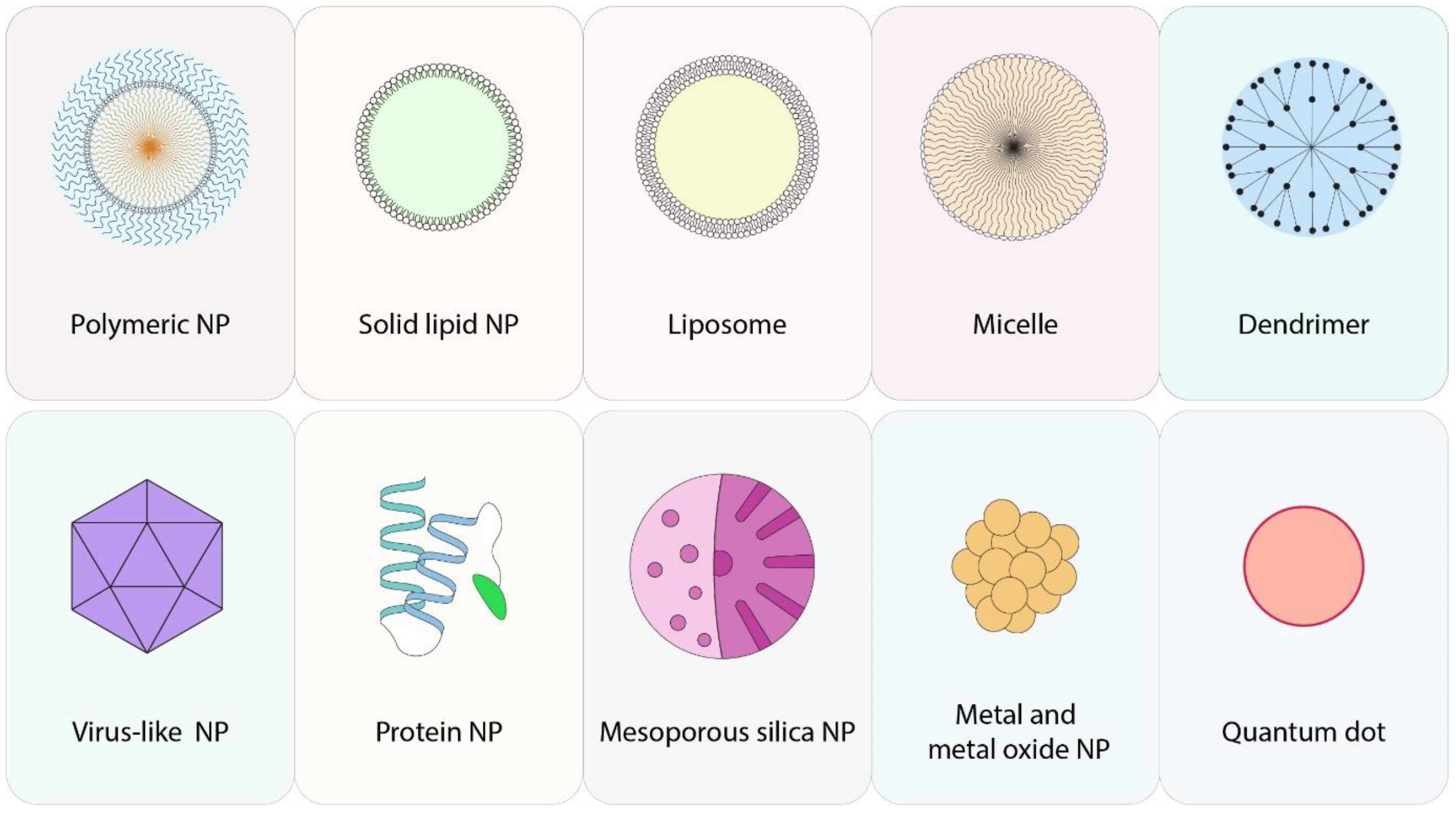
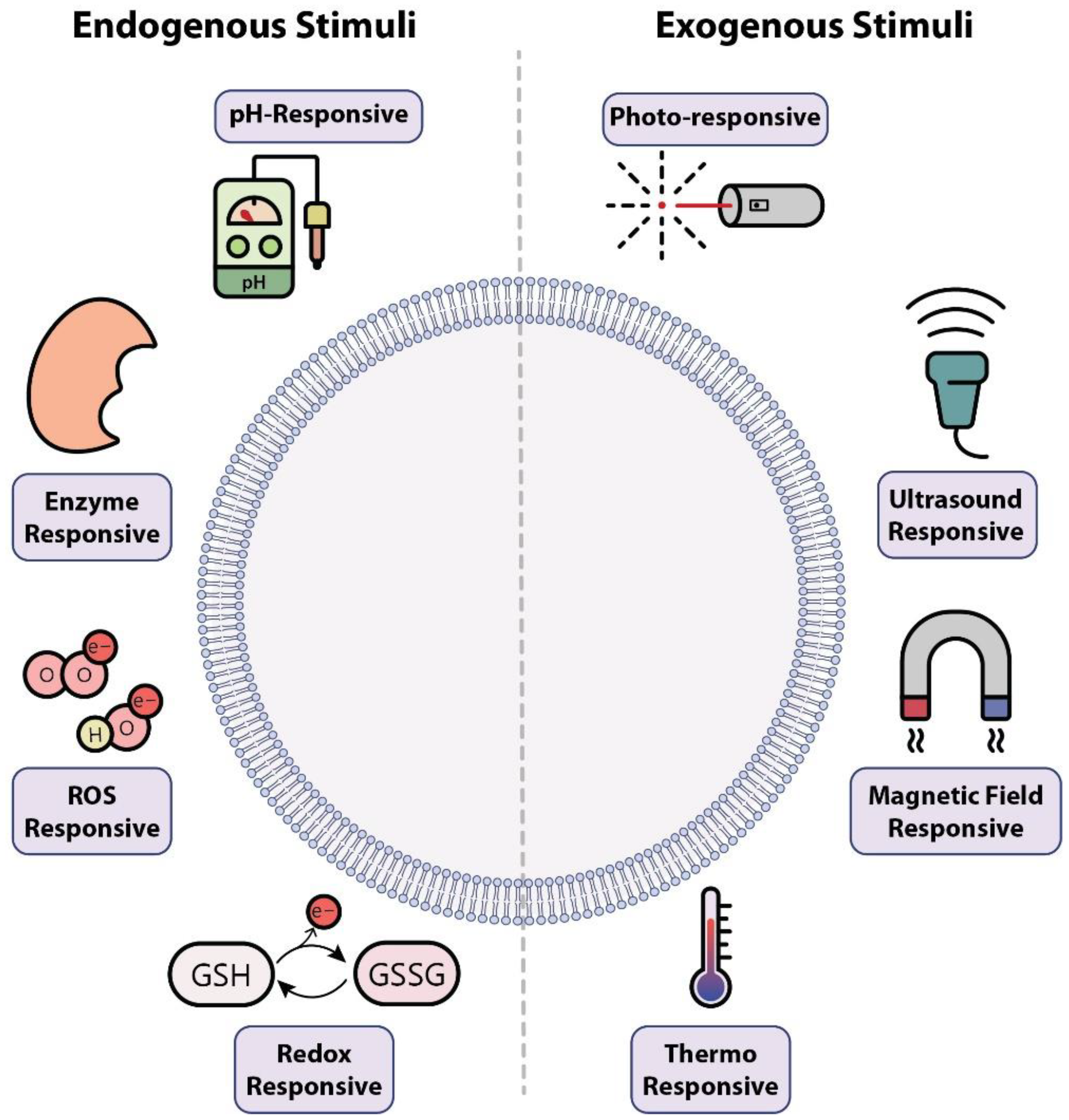
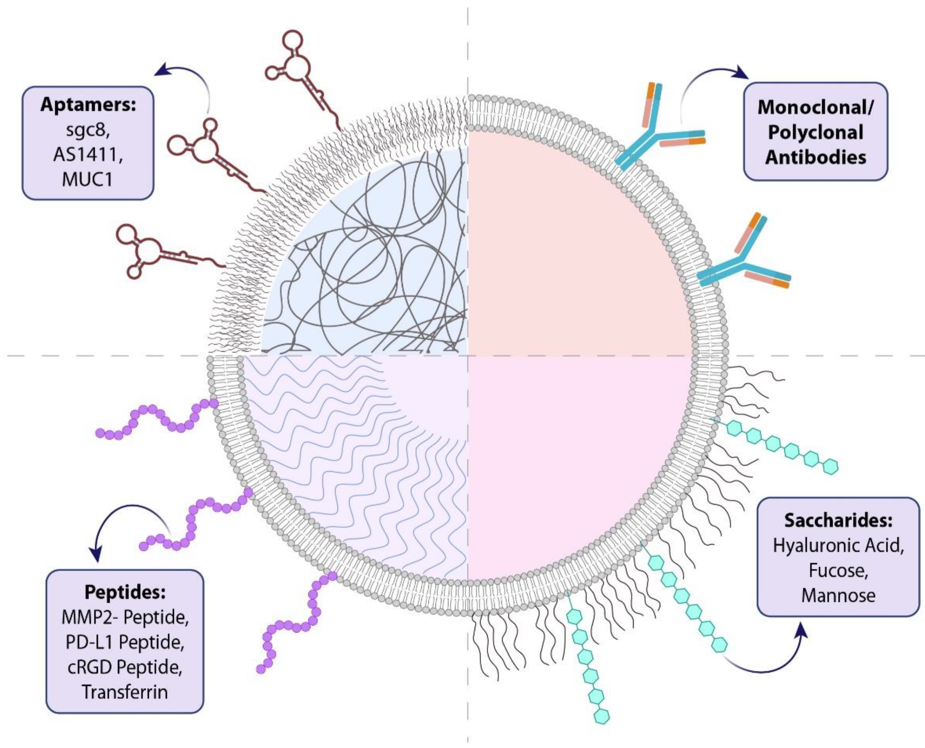
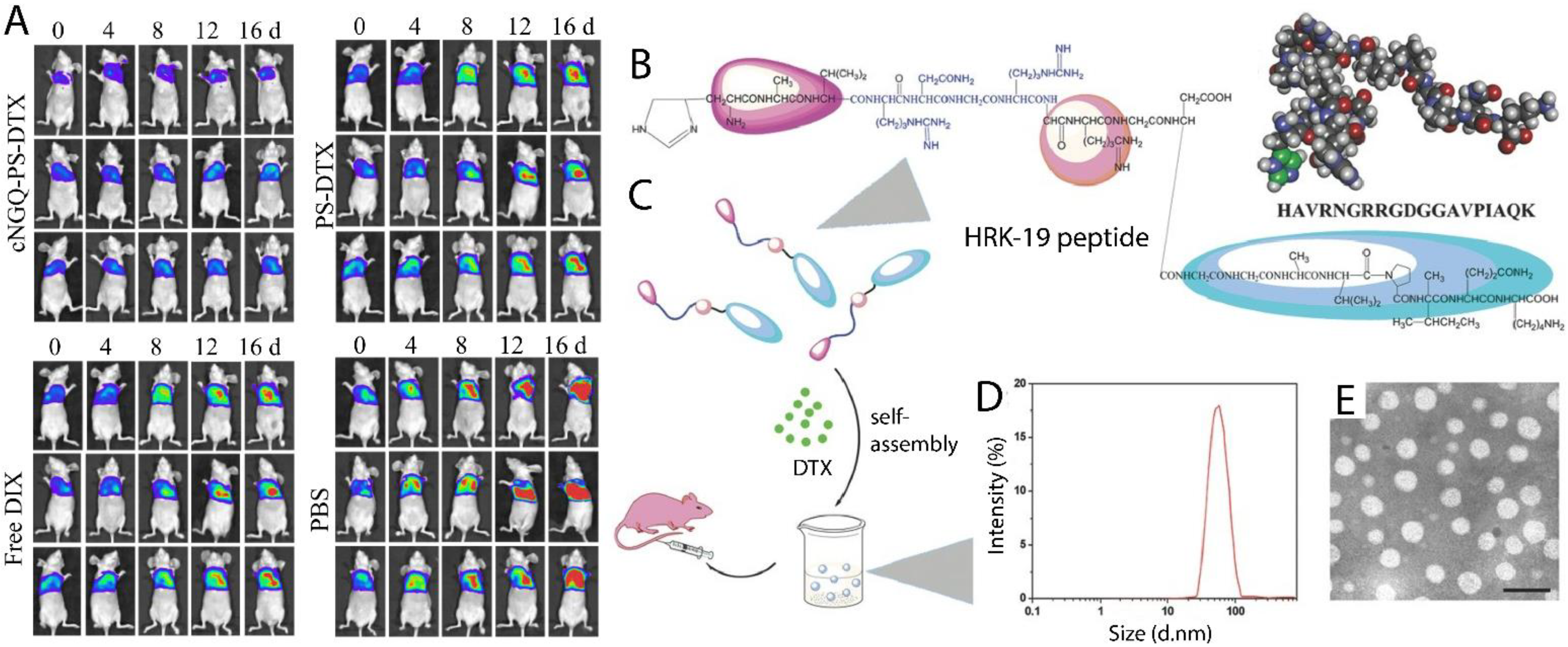
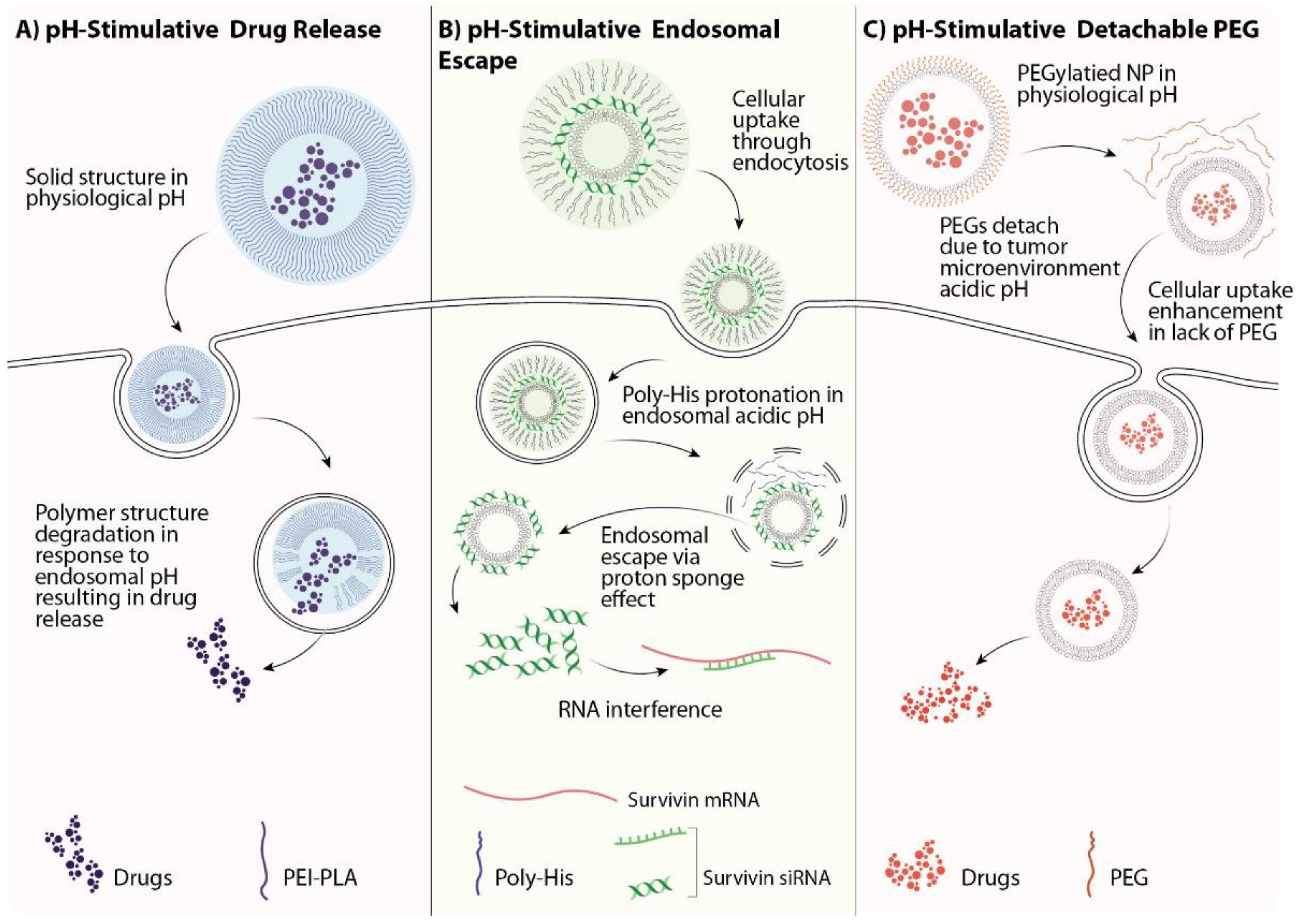
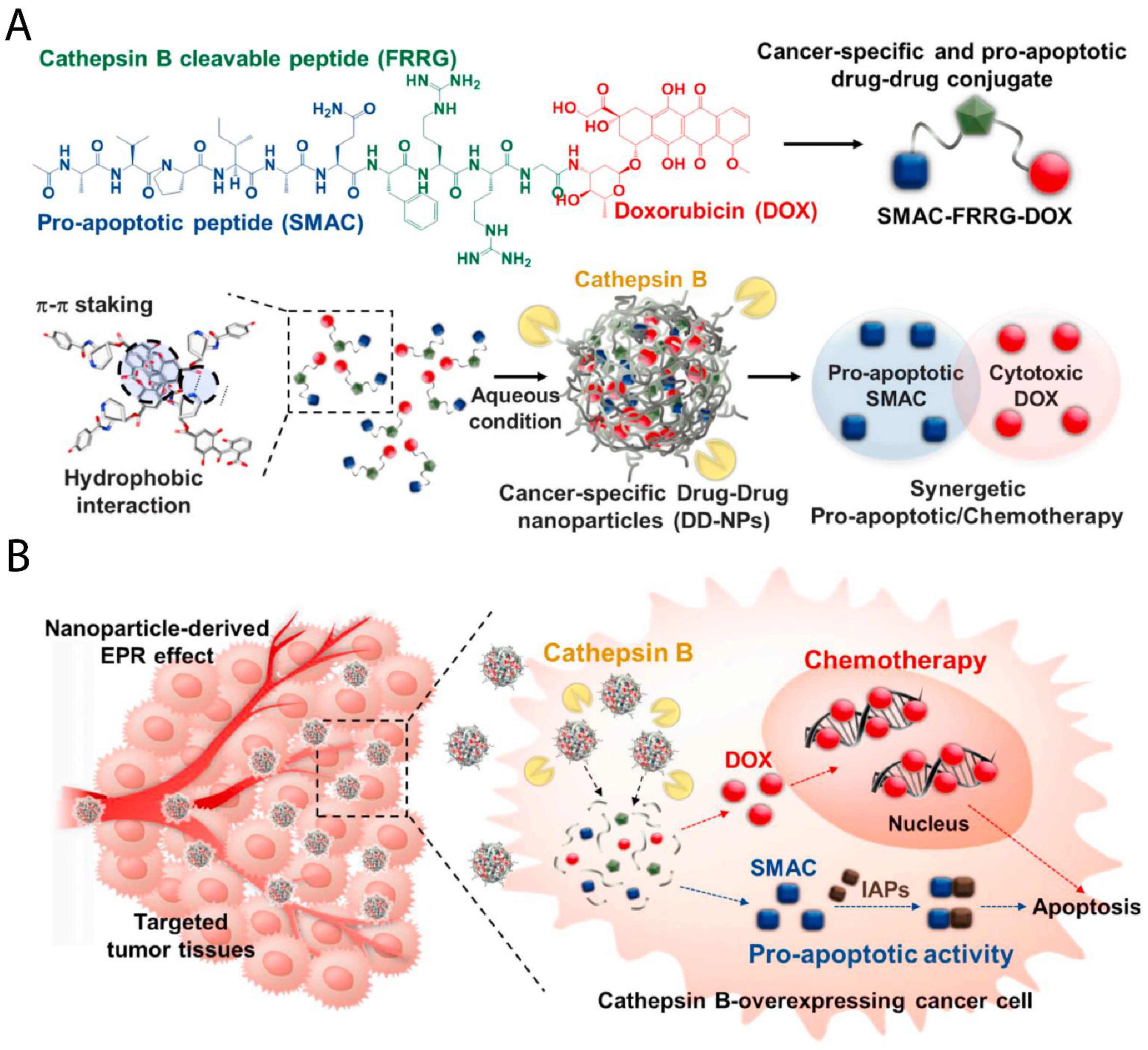
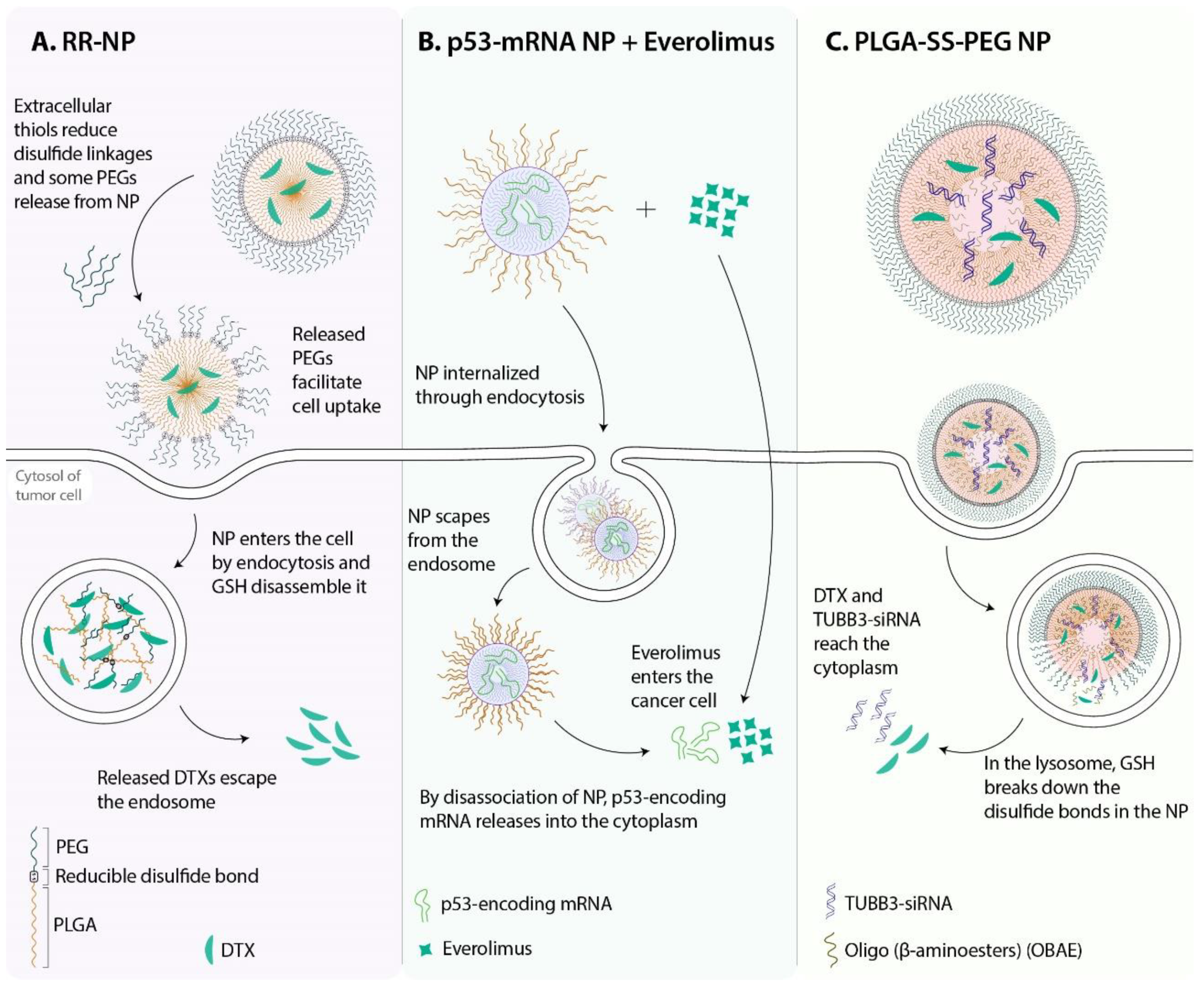
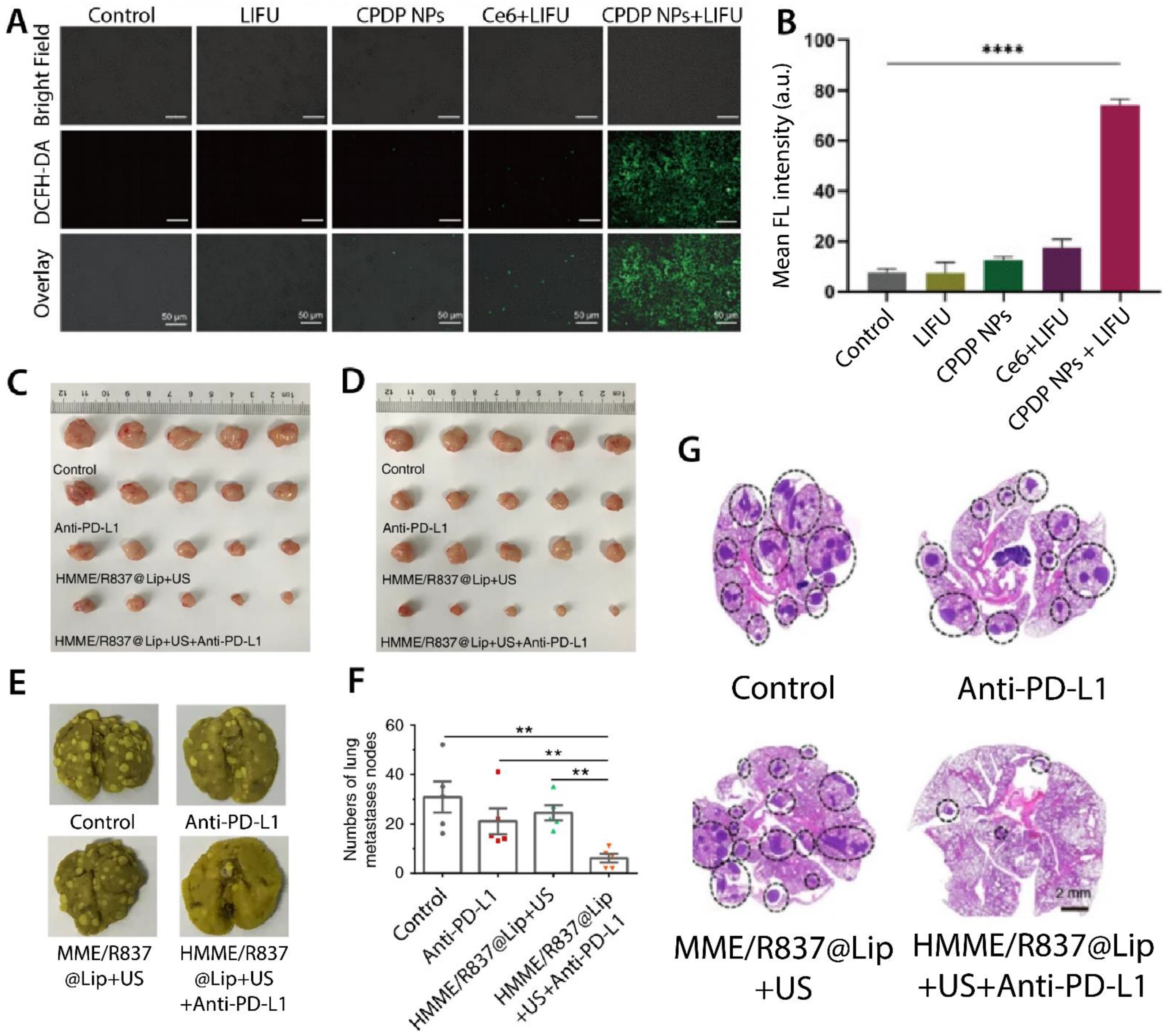
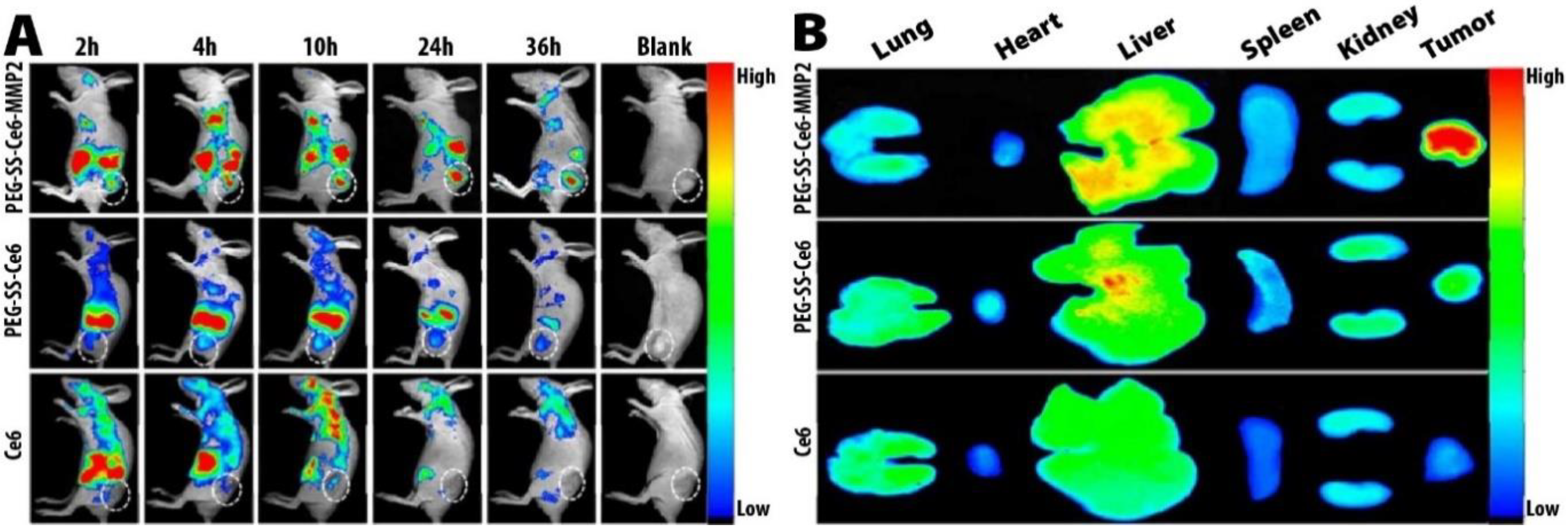
Table 1.
Recently developed targeted nanotherapeutics for lung cancer.
| Nanotherapeutic Name/Abbreviation | Target Cell | Targeting Agent | Target | Therapeutic Agent(s) | Ref. |
|---|---|---|---|---|---|
| Docetaxel (DTX)-loaded hyaluronic acid (HA) nanocapsules | A549 | HA | CD44 receptor | DTX | [41] |
| DTX-HPLGA | A549 | HA | CD44 receptor | DTX | [42] |
| HA@DOX@CNC | A549 | HA | CD44 receptor | Doxorubicin (DOX) | [43] |
| HA-CDDP/PMet | 4T1—Lewis lung carcinoma LLC—HepG2 | HA | CD44 receptor | Cisplatin—Metformin | [44] |
| Apt-Co-NPs | A549—SK-MES-1 | HA | CD44 receptor | DOX—Cisplatin | [45] |
| DTX-loaded ImI-PMs | A549 | α-Conotoxin ImI | α7-nAChR | DTX | [46] |
| CHC/DMC-CDDP/anti-CD133 | A549-ON | CD133-antibody | CD133 receptor | Cisplatin—demethoxycurcumin | [47] |
| DOC/peptide | A549 | HRK-19 peptide | Integrin αvβ3—CD13—E-cadherin | DTX | [48] |
| cRGD-PLGA@DOX | A549—HeLa | Cyclic arginine-glycine-aspartic acid polypeptide (cRGD) | Integrin αvβ3 | DOX | [49] |
| cRGD-LPP-Dox | A549 | cRGD | αvβ3 Integrin | DOX | [50] |
| RGDfC-Se@DOX | A549 | Cyclic peptide RGDfC | αvβ3 integrin | DOX | [51] |
| EGF DTX/RSV LPNs | HCC827—NCIH2135—HUVEC | Epidermal growth factor (EGF) | EGF receptor | DTX—resveratrol | [52] |
| DTX-CS-TPGS-CTX-NPs | A549 | Cetuximab | EGF receptor | Cetuximab—DTX | [53] |
| LPs-DTX-FA | A549—SPCA1 | Folic acid | Folate receptor | DTX | [54] |
| 64Cu-NOTA-GO-FSHR-mAb | cbgLuc-MDA-MB-231 | Monoclonal antibody (mAb) | Follicle-stimulating hormone receptor (FSHR) | DOX—mAb | [55] |
| C-DVM | 4T1 | CREKA peptides | Fibronectin | DOX—Vinorelbine | [56] |
| DOX-DNA | A549—4T1 | AS1411 aptamer | Nucleolin | FOXM1 aptamer—DOX | [57] |
| DL-NCs | A549/CDDP | D-α-tocopheryl polyethylene glycol succinate (TPGS)—transferrin | Transferrin receptor | Cisplatin—TPGS | [58] |
| NAG-Dend-CPT | A549 | N-acetylD-glucosamine (NAG) | Glucose transporters—lectin receptors | Camptothecin | [59] |
Table 2.
Recent smart nanotherapeutics developed for lung cancer that are responsive to two or more stimuli.
Table 2.
Recent smart nanotherapeutics developed for lung cancer that are responsive to two or more stimuli.
| Nanotherapeutic Name/Abbreviation | Stimuli | Anticancer Agent(s) | In Vitro | In Vivo | Ref. |
|---|---|---|---|---|---|
| PAHD | pH—enzyme (Heparanase) | DTX—aspirin | 4T1 | 4T1 tumor-bearing mice | [121] |
| PM@THL | pH—enzyme (matrix metalloproteinase (MMP)) | PTX- thioridazine—HY19991 | MCF-7 | Mice bearing metastatic MCF-7 tumors | [119] |
| Fe3O4@SiO2@DOX | pH—Photothermal | DOX | A549 | - | [123] |
| mPEG-PAAV/IR780 + DOX micelle | pH—Photothermal | DOX | 4T1 | 4T1 tumor-bearing mice | [124] |
| TOCM | pH—redox | Cisplatin—TPGS | A549DDP | H22 tumor-bearing mice | [125] |
| PTXL-ss-PMAGP-GEM/NAG NLC | pH—redox | Gemcitabine—Paclitaxel | A549—LTEP-a-2 | A549 tumor-bearing nude mice | [126] |
| PSPm(PTX/CDDP) | pH—redox | PTX—Cisplatin | NCI-H520—CRL-5802—NCI-H358 | - | [127] |
| DOX-N-NPs | pH—redox | DOX | A549—MCF-7 | MCF-7 tumor-bearing mice | [128] |
| GP-NA | pH—redox | Ultra-small platinum nanoparticles—Gem | A549—NCI-H1299 | NCI-H1299 tumor-bearing mice—A549 tumor-bearing nude mice | [129] |
| PPT/D(DMA)@DOX | pH—ROS | DOX | A549 | A549 cell tumor-bearing mice model | [117] |
| SFR/Ce6-loaded PEG-PPMT | pH—ROS—enzyme | Sorafenib chlorin e6 (Ce6) | A549 | A549 xenografted in nude mice | [122] |
| ZnPc/CPT-TPPNP | ROS—photodynamic | Camptothecin | NCI-H460 | NCI-H460 tumor-bearing mice | [130] |
| NP(Se) | Redox—H2O2 | Cisplatin prodrug | A549, A549DDP, 7404DDP | Patient-derived xenograft models of hepatic carcinoma and multidrug-resistant lung cancer | [118] |
Table 3.
Recent smart nanotherapeutics for lung cancer that are simultaneously targeted and stimuli-responsive.
Table 3.
Recent smart nanotherapeutics for lung cancer that are simultaneously targeted and stimuli-responsive.
| Nanotherapeutic Name/Abbreviation | Targeting Agent | Target | Stimuli | Drug | Therapy | Application | Cell/Animal Model | Ref. |
|---|---|---|---|---|---|---|---|---|
| HA-ZnO-DOX | Hyaluronic acid | CD44 receptor | pH | DOX | Chemotherapy | Therapy | A549 cell line | [137] |
| HACE | Hyaluronic acid | CD44 receptor | Photo | Ce6 | Photodynamic therapy | Therapy and imaging | A549 cell line, A549 tumor-bearing nude mice model | [135] |
| DOX/HA-ss-DOX micelles | Hyaluronic acid | CD44 receptor | pH, redox | DOX | Chemotherapy | Therapy | A549 cell line, xenograft model | [138] |
| DTX/PPN@PPY@HA | Hyaluronic acid | CD44 receptor | Photo, thermo | DTX—Polypyrrole | Chemotherapy, photothermal therapy | Therapy and imaging | A549 cell line, 4T1 tumor-bearing mice model | [136] |
| HPGBCA | Hyaluronic acid | CD44 receptor | Photo, enzyme, ROS | Afatinib, Ce6 | Chemotherapy, photothermal therapy, photodynamic therapy | Therapy | A549 and 3T3 cell lines, A549 tumor-bearing nude mice | [139] |
| HA-GC-DOX/CXB | Hyaluronic acid | CD44 receptor | pH | DOX, Celecoxib | Chemotherapy | Therapy | A549-Luc cell line, Murine A549-Luc xenograft model | [140] |
| sp-PA | Biotin | Biotin receptor | Photo, ROS | PTX, silicon 2,3-naphthalocyanine bis | Chemotherapy, photodynamic therapy | Therapy | A549 cell line, Xenograft tumor mouse model | [141] |
| CS-nanogels | Chondroitin sulfate | CD44 receptor | pH, thermo | Cisplatin | Chemotherapy | Therapy | A549 cell line, A549 tumor-bearing nude mice model | [142] |
| CCG-NP | Concanavalin A | Mannose receptor | MMP2 | Cisplatin | Chemotherapy | Therapy | A549 cell line | [143] |
| Tf-SS-Afa-LPNs | Transferrin | Transferrin receptor | Redox | Afatinib | Chemotherapy | Therapy | H1975, and PC-9 cell lines, lung cancer-bearing mice | [144] |
| UA@M-CS-FA | Folic acid | Folate receptor | pH | Ursolic acid | Chemotherapy | Therapy | HeLa and HepG2 cell lines, HeLa bearing tumor model | [145] |
| FA-BSA@DA | Folic acid | Folate receptor | ROS | Aspirin prodrug | Chemotherapy | Therapy and imaging | A549, MCF-7, and MCF-7 cell lines, breast cancer xenograft tumor models with lung metastasis | [146] |
| MDNP | Folic acid | Folate receptor | pH, photo | Gemcitabine, NU7441 | Chemotherapy, radiotherapy | Therapy | A549 and H460 lung cancer cell lines, H460 tumor-bearing athymic nude mice | [147] |
| FA/Tf-CDDP-NPs | Folic acid, transferrin | Folate receptor | GSH | Cisplatin | Chemotherapy | Therapy | A549 cell line, A549 tumor-bearing nude mice model | [148] |
| TBP@DOX | Fucoidan | P-selectin | Microwave | DOX | Chemotherapy | Therapy | HepG2 and H22 cell lines, HepG2 and H22 xenograft tumor models with lung metastasis | [149] |
| GEM-FGONS | Fucose | Fucose receptor | pH | Gemcitabine hydrochloride | Chemotherapy | Therapy | MDA-MB-231 and A549 cell lines | [150] |
| PSO-HGNPs-DOX | Polyoxyethylene sorbitol oleate | Low-density lipoprotein receptor | Photo, thermo | DOX | Chemotherapy, radiotherapy, photothermal therapy | Therapy | A549 cell line, A549 human lung cancer-bearing mouse model | [151] |
| Apt@DP-DOX-MCN | MUC1 aptamer | Cancer cell | pH, GSH | DOX | Chemotherapy | Therapy | A549 and MCF-7 cell lines | [152] |
| Apt-Fe3O4@C@DOX | sgc8 aptamer | Cancer cell | pH, thermo | DOX | Chemotherapy, photothermal therapy | Therapy and imaging | A549 cell line, A549 tumor-bearing nude mice model | [153] |
| Au-siRNA-PAA-AS1411 | AS1411 aptamer | Nucleolin receptors | Photo, MMP2, magnetic field | DOX, siRNA | Chemotherapy, gene therapy, photothermal therapy | Therapy | NCI-H889 cell line, lung cancer orthotopic murine model | [154] |
| CDGM | MMP2 peptide | MMP2 | pH, photo | DOX, Ce6 | Chemotherapy, photodynamic therapy | Therapy and imaging | A549 cell line, A549 tumor-bearing nude mice model | [133] |
| PEG-SS-Ce6-MMP2 | MMP2 peptide | MMP2 | Photo, redox | Ce6 | Photodynamic therapy | Therapy and imaging | A549 cell line, A549 tumor-bearing nude mice model | [132] |
| GNS@BSA/I-MMP2 | MMP2 peptide | MMP2 | Photo | IR 780 iodide | Photodynamic therapy, photothermal therapy | Therapy and imaging | A549 cell line, A549 tumor-bearing nude mice model | [131] |
| GNPs@PEG/Ce6-P | PD-L1 peptide | PD-L1 | Photo | Ce6 | Photodynamic therapy | Therapy and imaging | HCC827 cell line, HCC827 tumor-bearing mice | [134] |
| cNGQ-PS-Dox | cNGQ peptide | α3β1 integrin receptor | Redox | DOX | Chemotherapy | Therapy | A549 cell line, A549 human lung cancer-bearing nude mice model | [155] |
| cPCPL/siRNA/ETP | cRGDyC peptide | - | pH, redox | EZH2 siRNA—Etoposide | Chemotherapy, gene therapy | Therapy | luc-A549 cell line, nude mice bearing orthotopic NSCLC | [156] |
| cNGQ-PS-DTX | cNGQGEQc peptide | α3β1 integrin receptor | Redox | DTX | Chemotherapy | Therapy | A549 cell line, orthotopic A549 xenografts | [63] |
| siBec1@PPN | cRGD peptide | α3β1 integrin receptor | GSH | Cisplatin—Beclin1 siRNA | Chemotherapy, gene Therapy | Therapy | A549 cell line, Cisplatin-resistant A549 tumor on xenograft mice models | [157] |
| RGD-ss-PTX/CDDP LPNs | RGD peptide | αvβ3 integrin | Redox | PTX, Cisplatin | Chemotherapy | Therapy | A549 and NCI-H1299 cell lines, A549 tumor-bearing mice | [158] |
| U11-DOX/CUR | U11 peptide | Urokinase plasminogen activator receptor | pH | DOX, Curcumin | Chemotherapy | Therapy | A549/ADR cells line, mice bearing A549/ADR cells cancer model | [159] |
| T7-DSNPs/9291 | T7 peptide | Transferrin receptor | GSH | Osimertinib, DOX | Chemotherapy | Therapy | PC-9 cell line, PC-9 tumor nude mice | [160] |
Table 4.
Clinical trials performed with nanotherapeutics in lung cancer patients.
| Trade Name | Particle Type | Therapeutic Agent | Indication | NCT Number | Phase | Status |
|---|---|---|---|---|---|---|
| Abraxane | Albumin based nanoparticle | Paclitaxel | Non-squamous NSCLC | NCT02264990 | Phase III | Completed |
| NCT03169335 | Phase III | Completed | ||||
| NSCLC | NCT03594747 | Phase III | Ongoing | |||
| NCT03296163 | Phase III | Completed | ||||
| NCT02754882 | Phase III | Completed | ||||
| BIND-014 | Polymeric | DTX | Advanced or metastatic cancer, including lung cancer | NCT01300533 | Phase I | Completed |
| NSCLC | NCT02283320 | Phase II | Completed | |||
| NCT01792479 | Phase II | Completed | ||||
| EP0057 (CRLX101) | Polymeric | Camptothecin | NSCLC | NCT01380769 | Phase II | Completed |
| Lung Neoplasms, Small Cell Lung Cancer | NCT02769962 | Phae I/II | Ongoing | |||
| Genexol-PM | Polymeric micelle | Paclitaxel | NSCLC | NCT01770795 | Phase II | Completed |
| NCT01023347 | Phase II | Completed | ||||
| LY01610 | Liposome | Irinotecan hydrochloride | Small Cell Lung Cancer | NCT04381910 | Phase II | Ongoing |
| NBTXR3 | Hafnium Oxide-containing Nanoparticles | NSCLC | NCT03589339 | Phase I | Ongoing | |
| Recurrent NSCLC | NCT04505267 | Phase I | Ongoing | |||
| NC-6004 | Micellar | Cisplatin | Solid tumors | NCT02240238 | Phase I/II | Completed |
| NK012 | Polymeric micelle | SN-38 | Small cell lung cancer | NCT00951613 | Phase II | Completed |
| NKTR-102 | Polymeric | Irinotecan | NSCLC | NCT01773109 | Phase II | Completed |
| Lung and breast cancer | NCT02312622 | Phase II | Completed | |||
| Recurrent small cell lung carcinoma | NCT01876446 | Phase II | Completed | |||
| Oncoprex (DOTAP:Chol-fus1) | Liposomal | FUS1 gene | NSCLC | NCT00059605 | Phase I | Completed |
| NCT01455389 | Phase I/II | Ongoing | ||||
| NCT04486833 | Phase I/II | Ongoing | ||||
| Onivyde | Liposomal | Topotecan | Small cell lung cancer | NCT03088813 | Phase II/III | Ongoing |
| Pm-Pac | Polymeric micellar | Paclitaxel | NSCLC | NCT02667743 | Phase III | Completed |
| SGT-53 | Cationic liposome | Wild-type p53 DNA sequence | Neoplasm | NCT00470613 | Phase I | Completed |
| TargomiRs | Targeted minicells | miRNA | NSCLC | NCT02369198 | Phase I | Completed |
| Taxoprexin | DHA-bonded paclitaxel | NSCLC | NCT00243867 | Phase III | Completed | |
| TLI | Liposomal | Topotecan | Small cell lung cancer | NCT00765973 | Phase I | Completed |
Publisher’s Note: MDPI stays neutral with regard to jurisdictional claims in published maps and institutional affiliations. |
© 2021 by the authors. Licensee MDPI, Basel, Switzerland. This article is an open access article distributed under the terms and conditions of the Creative Commons Attribution (CC BY) license (https://creativecommons.org/licenses/by/4.0/).
Share and Cite
MDPI and ACS Style
Doroudian, M.; Azhdari, M.H.; Goodarzi, N.; O’Sullivan, D.; Donnelly, S.C. Smart Nanotherapeutics and Lung Cancer. Pharmaceutics 2021, 13, 1972. https://doi.org/10.3390/pharmaceutics13111972
AMA Style
Doroudian M, Azhdari MH, Goodarzi N, O’Sullivan D, Donnelly SC. Smart Nanotherapeutics and Lung Cancer. Pharmaceutics. 2021; 13(11):1972. https://doi.org/10.3390/pharmaceutics13111972
Chicago/Turabian StyleDoroudian, Mohammad, Mohammad H. Azhdari, Nima Goodarzi, David O’Sullivan, and Seamas C. Donnelly. 2021. "Smart Nanotherapeutics and Lung Cancer" Pharmaceutics 13, no. 11: 1972. https://doi.org/10.3390/pharmaceutics13111972
Note that from the first issue of 2016, this journal uses article numbers instead of page numbers. See further details here.