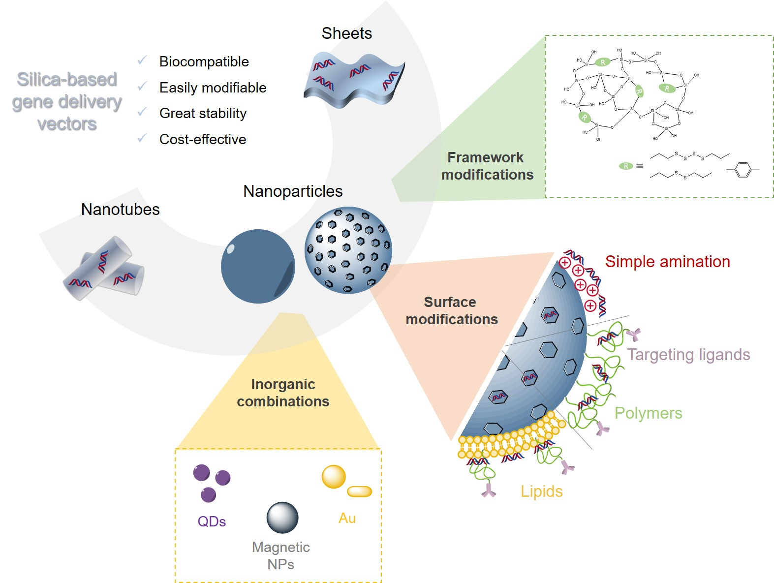
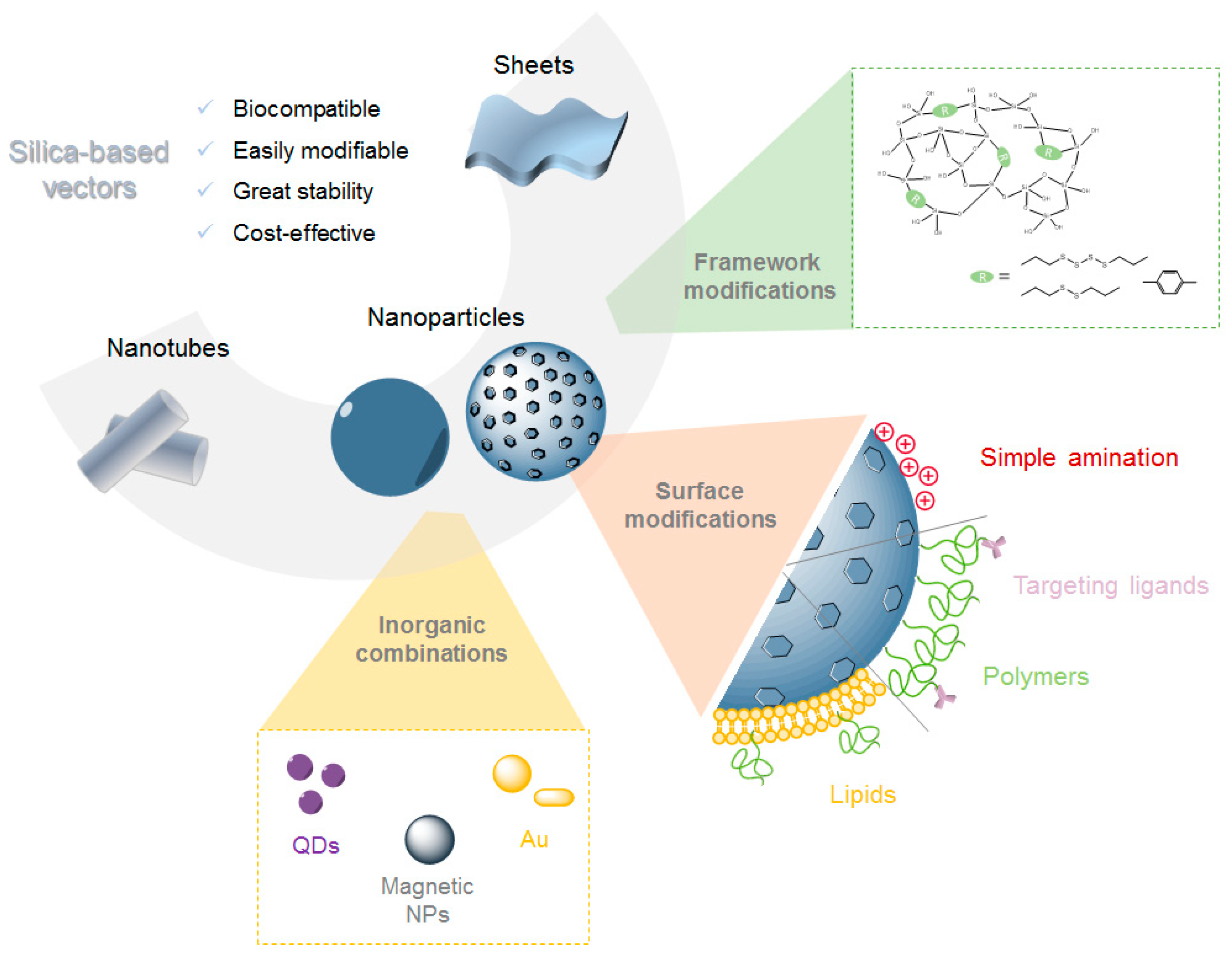
Silica-Based Gene Delivery Systems: From Design to Therapeutic Applications


Abstract
:
1. Introduction
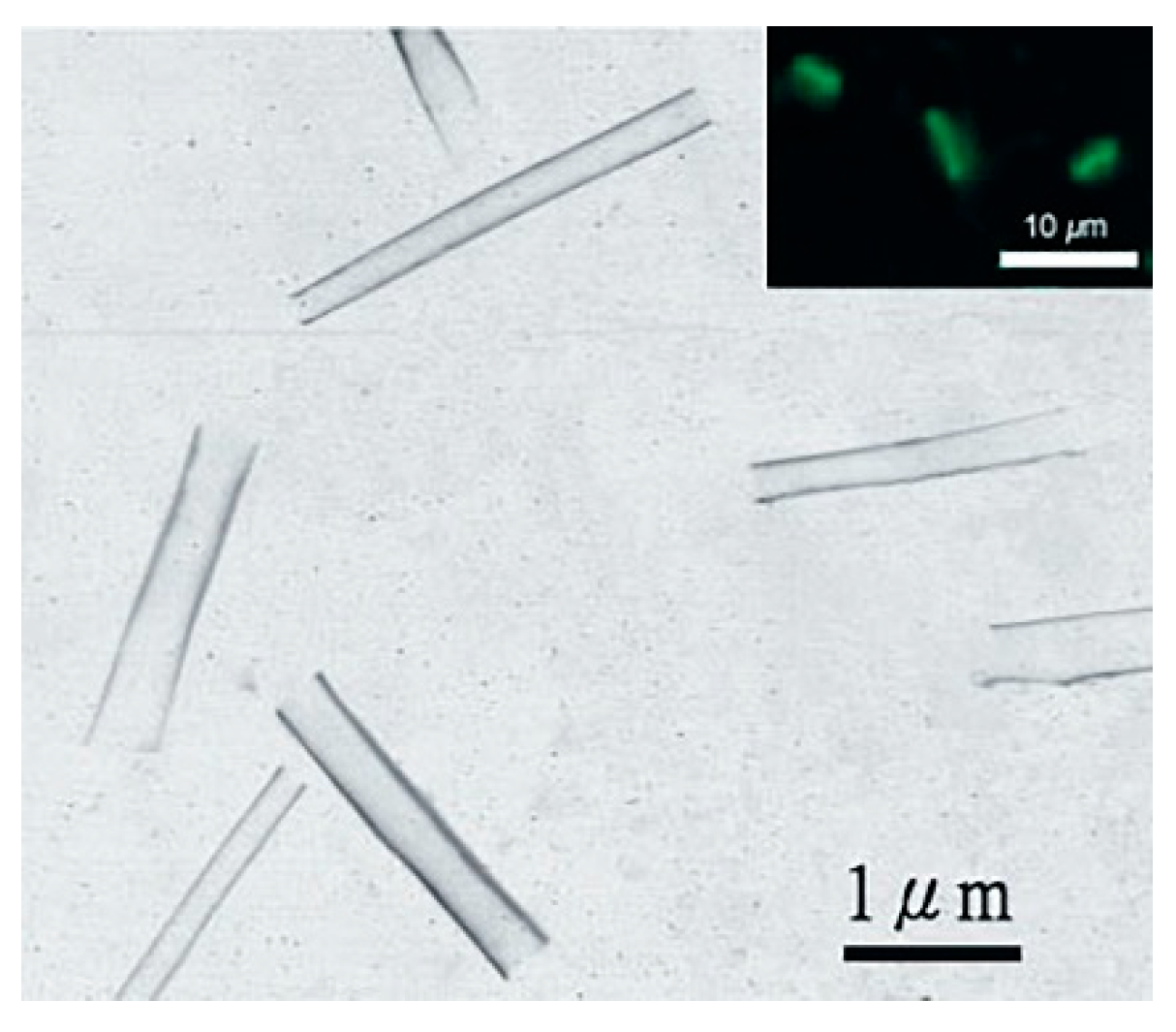
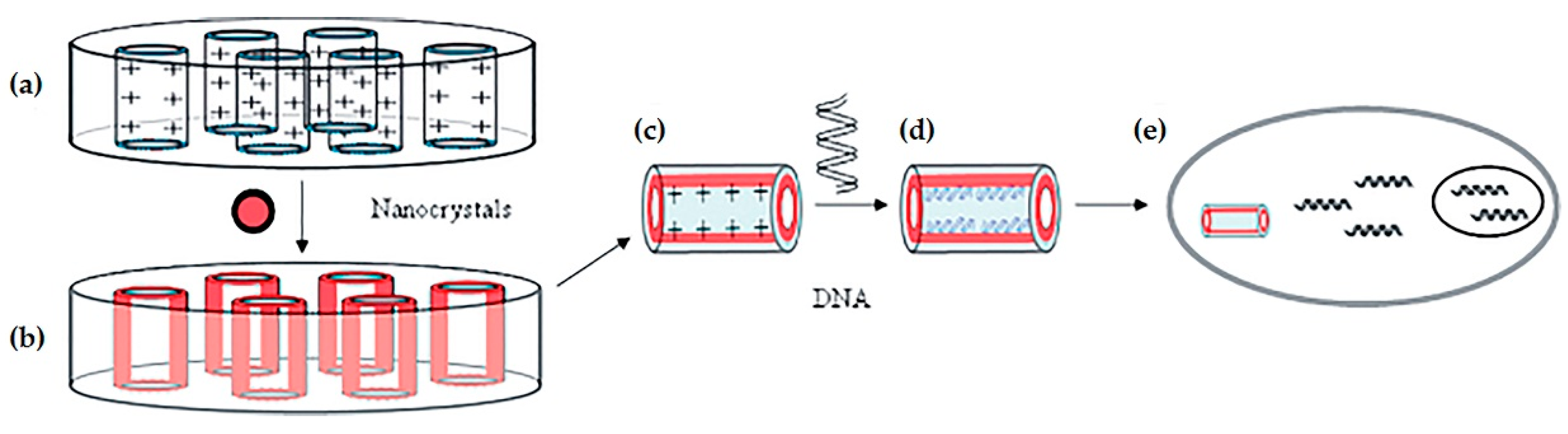
2. Silica Nanotubes
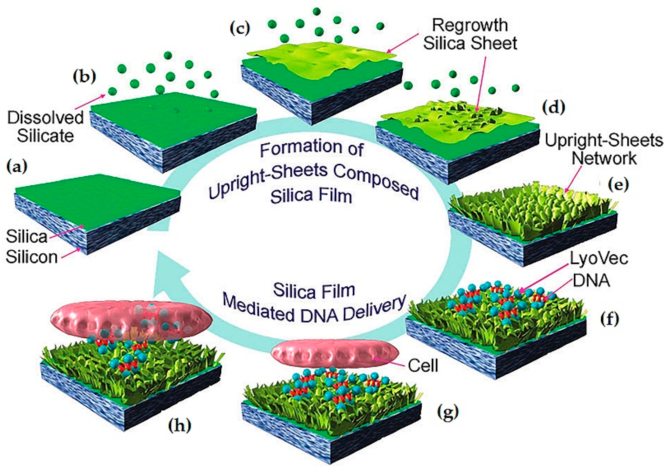
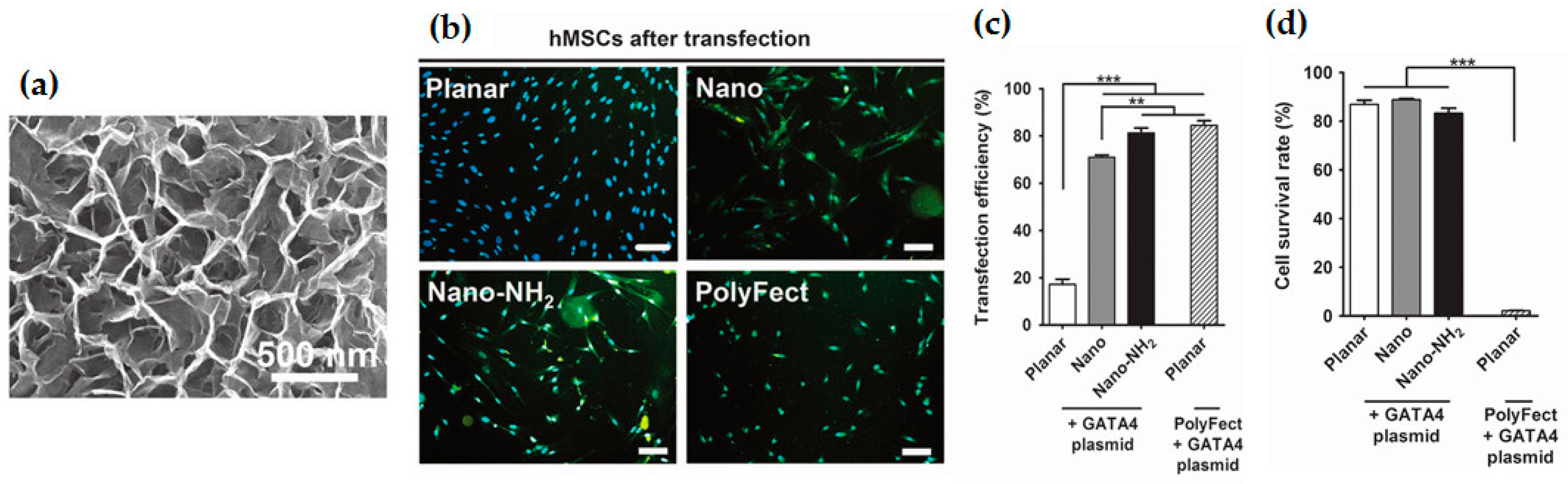
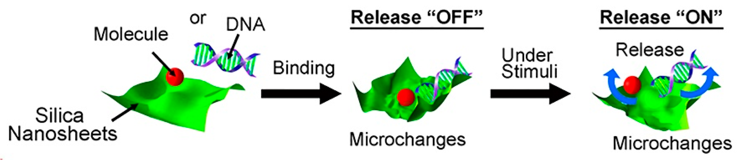
3. Silica Sheets
4. Silica Nanoparticles
4.1. Non-Porous Silica Nanoparticles
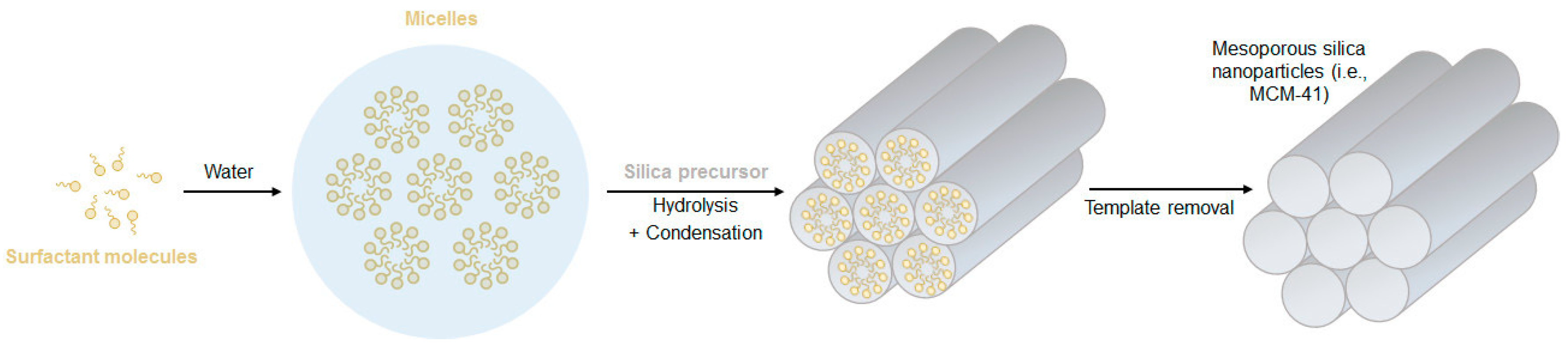
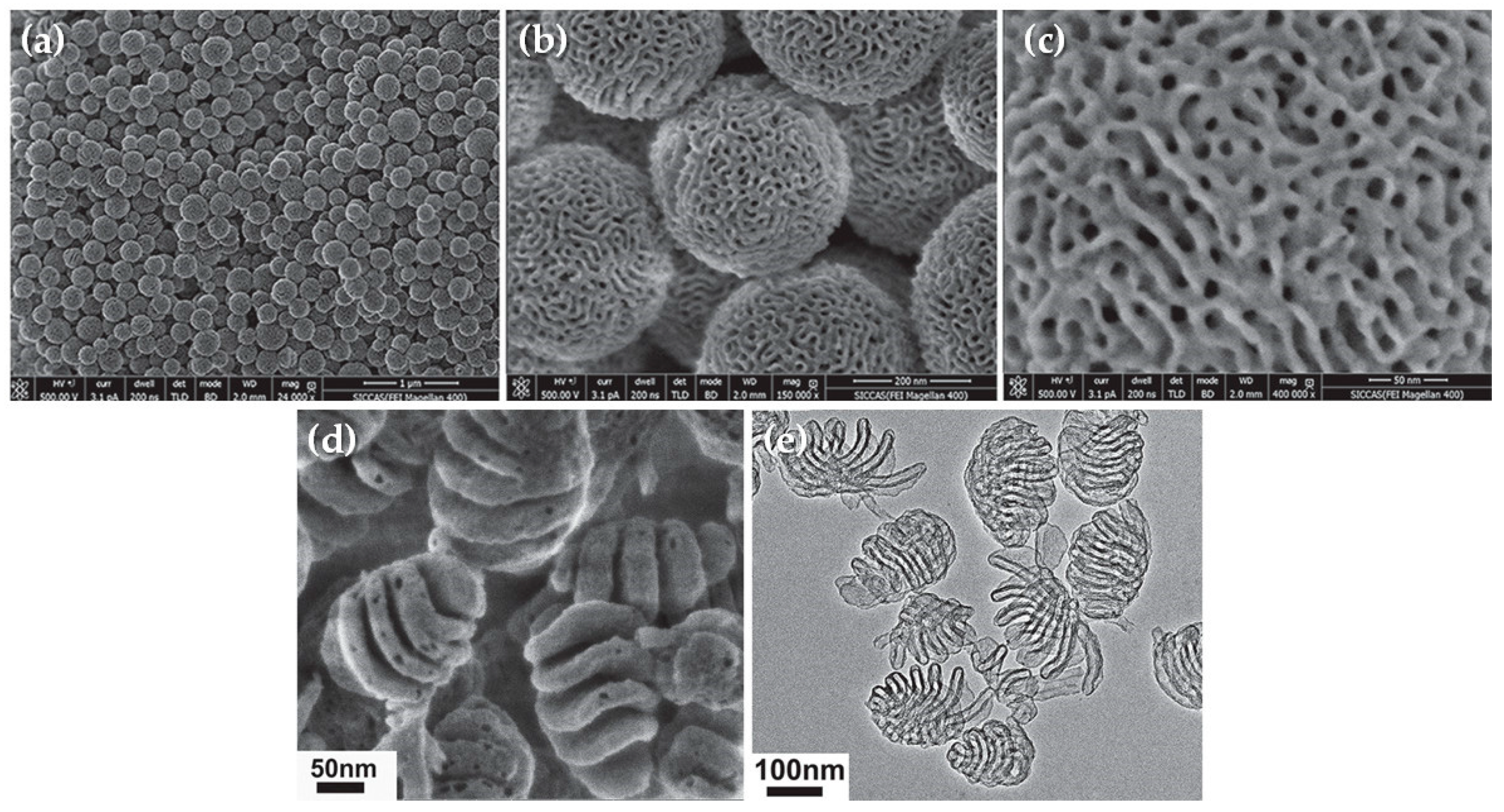
4.2. Porous: Mesoporous Silica Nanoparticles
Organosilica Nanoparticles
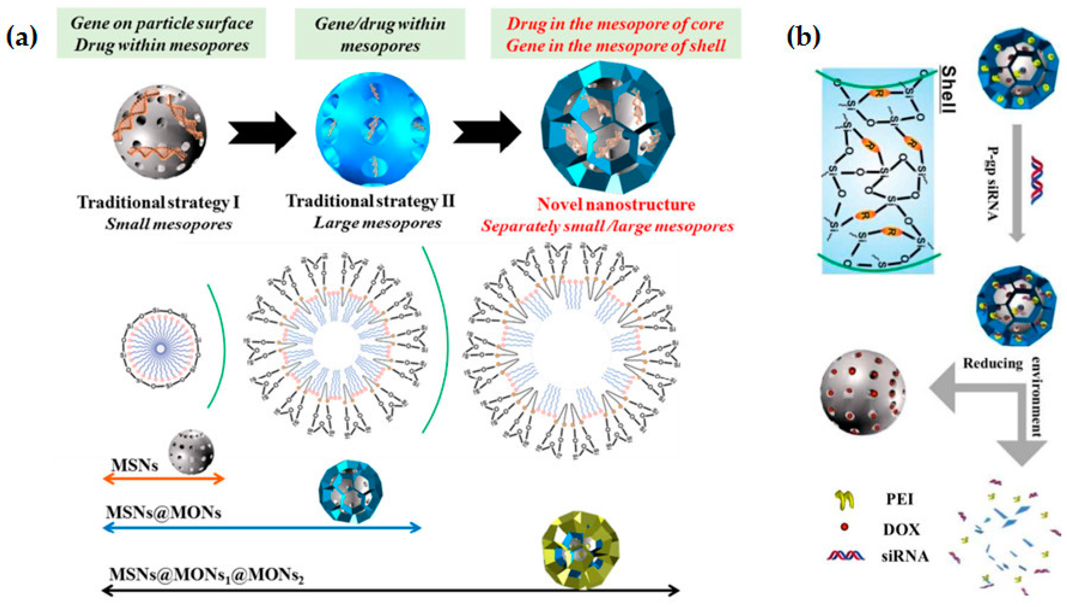
5. Hybrid Silica Nanoparticles
5.1. Polymer Modified Silica Nanoparticles
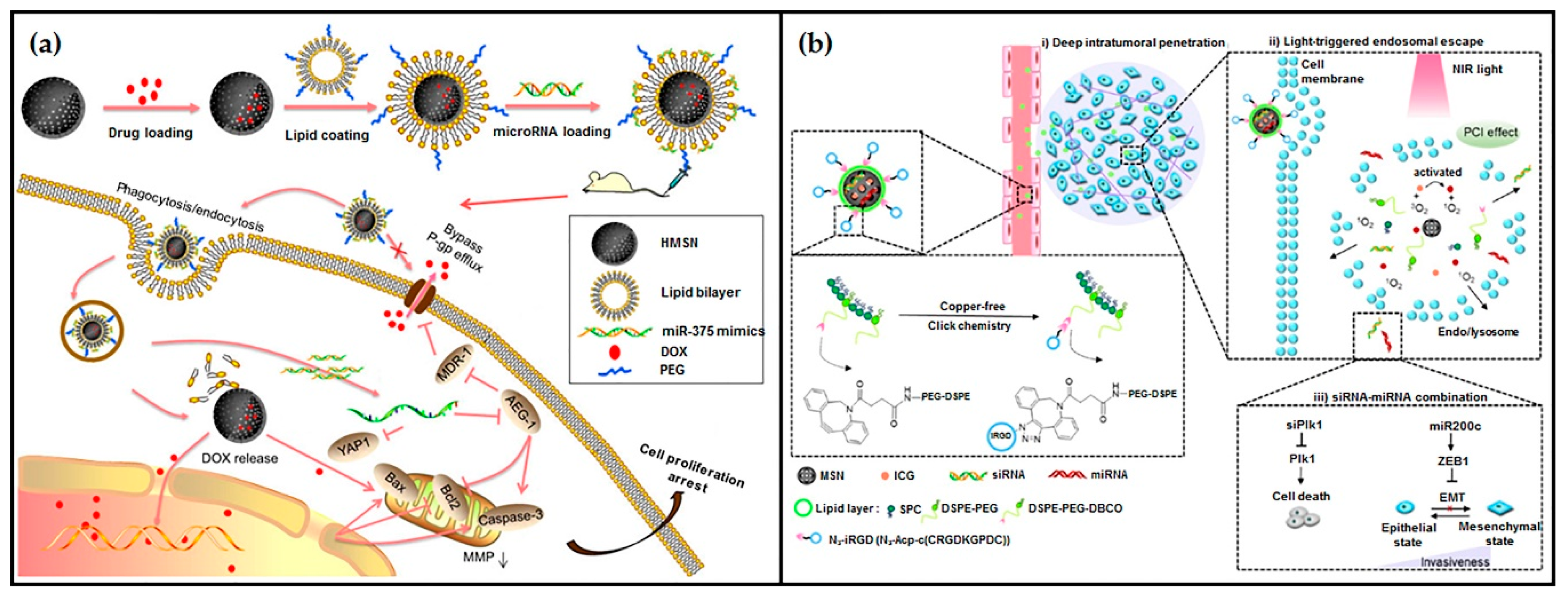
5.2. Lipid Modified Silica Nanoparticles
5.3. Magnetic Silica Nanoparticles
5.4. Other Silica-Based Nanocomposites
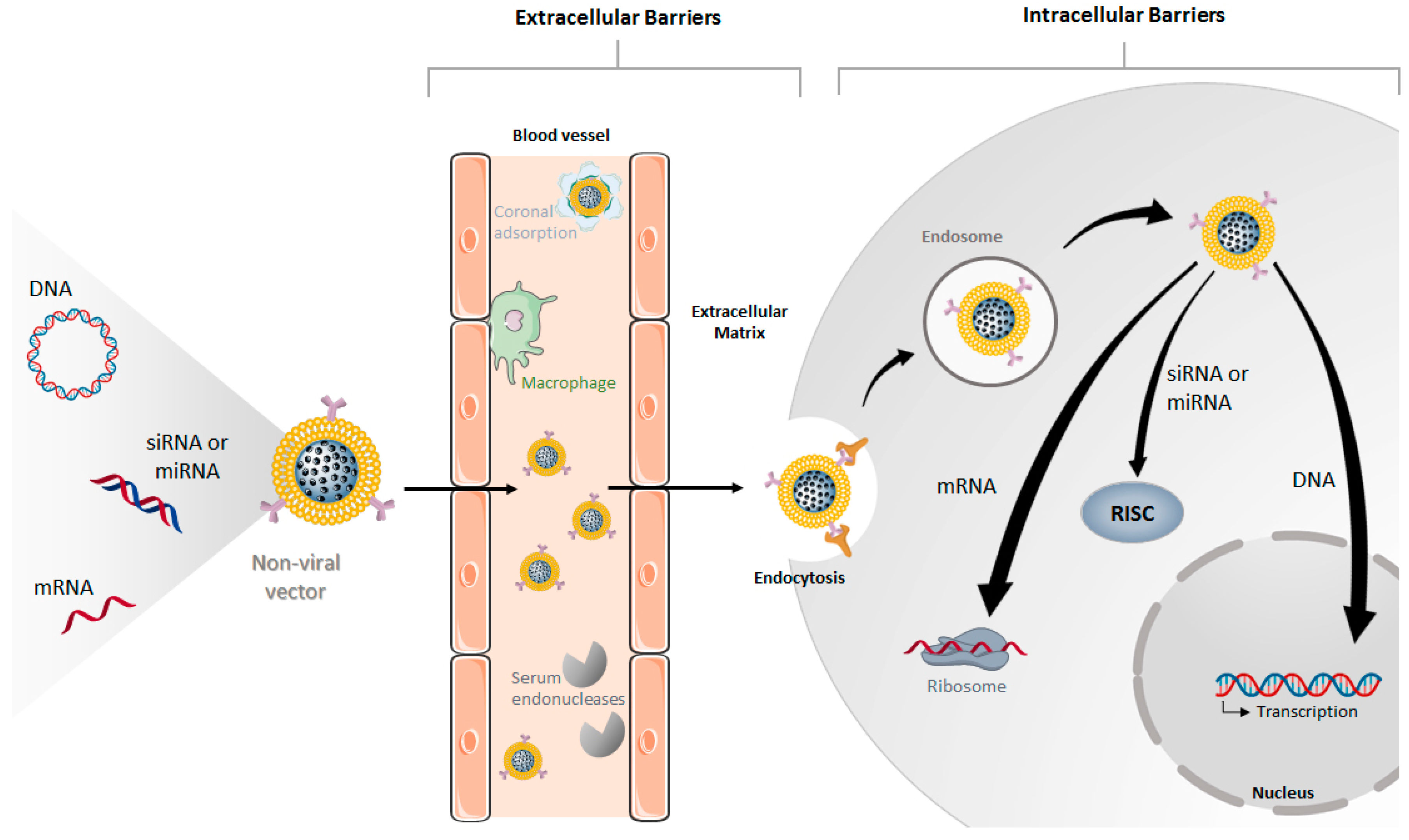
6. Cellular Uptake and Intracellular Fate of Silica-Based Vectors
7. Biocompatibility of Silica-Based Materials
8. Silica-Based Systems as an Alternative to Other Vectors
9. Conclusions
Funding
Conflicts of Interest
References
- Shim, G.; Kim, D.; Le, Q.-V.; Park, G.T.; Kwon, T.; Oh, Y.-K. Nonviral Delivery Systems For Cancer Gene Therapy: Strategies And Challenges. Curr. Gene Ther. 2018, 18, 3–20. [Google Scholar] [CrossRef]
- Keles, E.; Song, Y.; Du, D.; Dong, W.J.; Lin, Y. Recent progress in nanomaterials for gene delivery applications. Biomater. Sci. 2016, 4, 1291–1309. [Google Scholar] [CrossRef] [PubMed]
- Loh, X.J.; Lee, T.-C.; Dou, Q.; Deen, G.R. Utilising inorganic nanocarriers for gene delivery. Biomater. Sci. 2016, 4, 70–86. [Google Scholar] [CrossRef] [PubMed]
- Yin, H.; Kanasty, R.L.; Eltoukhy, A.A.; Vegas, A.J.; Dorkin, J.R.; Anderson, D.G. Non-viral vectors for gene-based therapy. Nat. Rev. Genet. 2014, 15, 541–555. [Google Scholar] [CrossRef]
- Wang, Y.; Huang, L. Composite Nanoparticles for Gene Delivery. In Advances in Genetics; Elsevier: Amsterdam, The Netherlands, 2014; Vol. 88, pp. 111–137. [Google Scholar]
- Guo, K.; Zhao, X.; Dai, X.; Zhao, N.; Xu, F.J. Organic/inorganic nanohybrids as multifunctional gene delivery systems. J. Gene Med. 2019, 21. [Google Scholar] [CrossRef] [Green Version]
- Riley, M.; Vermerris, W. Recent Advances in Nanomaterials for Gene Delivery—A Review. Nanomaterials 2017, 7, 94. [Google Scholar] [CrossRef] [Green Version]
- Watermann, A.; Brieger, J. Mesoporous silica nanoparticles as drug delivery vehicles in cancer. Nanomaterials 2017, 7, 189. [Google Scholar] [CrossRef] [Green Version]
- Yang, X.; Tang, H.; Cao, K.; Song, H.; Sheng, W.; Wu, Q. Templated-assisted one-dimensional silica nanotubes: Synthesis and applications. J. Mater. Chem. 2011, 21, 6122–6135. [Google Scholar] [CrossRef]
- Barkat, A.; Beg, S.; Panda, S.K.; S Alharbi, K.; Rahman, M.; Ahmed, F.J. Functionalized mesoporous silica nanoparticles in anticancer therapeutics. Semin. Cancer Biol. 2019. [Google Scholar] [CrossRef]
- Zhou, Y.; Quan, G.; Wu, Q.; Zhang, X.; Niu, B.; Wu, B.; Huang, Y.; Pan, X.; Wu, C. Mesoporous silica nanoparticles for drug and gene delivery. Acta Pharm. Sin. B 2018, 8, 165–177. [Google Scholar] [CrossRef]
- Benezra, M.; Penate-Medina, O.; Zanzonico, P.B.; Schaer, D.; Ow, H.; Burns, A.; DeStanchina, E.; Longo, V.; Herz, E.; Iyer, S.; et al. Multimodal silica nanoparticles are effective cancer-targeted probes in a model of human melanoma. J. Clin. Invest. 2011, 121, 2768–2780. [Google Scholar] [CrossRef] [Green Version]
- Ow, H.; Larson, D.R.; Srivastava, M.; Baird, B.A.; Webb, W.W.; Wiesner, U. Bright and Stable Core−Shell Fluorescent Silica Nanoparticles. Nano Lett. 2005, 5, 113–117. [Google Scholar] [CrossRef] [PubMed]
- Phillips, E.; Penate-Medina, O.; Zanzonico, P.B.; Carvajal, R.D.; Mohan, P.; Ye, Y.; Humm, J.; Gönen, M.; Kalaigian, H.; Schöder, H.; et al. Clinical translation of an ultrasmall inorganic optical-PET imaging nanoparticle probe. Sci. Transl. Med. 2014, 6, 1–10. [Google Scholar] [CrossRef] [PubMed] [Green Version]
- Bansal, K.K.; Mishra, D.K.; Rosling, A.; Rosenholm, J.M. Therapeutic potential of polymer-coated mesoporous silica nanoparticles. Appl. Sci. 2020, 10, 289. [Google Scholar] [CrossRef] [Green Version]
- Wang, L.; Zhao, W.; Tan, W. Bioconjugated silica nanoparticles: Development and applications. Nano Res. 2008, 1, 99–115. [Google Scholar] [CrossRef] [Green Version]
- Anselmo, A.C.; Mitragotri, S. Nanoparticles in the clinic: An update. Bioeng. Transl. Med. 2019, 4, 1–16. [Google Scholar] [CrossRef] [Green Version]
- Levina, A.S.; Repkova, M.N.; Ismagilov, Z.R.; Zarytova, V.F. Methods of the synthesis of silicon-containing nanoparticles intended for nucleic acid delivery. Eurasian Chem. J. 2018, 20, 177–194. [Google Scholar] [CrossRef]
- Du, X.; Kleitz, F.; Li, X.; Huang, H.; Zhang, X.; Qiao, S.Z. Disulfide-Bridged Organosilica Frameworks: Designed, Synthesis, Redox-Triggered Biodegradation, and Nanobiomedical Applications. Adv. Funct. Mater. 2018, 28. [Google Scholar] [CrossRef] [Green Version]
- Kamegawa, R.; Naito, M.; Miyata, K. Functionalization of silica nanoparticles for nucleic acid delivery. Nano Res. 2018, 11, 5219–5239. [Google Scholar] [CrossRef]
- Shen, J.; Zhang, W.; Qi, R.; Mao, Z.W.; Shen, H. Engineering functional inorganic-organic hybrid systems: Advances in siRNA therapeutics. Chem. Soc. Rev. 2018, 47, 1969–1995. [Google Scholar] [CrossRef] [PubMed]
- Chen, C.C.; Liu, Y.C.; Wu, C.H.; Yeh, C.C.; Su, M.T.; Wu, Y.C. Preparation of fluorescent silica nanotubes and their application in gene delivery. Adv. Mater. 2005, 17, 404–407. [Google Scholar] [CrossRef]
- Namgung, R.; Zhang, Y.; Fang, Q.L.; Singha, K.; Lee, H.J.; Kwon, I.K.; Jeong, Y.Y.; Park, I.K.; Son, S.J.; Kim, W.J. Multifunctional silica nanotubes for dual-modality gene delivery and MR imaging. Biomaterials 2011, 32, 3042–3052. [Google Scholar] [CrossRef] [PubMed]
- Slowing, I.; Trewyn, B.G.; Lin, V.S.Y. Effect of surface functionalization of MCM-41-type mesoporous silica nanoparticles on the endocytosis by human cancer cells. J. Am. Chem. Soc. 2006, 128, 14792–14793. [Google Scholar] [CrossRef] [PubMed] [Green Version]
- Martin, C.R.; Kohli, P. The emerging field of nanotube biotechnology. Nat. Rev. Drug Discov. 2003, 2, 29–37. [Google Scholar] [CrossRef]
- Chen, S.; Zhang, Q.; Jia, L.; Du, X.; Hanagata, N. A facilely controlled length, cytotoxicity, length-dependent and cell type-dependent cellular uptake of silica nanotubes and their applications in the delivery of immunostimulatory CpG oligodeoxynucleotides. J. Mater. Chem. B 2015, 3, 7246–7254. [Google Scholar] [CrossRef]
- Yu, J.; Bai, X.; Suh, J.; Sang, B.L.; Sang, J.S. Mechanical capping of silica nanotubes for encapsulation of molecules. J. Am. Chem. Soc. 2009, 131, 15574–15575. [Google Scholar] [CrossRef]
- Kraszewski, S.; Picaud, F.; Elhechmi, I.; Gharbi, T.; Ramseyer, C. How long a functionalized carbon nanotube can passively penetrate a lipid membrane. Carbon N. Y. 2012, 50, 5301–5308. [Google Scholar] [CrossRef]
- Huang, N.C.; Ji, Q.; Yamazaki, T.; Nakanishi, W.; Hanagata, N.; Ariga, K.; Hsu, S.H. Gene transfer on inorganic/organic hybrid silica nanosheets. Phys. Chem. Chem. Phys. 2015, 17, 25455–25462. [Google Scholar] [CrossRef]
- Ji, Q.; Yamazaki, T.; Hanagata, N.; Lee, M.V.; Hill, J.P.; Ariga, K. Silica-based gene reverse transfection: An upright nanosheet network for promoted DNA delivery to cells. Chem. Commun. 2012, 48, 8496–8498. [Google Scholar] [CrossRef]
- Huang, N.C.; Ji, Q.; Ariga, K.; Hsu, S.H. Nanosheet transfection: Effective transfer of naked DNA on silica glass. NPG Asia Mater. 2015, 7, 1–9. [Google Scholar] [CrossRef] [Green Version]
- Ji, Q.; Yamazaki, T.; Sun, J.; Górecka, Z.; Huang, N.C.; Hsu, S.H.; Shrestha, L.K.; Hill, J.P.; Ariga, K. Spongelike Porous Silica Nanosheets: From “soft” Molecular Trapping to DNA Delivery. ACS Appl. Mater. Interfaces 2017, 9, 4509–4518. [Google Scholar] [CrossRef]
- Stöber, W.; Fink, A.; Bohn, E. Controlled growth of monodisperse silica spheres in the micron size range. J. Colloid Interface Sci. 1968, 26, 62–69. [Google Scholar] [CrossRef]
- Tang, L.; Cheng, J. Nonporous silica nanoparticles for nanomedicine application. Nano Today 2013, 8, 290–312. [Google Scholar] [CrossRef] [PubMed] [Green Version]
- Luo, D.; Saltzman, W.M. Synthetic DNA delivery systems. Nat. Biotechnol. 2000, 18, 33–37. [Google Scholar] [CrossRef]
- Luo, D.; Han, E.; Belcheva, N.; Saltzman, W.M. A self-assembled, modular DNA delivery system mediated by silica nanoparticles. J. Control. Release 2004, 95, 333–341. [Google Scholar] [CrossRef] [PubMed]
- Luo, D.; Saltzman, W.M. Nonviral gene delivery: Thinking of silica. Gene Ther. 2006, 13, 585–586. [Google Scholar] [CrossRef] [PubMed]
- Guo, C.; Gemeinhart, R.A. Assessment of a Modular Transfection System Based upon Cellular Localization of DNA. Mol. Pharm. 2004, 1, 309–316. [Google Scholar] [CrossRef]
- Kneuer, C.; Sameti, M.; Haltner, E.G.; Schiestel, T.; Schirra, H.; Schmidt, H.; Lehr, C.M. Silica nanoparticles modified with aminosilanes as carriers for plasmid DNA. Int. J. Pharm. 2000, 196, 257–261. [Google Scholar] [CrossRef] [Green Version]
- Kneuer, C.; Sameti, M.; Bakowsky, U.; Schiestel, T.; Schirra, H.; Schmidt, H.; Lehr, C.M. A nonviral DNA delivery system based on surface modified silica-nanoparticles can efficiently transfect cells in vitro. Bioconjug. Chem. 2000, 11, 926–932. [Google Scholar] [CrossRef] [Green Version]
- Ravi Kumar, M.N.V.; Sameti, M.; Mohapatra, S.S.; Kong, X.; Lockey, R.F.; Bakowsky, U.; Lindenblatt, G.; Schmidt, H.; Lehr, C.M. Cationic silica nanoparticles as gene carriers: Synthesis, characterization and transfection efficiency In vitro and In vivo. J. Nanosci. Nanotechnol. 2004, 4, 876–881. [Google Scholar] [CrossRef]
- Bharali, D.J.; Klejbor, I.; Stachowiak, E.K.; Dutta, P.; Roy, I.; Kaur, N.; Bergey, E.J.; Prasad, P.N.; Stachowiak, M.K. Organically modified silica nanoparticles: A nonviral vector for in vivo gene delivery and expression in the brain. Proc. Natl. Acad. Sci. USA 2005, 102, 11539–11544. [Google Scholar] [CrossRef] [Green Version]
- Reinhardt, N.; Adumeau, L.; Lambert, O.; Ravaine, S.; Mornet, S. Quaternary ammonium groups exposed at the surface of silica nanoparticles suitable for DNA complexation in the presence of cationic lipids. J. Phys. Chem. B 2015, 119, 6401–6411. [Google Scholar] [CrossRef] [PubMed]
- Rejeeth, C.; Salem, A. Novel luminescent silica nanoparticles (LSN): P53 gene delivery system in breast cancer in vitro and in vivo. J. Pharm. Pharmacol. 2012, 68, 305–315. [Google Scholar] [CrossRef] [PubMed]
- Xiao, X.; He, Q.; Huang, K. Novel amino-modified silica nanoparticles as efficient vector for hepatocellular carcinoma gene therapy. Med. Oncol. 2010, 27, 1200–1207. [Google Scholar] [CrossRef] [PubMed]
- Yu, M.; Niu, Y.; Zhang, J.; Zhang, H.; Yang, Y.; Taran, E.; Jambhrunkar, S.; Gu, W.; Thorn, P.; Yu, C. Size-dependent gene delivery of amine-modified silica nanoparticles. Nano Res. 2016, 9, 291–305. [Google Scholar] [CrossRef]
- Chang, J.H.; Tsai, P.H.; Chen, W.; Chiou, S.H.; Mou, C.Y. Dual delivery of siRNA and plasmid DNA using mesoporous silica nanoparticles to differentiate induced pluripotent stem cells into dopaminergic neurons. J. Mater. Chem. B 2017, 5, 3012–3023. [Google Scholar] [CrossRef] [PubMed]
- Zhao, S.; Xu, M.; Cao, C.; Yu, Q.; Zhou, Y.; Liu, J. A redox-responsive strategy using mesoporous silica nanoparticles for co-delivery of siRNA and doxorubicin. J. Mater. Chem. B 2017, 5, 6908–6919. [Google Scholar] [CrossRef]
- Zheng, G.; Zhao, R.; Xu, A.; Shen, Z.; Chen, X.; Shao, J. Co-delivery of sorafenib and siVEGF based on mesoporous silica nanoparticles for ASGPR mediated targeted HCC therapy. Eur. J. Pharm. Sci. 2018, 111, 492–502. [Google Scholar] [CrossRef]
- Choi, E.; Lee, J.; Kwon, I.C.; Lim, D.K.; Kim, S. Cumulative directional calcium gluing between phosphate and silicate: A facile, robust and biocompatible strategy for siRNA delivery by amine-free non-positive vector. Biomaterials 2019, 209, 126–137. [Google Scholar] [CrossRef]
- Kim, M.H.; Na, H.K.; Kim, Y.K.; Ryoo, S.R.; Cho, H.S.; Lee, K.E.; Jeon, H.; Ryoo, R.; Min, D.H. Facile synthesis of monodispersed mesoporous silica nanoparticles with ultralarge pores and their application in gene delivery. ACS Nano 2011, 5, 3568–3576. [Google Scholar] [CrossRef]
- Gao, F.; Botella, P.; Corma, A.; Blesa, J.; Dong, L. Monodispersed mesoporous silica nanoparticles with very large pores for enhanced adsorption and release of DNA. J. Phys. Chem. B 2009, 113, 1796–1804. [Google Scholar] [CrossRef] [PubMed]
- Wu, M.; Meng, Q.; Chen, Y.; Zhang, L.; Li, M.; Cai, X.; Li, Y.; Yu, P.; Zhang, L.; Shi, J. Large Pore-Sized Hollow Mesoporous Organosilica for Redox-Responsive Gene Delivery and Synergistic Cancer Chemotherapy. Adv. Mater. 2016, 28, 1963–1969. [Google Scholar] [CrossRef] [PubMed]
- Meka, A.K.; Niu, Y.; Karmakar, S.; Hartono, S.B.; Zhang, J.; Lin, C.X.C.; Zhang, H.; Whittaker, A.; Jack, K.; Yu, M.; et al. Facile Synthesis of Large-Pore Bicontinuous Cubic Mesoporous Silica Nanoparticles for Intracellular Gene Delivery. ChemNanoMat 2016, 2, 220–225. [Google Scholar] [CrossRef]
- Niu, D.; Liu, Z.; Li, Y.; Luo, X.; Zhang, J.; Gong, J.; Shi, J. Monodispersed and ordered large-pore mesoporous silica nanospheres with tunable pore structure for magnetic functionalization and gene delivery. Adv. Mater. 2014, 26, 4947–4953. [Google Scholar] [CrossRef]
- Du, X.; Li, X.; Xiong, L.; Zhang, X.; Kleitz, F.; Qiao, S.Z. Mesoporous silica nanoparticles with organo-bridged silsesquioxane framework as innovative platforms for bioimaging and therapeutic agent delivery. Biomaterials 2016, 91, 90–127. [Google Scholar] [CrossRef]
- Wu, M.; Meng, Q.; Chen, Y.; Du, Y.; Zhang, L.; Li, Y.; Zhang, L.; Shi, J. Large-pore ultrasmall mesoporous organosilica nanoparticles: Micelle/precursor co-templating assembly and nuclear-targeted gene delivery. Adv. Mater. 2015, 27, 215–222. [Google Scholar] [CrossRef]
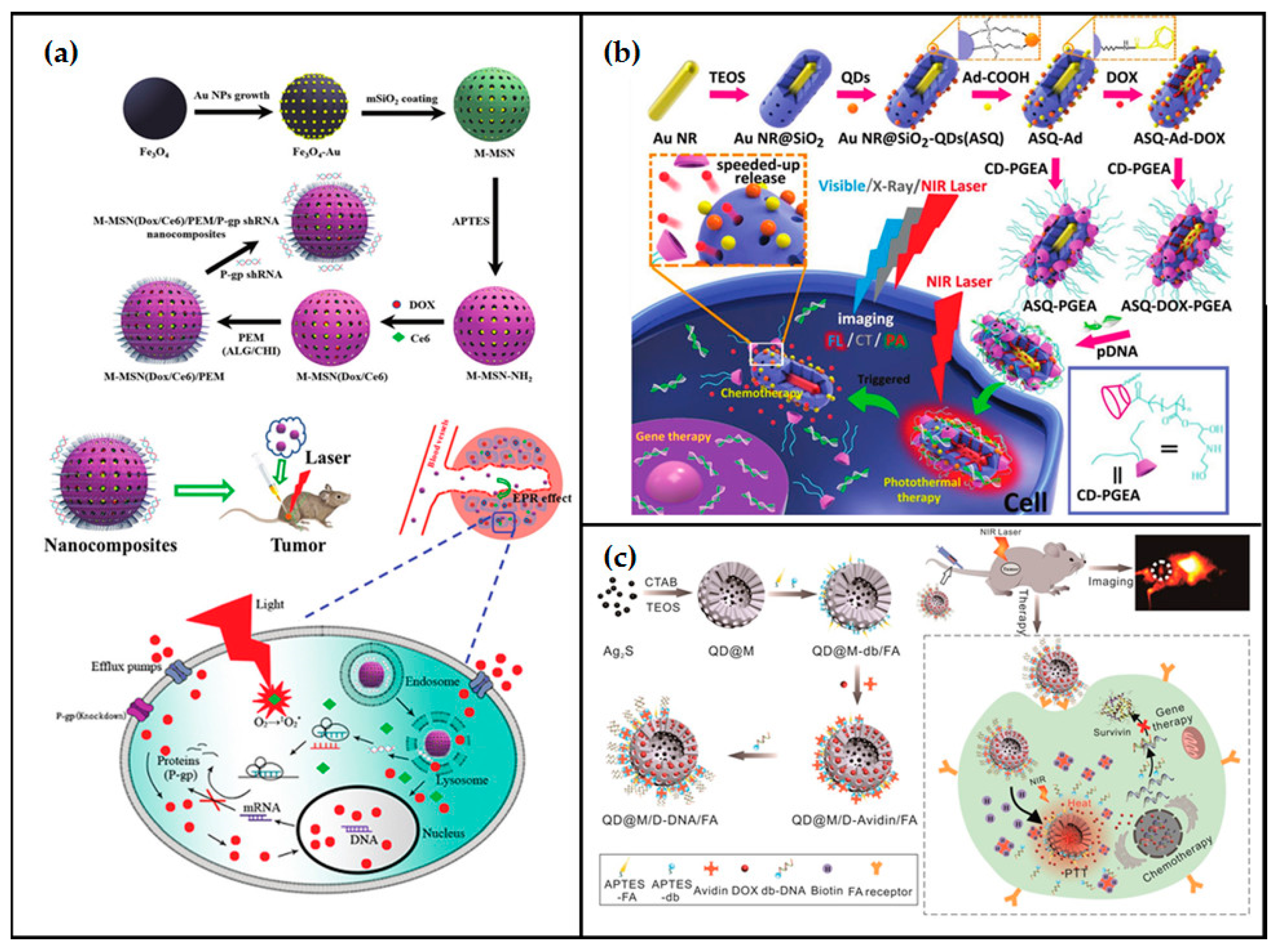
- Sun, L.; Wang, D.; Chen, Y.; Wang, L.; Huang, P.; Li, Y.; Liu, Z.; Yao, H.; Shi, J. Core-shell hierarchical mesostructured silica nanoparticles for gene/chemo-synergetic stepwise therapy of multidrug-resistant cancer. Biomaterials 2017, 133, 219–228. [Google Scholar] [CrossRef]
- Zhang, B.; Liu, Q.; Liu, M.; Shi, P.; Zhu, L.; Zhang, L.; Li, R. Biodegradable hybrid mesoporous silica nanoparticles for gene/chemo-synergetic therapy of breast cancer. J. Biomater. Appl. 2019, 33, 1382–1393. [Google Scholar] [CrossRef] [PubMed]
- Zhang, Q.; Shen, C.; Zhao, N.; Xu, F.J. Redox-Responsive and Drug-Embedded Silica Nanoparticles with Unique Self-Destruction Features for Efficient Gene/Drug Codelivery. Adv. Funct. Mater. 2017, 27, 1–12. [Google Scholar] [CrossRef]
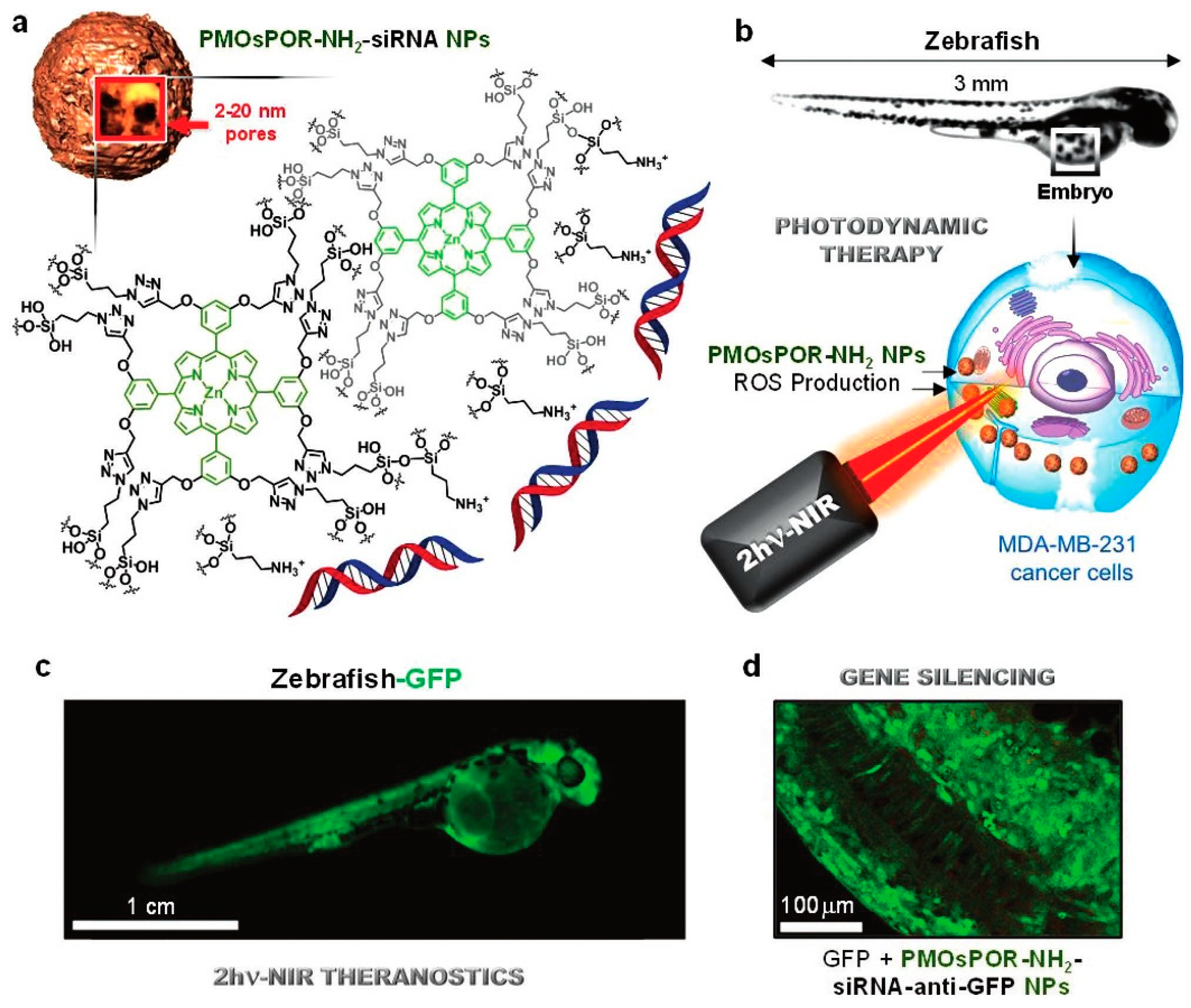
- Mauriello Jimenez, C.; Aggad, D.; Croissant, J.G.; Tresfield, K.; Laurencin, D.; Berthomieu, D.; Cubedo, N.; Rossel, M.; Alsaiari, S.; Anjum, D.H.; et al. Porous Porphyrin-Based Organosilica Nanoparticles for NIR Two-Photon Photodynamic Therapy and Gene Delivery in Zebrafish. Adv. Funct. Mater. 2018, 28, 1–12. [Google Scholar] [CrossRef]
- Ekineker, G.; Nguyen, C.; Bayir, S.; Dominguez Gil, S.; Işci, Ü.; Daurat, M.; Godefroy, A.; Raehm, L.; Charnay, C.; Oliviero, E.; et al. Phthalocyanine-based mesoporous organosilica nanoparticles: NIR photodynamic efficiency and siRNA photochemical internalization. Chem. Commun. 2019, 55, 11619–11622. [Google Scholar] [CrossRef] [PubMed]
- Santo, D.; Cordeiro, R.A.; Sousa, A.; Serra, A.; Coelho, J.F.J.; Faneca, H. Combination of Poly[(2-dimethylamino)ethyl methacrylate] and Poly(β-amino ester) Results in a Strong and Synergistic Transfection Activity. Biomacromolecules 2017, 18, 3331–3342. [Google Scholar] [CrossRef]
- Cordeiro, R.A.; Santo, D.; Farinha, D.; Serra, A.; Faneca, H.; Coelho, J.F.J. High transfection efficiency promoted by tailor-made cationic tri-block copolymer-based nanoparticles. Acta Biomater. 2017, 47, 113–123. [Google Scholar] [CrossRef] [PubMed]
- Ngamcherdtrakul, W.; Morry, J.; Gu, S.; Castro, D.J.; Goodyear, S.M.; Sangvanich, T.; Reda, M.M.; Lee, R.; Mihelic, S.A.; Beckman, B.L.; et al. Cationic polymer modified mesoporous silica nanoparticles for targeted siRNA delivery to HER2+ breast cancer. Adv. Funct. Mater. 2015, 25, 2646–2659. [Google Scholar] [CrossRef] [PubMed] [Green Version]
- Hom, C.; Lu, J.; Liong, M.; Luo, H.; Li, Z.; Zink, J.I.; Tamanoi, F. Mesoporous silica nanoparticles facilitate delivery of siRNA to shutdown signaling pathways in mammalian cells. Small 2010, 6, 1185–1190. [Google Scholar] [CrossRef] [PubMed] [Green Version]
- Zarei, H.; Kazemi Oskuee, R.; Hanafi-Bojd, M.Y.; Gholami, L.; Ansari, L.; Malaekeh-Nikouei, B. Enhanced gene delivery by polyethyleneimine coated mesoporous silica nanoparticles. Pharm. Dev. Technol. 2019, 24, 127–132. [Google Scholar] [CrossRef] [PubMed]
- Babaei, M.; Eshghi, H.; Abnous, K.; Rahimizadeh, M.; Ramezani, M. Promising gene delivery system based on polyethylenimine-modified silica nanoparticles. Cancer Gene Ther. 2017, 24, 156–164. [Google Scholar] [CrossRef]
- Zhang, X.; Zhang, J.; Quan, G.; Yang, P.; Pan, X.; Wu, C. The Serum-Resistant Transfection Evaluation and Long-Term Stability of Gene Delivery Dry Powder Based on Mesoporous Silica Nanoparticles and Polyethyleneimine by Freezing-Drying. AAPS PharmSciTech 2017, 18, 1536–1543. [Google Scholar] [CrossRef]
- Xia, T.; Kovochich, M.; Liong, M.; Meng, H.; Kabehie, S.; George, S.; Zink, J.I.; Nel, A.E. Polyethyleneimine coating enhances the cellular uptake of mesoporous silica nanoparticles and allows safe delivery of siRNA and DNA constructs. ACS Nano 2009, 3, 3273–3286. [Google Scholar] [CrossRef]
- Prabhakar, N.; Zhang, J.; Desai, D.; Casals, E.; Gulin-Sarfraz, T.; Näreoja, T.; Westermarck, J.; Rosenholm, J.M. Stimuli-responsive hybrid nanocarriers developed by controllable integration of hyperbranched PEI with mesoporous silica nanoparticles for sustained intracellular siRNA delivery. Int. J. Nanomedicine 2016, 11, 6591–6608. [Google Scholar] [CrossRef] [Green Version]
- Slita, A.; Egorova, A.; Casals, E.; Kiselev, A.; Rosenholm, J.M. Characterization of modified mesoporous silica nanoparticles as vectors for siRNA delivery. Asian J. Pharm. Sci. 2018, 13, 592–599. [Google Scholar] [CrossRef] [PubMed]
- Ngamcherdtrakul, W.; Sangvanich, T.; Reda, M.; Gu, S.; Bejan, D.; Yantasee, W. Lyophilization and stability of antibody-conjugated mesoporous silica nanoparticle with cationic polymer and PEG for siRNA delivery. Int. J. Nanomedicine 2018, 13, 4015–4027. [Google Scholar] [CrossRef] [Green Version]
- Oupicky, D.; Bhattarai, S.R.; Muthuswamy, E.; Wani, A.; Brichacek, M.; Castañeda, A.L.; Brock, S.L. Enhanced gene and siRNA delivery by polycation-modified mesoporous silica nanoparticles loaded with chloroquine. Pharm. Res. 2010, 27, 2556–2568. [Google Scholar]
- Lin, X.; Zhao, N.; Yan, P.; Hu, H.; Xu, F.J. The shape and size effects of polycation functionalized silica nanoparticles on gene transfection. Acta Biomater. 2015, 11, 381–392. [Google Scholar] [CrossRef]
- Lin, J.T.; Liu, Z.K.; Zhu, Q.L.; Rong, X.H.; Liang, C.L.; Wang, J.; Ma, D.; Sun, J.; Wang, G.H. Redox-responsive nanocarriers for drug and gene co-delivery based on chitosan derivatives modified mesoporous silica nanoparticles. Colloids Surfaces B Biointerfaces 2017, 155, 41–50. [Google Scholar] [CrossRef]
- Kar, M.; Tiwari, N.; Tiwari, M.; Lahiri, M.; Gupta, S. Sen Poly-L-arginine grafted silica mesoporous nanoparticles for enhanced cellular uptake and their application in DNA delivery and controlled drug release. Part. Part. Syst. Charact. 2013, 30, 166–179. [Google Scholar] [CrossRef]
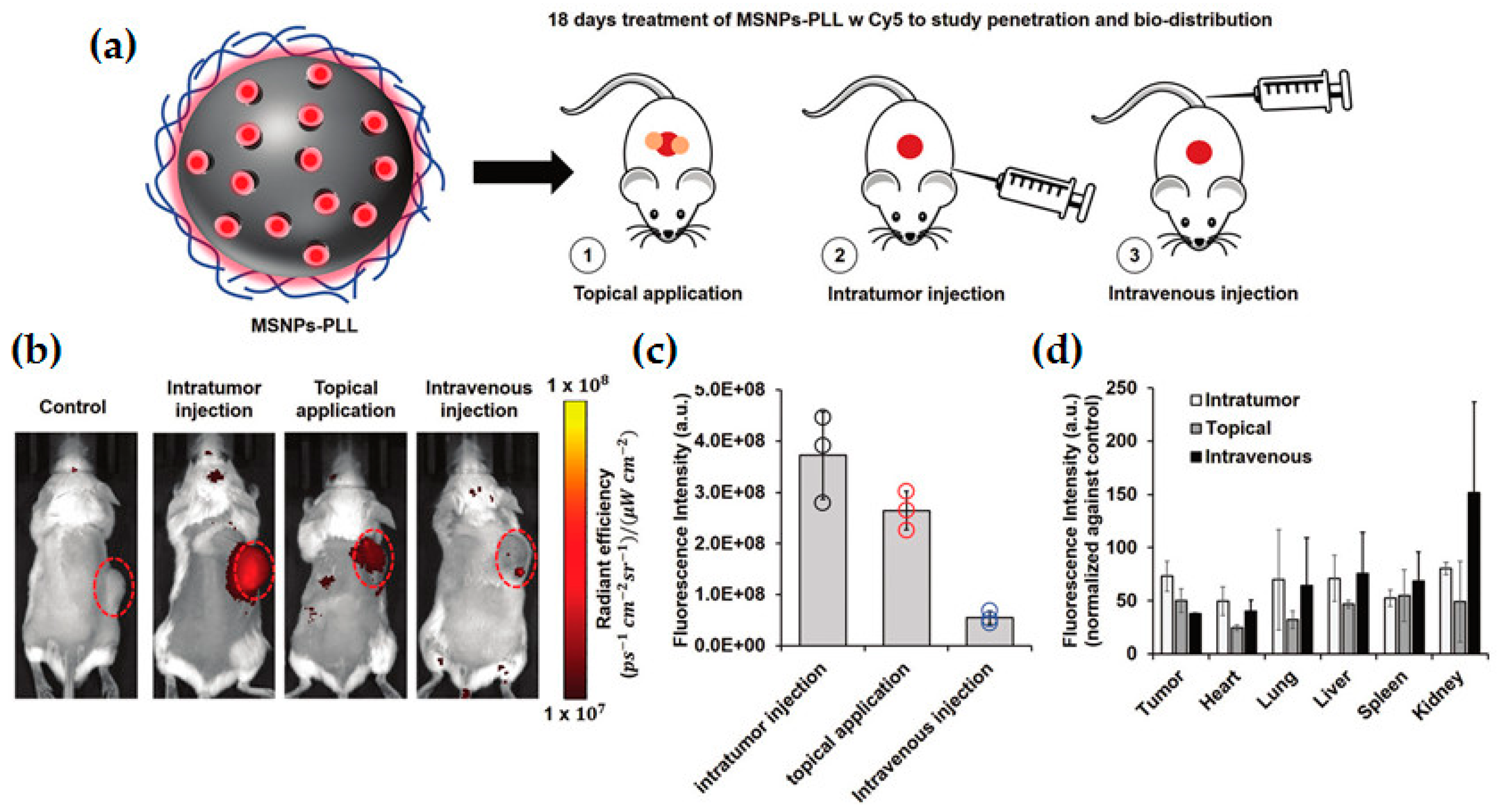
- Lio, D.C.S.; Liu, C.; Oo, M.M.S.; Wiraja, C.; Teo, M.H.Y.; Zheng, M.; Chew, S.W.T.; Wang, X.; Xu, C. Transdermal delivery of small interfering RNAs with topically applied mesoporous silica nanoparticles for facile skin cancer treatment. Nanoscale 2019, 11, 17041–17051. [Google Scholar] [CrossRef] [PubMed]
- Patel, S.; Kim, J.; Herrera, M.; Mukherjee, A.; Kabanov, A.V.; Sahay, G. Brief update on endocytosis of nanomedicines. Adv. Drug Deliv. Rev. 2019, 144, 90–111. [Google Scholar] [CrossRef] [PubMed]
- Freeman, E.C.; Weiland, L.M.; Meng, W.S. Modeling the proton sponge hypothesis: Examining proton sponge effectiveness for enhancing intracellular gene delivery through multiscale modeling. J. Biomater. Sci. Polym. Ed. 2013, 24, 398–416. [Google Scholar] [CrossRef] [Green Version]
- Möller, K.; Müller, K.; Engelke, H.; Bräuchle, C.; Wagner, E.; Bein, T. Highly efficient siRNA delivery from core-shell mesoporous silica nanoparticles with multifunctional polymer caps. Nanoscale 2016, 8, 4007–4019. [Google Scholar] [CrossRef] [PubMed] [Green Version]
- Li, R.; Liu, T.; Wang, K. Hyaluronic modified and amine-functionalized silica nanoparticles as intracellular siRNA delivery carriers in lung cancer gene therapy. Int. J. Clin. Exp. Med. 2016, 9, 10191–10200. [Google Scholar]
- Shi, X.L.; Li, Y.; Zhao, L.M.; Su, L.W.; Ding, G. Delivery of MTH1 inhibitor (TH287) and MDR1 siRNA via hyaluronic acid-based mesoporous silica nanoparticles for oral cancers treatment. Colloids Surf. B Biointerfaces 2019, 173, 599–606. [Google Scholar] [CrossRef] [PubMed]
- Li, Y.; Duo, Y.; Zhai, P.; He, L.; Zhong, K.; Zhang, Y.; Huang, K.; Luo, J.; Zhang, H.; Yu, X. Targeted delivery of anti-mir-155 by functionalized mesoporous silica nanoparticles for colorectal cancer therapy. Nanomedicine 2018, 13, nnm-2017-0353. [Google Scholar] [CrossRef] [PubMed]
- Liberman, A.; Mendez, N.; Trogler, W.C.; Kummel, A.C. Synthesis and surface functionalization of silica nanoparticles for nanomedicine. Surf. Sci. Rep. 2014, 69, 132–158. [Google Scholar] [CrossRef] [Green Version]
- Yang, Y.; Yu, C. Advances in silica based nanoparticles for targeted cancer therapy. Nanomed. Nanotechnol. Biol. Med. 2016, 12, 317–332. [Google Scholar] [CrossRef]
- Morry, J.; Ngamcherdtrakul, W.; Gu, S.; Reda, M.; Castro, D.J.; Sangvanich, T.; Gray, J.W.; Yantasee, W. Targeted treatment of metastatic breast cancer by PLK1 siRNA delivered by an antioxidant nanoparticle platform. Mol. Cancer Ther. 2017, 16, 763–772. [Google Scholar] [CrossRef] [PubMed] [Green Version]
- Zhou, S.; Ding, C.; Wang, C.; Fu, J. UV-light cross-linked and pH de-cross-linked coumarin-decorated cationic copolymer grafted mesoporous silica nanoparticles for drug and gene co-delivery in vitro. Mater. Sci. Eng. C 2020, 108, 110469. [Google Scholar] [CrossRef]
- Keasberry, N.A.; Yapp, C.W.; Idris, A. Mesoporous silica nanoparticles as a carrier platform for intracellular delivery of nucleic acids. Biochem. 2017, 82, 655–662. [Google Scholar] [CrossRef] [PubMed]
- Zhu, Y.; Meng, W.; Gao, H.; Hanagata, N. Hollow mesoporous silica/poly(l-lysine) particles for codelivery of drug and gene with enzyme-triggered release property. J. Phys. Chem. C 2011, 115, 13630–13636. [Google Scholar] [CrossRef] [Green Version]
- Ahir, M.; Upadhyay, P.; Ghosh, A.; Sarker, S.; Bhattacharya, S.; Gupta, P.; Ghosh, S.; Chattopadhyay, S.; Adhikary, A. Delivery of dual miRNA through CD44-targeted mesoporous silica nanoparticles for enhanced and effective triple-negative breast cancer therapy. Biomater. Sci. 2020. [Google Scholar] [CrossRef]
- Passadouro, M.; Faneca, H. Managing Pancreatic Adenocarcinoma: A Special Focus in MicroRNA Gene Therapy. Int. J. Mol. Sci. 2016, 17, 718. [Google Scholar] [CrossRef] [PubMed] [Green Version]
- Simões, S.; Filipe, A.; Faneca, H.; Mano, M.; Penacho, N.; Düzgünes, N.; de Lima, M.P. Cationic liposomes for gene delivery. Expert Opin. Drug Deliv. 2005, 2, 237–254. [Google Scholar] [CrossRef] [PubMed]
- Van Schooneveld, M.M.; Vucic, E.; Koole, R.; Zhou, Y.; Stocks, J.; Cormode, D.P.; Tang, C.Y.; Gordon, R.E.; Nicolay, K.; Meijerink, A.; et al. Improved biocompatibility and pharmacokinetics of silica nanoparticles by means of a lipid coating: A multimodality investigation. Nano Lett. 2008, 8, 2517–2525. [Google Scholar] [CrossRef] [PubMed] [Green Version]
- Wang, L.-S.; Wu, L.-C.; Lu, S.-Y.; Chang, L.-L.; Teng, I.-T.; Yang, C.-M.; Ho, J.A. Biofunctionalized Phospholipid-Capped Mesoporous Silica Nanoshuttles for Targeted Drug Delivery: Improved Water Suspensibility and Decreased Nonspecific Protein Binding. ACS Nano 2010, 4, 4371–4379. [Google Scholar] [CrossRef]
- Roggers, R.A.; Lin, V.S.Y.; Trewyn, B.G. Chemically reducible lipid bilayer coated mesoporous silica nanoparticles demonstrating controlled release and HeLa and normal mouse liver cell biocompatibility and cellular internalization. Mol. Pharm. 2012, 9, 2770–2777. [Google Scholar] [CrossRef]
- Cauda, V.; Engelke, H.; Sauer, A.; Arcizet, D.; Bräuchle, C.; Rädler, J.; Bein, T. Colchicine-loaded lipid bilayer-coated 50 nm mesoporous nanoparticles efficiently induce microtubule depolymerization upon cell uptake. Nano Lett. 2010, 10, 2484–2492. [Google Scholar] [CrossRef] [PubMed]
- Desai, D.; Zhang, J.; Sandholm, J.; Lehtimäki, J.; Grönroos, T.; Tuomela, J.; Rosenholm, J.M. Lipid Bilayer-Gated Mesoporous Silica Nanocarriers for Tumor-Targeted Delivery of Zoledronic Acid in vivo. Mol. Pharm. 2017, 14, 3218–3227. [Google Scholar] [CrossRef]
- Durfee, P.N.; Lin, Y.S.; Dunphy, D.R.; Muñiz, A.J.; Butler, K.S.; Humphrey, K.R.; Lokke, A.J.; Agola, J.O.; Chou, S.S.; Chen, I.M.; et al. Mesoporous Silica Nanoparticle-Supported Lipid Bilayers (Protocells) for Active Targeting and Delivery to Individual Leukemia Cells. ACS Nano 2016, 10, 8325–8345. [Google Scholar] [CrossRef]
- Meng, H.; Wang, M.; Liu, H.; Liu, X.; Situ, A.; Wu, B.; Ji, Z.; Chang, C.H.; Nel, A.E. Use of a Lipid-Coated Mesoporous Silica Nanoparticle Platform for Synergistic Gemcitabine and Paclitaxel Delivery to Human Pancreatic Cancer in Mice. ACS Nano 2015, 9, 3540–3557. [Google Scholar] [CrossRef] [PubMed] [Green Version]
- Shi, H.; Liu, S.; Cheng, J.; Yuan, S.; Yang, Y.; Fang, T.; Cao, K.; Wei, K.; Zhang, Q.; Liu, Y. Charge-Selective Delivery of Proteins Using Mesoporous Silica Nanoparticles Fused with Lipid Bilayers. ACS Appl. Mater. Interfaces 2019, 11, 3645–3653. [Google Scholar] [CrossRef] [PubMed]
- Xue, H.; Yu, Z.; Liu, Y.; Yuan, W.; Yang, T.; You, J.; He, X.; Lee, R.J.; Li, L.; Xu, C. Delivery of miR-375 and doxorubicin hydrochloride by lipid-coated hollow mesoporous silica nanoparticles to overcome multiple drug resistance in hepatocellular carcinoma. Int. J. Nanomedicine 2017, 12, 5271–5287. [Google Scholar] [CrossRef] [PubMed] [Green Version]
- Wang, Y.; Xie, Y.; Kilchrist, K.V.; Li, J.; Duvall, C.L.; Oupický, D. Endosomolytic and Tumor-Penetrating Mesoporous Silica Nanoparticles for siRNA/miRNA Combination Cancer Therapy. ACS Appl. Mater. Interfaces 2020, 12, 4308–4322. [Google Scholar] [CrossRef] [PubMed]
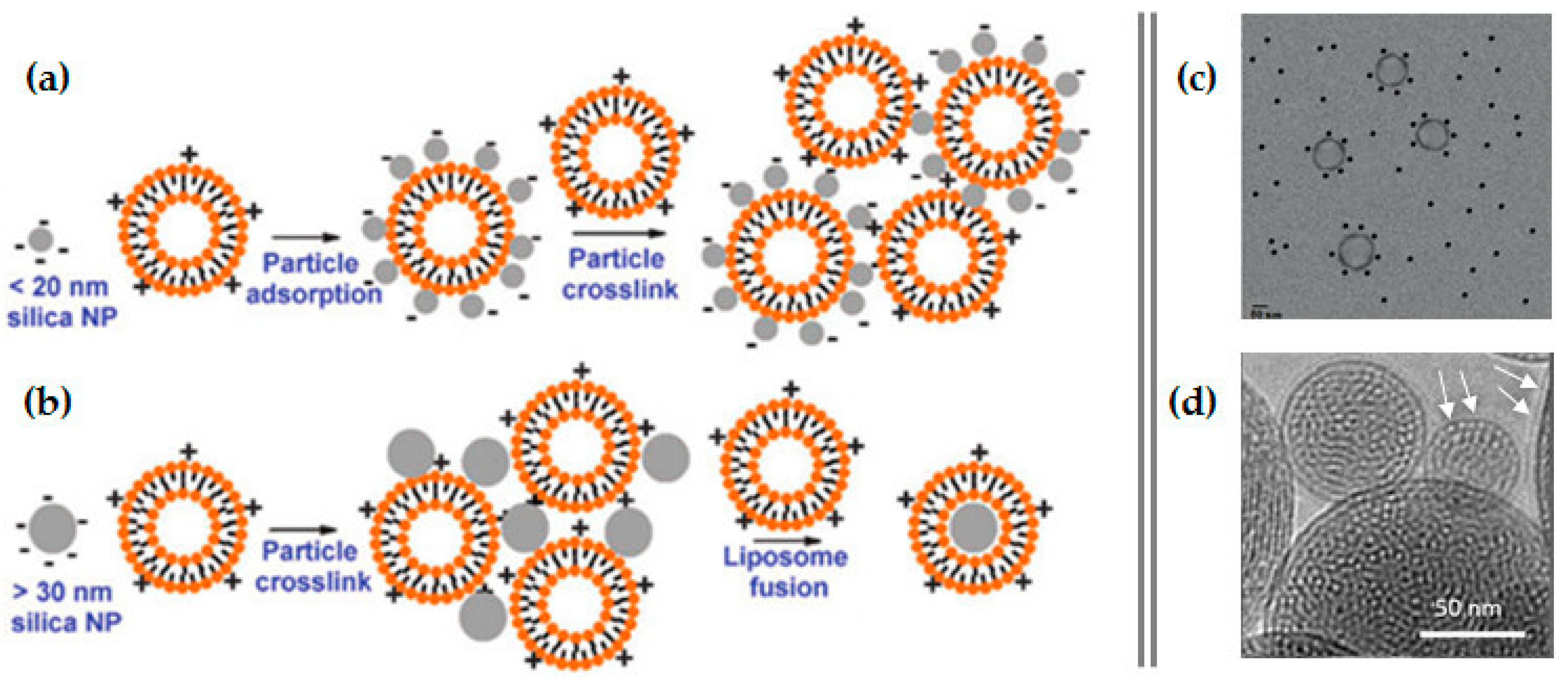
- Liu, J.; Stace-Naughton, A.; Brinker, C.J. Silica nanoparticle supported lipid bilayers for gene delivery. Chem. Commun. 2009, 5100–5102. [Google Scholar] [CrossRef]
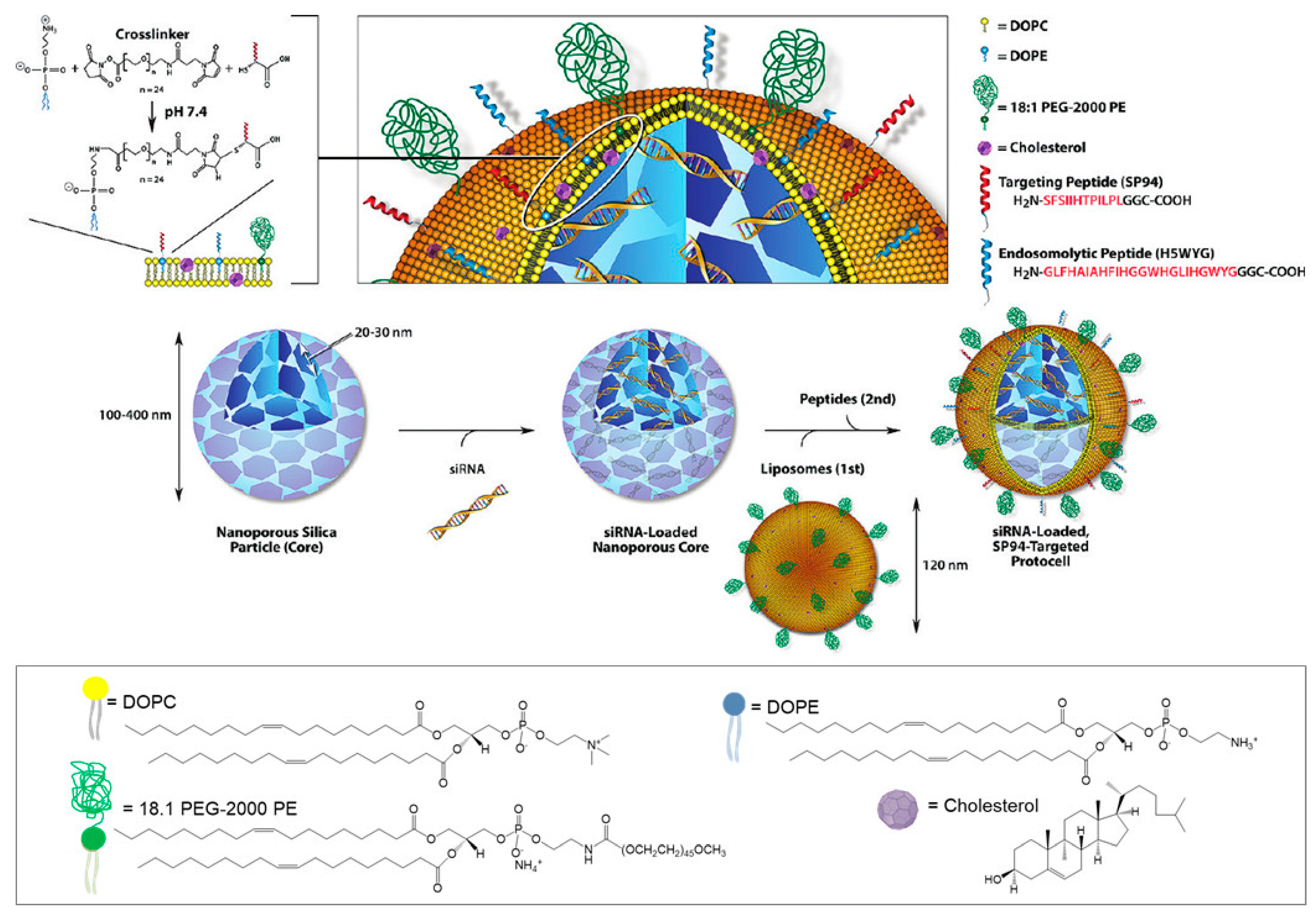
- Ashley, C.E.; Carnes, E.C.; Phillips, G.K.; Padilla, D.; Durfee, P.N.; Brown, P.A.; Hanna, T.N.; Liu, J.; Phillips, B.; Carter, M.B.; et al. The targeted delivery of multicomponent cargos to cancer cells by nanoporous particle-supported lipid bilayers. Nat. Mater. 2011, 10, 389–397. [Google Scholar] [CrossRef] [Green Version]
- Ashley, C.E.; Carnes, E.C.; Epler, K.E.; Padilla, D.P.; Phillips, G.K.; Castillo, R.E.; Wilkinson, D.C.; Wilkinson, B.S.; Burgard, C.A.; Kalinich, R.M.; et al. Delivery of small interfering RNA by peptide-targeted mesoporous silica nanoparticle-supported lipid bilayers. ACS Nano 2012, 6, 2174–2188. [Google Scholar] [CrossRef] [PubMed]
- Savarala, S.; Ahmed, S.; Ilies, M.A.; Wunder, S.L. Stabilization of soft lipid colloids: Competing effects of nanoparticle decoration and supported lipid bilayer formation. ACS Nano 2011, 5, 2619–2628. [Google Scholar] [CrossRef]
- Dengler, E.C.; Liu, J.; Kerwin, A.; Torres, S.; Olcott, C.M.; Bowman, B.N.; Armijo, L.; Gentry, K.; Wilkerson, J.; Wallace, J.; et al. Mesoporous silica-supported lipid bilayers (protocells) for DNA cargo delivery to the spinal cord. J. Control. Release 2013, 168, 209–224. [Google Scholar] [CrossRef] [PubMed] [Green Version]
- Colapicchioni, V.; Palchetti, S.; Pozzi, D.; Marini, E.S.; Riccioli, A.; Ziparo, E.; Papi, M.; Amenitsch, H.; Caracciolo, G. Killing cancer cells using nanotechnology: Novel poly(I:C) loaded liposome-silica hybrid nanoparticles. J. Mater. Chem. B 2015, 3, 7408–7416. [Google Scholar] [CrossRef]
- Lee, J.E.; Lee, N.; Kim, H.; Kim, J.; Choi, S.H.; Kim, J.H.; Kim, T.; Song, I.C.; Park, S.P.; Moon, W.K.; et al. Uniform Mesoporous Dye-Doped Silica Nanoparticles Decorated with Multiple Magnetite Nanocrystals for Simultaneous Enhanced Magnetic Resonance Imaging, Fluorescence Imaging, and Drug Delivery. J. Am. Chem. Soc. 2010, 132, 552–557. [Google Scholar] [CrossRef]
- Li, E.; Yang, Y.; Hao, G.; Yi, X.; Zhang, S.; Pan, Y.; Xing, B.; Gao, M. Multifunctional Magnetic Mesoporous Silica Nanoagents for in vivo Enzyme-Responsive Drug Delivery and MR Imaging. Nanotheranostics 2018, 2, 233–242. [Google Scholar] [CrossRef] [Green Version]
- Iqbal, M.Z.; Ma, X.; Chen, T.; Zhang, L.; Ren, W.; Xiang, L.; Wu, A. Silica-coated super-paramagnetic iron oxide nanoparticles (SPIONPs): A new type contrast agent of T 1 magnetic resonance imaging (MRI). J. Mater. Chem. B 2015, 3, 5172–5181. [Google Scholar] [CrossRef]
- Vaz-Ramos, J.; Cordeiro, R.; Castro, M.M.C.A.; Geraldes, C.F.G.C.; Costa, B.F.O.; Faneca, H.; Durães, L. Supercritically dried superparamagnetic mesoporous silica nanoparticles for cancer theranostics. Mater. Sci. Eng. C 2020, 111124. [Google Scholar] [CrossRef] [PubMed]
- Pisani, C.; Rascol, E.; Dorandeu, C.; Charnay, C.; Guari, Y.; Chopineau, J.; Devoisselle, J.M.; Prat, O. Biocompatibility assessment of functionalized magnetic mesoporous silica nanoparticles in human HepaRG cells. Nanotoxicology 2017, 11, 871–890. [Google Scholar] [CrossRef]
- Hartono, S.B.; Yu, M.; Gu, W.; Yang, J.; Strounina, E.; Wang, X.; Qiao, S.; Yu, C. Synthesis of multi-functional large pore mesoporous silica nanoparticles as gene carriers. Nanotechnology 2014, 25. [Google Scholar] [CrossRef]
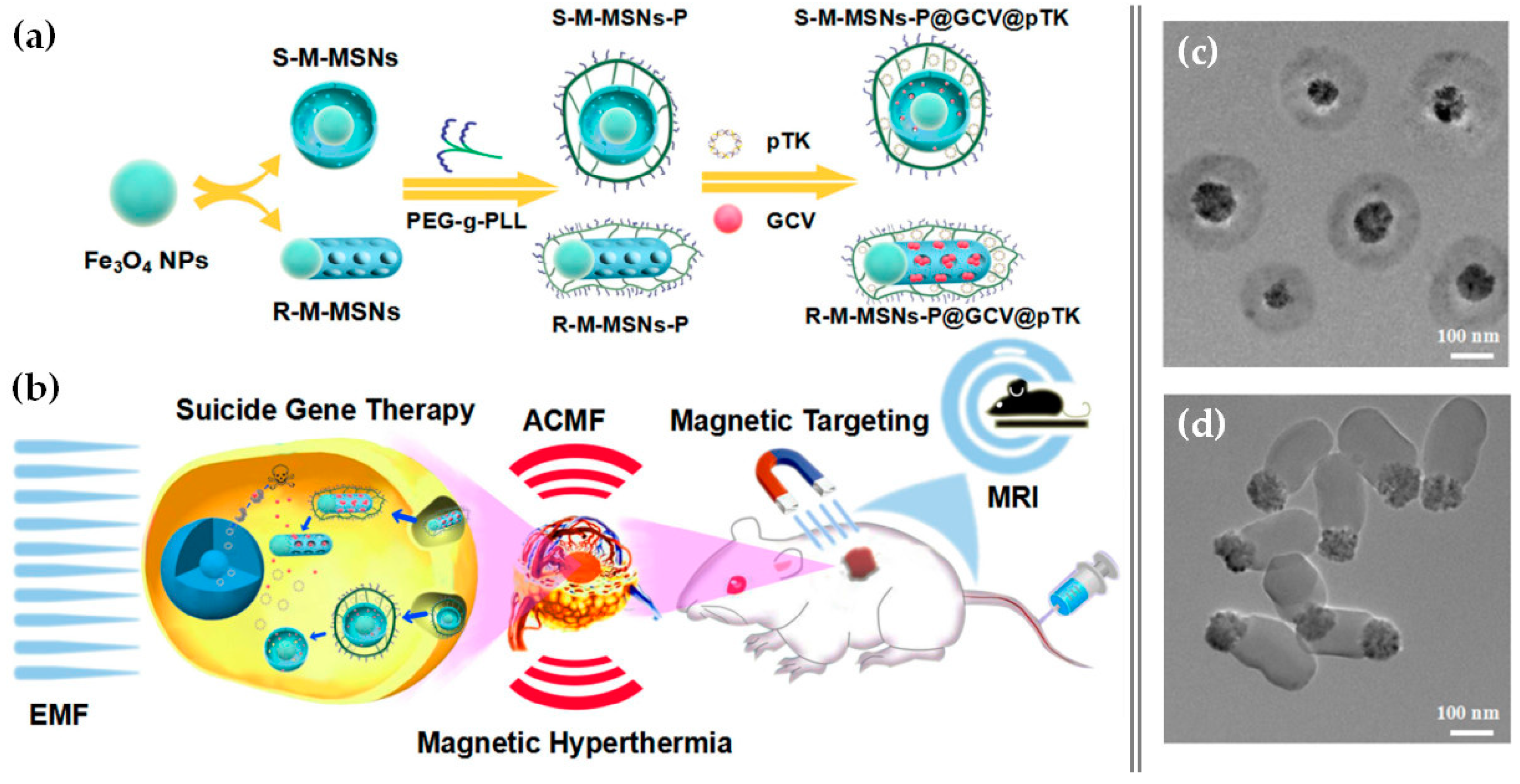
- Wang, Z.; Chang, Z.; Lu, M.; Shao, D.; Yue, J.; Yang, D.; Zheng, X.; Li, M.; He, K.; Zhang, M.; et al. Shape-controlled magnetic mesoporous silica nanoparticles for magnetically-mediated suicide gene therapy of hepatocellular carcinoma. Biomaterials 2018, 154, 147–157. [Google Scholar] [CrossRef] [PubMed]
- Wang, Y.; Gu, H. Core-shell-type magnetic mesoporous silica nanocomposites for bioimaging and therapeutic agent delivery. Adv. Mater. 2015, 27, 576–585. [Google Scholar] [CrossRef] [PubMed]
- Xiong, L.; Bi, J.; Tang, Y.; Qiao, S.Z. Magnetic Core-Shell Silica Nanoparticles with Large Radial Mesopores for siRNA Delivery. Small 2016, 12, 4735–4742. [Google Scholar] [CrossRef]
- Kim, J.; Lee, J.E.; Lee, J.; Yu, J.H.; Kim, B.C.; An, K.; Hwang, Y.; Shin, C.-H.; Park, J.-G.; Kim, J.; et al. Magnetic Fluorescent Delivery Vehicle Using Uniform Mesoporous Silica Spheres Embedded with Monodisperse Magnetic and Semiconductor Nanocrystals. J. Am. Chem. Soc. 2006, 128, 688–689. [Google Scholar] [CrossRef]
- Li, X.; Chen, Y.; Wang, M.; Ma, Y.; Xia, W.; Gu, H. A mesoporous silica nanoparticle - PEI - Fusogenic peptide system for siRNA delivery in cancer therapy. Biomaterials 2013, 34, 1391–1401. [Google Scholar] [CrossRef]
- Chen, Y.; Gu, H.; Zhang, D.S.Z.; Li, F.; Liu, T.; Xia, W. Highly effective inhibition of lung cancer growth and metastasis by systemic delivery of siRNA via multimodal mesoporous silica-based nanocarrier. Biomaterials 2014, 35, 10058–10069. [Google Scholar] [CrossRef] [PubMed]
- Chen, Y.; Wang, X.; Liu, T.; Zhang, D.S.Z.; Wang, Y.; Gu, H.; Di, W. Highly effective antiangiogenesis via magnetic mesoporous silica-based siRNA vehicle targeting the VEGF gene for orthotopic ovarian cancer therapy. Int. J. Nanomedicine 2015, 10, 2579–2594. [Google Scholar] [PubMed] [Green Version]
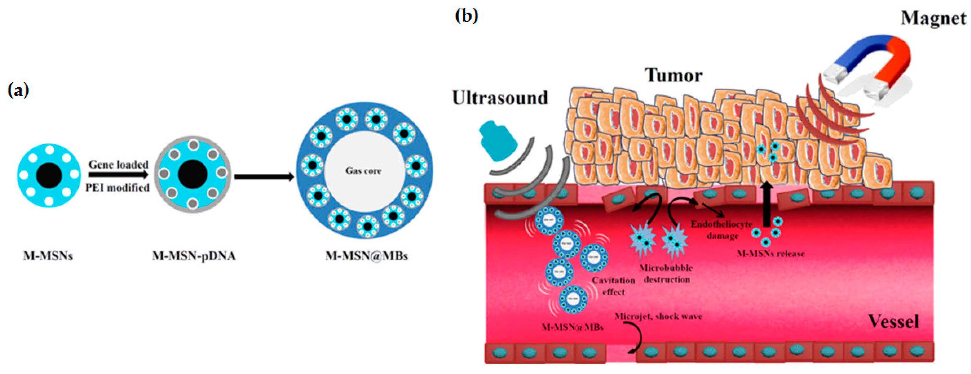
- Du, M.; Chen, Y.; Tu, J.; Liufu, C.; Yu, J.; Yuan, Z.; Gong, X.; Chen, Z. Ultrasound Responsive Magnetic Mesoporous Silica Nanoparticle-Loaded Microbubbles for Efficient Gene Delivery. ACS Biomater. Sci. Eng. 2020. [Google Scholar] [CrossRef]
- Yang, H.; Chen, Y.; Chen, Z.; Geng, Y.; Xie, X.; Shen, X.; Li, T.; Li, S.; Wu, C.; Liu, Y. Chemo-photodynamic combined gene therapy and dual-modal cancer imaging achieved by pH-responsive alginate/chitosan multilayer-modified magnetic mesoporous silica nanocomposites. Biomater. Sci. 2017, 5, 1001–1013. [Google Scholar] [CrossRef] [PubMed]
- Duan, S.; Yang, Y.; Zhang, C.; Zhao, N.; Xu, F.J. NIR-Responsive Polycationic Gatekeeper-Cloaked Hetero-Nanoparticles for Multimodal Imaging-Guided Triple-Combination Therapy of Cancer. Small 2017, 13, 1–10. [Google Scholar] [CrossRef] [PubMed]
- Li, C.; Yang, X.Q.; Zhang, M.Z.; Song, Y.Y.; Cheng, K.; An, J.; Zhang, X.S.; Xuan, Y.; Liu, B.; Zhao, Y. Di In vivo imaging-guided nanoplatform for tumor targeting delivery and combined chemo-, gene- and photothermal therapy. Theranostics 2018, 8, 5662–5675. [Google Scholar] [CrossRef]
- Ziello, J.E.; Huang, Y.; Jovin, I.S. Cellular Endocytosis and Gene Delivery. Mol. Med. 2010, 16, 222–229. [Google Scholar] [CrossRef]
- El-Sayed, A.; Harashima, H. Endocytosis of Gene Delivery Vectors: From Clathrin-dependent to Lipid Raft-mediated Endocytosis. Mol. Ther. 2013, 21, 1118–1130. [Google Scholar] [CrossRef] [Green Version]
- Huang, X.; Teng, X.; Chen, D.; Tang, F.; He, J. The effect of the shape of mesoporous silica nanoparticles on cellular uptake and cell function. Biomaterials 2010, 31, 438–448. [Google Scholar] [CrossRef] [PubMed]
- Lu, F.; Wu, S.-H.; Hung, Y.; Mou, C.-Y. Size Effect on Cell Uptake in Well-Suspended, Uniform Mesoporous Silica Nanoparticles. Small 2009, 5, 1408–1413. [Google Scholar] [CrossRef] [PubMed]
- Zhu, J.; Tang, J.; Zhao, L.; Zhou, X.; Wang, Y.; Yu, C. Ultrasmall, Well-Dispersed, Hollow Siliceous Spheres with Enhanced Endocytosis Properties. Small 2010, 6, 276–282. [Google Scholar] [CrossRef]
- Kim, S.; Na, H.-K.; Won, C.; Min, D.-H. In-depth study on the gene silencing capability of silica nanoparticles with different pore sizes: Degree and duration of RNA interference. RSC Adv. 2016, 6, 27143–27150. [Google Scholar] [CrossRef]
- Pinese, C.; Lin, J.; Milbreta, U.; Li, M.; Wang, Y.; Leong, K.W.; Chew, S.Y. Sustained delivery of siRNA/mesoporous silica nanoparticle complexes from nanofiber scaffolds for long-term gene silencing. Acta Biomater. 2018, 76, 164–177. [Google Scholar] [CrossRef] [PubMed]
- Hartono, S.B.; Gu, W.; Kleitz, F.; Liu, J.; He, L.; Middelberg, A.P.J.; Yu, C.; Lu, G.Q.; Qiao, S.Z. Poly-l-lysine Functionalized Large Pore Cubic Mesostructured Silica Nanoparticles as Biocompatible Carriers for Gene Delivery. ACS Nano 2012, 6, 2104–2117. [Google Scholar] [CrossRef] [PubMed] [Green Version]
- Brevet, D.; Hocine, O.; Delalande, A.; Raehm, L.; Charnay, C.; Midoux, P.; Durand, J.-O.; Pichon, C. Improved gene transfer with histidine-functionalized mesoporous silica nanoparticles. Int. J. Pharm. 2014, 471, 197–205. [Google Scholar] [CrossRef] [PubMed]
- Radu, D.R.; Lai, C.-Y.; Jeftinija, K.; Rowe, E.W.; Jeftinija, S.; Lin, V.S.-Y. A Polyamidoamine Dendrimer-Capped Mesoporous Silica Nanosphere-Based Gene Transfection Reagent. J. Am. Chem. Soc. 2004, 126, 13216–13217. [Google Scholar] [CrossRef] [PubMed]
- Li, Y.; Hei, M.; Xu, Y.; Qian, X.; Zhu, W. Ammonium salt modified mesoporous silica nanoparticles for dual intracellular-responsive gene delivery. Int. J. Pharm. 2016, 511, 689–702. [Google Scholar] [CrossRef]
- Leidner, A.; Weigel, S.; Bauer, J.; Reiber, J.; Angelin, A.; Grösche, M.; Scharnweber, T.; Niemeyer, C.M. Biopebbles: DNA-Functionalized Core-Shell Silica Nanospheres for Cellular Uptake and Cell Guidance Studies. Adv. Funct. Mater. 2018, 28, 1707572. [Google Scholar] [CrossRef]
- Roy, I.; Ohulchanskyy, T.Y.; Bharali, D.J.; Pudavar, H.E.; Mistretta, R.A.; Kaur, N.; Prasad, P.N. Optical tracking of organically modified silica nanoparticles as DNA carriers: A nonviral, nanomedicine approach for gene delivery. Proc. Natl. Acad. Sci. USA 2005, 102, 279–284. [Google Scholar] [CrossRef] [PubMed] [Green Version]
- Li, T.; Shen, X.; Geng, Y.; Chen, Z.; Li, L.; Li, S.; Yang, H.; Wu, C.; Zeng, H.; Liu, Y. Folate-Functionalized Magnetic-Mesoporous Silica Nanoparticles for Drug/Gene Codelivery To Potentiate the Antitumor Efficacy. ACS Appl. Mater. Interfaces 2016, 8, 13748–13758. [Google Scholar] [CrossRef] [PubMed]
- Park, I.Y.; Kim, I.Y.; Yoo, M.K.; Choi, Y.J.; Cho, M.-H.; Cho, C.S. Mannosylated polyethylenimine coupled mesoporous silica nanoparticles for receptor-mediated gene delivery. Int. J. Pharm. 2008, 359, 280–287. [Google Scholar] [CrossRef] [PubMed]
- Zanta, M.-A.; Boussif, O.; Adib, A.; Behr, J.-P. In Vitro Gene Delivery to Hepatocytes with Galactosylated Polyethylenimine. Bioconjug. Chem. 1997, 8, 839–844. [Google Scholar] [CrossRef] [PubMed]
- Gandra, N.; Wang, D.-D.; Zhu, Y.; Mao, C. Virus-Mimetic Cytoplasm-Cleavable Magnetic/Silica Nanoclusters for Enhanced Gene Delivery to Mesenchymal Stem Cells. Angew. Chemie Int. Ed. 2013, 52, 11278–11281. [Google Scholar] [CrossRef] [PubMed] [Green Version]
- Mykhaylyk, O.; Sobisch, T.; Almstätter, I.; Sanchez-Antequera, Y.; Brandt, S.; Anton, M.; Döblinger, M.; Eberbeck, D.; Settles, M.; Braren, R.; et al. Silica-iron oxide magnetic nanoparticles modified for gene delivery: A search for optimum and quantitative criteria. Pharm. Res. 2012, 29, 1344–1365. [Google Scholar] [CrossRef] [PubMed]
- Singh, P.; Srivastava, S.; Singh, S.K. Nanosilica: Recent Progress in Synthesis, Functionalization, Biocompatibility, and Biomedical Applications. ACS Biomater. Sci. Eng. 2019, 5, 4882–4898. [Google Scholar] [CrossRef]
- Narayan, R.; Nayak, U.Y.; Raichur, A.M.; Garg, S. Mesoporous silica nanoparticles: A comprehensive review on synthesis and recent advances. Pharmaceutics 2018, 10, 118. [Google Scholar] [CrossRef] [Green Version]
- Mebert, A.M.; Baglole, C.J.; Desimone, M.F.; Maysinger, D. Nanoengineered silica: Properties, applications and toxicity. Food Chem. Toxicol. 2017, 109, 753–770. [Google Scholar] [CrossRef]
- Croissant, J.G.; Fatieiev, Y.; Almalik, A.; Khashab, N.M. Mesoporous Silica and Organosilica Nanoparticles: Physical Chemistry, Biosafety, Delivery Strategies, and Biomedical Applications. Adv. Healthc. Mater. 2018, 7, 1–75. [Google Scholar] [CrossRef] [Green Version]
- Lehman, S.E.; Morris, A.S.; Mueller, P.S.; Salem, A.K.; Grassian, V.H.; Larsen, S.C. Silica nanoparticle-generated ROS as a predictor of cellular toxicity: Mechanistic insights and safety by design. Environ. Sci. Nano 2016, 3, 56–66. [Google Scholar] [CrossRef] [PubMed] [Green Version]
- Kim, I.Y.; Joachim, E.; Choi, H.; Kim, K. Toxicity of silica nanoparticles depends on size, dose, and cell type. Nanomed. Nanotechnol. Biol. Med. 2015, 11, 1407–1416. [Google Scholar] [CrossRef] [PubMed]
- Yu, K.O.; Grabinski, C.M.; Schrand, A.M.; Murdock, R.C.; Wang, W.; Gu, B.; Schlager, J.J.; Hussain, S.M. Toxicity of amorphous silica nanoparticles in mouse keratinocytes. J. Nanoparticle Res. 2009, 11, 15–24. [Google Scholar] [CrossRef]
- Li, Z.; Zhang, Y.; Feng, N. Mesoporous silica nanoparticles: Synthesis, classification, drug loading, pharmacokinetics, biocompatibility, and application in drug delivery. Expert Opin. Drug Deliv. 2019, 16, 219–237. [Google Scholar] [CrossRef]
- Murugadoss, S.; Lison, D.; Godderis, L.; Van Den Brule, S.; Mast, J.; Brassinne, F.; Sebaihi, N.; Hoet, P.H. Toxicology of silica nanoparticles: An update. Arch. Toxicol. 2017, 91, 2967–3010. [Google Scholar] [CrossRef] [PubMed]
- Yang, Y.; Du, X.; Wang, Q.; Liu, J.; Zhang, E.; Sai, L.; Peng, C.; Lavin, M.F.; Yeo, A.J.; Yang, X.; et al. Mechanism of cell death induced by silica nanoparticles in hepatocyte cells is by apoptosis. Int. J. Mol. Med. 2019, 44, 903–912. [Google Scholar] [CrossRef] [PubMed] [Green Version]
- Li, J.; Yang, H.; Sha, S.; Li, J.; Zhou, Z.; Cao, Y. Evaluation of in vitro toxicity of silica nanoparticles (NPs) to lung cells: Influence of cell types and pulmonary surfactant component DPPC. Ecotoxicol. Environ. Saf. 2019, 186, 109770. [Google Scholar] [CrossRef] [PubMed]
- Chauhan, S.; Manivasagam, G.; Kumar, P.; Ambasta, R.K. Cellular Toxicity of Mesoporous Silica Nanoparticle in SHSY5Y and BMMNCs Cell. Pharm. Nanotechnol. 2018, 6, 245–252. [Google Scholar] [CrossRef]
- Zhang, Q.; Xu, H.; Zheng, S.; Su, M.; Wang, J. Genotoxicity of mesoporous silica nanoparticles in human embryonic kidney 293 cells. Drug Test. Anal. 2015, 7, 787–796. [Google Scholar] [CrossRef] [PubMed]
- Královec, K.; Havelek, R.; Kročová, E.; Kučírková, L.; Hauschke, M.; Bartáček, J.; Palarčík, J.; Sedlák, M. Silica coated iron oxide nanoparticles-induced cytotoxicity, genotoxicity and its underlying mechanism in human HK-2 renal proximal tubule epithelial cells. Mutat. Res. - Genet. Toxicol. Environ. Mutagen. 2019. [Google Scholar] [CrossRef]
- Chen, L.; Liu, J.; Zhang, Y.; Zhang, G.; Kang, Y.; Chen, A.; Feng, X.; Shao, L. The toxicity of silica nanoparticles to the immune system. Nanomedicine 2018, 13, 1939–1962. [Google Scholar] [CrossRef] [PubMed] [Green Version]
- Yazdimamaghani, M.; Moos, P.J.; Dobrovolskaia, M.A.; Ghandehari, H. Genotoxicity of amorphous silica nanoparticles: Status and prospects. Nanomed. Nanotechnol. Biol. Med. 2019, 16, 106–125. [Google Scholar] [CrossRef] [PubMed]
- Kharlamov, A.N.; Tyurnina, A.E.; Veselova, V.S.; Kovtun, O.P.; Shur, V.Y.; Gabinsky, J.L. Silica-gold nanoparticles for atheroprotective management of plaques: Results of the NANOM-FIM trial. Nanoscale 2015, 7, 8003–8015. [Google Scholar] [CrossRef] [PubMed]
- Sapre, A.A.; Yong, G.; Yeh, Y.; Ruff, L.E.; Plaut, J.S.; Sayar, Z.; Agarwal, A.; Martinez, J.; Nguyen, T.N.; Liu, Y.; et al. Silica cloaking of adenovirus enhances gene delivery while reducing immunogenicity. J. Control. Release 2019, 297, 48–59. [Google Scholar] [CrossRef] [PubMed]


| Name | Particle Type | Application | Study Number | Submission Date | Status |
|---|---|---|---|---|---|
| C-Dots | Silica NPS with a NIR fluorophore, PEG coating, and a 124I-labeled cRGDY targeting peptide | PET and fluorescent imaging of melanoma and malignant brain tumors | NCT01266096 | 24 December 2010 | Active, not recruiting |
| NCT02106598 | 8 April 2014 | Recruiting | |||
| NCT03465618 | 14 March 2018 | Recruiting | |||
| Silica NPS with a NIR fluorophore, PEG coating, and a 64Cu-labeled PSMA-targeting particle tracer | Image-Guided Surgical Treatment of Prostate Cancer | NCT04167969 | 19 November 2019 | Recruiting | |
| AuroLase® | PEG-coated silica-gold nanoshells for NIR facilitated thermal ablation | Thermal ablation of solid primary and/or metastatic lung tumors | NCT00848042 | 20 February 2009 | Completed |
| NCT01679470 | 6 September 2012 | Terminated | |||
| NCT02680535 | 11 February 2016 | Active, not recruiting | |||
| n.d. | Silica-gold NPS and iron-bearing silica-gold NPS | Plasmonic photothermal therapy (PPTT) of atherosclerotic lesions | NCT01270139 | 5 January 2011 | Completed |
| Size (nm) | Pore-Enlarging Method | Surface Modification | Ref. |
|---|---|---|---|
| MSNs: 250 Pores: 23 |
| APTES | [51] |
| MSNs: 200–400 Pores: 9 |
(fluorocarbon + hydrocarbon) | Octadecyl group | [54] |
| MSNs: 70–300 Pores: 20 |
| APTMS | [52] |
| MSNs: 200–300 Pores: ~15 |
| APTES | [55] |
| MSNs: ~400 Pores: 24 |
| APTES + PβAE | [53] |
| Silica Nanoparticle Framework | Polymeric Surface Modifying Agent | Payload | Target Gene | Tested Cell Type | In Vivo Testing | Ref. |
|---|---|---|---|---|---|---|
| MSNs smaller than 130 nm and pore size of ~2.5 nm | PEI (1.3 kDa) | siRNA | eGFP, Akt, K-ras | PANC-1 | - | [66] |
| Phosphonated MSNs with d ~165 nm | PEI (10 and 25 kDa) | CQ and pDNA | GFP | Neuro-2A | - | [67] |
| Polyamine-functionalized silica NPs of ~10 nm | PEI (1.8, 10 and 25 kDa) | pDNA | pEGFP-N1 | Neuro-2A | - | [68] |
| Amine-functionalized MSNs of ~100 nm with average pore size of 4.58 nm | PEI (25 kDa) | pDNA | pEGFP-C1 | 293T and A549 | - | [69] |
| MSNs with 100–130 nm diameter and pore size of ~2.5 nm | PEI (0.6 to 25 kDa) | siRNA, pDNA and paclitaxel | GFP | PANC-1, BxPC3, RAW 264.7, BEAS-2B and HEPA-1 | Mice | [70] |
| MSNs with 47 nm diameter | PEI (1.8 and 10 kDa) + PEG | siRNA and antibody (trastuzumab) | Luc, HER2 | HER2+ (MDA-MB-31-H2N-luc, BT474, SKBR3, HCC1954, and JIMT-1) and HER2− (MCF-7, MDA-MB-231, MDA-MB-468, MCF-10a, HepG2, HEK-293) | Orthotopic HCC1954 tumor-bearing SCID mice | [65,73] |
| Amino-functionalized MSNs of 70 nm and pore size ~4.8 nm | PEI + hbPEI (1.3 kDa) | siRNA and oligonucleotides | GFP | MDA-MB-231 | - | [71,72] |
| MSNs with an average diameter of 56.7 nm and pore size of 2–3 nm | PEG + PDMAEMA or PEG + PDEAEMA | CQ and pDNA or siRNA | Luc, GAPDH | B16F10 | - | [74] |
| Solid and hollow NPs ~100 nm; Nanorods with d~100 nm and l~300 nm; Chiral nanorods d~100 nm and l~200 nm or ~300 nm | PDMAEMA | pDNA | pRL-CMV, pEGFP-N1 | COS-7 and HEPG2 | - | [75] |
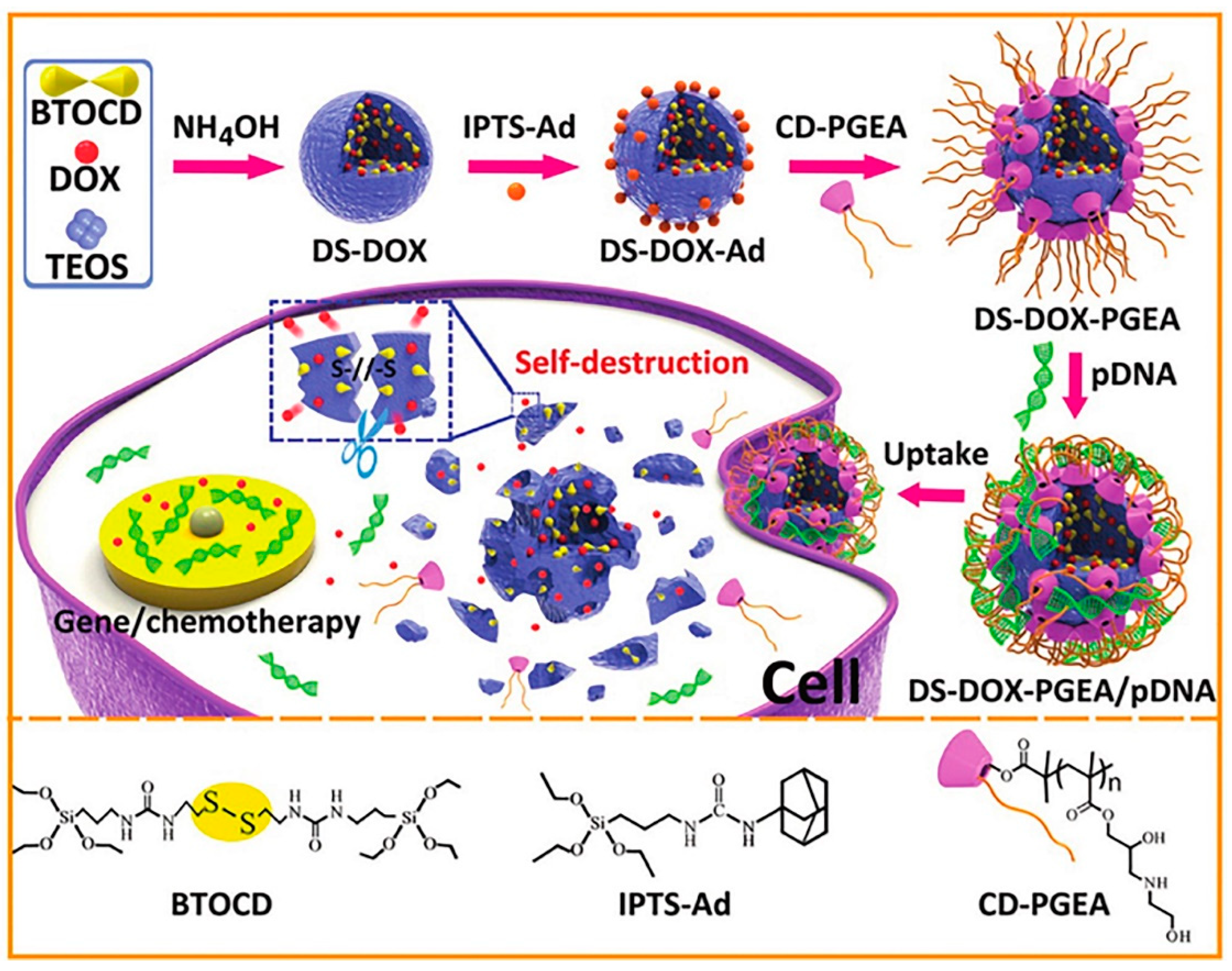
| Disulfide-bridged silica nanoparticles with incorporated DOX | CD-PGEA | pDNA and DOX | pRL-CMV, pEGFP, p53 | HepG2 and C6 | C6 glioma tumor- bearing BALB/c nude mice | [60] |
| Amine-functionalized MSNs of ~80 nm | PAMAM dendrimer conjugated chitosan | pDNA and DOX | p53 | Hela | - | [76] |
| Hollow MSNs with ultra-large pores of ~24 nm | Poly(β-amino ester) | siRNA and DOX | P-gp | MCF-7/ADR | MCF-7/ADR tumor-bearing BALB/c nude mice | [53] |
| Azide-functionalized MSNs of 35–60 nm with 2.6 nm pore size | Poly l-arginine | pDNA and DOX | mCherry | HeLa and A549 | - | [77] |
| MSNs of ~200 nm with 4 nm pore size | Poly l-lysine | siRNA, oligonucleotides | GAPDH, CTGF, TGFβR-1 | RT3 | SCID mice | [78] |
| MSNs 100–200 nm with pore sizes of 4–5 nm: internally lined with amino groups and externally with mercapto groups or disulfide-bridged amino groups | Block copolymer containing cationic artificial amino acids and oleic acid blocks | siRNA | eGFP-Luc | KB, MCF-7 | - | [81] |
| Amine-functionalized silica NPs with 90 nm | Hyaluronic acid | siRNA and pDNA | GFP | A549 | Tumor-bearing BALB/c nude mice | [82] |
| Amine-functionalized MSNs of ~140 nm | Hyaluronic acid | siRNA and TH287 | MDR1 | CAL27 | Tumor-bearing BALB/c nude mice | [83] |
| Amine-functionalized MSNs of ~125 nm | Polymerized dopamine | Anti-miRNA and AS1411 aptamer | miR-155 | Human CRC (SW480, HT-29, SW620, Lovo, and Caco-2) and NCM460 | SW480 tumor-bearing BALB/c nude mice | [84] |
| Silica-Based Vector | Other Vectors | Main Comparative Results | Ref. | ||
|---|---|---|---|---|---|
| Performance | Safety | ||||
| Nanotubes functionalized with bPEI (1.8 kDa) | Polymers | bPEI (1.8 or 25 kDa) | In both HeLa and NIH3T3 cells, at N/P = 10, bPEI-nanotubes showed:
| In both HeLa and NIH3T3 cells:
| [23] |
| Amine-coated solid NPs | PEI (60 kDa) | Silica NPs reached 30% of the transfection efficiency achieved with PEI in Cos-1 cells. | At concentrations used for transfection, the use of PEI led to 50% reduction in cell viability, while no significant toxicity was detected using silica NPs. | [40] | |
| HA-coated solid NPs | PEI (25 kDa) | The gene silencing effect observed with silica NPs was efficient and similar to the effect obtained with PEI. | On A549 cells, silica NPs presented low cytotoxicity (<20%) even at 128 μg/mL; PEI presented an IC50 value of about 8 μg/mL. | [82] | |
| Amine-coated upright nanosheets | PolyFect (activated dendrimers) | Transfection efficiency in hMSCs was ~85% for both. | Cell viability in hMSCs was 80–90% without PolyFect and 3% with PolyFect. | [31] | |
| MSN-hbPEI | Peptide | Peptide-based vector L1 with ligand and PEI 25 kDa | The transfection using PEI-coated MSNs reached more than 2-fold GFP intensity (%) in MDA-MB-231 cells than that using both vector L1 and PEI. | Even at the higher concentration of 41 µg/mL, the nanoparticles did not exhibit any hemolytic activity. Vector L1 and PEI were not tested. | [72] |
| Amine-coated solid NPs | Lipids | LipofectinTM | Silica NPs showed higher transfection efficiency than LipofectinTM in MCF-7 cells. When the ratio of silica:DNA was 9:1, GFP expression was 2-fold higher than with LipofectinTM. | No tissue or cellular damages were observed in the mice injected with the pDNA/silica nanoparticles. | [44] |
| Positively charged MSNs | Lipofectamine® 2000 | MSNs were more effective than Lipofectamine 2000 in Rex1 knockdown. | On iPSCs:
| [47] | |
| Amino-coated large-pore MSNs | Lipofectamine® 2000 | No significant differences were observed between the transfection efficiencies of MSNs and Lipofectamine® 2000 in SMMC-7721, HepG2 and Huh7 cells. | MSNs caused almost no toxic effects to SMMC-7221 cells at concentrations lower than 400 µg/mL in both 24 and 72 h incubations. Lipofectamine® 2000 was not tested. | [55] | |
| Octadecyl-coated large-pore MSNs | OligofectamineTM | The delivery efficiency of siRNA was not significantly different between MSNs and Oligo and was dose-dependent in both cases. | HCT116 cells showed negligible cytotoxic effect up to 40 μg/mL for both coated and uncoated MSNs. Oligo was not tested. | [54] | |
| PEI-coated large pore magnetic MSNs | OligofectamineTM | The delivery of siRNA to KHOS cells by MSN-based carrier led to a cell viability inhibition of 80%, while the same dose of siRNA delivered by Oligo resulted in only 50% cell viability reduction. | Both MSN-based carriers and Oligofectamine caused no significant cytotoxicity to KHOS cells. | [115] | |
| MSN-hbPEI | Lipofectamine RNAiMax | Transfection with MSNs showed a time-dependent reduction in cell viability similar to Lipofectamine. There was no significant difference between their efficacies. | MSN-based carriers did not contribute to any significant toxicity to MDA-MB-231 cells after 72 h of incubation in concentrations of 2–50 µg/mL. Lipofectamine was not tested. | [71] | |
| DOPC Protocell | DOPC and DOTAP liposomes |
| DOPC protocells revealed much lower cytotoxicity than DOTAP liposomes in Hep3B cells. | [106] | |
| Amine-coated solid NPs | Viral | HSV-1 | Silica NPs transfection efficiency was equal or higher than that obtained using HSV-1 viral vectors. | Tissue damage was observed during intra-brain gene transfer using the viral vector, while no toxic effects were observed in mice treated with silica NPs even 4 weeks after transfection. | [42] |
© 2020 by the authors. Licensee MDPI, Basel, Switzerland. This article is an open access article distributed under the terms and conditions of the Creative Commons Attribution (CC BY) license (http://creativecommons.org/licenses/by/4.0/).
Share and Cite
Carvalho, A.M.; Cordeiro, R.A.; Faneca, H. Silica-Based Gene Delivery Systems: From Design to Therapeutic Applications. Pharmaceutics 2020, 12, 649. https://doi.org/10.3390/pharmaceutics12070649
Carvalho AM, Cordeiro RA, Faneca H. Silica-Based Gene Delivery Systems: From Design to Therapeutic Applications. Pharmaceutics. 2020; 12(7):649. https://doi.org/10.3390/pharmaceutics12070649
Chicago/Turabian StyleCarvalho, Ana Maria, Rosemeyre A. Cordeiro, and Henrique Faneca. 2020. "Silica-Based Gene Delivery Systems: From Design to Therapeutic Applications" Pharmaceutics 12, no. 7: 649. https://doi.org/10.3390/pharmaceutics12070649