Next Article in Journal
Chitosan Film Containing Mansoa hirsuta Fraction for Wound Healing
Next Article in Special Issue
Investigation of Cytotoxicity and Cell Uptake of Cationic Beta-Cyclodextrins as Valid Tools in Nasal Delivery
Previous Article in Journal
Influence of the Polymer Glass Transition Temperature and Molecular Weight on Drug Amorphization Kinetics Using Ball Milling
Previous Article in Special Issue
Optimization of Curcumin Nanocrystals as Promising Strategy for Nose-to-Brain Delivery Application
 
 
Retraction published on 29 January 2024, see Pharmaceutics 2024, 16(2), 189.
Article

RETRACTED: Intranasal Niosomal In Situ Gel as a Promising Approach for Enhancing Flibanserin Bioavailability and Brain Delivery: In Vitro Optimization and Ex Vivo/In Vivo Evaluation

by
Usama A. Fahmy
1,2,
Shaimaa M. Badr-Eldin
1,3,*,
Osama A. A. Ahmed
1,2,
Hibah M. Aldawsari
1,4,
Singkome Tima
5,
Hani Z. Asfour
6,
Mohammed W. Al-Rabia
6,
Aya A. Negm
7,
Muhammad H. Sultan
8,
Osama A. A. Madkhali
8 and
Nabil A. Alhakamy
1,2,4,9
1
Department of Pharmaceutics, Faculty of Pharmacy, King Abdulaziz University, Jeddah 21589, Saudi Arabia
2
Advanced Drug Delivery Research Group, Faculty of Pharmacy, King Abdulaziz University, Jeddah 21589, Saudi Arabia
3
Department of Pharmaceutics and Industrial Pharmacy, Faculty of Pharmacy, Cairo University, Cairo 11562, Egypt
4
Center of Excellence for Drug Research and Pharmaceutical Industries, King Abdulaziz University, Jeddah 21589, Saudi Arabia
5
Department of Medical Technology, Faculty of Associated Medical Sciences, Chiang Mai University, Chiang Mai 50200, Thailand
6
Department of Medical Microbiology and Parasitology, Faculty of Medicine, King Abdulaziz University, Jeddah 21589, Saudi Arabia
7
Department of Pharmacognosy, Faculty of Pharmacy, Zagazig University, Zagazig 44518, Egypt
8
Department of Pharmaceutics, College of Pharmacy, Jazan University, Jazan 45142, Saudi Arabia
9
King Fahd Medical Research Center, King Abdulaziz University, Jeddah 21589, Saudi Arabia
*
Author to whom correspondence should be addressed.
Pharmaceutics 2020, 12(6), 485; https://doi.org/10.3390/pharmaceutics12060485
Submission received: 3 April 2020 / Revised: 18 May 2020 / Accepted: 23 May 2020 / Published: 27 May 2020 / Retracted: 29 January 2024
(This article belongs to the Special Issue Nose to Brain Delivery (Volume II))

Abstract

:
Flibanserin (FLB) is a multifunctional serotonergic agent that was recently approved by the FDA for the oral treatment of premenopausal women with hypoactive sexual desire disorder. FLB is a centrally acting drug that has a low oral bioavailability of 33% owing to its exposure to the hepatic first-pass effect, as well as its pH-dependent solubility, which could be an obstacle hindering the drug dissolution and absorption via mucosal barriers. Thus, this work aimed at overcoming the aforementioned drawbacks and promoting the nose-to-brain delivery of FLB via the formulation of an intra-nasal in situ niosomal gel. The Box–Behnken design was employed to study the impact of Span® 85 concentration (X1), hydration time (X2), and pH of the hydrating buffer (X3) on the vesicle size and drug entrapment. The optimized formulation exhibited a spherical shape with a vesicular size of 46.35 nm and entrapment efficiency of 92.48%. The optimized FLB niosomes integrated into gellan gum-based in situ gel exhibited enhanced ex vivo permeation and improved plasma and brain concentrations after nasal administration in rats compared to raw FLB. These findings highlight the capability of the proposed intra-nasal FLB niosomal in situ gel to boost the drug bioavailability and to promote its direct delivery to the brain.

1. Introduction

Flibanserin (FLB; 2H-benzimidazol-2-one, 1,3-dihydro-1-[2-[4-[3(tri-fluoromethyl) phenyl]-1-piperazinyl]ethyl] was approved by US FDA in August 2015 under the trade name of Addyi® for treating premenopausal woman with generalized hypoactive sexual desire disorder (HSDD), which is the most common form of female sexual dysfunction that is associated with emotional distress and relationship problems [1,2]. FLB is a non-hormonal drug that selectively binds to serotonin receptors in the central nervous system (CNS). FLB acts as both serotonin agonist (5-HT1A) and antagonist (5-HT2A). It acts by decreasing serotonin and increasing dopamine and norepinephrine; this effect on the mentioned neurotransmitters is important for a healthy sexual response [3]. FLB has a half-life of 11 h with a rapid rise of plasma concentration within the first hour after oral administration followed by a gradual decrease thereafter. However, the drug has a low oral bioavailability of 33% [1,4]. This reduced bioavailability might be ascribed to FLB’s considerable presystemic metabolism and low aqueous solubility (≈ 4.312 mg/L at 25 °C). Moreover, possible drug degradation after oral administration by the acidic environment of gastrointestinal (GI) tract, mucosal enzymes, as well as the intestinal barriers could be challenging factors that contribute to FLB low bioavailability [5].
Nano-sized-based drug delivery systems have been extensively studied during the past few decades as a new strategy for circumventing the poor bioavailability of various pharmaceutical drugs. Nanoparticulate delivery systems, including polymeric nanoparticles, liposomes, micelles, dendrimer, solid lipid nanoparticles, and niosomes have been widely used. Vesicular carriers provided additional benefits, such as the defense of active agents against enzymatic and chemical degradation and the possibility of increasing the time of distribution of drugs in the bloodstream [6,7,8].
Niosomes are self-assembled nonionic surfactant-based vesicular systems that have attracted attention as successful drug delivery systems in the last decades [9,10]. The lamellar bilayer structure of niosomes with the amphiphilic property of the surfactants can improve the solubility of hydrophobic drugs by partitioning into the lipophilic domain of the bilayers and consequently enhancing their absorption [11]. Niosomes have many benefits such as reduced toxicity owing to their nonionic nature, the ability to carry both lipophilic and hydrophilic drugs owing to their core–shell structure, their high permeation through biological membranes, and their capability of prolonging the circulation of entrapped drugs. Compared to phospholipid-based vesicles, niosomes demonstrate greater physical stability and reduced handling and storage costs. Moreover, niosomes have been reported as efficient carriers for targeting drugs and increasing their accumulation in organs as liver and brain [12,13,14,15].
To deliver the drugs to the CNS, three different administration routes were suggested including, systemic absorption through the blood–brain barrier (BBB), intra-cerebroventricular (ICV) administration, and intra-nasal administration [16]. Intra-nasal administration has been recently regarded as the method of choice for drug delivery to the CNS due to several advantages such as non-invasiveness, rapid, and targeted drug delivery to the CNS via the olfactory epithelium and trigeminal region, and consequently reduced systemic side effects [17,18]. Niosomes can be useful for brain delivery; in reality, polysorbates (Tweens) used in the preparation of vesicles can serve as an anchor for apolipoprotein E (apo E) from blood plasma. Particles tend to mimic LDL and interact with the LDL receptor that contributes to endothelial cells being absorbed. Following this, the drug may be released into these cells and distributed to the inside of the brain, or the particles may be transcytosed. In addition, such surfactants have a strong hydrophilic–lipophilic balance (HLB) content, so they require an equimolar amount of cholesterol to acquire a vesicular bilayer and are able to cross the BBB by interacting with the LDL receptor. Design of experiments (DOE) has emerged as a powerful and economic statistical approach that yields more data with applying a reduced number of runs [19]. Experimental design has the advantage of detecting the relative significance of different factors affecting the formulation. Box–Behnken is an independent, rotatable, or nearly rotatable three-level response surface design that has been widely applied for optimization in the pharmaceutical area [20]. It can generate higher order response surfaces with fewer runs compared to normal factorial.
This work aimed at combining the beneficial delivery characteristics of niosomes, the advantages of intra-nasal administration, and the potential of nanotechnology for the direct targeting of FLB to the CNS, as well as enhancing its bioavailability. In this study, the Box–Behnken design has been utilized for the formulation and in vitro optimization of FLB niosomes. The optimized formulation of FLB-NI was evaluated for shape, vesicle size, and stability. Then, the optimized formulation was integrated into the gellan gum-based in situ gel. The prepared gel was assessed for ex vivo permeation and in vivo pharmacokinetic performance in Wistar rats. The positive ability of niosomes to encapsulate both hydrophilic and lipophilic compounds and to cross the BBB reflected our logical design of niosomal formulations that are useful for achieving a BBB crossing and thus for the CNS delivery of drugs.

2. Materials and Methods

2.1. Materials

Flibanserin was purchased from Qingdao Sigma Chemical Co., Ltd. (Qingdao, China). Span® 85 (sorbitan trioleate) and cholesterol were purchased from Sigma-Aldrich Co. (St Louis, MO, USA). Ethanol was purchased from EMD Millipore (Billerica, MA, USA). All other reagents and chemicals were of analytical grade.

2.2. Experimental Design and Optimization

A three-variable Box–Behnken design was applied to optimize FLB niosomes using Design-Expert software (Version 12; Stat-Ease Inc., Minneapolis, MN, USA). The cholesterol (CH) amount was fixed (550 mg) in all formulations, while Span® 85 concentration (X1, %), hydration time (X2, min), and hydrating buffer pH (X3) were studied as variables in the range of 1–6 µM, 15–60 min, and pH range from 3 to 9 [21], respectively. The ranges used in the study were selected based on a preliminary study (data not shown). The vesicle size (Y1, nm) and drug entrapment efficiency (Y2, %) were investigated as response parameters. The coded levels of each factor and their corresponding actual values are compiled in Table 1. A total of 17 experimental runs were generated according to the experimental design. The different combination of variables levels and the measured responses for the experimental runs are depicted in Table 2. The measured responses were subjected to statistical analysis using Analysis of Variance (ANOVA) at a 95% level of significance. Different sequential models including linear, 2FI, and quadratic and cubic were analyzed for the test of fit to derive the relation between the investigated variables and the niosomes’ vesicle size. Diagnostic plots were utilized to confirm the validity of the selected model for each response. The polynomial equation representing the best-fitting model was computed for each response. Three-dimensional response and cube plots were generated to assess the effect of the investigated variables and the interaction between them.

2.3. Preparation of FLB Niosomes

FLB niosomes were prepared according to a previously reported method with slight modification [13]. Briefly, accurately weighed amounts of FLB (50 mg), cholesterol (CH, 550 mg), and Span® 85 were dissolved in ethanol (10 mL) in a round-bottom flask. Then, the ethanolic solution was subjected to evaporation under reduced pressure (R-200; BÜCHI Labortechnik AG, Flawil, Switzerland) at 45 °C for 2 h to remove the organic solvent. After complete removal of the solvent, the niosmal dried film formed on the flask’s inner wall was hydrated using phosphate buffer (10 mL) under normal pressure; the hydration time and the pH of the buffer used were specified according to the experimental design. For vesicle size diminution, the formed niosomal dispersion was subjected to sonication for 60 s using a Sonics Vibra Cell tapered microtip of amplitude 40%, 750 watt, 20 KHZ (Sonics & materials Inc., Newtown, CT, USA). The niosomal dispersion was kept overnight at 4 °C for maturation prior to further investigations [22,23].

2.4. Vesicle Size Measurement

The dynamic light scattering technique was employed for the determination of FLB niosomes’ vesicles size (z-average) using a Zetatrac Particle size analyzer (Microtrac ® Inc., Montgomeryville, PA, USA). Five replication measurements were used to calculate the average vesicle size after the dilution of 1 mL of each niosomal formulation with 20 mL of deionized water. The used equipment was standardized through standard solution (monomodal aqueous suspensions of silica nanoparticles) provided by the manufacturer in addition to regular maintenance checks by the manufacturer.

2.5. Determination of Entrapment Efficiency (EE %)

An indirect method was used to assess the drug entrapment efficiency (EE %) by calculating the difference between the total FLB added in the formulation and the residual amount in the supernatant after separating the niosomal dispersion. FLB niosomal suspension was centrifuged for 45 min at 400,000× g (Himac CP-NX ultracentrifuge, Hitachi, Minato, Tokyo, JAPAN). The supernatant was collected by filtration through a Millipore membrane filter (0.2 μm). High-performance liquid chromatography with an ultraviolet (UV) diode array detection (HPLC-DAD) method, developed in our laboratory, was used to determine the un-entrapped FLB. The method was validated in terms of linearity, accuracy, and precision. A high performance-liquid chromatography (HPLC) Agilent 1200 system with a diode array detector was used. The system was equipped with an autosampler, a quaternary pump, and a column compartment (Palo Alto, CA, USA). The system was equipped with ChemStation software (Rev. B.01.03 SR2(204)). Isocratic elution was employed at a flow rate of 0.6 mL/min. The utilized mobile system comprised acetonitrile/water containing 0.1% formic acid (9:1). A volume of 5 µL was injected on a Zorbax Extend C18 column (4.6 × 150 mm, 5 µm) for LC-DAD analysis. The detection was carried out at 260 nm, and the concentration of FLB in the injected samples was calculated with reference to the constructed calibration curve. The assay showed acceptable linearity over the range of 10–500 µg/mL with a correlation coefficient of 0.9994. The assay exhibited acceptable accuracy with a mean recovery percentage of 96.32% and adequate precision with a relative standard deviation (RSD) of 2.3%–4.9% and 1.9%–3.7% for intra-day assay and the inter-day validation, respectively. FLB EE% was computed using the following equation:
F L B   E E % = ( T o t a l   a m o u n t   o f   F L B   a d d e d A m o u n t   o f   F L B   i n   t h e   s u p e r n a t a n t T o t a l   a m o u n t   o f   F L B   a d d e d ) × 100

2.6. Optimization of FLB Niosomes

Numerical optimization was applied and desirability was computed for prediction of the composition of the optimized FLB niosomes formulation with minimized vesicle size and maximized drug entrapment. The optimization procedure was performed utilizing Design-Expert software (Version 12; Stat-Ease Inc., Minneapolis, MN, USA). The proposed optimum formulation was prepared and evaluated in triplicate to test for the validity of the proposed optimal variables’ levels and the predicted responses.

2.7. Characterization of Optimized FLB Niosomes

The shape of the optimized FLB niosomes was examined utilizing transmission electron microscopy; TEM (Philips XL30, Eindhoven, Netherlands) operated at 200 KV. A diluted niosomal dispersion sample was placed on a carbon-coated grid for 2 min to allow adsorption. Then, the adsorbed niosomes were negatively stained with uranyl acetate and left for air drying prior to visualization.
The physical stability of the optimized FLB niosomes was studied by subjection to three freeze–thaw cycles (between −20 °C and +25 °C). The vesicle size and EE % of the formulation before and after the cycles were measured and compared statistically using the paired student t-test at P < 0.05.

2.8. Preparation of FLB Niosomal In Situ Intra-Nasal Gel

Gellan gum dispersion (0.7%, w/v) was prepared by sprinkling over boric acid/borax buffer (pH 7.4) at 80 °C while stirring. The mixture was stirred continuously until a clear dispersion was obtained. After allowing to cool, optimized FLB niosomes were incorporated into gellan gum dispersion to attain an FLB concentration of 10 mg/g. Then, the niosomal loaded in situ gel was kept for 12 h at 4 °C before evaluation to allow the removal of entrapped air bubbles within the gel during preparation. Raw FLB loaded in situ gel was prepared at the same concentration as a control for comparison. The prepared FLB niosomal in situ nasal gel formulation was evaluated for viscosity and gelation before and after the addition of simulated nasal fluid (SNF) [24,25]. The viscosity was evaluated using a Brookfield digital viscometer (Brookfield Engineering Laboratories, Middleborough, MA, USA). The apparent viscosity was measured at 10 rpm after 30 s before and after gelation.

2.9. Ex Vivo Permeation

An automated Franz diffusion cell vertical diffusion cell test system (Microette Plus Hanson research, CA, USA) was utilized for performing an ex vivo permeation study via fresh excised bovine nasal mucosa obtained from the slaughterhouse. Chloroform-methanol (2:1, v/v) was used for the delipidization of mucosa for 45 min. [26,27]. Mucosal samples were mounted between the two chambers of the diffusion cell with an effective diffusional area of 1.76 cm2. Either optimized FLB niosomal loaded in situ gel or control raw FLB loaded gel (0.1 g) was placed in direct contact to the mucosa in the donor chamber. The receptor chamber was occupied with 7 mL of simulated nasal fluid (SNF), pH 6.5 as a diffusion medium. SNF was composed of 7.45 mg/mL NaCl, 1.29 mg/mL KCl, and 0.32 mg/mL CaCl2·2H2O [25]. The temperature was maintained at 35°C ± 0.5°C throughout the study, and the agitation rate was set to 400 rpm [28,29]. At predetermined intervals of 0.5, 1, 2, 4, 6, and 8 h, aliquots of 1.5 mL were withdrawn and replaced with fresh diffusion medium. The samples were analyzed using high-performance liquid chromatography with an ultraviolet (UV) diode array detection (HPLC-DAD). A mean cumulative amount of FLB permeated through the mucosal surface per unit surface area (µg/cm2) was plotted as a function of time. Then, permeation parameters, namely steady-state flux (Jss; µg/cm2.h), permeability (Pc), and diffusion (D) coefficients were computed [30,31].

2.10. In Vivo Pharmacokinetic Assessment

2.10.1. Study Protocol and Sample Preparation

The pharmacokinetic parameters of the FLB following the intra-nasal administration of optimized FLB niosomal in situ gel compared to control raw FLB gel were assessed in male Wistar rats weighing between 200 and 250 g. The study protocol has been revised and approved by The Research Ethics Committee in The Faculty of Pharmacy, King Abdulaziz University with approval no. (PH-124-41). The committee ensures that animal use complies with the European Union Directive 2010/63/EU on animal welfare and the Guiding Principles on animal welfare (DHEW publication NIH 80-23). The study design comprised two animal groups (I and II, n = 42 for each). Group I (test group) received optimized FLB niosomal in situ gel, while group II (positive control) received raw FLB in situ gel. All the rats received via the intra-nasal route an FLB dose of 10 mg/kg as previously reported [32]. The gel was distributed equally between the right and left nostrils using a micropipette (10–100 µL capacity), with its tip placed 1 cm deep inside the nasal cavity. A collection of blood samples (100 µL) was performed at 0, 0.5, 1, 2, 3, 6, and 8 h following administration via the tail vein. The centrifugation of blood samples was performed at 7100× g (Eppendorf Centrifuge, Model 5425, Eppendorf-Netheler-Hinz GmbH 2000, Hamburg, Germany) for 5 min to separate plasma. Six rats from each group were scarified at each time interval, and the whole brain was removed from the cut open skull, washed three times with saline, and weighed. Extraction was performed by homogenizing the brain tissues with phosphate buffer saline (pH 7.4) at 7000 rpm for 2 min (T 25 digital ULTRA-TURRAX®, Staufen, Germany). The separated plasma and the homogenized brain samples were stored at –80 °C until analysis [33].

2.10.2. In Vivo Assay

The specified volume of plasma or brain homogenate was mixed with 50 μL of valsartan solution (625 ng/μL) as an internal standard and 1 mL of acetonitrile in a screw-capped test tube. The mixture was vortexed for 1 min and then centrifuged at 7500× g for 8 min. An aliquot of about 500 μL of the clear supernatant was transferred to a total recovery auto sampler vial, and a volume of 7 μL was injected for liquid chromatography-mass spectrometry coupled to diode array detection (LC-MS/MS-DAD) analysis. The MS system was connected to an HPLC Agilent 1200 system equipped with an auto sampler, a quaternary pump, and a column compartment (Palo Alto, CA, USA). The system was equipped with ChemStation software (Rev. B.01.03 SR2 (204)). The IT-MS was controlled using 6300 series trap control version 6.2 Build No. 62.24 (Bruker Daltonik GmbH), and the general MS adjustments were capillary voltage, 4200 V; nebulizer pressure, 37 psi; drying gas flow rate, 12 L/min; gas temperature, 330 °C; ion charge control (ICC) smart target, 200,000; and max accumulation time, 200 milliseconds (ms). For quantitative monitoring, single positive molar ion mode was applied at each programmed time segment, 0–4.0 min, m/z 391.2 for FLB and 4.0–10 min, m/z 436.3 [M+H]+ for valsartan (internal standard). Isocratic elution was conducted at a flow rate of 0.5 mL/min with a mobile system composed of 52% acetonitrile and 48% water containing 0.1% formic acid. The stationary phase was Thermo C18 (250 × 4.6 mm2, 5 μm particle size) column. FLB content in the assayed samples was quantified with reference to the constructed calibration curve.

2.10.3. Pharmacokinetic and Statistical Analysis

Pharmacokinetic parameters including the maximum plasma concentration (Cmax), time to maximum plasma concentration (Tmax), and area under the plasma concentration-time curve (AUC) were computed using Kinetica ™ software (Version 4; Thermo Fisher Scientific, Waltham, MA, USA). Both Cmax and AUC were expressed as mean ± standard deviation, while Tmax was presented as the median. Statistical analysis of the measured plasma concentrations and the calculated parameters was performed using using Prism® (version 8.4.0, GraphPad Software Inc., La Jolla, CA, USA) at P < 0.05. Two-way ANOVA followed by Sidak’s multiple comparisons test was performed to analyze the plasma concentrations. An unpaired t-test was performed for Cmax and AUC, while the nonparametric Mann–Whitney U test (Wilcoxon rank sum test) was performed for Tmax.

3. Results and Discussion

3.1. Experimental Design

In this study, a three-factor, three-level Box–Behnken design was utilized for the formulation and optimization of FLB niosomes with minimized vesicle size and maximized entrapment efficiency. The software yielded 17 runs, representing 12 midpoints of the edges of the design space and 5 center nodes, as shown in Table 2.

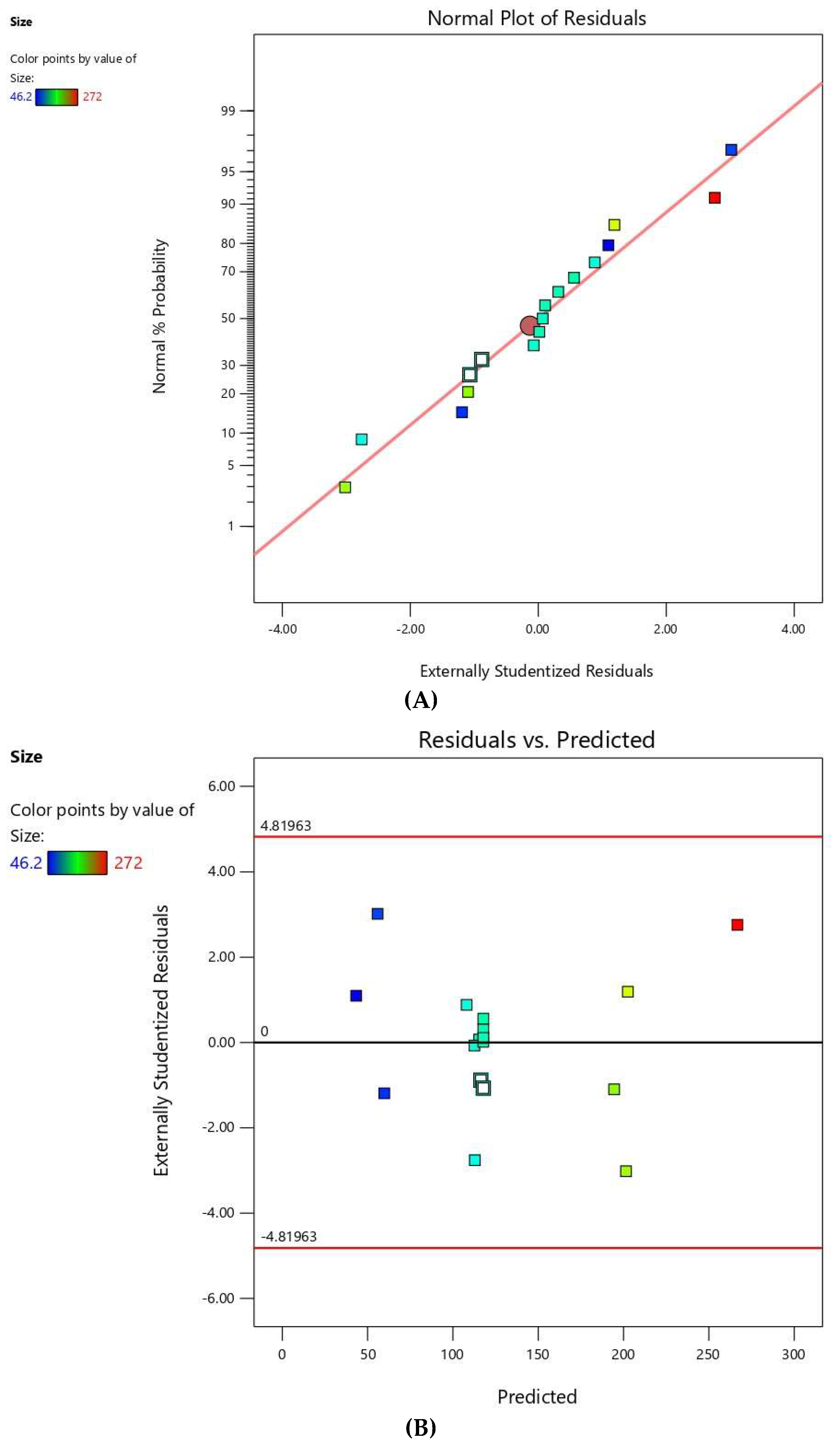
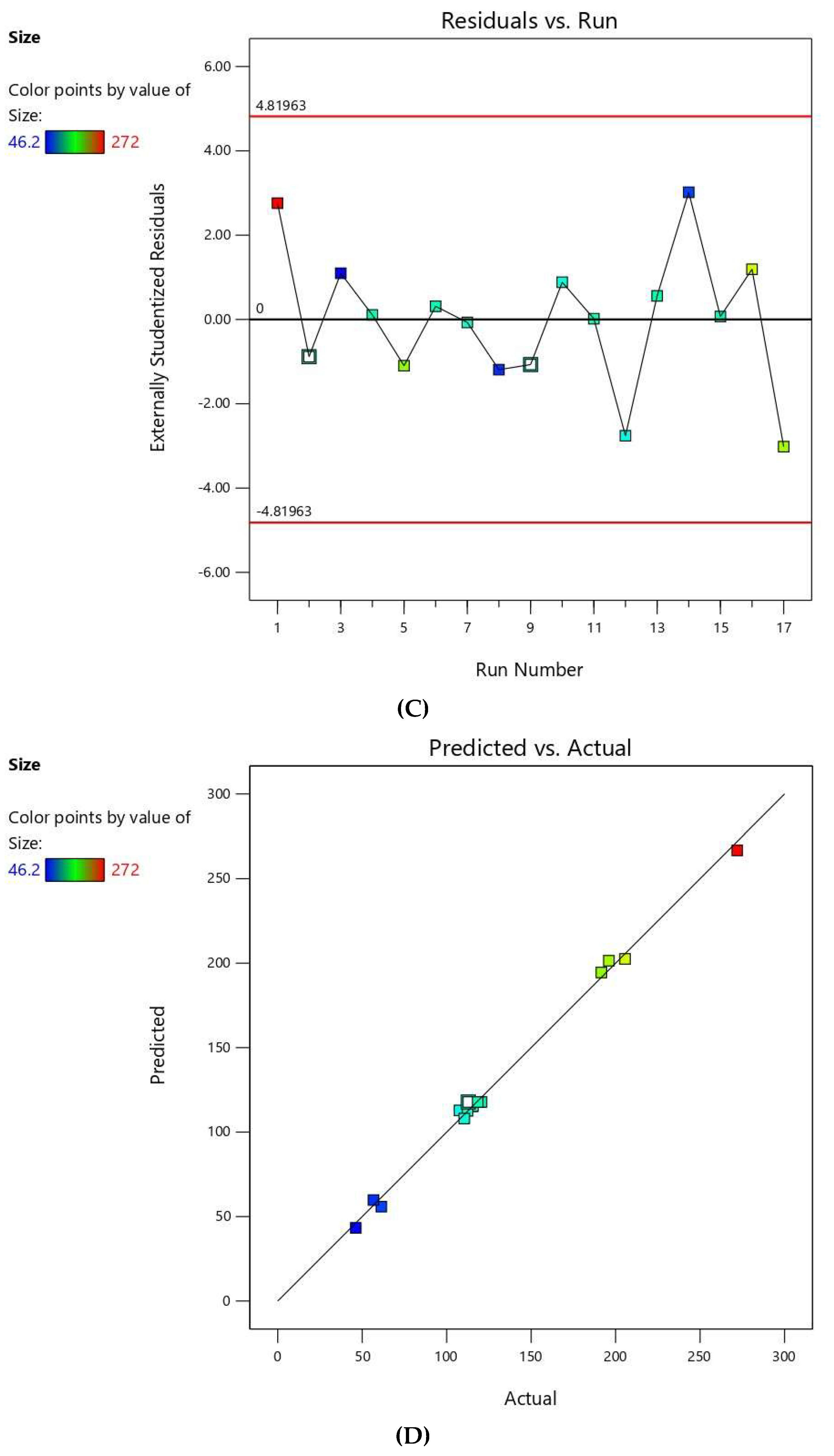
3.1.1. Sequential Model Selection and Diagnostic Analysis

Based on the highest correlation coefficient (R2) and the lowest predicted residual error sum of squares (PRESS), the data of both vesicle size and entrapment efficiency fitted to the quadratic model, as shown in Table 3. In addition, there was a reasonable agreement between the predicted and adjusted R2 indicating the validity of the model. Furthermore, adequate precision for both responses was greater than 4, indicating an adequate signal-to-noise ratio; this ratio implies the suitability of the selected model to explore the design space.
The residuals were computed as the difference between the actual and predicted values of the responses. Diagnostic plots for both responses were generated to assess the goodness of fit of the selected model and ensure its credibility. Normal probability plots of residuals, Figure 1A and Figure 2A, exhibited a linear pattern indicating that the residuals are normally distributed and thus, the data do not need transformation. Externally studentized residuals versus predicted responses’ values, Figure 1B and Figure 2B, showed that color points depicting the values of the responses were sited within the limits close to the zero-axis and scattered randomly reflecting the absence of constant error. Figure 1C and Figure 2C representing the residual versus run plots depicted a random and uniform scatter of points, indicating no lurking variable affected any of the response. Furthermore, predicted versus actual values of both responses’ plots, Figure 1D and Figure 2D, were highly linear, revealing that the observed responses showed good analogy to the predicted ones [34].

3.1.2. Statistical and Response Analysis for the Effect of Variables on Vesicle Size (Y1)

The size of vesicular carriers is an important parameter that displays significant influence on the release pattern and consequently permeation of drugs via the biological membranes. Generally, nanovesicles with a size less than 100 nm exhibit excellent nose-to-brain delivery [35]; however, some studies reported excellent pharmacokinetic data upon nasal administration of nanoparticles with sizes more than 100 nm [36]. FLB niosomes exhibited nano-sized vesicles with an average size ranging from 46.2 ± 2.14 to 272.4 ± 8.56 (Table 2). The relatively low computed standard deviation indicating homogenous and uniform distribution of the niosomal dispersions. The observed nano-size could play a crucial role in promoting the drug permeation via the nasal mucosa directly to the brain, in addition to, facilitating crossing the blood–brain barrier [37]. Analysis of variance (ANOVA) for the vesicles size confirmed the significance of the quadratic model as depicted by its F-value of 211.73 (P < 0.0001). The lack of fit F-value of 0.99 (P = 0.4821) implied a non-significant lack of fit relative to pure error, ensuring data fitting to the proposed model. The equation representing the quadratic model was generated in terms of coded factor as follows:
Y1 = 115.81 − 74.17 X1 − 1.39 X2 − 2.79 X3 − 4.87 X1X2 + 29.33 X1X3 − 0.9 X2X3 + 27.67 X12 −19.71 X22 + 16.99 X32.
The statistical analysis revealed that Span® 85 concentration (X1) has a significant negative effect on FLB niosomes size (P < 0.0001). The quadratic terms corresponding to the three investigated variables, in addition to the interaction term X1X3 corresponding to the interaction between span concentration and the pH of the hydrating buffer, were also found to be significant at a 95% significance level. Figure 3 illustrates the response surface and cube plots for the effects of the studied variables on vesicle size. It was evident that the vesicle size significantly decreases with increasing Span® concentration. This effect could be credited to the relatively low HLB value of Span® 85 employed in this study. The low HLB value could result in reduced surface energy, leading to less water uptake in the core, and consequently, reduced size [38]. The reduced size could also be explained by the possible formation of a micellar structure at higher surfactant concentrations, which is comparatively smaller in size [11].

3.1.3. Statistical and Response Analysis for the Effect of Variables on EE% (Y2)

The ability of niosomes to entrap a considerable amount of FLB is crucial for its effective targeting to the CNS. EE % ranged from 51.7 ± 1.23 to 98.5 ± 4.67 (Table 2). It should be mentioned that the fraction of FLB, although a small fraction, could be adsorbed onto the 0.2 µm filter. Analysis of variance (ANOVA) for the EE ensured that the proposed quadratic model is significant as evidenced by its F-value of 478.91 (P < 0.0001). The lack of fit F-value of 0.83 (P = 0.5436) indicates that the lack of fit was non-significant in relation to pure error confirming the model validity. The equation corresponding to the quadratic model was generated in terms of the coded factor as follows:
Y2 = 68.90 + 17.65 X1 + 2.35 X2 + 5.60 X3 + 1.45 X1X2 + 2.10 X1X3 − 0.40 X2X3 + 4.22 X12 − 0.58 X22 − 0.13 X32.
The statistical analysis revealed that all the investigated variables, namely, Span® 85 concentration (X1), hydration time (X2), and pH of hydrating buffer (X3) have significant effects on the drug EE % (P < 0.0001). The quadratic terms corresponding to the Span® concentration (X12), in addition to the interaction terms X1X2 and X1X3 corresponding to the interaction between Span concentration and either the hydration time or pH of the hydrating buffer were also found to be significant at P < 0.05. Figure 4 illustrates the response surface and cube plots for the effects of the studied variables on EE %. A significant increase of EE with increasing Span® concentration is evident. The observed synergistic effect of increasing the surfactant on EE might be attributed to the hydrophobic nature of Span® as well as the varying surfactant-to-cholesterol ratio. Span® 85 is relatively highly hydrophobic with a small critical packing parameter. Accordingly, at low surfactant concentrations, a competition for the packing space within the bilayer might occur between the drug and cholesterol. This competition might result in exclusion of the drug during the assembly of amphiphiles into vesicles. Another possible explanation for the observed decreased entrapment on increasing the cholesterol-to-Span® ratio is based on the disruption of the vesicles’ regular membranes, taking into account that the hydrophobic Span® requires only a limited amount of cholesterol to yield the appropriate membrane curvature for vesicular structure [39]. In a previous study, Aboughaly and Abdelbary [13] similarly reported enhanced methotrexate entrapment in niosomes with increasing Span® concentration relative to cholesterol and attributed this increase to the formation less permeable niosomal membrane.
The hydration time also positively influenced the EE % at P < 0.05. This observation was in accordance with previous studies [13,40]. Furthermore, increasing the pH of the hydrating buffer caused a significant increase in the EE of the FLB. This effect could be interpreted on the basis of drug partitioning between the lipid layer and the hydration medium. FLB is a weakly basic drug with a pH-dependent solubility; [41]. Accordingly, increasing pH favor unionized species of the drug, leading to a higher o/w partition coefficient and consequently increased entrapment. On the contrary, at lower pH values, FLB base protonation is favored, leading to confining of the drug in the hydration medium with consequent reduced entrapment [42].

3.1.4. Optimization of FLB Niosomes

After employing the vesicle size and EE constraints, the optimized levels of the variables were predicted utilizing numerical optimization with an overall desirability of 0.961. The desirability function approaching one implies that the combination of the predicted levels of the variables will mostly achieve the goals set for the responses. The optimized formulation was prepared and evaluated. The percentage error between the predicted and observed responses was relatively low, showing the validity of the optimization technique. Therefore, the optimized formulation was subjected to further investigations. The variables’ levels, predicted responses, and observed responses for the optimized formulation are depicted in Table 4. It is worthy to mention the pka of flibanserin = 12.91 (strongest acidic) and 7.03 (strongest basic) [41]. At the selected hydration medium pH of the optimized formula (pH 6.6), the strongest acidic group is almost completely unionized >99.999%, while the basic group is 71% unionized. This explains the increased entrapment of the optimized formulation.

3.2. Transmission Electron Microscopy (TEM)

Transmission electron microscopic (TEM) analysis has been proved as a successful tool for investigating the shape, size, and structure of vesicular systems. A representative TEM micrograph of the optimized FB niosomes is shown in Figure 5. The niosomes revealed a spherical shape with a definite wall. In addition, the observed size was in reasonable accordance with the results obtained by the dynamic light scattering technique. The micrograph also shows the presence of multi-lamellar vesicles. The stability of the multiple bilayers as well as their rigidity are primarily controlled by the hydrogen bonding between the ester group of Span® and the hydroxyl group of cholesterol. Previous studies have reported the formation of multi-lamellar niosomes [43,44].

3.3. Physical Stability

The optimized formulation exhibited adequate physical stability after subjection to freeze–thaw cycles with slight changes in both vesicle size and EE% (supplementary data (Table S1)). A paired Student-t test revealed that the recorded slight changes were statistically non-significant (P > 0.05).

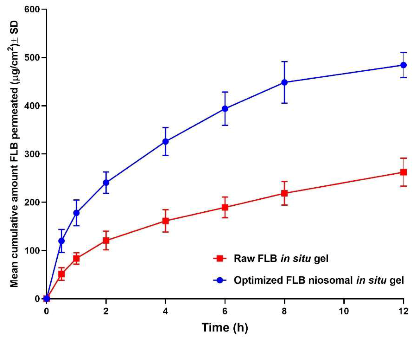
3.4. Characterization and Ex Vivo Permeation of FLB Niosomal In Situ Gel

The in situ gel formulation loaded with optimized FB niosomes exhibited appropriate viscosity of 117 ± 0.98 cps that permits facile instillation into the nose. After exposure to SNF, the formulation was converted from viscous solution into gel, as depicted by the increased gelling factor [30]. The viscosity of the formulation after gelation was increased to 435 ± 1.78 cps. This gelation is favored for adequate drug residence in the nasal cavity; it might be ascribed to the cross-linking action of the SNF cations on the gellan gum helices [45].
Ex vivo permeation study was performed using simulated nasal fluid, PH 6.5, for both FLB niosomal in situ nasal gel compared to raw FLB in situ gel. The mean cumulative ex vivo FLB amount permeated versus time across freshly excised bovine nasal mucosa is graphically illustrated in Figure 6. The permeation parameters were computed and compiled in Table 5. The permeation profile obviously shows that the in situ gel loaded with optimized FLB niosomes could significantly enhance the cumulative amount of permeated FLB (85.19% ± 4.59) compared to the control in situ gel loaded with raw FLB (46.17% ± 5.10). The gradual release displayed by both formulations could be ascribed to the integration of either the drug or the drug-loaded niosomes into the in situ gelling matrix formed upon exposure to the SNF. An unpaired Student-t test revealed that the computed diffusion and permeability coefficients of FLB niosomal gel were significantly higher than those of the control gel at P < 0.05. In addition, the niosomal gel exhibited an enhancement factor of 1.84 compared to the raw FLB gel. The enhanced permeation of FLB from the niosomal in situ gel could be credited to the enhanced solubility of the drug by the niosomes. The amphiphilc characters of cholesterol with the surfactant together form hydrogen bonding that improves the FLB content and solubility within the niosomal core, which leads to improved drug release [16]. In addition, the lipidic property, nano-size of the niosomes, and the presence of nonionic surfactant (Span®) that act as permeation enhancers could contribute to the enhanced diffusion of the drug via the nasal mucosa [14].

3.5. In Vivo Assessment

FLB concentrations spiked in plasma and brain homogenate showed a linear relationship with the peak area ratios, with coefficients (R) of 0.9992 and 0.9984, respectively. The assay showed an adequate precision with relative standard deviation (RSD) being in the range of 4.1%–6.9% and 7.3%–8.9% for intra-day assay and the inter-day assay, respectively. Mean extraction recovery was 94.8 ± 5.4% and 92.6 ± 7.6% for FLB-spiked plasma and brain samples, respectively. The average FLB concentrations in rats’ plasma and brain following the intra-nasal administration of optimized FLB niosomal in situ gel and raw FLB in situ gel are graphically illustrated in Figure 7. As depicted in Table 6, the optimized FLB niosomal in situ gel demonstrated significantly higher Cmax and AUC in both plasma and brain (P < 0.05) compared to the control in situ gel with a 3.67 and 5.52 fold relative increased relative availability, respectively. These results correlate with the ex vivo permeation data. Additionally, the niosomal gel demonstrated significantly lower plasma Tmax, indicating rapid absorption into systemic circulation. On the other hand, the niosomal gel showed gradual accumulation in the brain tissues with significantly higher Tmax (P < 0.05) indicating controlled and sustained action of the drug.
The higher absorption extent of FLB from the niosomal gel compared to the raw FLB gel could be attributed the capability of niosomes to improve drug solubility and permeation across nasal mucosa. This observation is in good agreement with the ex vivo permeation data. The proposed enhanced drug permeation across nasal mucosa could be interpreted on the basis of the niosomes’ nano-size and lipophilic nature, which facilitate the passage of drug across the nasal mucosal epithelium with consequent efficient delivery into the bloodstream [33]. Furthermore, the increased concentration of the drug in the brain foreshadows the ability of the proposed niosomes to enhance the direct delivery of the drug to the CNS delivery through the nasal olfactory region BBB [14]. The improved CNS delivery could be ascribed the ability of the intact lipid-based niosomes to pass the BBB by receptor-mediated endocytosis. Then, the drug uptake into the brain cells could be achieved by passive diffusion through endothelial cells [46]. Furthermore, the protection of the drug from fast excretion and metabolism via encapsulation into the nano-sized vesicles could also contribute to the improved CNS delivery [33].

4. Conclusions

The Box–Behnken design has been successfully applied for the formulation and optimization of FLB niosomes. Surfactant concentration has significantly influenced the niosomes’ vesicle size and drug entrapment, while the hydration time and pH exhibited a significant effect on only the entrapment efficiency. The optimized niosomal formulation with minimized vesicle size and maximized drug entrapment showed adequate physical stability and enhanced ex vivo permeation upon integration into gellan gum in situ gel compared to raw drug. Further in vivo studies revealed that optimized FLB niosomal in situ gel demonstrated higher plasma and brain concentrations compared to control in situ gel loaded with raw drug. Accordingly, the proposed optimized FLB niosomal in situ gel could be regarded as a promising delivery system to improve the delivery of the drug to the brain and to overcome its low oral bioavailability.

Supplementary Materials

The following are available online at https://www.mdpi.com/1999-4923/12/6/485/s1, Table S1. Freeze-thaw cycles for the stability study of FLB Niosomes.

Author Contributions

Conceptualization, U.A.F., O.A.A.A.; methodology, U.A.F., O.A.A.A.; software, S.M.B.-E.; validation, H.M.A., S.T., H.Z.A., N.A.A.; formal analysis, S.M.B.-E.; investigation, U.A.F., O.A.A.A.; resources, M.W.A.-R., A.A.N., H.M.A.; data curation, U.A.F., N.A.A.; writing—original draft preparation, U.A.F., S.M.B.-E.; writing—review and editing, O.A.A.A., S.M.B.-E., O.A.A.M., M.H.S.; visualization, H.M.A., S.M.B.-E.; supervision, O.A.A.A.; project administration, N.A.A.; funding acquisition, N.A.A. All authors have read and agreed to the published version of the manuscript.

Funding

This research was funded by was funded by the Deanship of Scientific Research (DSR) at King Abdulaziz University, Jeddah, under grant no. (RG-6–166–38).

Acknowledgments

This project was funded by the Deanship of Scientific Research (DSR) at King Abdulaziz University, Jeddah, under grant no. (RG-6–166–38). The authors, therefore, acknowledge with thanks DSR for technical and financial support.

Conflicts of Interest

The authors declare no conflict of interest.

References

  1. Gelman, F.; Atrio, J. Flibanserin for hypoactive sexual desire disorder: Place in therapy. Ther. Adv. Chronic Dis. 2017, 8, 16–25. [Google Scholar] [CrossRef]
  2. Vallejos, X.; Wu, C. Flibanserin: A novel, nonhormonal agent for the treatment of hypoactive sexual desire disorder in premenopausal women. J. Pharm. Pract. 2017, 30, 256–260. [Google Scholar] [CrossRef] [PubMed]
  3. Allers, K.A.; Dremencov, E.; Ceci, A.; Flik, G.; Ferger, B.; Cremers, T.I.F.H.; Ittrich, C.; Sommer, B. Acute and repeated flibanserin administration in female rats modulates monoamines differentially across brain areas: A microdialysis study. J. Sex. Med. 2010, 7, 1757–1767. [Google Scholar] [CrossRef] [PubMed]
  4. Dooley, E.M.; Miller, M.K.; Clayton, A.H. Flibanserin: From Bench to Bedside. Sex. Med. Rev. 2017, 5, 461–469. [Google Scholar] [CrossRef] [PubMed]
  5. El-Kattan, A.; Varma, M. Oral Absorption, Intestinal Metabolism and Human Oral Bioavailability. In Topics on Drug Metabolism; James Paxton, Ed.; IntechOpen: London, UK, 2012. [Google Scholar]
  6. Bragagni, M.; Mennini, N.; Furlanetto, S.; Orlandini, S.; Ghelardini, C.; Mura, P. Development and characterization of functionalized niosomes for brain targeting of dynorphin-B. Eur. J. Pharm. Biopharm. 2014, 87, 73–79. [Google Scholar] [CrossRef]
  7. Chowdhury, A.; Kunjiappan, S.; Panneerselvam, T.; Somasundaram, B.; Bhattacharjee, C. Nanotechnology and nanocarrier-based approaches on treatment of degenerative diseases. Int. Nano Lett. 2017, 7, 91–122. [Google Scholar] [CrossRef]
  8. Bozzuto, G.; Molinari, A. Liposomes as nanomedical devices. Int. J. Nanomed. 2015, 10, 975–999. [Google Scholar] [CrossRef]
  9. Ge, X.; Wei, M.; He, S.; Yuan, W.-E. Advances of Non-Ionic Surfactant Vesicles (Niosomes) and Their Application in Drug Delivery. Pharmaceutics 2019, 11, 55. [Google Scholar] [CrossRef]
  10. Bartelds, R.; Nematollahi, M.H.; Pols, T.; Stuart, M.C.A.; Pardakhty, A.; Asadikaram, G.; Poolman, B. Niosomes, an alternative for liposomal delivery. PLoS ONE 2018, 13, e0194179. [Google Scholar] [CrossRef]
  11. Qumbar, M.; Imam, S.S.; Ali, J.; Ahmad, J.; Ali, A. Formulation and optimization of lacidipine loaded niosomal gel for transdermal delivery: In-vitro characterization and in-vivo activity. Biomed. Pharmacother. 2017, 93, 255–266. [Google Scholar] [CrossRef]
  12. Azmin, M.N.; Florence, A.T.; Handjani-Vila, R.M.; Stuart, J.F.B.; Vanlerberghe, G.; Whittaker, J.S. The effect of non-ionic surfactant vesicle (niosome) entrapment on the absorption and distribution of methotrexate in mice. J. Pharm. Pharmacol. 1985, 37, 237–242. [Google Scholar] [CrossRef]
  13. Abdelbary, A.A.; Aboughaly, M.H.H. Design and optimization of topical methotrexate loaded niosomes for enhanced management of psoriasis: Application of Box-Behnken design, in-vitro evaluation and in-vivo skin deposition study. Int. J. Pharm. 2015, 485, 235–243. [Google Scholar] [CrossRef]
  14. Abou-Taleb, H.A.; Khallaf, R.A.; Abdel-Aleem, J.A. Intranasal niosomes of nefopam with improved bioavailability: Preparation, optimization, and in-vivo evaluation. Drug Des. Dev. Ther. 2018, 12, 3501–3516. [Google Scholar] [CrossRef] [PubMed]
  15. Zidan, A.S.; Hosny, K.M.; Ahmed, O.A.A.; Fahmy, U.A. Assessment of simvastatin niosomes for pediatric transdermal drug delivery. Drug Deliv. 2016, 23, 1536–1549. [Google Scholar] [CrossRef] [PubMed]
  16. Gharbavi, M.; Amani, J.; Kheiri-Manjili, H.; Danafar, H.; Sharafi, A. A Promising Nanocarrier for Natural Drug Delivery through Blood-Brain Barrier. Adv. Pharmacol. Sci. 2018, 2018, 6847971. [Google Scholar] [CrossRef] [PubMed]
  17. Grassin-Delyle, S.; Buenestado, A.; Naline, E.; Faisy, C.; Blouquit-Laye, S.; Couderc, L.; Guen, M.; Fischler, M.; Devillier, P. Intraasal drug delivery: An efficient and non-invasive route for systemic admistration: Focus on opioids. Pharmacol. Ther. 2012, 134, 366–379. [Google Scholar] [CrossRef]
  18. Talegaonkar, S.; Mishra, P. Intranasal delivery: An approach to bypass the blood brain barrier. Indian J. Pharmacol. 2004, 36, 140–147. [Google Scholar]
  19. Liu, C.H.; Wu, C.T.; Fang, J.Y. Characterization and formulation optimization of solid lipid nanoparticles in vitamin K1 delivery. Drug Dev. Ind. Pharm. 2010, 36, 751–761. [Google Scholar] [CrossRef]
  20. Khuri, A.I.; Mukhopadhyay, S. Response surface methodology. WIREs Comput. Stat. 2010, 2, 128–149. [Google Scholar] [CrossRef]
  21. Gong, W.J.; Nadzir, M.M.; Hisham, S.F.; Kalidas, S.R. Size, Entrapment Efficiency and Stability of Curcumin Niosomes Prepared at Different pH Conditions. Asian J. Sci. Res. 2020, 13, 23–28. [Google Scholar]
  22. Kumbhar, D.; Wavikar, P.; Vavia, P. Niosomal gel of lornoxicam for topical delivery: In vitro assessment and pharmacodynamic activity. AAPS PharmSciTech 2013, 14, 1072–1082. [Google Scholar] [CrossRef] [PubMed]
  23. Hashim, I.I.A.; El-Magd, N.F.A.; El-Sheakh, A.R.; Hamed, M.F.; El-Gawad, A.E.G.H.A. Pivotal role of acitretin nanovesicular gel for effective treatment of psoriasis: Ex vivo–in vivo evaluation study. Int. J. Nanomed. 2018, 13, 1059–1079. [Google Scholar] [CrossRef] [PubMed]
  24. Sayed, E.G.; Hussein, A.K.; Khaled, K.A.; Ahmed, O.A.A. Improved corneal bioavailability of ofloxacin: Biodegradable microsphere-loaded ion-activated in situ gel delivery system. Drug Des. Devel. Ther. 2015, 9, 1427–1435. [Google Scholar]
  25. Farid, R.M.; Etman, M.A.; Nada, A.H.; Ebian, A.E.A.R. Formulation and In Vitro Evaluation of Salbutamol Sulphate In Situ Gelling Nasal Inserts. AAPS PharmSciTech 2013, 14, 712–718. [Google Scholar] [CrossRef] [PubMed]
  26. Karasulu, E.; Yavaşoğlu, A.; Evrenşanal, Z.; Uyanıkgil, Y.; Karasulu, H.Y. Permeation Studies and Histological Examination of Sheep Nasal Mucosa Following Administration of Different Nasal Formulations with or without Absorption Enhancers. Drug Deliv. 2008, 15, 219–225. [Google Scholar] [CrossRef] [PubMed]
  27. Nour, S.A.; Abdelmalak, N.S.; Naguib, M.J.; Rashed, H.M.; Ibrahim, A.B. Intranasal brain-targeted clonazepam polymeric micelles for immediate control of status epilepticus: In vitro optimization, ex vivo determination of cytotoxicity, in vivo biodistribution and pharmacodynamics studies. Drug Deliv. 2016, 23, 3681–3695. [Google Scholar] [CrossRef]
  28. Karavasili, C.; Bouropoulos, N.; Sygellou, L.; Amanatiadou, E.P.; Vizirianakis, I.S.; Fatouros, D.G. PLGA/DPPC/trimethylchitosan spray-dried microparticles for the nasal delivery of ropinirole hydrochloride: In vitro, ex vivo and cytocompatibility assessment. Mater. Sci. Eng. C 2016, 59, 1053–1062. [Google Scholar] [CrossRef]
  29. Naguib, M.J.; Salah, S.; Abdel Halim, S.A.; Badr-Eldin, S.M. Investigating the potential of utilizing glycerosomes as a novel vesicular platform for enhancing intranasal delivery of lacidipine. Int. J. Pharm. 2020, 582, 119302. [Google Scholar] [CrossRef]
  30. Ahmed, O.A.A.; Badr-Eldin, S.M. In situ misemgel as a multifunctional dual-absorption platform for nasal delivery of raloxifene hydrochloride: Formulation, characterization, and in vivo performance. Int. J. Nanomed. 2018, 13, 6325–6335. [Google Scholar] [CrossRef]
  31. Pund, S.; Rasve, G.; Borade, G. Ex vivo permeation characteristics of venlafaxine through sheep nasal mucosa. Eur. J. Pharm. Sci. 2013, 48, 195–201. [Google Scholar] [CrossRef]
  32. Lazenka, M.F.; Blough, B.E.; Negus, S.S. Preclinical Abuse Potential Assessment of Flibanserin: Effects on Intracranial Self-Stimulation in Female and Male Rats. J. Sex. Med. 2016, 13, 338–349. [Google Scholar] [CrossRef] [PubMed]
  33. Al Asmari, A.K.; Ullah, Z.; Tariq, M.; Fatani, A. Preparation, characterization, and in vivo evaluation of intranasally administered liposomal formulation of donepezil. Drug Des. Dev. Ther. 2016, 10, 205–215. [Google Scholar]
  34. Ahmed, O.A.A.; El-Say, K.M.; Aljaeid, B.M.; Badr-Eldin, S.M.; Ahmed, T.A. Optimized vinpocetine-loaded vitamin E D-α-tocopherol polyethylene glycol 1000 succinate-alpha lipoic acid micelles as a potential transdermal drug delivery system: In vitro and ex vivo studies. Int. J. Nanomed. 2018, 14, 33–43. [Google Scholar] [CrossRef] [PubMed]
  35. Subbiah, R.; Ramalingam, P.; Ramasundaram, S.; Kim, D.Y.; Park, K.; Ramasamy, M.K.; Choi, K.J. N,N,N-Trimethyl chitosan nanoparticles for controlled intranasal delivery of HBV surface antigen. Carbohydr. Polym. 2012, 89, 1289–1297. [Google Scholar] [CrossRef] [PubMed]
  36. Alex, A.T.; Joseph, A.; Shavi, G.; Rao, J.V.; Udupa, N. Development and evaluation of carboplatin-loaded PCL nanoparticles for intranasal delivery. Drug Deliv. 2014, 23, 1–10. [Google Scholar] [CrossRef] [PubMed]
  37. Phukan, K.; Nandy, M.; B Sharma, R.; K Sharma, H. Nanosized Drug Delivery Systems for Direct Nose to Brain Targeting: A Review. Recent Pat. Drug Deliv. Formul. 2016, 10, 156–164. [Google Scholar] [CrossRef] [PubMed]
  38. Khan, M.I.; Madni, A.; Ahmad, S.; Mahmood, M.A.; Rehman, M.; Ashfaq, M. Formulation design and characterization of a non-ionic surfactant based vesicular system for the sustained delivery of a new chondroprotective agent. Braz. J. Pharm. Sci. 2015, 51, 607–616. [Google Scholar] [CrossRef]
  39. Moghddam, S.R.M.; Ahad, A.; Aqil, M.; Imam, S.S.; Sultana, Y. Formulation and optimization of niosomes for topical diacerein delivery using 3-factor, 3-level Box-Behnken design for the management of psoriasis. Mater. Sci. Eng. C 2016, 69, 789–797. [Google Scholar] [CrossRef]
  40. Yeo, L.K.; Chaw, C.S.; Elkordy, A.A. The effects of hydration parameters and co-surfactants on methylene blue-loaded niosomes prepared by the thin film hydration method. Pharmaceuticals 2019, 12, 46. [Google Scholar] [CrossRef]
  41. Brickl, R.S.; Boni, J.; Wagner, K.G. Formulations of Flibanserin. U.S. Patents US20110045090A1, 24 Feburary 2011. [Google Scholar]
  42. Nounou, M.M.; El-Khordagui, L.K.; Khalafallah, N. Effect of various formulation variables on the encapsulation and stability of dibucaine base in multilamellar vesicles. Acta Pol. Pharm. 2005, 62, 369–379. [Google Scholar]
  43. Garcia-Salinas, S.; Himawan, E.; Mendoza, G.; Arruebo, M.; Sebastian, V. Rapid on-Chip Assembly of Niosomes: Batch versus Continuous Flow Reactors. ACS Appl. Mater. Interfaces 2018, 10, 19197–19207. [Google Scholar] [CrossRef] [PubMed]
  44. Guinedi, A.S.; Mortada, N.D.; Mansour, S.; Hathout, R.M. Preparation and evaluation of reverse-phase evaporation and multilamellar niosomes as ophthalmic carriers of acetazolamide. Int. J. Pharm. 2005, 306, 71–82. [Google Scholar] [CrossRef] [PubMed]
  45. Paulsson, M.; Hägerström, H.; Edsman, K. Rheological studies of the gelation of deacetylated gellan gum (Gelrite(®)) in physiological conditions. Eur. J. Pharm. Sci. 1999, 9, 99–105. [Google Scholar] [CrossRef]
  46. De, A.; Venkatesh, N.; Senthil, M.; Sanapalli, B.K.R.; Shanmugham, R.; Karri, V.V.S.R. Smart niosomes of temozolomide for enhancement of brain targeting. Nanobiomedicine 2018, 5. [Google Scholar] [CrossRef] [PubMed]
Figure 1. Diagnostic plots for vesicle size of FLB niosomes (A); normal probability plot (B); studentized residuals vs. predicted values plot; (C) externally studentized residuals vs. run number plot; and (D) predicted vs. actual values plot.
Pharmaceutics 12 00485 g001aPharmaceutics 12 00485 g001b
Figure 2. Diagnostic plots for entrapment efficiency of FLB niosomes (A); normal probability plot (B); studentized residuals vs. predicted values plot (C); externally studentized residuals vs. run number plot; and (D) predicted vs. actual values plot.
Pharmaceutics 12 00485 g002aPharmaceutics 12 00485 g002b
Figure 3. Response 3D plots (AC) and cube plot (D) for the effect of Span® 85 concentration (X1), Hydration time (X2), and hydrating buffer pH (X3) on the vesicle size of FLB niosomes.
Pharmaceutics 12 00485 g003aPharmaceutics 12 00485 g003bPharmaceutics 12 00485 g003c
Figure 4. Response 3D plots (AC) and cube plot (D) for the effect of Span® 85 concentration (X1), hydration time (X2), and hydrating buffer pH (X3) on the entrapment efficiency of FLB niosomes.
Pharmaceutics 12 00485 g004aPharmaceutics 12 00485 g004b
Figure 5. Transmission electron microscope (TEM) of optimized FLB niosomes, 25,000× magnification.
Pharmaceutics 12 00485 g005
Figure 6. Ex vivo permeation profile of optimized FLB niosomal in situ nasal gel compared to raw FLB in situ gel in simulated nasal fluid, pH 6.5 at 35°C (Results presented as mean ± SD, n = 3).
Pharmaceutics 12 00485 g006
Figure 7. Mean (A) plasma concentrations and (B) brain concentrations versus time of FLB in rats after nasal administration of FLB niosomal in situ gel compared to control raw FLB in situ gel at a dose of 10 mg/kg. (Results presented as mean ± SD, n = 6). * Significant at P < 0.05, Sidak’s multiple comparisons test compared to raw FLB gel.
Pharmaceutics 12 00485 g007
Table 1. Independent variables and responses used in the Box–Behnken design for the formulation and optimization of FLB niosomes.
Independent VariablesLevels
(−1)(0)(+1)
X1: Span® 85 concentration (µM)1.03.06.0
X2: Hydration time (min.)15.037.560.0
X3: Hydrating buffer pH3.06.09.0
ResponsesDesirability Constraints
Y1: Vesicle size (nm)Minimize
Y2: Entrapment efficiency (%)Maximize
Table 2. Experimental runs, variables levels, and observed responses of FLB niosomes prepared according to the Box–Behnken design.
Experimental Run #Independent VariablesVesicle Size
(nm) ± SD #
Entrapment Efficiency (%) ± SD &
Span® 85 conc. (µM)Hydration Time (min.)Hydrating Buffer pH
F11.0037.503.00272.4 ± 8.5651.7 ± 1.23
F23.5015.003.00114.3 ± 2.3560.3 ± 1.98
F36.0060.006.0046.2 ± 2.1494.3 ± 1.56
F43.5037.506.00118.4 ± 5.2868.8 ± 0.98
F51.0015.006.00191.6 ± 3.5653.7 ± 0.78
F63.5037.506.00119.4 ± 1.4769.9 ± 3.42
F73.5015.009.00112.4 ± 1.8971.5 ± 2.67
F86.0037.503.0056.7 ± 2.1982.3 ± 3.99
F93.5037.506.00102.8 ± 1.5968.6 ± 3.12
F103.5060.009.00110.4 ± 3.3675.3 ± 2.59
F113.5037.506.00117.9 ± 1.9867.7 ± 2.11
F126.0037.509.00107.6 ± 2.1198.5 ± 4.67
F133.5037.506.00120.8 ± 0.9869.5 ± 2.54
F146.0015.006.0061.3 ± 4.4586.6 ± 4.17
F153.5060.003.00115.6 ± 3.6565.7 ± 2.97
F161.0037.509.00205.6 ± 5.4359.5 ± 3.32
F171.0060.006.00196.2 ± 6.4355.6 ± 2.47
Abbreviations: FLB, Flibanserin; SD, standard deviation; Results are presented as mean ± SD, # n = 5, & n = 3.
Table 3. Statistical analysis of FLB niosomes responses according to the quadratic model.
ResponsesSequential P-ValueLack of Fit P-ValueR2Adjusted R2Predicted R2Adequate PrecisionPRESSSignificant Terms
Y1: Vesicle size (nm)<0.00010.48210.99300.98400.946139.72312905.48X1, X1X3, X12, X22, X32
Y2: Entrapment Efficiency (%)0.00010.54360.99840.99630.988573.990833.33X1, X2, X3, X1X2, X1X3, X12
Abbreviations: FLB, Flibanserin; PRESS, predicted residual error sum of squares.
Table 4. Optimized variables levels of the optimized FLB niosomes and its predicted and observed responses.
VariablesX1: Span® 85 conc. (µM)X2: Hydration Time (min.)X3: Hydrating Buffer pH
Optimum values6.060.06.6
Predicted ValueObserved ValueError %
Vesicle size (nm)49.1546.355.69
Entrapment efficiency (%)95.4592.483.18
Abbreviations: FLB, Flibanserin.
Table 5. Ex vivo permeation parameters of intra-nasal in situ gel loaded with optimized FLB niosomes compared to raw FLB in situ gel.
FormulationDmax
(μg) ± SD
Jss
(μg/cm2 h)
Pc
(cm/h)
D
(cm2/h)
Enhancement Factor
FLB niosomal in situ gel851.99 ± 45.9931.450.03140.01471.84
Raw FLB in situ gel461.71 ± 50.9817.410.01740.0044-----
Abbreviations: FLB, Flibanserin; Dmax, maximum amount of drug permeated; Jss, steady-state flux; Pc, permeability coefficient; D, diffusion coefficient; SD, standard deviation.
Table 6. In vivo pharmacokinetic parameters following intra-nasal administration of optimized FLB niosomal in situ gel compared to raw FLB in situ gel.
Pharmacokinetic
Parameter
Plasma DataBrain Data
Raw FLB In Situ GelFLB Niosomal In Situ GelRaw FLB In Situ GelFLB Niosomal In Situ Gel
Cmax &
(ng/mL, plasma) (ng/g, brain)
123.88 ± 3.99423.21 ± 60.06 #7.90 ± 1.2025.00 ± 5.11#
AUC0–∞ &
(ng.hr/mL, plasma) (ng.hr/ng, brain)
245.62 ± 42.34901.43 ± 177.47 #31.39 ± 2.98173.36 ± 21.32 #
Tmax (h)^1.000.50 $3.006.00 $
Relative bioavailability¥----367.00%---552.28%
Abbreviations: Flibanserin; FLB; SD: standard deviation; SD, standard deviation; AUC0–∞, area under concentration–time curve from time zero to infinity. & Data represent the mean value ± standard deviation (SD), n = 6; ^ Data represent the median; # Significant at p < 0.05, unpaired t test (two-tailed) compared to raw FLB gel; $ Significant at p < 0.05, Mann–Whitney test compared to raw FLB gel; ¥ Relative to raw FLB gel.

