Odorless Glutathione Microneedle Patches for Skin Whitening


Abstract
:1. Introduction
2. Materials and Methods
2.1. Materials
2.2. Cell Culture
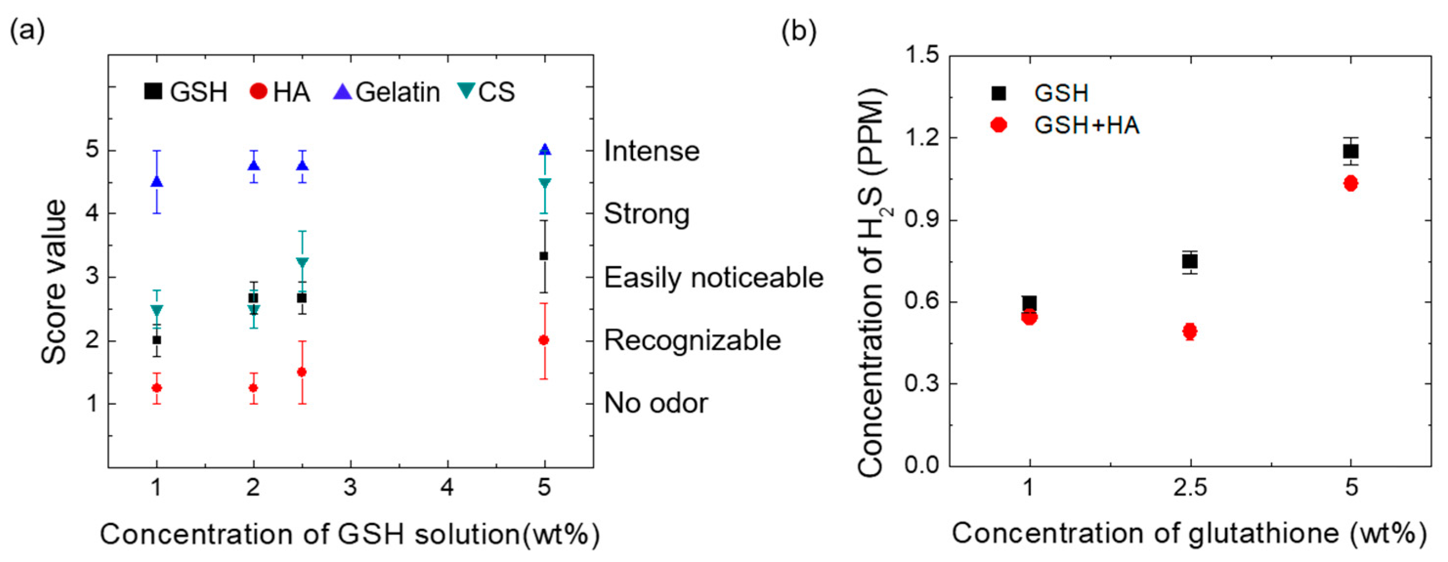
2.3. Odor Tests
2.4. Gas Chromatography (GC) Measurements
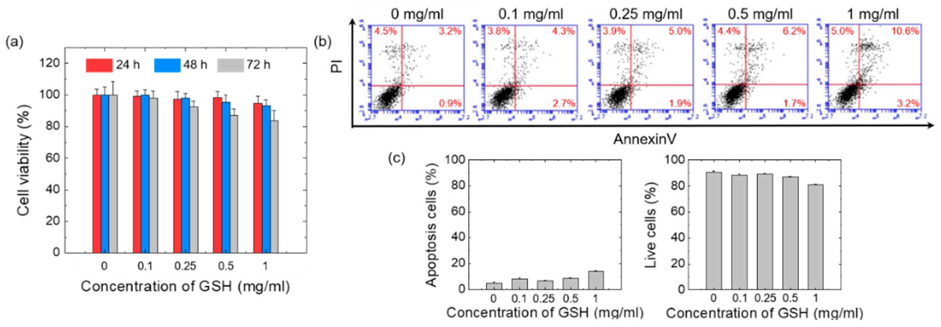
2.5. Cytotoxicity Tests
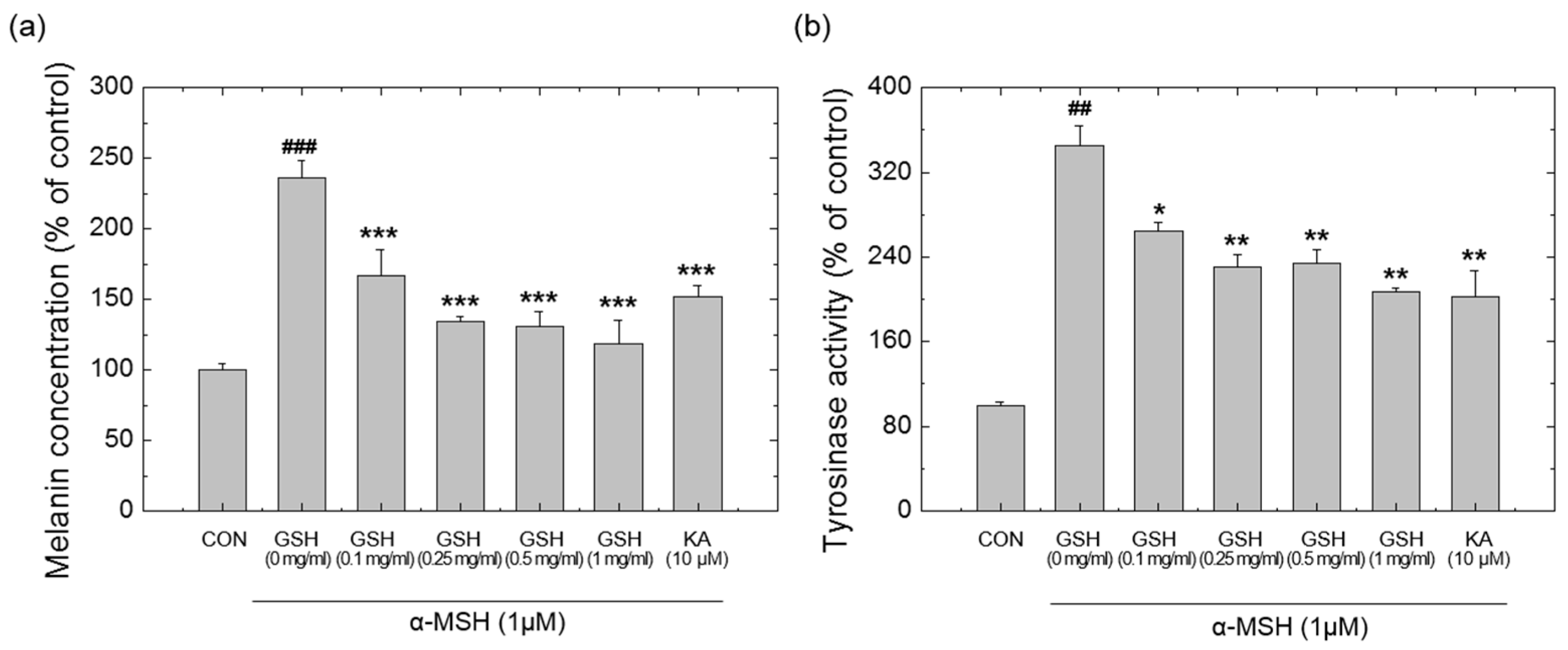
2.6. Determination of Cellular Melanin Content and Tyrosinase Activity
2.7. Fabrication of GSH-MN Patches
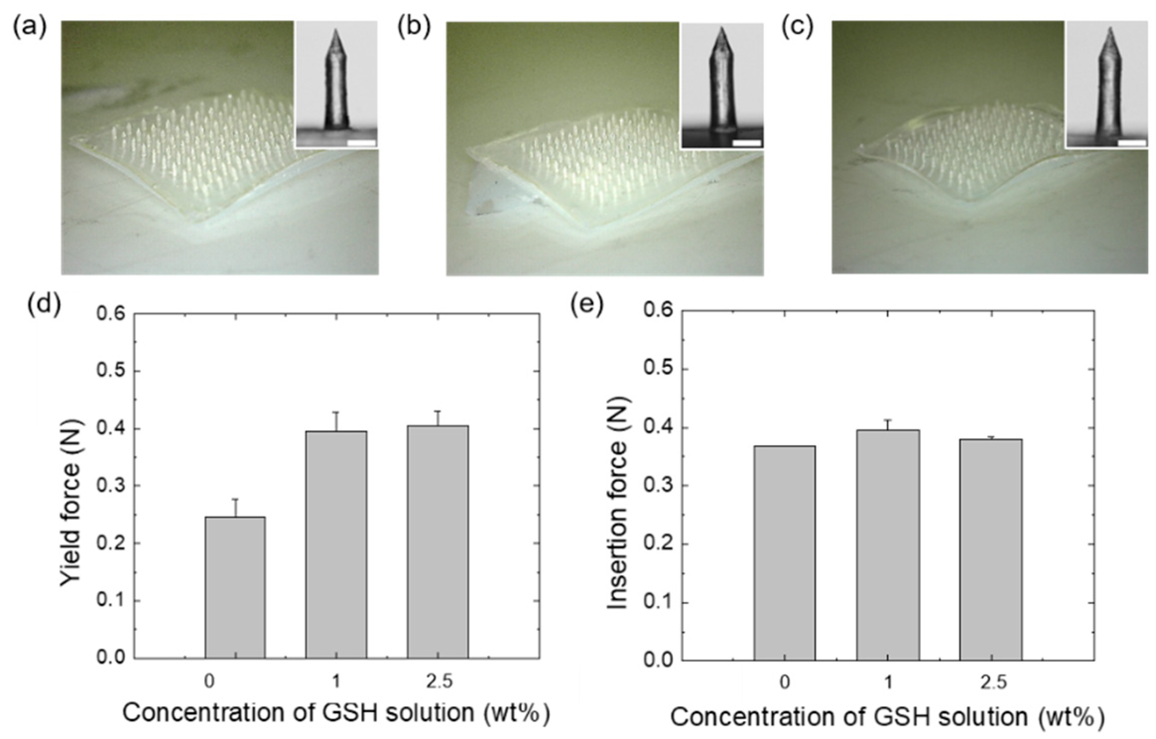
2.8. Fracture Tests
2.9. Skin Insertion Test
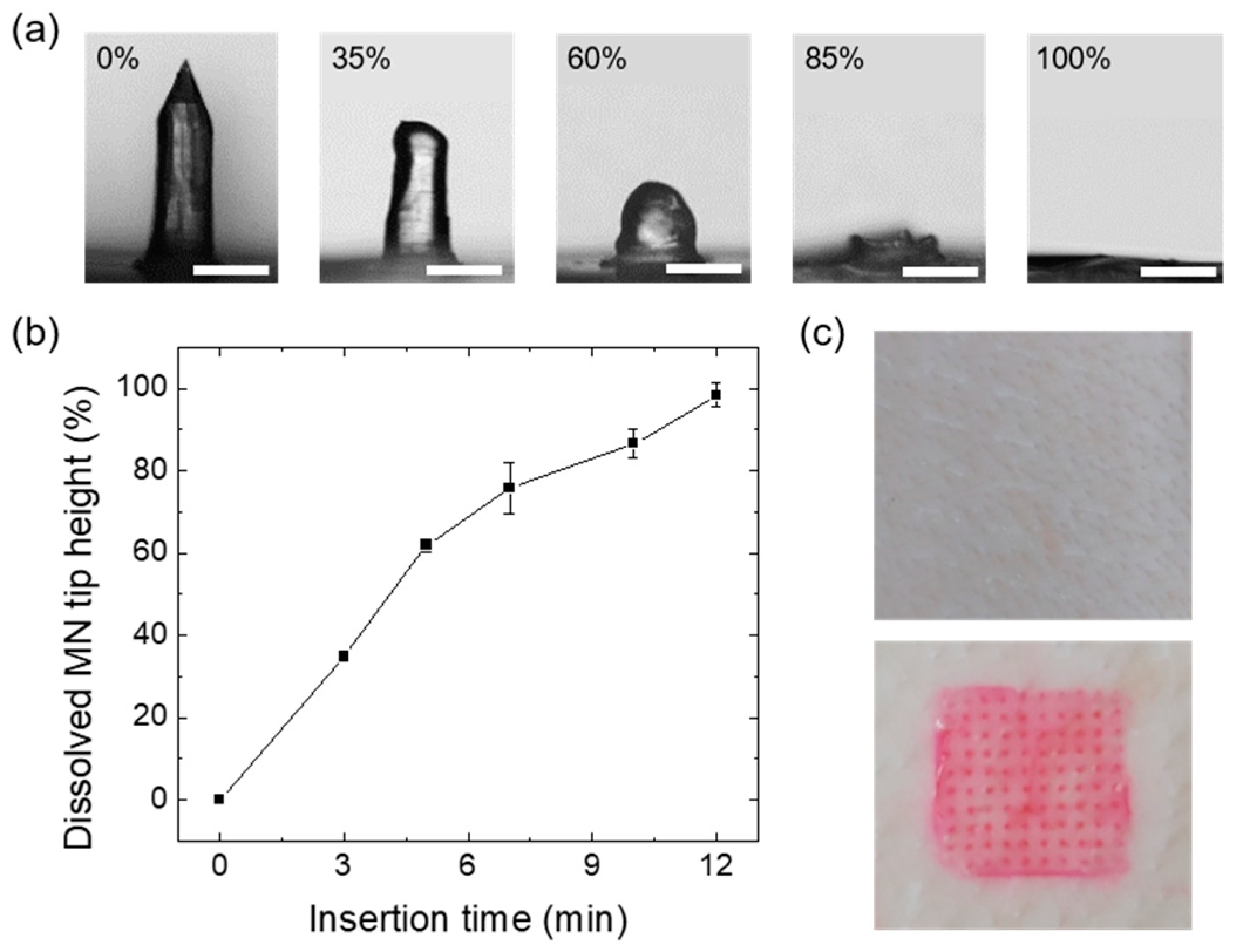
2.10. Dissolution Test
2.11. Loading Capacity and Encapsulation Efficiency
2.12. In Vitro GSH Skin Permeation Tests
2.13. Statistical Analysis
3. Results and Discussion
3.1. Screening Tests to Select Deodorizable Polymers
3.2. Effect of GSH on Cytotoxicity and Tyrosinase Activity
3.3. Fabrication of GSH-loaded HA MNs and Their Mechanical Properties
3.4. In Vitro Dissolution Tests of the GSH-HA MNs
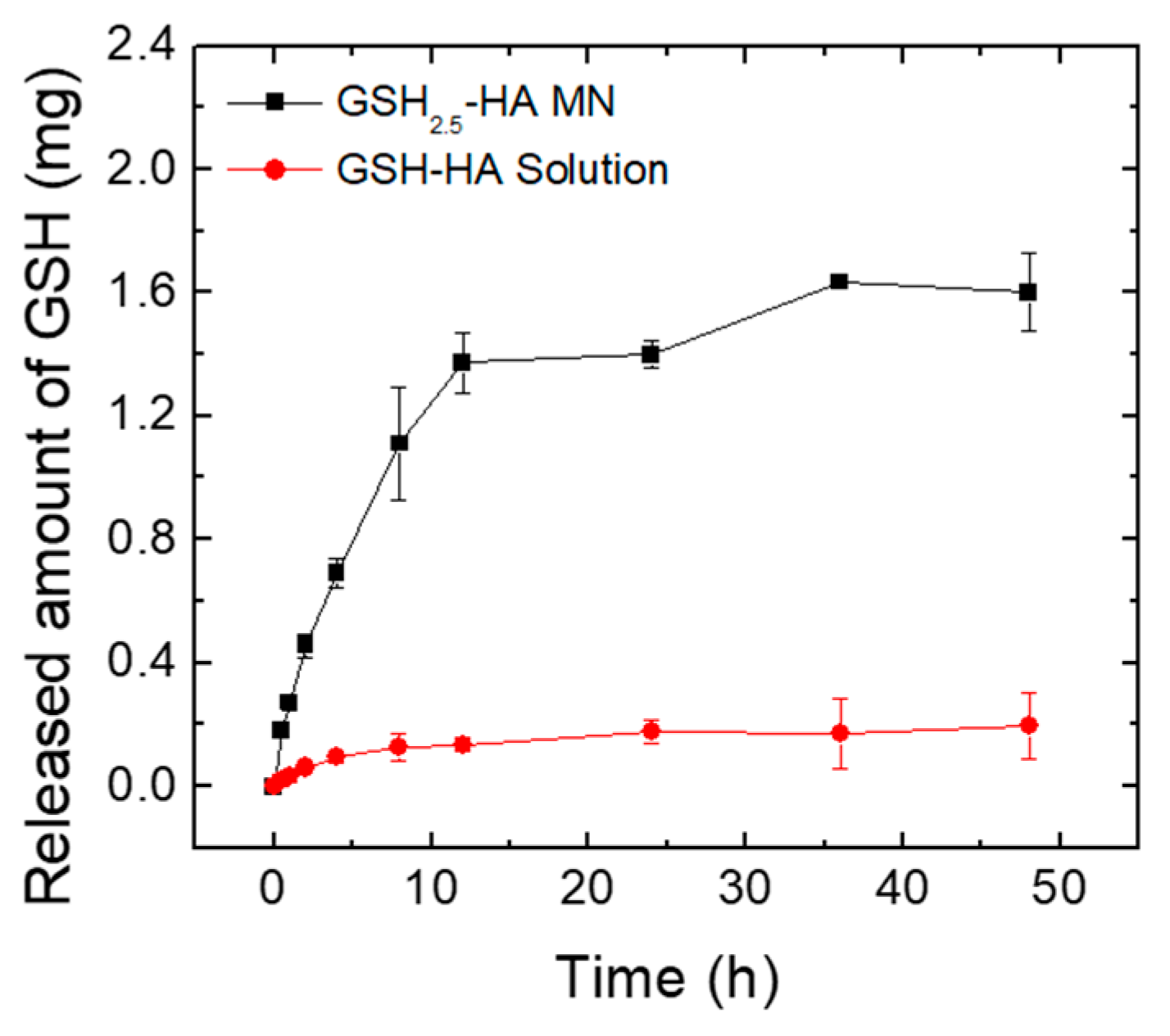
3.5. Ex Vivo Skin Permeation Test of GSH-HA MN Patches
4. Conclusions
Supplementary Materials
Author Contributions
Funding
Conflicts of Interest
References
- Forman, H.J.; Zhang, H.; Rinna, A. Glutathione: Overview of its protective roles, measurement, and biosynthesis. Mol. Asp. Med. 2009, 30, 1–12. [Google Scholar] [CrossRef] [Green Version]
- Hayes, J.D.; McLellan, L.I. Glutathione and glutathione-dependent enzymes represent a co-ordinately regulated defence against oxidative stress. Free Radic. Res. 1999, 31, 273–300. [Google Scholar] [CrossRef]
- Rahman, I.; Kode, A.; Biswas, S.K. Assay for quantitative determination of glutathione and glutathione disulfide levels using enzymatic recycling method. Nat. Protoc. 2006, 1, 3159. [Google Scholar] [CrossRef]
- Yano, H. Comparison of Oxidized and Reduced Glutathione in the Bread-Making Qualities of Rice Batter. J. Food Sci. 2012, 77, C182–C188. [Google Scholar] [CrossRef] [PubMed]
- Weschawalit, S.; Thongthip, S.; Phutrakool, P.; Asawanonda, P. Glutathione and its antiaging and anti-melanogenic effects. Clin. Cosmet. Investig. Dermatol. 2017, 10, 147. [Google Scholar] [CrossRef] [PubMed] [Green Version]
- Villarama, C.D.; Maibach, H.I. Glutathione as a depigmenting agent: An overview. Int. J. Cosmet. Sci. 2005, 27, 147–153. [Google Scholar] [CrossRef] [PubMed]
- Arjinpathana, N.; Asawanonda, P. Glutathione as an oral whitening agent: A randomized, double-blind, placebo-controlled study. J. Dermatol. Treat. 2012, 23, 97–102. [Google Scholar] [CrossRef] [PubMed]
- Droge, W.; Breitkreutz, R. Glutathione and immune function. Proc. Nutr. Soc. 2000, 59, 595–600. [Google Scholar] [CrossRef] [PubMed]
- Patrick, L. Mercury toxicity and antioxidants: Part 1: Role of glutathione and alpha-lipoic acid in the treatment of mercury toxicity. Altern. Med. Rev. 2002, 7, 456–472. [Google Scholar]
- Chen, J.Z.; Kadlubar, F.F. A new clue to glaucoma pathogenesis. Am. J. Med. 2003, 114, 697–698. [Google Scholar] [CrossRef]
- Hassan, M.Q.; Hadi, R.A.; Al-Rawi, Z.S.; Padron, V.A.; Stohs, S.J. The glutathione defense system in the pathogenesis of rheumatoid arthritis. J. Appl. Toxicol. 2001, 21, 69–73. [Google Scholar] [CrossRef] [PubMed]
- Seong, K.Y.; Seo, M.S.; Hwang, D.Y.; O’Cearbhaill, E.D.; Sreenan, S.; Karp, J.M.; Yang, S.Y. A self-adherent, bullet-shaped microneedle patch for controlled transdermal delivery of insulin. J. Control. Release 2017, 265, 48–56. [Google Scholar] [CrossRef] [PubMed]
- Jin, J.; Zhu, L.L.; Chen, M.; Xu, H.M.; Wang, H.F.; Feng, X.Q.; Zhu, X.P.; Zhou, Q. The optimal choice of medication administration route regarding intravenous, intramuscular, and subcutaneous injection. Patient Prefer. Adherence 2015, 9, 923–942. [Google Scholar] [PubMed] [Green Version]
- Bouwstra, J.A.; Honeywell-Nguyen, P.L.; Gooris, G.S.; Ponec, M. Structure of the skin barrier and its modulation by vesicular formulations. Prog. Lipid Res. 2003, 42, 1–36. [Google Scholar] [CrossRef]
- Jiskoot, W.; Randolph, T.W.; Volkin, D.B.; Middaugh, C.R.; Schöneich, C.; Winter, G.; Friess, W.; Crommelin, D.J.; Carpenter, J.F. Protein instability and immunogenicity: Roadblocks to clinical application of injectable protein delivery systems for sustained release. J. Pharm. Sci. 2002, 101, 946–954. [Google Scholar] [CrossRef]
- Liu, S.; Jin, M.N.; Quan, Y.S.; Kamiyama, F.; Katsumi, H.; Sakane, T.; Yamamoto, A. The development and characteristics of novel microneedle arrays fabricated from hyaluronic acid, and their application in the transdermal delivery of insulin. J. Control. Release 2012, 161, 933–941. [Google Scholar] [CrossRef]
- Wang, C.; Ye, Y.; Hochu, G.M.; Sadeghifar, H.; Gu, Z. Enhanced cancer immunotherapy by microneedle patch-assisted delivery of anti-PD1 antibody. Nano Lett. 2016, 16, 2334–2340. [Google Scholar] [CrossRef]
- Dassanayake, R.S.; Acharya, S.; Abidi, N. Biopolymer-based materials from polysaccharides: Properties, processing, characterization and sorption applications. In Advanced Sorption Process Applications; Edebali, S., Ed.; IntechOpen: London, UK, 2018; pp. 1–24. [Google Scholar]
- Lee, J.W.; Choi, S.O.; Felner, E.I.; Prausnitz, M.R. Dissolving microneedle patch for transdermal delivery of human growth hormone. Small 2011, 7, 531–539. [Google Scholar] [CrossRef] [Green Version]
- Donnelly, R.F.; Singh, T.R.R.; Garland, M.J.; Migalska, K.; Majithiya, R.; McCrudden, C.M.; Kole, P.L.; Mahmood, T.M.T.; McCarthy, H.O.; Woolfson, A.D. Hydrogel-forming microneedle arrays for enhanced transdermal drug delivery. Adv. Funct. Mater. 2012, 22, 4879–4890. [Google Scholar] [CrossRef] [Green Version]
- Kim, J.D.; Kim, M.; Yang, H.; Lee, K.; Jung, H. Droplet-born air blowing: Novel dissolving microneedle fabrication. J. Control. Release 2013, 170, 430–436. [Google Scholar] [CrossRef]
- Kim, H.; Seong, K.Y.; Lee, J.H.; Park, W.; Yang, S.Y.; Hahn, S.K. Biodegradable microneedle patch delivering antigenic peptide-hyaluronate conjugate for cancer immunotherapy. ACS Biomater. Sci. Eng. 2019, 5, 5150–5158. [Google Scholar] [CrossRef]
- Du, X.; Song, M.; Rouseff, R. Identification of new strawberry sulfur volatiles and changes during maturation. J. Agric. Food Chem. 2011, 59, 1293–1300. [Google Scholar] [CrossRef] [PubMed]
- Boczkaj, G.; Kaminski, M.; Przyjazny, A. Process control and investigation of oxidation kinetics of postoxidative effluents using gas chromatography with pulsed flame photometric detection (GC-PFPD). Ind. Eng. Chem. Res. 2010, 49, 12654–12662. [Google Scholar] [CrossRef]
- Davis, S.P.; Landis, B.J.; Adams, Z.H.; Allen, M.G.; Prausnitz, M.R. Insertion of microneedles into skin: Measurement and prediction of insertion force and needle fracture force. J. Biomech. 2004, 37, 1155–1163. [Google Scholar] [CrossRef] [PubMed]
- Chandrasekharan, A.; Hwang, Y.J.; Seong, K.Y.; Park, S.; Kim, S.; Yang, S.Y. Acid-treated water-soluble chitosan suitable for microneedle-assisted intracutaneous drug delivery. Pharmaceutics 2019, 11, 209. [Google Scholar] [CrossRef] [PubMed] [Green Version]
- Kim, B.; Seong, K.Y.; You, I.; Selvaraj, V.; Yim, S.G.; O’Cearbhaill, E.D.; Jeong, U.; Yang, S.Y. Touch-actuated transdermal delivery patch for quantitative skin permeation control. Sens. Actuators B Chem. 2018, 256, 18–26. [Google Scholar] [CrossRef]
- Jensen, G.A.; Adams, D.F.; Stern, H. Absorption of hydrogen sulfide and methyl mercaptan from dilute gas mixtures. J. Air Pollut. Control Assoc. 1966, 16, 248–253. [Google Scholar] [CrossRef]
- Onda, K.; Takeuchi, H.; Kobayashi, T.; Yokota, K. Simultaneous Absorption of Hydrogen Sulphide and Carbon Dioxide in Aqueous Sodium Hydroxide Solutions. J. Chem. Eng. Jpn. 1972, 5, 27–33. [Google Scholar] [CrossRef] [Green Version]
- Kabakov, A.E.; Gavai, V.L. Cell death and survival assays. In Chaperones; Humana Press: New York, NY, USA, 2018; pp. 107–127. [Google Scholar]
- Samant, P.P.; Prausnitz, M.R. Mechanisms of sampling interstitial fluid from skin using a microneedle patch. Proc. Natl. Acad. Sci. USA 2018, 115, 4583–4588. [Google Scholar] [CrossRef] [Green Version]
- Yang, S.Y.; O’Cearbhaill, E.D.; Sisk, G.C.; Park, K.M.; Cho, W.K.; Villiger, M.; Bouma, B.E.; Pomahac, B.; Karp, J.M. A bio-inspired swellable microneedle adhesive for mechanical interlocking with tissue. Nat. Commun. 2013, 4, 1702. [Google Scholar] [CrossRef] [Green Version]
- Migdadi, E.M.; Courtenay, A.J.; Tekko, I.A.; McCrudden, M.T.; Kearney, M.C.; McAlister, E.; McCarthy, H.O.; Donnelly, R.F. Hydrogel-forming microneedles enhance transdermal delivery of metformin hydrochloride. J. Control. Release 2018, 285, 142–151. [Google Scholar] [CrossRef] [PubMed]


| GSH-Loaded MN Patch | GSH Amount Loaded in 100 MN Tips (mg) | Loading Capacity of GSH (%) | Encapsulation Efficiency of GSH (%) |
|---|---|---|---|
| GSH1-HA MN | 0.29 ± 0.03 | 11.26 ± 1.24 | 8.36 ± 0.92 |
| GSH2.5-HA MN | 0.56 ± 0.03 | 21.33 ± 1.13 | 6.34 ± 0.34 |
© 2020 by the authors. Licensee MDPI, Basel, Switzerland. This article is an open access article distributed under the terms and conditions of the Creative Commons Attribution (CC BY) license (http://creativecommons.org/licenses/by/4.0/).
Share and Cite
Lee, Y.; Kumar, S.; Kim, S.H.; Seong, K.-Y.; Lee, H.; Kim, C.; Jung, Y.-S.; Yang, S.Y. Odorless Glutathione Microneedle Patches for Skin Whitening. Pharmaceutics 2020, 12, 100. https://doi.org/10.3390/pharmaceutics12020100
Lee Y, Kumar S, Kim SH, Seong K-Y, Lee H, Kim C, Jung Y-S, Yang SY. Odorless Glutathione Microneedle Patches for Skin Whitening. Pharmaceutics. 2020; 12(2):100. https://doi.org/10.3390/pharmaceutics12020100
Chicago/Turabian StyleLee, Yechan, Sujeet Kumar, Sou Hyun Kim, Keum-Yong Seong, Hyeseon Lee, Chaerin Kim, Young-Suk Jung, and Seung Yun Yang. 2020. "Odorless Glutathione Microneedle Patches for Skin Whitening" Pharmaceutics 12, no. 2: 100. https://doi.org/10.3390/pharmaceutics12020100