Non-Invasive Delivery of Nano-Emulsified Sesame Oil-Extract of Turmeric Attenuates Lung Inflammation


 ,
,  and
and 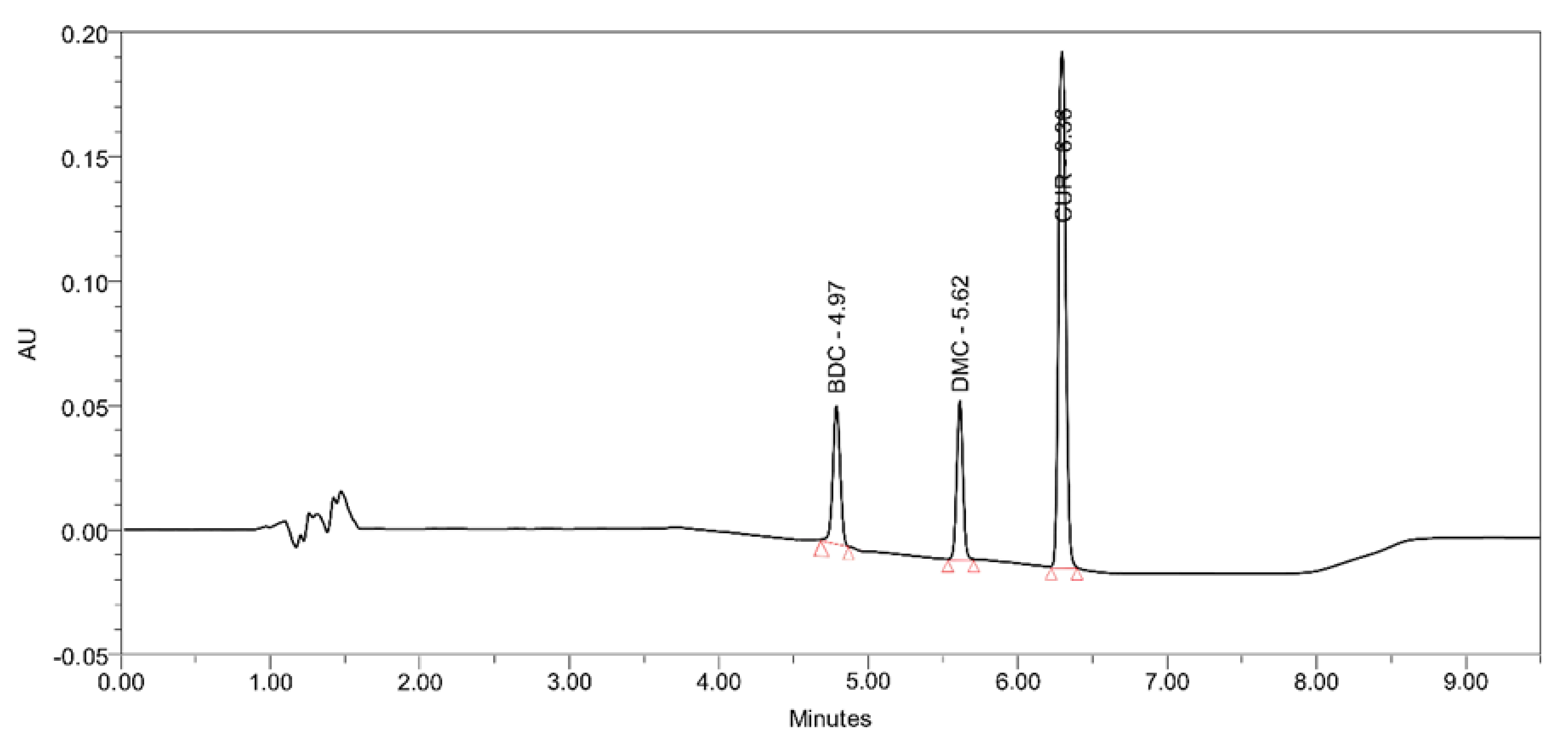
Abstract
:1. Introduction
2. Methods and Methods
2.1. Experimental Materials
2.2. Extraction of Turmeric in Edible Oils
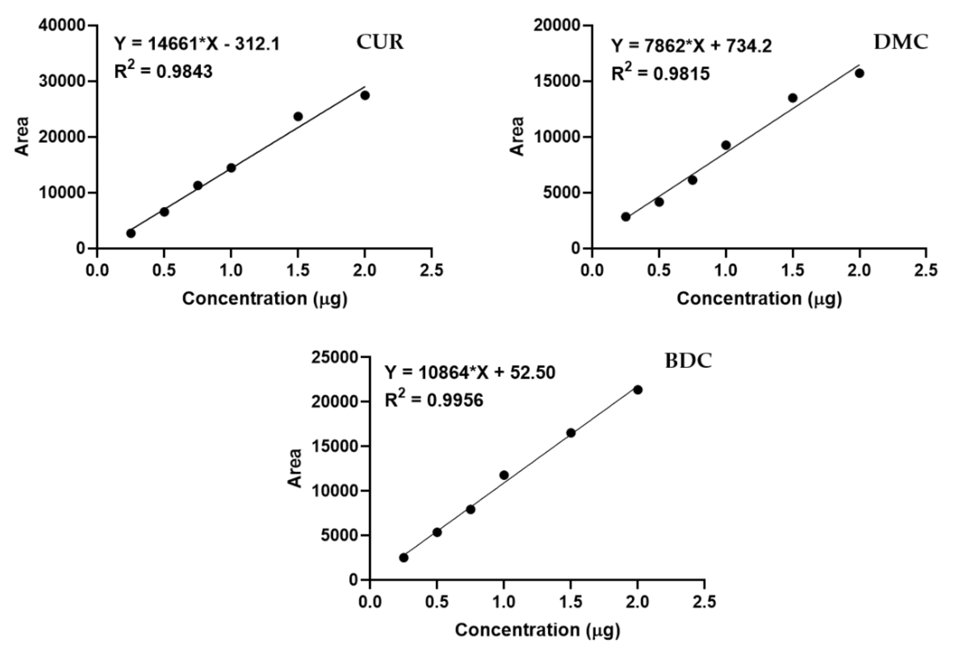
2.3. Determination of Curcuminoids in Oils
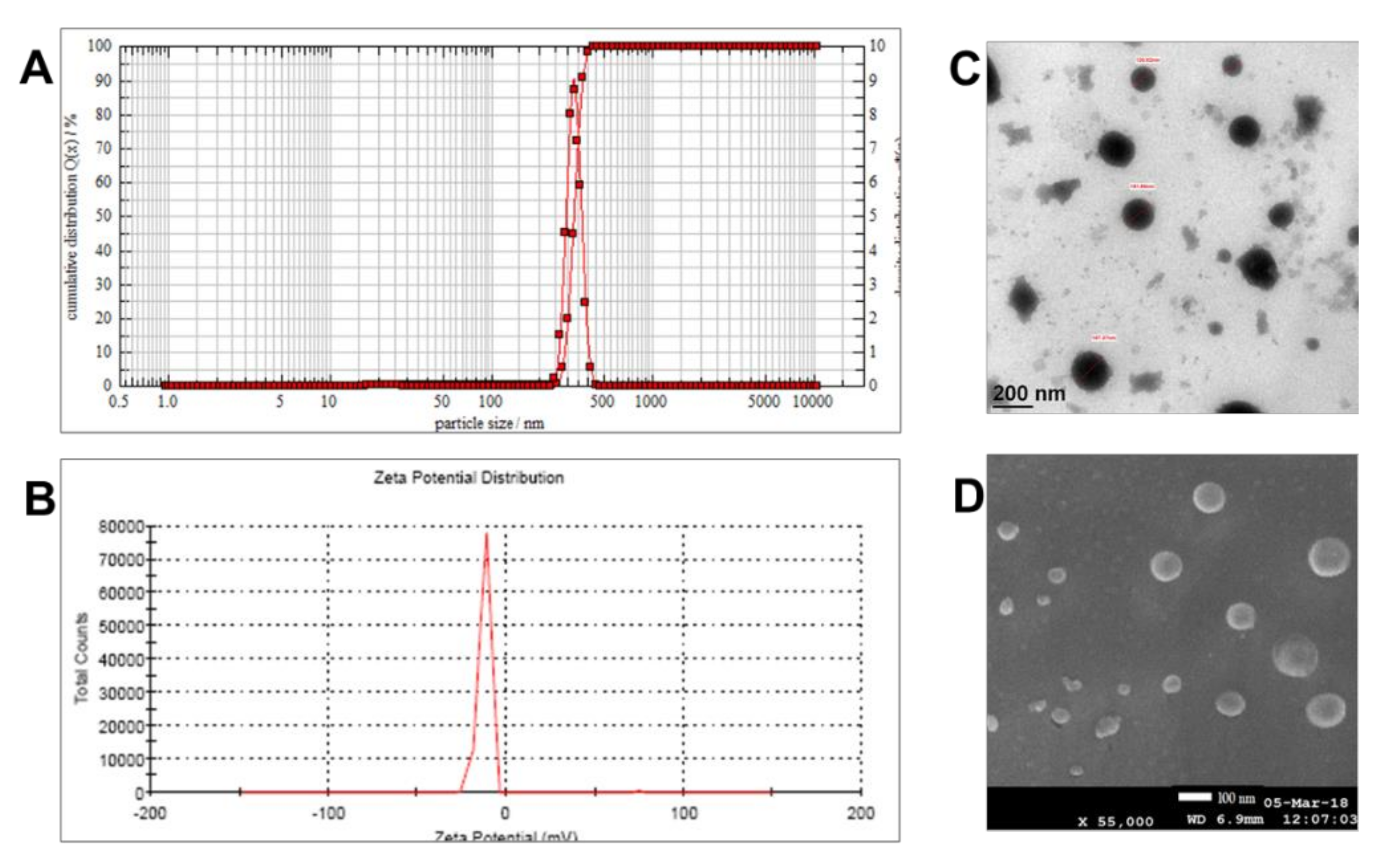
2.4. Preparation of Nano-Emulsion and Characterization
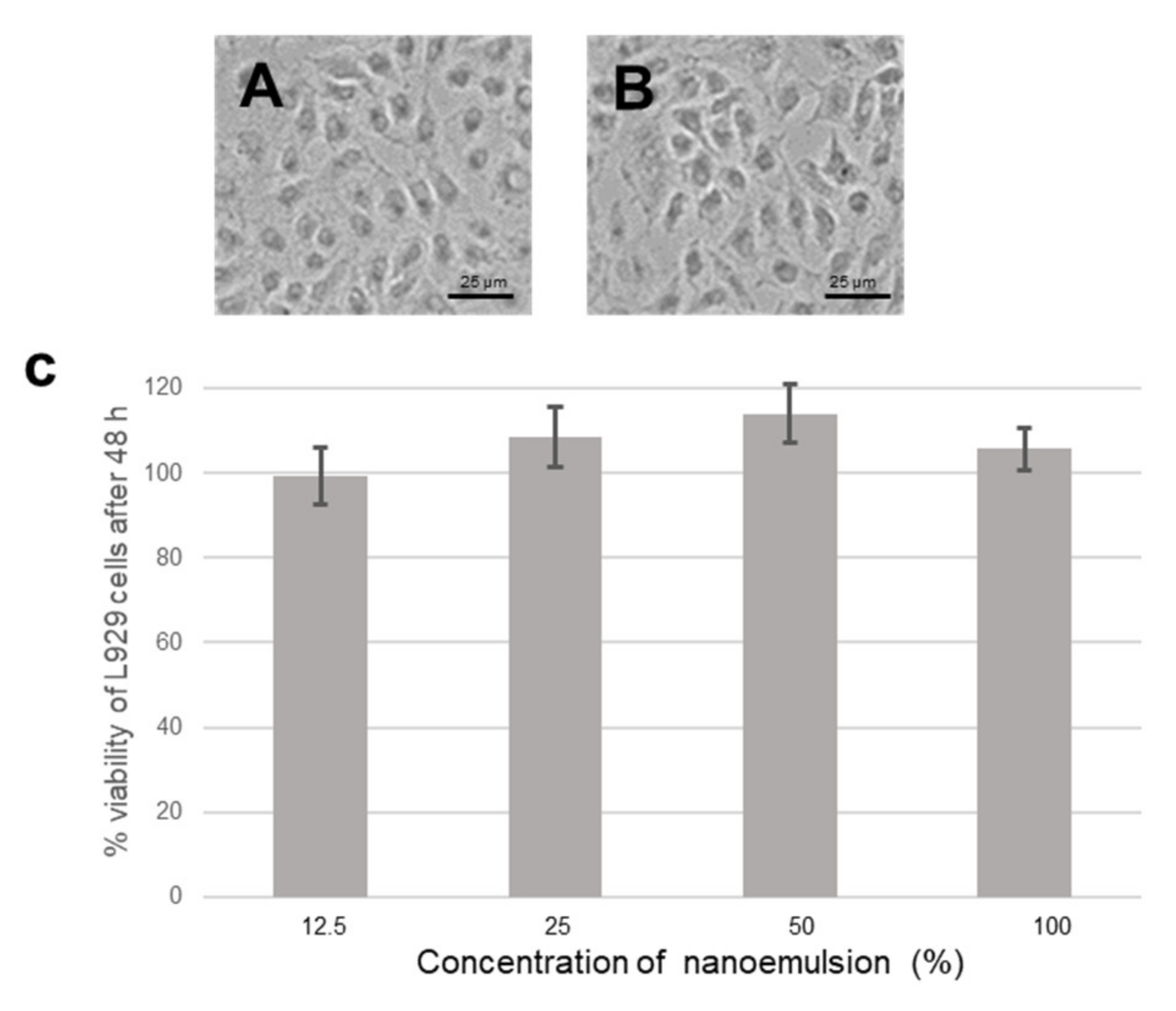
2.5. In Vitro Biocompatibility Testing
2.6. Experimental Animals
2.7. Stimulation of Allergic Symptoms and Treatment
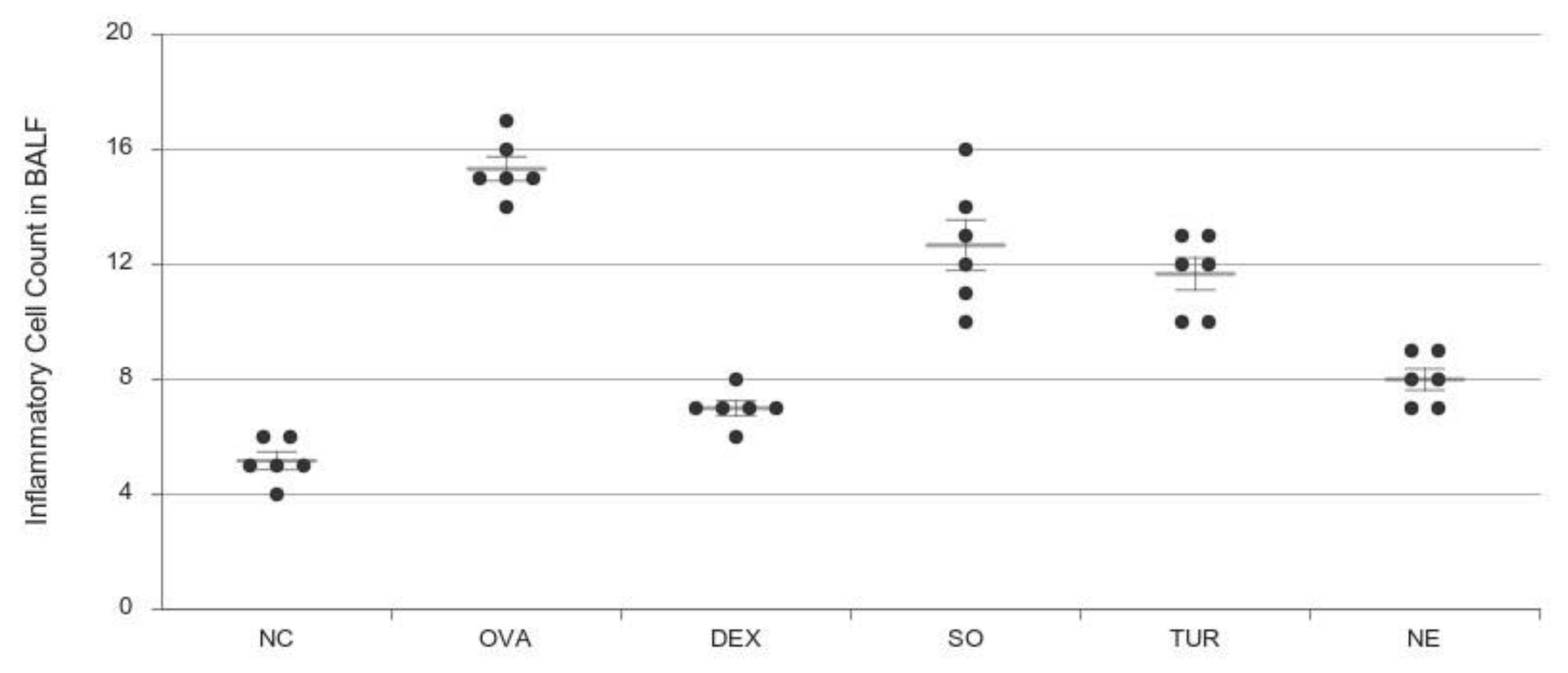
2.8. BALF Collection and Inflammatory Cell Count
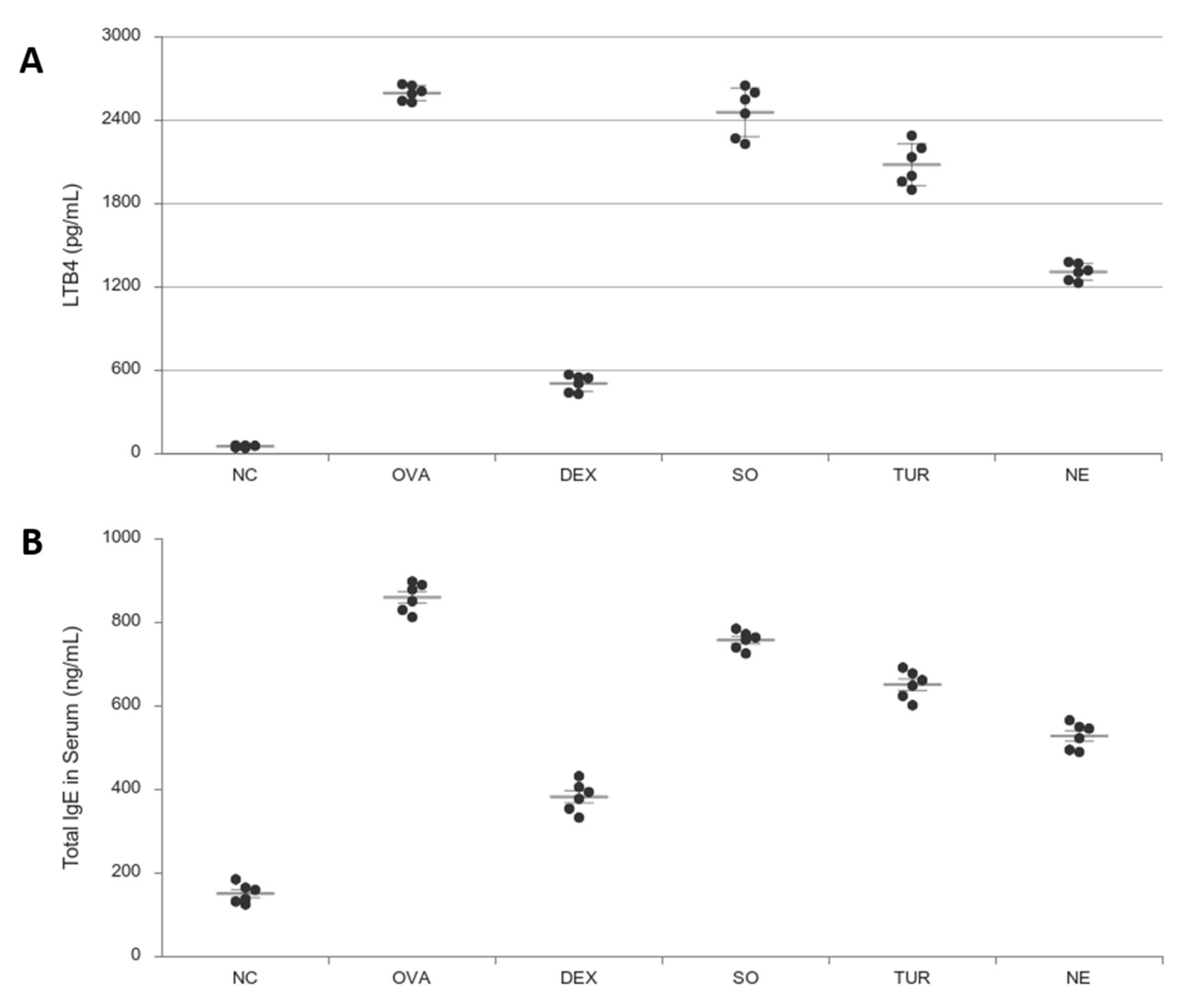
2.9. Determination of Total Levels of Ovalbumin—Specific IgE and Leukotriene LTB
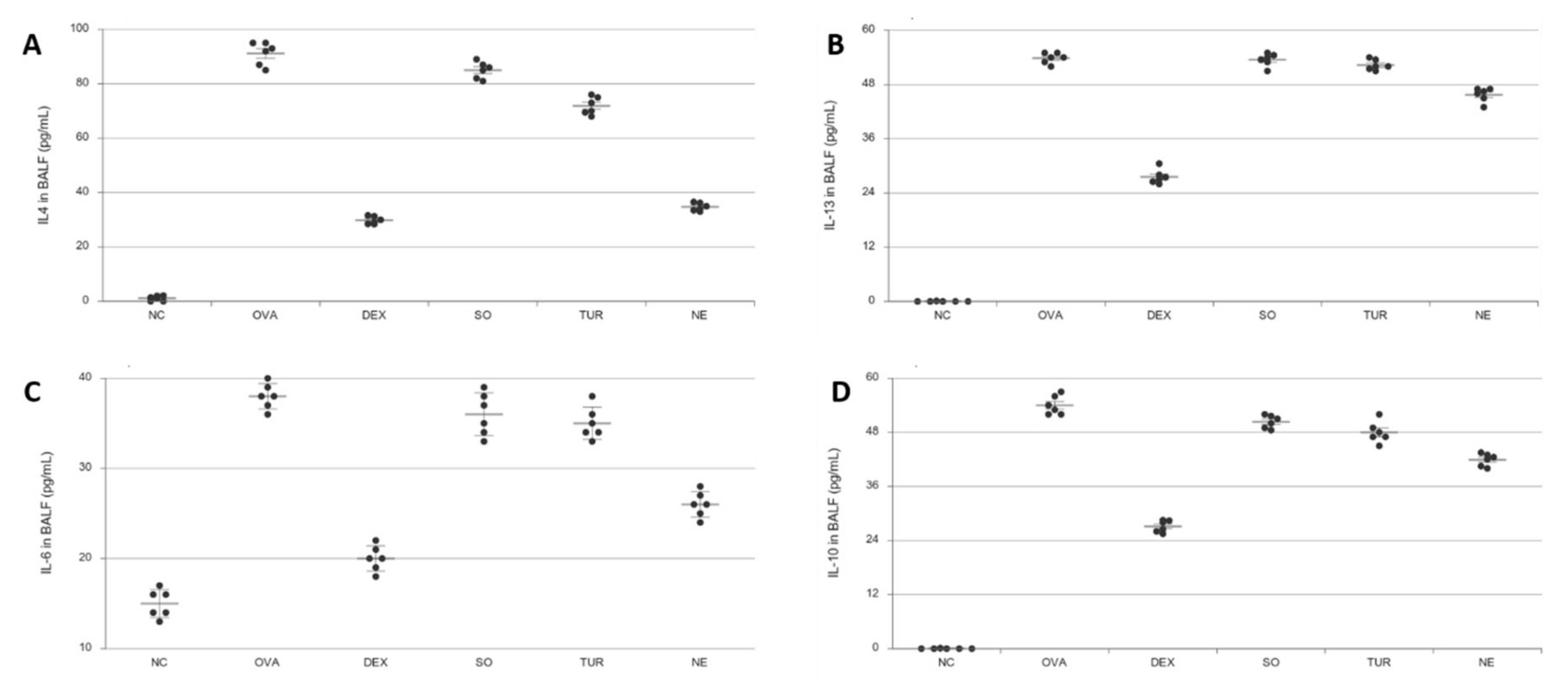
2.10. Measurement of Inflammatory Cytokines in BALF
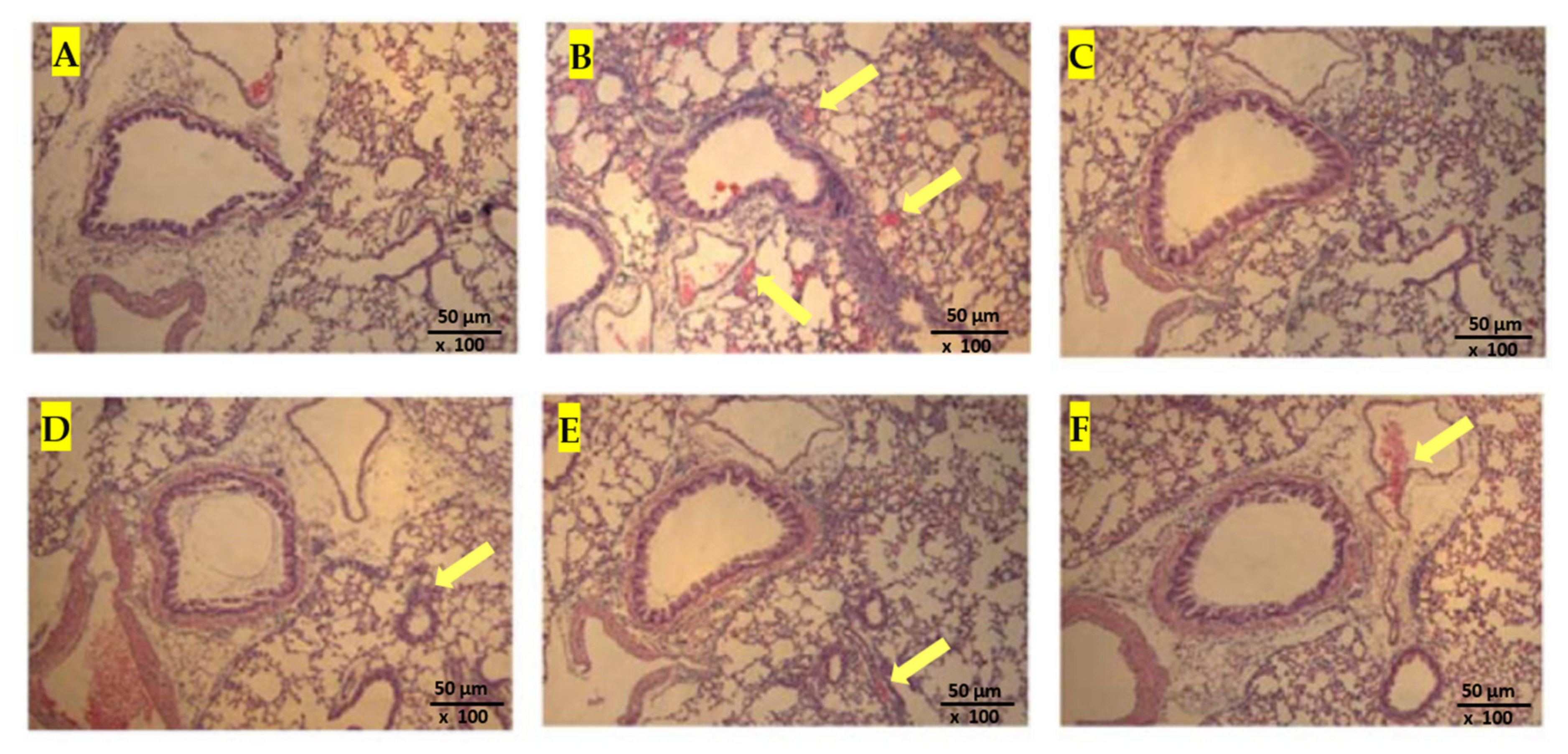
2.11. Histological Analysis
2.12. Flow Cytometry
2.13. Statistical Analysis
3. Results
3.1. Extraction of Curcuminoids and Estimation of Contents
3.2. Characterization of Nano-Emulsion
3.3. In Vitro Biocompatibility Testing
3.4. Leukocyte Count in BALF
3.5. Measurement of Leukotriene B4 (LTB4) and IgE
3.6. Determination of Pro-Inflammatory and Anti-Inflammatory Cytokines
3.7. Histopathology of Lungs
4. Discussion
5. Conclusions
Supplementary Materials
Author Contributions
Funding
Acknowledgments
Conflicts of Interest
References
- Wong, J.; Magun, B.E.; Wood, L.J. Lung inflammation caused by inhaled toxicants: A review. Int. J. Chronic Obstr. Pulm. Dis. 2016, 11, 1391–1401. [Google Scholar] [CrossRef] [PubMed] [Green Version]
- Madl, A.K.; Pinkerton, K.E. Health effects of inhaled engineered and incidental nanoparticles. Crit. Rev. Toxicol. 2009, 39, 629–658. [Google Scholar] [CrossRef] [PubMed]
- Bernard, G.R. Acute respiratory distress syndrome: A historical perspective. Am. J. Respir. Crit. Care Med. 2005, 172, 798–806. [Google Scholar] [CrossRef] [PubMed] [Green Version]
- Tisoncik, J.R.; Korth, M.J.; Simmons, C.P.; Farrar, J.; Martin, T.R.; Katze, M.G. Into the eye of the cytokine storm. Microbiol. Mol. Biol. Rev. MMBR 2012, 76, 16–32. [Google Scholar] [CrossRef] [Green Version]
- Christenson, S.A.; Steiling, K.; van den Berge, M.; Hijazi, K.; Hiemstra, P.S.; Postma, D.S.; Lenburg, M.E.; Spira, A.; Woodruff, P.G. Asthma-COPD overlap. Clinical relevance of genomic signatures of type 2 inflammation in chronic obstructive pulmonary disease. Am. J. Respir. Crit. Care Med. 2015, 191, 758–766. [Google Scholar] [CrossRef] [Green Version]
- Leung, J.M.; Sin, D.D. Asthma-COPD overlap syndrome: Pathogenesis, clinical features, and therapeutic targets. BMJ Clin. Res. Ed. 2017, 358, j3772. [Google Scholar] [CrossRef]
- Murrison, L.B.; Brandt, E.B.; Myers, J.B.; Hershey, G. Environmental exposures and mechanisms in allergy and asthma development. J. Clin. Investig. 2019, 129, 1504–1515. [Google Scholar] [CrossRef] [Green Version]
- Aggarwal, B.B.; Prasad, S.; Reuter, S.; Kannappan, R.; Yadev, V.R.; Park, B.; Kim, J.H.; Gupta, S.C.; Phromnoi, K.; Sundaram, C.; et al. Identification of novel anti-inflammatory agents from Ayurvedic medicine for prevention of chronic diseases: “reverse pharmacology” and “bedside to bench” approach. Curr. Drug Targets 2011, 12, 1595–1653. [Google Scholar] [CrossRef] [Green Version]
- Xu, X.Y.; Meng, X.; Li, S.; Gan, R.Y.; Li, Y.; Li, H.B. Bioactivity, health benefits, and related molecular mechanisms of curcumin: Current progress, challenges, and perspectives. Nutrients 2018, 10, 1553. [Google Scholar] [CrossRef] [Green Version]
- Hsu, D.Z.; Liu, C.T.; Chu, P.Y.; Li, Y.H.; Periasamy, S.; Liu, M.Y. Sesame oil attenuates ovalbumin-induced pulmonary edema and bronchial neutrophilic inflammation in mice. BioMed Res. Int. 2013, 2013, 1–7. [Google Scholar] [CrossRef]
- Basnet, P.; Skalko-Basnet, N. Curcumin: An anti-inflammatory molecule from a curry spice on the path to cancer treatment. Molecules 2011, 16, 4567–4598. [Google Scholar] [CrossRef] [PubMed] [Green Version]
- Nader, M.A.; El-Awady, M.S.; Shalaby, A.A.; El-Agamy, D.S. Sitagliptin exerts anti-inflammatory and anti-allergic effects in ovalbumin-induced murine model of allergic airway disease. Naunyn-Schmiedeberg’s Arch. Pharmacol. 2012, 385, 909–919. [Google Scholar] [CrossRef] [PubMed]
- Paul, P.; Majhi, S.; Mitra, S.; Banerjee, E.R. Immuno-modulatory and therapeutic effect of curcumin in an allergen-sensitized murine model of chronic asthma. J. Clin. Cell. Immunol. 2018, 9, 551. [Google Scholar] [CrossRef]
- Wang, J.; Li, R.; Peng, Z.; Hu, B.; Rao, X.; Li, J. HMGB1 participates in LPS-induced acute lung injury by activating the AIM2 inflammasome in macrophages and inducing polarization of M1 macrophages via TLR2, TLR4, and RAGE/NF-κB signaling pathways. Int. J. Mol. Med. 2020, 45, 61–80. [Google Scholar] [CrossRef] [PubMed]
- Jangle, R.D.; Thorat, B.N. Reversed-phase high-performance liquid chromatography method for analysis of curcuminoids and curcuminoid-loaded liposome formulation. Indian J. Pharm. Sci. 2013, 75, 60. [Google Scholar] [CrossRef] [Green Version]
- Scoditti, E.; Massaro, M.; Garbarino, S.; Toraldo, D.M. Role of diet in chronic obstructive pulmonary disease prevention and treatment. Nutrients 2019, 11, 1357. [Google Scholar] [CrossRef] [Green Version]
- Rodríguez-Rodríguez, E.; Ortega, R.M.; Andrés, P.; Aparicio, A.; González-Rodríguez, L.G.; López-Sobaler, A.M.; Navia, B.; Perea, J.M.; Rodríguez-Rodríguez, P. Antioxidant status in a group of institutionalised elderly people with chronic obstructive pulmonary disease. Br. J. Nutr. 2016, 115, 1740–1747. [Google Scholar] [CrossRef] [Green Version]
- Pulido-Moran, M.; Moreno-Fernandez, J.; Ramirez-Tortosa, C.; Ramirez-Tortosa, M. Curcumin and health. Molecules 2016, 21, 264. [Google Scholar] [CrossRef]
- Sorrenti, V.; Contarini, G.; Sut, S.; Dall’Acqua, S.; Confortin, F.; Pagetta, A.; Zusso, M. Curcumin prevents acute neuroinflammation and long-term memory impairment induced by systemic lipopolysaccharide in mice. Front. Pharmacol. 2018, 9, 183. [Google Scholar] [CrossRef] [Green Version]
- Wang, X.; An, X.; Wang, X.; Bao, C.; Li, J.; Yang, D.; Bai, C. Curcumin ameliorated ventilator-induced lung injury in rats. Biomed. Pharmacother. 2018, 98, 754–761. [Google Scholar] [CrossRef]
- Hui-Yin, Y.; Ahmad, N.; Azmi, N.; Makmor-Bakry, M. Curcumin: The molecular mechanisms of action in inflammation and cell death during kainate-induced epileptogenesis. Indian J. Pharm. Educ. Res. 2018, 52, 32–41. [Google Scholar] [CrossRef] [Green Version]
- Magotra, A.; Kotwal, P.; Bhatt, S.; Dogra, A.; Singh, G.; Nandi, U. Impact of concomitantly administered curcumin on pharmacokinetics of daclatasvir in mice under the frame of herb-drug interaction. Indian J. Pharm. Educ. Res. 2018, 52, S11–S15. [Google Scholar] [CrossRef]
- Guerrero, S.; Inostroza-Riquelme, M.; Contreras-Orellana, P.; Diaz-Garcia, V.; Lara, P.; Vivanco-Palma, A.; Cárdenas, A.; Miranda, V.; Robert, P.; Leyton, L.; et al. Curcumin-loaded nanoemulsion: A new safe and effective formulation to prevent tumor reincidence and metastasis. Nanoscale 2018, 10, 22612–22622. [Google Scholar] [CrossRef] [PubMed]
- Chang, H.B.; Chen, B.H. Inhibition of lung cancer cells A549 and H460 by curcuminoid extracts and nano-emulsions prepared from Curcuma longa Linnaeus. Int. J. Nanomed. 2015, 10, 5059–5080. [Google Scholar] [CrossRef] [Green Version]
- Joe, B.; Lokesh, B.R. Role of capsaicin, curcumin and dietary n—3 fatty acids in lowering the generation of reactive oxygen species in rat peritoneal macrophages. Biochim. Biophys. Acta BBA Mol. Cell Res. 1994, 1224, 255–263. [Google Scholar] [CrossRef]
- Ramkumar, M.; Rajasankar, S.; Gobi, V.V.; Janakiraman, U.; Manivasagam, T.; Thenmozhi, A.J.; Guillemin, G.J. Demethoxycurcumin, a natural derivative of curcumin abrogates rotenone-induced dopamine depletion and motor deficits by its antioxidative and anti-inflammatory properties in Parkinsonian rats. Pharmacogn. Mag. 2018, 14, 9. [Google Scholar] [CrossRef] [PubMed]
- Simon, J.E.; Chadwick, A.F. Herbs: An Indexed Bibliography 1971–1980: The Scientific Literature on Selected Herbs and Aromatic and Medicinal Plants of the Temperature Zone (No. 581.634063 S55); Archon Books: San Francisco, CA, USA, 1984; Available online: http://www.hort.purdue.edu/newcrop/medaro/factsheets/sesame.html (accessed on 28 July 2020).
- Sarma, L.; Chakraborty, S.; Duary, R.K. Solvent-based microwave-assisted extraction and identification of bioactive compounds from Sesamum indicum leaves using particle swarm optimization-integrated response surface methodology. Pharmacogn. Mag. 2018, 14, 275. [Google Scholar] [CrossRef]
- Hsu, D.Z.; Su, S.B.; Chien, S.P.; Chiang, P.J.; Li, Y.H.; Lo, Y.J.; Liu, M.Y. Effect of sesame oil on oxidative-stress-associated renal injury in endotoxemic rats: Involvement of nitric oxide and proinflammatory cytokines. Shock 2005, 24, 276–280. [Google Scholar] [CrossRef]
- Hsu, D.Z.; Chen, K.T.; Chien, S.P. Sesame oil attenuates acute iron-induced lipid peroxidation-associated hepatic damage in mice. Shock 2006, 26, 625–630. [Google Scholar] [CrossRef]
- Beigelman, A.; Gunsten, S.; Mikols, C.L.; Vidavsky, I.; Cannon, C.L.; Brody, S.L.; Walter, M.J. Azithromycin attenuates airway inflammation in a noninfectious mouse model of allergic asthma. Chest 2009, 136, 498–506. [Google Scholar] [CrossRef]
- Galli, S.J.; Tsai, M.; Piliponsky, A.M. The development of allergic inflammation. Nature 2008, 454, 445–454. [Google Scholar] [CrossRef] [PubMed] [Green Version]
- Bobic, S.; Seys, S.; De Vooght, V.; Callebaut, I.; Hox, V.; Dooms, C.; Bullens, D.M. Placental growth factor contributes to bronchial neutrophilic inflammation and edema in allergic asthma. Am. J. Respir. Cell Mol. Biol. 2012, 46, 781–789. [Google Scholar] [CrossRef] [PubMed] [Green Version]
- Creţu, E.; Trifan, A.; Vasincu, A.; Miron, A. Plant-derived anticancer agents—Curcumin in cancer prevention and treatment. Rev. Med. Chir. Soc. Med. Nat. Iasi 2012, 116, 1223–1229. [Google Scholar] [PubMed]
- Yang, G.; Hamacher, J.; Gorshkov, B. The dual role of TNF in pulmonary edema. J. Cardiovasc. Dis. Res. 2010, 1, 29–36. [Google Scholar] [CrossRef] [PubMed] [Green Version]
- Kuhl, K.; Hanania, N.A. Targeting IgE in asthma. Curr. Opin. Pulm. Med. 2012, 18, 1–5. [Google Scholar] [CrossRef] [PubMed]
- Cho, J.W.; Lee, K.S.; Kim, C.W. Curcumin attenuates the expression of IL-1beta, IL-6, and TNF-alpha as well as cyclin E in TNF-alpha-treated HaCaT cells; NF-kappaB and MAPKs as potential upstream targets. Int. J. Mol. Med. 2007, 19, 469–474. [Google Scholar]
- Jin, C.Y.; Lee, J.D.; Park, C.; Choi, Y.H.; Kim, G.Y. Curcumin attenuates the release of pro-inflammatory cytokines in lipopolysaccharide-stimulated BV2 microglia. Acta Pharmacol Sin. 2007, 28, 1645–1651. [Google Scholar] [CrossRef] [Green Version]
- Moqbel, R.; Odemuyiwa, S.O. Allergy, asthma, and inflammation: Which inflammatory cell type is more important? Allergy Asthma Clin. Immunol. 2008, 4, 15–156. [Google Scholar] [CrossRef] [Green Version]
- Kurup, V.P.; Barrios, C.S. Immunomodulatory effects of curcumin in allergy. Mol. Nutr. Food Res. 2008, 52, 1031–1039. [Google Scholar] [CrossRef]
- Chong, L.; Zhang, W.; Nie, Y.; Yu, G.; Liu, L.; Lin, L.; Li, C. Protective effect of curcumin on acute airway inflammation of allergic asthma in mice through Notch1–GATA3 signaling pathway. Inflammation 2014, 37, 1476–1485. [Google Scholar] [CrossRef] [Green Version]
- Sugano, M. Sesamin: A multifunctional gift from nature. J. Chin. Nutr. Sci. 1993, 18, 1–11. [Google Scholar]
- Ma, C.; Ma, Z.; Fu, Q.; Ma, S. Curcumin attenuates allergic airway inflammation by regulation of CD4+ CD25+ regulatory T cells (Tregs)/Th17 balance in ovalbumin-sensitized mice. Fitoterapia 2013, 87, 57–64. [Google Scholar] [CrossRef] [PubMed]


Publisher’s Note: MDPI stays neutral with regard to jurisdictional claims in published maps and institutional affiliations. |
© 2020 by the authors. Licensee MDPI, Basel, Switzerland. This article is an open access article distributed under the terms and conditions of the Creative Commons Attribution (CC BY) license (http://creativecommons.org/licenses/by/4.0/).
Share and Cite
Rasool, S.T.; Alavala, R.R.; Kulandaivelu, U.; Sreeharsha, N. Non-Invasive Delivery of Nano-Emulsified Sesame Oil-Extract of Turmeric Attenuates Lung Inflammation. Pharmaceutics 2020, 12, 1206. https://doi.org/10.3390/pharmaceutics12121206
Rasool ST, Alavala RR, Kulandaivelu U, Sreeharsha N. Non-Invasive Delivery of Nano-Emulsified Sesame Oil-Extract of Turmeric Attenuates Lung Inflammation. Pharmaceutics. 2020; 12(12):1206. https://doi.org/10.3390/pharmaceutics12121206
Chicago/Turabian StyleRasool, Sahibzada Tasleem, Rajasekhar Reddy Alavala, Umasankar Kulandaivelu, and Nagaraja Sreeharsha. 2020. "Non-Invasive Delivery of Nano-Emulsified Sesame Oil-Extract of Turmeric Attenuates Lung Inflammation" Pharmaceutics 12, no. 12: 1206. https://doi.org/10.3390/pharmaceutics12121206