Review

Blood–Brain Barrier Modulation to Improve Glioma Drug Delivery

1
Department of Chemical and Biological Engineering, University of Wisconsin-Madison, Madison, WI 53706, USA
2
Department of Neurological Surgery, University of Wisconsin-Madison, 600 Highland Avenue, Madison, WI 53792, USA
*
Author to whom correspondence should be addressed.
Pharmaceutics 2020, 12(11), 1085; https://doi.org/10.3390/pharmaceutics12111085
Submission received: 29 October 2020 / Revised: 9 November 2020 / Accepted: 10 November 2020 / Published: 12 November 2020
(This article belongs to the Special Issue New Drug Delivery across the Blood–Brain Barrier)

Abstract

:
The blood–brain barrier (BBB) is formed by brain microvascular endothelial cells that are sealed by tight junctions, making it a significant obstacle for most brain therapeutics. The poor BBB penetration of newly developed therapeutics has therefore played a major role in limiting their clinical success. A particularly challenging therapeutic target is glioma, which is the most frequently occurring malignant brain tumor. Thus, to enhance therapeutic uptake in tumors, researchers have been developing strategies to modulate BBB permeability. However, most conventional BBB opening strategies are difficult to apply in the clinical setting due to their broad, non-specific modulation of the BBB, which can result in damage to normal brain tissue. In this review, we have summarized strategies that could potentially be used to selectively and efficiently modulate the tumor BBB for more effective glioma treatment.

Graphical Abstract

1. Introduction

Gliomas are one of the more frequent tumors occurring in the central nervous systems (CNS), accounting for over 32% of primary CNS cancers and over 77% of primary malignant brain cancers [1]. Gliomas include glioblastoma (GBM), a highly-aggressive brain tumor characterized by its rapid proliferation and angiogenesis, leading to a high mortality and making it the leading cause of cancer-related death in young adults aged 20–39 years and the second leading cause of death in children with cancer [2]. The prognosis of GBM patients is quite poor, with a median survival of approximately 14.6 months [3], and 5 year and 10 year survival rates of 5.0% and 2.6%, respectively [4]. Despite these challenges, new therapeutic regimens have been few. Although many new highly potent cytotoxic agents have been developed, systemic administration of these drugs has not increased the median survival of glioma patients in large part due to their poor BBB penetration [3]. The BBB is a highly specialized vascular interface that protects the neuronal environment from most blood-borne materials, and oftentimes the BBB excludes therapeutics. The BBB is formed by brain microvascular endothelial cells (BMECs) that work in concert with other cells of the neurovascular unit such as pericytes, astrocytes and neurons to regulate the movement of molecules and cells between blood and brain (Figure 1A). BMECs are non-fenestrated endothelial cells connected by junctional complexes that include adherens junction proteins (AJ, such as VE-cadherin) and tight junction (TJ) proteins, such as claudins, occludin and zonula occludens protein 1 (ZO-1). These properties, along with low levels of vesicular trafficking, help form a physical barrier to diffusion of material from the blood into the brain. Brain accumulation of hydrophilic small molecules and large-molecule biologics, such as siRNA/mRNA, monoclonal antibodies and antibody–drug conjugates are significantly blocked by this physical barrier [5,6]. For lipophilic small molecules that can diffuse across the plasma membranes of BMECs, efflux transporters expressed in BMECs often pump the drugs back into the bloodstream, acting as an active barrier [7]. These passive and active barrier properties presented by the BBB make it difficult to achieve therapeutic drug concentrations when treating glioma. Thus, it is of importance to enhance drug delivery across the BBB in order to improve glioma therapy and prognosis.
Despite the substantial barrier imparted by the BBB, its integrity can be disrupted by several pathologic conditions, including glioma. In the clinic, BBB disruption in glioma has been observed by accumulation of gadolinium-based magnetic resonance imaging (MRI) contrast agents within tumor regions [8]. A significant leakage of Evans blue and gadolinium at the glioma site also indicates BBB disruption [9]. This leaky glioma BBB caused many to suggest that the BBB is no longer limiting drug delivery and hence efficacy of therapies in treating GBM [5]. However, any drug delivery enhancement that results from BBB leakage is not enough to cure GBM. In fact, even when drugs help treat the BBB-disrupted tumor bulk or when neurosurgeons perform a gross total resection of all BBB disrupted regions of the tumor core indicated by contrast-enhancing agent, the tumor will recur within months in nearly all of these GBM patients [10]. While there is BBB disruption in GBM, numerous studies have shown that this disruption is heterogeneous in GBM patients [11,12,13,14]. As a result of the heterogeneous disruption and the highly-invasive nature of GBM, tumors can be found in areas with an intact BBB in the tumor rim (Figure 1B), where this intact BBB can still limit the distribution of therapeutic drugs [15]. As such, in this review, we will refer to the heterogeneously permeable vasculature of the tumor including both the tumor core and rim as the “tumor BBB”. Because of the tumor lying in areas behind the comparatively healthy tumor BBB in the tumor rim and invasive margins, many novel glioma therapeutics have failed in the clinic, in part because of the inability to reach effective drug concentrations [7]. Thus, despite tumor BBB disruption in the tumor core, it is still essential to establish strategies that can mediate delivery of therapeutic agents to the tumor rim and invasive margins when treating GBM.
A variety of strategies have been suggested to overcome the tumor BBB, thus improving drug delivery in treating glioma [16]. These include approaches to increase drug permeability through chemical modification, inhibition of efflux transporters, transcytosis via targeting of endogenous BBB transporters/receptors, and osmotic BBB disruption, among others and these have been reviewed elsewhere [16,17,18]. Such strategies can often lead to broad drug distribution in the brain, which can result in damage to the non-diseased tissue. For example, while osmotic disruption has met partial clinical success for the treatment of GBM, non-selective opening of the BBB induced by mannitol can also result in various complications including epilepsy and brain edema [19]. Thus, selective and efficient opening of the tumor BBB at the site of glioma would be beneficial for the targeting of cytotoxic therapies and minimization of side effects [10]. In this review, we will discuss drug delivery strategies that hinge on selective targeting of pathological tumor BBB disruption or selective biochemical or physical modulation of tumor BBB permeability to allow for enhanced, localized drug uptake to glioma.

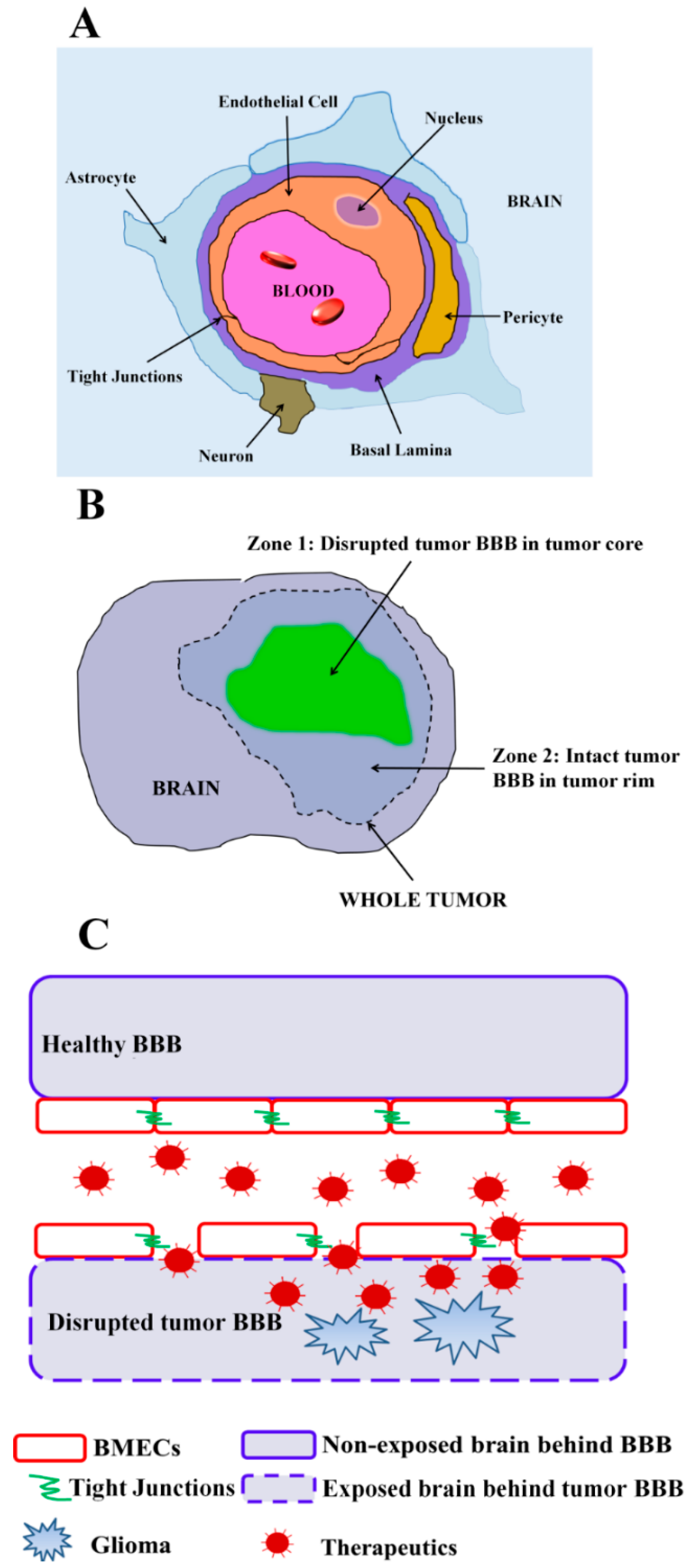
2. Leveraging Pathological BBB Disruption in Glioma

As described above, tumor BBB disruption in glioma is heterogeneous and is quite dependent on the pathological development or stage of glioma [20]. In addition to gadolinium-contrast MRI, the permeability of 14C-sucrose in rat GBM area was 25-fold higher than that in the normal brain tissue [21]. Furthermore, intracarotid injection of anti-tumor drug, cisplatin, demonstrated a 10-fold higher distribution in GBM area compared with the control normal brain tissue [21]. Increased tumor BBB disruption tends to correlate with higher-grade or more malignant glioma in human patients [22], and a systematic study in mice using real-time MRI indicated that the tumor BBB remains intact in earlier glioma stages but becomes significantly disrupted in the tumor core in late stages of glioma progression [23]. In part, this is a result of glioma malignancy grade being positively correlated with angiogenesis, leading to more immature and higher permeability vessels within the tumor [24,25]. Thus, tumor BBB leakiness in the pathological progression of glioma has been used as a surrogate target for selective drug delivery into the tumor site during therapy [26,27]. To date, two main treatment strategies have been explored to exploit tumor BBB leakiness in the tumor core, one that relies on passive drug accumulation at sites of tumor BBB disruption and another that leverages specific targeting of glioma regions with disrupted tumor BBB.

2.1. Passive Drug Accumulation at Sites of Tumor BBB Disruption

First, the ability for normally BBB-impermeant drugs to accumulate at sites of tumor BBB disruption is suggested by the selective accumulation of gadolinium-based MRI contrast agents (normally 500–1000 Dalton) in the tumor bulk in areas of disrupted tumor BBB [8]. It is therefore also possible to deliver drugs directly to the leaky tumor regions with similar molecular weight or size. Recently, Mittapalli and colleagues employed quantitative fluorescent microscopy in a rat GBM model, in which simultaneous administration of multi-sized tracers (up to 625 kDa) were used to determine vascular permeability [28]. Three molecules ranging from 100 to 70 kDa penetrated into the GBM bulk at rates similar to their diffusion in water, suggesting that these solutes freely diffuse from the blood to the GBM site across disrupted vascular pores without steric restriction, and calculated pore diameters were >140 nm [28]. In another study, a panel of differentially-sized nanoparticles was used to measure the tumor BBB permeability threshold in an RG-2 glioma model and it was found that 330 kDa and smaller nanoparticles were able to pass the disrupted tumor BBB and enter the tumor [29]. Despite the core tumor BBB leakiness, small-molecule drugs typically fail to accumulate to therapeutic concentrations [10]. The challenge is even more substantial for those drugs that are substrates for efflux transporters present both at the tumor BBB and in tumor cells. For instance, approved anti-tumor drugs for treating GBM such as temozolomide, topotecan, etoposode, irinotecan and vincristine are substrates of various efflux transporters expressed on the membrane of GBM cells and BMECs [10,30,31,32,33]. To help increase the concentration of drugs at sites of disrupted tumor BBB and overcome drug efflux transporters, nanoparticles loaded with anti-tumor drugs have been deployed [33,34]. Importantly, the pore size of the tumor BBB openings in the tumor core can be as large as 500 nm [35], which can allow nanoparticles to traverse the disrupted BBB and accumulate in tissue. The most-studied nanoparticulate systems are liposomal in nature. Liposomal encapsulation of many anti-cancer drugs such as doxorubicin have proven efficacious in animal models of GBM [36,37], and multiple clinical trials [38,39,40,41,42] have tested liposome-loaded doxorubicin for treatment of malignant glioma. Unfortunately, despite the promise of such formulations, reliance on passive diffusion and accumulation of drug-loaded liposomes has not yet produced durable therapeutic outcomes in the clinic.

2.2. Targeted Drug Delivery at Sites of Tumor BBB Disruption

A potential strategy to further the accumulation of drugs in the disrupted region is to enhance the retention of drugs. The normally non-exposed brain tumor tissue behind an intact BBB would be exposed after pathological disruption of the BBB in the tumor core. This exposure would render brain extracellular matrix (ECM) and cellular components in the tumor microenvironment accessible to circulating therapeutics, offering opportunities for selective targeting to the tumor BBB-disrupted glioma region (Figure 1C). For instance, our laboratory has identified targeting molecules derived from variable lymphocyte receptors that target both brain ECM and brain tumor ECM. P1C10, the lead candidate, specifically accumulated in regions of orthotopic GBM tumors with disrupted tumor BBB, with minimal uptake in normal brain or peripheral organs [43]. Importantly, P1C10-targeted doxorubicin-loaded liposomes selectively accumulated in tumor BBB-disrupted regions, significantly improving survival over mock-targeted controls, indicating the value of targeting and retention in the disrupted regions [43]. In addition to brain ECM, many other components are also exposed after tumor BBB disruption, including but not limited to components from astrocytes, pericytes, microglia and glioma cells themselves. For example, receptors that are highly expressed on the surface of glioma cells can allow for targeted delivery of agents into tumor BBB-disrupted regions, including interleukin-4 receptor (IL-4R) [44], interleukin-13 receptor (IL-13R) [45,46,47] and neurokinin-1 (NK-1) [48]. For example, CRKRLDRNC peptide (AP1 peptide), selected via phage-display technology, was reported to selectively bind to IL-4R [28]. This tumor-homing peptide, AP1, can target doxorubicin-loaded nanoparticles to accumulate in glioma to a larger extent than untargeted nanoparticles, and achieve better therapeutic outcomes in glioma-bearing mice [44]. Similarly, a peptide derived from IL-13 protein was established to be able to specially bind with IL-13Rα2, which was highly expressed on GBM cells but not on normal brain tissues [45,46,47]. This IL-13 peptide was used to target docetaxel-loaded nanoparticles, increasing uptake in orthotropic GBM tumors [49]. Some other successful attempts to utilize the IL-13-targeting peptide to enhance delivery of GBM therapy have also been reported [47,50,51,52,53,54]. Neurokinin-1 (NK-1) receptors were also found to be selectively overexpressed in several malignant tumors including GBM [48]. Substance P peptide (RPCPQQFFGLM), one of the NK-1-binding ligands, has been exploited to target albumin- and dendrimer-based nanoparticles to GBM and prolong the survival for mice bearing GBM [55,56]. While these studies targeting drug-loaded particles to tumor cells did not explicitly mention the route of entry into the tumors as via the disrupted tumor BBB, it is unlikely that these particles were able to appreciably cross the BBB in tumor regions with an intact BBB. Thus, when targeting the pathologically disrupted BBB, it can be beneficial to enhance drug retention by targeting components of the exposed tumor microenvironment.

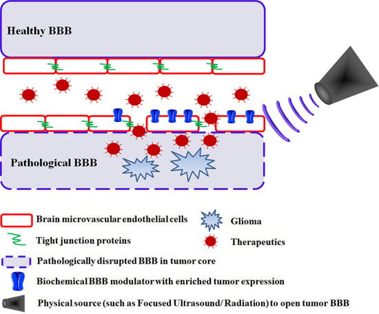
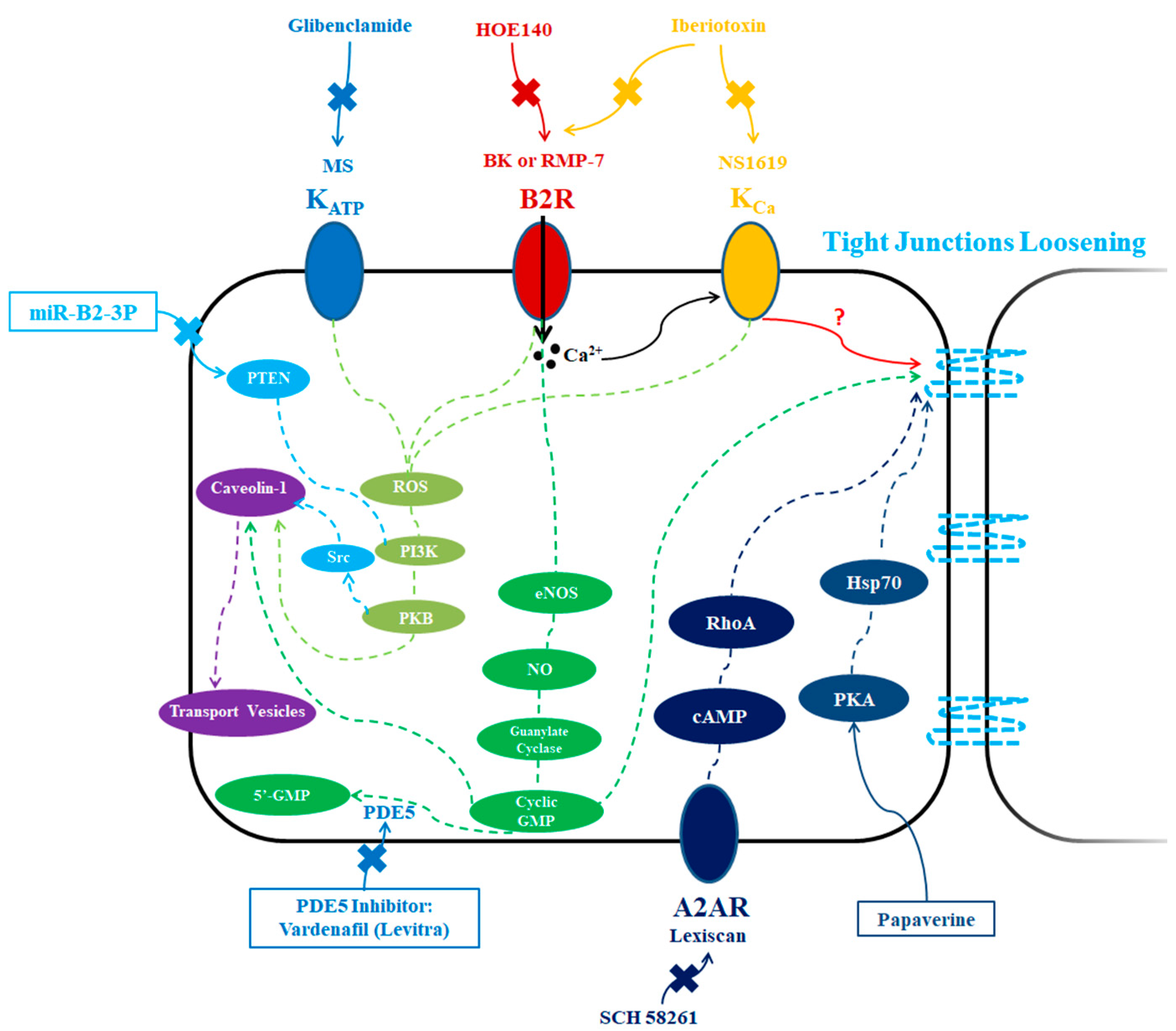
3. Biochemical Modulation

Pathological BBB disruption is heterogeneous throughout a brain tumor, with brain tumor cells lying behind a relatively intact tumor BBB in the tumor rim in addition to infiltrative cells that can be found outside the tumor margins. Thus, reliance on either passive or targeted delivery to only the pathologically disrupted tumor BBB regions in the glioma core will not eliminate all tumor cells, and a subsequent glioma recurrence will be likely. It therefore remains important to establish strategies that can target the tumor BBB throughout the whole tumor volume. In this section, several methods involving selective biochemical modulation of the tumor BBB are summarized (Table 1).

3.1. ATP-Sensitive Potassium Channel Activators

Minoxidil sulfate (MS), a selective activator of ATP-sensitive potassium (KATP) channels, was able to selectively increase the tumor BBB permeability in glioma via a transcellular pathway, and could be attenuated by glibenclamide, a selective inhibitor of KATP channels [57,58] (Figure 2). KATP channels are overexpressed in tumor vascular endothelial cells with low expression in normal brain endothelial cells, leading to a selective permeability increase in the brain tumor area [57]. In terms of the mechanism for increased tumor BBB permeability, pinocytosis might be involved as increased formation of pinocytotic vesicles was confirmed in both brain tumor endothelium and tumor cells in vivo [57]. Moreover, it has been shown that MS leads to increased caveolin-1 expression at tumor sites, again suggesting that elevated transcellular endocytosis processes could be leading to selective tumor BBB permeability within the tumors [58]. Interestingly, reactive oxygen species (ROS) appeared to be involved in this process, since a ROS scavenger (N-2-mercaptopropionyl glycine, MPG) could significantly decrease the effects of MS on caveolin-1 protein expression in the glioma region [58] (Figure 2). Another study using a rat brain glioma (C6) model also identified that MS induced tumor BBB selective opening in the tumor volume in a time-dependent manner by downregulating the expression of TJ proteins, including occludin and claudin-5, and through activation of signaling cascades involving ROS/RhoA/PI3K/PKB [59]. Because of the increased, selective tumor BBB permeability, it has been possible to improve glioma drug uptake for drugs of varying sizes, including anti-HER2 monoclonal antibody and carboplatin, with carboplatin significantly increasing survival in rats bearing RG-2 glioma [57].

3.2. Calcium-Activated Potassium Channel Activators

Similarly, intravenous injection of NS1619, an agonist of calcium-activated potassium (KCa) channels, can selectively enhance tumor BBB permeability in glioma, but not BBB permeability in the normal brain tissue [60,61] (Figure 2). Tumor BBB selectivity was thought to be achieved because there is a lower or absent expression of Kca channels in the normal brain tissue area compared with that in tumor and tumor vessels [60,61,62,63]. For example, mRNA and protein analyses showed that Kca channel subunit alpha-1 (KCNMA1) was amplified in 90% of high-grade gliomas samples from patients, as well as in the human high-grade glioma cell line U-87 [63]. However, no amplification of KCNMA1 was found in normal human brain tissues [63]. This suggests that Kca channels could serve as an effective target for selective biochemical tumor BBB modulation and enhance the delivery of chemotherapeutics to glioma [60,61]. For example, the penetration of [14C]-carboplatin and anti-HER2 monoclonal antibody into the tumor volume was significantly increased by co-administration of KCa channel agonist, NS1619, to glioma-bearing Wistar rats via intracarotid infusion, leading to an enhanced survival [61]. In addition, when co-administered with NS1619 via intravenous injection, temozolomide (TMZ) and trastuzumab (anti-HER2 antibody) led to increased survival of mice bearing glioma [64]. The underlying mechanism responsible for NS1619-mediated tumor BBB permeability increases in glioma appears to depend on increased endocytotic, transcellular processes [60,61]. NS1619 treatment increased the number of transport vesicles in the cytoplasm of tumor and brain tumor BBB endothelial cells in vivo, suggesting a role for these transport vesicles in KCa channel-mediated selective modulation in tumor BBB permeability [60,61,64]. It was found that NS1619 could selectively modulate the protein expression of caveolin-1 through ROS/PI3K/PKB/FoxO1 signaling in brain capillary endothelial cells in tumor area of rat brain glioma (C6) model in a time dependent fashion, resulting in increased caveolae-mediated cholera toxin subunit B endocytosis into tumor microvessels [65] (Figure 2). Interestingly, BBB permeability induced by bradykinin (see below), nitric oxide (NO) donors or an agonist of soluble guanylate cyclase (sGC) could all be attenuated by co-infusion of KCa channel antagonist, iberiotoxin, indicating KCa channels might serve as a hub for several strategies used for biochemical modulation of tumor BBB permeability in glioma [60] (Figure 2).

3.3. Phosphodiesterase 5 (PDE5) Inhibitors

Cyclic guanosine monophosphate (cGMP), has been shown to be involved in the modulation of vascular permeability [66]. Phosphodiesterase 5 (PDE5) inhibitor can selectively inhibit the degradation of cGMP [67], and the resulting accumulation of intracellular cGMP can result in increased permeability of brain capillaries, particularly in the brain tumor volume [66,68] (Figure 2). PDE5 was found to be strongly expressed in GBM cells isolated from patients [69]. In this way, administration of PDE5 inhibitor was able to selectively modulate the tumor BBB [68]. The tumor uptake of the chemotherapeutic agent, adriamycin, was selectively increased after oral administration of a PDE5 inhibitor, vardenafil (Levitra), without a significant increase in normal brain, leading to a significantly increased survival of rats bearing 9 L gliosarcoma tumors [68]. Similarly, increased cGMP levels were correlated with the enhanced tumor BBB permeability [68]. The selective increase in tumor BBB permeability appears to be induced by enhanced vesicular transport across blood vessels at the tumor margins, and could be attenuated by iberiotoxin, a selective inhibitor for KCa channels, which are also effectors in cGMP signaling [68]. Moreover, inhibition of caveolae-mediated transcytosis or macropinocytosis could attenuate PDE5 inhibitor-mediated uptake of Herceptin-AlexaFluor-680 in cultured mouse brain endothelial cells, suggesting the possible involvement of these transcellular pathways in PDE5-mediated effects [70].

3.4. Bradykinin Type 2 Receptor Activators

Low doses of bradykinin (BK), an activator of bradykinin type 2 (B2) receptors, was demonstrated to selectively and transiently enhance the permeability of the tumor BBB for six tracers with varying molecular sizes in experimental rats bearing RG2 glioma [71] (Figure 2). Importantly, bradykinin does not induce breakdown of the normal BBB unless at very high doses [72]. For instance, 70 kDa dextran had a 12-fold higher permeability in the microvasculature within the glioma in the BK-administered group compared with saline-administered group, while no effect of BK was found for permeability of normal brain microvasculature [71]. RMP-7 (also called cereport or labradimil), the first selective peptide agonist of B2 receptor applied in clinical trials, was confirmed to be able to selectively increase the tumor BBB transport of a various of drugs into brain GBM area in preclinical studies and in clinical trials [73]. The proteolytic lability of BK limited its further application in clinic [74]. Thus, a new analog of bradykinin named retro-inverso bradykinin (RI-BK) was developed, having a 40× higher binding affinity to B2 receptor compared with BK and a resistance to proteolysis [74]. RI-BK could increase the accumulation of coumarin-6-loaded micelles in glioma, but not in normal brain tissue, thus enhancing the anti-glioma effects in mice [74]. The transient BBB opening in the tumor volume mediated by RI-BK was size dependent, with gold nanoparticles of 70 nm having the maximal tumor uptake [75]. As mentioned, the BK- or bradykinin analog-induced tumor BBB opening is transient, usually lasting less than 20–25 min [71,73,76]. The selectivity of BK and BK analog treatment for the tumor region appears to be based on the expression levels of B2 receptors within brain tumor cells [77,78,91] and microvascular endothelial cells in the tumor [79,80]. In clinical isolates, there was high expression of B2R in glioma which increased along with tumor grade [79]. Moreover, a strong immunochemistry staining of B2R was found at both luminal and abluminal surfaces of endothelial cells in tumor microvessels, while the staining of B2R was largely reduced or even absent in microvessels adjacent or outside of the tumor regions [79].
The underlying mechanism for bradykinin-induced permeability increases at tumor sites is a complex process, possibly including transcytosis, TJ loosening, KATP channels, Ca2+ flux, nitric oxide synthase (NOS), NO, and cGMP (Figure 2). The caveolae-1- and caveolae-2-mediated transcellular pathways might be involved in the bradykinin-induced tumor BBB permeability increase in brain tumors [92]. A significantly enhanced expression of caveolin-1 was found after 15, 30 and 60 min RI-BK intra-arterial infusion, suggesting an increase in transcellular transport [75]. In addition, after BK treatment, the expression levels of ZO-1, claudin-5 and occludin were downregulated and the cytoskeleton was rearranged, suggesting an increase in junctional leakiness [81]. RI-BK or BK-induced selective tumor BBB opening in the tumor volume might also be related with the decreased ZO-1 expression and distribution as well as the depolymerization of F-actin [75]. Involvement of accelerated KATP formation in BK-induced tumor BBB permeability has been reported [93]. BK could also increase intracellular Ca2+ level by releasing Ca2+ from internal sites or enhancing Ca2+ influx [94]. It is possible that the increased intracellular Ca2+ might further activate nitric oxide synthase (NOS) and increase NO production. For example, the BK analog, R523, could increase the penetration of various hydrophilic macromolecular agents across the tumor BBB, and the increased permeability could be inhibited by an inhibitor of nitric oxide (NO) synthase, L-NA [80,95], suggesting a complex underlying mechanism. In addition, the effects of BK could be attenuated by pretreating with a NOS inhibitor, L-NAME [96]. Decreased cGMP formation induced by an inhibitor of soluble guanylate cyclase (sGC), LY83583, could block BK- induced BBB permeability [97]. Conversely, NO could also activate sGC, further increasing the formation of cGMP [98], indicating NO might be a core factor in the modulation of BK-induced BBB permeability [80,99]. Furthermore, NO-producing agents, including L-arginine and hydroxyurea, could increase tumor permeability in rats bearing 9 L gliosarcoma, and involved the change of eNOS and cGMP levels in the tumor region [100]. The findings suggest that use of oral NO donors may be a strategy to enhance the delivery of chemotherapeutics to malignant brain tumors [100].

3.5. Adenosine 2A Receptor Activators

Adenosine is an endogenous ligand for the adenosine 2A receptor (A2AR) which belongs to the family of G protein-coupled receptors [82]. Activating the vascular A2AR signaling pathway using Lexiscan, an FDA-approved selective A2AR agonist, can significantly and transiently enhance BBB permeability in mice and this permeability is attenuated in transgenic mice lacking A2AR [82,83] (Figure 2). Importantly, it was found that A2AR expression was higher in human glioma margins than that of peritumor normal brain area, suggesting it may be possible for A2AR agonists to selectively alter tumor BBB permeability in glioma patients [20]. In addition, in preclinical studies, A2AR agonists produced no obvious side effects even after multiple repeats of BBB modulation [84,85]. However, although Lexiscan has been shown to be effective in rodents to improve the BBB penetration of agents with various sizes via activating A2AR, limited success has been achieved in clinic [101,102]. For example, Lexiscan (0.4 mg/kg) failed to increase temozolomide concentrations in the brain or GBM volume in human patients [101], and more efforts to explore alternative doses and schedules might be warranted. In terms of mechanism, TJ and cytoskeletal proteins are of vital importance in regulating A2AR-mediated BBB permeability [82]. For example, A2AR activation and commensurate increased BBB permeability were mainly mediated by the downregulation of junctional proteins such as VE-cadherin and claudin-5, as well as the reorganization of cytoskeletal actin, which was related with cAMP/RhoA signaling [82,83].

3.6. Papaverine

Papaverine is a natural opioid that can cause a reversible increase in tumor BBB permeability after intra-arterial infusion [86,87,88]. In particular, Evans blue and 14C sucrose extravasation were increased after papaverine infusion, and recovered to control levels after 5 h, indicating that papaverine could temporally open the BBB, particularly in the brain tumor region when using a C6 glioma rat model [86,89]. An enhanced tumor uptake of chemotherapeutic, Bis-chloronitrosourea, was achieved for patients with malignant glioma when combined with papaverine [88]. The involvement of protein kinase A (PKA) and heat shock protein 70 (HSP70) has been demonstrated to be involved in this reversible increase in tumor BBB permeability in rats bearing C6 glioma [89] (Figure 2). A combination of western blotting and immunochemistry analysis also demonstrated that papaverine could downregulate claudin-5, occludin and F-actin expression by tumor microvessels, but not in normal brain tissue, suggesting that the tumor BBB opening was due to TJ dysfunction [89].

3.7. microRNAs

Recently, microRNAs (miRNAs) have been identified as important regulatory factors for various biological processes, including vascular permeability. In particular, miR-132-3p expression was found to be upregulated in glioma endothelial cells, and therefore could be a novel target for selective tumor BBB modulation in brain tumors [90]. Researchers have further identified miR-132-3p as an important miRNA in selectively increasing tumor BBB permeability in glioma and overexpression of this miRNA yielded increased uptake of doxorubicin in rat C6 glioma tumors [90]. In vitro studies indicated that endocytosis of cholera toxin subunit B and FITC-bovine serum albumin were significantly increased by miR-132-3p, via PTEN/PI3K/PKB/Src/Caveolin-1 signaling pathways [90] (Figure 2).
While the aforementioned biochemical strategies to control tumor BBB permeability have in many cases exhibited selective increases in BBB permeability and commensurate increases in drug uptake, clinical success has been limited. More potent drugs or targeting ligands are likely needed to pair with these approaches. In addition, it may also be possible that the combination of these biochemical approaches for tumor BBB modulation with selective targeting of the exposed tissue as described in the Pathological BBB Disruption section could enhance efficacy.

4. Physical Modulation

Another strategy to target BBB permeability to tumor regions is the use of physical disruption methods that employ various forms of electromagnetic radiation and ultrasound. These approaches can be widely applicable for drug delivery and have been proposed to efficiently and selectively allow drug passage into the tumor margin with minimal damage, including in several clinical applications (Table 2).

4.1. Electromagnetic Pulse (EMP)

Radiofrequency electromagnetic radiation (EMP) has been shown to increase the permeability of BBB for agents that normally do not pass the BBB such as Evans blue [103,104]. The induced permeability appears reversible as EMP exposure of an in vitro BBB model consisting of cocultured primary rat brain microvascular endothelial cells and primary rat astrocytes increased permeability and the effect was normalized after 24 h [105]. Further, in vivo studies have demonstrated that EMP exposure was able to transiently disrupt the BBB in healthy rats, with BBB permeability increased at 1 h, peaking at 3 h, and recovering at 12 h using Evans blue, endogenous albumin and lanthanum nitrate as tracers [106,107]. The chemotherapeutic 1-(2-chlorethyl)-cyclohexyl-nitrosourea (CCNU, lomustine) could be delivered to the brain tumor volume when glioma-bearing rats were exposed to EMP, enhancing tumor toxicity without significant side effects in normal brain tissue [108]. Although whole body exposure is currently used to administer EMP, there is some evidence of tumor BBB selectivity [108], and further work is required to determine the basis of such selectivity. In terms of mechanism, in vitro BBB permeability modulation by EMP was shown to be mediated by alteration in protein expression levels of the ZO-1 and claudin-5 TJ proteins [105]. EMP exposure in healthy rats also significantly decreased the gene and protein expression of TJ proteins, occludin and ZO-1, and increased the expression of matrix metalloproteinase such as MMP-2 and MMP-9, which were suggested to drive the observed changes in BBB permeability [107].

4.2. Laser-Induced Thermal Therapy (LITT)

Laser-induced thermal therapy (LITT) is another strategy being applied in treating brain tumors and employs a stereotactically implanted laser source within the tumor volume. Currently, LITT has been used in a thermal ablation modality, and pilot clinical studies have confirmed its effectiveness and feasibility in patients suffering recurrent brain tumors, with therapeutic effects monitored by real-time MRI [109]. In addition, it has been shown that LITT can induce a local, selective opening of the healthy BBB as demonstrated by extravasation of Evans blue, fibrinogen and IgM, and LITT further facilitated the passage of anti-tumor drugs such as paclitaxel into rat brains [110]. The BBB was observed to be opening at 1.5 h after LITT and it reached the peak value at 2.5 h after LITT [110]. It has been suggested that membrane defects in the capillary endothelium during LITT might be the direct cause of BBB opening and that these defects might be related to LITT-induced hyperthermia [110]. Other studies have also demonstrated the possible influence of hyperthermia on BBB integrity, showing that the breakdown can occur as brain temperatures rise over 41 °C [111,112]. It has also been suggested that polyamine synthesis could play a role in LITT-induced BBB opening as hyperthermia has been shown to stimulate increased polyamine synthesis [110,113]. Heat stress can also cause BBB opening that is related to a significant upregulation of NOS activity in the CNS [114]. To date, there have been no reports of specific changes in TJ proteins upon LITT-induced BBB opening [137]. While LITT has been demonstrated to mediate a local, selective healthy BBB opening, it has yet to be tested for selective tumor BBB opening and potential improvements in tumor drug uptake.

4.3. Radiotherapy: Synchrotron Microbeam Radiation Therapy (MRT)

Conventional radiotherapy can increase BBB permeability in preclinical animal models and clinical trials [115,116,117], enhancing the efficacy of systemic administration of chemotherapeutic drugs in treating brain tumors such as glioma [116,118,119]. In a clinical study, irradiation with 20 to 40 Grays induced tumor BBB opening and facilitated the entry of chemotherapeutics such as methotrexate in glioblastoma patients, increasing survival time [117]. Apart from conventional radiotherapy, microbeam radiation therapy (MRT) is a different form of radiotherapy that has been used to alter the tumor BBB that relies on radiation delivery strategies to focus the irradiation to the tumor site [120,121]. In this way, MRT has been shown to selectively attenuate or even block tumor growth in several tumor models, leading to an increased animal survival with a high tolerance of normal tissues to MRT [120,122]. Interestingly, from the point of view of this review, exposure to MRT was shown to make the tumor BBB more permeable to low-molecular-weight contrast medium [119], and MRT therapy did not modulate the BBB permeability in contralateral brain, but selectively increased the tumor BBB permeability in a rat model of intracranial GBM [123]. Real-time MRI also demonstrated that Gd-DTPA uptake could be increased in the initially non-enhanced tumor area but not in the surrounding normal brain tissue during MRT, indicating selective effects on the tumor BBB [138]. Although the underlying mechanism responsible for increased MRT-induced tumor BBB permeability is not yet clear, conventional radiation has been associated with the decreased expression and rearrangement of TJ proteins including claudin-5, ZO-1 and beta-catenin [115]. Despite the aforementioned evidence that MRT can lead to a selective increase in tumor BBB permeability, the strategy has yet to be combined with co-administration of a chemotherapeutic and efficacy demonstrated in a brain tumor model.

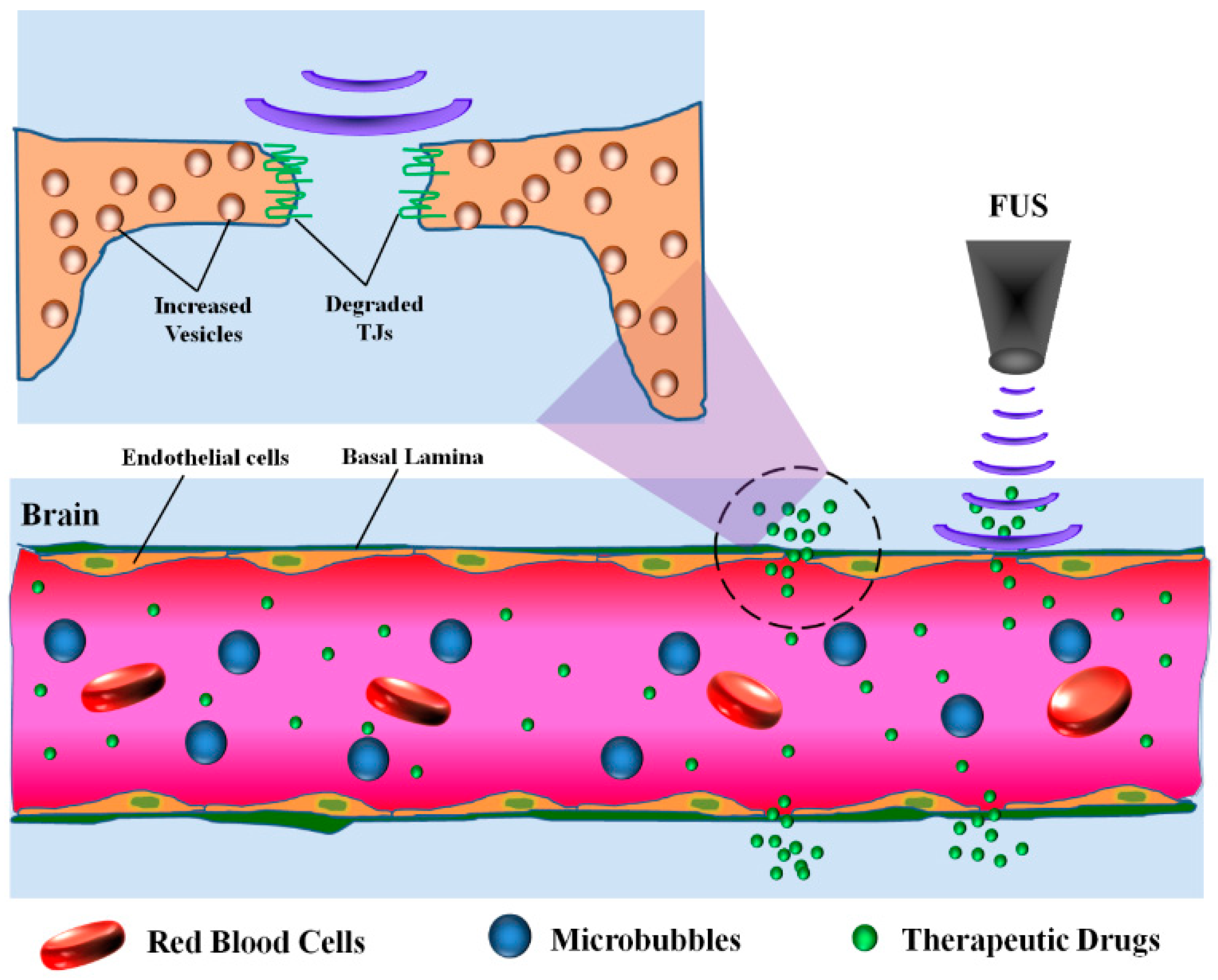
4.4. Focused Ultrasound (FUS)

FUS has been rapidly developing as a promising noninvasive method for localized BBB opening for treatment of CNS diseases (Figure 3) [139,140]. The physical interactions between systemically administered microbubbles and focused ultrasonic waves enable the transient and reversible disruption of the BBB in targeted brain regions [125,126]. To date, FUS has been applied for enhancing tumor BBB penetration of anti-tumor drugs such as doxorubicin, temozolomide, carboplatin and paclitaxel into glioma [20,127,128,129]. For example, regardless of its anti-glioma potency, paclitaxel has showed little benefit in vivo due to its poor BBB penetration, and FUS was shown to increase paclitaxel penetration across the tumor BBB and improve its anti-glioma effects [130]. A clinical FUS device was developed and tested for the delivery of carboplatin to the brain in rats bearing F98 glioma model, leading to 1.7-fold and 3.3-fold higher MRI contrast agent signal in the center and margin of tumor, respectively, compared to tumor without FUS [131]. FUS can also increase tumor uptake of biologicals and complex nanostructures such as IgG and gold particles [141,142,143,144,145], and has been shown to be a relatively safe, well-tolerated approach for healthy BBB and tumor BBB opening in preclinical animal models [132,133], and in clinical trials where improved drug efficacy was observed [129,134]. Given that FUS is showing early promise in terms of clinical implementation, safety is being more extensively evaluated. FUS-induced barrier opening can last for several hours, with a barrier restoration time that depends on the molecular size of the tracer or therapeutics [125]. Although the extravasation of albumin and red blood cells can be seen in FUS treated regions, they can be rapidly cleared by glia and it has been suggested that FUS does not lead to ischemia or adverse behavioral effects [146]. However, microbubble oscillation induces mechanical shear forces that might still be of potential risk, and cause excessive immune reactions and brain hemorrhage [132,147]. It is also been reported that unpredictable hot spots might exist in brain regions receiving FUS [148]. In terms of mechanism, FUS can lead to upregulation of cellular machinery in charge of transcellular transport, including caveolin-1 showing a peak value at 1 h after sonication, with a commensurate downregulation of TJ proteins such as claudin-1, claudin-5 and occludin [135]. Indeed, both transcytosis and free passage via the affected BBB endothelial cells have been shown to be involved in FUS-mediated BBB opening, and the mechanisms may be dependent on the intensity of the ultrasound [126].

5. Conclusions and Future Perspectives

Existing as an interface between the blood and brain tumor tissue, the tumor BBB hinders the glioma uptake of many therapeutics. While methods to globally open the BBB have been proposed to enhance drug penetration into the brain tumor volume, they are often met with challenges resulting from their non-specific nature of BBB opening, which could damage normal brain tissue. By contrast, the methods described here have the potential to selectively and efficiently modulate the tumor BBB in glioma. Although pathological disruption does partially expose regions of the brain tumor core to the systemic circulation, the extent of disruption can be heterogeneous across the tissue, and tumor cells may reside behind a relatively intact tumor BBB. While we believe that targeting and retention of therapeutic cargo in the disrupted tumor BBB regions can clearly lead to improved glioma treatment, it will likely fail to eradicate all tumor cells, particularly in the tumor rim. Thus, such approaches will need to be combined with biochemical or physical approaches to further selectively open the tumor BBB. Importantly, blood vessels in tumor tissue can have differential molecular expression profiles compared with normal brain blood vessels and biochemical modulators could potentially take advantage of these differences for selective BBB opening. The search for additional pathways that can be used to selectively modulate tumor BBB permeability especially at the tumor margins should continue, as those described here have shown limited clinical success despite being investigated for over 20 years. In addition, several physical methods can be used to stereotactically target the brain tumor volume, thereby generating localized BBB disruption both within and surrounding the tumor volume. It is our opinion that FUS, in particular, offers a wealth of opportunities for enhanced glioma delivery, especially given the strides that have been made to evaluate its clinical safety. Regardless of choice, if the tumor BBB can be reversibly disrupted throughout the entire tumor volume by biochemical or physical means, the various structural and cellular components normally invisible to the circulation, including ECM, can be exposed. Targeting of exposed components may allow therapeutic accumulation throughout the entire tumor volume. Moving forward, one could envision combining new and improved targeted therapy strategies with optimized strategies of BBB disruption to more selectively and efficaciously deliver medicine to those suffering from glioma.

Author Contributions

Conceptualization, writing and editing were done by H.L. and E.V.S. All authors have read and agreed to the published version of the manuscript.

Funding

This work was in part supported by National Institutes of Health grant NS099158.

Conflicts of Interest

E.V.S. is an author on a US patent application regarding the P1C10 variable lymphocyte receptor referenced in the pathological disruption section. The funders had no role in the design of the study; in the collection, analyses, or interpretation of data; in the writing of the manuscript, or in the decision to publish the results.

References

  1. Cohen, A.L.; Colman, H. Glioma biology and molecular markers. Cancer Treat. Res. 2015, 163, 15–30. [Google Scholar] [PubMed]
  2. Mrugala, M.M. Advances and challenges in the treatment of glioblastoma: A clinician’s perspective. Discov. Med. 2013, 15, 221–230. [Google Scholar] [PubMed]
  3. Taylor, O.G.; Brzozowski, J.S.; Skelding, K.A. Glioblastoma Multiforme: An Overview of Emerging Therapeutic Targets. Front. Oncol. 2019, 9, 963. [Google Scholar] [CrossRef] [PubMed] [Green Version]
  4. Ostrom, Q.T.; Gittleman, H.; Liao, P.; Rouse, C.; Chen, Y.; Dowling, J.; Wolinsky, Y.; Kruchko, C.; Barnholtz-Sloan, J. CBTRUS Statistical Report: Primary Brain and Central Nervous System Tumors Diagnosed in the United States in 2007–2011. Neuro-Oncology 2014, 16, iv1–iv63. [Google Scholar] [CrossRef] [PubMed]
  5. Parrish, K.E.; Sarkaria, J.N.; Elmquist, W.F. Improving drug delivery to primary and metastatic brain tumors: Strategies to overcome the blood-brain barrier. Clin. Pharmacol. Ther. 2015, 97, 336–346. [Google Scholar] [CrossRef]
  6. Cardoso, F.L.; Brites, D.; Brito, M.A. Looking at the blood–brain barrier: Molecular anatomy and possible investigation approaches. Brain Res. Rev. 2010, 64, 328–363. [Google Scholar] [CrossRef]
  7. Agarwal, S.; Sane, R.; Oberoi, R.; Ohlfest, J.R.; Elmquist, W.F. Delivery of molecularly targeted therapy to malignant glioma, a disease of the whole brain. Expert Rev. Mol. Med. 2011, 13, e17. [Google Scholar] [CrossRef] [Green Version]
  8. Cao, Y.; Sundgren, P.C.; Tsien, C.I.; Chenevert, T.T.; Junck, L. Physiologic and Metabolic Magnetic Resonance Imaging in Gliomas. J. Clin. Oncol. 2006, 24, 1228–1235. [Google Scholar] [CrossRef]
  9. Prabhu, S.S.; Broaddus, W.C.; Oveissi, C.; Berr, S.S.; Gillies, G.T. Determination of intracranial tumor volumes in a rodent brain using magnetic resonance imaging, evans blue, and histology: A comparative study. IEEE Trans. Bio-Med. Eng. 2000, 47, 259–265. [Google Scholar] [CrossRef]
  10. Oberoi, R.K.; Parrish, K.E.; Sio, T.T.; Mittapalli, R.K.; Elmquist, W.F.; Sarkaria, J.N. Strategies to improve delivery of anticancer drugs across the blood–brain barrier to treat glioblastoma. Neuro-Oncology 2015, 18, 27–36. [Google Scholar] [CrossRef] [Green Version]
  11. Choi, Y.; Kim, D.W.; Lee, S.-K.; Chang, J.H.; Kang, S.-G.; Kim, E.-H.; Kim, S.H.; Rim, T.H.; Ahn, S.S. The Added Prognostic Value of Preoperative Dynamic Contrast-Enhanced MRI Histogram Analysis in Patients with Glioblastoma: Analysis of Overall and Progression-Free Survival. Am. J. Neuroradiol. 2015, 36, 2235–2241. [Google Scholar] [CrossRef] [PubMed] [Green Version]
  12. Arevaloperez, J.; Thomas, A.; Kaley, T.J.; Lyo, J.K.; Peck, K.K.; Holodny, A.; Mellinghoff, I.K.; Shi, W.; Zhang, Z.; Young, R. T1-Weighted Dynamic Contrast-Enhanced MRI as a Noninvasive Biomarker of Epidermal Growth Factor Receptor vIII Status. Am. J. Neuroradiol. 2015, 36, 2256–2261. [Google Scholar] [CrossRef] [PubMed] [Green Version]
  13. Santarosa, C.; Castellano, A.; Conte, G.M.; Cadioli, M.; Iadanza, A.; Terreni, M.R.; Franzin, A.; Bello, L.; Caulo, M.; Falini, A.; et al. Dynamic contrast-enhanced and dynamic susceptibility contrast perfusion MR imaging for glioma grading: Preliminary comparison of vessel compartment and permeability parameters using hotspot and histogram analysis. Eur. J. Radiol. 2016, 85, 1147–1156. [Google Scholar] [CrossRef] [PubMed]
  14. Law, M.; Yang, S.; Babb, J.S.; A Knopp, E.; Golfinos, J.G.; Zagzag, D.; Johnson, G. Comparison of cerebral blood volume and vascular permeability from dynamic susceptibility contrast-enhanced perfusion MR imaging with glioma grade. Am. J. Neuroradiol. 2004, 25, 746–755. [Google Scholar] [PubMed]
  15. Onda, K.; Tanaka, R.; Takahashi, H.; Takeda, N.; Ikuta, F. Cerebral Glioblastoma with Cerebrospinal Fluid Dissemination: A Clinicopathological Study of 14 Cases Examined by Complete Autopsy. Neurosurg. 1989, 25, 533–540. [Google Scholar] [CrossRef]
  16. Erdlenbruch, B.; Alipour, M.; Fricker, G.; Miller, D.S.; Kugler, W.; Eibl, H.; Lakomek, M. Alkylglycerol opening of the blood-brain barrier to small and large fluorescence markers in normal and C6 glioma-bearing rats and isolated rat brain capillaries. Br. J. Pharmacol. 2003, 140, 1201–1210. [Google Scholar] [CrossRef] [Green Version]
  17. Abdul Razzak, R.; Florence, G.J.; Gunn-Moore, F.J. Approaches to CNS Drug Delivery with a Focus on Transporter-Mediated Transcytosis. Int. J. Mol. Sci. 2019, 20, 3108. [Google Scholar] [CrossRef] [Green Version]
  18. Georgieva, J.V.; Hoekstra, D.; Zuhorn, I.S. Smuggling Drugs into the Brain: An Overview of Ligands Targeting Transcytosis for Drug Delivery across the Blood–Brain Barrier. Pharmaceutics 2014, 6, 557–583. [Google Scholar] [CrossRef] [Green Version]
  19. Marchi, N.; Angelov, L.; Masaryk, T.; Fazio, V.; Granata, T.; Hernandez, N.; Hallene, K.; Diglaw, T.; Franic, L.; Najm, I.; et al. Seizure-Promoting Effect of Blood? Brain Barrier Disruption. Epilepsia 2007, 48, 732–742. [Google Scholar] [CrossRef] [Green Version]
  20. Gao, X.; Yue, Q.; Liu, Y.; Fan, D.; Fan, K.; Li, S.; Qian, J.; Han, L.; Fang, F.; Xu, F.; et al. Image-guided chemotherapy with specifically tuned blood brain barrier permeability in glioma margins. Theranostics 2018, 8, 3126–3137. [Google Scholar] [CrossRef]
  21. Ichimura, K.; Ohno, K.; Aoyagi, M.; Tamaki, M.; Suzuki, R.; Hirakawa, K. Capillary permeability in experimental rat glioma and effects of intracarotid CDDP administration on tumor drug delivery. J. Neuro-Oncol. 1993, 16, 211–215. [Google Scholar] [CrossRef] [PubMed]
  22. Jain, R. Measurements of tumor vascular leakiness using DCE in brain tumors: Clinical applications. NMR Biomed. 2013, 26, 1042–1049. [Google Scholar] [CrossRef] [PubMed]
  23. Aprile, I.; Giovannelli, G.; Fiaschini, P.; Muti, M.; Kouleridou, A.; Caputo, N. High- and low-grade glioma differentiation: The role of percentage signal recovery evaluation in MR dynamic susceptibility contrast imaging. La Radiol. Medica 2015, 120, 967–974. [Google Scholar] [CrossRef] [PubMed]
  24. Gerlowski, L.E.; Jain, R.K. Microvascular permeability of normal and neoplastic tissues. Microvasc. Res. 1986, 31, 288–305. [Google Scholar] [CrossRef]
  25. Nugent, L.J.; Jain, R.K. Extravascular diffusion in normal and neoplastic tissues. Cancer Res. 1984, 44, 238–244. [Google Scholar]
  26. Provenzale, J.M.; Mukundan, S.; Dewhirst, M. The Role of Blood-Brain Barrier Permeability in Brain Tumor Imaging and Therapeutics. Am. J. Roentgenol. 2005, 185, 763–767. [Google Scholar] [CrossRef]
  27. Umlauf, B.J.; Shusta, E.V. Exploiting BBB disruption for the delivery of nanocarriers to the diseased CNS. Curr. Opin. Biotechnol. 2019, 60, 146–152. [Google Scholar] [CrossRef]
  28. Mittapalli, R.K.; Adkins, C.E.; Bohn, K.A.; Mohammad, A.S.; Lockman, J.A.; Lockman, P.R. Quantitative Fluorescence Microscopy Measures Vascular Pore Size in Primary and Metastatic Brain Tumors. Cancer Res. 2016, 77, 238–246. [Google Scholar] [CrossRef] [Green Version]
  29. Sarin, H.; Kanevsky, A.S.; Wu, H.; Brimacombe, K.R.; Fung, S.H.; Sousa, A.A.; Auh, S.; Wilson, C.M.; Sharma, K.; A Aronova, M.; et al. Effective transvascular delivery of nanoparticles across the blood-brain tumor barrier into malignant glioma cells. J. Transl. Med. 2008, 6, 80. [Google Scholar] [CrossRef] [Green Version]
  30. Goldwirt, L.; Beccaria, K.; Carpentier, A.; Farinotti, R.; Fernandez, C. Irinotecan and temozolomide brain distribution: A focus on ABCB1. Cancer Chemother. Pharmacol. 2014, 74, 185–193. [Google Scholar] [CrossRef]
  31. De Vries, N.A.; Zhao, J.; Kroon, E.; Buckle, T.; Beijnen, J.H.; Van Tellingen, O. P-Glycoprotein and Breast Cancer Resistance Protein: Two Dominant Transporters Working Together in Limiting the Brain Penetration of Topotecan. Clin. Cancer Res. 2007, 13, 6440–6449. [Google Scholar] [CrossRef] [PubMed] [Green Version]
  32. Dréan, A.; Goldwirt, L.; Verreault, M.; Canney, M.; Schmitt, C.; Guehennec, J.; Delattre, J.-Y.; Carpentier, A.; Idbaih, A. Blood-brain barrier, cytotoxic chemotherapies and glioblastoma. Expert Rev. Neurother. 2016, 16, 1285–1300. [Google Scholar] [CrossRef] [PubMed]
  33. Wang, D.; Wang, C.; Wang, L.; Chen, Y. A comprehensive review in improving delivery of small-molecule chemotherapeutic agents overcoming the blood-brain/brain tumor barriers for glioblastoma treatment. Drug Deliv. 2019, 26, 551–565. [Google Scholar] [CrossRef] [PubMed]
  34. Al-Ahmady, Z.S. Selective drug delivery approaches to lesioned brain through blood brain barrier disruption. Expert Opin. Drug Deliv. 2018, 15, 335–349. [Google Scholar] [CrossRef]
  35. Front, D.; Israel, O.; Kohn, S.; Nir, I. The blood-tissue barrier of human brain tumors: Correlation of scintigraphic and ultrastructural findings: Concise communication. J. Nucl. Med. 1984, 25, 461–465. [Google Scholar]
  36. Siegal, T.; Horowitz, A.; Gabizon, A. Doxorubicin encapsulated in sterically stabilized liposomes for the treatment of a brain tumor model: Biodistribution and therapeutic efficacy. J. Neurosurg. 1995, 83, 1029–1037. [Google Scholar] [CrossRef] [Green Version]
  37. Krauze, M.T.; Noble, C.O.; Kawaguchi, T.; Drummond, D.; Kirpotin, D.B.; Yamashita, Y.; Kullberg, E.; Forsayeth, J.; Park, J.W.; Bankiewicz, K.S. Convection-enhanced delivery of nanoliposomal CPT-11 (irinotecan) and PEGylated liposomal doxorubicin (Doxil) in rodent intracranial brain tumor xenografts. Neuro-Oncology 2007, 9, 393–403. [Google Scholar] [CrossRef]
  38. Fabel, K.; Dietrich, J.; Hau, P.; Wismeth, C.; Winner, B.; Przywara, S.; Steinbrecher, A.; Ullrich, W.; Bogdahn, U. Long-term stabilization in patients with malignant glioma after treatment with liposomal doxorubicin. Cancer 2001, 92, 1936–1942. [Google Scholar] [CrossRef]
  39. Hau, P.; Fabel, K.; Baumgart, U.; Rümmele, P.; Grauer, O.; Bock, A.; Dietmaier, C.; Dietmaier, W.; Dietrich, J.; Dudel, C.; et al. Pegylated liposomal doxorubicin-efficacy in patients with recurrent high-grade glioma. Cancer 2004, 100, 1199–1207. [Google Scholar] [CrossRef]
  40. Chua, S.L.; A Rosenthal, M.; Wong, S.S.; Ashley, D.M.; Woods, A.-M.; Dowling, A.J.; Cher, L.M. Phase 2 study of temozolomide and Caelyx in patients with recurrent glioblastoma multiforme. Neuro-Oncology 2004, 6, 38–43. [Google Scholar] [CrossRef]
  41. Glas, M.; Koch, H.; Hirschmann, B.; Jauch, T.; Steinbrecher, A.; Herrlinger, U.; Bogdahn, U.; Hau, P. Pegylated Liposomal Doxorubicin in Recurrent Malignant Glioma: Analysis of a Case Series. Oncology 2007, 72, 302–307. [Google Scholar] [CrossRef] [PubMed]
  42. Beier, C.P.; Schmid, C.; Gorlia, T.; Kleinletzenberger, C.; Beier, D.; Grauer, O.; Steinbrecher, A.; Hirschmann, B.; Brawanski, A.; Dietmaier, C.; et al. RNOP-09: Pegylated liposomal doxorubicine and prolonged temozolomide in addition to radiotherapy in newly diagnosed glioblastoma - a phase II study. BMC Cancer 2009, 9, 1–10. [Google Scholar] [CrossRef] [PubMed] [Green Version]
  43. Umlauf, B.J.; A Clark, P.; Lajoie, J.M.; Georgieva, J.V.; Bremner, S.; Herrin, B.R.; Kuo, J.S.; Shusta, E.V. Identification of variable lymphocyte receptors that can target therapeutics to pathologically exposed brain extracellular matrix. Sci. Adv. 2019, 5, eaau4245. [Google Scholar] [CrossRef] [PubMed] [Green Version]
  44. Sun, Z.; Yan, X.; Liu, Y.; Huang, L.; Kong, C.; Qu, X.; Wang, M.; Gao, R.; Qin, H.-L. Application of dual targeting drug delivery system for the improvement of anti-glioma efficacy of doxorubicin. Oncotarget 2017, 8, 58823–58834. [Google Scholar] [CrossRef] [PubMed] [Green Version]
  45. Jarboe, J.S.; Johnson, K.R.; Choi, Y.; Lonser, R.R.; Park, J.K. Expression of Interleukin-13 Receptor α2 in Glioblastoma Multiforme: Implications for Targeted Therapies. Cancer Res. 2007, 67, 7983–7986. [Google Scholar] [CrossRef] [PubMed] [Green Version]
  46. Debinski, W.; Gibo, D.M.; Slagle, B.; Powers, S.K.; Gillespie, G.Y. Receptor for interleukin 13 is abundantly and specifically over-expressed in patients with glioblastoma multiforme. Int. J. Oncol. 1999, 15. [Google Scholar] [CrossRef] [PubMed]
  47. Madhankumar, A.; Slagle-Webb, B.; Mintz, A.; Sheehan, J.M.; Connor, J.R. Interleukin-13 receptor-targeted nanovesicles are a potential therapy for glioblastoma multiforme. Mol. Cancer Ther. 2006, 5, 3162–3169. [Google Scholar] [CrossRef] [Green Version]
  48. Muñoz, M.; Coveñas, R.; Esteban, F.; Redondo, M. The substance P/NK-1 receptor system: NK-1 receptor antagonists as anti-cancer drugs. J. Biosci. 2015, 40, 441–463. [Google Scholar] [CrossRef]
  49. Gao, H.; Zhang, S.; Yang, Z.; Cao, S.; Jiang, X.; Pang, Z. In vitro and in vivo intracellular distribution and anti-glioblastoma effects of docetaxel-loaded nanoparticles functioned with IL-13 peptide. Int. J. Pharm. 2014, 466, 8–17. [Google Scholar] [CrossRef]
  50. Gao, H.; Yang, Z.; Zhang, S.; Cao, S.; Shen, S.; Pang, Z.; Jiang, X. Ligand modified nanoparticles increases cell uptake, alters endocytosis and elevates glioma distribution and internalization. Sci. Rep. 2013, 3, srep02534. [Google Scholar] [CrossRef] [Green Version]
  51. Gao, H.; Yang, Z.; Zhang, S.; Cao, S.; Pang, Z.; Yang, X.; Jiang, X. Glioma-homing peptide with a cell-penetrating effect for targeting delivery with enhanced glioma localization, penetration and suppression of glioma growth. J. Control. Release 2013, 172, 921–928. [Google Scholar] [CrossRef] [PubMed]
  52. Madhankumar, A.B.; Mintz, A.; Debinski, W. Interleukin 13 mutants of enhanced avidity toward the glioma-associated receptor, IL13Ralpha2. Neoplasia 2004, 6, 15–22. [Google Scholar] [CrossRef] [Green Version]
  53. Guo, X.; Wu, G.; Wang, H.; Xu, B. Pep-1&borneol–Bifunctionalized Carmustine-Loaded Micelles Enhance Anti-Glioma Efficacy through Tumor-Targeting and BBB-Penetrating. J. Pharm. Sci. 2019, 108, 1726–1735. [Google Scholar] [CrossRef] [PubMed]
  54. Jiang, Y.; Lv, L.; Shi, H.; Hua, Y.; Lv, W.; Wang, X.; Xin, H.; Xu, Q. PEGylated Polyamidoamine dendrimer conjugated with tumor homing peptide as a potential targeted delivery system for glioma. Colloids Surf. B Biointerfaces 2016, 147, 242–249. [Google Scholar] [CrossRef] [PubMed]
  55. Ruan, C.; Liu, L.; Lu, Y.; Zhang, Y.; He, X.; Chen, X.; Zhang, Y.; Chen, Q.; Guo, Q.; Sun, T.; et al. Substance P-modified human serum albumin nanoparticles loaded with paclitaxel for targeted therapy of glioma. Acta Pharm. Sin. B 2018, 8, 85–96. [Google Scholar] [CrossRef]
  56. Sun, T.; Jiang, X.; Wang, Q.; Chen, Q.; Lu, Y.; Liu, L.; Zhang, Y.; He, X.; Ruan, C.; Zhang, Y.; et al. Substance P Mediated DGLs Complexing with DACHPt for Targeting Therapy of Glioma. ACS Appl. Mater. Interfaces 2017, 9, 34603–34617. [Google Scholar] [CrossRef] [PubMed]
  57. Ningaraj, N.S.; Rao, M.K.; Black, K.L. Adenosine 5’-triphosphate-sensitive potassium channel-mediated blood-brain tumor barrier permeability increase in a rat brain tumor model. Cancer Res. 2003, 63, 8899–8911. [Google Scholar]
  58. Gu, Y.-T.; Xue, Y.-X.; Zhang, H.; Li, Y.; Liang, X.-Y. Adenosine 5′-Triphosphate-Sensitive Potassium Channel Activator Induces the Up-Regulation of Caveolin-1 Expression in a Rat Brain Tumor Model. Cell. Mol. Neurobiol. 2011, 31, 629–634. [Google Scholar] [CrossRef]
  59. Gu, Y.-T.; Xue, Y.; Wang, Y.; Wang, J.-H.; Chen, X.; Shangguan, Q.-R.; Lian, Y.; Zhong, L.; Meng, Y.-N. Minoxidil sulfate induced the increase in blood–brain tumor barrier permeability through ROS/RhoA/PI3K/PKB signaling pathway. Neuropharmacology 2013, 75, 407–415. [Google Scholar] [CrossRef]
  60. Ningaraj, N.S.; Rao, M.; Hashizume, K.; Asotra, K.; Black, K.L. Regulation of Blood-Brain Tumor Barrier Permeability by Calcium-Activated Potassium Channels. J. Pharmacol. Exp. Ther. 2002, 301, 838–851. [Google Scholar] [CrossRef] [Green Version]
  61. Ningaraj, N.S.; Rao, M.; Black, K.L. Calcium-dependent potassium channels as a target protein for modulation of the blood-brain tumor barrier. Drug News Perspect. 2003, 16, 291. [Google Scholar] [CrossRef] [PubMed]
  62. Hu, J.; Yuan, X.; Ko, M.K.; Yin, D.; Sacapano, M.R.; Wang, X.; Konda, B.; Espinoza, A.; Prosolovich, K.; Ong, J.M.; et al. Calcium-activated potassium channels mediated blood-brain tumor barrier opening in a rat metastatic brain tumor model. Mol. Cancer 2007, 6, 22. [Google Scholar] [CrossRef] [PubMed] [Green Version]
  63. Ningaraj, N.; Khaitan, D. Evidence of calcium-activated potassium channel subunit alpha-1 as a key promoter of glioma growth and tumorigenicity. Glioma 2019, 2, 46. [Google Scholar] [CrossRef]
  64. Ningaraj, N.S.; Sankpal, U.T.; Khaitan, D.; Meister, E.A.; Vats, T.S. Modulation of KCa channels increases anticancer drug delivery to brain tumors and prolongs survival in xenograft model. Cancer Biol. Ther. 2009, 8, 1924–1933. [Google Scholar] [CrossRef] [Green Version]
  65. Cai, R.-P.; Xue, Y.-X.; Huang, J.; Wang, J.-H.; Wang, J.-H.; Zhao, S.-Y.; Guan, T.-T.; Zhang, Z.; Gu, Y.-T. NS1619 regulates the expression of caveolin-1 protein in a time-dependent manner via ROS/PI3K/PKB/FoxO1 signaling pathway in brain tumor microvascular endothelial cells. J. Neurol. Sci. 2016, 369, 109–118. [Google Scholar] [CrossRef] [Green Version]
  66. Michel, C. Capillaries, Caveolae, Calcium and Cyclic Nucleotides: A New Look at Microvascular Permeability. J. Mol. Cell. Cardiol. 1998, 30, 2541–2546. [Google Scholar] [CrossRef]
  67. Juilfs, D.M.; Soderling, S.; Burns, F.; Beavo, J.A. Cyclic GMP as substrate and regulator of cyclic nucleotide phosphodiesterases (PDEs). Rev. Physiol. Biochem. Pharmacol. 1999, 135, 67–104. [Google Scholar] [CrossRef]
  68. Black, K.L.; Yin, D.; Ong, J.M.; Hu, J.; Konda, B.M.; Wang, X.; Ko, M.K.; Bayan, J.A.; Sacapano, M.R.; Espinoza, A.; et al. PDE5 inhibitors enhance tumor permeability and efficacy of chemotherapy in a rat brain tumor model. Brain Res. 2008, 1230, 290–302. [Google Scholar] [CrossRef] [Green Version]
  69. Cesarini, V.; Martini, M.; Vitiani, L.R.; Gravina, G.L.; Di Agostino, S.; Graziani, G.; D’Alessandris, Q.G.; Pallini, R.; LaRocca, L.M.; Rossi, P.; et al. Type 5 phosphodiesterase regulates glioblastoma multiforme aggressiveness and clinical outcome. Oncotarget 2017, 8, 13223–13239. [Google Scholar] [CrossRef]
  70. Hu, J.; Ljubimova, J.Y.; Inoue, S.; Konda, B.; Patil, R.; Ding, H.; Espinoza, A.; Wawrowsky, K.A.; Patil, C.; Ljubimov, A.V.; et al. Phosphodiesterase Type 5 Inhibitors Increase Herceptin Transport and Treatment Efficacy in Mouse Metastatic Brain Tumor Models. PLoS ONE 2010, 5, e10108. [Google Scholar] [CrossRef] [Green Version]
  71. Inamura, T.; Black, K.L. Bradykinin Selectively Opens Blood-Tumor Barrier in Experimental Brain Tumors. Br. J. Pharmacol. 1994, 14, 862–870. [Google Scholar] [CrossRef] [Green Version]
  72. Raymond, J.J.; Robertson, D.M.; Dinsdale, H.B. Pharmacological Modification of Bradykinin Induced Breakdown of the Blood-brain Barrier. Can. J. Neurol. Sci. 1986, 13, 214–220. [Google Scholar] [CrossRef] [PubMed] [Green Version]
  73. Emerich, D.F.; Dean, R.L.; Osborn, C.; Bartus, R.T. The Development of the Bradykinin Agonist Labradimil as a Means to Increase the Permeability of the Blood-Brain Barrier. Clin. Pharmacokinet. 2001, 40, 105–123. [Google Scholar] [CrossRef] [PubMed]
  74. Xie, Z.; Shen, Q.; Xie, C.; Lu, W.; Peng, C.; Wei, X.; Li, X.; Su, B.; Gao, C.; Liu, M. Retro-inverso bradykinin opens the door of blood–brain tumor barrier for nanocarriers in glioma treatment. Cancer Lett. 2015, 369, 144–151. [Google Scholar] [CrossRef] [PubMed] [Green Version]
  75. Su, B.; Wang, R.; Xie, Z.; Ruan, H.; Li, J.; Xie, C.; Lu, W.; Wang, J.; Wang, D.; Liu, M. Effect of Retro-Inverso Isomer of Bradykinin on Size-Dependent Penetration of Blood-Brain Tumor Barrier. Small 2018, 14. [Google Scholar] [CrossRef]
  76. Bartus, R.; Elliott, P.; Hayward, N.; Dean, R.; McEwen, E.; Fisher, S. Permeability of the blood brain barrier by the bradykinin agonist, RMP-7: Evidence for a sensitive, auto-regulated, receptor-mediated system. Immunopharmacology 1996, 33, 270–278. [Google Scholar] [CrossRef]
  77. Uchida, M.; Chen, Z.; Liu, Y.; Black, K.L. Overexpression of bradykinin type 2 receptors on glioma cells enhances bradykinin-mediated blood–brain tumor barrier permeability increase. Neurol. Res. 2002, 24, 739–746. [Google Scholar] [CrossRef]
  78. Liu, Y.; Hashizume, K.; Chen, Z.; Samoto, K.; Ningaraj, N.; Asotra, K.; Black, K.L. Correlation between bradykinin-induced blood–tumor barrier permeability and B2 receptor expression in experimental brain tumors. Neurol. Res. 2001, 23, 379–387. [Google Scholar] [CrossRef]
  79. Zhao, Y.; Xue, Y.; Liu, Y.; Fu, W.; Jiang, N.; An, P.; Wang, P.; Yang, Z.; Wang, Y. Study of correlation between expression of bradykinin B 2 receptor and pathological grade in human gliomas. Br. J. Neurosurg. 2005, 19, 322–326. [Google Scholar] [CrossRef]
  80. Cote, J.; Savard, M.; Bovenzi, V.; Dubuc, C.; Tremblay, L.; Tsanaclis, A.M.; Fortin, D.; Lepage, M.; Gobeil, F., Jr. Selective tumor blood-brain barrier opening with the kinin B2 receptor agonist [Phe(8)psi(CH(2)NH)Arg(9)]-BK in a F98 glioma rat model: An MRI study. Neuropeptides 2010, 44, 177–185. [Google Scholar] [CrossRef]
  81. Liu, L.B.; Xue, Y.X.; Liu, Y.H.; Wang, Y.B. Bradykinin increases blood-tumor barrier permeability by down-regulating the expression levels of ZO-1, occludin, and claudin-5 and rearranging actin cytoskeleton. J. Neurosci. Res. 2008, 86, 1153–1168. [Google Scholar] [CrossRef] [PubMed]
  82. Carman, A.J.; Mills, J.H.; Krenz, A.; Kim, D.-G.; Bynoe, M.S. Adenosine Receptor Signaling Modulates Permeability of the Blood-Brain Barrier. J. Neurosci. 2011, 31, 13272–13280. [Google Scholar] [CrossRef] [PubMed]
  83. Kim, D.-G.; Bynoe, M.S. A2A Adenosine Receptor Regulates the Human Blood-Brain Barrier Permeability. Mol. Neurobiol. 2014, 52, 664–678. [Google Scholar] [CrossRef] [PubMed] [Green Version]
  84. Zheng, S.; Bai, Y.-Y.; Liu, Y.; Gao, X.; Li, Y.; Changyi, Y.; Wang, Y.; Chang, D.; Ju, S.-H.; Li, C. Salvaging brain ischemia by increasing neuroprotectant uptake via nanoagonist mediated blood brain barrier permeability enhancement. Biomaterials 2015, 66, 9–20. [Google Scholar] [CrossRef]
  85. Jin, Q.; Cai, Y.; Li, S.; Liu, H.; Zhou, X.; Lu, C.; Gao, X.; Qian, J.; Zhang, J.; Ju, S.; et al. Edaravone-Encapsulated Agonistic Micelles Rescue Ischemic Brain Tissue by Tuning Blood-Brain Barrier Permeability. Theranostics 2017, 7, 884–898. [Google Scholar] [CrossRef] [Green Version]
  86. Bhattacharjee, A.K.; Kondoh, T.; Nagashima, T.; Ikeda, M.; Ehara, K.; Tamaki, N. Quantitative Analysis of Papaverine-Mediated Blood–Brain Barrier Disruption in Rats. Biochem. Biophys. Res. Commun. 2001, 289, 548–552. [Google Scholar] [CrossRef]
  87. Platz, J.; Barath, K.; Keller, E.; Valavanis, A. Disruption of the blood-brain barrier by intra-arterial administration of papaverine: A technical note. Neuroradiology 2008, 50, 1035–1039. [Google Scholar] [CrossRef] [Green Version]
  88. Xue, H.; Wang, H.; Kong, L.; Zhou, H. Opening blood-brain-barrier by intracarotid infusion of papaverine in treatment of malignant cerebral glioma. Chin. Med. J. 1998, 111, 751–753. [Google Scholar]
  89. Wang, Z.-H.; Xue, Y.; Liu, Y.-H. The modulation of protein kinase A and heat shock protein 70 is involved in the reversible increase of blood–brain tumor barrier permeability induced by papaverine. Brain Res. Bull. 2010, 83, 367–373. [Google Scholar] [CrossRef]
  90. Gu, Y.; Cai, R.; Zhang, C.; Xue, Y.; Pan, Y.; Wang, J.; Zhang, Z. miR-132-3p boosts caveolae-mediated transcellular transport in glioma endothelial cells by targeting PTEN/PI3K/PKB/Src/Cav-1 signaling pathway. FASEB J. Off. Publ. Fed. Am. Soc. Exp. Biol. 2019, 33, 441–454. [Google Scholar] [CrossRef]
  91. Wang, Y.-B.; Peng, C.; Liu, Y.-H. Low dose of bradykinin selectively increases intracellular calcium in glioma cells. J. Neurol. Sci. 2007, 258, 44–51. [Google Scholar] [CrossRef] [PubMed]
  92. Liu, L.B.; Xue, Y.X.; Liu, Y.H. Bradykinin increases the permeability of the blood-tumor barrier by the caveolae-mediated transcellular pathway. J. Neuro-Oncol. 2010, 99, 187–194. [Google Scholar] [CrossRef] [PubMed]
  93. Zhang, H.; Gu, Y.T.; Xue, Y.X. Bradykinin-induced blood-brain tumor barrier permeability increase is mediated by adenosine 5’-triphosphate-sensitive potassium channel. Brain Res. 2007, 1144, 33–41. [Google Scholar] [CrossRef] [PubMed]
  94. Hall, J.M. Bradykinin receptors. Gen. Pharmacol. 1997, 28, 1–6. [Google Scholar] [CrossRef]
  95. Venema, R.C. Post-translational mechanisms of endothelial nitric oxide synthase regulation by bradykinin. Int. Immunopharmacol. 2002, 2, 1755–1762. [Google Scholar] [CrossRef]
  96. Nakano, S.; Matsukado, K.; Black, K.L. Increased brain tumor microvessel permeability after intracarotid bradykinin infusion is mediated by nitric oxide. Cancer Res. 1996, 56, 4027–4031. [Google Scholar]
  97. Sugita, M.; Black, K.L. Cyclic GMP-specific phosphodiesterase inhibition and intracarotid bradykinin infusion enhances permeability into brain tumors. Cancer Res. 1998, 58, 914–920. [Google Scholar]
  98. Khan, F.; Pearson, R.J.; Newton, D.J.; Belch, J.J.; Butler, A.R. Chemical synthesis and microvascular effects of new nitric oxide donors in humans. Clin. Sci. 2003, 105, 577–584. [Google Scholar] [CrossRef] [Green Version]
  99. Liu, Y.; Hashizume, K.; Samoto, K.; Sugita, M.; Ningaraj, N.; Asotra, K.; Black, K.L. Repeated, short-term ischemia augments bradykinin-mediated opening of the blood–tumor barrier in rats with RG2 glioma. Neurol. Res. 2001, 23, 631–640. [Google Scholar] [CrossRef]
  100. Yin, D.; Wang, X.; Konda, B.; Ong, J.M.; Hu, J.; Sacapano, M.R.; Ko, M.K.; Espinoza, A.J.; Irvin, D.K.; Shu, Y.; et al. Increase in Brain Tumor Permeability in Glioma-Bearing Rats with Nitric Oxide Donors. Clin. Cancer Res. 2008, 14, 4002–4009. [Google Scholar] [CrossRef] [Green Version]
  101. Jackson, S.; Weingart, J.; Nduom, E.K.; Harfi, T.T.; George, R.T.; McAreavey, D.; Ye, X.; Anders, N.M.; Peer, C.; Figg, W.D.; et al. The effect of an adenosine A2A agonist on intra-tumoral concentrations of temozolomide in patients with recurrent glioblastoma. Fluids Barriers CNS 2018, 15, 2. [Google Scholar] [CrossRef] [PubMed] [Green Version]
  102. Jackson, S.; George, R.T.; Lodge, M.A.; Piotrowski, A.; Wahl, R.L.; Gujar, S.K.; Grossman, S. The effect of regadenoson on the integrity of the human blood-brain barrier, a pilot study. J. Neuro-Oncol. 2017, 132, 513–519. [Google Scholar] [CrossRef] [PubMed]
  103. Sırav, B.; Seyhan, N. Effects of GSM modulated radio-frequency electromagnetic radiation on permeability of blood–brain barrier in male & female rats. J. Chem. Neuroanat. 2016, 75, 123–127. [Google Scholar] [CrossRef] [PubMed]
  104. Salford, L.G.; Brun, A.; Sturesson, K.; Eberhardt, J.L.; Persson, B. Permeability of the blood-brain barrier induced by 915 MHz electromagnetic radiation, continuous wave and modulated at 8, 16, 50, and 200 Hz. Microsc. Res. Tech. 1994, 27, 535–542. [Google Scholar] [CrossRef]
  105. Zhou, J.X.; Ding, G.R.; Zhang, J.; Zhou, Y.C.; Zhang, Y.J.; Guo, G.Z. Detrimental effect of electromagnetic pulse exposure on permeability of in vitro blood-brain-barrier model. Biomed. Environ. Sci. 2013, 26, 128–137. [Google Scholar] [PubMed]
  106. Ding, G.-R.; Li, K.-C.; Wang, X.-W.; Zhou, Y.-C.; Qiu, L.-B.; Tan, J.; Xu, S.-L.; Guo, G.-Z. Effect of Electromagnetic Pulse Exposure on Brain Micro Vascular Permeability in Rats. Biomed. Environ. Sci. 2009, 22, 265–268. [Google Scholar] [CrossRef]
  107. Zhang, Y.-M.; Zhou, Y.; Qiu, L.B.; Ding, G.R.; Pang, X.F. Altered expression of matrix metalloproteinases and tight junction proteins in rats following PEMF-induced BBB permeability change. Biomed. Environ. Sci. 2012, 25, 197–202. [Google Scholar]
  108. Li, K.; Zhang, K.; Xu, S.; Wang, X.; Zhou, Y.; Zhou, Y.; Gao, P.; Lin, J.; Ding, G.; Guo, G.-Z. EMP-induced BBB-disruption enhances drug delivery to glioma and increases treatment efficacy in rats. Bioelectromagnetics 2017, 39, 60–67. [Google Scholar] [CrossRef]
  109. Carpentier, A.; Chauvet, D.; Reina, V.; Beccaria, K.; LeClerq, D.; McNichols, R.J.; Gowda, A.; Cornu, P.; Delattre, J.-Y. MR-guided laser-induced thermal therapy (LITT) for recurrent glioblastomas. Lasers Surg. Med. 2012, 44, 361–368. [Google Scholar] [CrossRef]
  110. Sabel, M.; Rommel, F.; Kondakci, M.; Gorol, M.; Willers, R.; Bilzer, T. Locoregional opening of the rodent blood-brain barrier for paclitaxel using Nd:YAG laser-induced thermo therapy: A new concept of adjuvant glioma therapy? Lasers Surg. Med. 2003, 33, 75–80. [Google Scholar] [CrossRef]
  111. Ikeda, N.; Hayashida, O.; Kameda, H.; Ito, H.; Matsuda, T. Experimental study on thermal damage to dog normal brain. Int. J. Hyperth. 1994, 10, 553–561. [Google Scholar] [CrossRef] [PubMed]
  112. Natah, S.S.; Srinivasan, S.; Pittman, Q.J.; Zhao, Z.; Dunn, J.F. Effects of acute hypoxia and hyperthermia on the permeability of the blood-brain barrier in adult rats. J. Appl. Physiol. 2009, 107, 1348–1356. [Google Scholar] [CrossRef] [PubMed] [Green Version]
  113. Harari, P.M.; Fuller, D.J.; Gerner, E.W. Heat shock stimulates polyamine oxidation by two distinct mechanisms in mammalian cell cultures. Int. J. Radiat. Oncol. 1989, 16, 451–457. [Google Scholar] [CrossRef]
  114. Alm, P.; Sharma, H.S.; Hedlund, S.; Sjoquist, P.O.; Westman, J. Nitric oxide in the pathophysiology of hyperthermic brain injury. Influence of a new anti-oxidant compound H-290/51. A pharmacological study using immunohistochemistry in the rat. Amino Acids 1998, 14, 95–103. [Google Scholar] [CrossRef] [PubMed]
  115. Sandor, N.; Walter, F.R.; Bocsik, A.; Santha, P.; Schilling-Toth, B.; Lener, V.; Varga, Z.; Kahan, Z.; Deli, M.A.; Safrany, G.; et al. Low dose cranial irradiation-induced cerebrovascular damage is reversible in mice. PLoS ONE 2014, 9, e112397. [Google Scholar] [CrossRef] [Green Version]
  116. van Vulpen, M.; Kal, H.B.; Taphoorn, M.J.; El-Sharouni, S.Y. Changes in blood-brain barrier permeability induced by radiotherapy: Implications for timing of chemotherapy? Rev. Oncol. Rep. 2002, 9, 683–688. [Google Scholar] [CrossRef] [Green Version]
  117. Qin, D.; Ou, G.; Mo, H.; Song, Y.; Kang, G.; Hu, Y.; Gu, X. Improved efficacy of chemotherapy for glioblastoma by radiation-induced opening of blood-brain barrier: Clinical results. Int. J. Radiat. Oncol. 2001, 51, 959–962. [Google Scholar] [CrossRef]
  118. Krueck, W.G.; Schmiedl, U.P.; Maravilla, K.R.; Spence, A.M.; Starr, F.L.; Kenney, J. MR assessment of radiation-induced blood-brain barrier permeability changes in rat glioma model. Am. J. Neuroradiol. 1994, 15, 625–632. [Google Scholar]
  119. Lemasson, B.; Serduc, R.; Maisin, C.; Bouchet, A.; Coquery, N.; Robert, P.; Le Duc, G.; Troprès, I.; Remy, C.; Barbier, E.L. Monitoring Blood-Brain Barrier Status in a Rat Model of Glioma Receiving Therapy: Dual Injection of Low-Molecular-Weight and Macromolecular MR Contrast Media 1. Radiology 2010, 257, 342–352. [Google Scholar] [CrossRef]
  120. Slatkin, D.N.; Spanne, P.; Dilmanian, F.A.; Gebbers, J.O.; Laissue, J.A. Subacute neuropathological effects of microplanar beams of x-rays from a synchrotron wiggler. Proc. Natl. Acad. Sci. USA 1995, 92, 8783–8787. [Google Scholar] [CrossRef] [Green Version]
  121. Crosbie, J.C.; Svalbe, I.; Midgley, S.M.; Yagi, N.; Rogers, P.A.; Lewis, R.A. A method of dosimetry for synchrotron microbeam radiation therapy using radiochromic films of different sensitivity. Phys. Med. Biol. 2008, 53, 6861–6877. [Google Scholar] [CrossRef] [PubMed]
  122. Serduc, R.; Vérant, P.; Vial, J.-C.; Farion, R.; Rocas, L.; Rémy, C.; Fadlallah, T.; Brauer, E.; Bravin, A.; Laissue, J.; et al. In vivo two-photon microscopy study of short-term effects of microbeam irradiation on normal mouse brain microvasculature. Int. J. Radiat. Oncol. 2006, 64, 1519–1527. [Google Scholar] [CrossRef] [PubMed]
  123. Bouchet, A.; Potez, M.; Coquery, N.; Rome, C.; Lemasson, B.; Bräuer-Krisch, E.; Rémy, C.; Laissue, J.A.; Barbier, E.L.; Djonov, V.; et al. Permeability of Brain Tumor Vessels Induced by Uniform or Spatially Microfractionated Synchrotron Radiation Therapies. Int. J. Radiat. Oncol. 2017, 98, 1174–1182. [Google Scholar] [CrossRef]
  124. Serduc, R.; van de Looij, Y.; Francony, G.; Verdonck, O.; van der Sanden, B.; Laissue, J.; Farion, R.; Brauer-Krisch, E.; Siegbahn, E.A.; Bravin, A.; et al. Characterization and quantification of cerebral edema induced by synchrotron x-ray microbeam radiation therapy. Phys. Med. Biol. 2008, 53, 1153–1166. [Google Scholar] [CrossRef] [PubMed]
  125. Carpentier, A.; Canney, M.; Vignot, A.; Reina, V.; Beccaria, K.; Horodyckid, C.; Karachi, C.; Leclercq, D.; Lafon, C.; Chapelon, J.-Y.; et al. Clinical trial of blood-brain barrier disruption by pulsed ultrasound. Sci. Transl. Med. 2016, 8, 343re2. [Google Scholar] [CrossRef]
  126. Sheikov, N.; McDannold, N.; Vykhodtseva, N.; Jolesz, F.; Hynynen, K. Cellular mechanisms of the blood-brain barrier opening induced by ultrasound in presence of microbubbles. Ultrasound Med. Biol. 2004, 30, 979–989. [Google Scholar] [CrossRef]
  127. Aryal, M.; Park, J.; Vykhodtseva, N.; Zhang, Y.-Z.; McDannold, N. Enhancement in blood-tumor barrier permeability and delivery of liposomal doxorubicin using focused ultrasound and microbubbles: Evaluation during tumor progression in a rat glioma model. Phys. Med. Biol. 2015, 60, 2511–2527. [Google Scholar] [CrossRef]
  128. Dréan, A.; Lemaire, N.; Bouchoux, G.; Goldwirt, L.; Canney, M.; Goli, L.; Bouzidi, A.; Schmitt, C.; Guehennec, J.; Verreault, M.; et al. Temporary blood–brain barrier disruption by low intensity pulsed ultrasound increases carboplatin delivery and efficacy in preclinical models of glioblastoma. J. Neuro-Oncol. 2019, 144, 33–41. [Google Scholar] [CrossRef] [Green Version]
  129. Mainprize, T.; Lipsman, N.; Huang, Y.; Meng, Y.; Bethune, A.; Ironside, S.; Heyn, C.; Alkins, R.; Trudeau, M.; Sahgal, A.; et al. Blood-Brain Barrier Opening in Primary Brain Tumors with Non-invasive MR-Guided Focused Ultrasound: A Clinical Safety and Feasibility Study. Sci. Rep. 2019, 9, 321. [Google Scholar] [CrossRef] [Green Version]
  130. Zhang, D.Y.; Dmello, C.; Chen, L.; Arrieta, V.A.; Gonzalez-Buendia, E.; Kane, J.R.; Magnusson, L.P.; Baran, A.; James, C.D.; Horbinski, C.; et al. Ultrasound-mediated Delivery of Paclitaxel for Glioma: A Comparative Study of Distribution, Toxicity, and Efficacy of Albumin-bound Versus Cremophor Formulations. Clin. Cancer Res. 2020, 26, 477–486. [Google Scholar] [CrossRef]
  131. McDannold, N.; Zhang, Y.; Supko, J.G.; Power, C.; Sun, T.; Peng, C.; Vykhodtseva, N.; Golby, A.J.; Reardon, D.A. Acoustic feedback enables safe and reliable carboplatin delivery across the blood-brain barrier with a clinical focused ultrasound system and improves survival in a rat glioma model. Theranostics 2019, 9, 6284–6299. [Google Scholar] [CrossRef] [PubMed]
  132. Aryal, M.; Vykhodtseva, N.; Zhang, Y.-Z.; Park, J.; McDannold, N. Multiple treatments with liposomal doxorubicin and ultrasound-induced disruption of blood–tumor and blood–brain barriers improve outcomes in a rat glioma model. J. Control. Release 2013, 169, 103–111. [Google Scholar] [CrossRef] [PubMed] [Green Version]
  133. Kovacs, Z.I.; Tu, T.-W.; Sundby, M.; Qureshi, F.; Lewis, B.K.; Jikaria, N.; Burks, S.R.; Frank, J.A. MRI and histological evaluation of pulsed focused ultrasound and microbubbles treatment effects in the brain. Theranostics 2018, 8, 4837–4855. [Google Scholar] [CrossRef] [PubMed]
  134. Idbaih, A.; Canney, M.; Belin, L.; Desseaux, C.; Vignot, A.; Bouchoux, G.; Asquier, N.; Law-Ye, B.; Leclercq, D.; Bissery, A.; et al. Safety and Feasibility of Repeated and Transient Blood-Brain Barrier Disruption by Pulsed Ultrasound in Patients with Recurrent Glioblastoma. Clin. Cancer Res. Off. J. Am. Assoc. Cancer Res. 2019, 25, 3793–3801. [Google Scholar] [CrossRef] [Green Version]
  135. Sheikov, N.; McDannold, N.; Sharma, S.; Hynynen, K. Effect of Focused Ultrasound Applied With an Ultrasound Contrast Agent on the Tight Junctional Integrity of the Brain Microvascular Endothelium. Ultrasound Med. Biol. 2008, 34, 1093–1104. [Google Scholar] [CrossRef] [Green Version]
  136. Alonso, A.; Reinz, E.; Jenne, J.W.; Fatar, M.; Schmidt-Glenewinkel, H.; Hennerici, M.G.; Meairs, S. Reorganization of gap junctions after focused ultrasound blood-brain barrier opening in the rat brain. J. Cereb. Blood Flow Metab. Off. J. Int. Soc. Cereb. Blood Flow Metab. 2010, 30, 1394–1402. [Google Scholar] [CrossRef] [Green Version]
  137. Patel, B.; Yang, P.H.; Kim, A.H. The effect of thermal therapy on the blood-brain barrier and blood-tumor barrier. Int. J. Hyperth. 2020, 37, 35–43. [Google Scholar] [CrossRef]
  138. Cao, Y.; Tsien, C.I.; Shen, Z.; Tatro, D.S.; Haken, R.K.T.; Kessler, M.L.; Chenevert, T.L.; Lawrence, T.S. Use of Magnetic Resonance Imaging to Assess Blood-Brain/Blood-Glioma Barrier Opening During Conformal Radiotherapy. J. Clin. Oncol. 2005, 23, 4127–4136. [Google Scholar] [CrossRef]
  139. Hynynen, K.; McDannold, N.; Vykhodtseva, N.; Jolesz, F.A. Noninvasive MR Imaging–guided Focal Opening of the Blood-Brain Barrier in Rabbits. Radiology 2001, 220, 640–646. [Google Scholar] [CrossRef]
  140. Chen, X.; Fan, W.; Lau, J.; Deng, L.; Shen, Z.; Chen, X. Emerging blood–brain-barrier-crossing nanotechnology for brain cancer theranostics. Chem. Soc. Rev. 2019, 48, 2967–3014. [Google Scholar] [CrossRef]
  141. Timbie, K.F.; Afzal, U.; Date, A.; Zhang, C.; Song, J.; Miller, G.W.; Suk, J.S.; Hanes, J.; Price, R.J. MR image-guided delivery of cisplatin-loaded brain-penetrating nanoparticles to invasive glioma with focused ultrasound. J. Control. Release 2017, 263, 120–131. [Google Scholar] [CrossRef] [PubMed]
  142. Wu, M.; Chen, W.; Chen, Y.; Zhang, H.; Liu, C.; Deng, Z.; Sheng, Z.; Chen, J.; Liu, X.; Yan, F.; et al. Focused Ultrasound-Augmented Delivery of Biodegradable Multifunctional Nanoplatforms for Imaging-Guided Brain Tumor Treatment. Adv. Sci. 2018, 5, 1700474. [Google Scholar] [CrossRef] [PubMed]
  143. Zhao, Y.-Z.; Lin, Q.; Wong, H.L.; Shen, X.-T.; Yang, W.; Xu, H.-L.; Mao, K.-L.; Tian, F.-R.; Yang, J.-J.; Xu, J.; et al. Glioma-targeted therapy using Cilengitide nanoparticles combined with UTMD enhanced delivery. J. Control. Release 2016, 224, 112–125. [Google Scholar] [CrossRef] [PubMed]
  144. Qu, F.; Wang, P.; Zhang, K.; Shi, Y.; Li, Y.; Li, C.; Lu, J.; Liu, Q.; Zheng, H. Manipulation of Mitophagy by “All-in-One” nanosensitizer augments sonodynamic glioma therapy. Autophagy 2019, 16, 1413–1435. [Google Scholar] [CrossRef]
  145. Hui, E.K.; Boado, R.J.; Pardridge, W.M. Tumor necrosis factor receptor-IgG fusion protein for targeted drug delivery across the human blood-brain barrier. Mol. Pharm. 2009, 6, 1536–1543. [Google Scholar] [CrossRef]
  146. McDannold, N.J.; Vykhodtseva, N.; Raymond, S.; Jolesz, F.A.; Hynynen, K. MRI-guided targeted blood-brain barrier disruption with focused ultrasound: Histological findings in rabbits. Ultrasound Med. Biol. 2005, 31, 1527–1537. [Google Scholar] [CrossRef]
  147. Fan, C.H.; Liu, H.L.; Ting, C.Y.; Lee, Y.H.; Huang, C.Y.; Ma, Y.J.; Wei, K.C.; Yen, T.C.; Yeh, C.K. Submicron-bubble-enhanced focused ultrasound for blood-brain barrier disruption and improved CNS drug delivery. PLoS ONE 2014, 9, e96327. [Google Scholar] [CrossRef]
  148. O’Reilly, M.A.; Huang, Y.; Hynynen, K. The impact of standing wave effects on transcranial focused ultrasound disruption of the blood–brain barrier in a rat model. Phys. Med. Biol. 2010, 55, 5251–5267. [Google Scholar] [CrossRef] [Green Version]
Figure 1. (A) Cross-sectional view of the neurovascular unit with the brain endothelial cells forming the blood–brain barrier (BMECs). Brain endothelial cells are connected by adherens and tight junctions, share a basement membrane with pericytes, make contacts with astrocyte endfeet and respond to neuronal cues. (B) Cartoon representation of the heterogeneity of tumor BBB permeability in glioma. Two zones can often be observed within the tumor volume—zone 1: disrupted tumor BBB in the tumor core imaged via MRI contrast leakage; zone 2: intact tumor BBB in the tumor rim area. (C) Cartoon depiction of drug delivery through normal and pathologically disrupted tumor BBB. Under normal conditions, the healthy and intact BBB separates the CNS from blood components, thereby preventing therapeutics from accessing the CNS. Under conditions in which glioma induces pathologic tumor BBB disruption, therapeutics can access the CNS via passive penetration or actively targeting the exposed components behind the disrupted tumor BBB.
Pharmaceutics 12 01085 g001
Figure 2. Biochemical modulators of tumor BBB permeability. To selectively open the BBB in the tumor volume, the following biochemical modulators have been applied: ATP-sensitive potassium channel (KATP channel) activator minoxidil sulfate (MS), calcium-activated potassium channel (KCa channel) activator NS1619, phosphodiesterase 5 (PDE5) inhibitor vardenafil (Levitra), bradykinin type 2 receptor (B2R) activator bradykinin (BK) or BK analogs such as RMP-7, adenosine 2A receptor (A2AR) agonist Lexiscan, papaverine, and microRNAs such as miR-B2-3P. These effects can be blocked in the presence of several inhibitors, such as glibenclamide for KATP channel, iberiotoxin for KCa channel, HOE or iberiotoxin for B2R, SCH58261 for A2AR. The depicted signaling cascades have been suggested to be involved in the regulation of permeability.
Pharmaceutics 12 01085 g002
Figure 3. Schematic of BBB disruption by focused ultrasound (FUS) and drug uptake into disrupted regions. Inset indicates that when microbubbles apply mechanical forces on endothelial cells, tight junctions can open and there can be increased transport vesicle formation.
Pharmaceutics 12 01085 g003
Table 1. Selective biochemical modulation for circumventing the tumor BBB in treating glioma.
Biochemical ModulationTumor Enriched Expression Applied DrugsFDA Approved Tight Junction EffectsVesicular Transport EffectsClinical Stage Refs
ATP-sensitive potassium channel (KATP channel)YesMinoxidil sulfateYesOccludin↓, Claudin-5↓Transport vesicles↑, Caveolin-1↑Preclinical[57,58,59]
Calcium-activated potassium channel (KCa channel)YesNS1619No_Transport vesicles↑, Caveolin-1↑Preclinical[60,61,62,63,64,65]
Phosphodiesterase 5 (PDE5)YesVardenafil (Levitra)Yes_Transport vesicles↑Preclinical[66,67,68,69,70]
Bradykinin type 2 receptor (B2R)YesBradykinin and analogsNoZO-1↓, Occludin↓, Claudin-5↓Caveolin-1↑Clinical[71,72,73,74,75,76,77,78,79,80,81]
Adenosine 2A receptor (A2AR)YesLexiscan YesOccludin↓, Claudin-5↓_Clinical[20,82,83,84,85]
Papaverine__NoOccludin↓, Claudin-5↓_Clinical[86,87,88,89]
microRNAs_miR-132-3pNo_Caveolin-1↑Preclinical[90]
Note: Up-regulated ↑; Down-regulated ↓.
Table 2. Selective physical modulation for circumventing the BBB in treating glioma.
Physical ModulationPhysical SourceInvasive or NoninvasiveTight Junction EffectsVesicular Transport EffectsClinical StageRefs
Electromagnetic pulse (EMP)Electromagnetic radiationNoninvasiveZO-1↓, Occludin↓, Claudin-5↓, MMP-2↑, MMP-9↑_Preclinical[103,104,105,106,107,108]
Laser-induced thermal therapy (LITT)LaserInvasive, under anesthesia__Clinical[109,110,111,112,113,114]
Radiotherapy: Synchrotron microbeam radiation therapy (MRT)X-ray beamsNoninvasiveZO-1↓, Claudin-5↓, beta-catenin↓_Preclinical[115,116,117,118,119,120,121,122,123,124]
Focused ultrasound (FUS)Ultrasonic waves
with microbubbles
NoninvasiveOccludin↓, Claudin-1↓, Claudin-5↓Transport vesicles↑, Caveolin-1↑Clinical[125,126,127,128,129,130,131,132,133,134,135,136]
Note: Up-regulated ↑; Down-regulated ↓.
Publisher’s Note: MDPI stays neutral with regard to jurisdictional claims in published maps and institutional affiliations.

