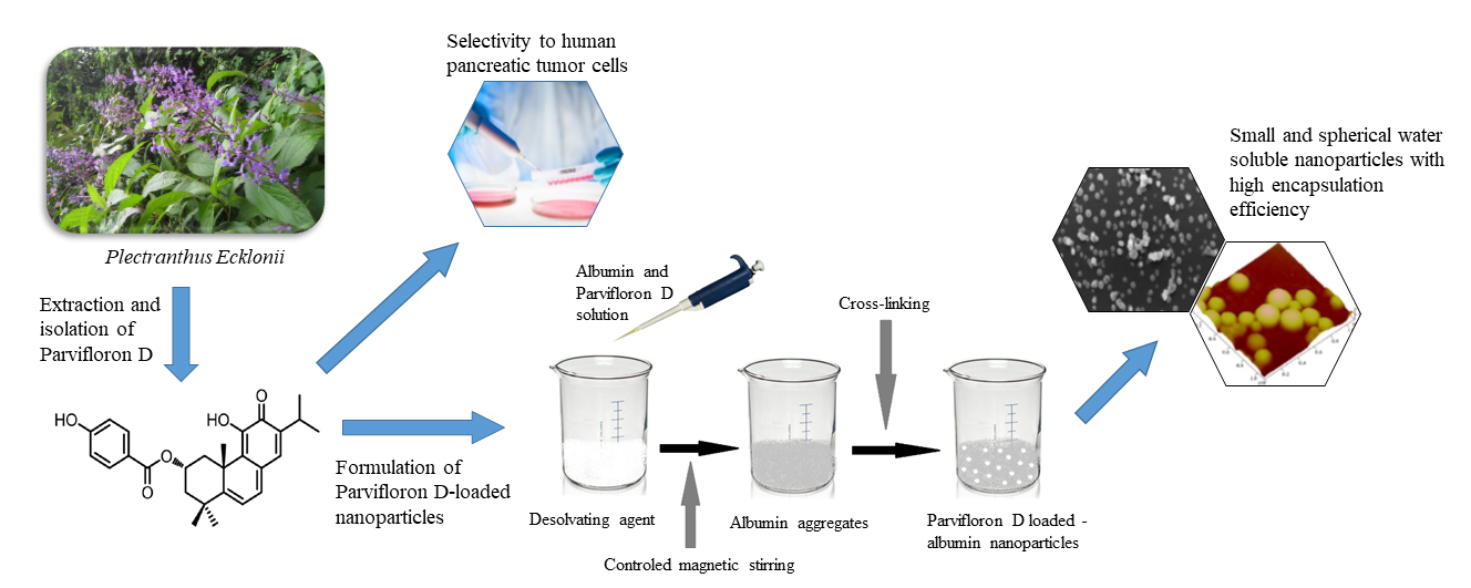
Development of Parvifloron D-Loaded Smart Nanoparticles to Target Pancreatic Cancer

 , , , , ,
  and
and 
Abstract
:
1. Introduction
2. Materials and Methods
2.1. Materials
2.2. Extraction and Isolation
2.2.1. Extraction
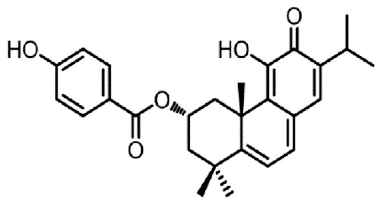
2.2.2. Isolation
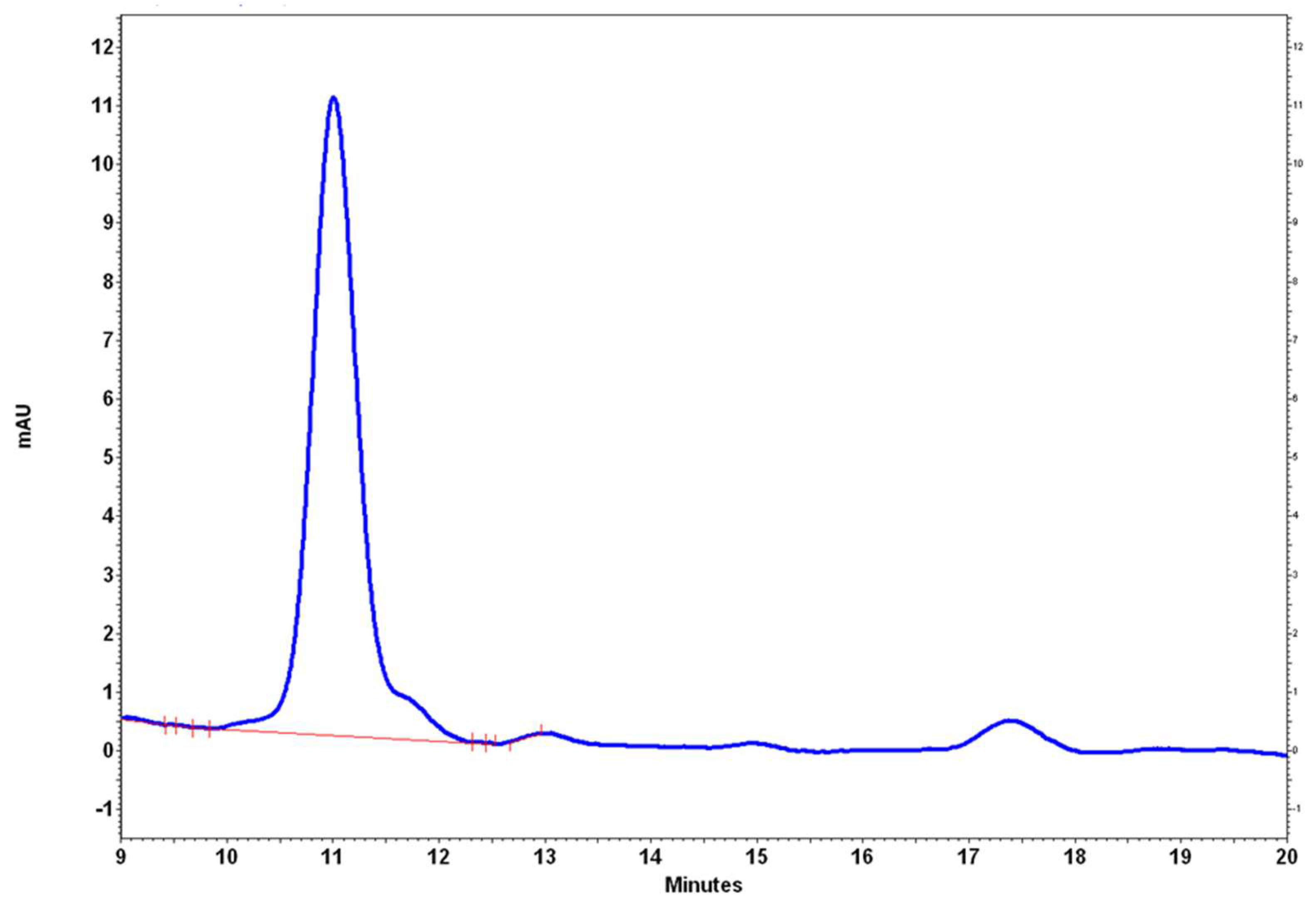
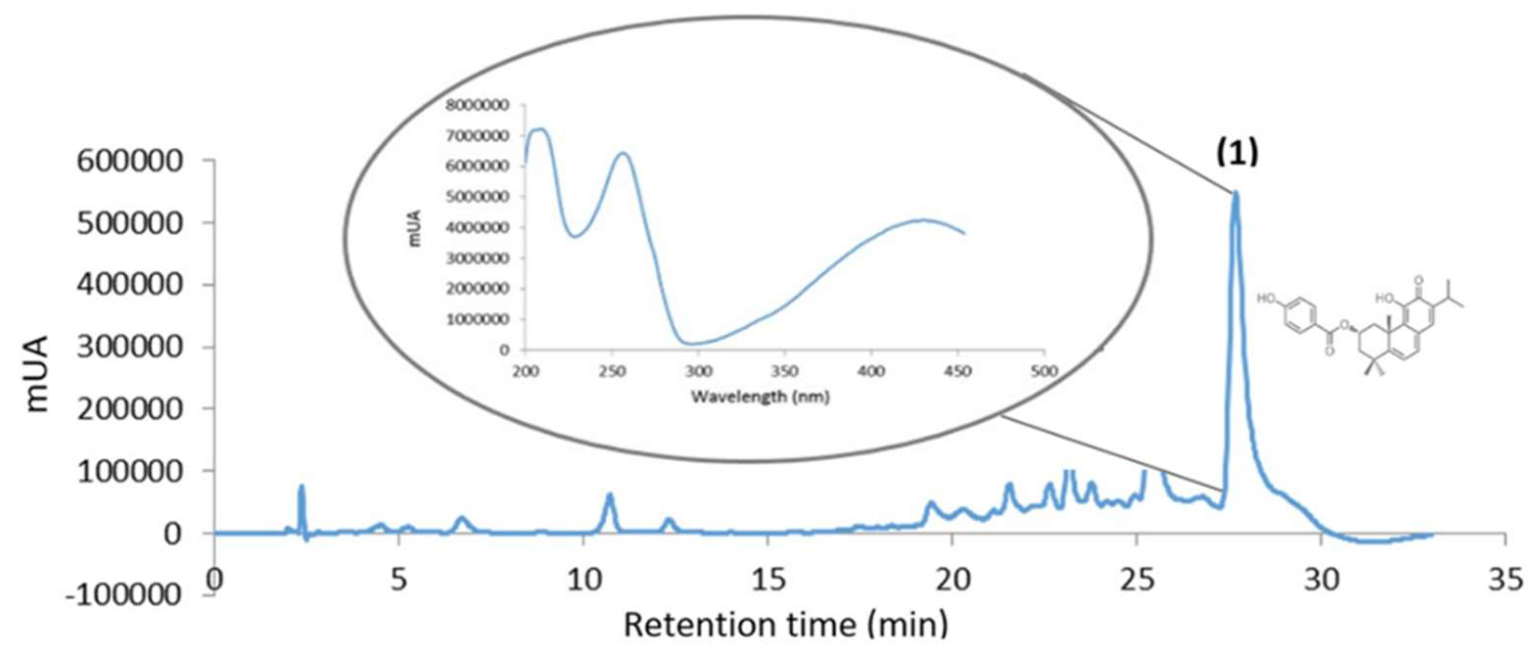
2.3. Parvifloron D Quantification by HPLC-DAD Analysis
2.4. Cell Culture and Cytotoxicity Assays
2.5. Parvifloron D Solubility Assays
2.6. Parvifloron D Encapsulation into a Biocompatible and Hydrophilic Nanomaterial
2.7. Determination of the Parvifloron D Encapsulation Efficiency by HPLC Analysis
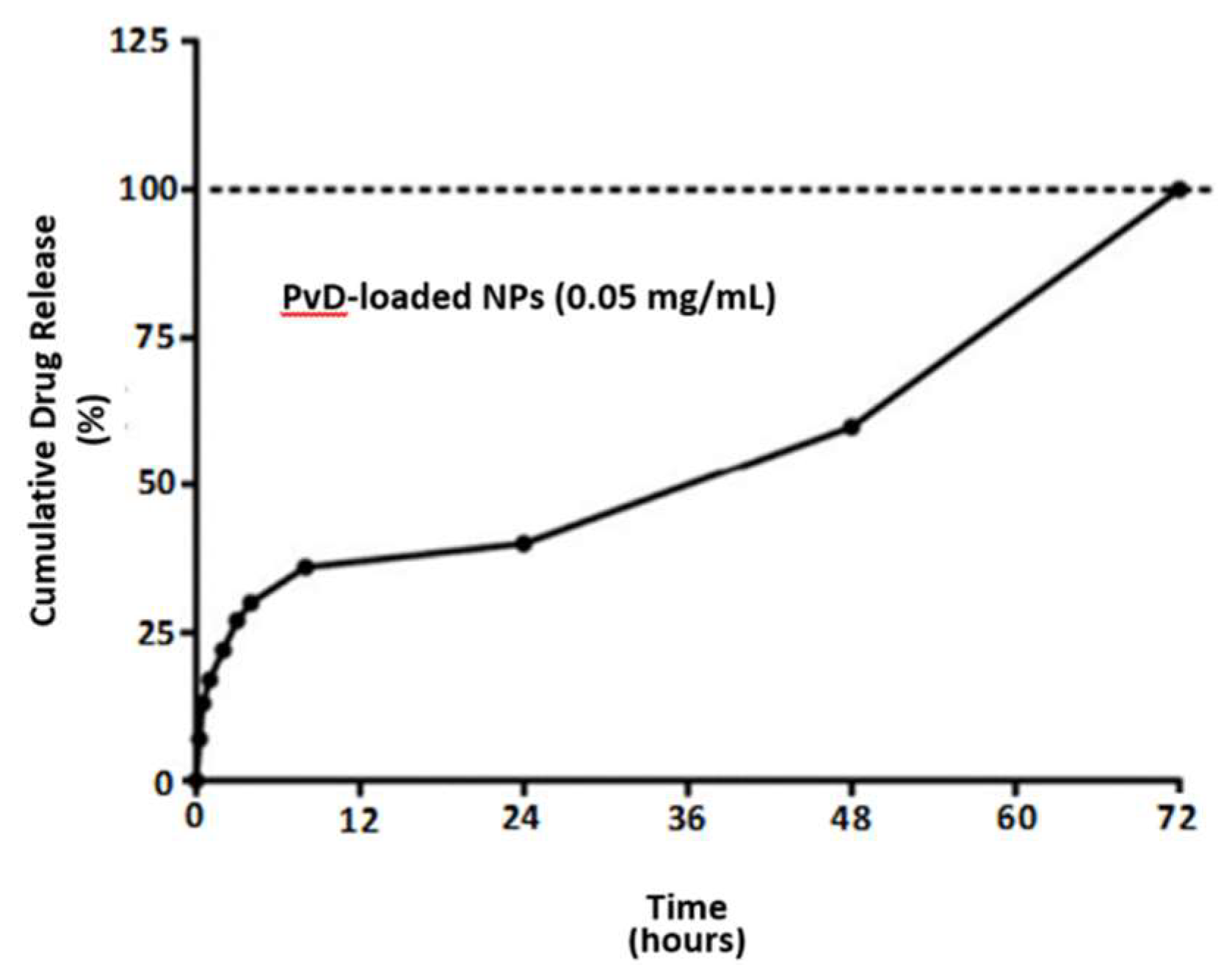
2.8. In Vitro Release Studies
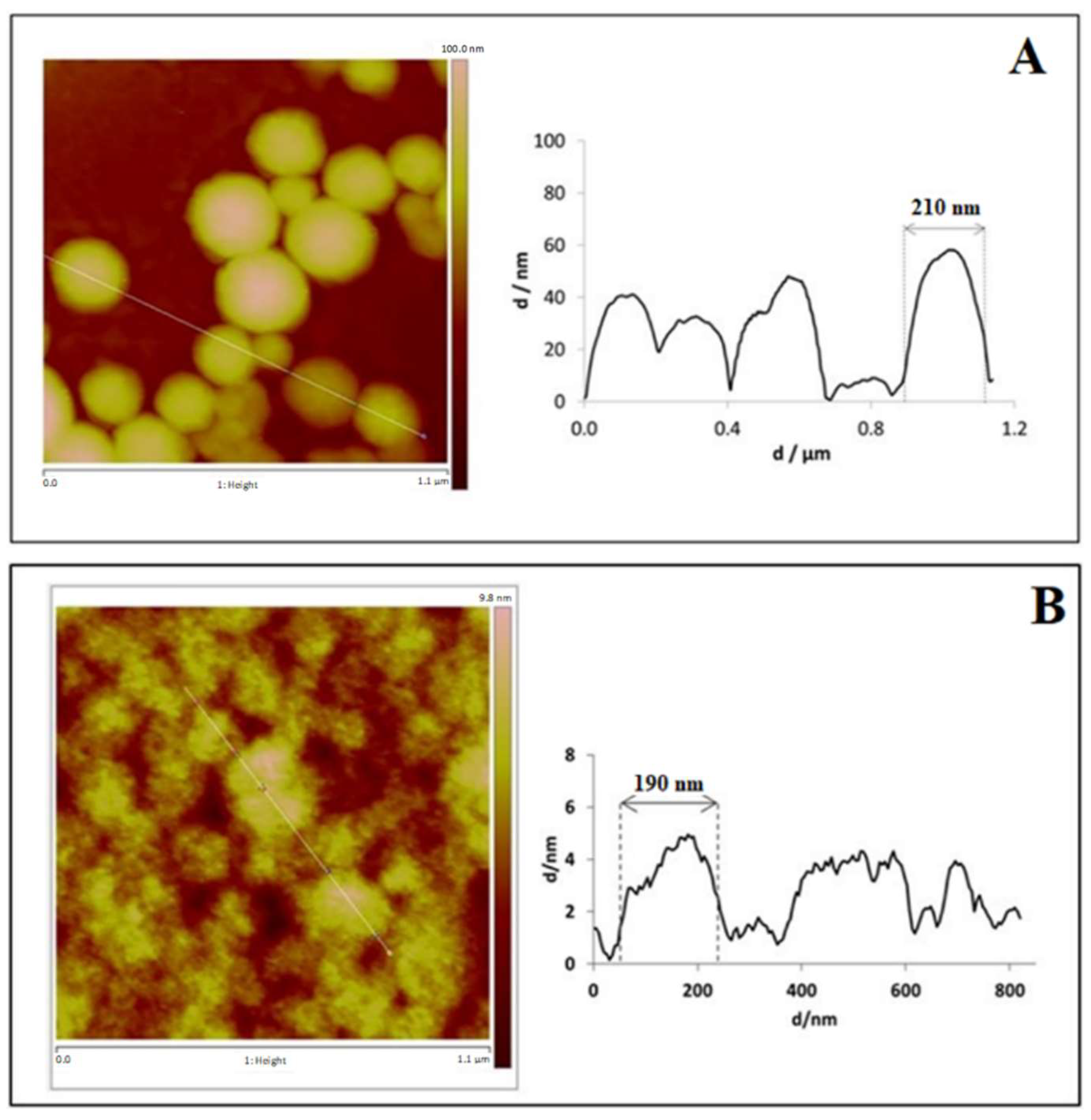
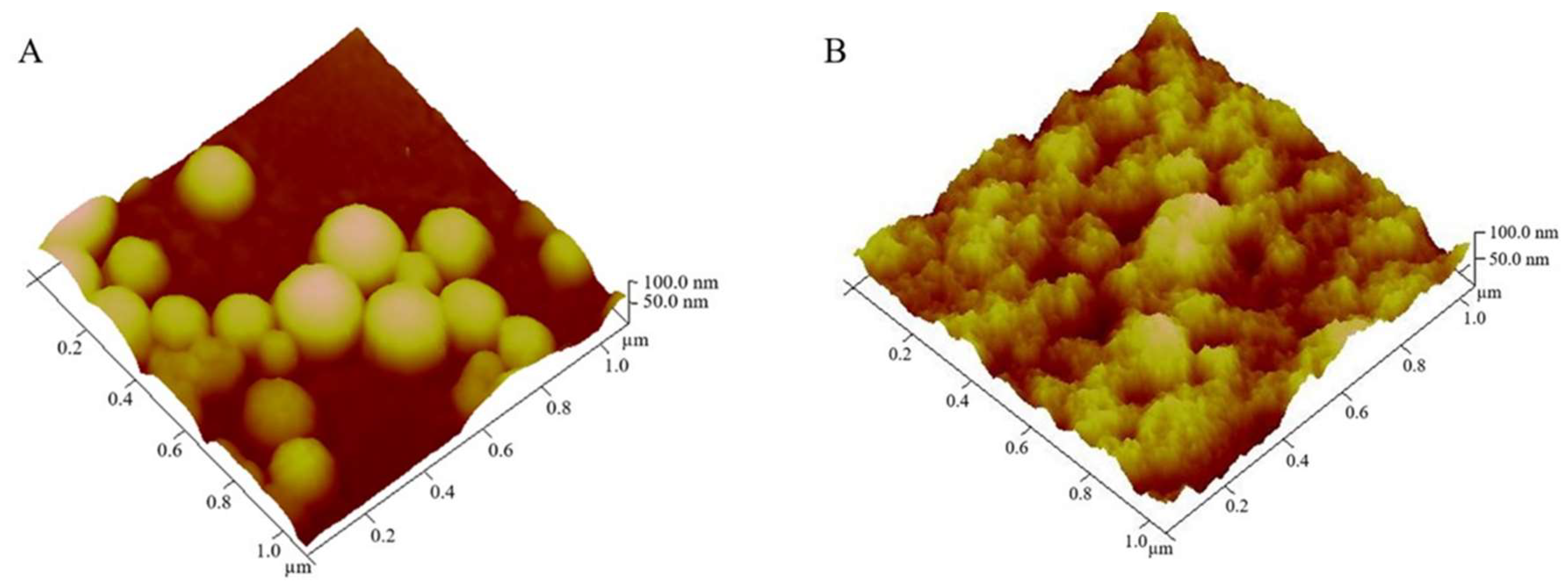
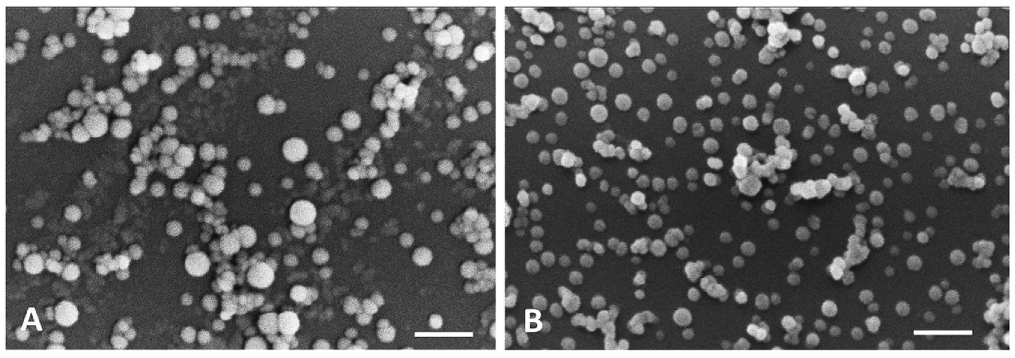
2.9. Physical and Morphological Characterization of the Nanoparticles: Dynamic Light Scattering (DLS), Scanning Electron Microscopy (SEM) and Atomic Force Microscopy (AFM)
2.10. Physicochemical Characterization of Nanoparticles Interaction Analysis by Fourier Transform Infrared (FT-IR)
2.11. Differential Scanning Calorimetry
3. Results and Discussion
3.1. Extraction and Isolation
3.2. Parvifloron D Quantification by HPLC-DAD
3.3. Cell culture and Cytotoxicity Assays
3.4. Nanoparticles Encapsulation Efficiency by HPLC Analysis
3.5. Parvifloron D Solubility Assays and In Vitro Release Studies
3.6. Physical and Morphological Characterization of the Nanoparticles: DLS, AFM, SEM
3.7. Physicochemical Characterization of Nanoparticles Interaction Analysis by FT-IR
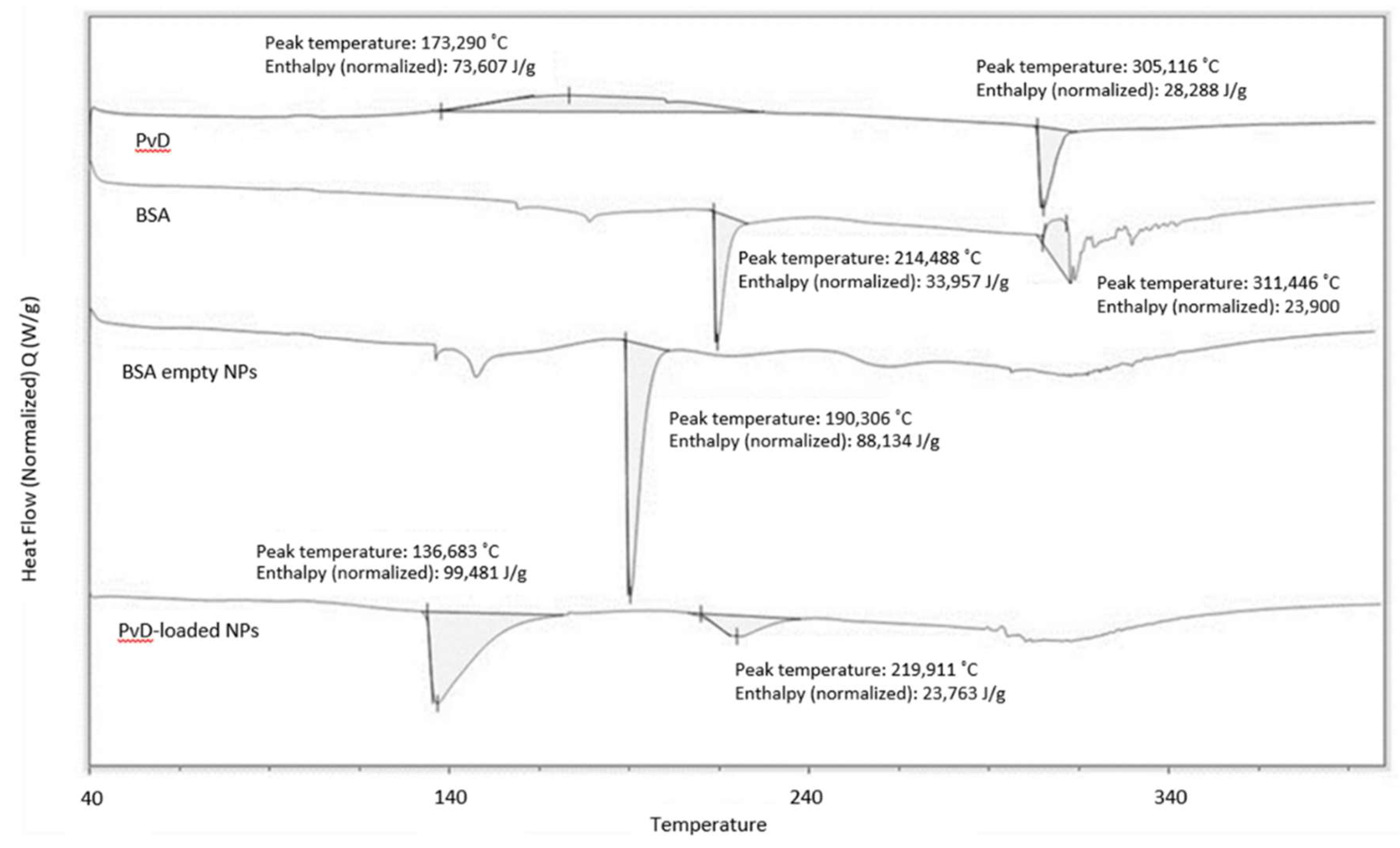
3.8. Differential Scanning Calorimetry
4. Conclusions
Supplementary Materials
Author Contributions
Funding
Acknowledgments
Conflicts of Interest
References
- Rahib, L.; Smith, B.D.; Aizenberg, R.; Rosenzweig, A.B.; Fleshman, J.M.; Matrisian, L.M. Projecting cancer incidence and deaths to 2030: The unexpected burden of thyroid, liver, and pancreas cancers in the united states. Cancer Res. 2014, 74, 2913–2921. [Google Scholar] [CrossRef] [PubMed]
- Niess, H.; Kleespies, A.; Andrassy, J.; Pratschke, S.; Angele, M.K.; Guba, M.; Jauch, K.-W.; Bruns, C.J. Pancreatic cancer in the elderly: Guidelines and individualized therapy. Der Chir. 2013, 84, 291–295. [Google Scholar] [CrossRef]
- Oberstein, P.E.; Olive, K.P. Pancreatic cancer: Why is it so hard to treat? Ther. Adv. Gastroenterol. 2013, 6, 321–337. [Google Scholar] [CrossRef] [PubMed]
- Rebelo, A.; Molpeceres, J.; Rijo, P.; Reis, C.P. Pancreatic Cancer Therapy Review: From Classic Therapeutic Agents to Modern Nanotechnologies. Curr. Drug Metab. 2017, 18, 346–359. [Google Scholar] [CrossRef] [PubMed]
- Neoptolemos, J.P.; Stocken, D.D.; Friess, H.; Bassi, C.; Dunn, J.A.; Hickey, H.; Beger, H.; Fernandez-Cruz, L.; Dervenis, C.; Lacaine, F.; et al. A Randomized Trial of Chemoradiotherapy and Chemotherapy after Resection of Pancreatic Cancer. N. Engl. J. Med. 2004, 350, 1200–1210. [Google Scholar] [CrossRef] [PubMed]
- Gourav, L.; Deepak-K., S.; Pawan-K., S.; Chand, M.P. Anticancer, antimicrobial and antifertility activities of some medicinal plants: A review. Med. Drug Res. 2015, 3, 7–11. [Google Scholar]
- Nicolai, M.; Pereira, P.; Vitor, R.F.; Pinto, C.; Roberto, A.; Rijo, P. Antioxidant activity and rosmarinic acid content of ultrasound-assisted ethanolic extracts of medicinal plants. Measurement 2016, 89, 328–332. [Google Scholar] [CrossRef]
- Burmistrova, O.; Perdomo, J.; Simões, M.F.; Rijo, P.; Quintana, J.; Estévez, F. The abietane diterpenoid parvifloron D from Plectranthus ecklonii is a potent apoptotic inducer in human leukemia cells. Phytomedicine 2015, 22, 1009–1016. [Google Scholar] [CrossRef] [PubMed]
- Simões, M.F.; Rijo, P.; Duarte, A.; Matias, D.; Rodríguez, B. An easy and stereoselective rearrangement of an abietane diterpenoid into a bioactive microstegiol derivative. Phytochem. Lett. 2010, 3, 234–237. [Google Scholar] [CrossRef]
- Burmistrova, O.; Simões, M.F.; Rijo, P.; Quintana, J.; Bermejo, J.; Estévez, F. Antiproliferative activity of abietane diterpenoids against human tumor cells. J. Nat. Prod. 2013, 76, 1413–1423. [Google Scholar] [CrossRef] [PubMed]
- Rosa, S.; Correia, V.; Ribeiro, I.; Rijo, P.; Saraiva, N.; Fernandes, A. In vitro antioxidant properties of the diterpenes Parvifloron D and 7α-acetoxy-6β-hydroxyroyleanone. Biomed. Biopharm. Res. 2015, 12, 59–67. [Google Scholar] [CrossRef]
- Bonifácio, B.V.; Silva, P.B.; Aparecido dos Santos Ramos, M.; Maria Silveira Negri, K.; Maria Bauab, T.; Chorilli, M. Nanotechnology-based drug delivery systems and herbal medicines: A review. Int. J. Nanomed. 2014, 9, 1–15. [Google Scholar] [CrossRef]
- Reis, C.P.; Figueiredo, I.V.; Carvalho, R.A.; Jones, J.; Nunes, P.; Soares, A.F.; Silva, C.F.; Ribeiro, A.J.; Veiga, F.J.; Damgé, C.; et al. Toxicological assessment of orally delivered nanoparticulate insulin. Nanotoxicology 2008, 2, 205–217. [Google Scholar] [CrossRef]
- Kouchakzadeh, H.; Shojaosadati, S.A.; Maghsoudi, A.; Farahani, E.V. Optimization of PEGylation Conditions for BSA Nanoparticles Using Response Surface Methodology. AAPS PharmSciTech 2010, 11, 1206–1211. [Google Scholar] [CrossRef] [PubMed] [Green Version]
- Abrantes, G.; Duarte, D.; Reis, C.P. An Overview of Pharmaceutical Excipients: Safe or Not Safe? J. Pharm. Sci. 2016, 105, 2019–2026. [Google Scholar] [CrossRef] [PubMed]
- Reis, C.P.; Damgé, C. Nanotechnology as a promising strategy for alternative routes of insulin delivery. Methods Enzymol. 2012, 508, 271–294. [Google Scholar] [CrossRef] [PubMed]
- Pinto Reis, C.; Silva, C.; Martinho, N.; Rosado, C. Drug carriers for oral delivery of peptides and proteins: Accomplishments and future perspectives. Ther. Deliv. 2013, 4, 251–265. [Google Scholar] [CrossRef] [PubMed]
- Harwood, L. “Dry-Column” Flash Chromatography. Aldrichim. Acta 1985, 18, 25. [Google Scholar]
- Gaspar-Marques, C.; Simões, M.F.; Valdeira, M.L.; Rodríguez, B. Terpenoids and phenolics from Plectranthus strigosus, bioactivity screening. Nat. Prod. Res. 2008, 22, 167–177. [Google Scholar] [CrossRef] [PubMed]
- Rijo, P.; Falé, P.L.; Serralheiro, M.L.; Simões, M.F.; Gomes, A.; Reis, C. Optimization of medicinal plant extraction methods and their encapsulation through extrusion technology. Measurement 2014, 58, 249–255. [Google Scholar] [CrossRef]
- Coelho, S.C.; Almeida, G.M.; Santos-Silva, F.; Pereira, M.C.; Coelho, M.A.N. Enhancing the efficiency of bortezomib conjugated to pegylated gold nanoparticles: An in vitro study on human pancreatic cancer cells and adenocarcinoma human lung alveolar basal epithelial cells. Expert Opin. Drug Deliv. 2016, 13, 1075–1081. [Google Scholar] [CrossRef] [PubMed]
- Hills, M.; Hudson, C.; Smith, P.G. Global monitoring of the resistance of malarial parasites to drugs: Statistical treatment of micro-test data. In Working Paper No. 2.8. 5 for the Informal Consultation on the Epidemiology of Drug Resistance of Malaria Parasites; World Health Organisation: Geneva, Switzerland, 1986. [Google Scholar]
- Silva, C.O.; Molpeceres, J.; Batanero, B.; Fernandes, A.S.; Saraiva, N.; Costa, J.G.; Rijo, P.; Figueiredo, I.V.; Faísca, P.; Reis, C.P. Functionalized diterpene parvifloron D-loaded hybrid nanoparticles for targeted delivery in melanoma therapy. Ther. Deliv. 2016, 7, 521–544. [Google Scholar] [CrossRef] [PubMed]
- Smith, R.; Tanford, C. Hydrophobicity of Long Chain n-Alkyl Carboxylic Acids, as Measured by Their Distribution Between Heptane and Aqueous Solutions. Proc. Natl. Acad. Sci. USA 1973, 70, 289–293. [Google Scholar] [CrossRef] [PubMed] [Green Version]
- Silva, P.J. Inductive and resonance effects on the acidities of phenol, enols, and carbonyl α-hydrogens. J. Org. Chem. 2009, 74, 914–916. [Google Scholar] [CrossRef] [PubMed]
- Bilia, A.R.; Piazzini, V.; Guccione, C.; Risaliti, L.; Asprea, M.; Capecchi, G.; Bergonzi, M.C. Improving on Nature: The Role of Nanomedicine in the Development of Clinical Natural Drugs. Planta Med. 2017, 83, 366–381. [Google Scholar] [CrossRef] [PubMed] [Green Version]
- Pinto Reis, C.; Neufeld, R.J.; Ribeiro, A.J.; Veiga, F. Nanoencapsulation I. Methods for preparation of drug-loaded polymeric nanoparticles. Nanomed. Nanotechnol. Biol. Med. 2006, 2, 8–21. [Google Scholar] [CrossRef] [PubMed] [Green Version]
- Weber, C.; Coester, C.; Kreuter, J.; Langer, K. Desolvation process and surface characterisation of protein nanoparticles. Int. J. Pharm. 2000, 194, 91–102. [Google Scholar] [CrossRef]
- Figueiredo, N.L.; Falé, P.L.; Madeira, P.J.A.; Florêncio, M.H.; Ascensão, L.; Serralheiro, M.L.M.; Lino, A.R.L. Phytochemical Analysis of Plectranthus sp. Extracts and Application in Inhibition of Dental Bacteria, Streptococcus sobrinus and Streptococcus mutans. Eur. J. Med. Plants 2014, 4, 794–809. [Google Scholar] [CrossRef]
- Yu, X.; Di, Y.; Xie, C.; Song, Y.; He, H.; Li, H.; Pu, X.; Lu, W.; Fu, D.; Jin, C. An in vitro and in vivo study of gemcitabine-loaded albumin nanoparticles in a pancreatic cancer cell line. Int. J. Nanomed. 2015, 10, 6825–6834. [Google Scholar] [CrossRef] [PubMed] [Green Version]
- Singh, A.; Xu, J.; Mattheolabakis, G.; Amiji, M. EGFR-targeted gelatin nanoparticles for systemic administration of gemcitabine in an orthotopic pancreatic cancer model. Nanomed. NBM 2015, 12, 589–600. [Google Scholar] [CrossRef] [PubMed]
- Acuna, A.; Jeffery, J.J.; Abril, E.R.; Nagle, R.B.; Guzman, R.; Pagel, M.D.; Meuillet, E.J. Nanoparticle delivery of an AKT/PDK1 inhibitor improves the therapeutic effect in pancreatic cancer. Int. J. Nanomed. 2014, 9, 5653–5665. [Google Scholar] [CrossRef]
- Mukai, S.; Moriya, S.; Hiramoto, M.; Kazama, H.; Kokuba, H.; Che, X.F.; Yokoyama, T.; Sakamoto, S.; Sugawara, A.; Sunazuka, T.; et al. Macrolides sensitize EGFR-TKI-induced non-apoptotic cell death via blocking autophagy flux in pancreatic cancer cell lines. Int. J. Oncol. 2016, 48, 45–54. [Google Scholar] [CrossRef] [PubMed]
- Wang, L.I.N.; Zhu, Z.; Zhang, W.; Zhang, W. Schedule-dependent cytotoxic synergism of pemetrexed and erlotinib in BXPC-3 and PANC-1 human pancreatic cancer cells. Exp. Ther. Med. 2011, 2, 969–975. [Google Scholar] [CrossRef] [PubMed] [Green Version]
- Wright, A.E.; Roth, G.P.; Hoffman, J.K.; Divlianska, D.B.; Pechter, D.; Sennett, S.H.; Guzmán, E.A.; Linley, P.; McCarthy, P.J.; Pitts, T.P.; et al. Isolation, synthesis, and biological activity of aphrocallistin, an adenine-substituted bromotyramine metabolite from the hexactinellida sponge Aphrocallistes beatrix. J. Nat. Prod. 2009, 72, 1178–1183. [Google Scholar] [CrossRef] [PubMed]
- Merlot, A.M.; Kalinowski, D.S.; Richardson, D.R. Unraveling the mysteries of serum albumin—More than just a serum protein. Front. Physiol. 2014, 5, 1–7. [Google Scholar] [CrossRef] [PubMed]
- Yameen, B.; Choi, W.I.; Vilos, C.; Swami, A.; Shi, J.; Farokhzad, O.C. Insight into nanoparticle cellular uptake and intracellular targeting. J. Control. Release 2015, 28, 485–499. [Google Scholar] [CrossRef] [PubMed]
- Marrese, M.; Guarino, V.; Ambrosio, L. Atomic Force Microscopy: A Powerful Tool to Address Scaffold Design in Tissue Engineering. J. Funct. Biomater. 2017, 8, 7. [Google Scholar] [CrossRef] [PubMed]
- Kowoll, T.; Müller, E.; Fritsch-Decker, S.; Hettler, S.; Störmer, H.; Weiss, C.; Gerthsen, D. Contrast of backscattered electron SEM images of nanoparticles on substrates with complex structure. Scanning 2017, 2017. [Google Scholar] [CrossRef] [PubMed]


| Cell line | IC50 (µM) ± SD |
|---|---|
| MCF-7 (breast cancer) | 35.1 ± 2.2 |
| HaCat (human keratinocyte) | 34.3 ± 4.1 |
| Caco-2 (Colon adenocarcinoma) | 32.1 ± 4.3 |
| INS-1E (rat pancreatic insulinoma) | 21.6 ± 0.5 |
| BxPC3 (human pancreatic adenocarcinoma) | 0.15 ± 0.1 |
| PANC-1 (human pancreatic adenocarcinoma) | 11.9 ± 0.7 |
| Functional Groups | O–H Carboxylic Acid (Stretching) | C–H Alkane (Stretching) | C=O Carbonyl (Stretching) | C=O Amide I (Stretching) | N–H Amide II (Bending) | C=C Aromatic (Stretching) | C–H Alkane (Bending) | C–O Alcohol (Stretching) | =C–H Alkene (Bending) |
|---|---|---|---|---|---|---|---|---|---|
| Compound | |||||||||
| BSA 1 | --- | --- | --- | 1654 | 1590 | --- | --- | --- | --- |
| PvD 2 | --- | 2871 | 1693 | --- | --- | 1510 | --- | --- | 850 |
| Glucose | 3350 | --- | --- | --- | --- | --- | 1456 | 1032 | --- |
| Physical mixture BSA + PvD | --- | 2871 | 1690 | 1658 | 1590 | 1515 | --- | --- | --- |
| Empty BSA-NPs 3 | 3000 | --- | --- | 1654 | 1540 | --- | --- | --- | --- |
| BSA-NPs loaded With PvD | 3000 | 2873 | --- | 1654 | 1540 | 1540 | --- | --- | 910 |
© 2018 by the authors. Licensee MDPI, Basel, Switzerland. This article is an open access article distributed under the terms and conditions of the Creative Commons Attribution (CC BY) license (http://creativecommons.org/licenses/by/4.0/).
Share and Cite
Santos-Rebelo, A.; Garcia, C.; Eleutério, C.; Bastos, A.; Castro Coelho, S.; Coelho, M.A.N.; Molpeceres, J.; S. Viana, A.; Ascensão, L.; Pinto, J.F.; et al. Development of Parvifloron D-Loaded Smart Nanoparticles to Target Pancreatic Cancer. Pharmaceutics 2018, 10, 216. https://doi.org/10.3390/pharmaceutics10040216
Santos-Rebelo A, Garcia C, Eleutério C, Bastos A, Castro Coelho S, Coelho MAN, Molpeceres J, S. Viana A, Ascensão L, Pinto JF, et al. Development of Parvifloron D-Loaded Smart Nanoparticles to Target Pancreatic Cancer. Pharmaceutics. 2018; 10(4):216. https://doi.org/10.3390/pharmaceutics10040216
Chicago/Turabian StyleSantos-Rebelo, Ana, Catarina Garcia, Carla Eleutério, Ana Bastos, Sílvia Castro Coelho, Manuel A. N. Coelho, Jesús Molpeceres, Ana S. Viana, Lia Ascensão, João F. Pinto, and et al. 2018. "Development of Parvifloron D-Loaded Smart Nanoparticles to Target Pancreatic Cancer" Pharmaceutics 10, no. 4: 216. https://doi.org/10.3390/pharmaceutics10040216