Article

Highly Efficient NO2 Sensors Based on Al-ZnOHF under UV Assistance

1
Key Laboratory for Liquid-Solid Structural Evolution and Processing of Materials, Ministry of Education and School of Materials Science and Engineering, Shandong University, Jinan 250061, China
2
Henan Joint International Research Laboratory of Nanocomposite Sensing Materials, School of Chemical and Environmental Engineering, Anyang Institute of Technology, Anyang 455000, China
3
School of Materials Science and Engineering, Qilu University of Technology (Shandong Academy of Sciences), Jinan 250353, China
*
Author to whom correspondence should be addressed.
Materials 2023, 16(9), 3577; https://doi.org/10.3390/ma16093577
Submission received: 5 April 2023 / Revised: 26 April 2023 / Accepted: 4 May 2023 / Published: 7 May 2023
(This article belongs to the Special Issue Application of Emerging Materials for Advanced Imaging and Sensing)

Abstract

:
Zinc hydroxyfluoride (ZnOHF) is a newly found resistive semiconductor used as a gas-sensing material with excellent selectivity to NO2 because of its unique energy band structure. In this paper, Al3+ doping and UV radiation were used to further improve the gas-sensing performance of ZnOHF. The optimized 0.5 at.% Al-ZnOHF sample exhibits improved sensitivity to 10 ppm NO2 at a lower temperature (100 °C) under UV assistance, as well as a short response/recovery time (35 s/96 s). The gas-sensing mechanism demonstrates that Al3+ doping increases electron concentration and promotes electron transfer of the nanorods by reducing the bandgap of ZnOHF, and the photogenerated electrons and holes with high activity under UV irradiation provide new reaction routes in the gas adsorption and desorption process, effectively promoting the gas-sensing process. The synergistic effect of Al3+ and UV radiation contribute to the enhanced performance of Al-ZnOHF.

1. Introduction

As a typical pollution gas, nitrogen dioxide (NO2) seriously threatens human health and the environment [1,2]. Exposure to NO2 with a concentration of over 200 μg/m3 in the short term or over 40 μg/m3 in the long term will pose a serious threat to the normal operation of the respiratory system [3]. Additionally, NO2 is also the main source of acid rain and photochemical smog. Hence, the development of gas sensors for efficiently detecting the concentration of NO2 in the atmosphere is highly significant and essential. In the research on NO2 sensors, resistance gas sensors stand out among different kinds of gas sensors due to their excellent sensitivity, selectivity, and stability [4,5,6], as well as their relatively low cost and easy operation [7]. As the core part of sensors, the sensing materials directly determine the performance of gas sensors.
Zinc hydroxyfluoride (ZnOHF) is a new type of semiconductor discovered in recent years which has gradually attracted the attention of researchers in various fields, such as photocatalysts [8,9], ultrasonic degradation [10] and solar cells [11], for its unique crystal and energy band structure [12,13]. In our previous research, we found that ZnOHF is an excellent gas-sensing material with good selectivity to NO2 gas [14]. The unique energy band structure of ZnOHF inhibits the adsorption and dissociation of oxygen molecules on its surface, and the little absorbed oxygen species on the surface of ZnOHF lead to its wide detection range to NO2, which makes ZnOHF a good candidate for NO2 detection among resistive semiconductors. However, the gas sensors based on ZnOHF still have room for improvement in performance; for example, the operating temperature of ZnOHF-based gas sensors is still high, and its sensitivity still has a certain gap compared with many other NO2-sensing materials. Therefore, further improvement for ZnOHF to obtain better sensing performance is necessary.
Light-activated gas sensors have been widely studied as an effective improvement method in recent years. Under the assistance of light irradiation, there will be more electrons and holes with high activity generated on the surface of materials, which is beneficial to the adsorption and desorption of target gases [15,16]. Wang et al. [17] found that ZnO nanoplates exhibited a great enhancement in sensitivity and recovery time in NO2 detection under intermittent UV irradiation. This phenomenon can be explained by the competitive adsorption of NO2 and O2 and the modulating action of gas adsorption. Zhang and his group [18] grew TiO2 nanoplates in situ with a highly active crystal plane on MXene (Ti3C2Tx) and obtained greatly enhanced gas-sensing performance to NH3 under UV light. The density functional theory revealed that this composite structure exhibits the highest adsorption affinity to NH3 with UV light assistance. Additionally, Tai’s group [19] also applied gamma-ray in NO2 detection, and they found that it can effectively enhance the sensitivity of ZnO nanorods at room temperature by constructing lots of defects. Bang et al. [20] presented a proton-beam irradiation method to engineer surface-point defects on ZnO in order to improve the preferential adsorption on the surface compared to water molecules so that it can detect NO2 with less humidity interference. As a kind of photocatalyst, ZnOHF has excellent photochemical properties, including suitable band gaps and strong light absorbance, which makes it possible to act as a better gas sensor of NO2 under UV radiation [21,22].
Ion doping is a promising approach in gas-sensing performance improvement. On the one hand, doping ions with different quantities of charges can effectively adjust the carrier concentration and energy band structure of semiconductors [23,24]. On the other hand, due to the difference in diameters between doping ions and original ions, ion doping can create lots of lattice imperfections. Some imperfections can act as efficient adsorption sites of oxygen molecules or target gas molecules, thus improving their gas-sensing performance [25]. Qin and his colleague [26] synthesized metal–organic-frameworks-derived Mn-doped Co3O4 porous nanosheets, which exhibited better CO-sensing performance than pure Co3O4 because of the oxygen vacancies generated in the ion doping process. Mokrushin et al. [27] doped Eu and Pr ions into ZnO powders and found that it not only improved the sensitivity but also obtained great humidity resistance performance. Cr3+-doped In2O3 was prepared by Sun et al. [28], and this material shows high sensitivity, a low detection limit, and short response/recovery time in NH3 sensing due to the increase in active sites and reduced material resistance. Al3+ ion is one of the common doping ions utilized in zinc-based materials’ modification. Since Al3+ has a higher charge than Zn2+, Al3+ doping can increase electron concentration, which will benefit the gas-sensing process [29,30]. Moreover, Al3+ has a similar diameter to Zn2+, indicating that Al3+ doping will not seriously damage the crystal structure of the host ZnOHF [31,32].
Herein, Al-ZnOHF nanorods with optimized Al3+ concentrations were prepared through a simple hydrothermal method. The NO2-sensing performance of Al3+-doped samples is significantly improved compared with pure ZnOHF under UV irradiation with a wavelength of 395 nm and a strength of 6 mW/cm2. Additionally, especially, the 0.5 at.% Al-ZnOHF sample exhibits the best gas-sensing properties. The NO2-sensing mechanism analysis revealed that the bandgap narrowing and improvement in UV absorbance after Al3+ doping are the main reasons for its performance enhancement.

2. Materials and Methods

2.1. Chemicals

All chemicals utilized in this work, including zinc acetate (Zn(CH3COO)2•2H2O), ammonium fluoride (NH4F), aluminum nitrate (Al(NO3)3•9H2O), and hexamethylenetetramine (HMT), were purchased from Sinopharm Chemical Reagent Co., Ltd. (Shanghai, China) and were of analytical grade, which can be used without any purification.

2.2. Synthesis of Al3+-doped ZnOHF

Al-ZnOHF nanorods with different molar ratios of Al3+ (0 at.%, 0.1 at.%, 0.2 at.%, 0.5 at.%, and 1 at.%) were synthesized via a simple hydrothermal method. Initially, 0.65 g Zn(CH3COO)2•2H2O, 0.42 g HMT and 0.051 g NH4F were dissolved in 15 mL of distilled water successively with continuous stirring to form a homogeneous solution. Different amounts of Al(NO3)3•9H2O were then added to the mixture with continuous stirring. After stirring for another 0.5 h, the solution was transferred into 20 mL Teflon-lined autoclaves and heated at 368 K for 2 h. The products were collected, purged with distilled water and ethyl alcohol, and then dried at 340 K for 6 h.

2.3. Characterizations

The crystal structures of samples were tested through X-ray powder defection (XRD, DMAX-2500PC, Regaku, Japan) equipped with Cu-Kα (λ = 1.542 Å) radiation with a step size of 0.02° and a scan rate of 10°/min in the range of 10–90°. The surface morphologies and micro-structure characterizations were carried out by scanning electron microscopy (SEM, SU-70, Hitachi, Japan) operated at an accelerating voltage of 15 kV and transmission electron microscopy (TEM, JEM 2100, JEOL Ltd., Akishima, Japan) operated at 200 kV. X-ray photoelectron spectrometer (XPS, AXIS Supra, Kratos, Japan) measurement (equipped with monochromatic Al-Kα radiation, hυ = 1486.6 eV) was utilized to analyze the valence state of elements and the chemical environment of atoms on the surface of samples. The UV-vis diffuse reflection spectrum (UV-vis DRS) was recorded on Agilent Cary 300/PE lambda 750S (America) ultraviolet spectrophotometer and was used to test UV absorption and bandgap of the samples.

2.4. Gas-Sensing Properties Test

The preparation method of gas-sensing units was explained as follows. First, the as-prepared samples were dispersed into distilled water with a weight ratio of 1:5 and treated with ultrasound for 10 min. Additionally, the slurry was dropped on the Al2O3 substrate, which has four Pt wires as the electrodes, and was dried at 80 °C. After repeating this step 2–4 times, the substrate was annealed at 200 °C for 3 h and then the electrodes were welded on the pedestal to form a gas-sensing unit. After that, the sensor was aged at 4 V for 7 days before the gas-sensing test. The gas-sensing properties of gas sensors based on various samples were tested via a WS-30B gas sensitivity instrument (Zhengzhou Winsen Electronics Co., Ltd., Zhengzhou, China) at an environmental temperature of 25 °C and humidity of 20–30%. The UV light was provided by LED light with wavelength of 395 nm (Ceaulight Co., Beijing, China). The response value of sensors (S) is calculated as S = Rg/Ra for oxidizing gases and S = Ra/Rg for reducing gases, where Ra and Rg are the resistance of the sensor in air and target gases, respectively. Additionally, the response and recovery times are defined as the times from gas input or pumped out until the response value changes to 90% of its maximum value.

3. Results and Discussion

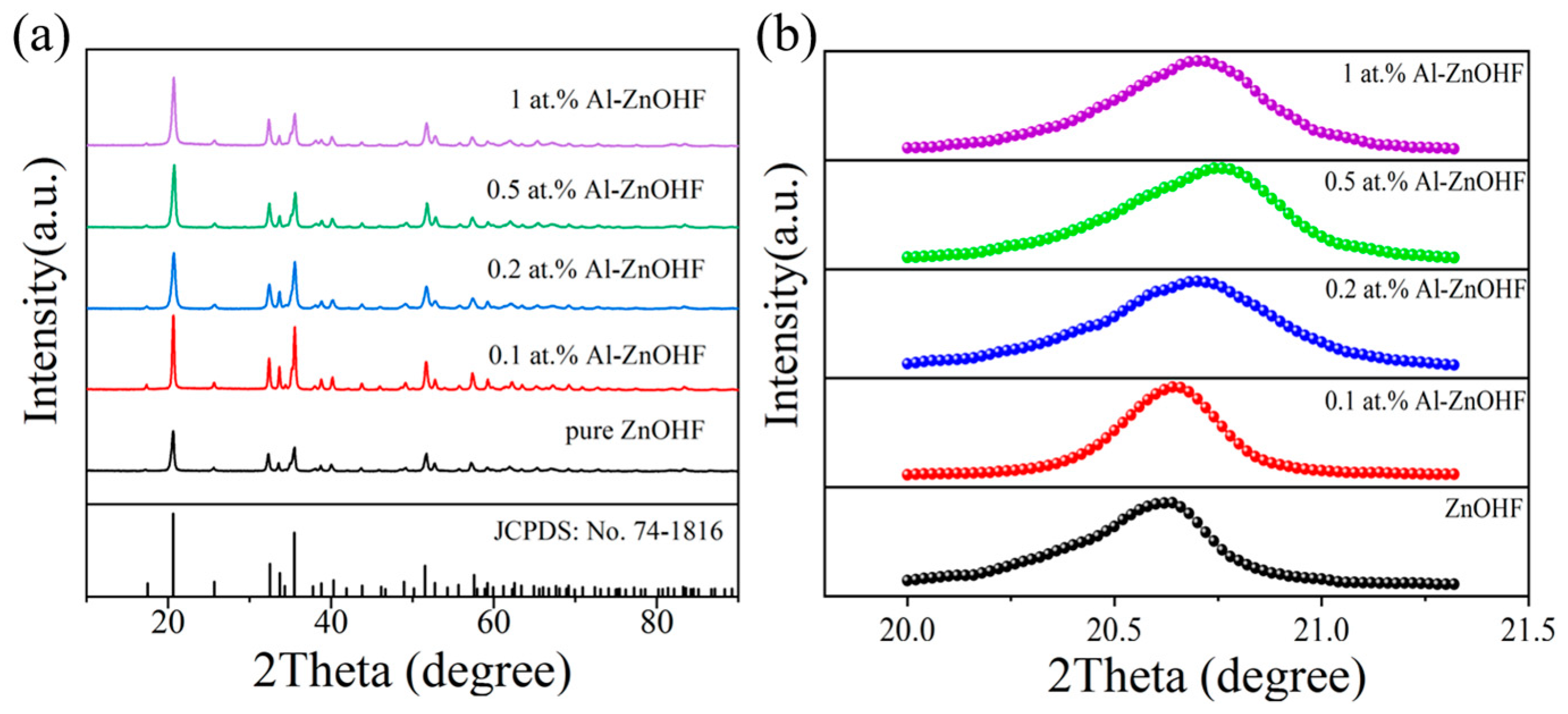
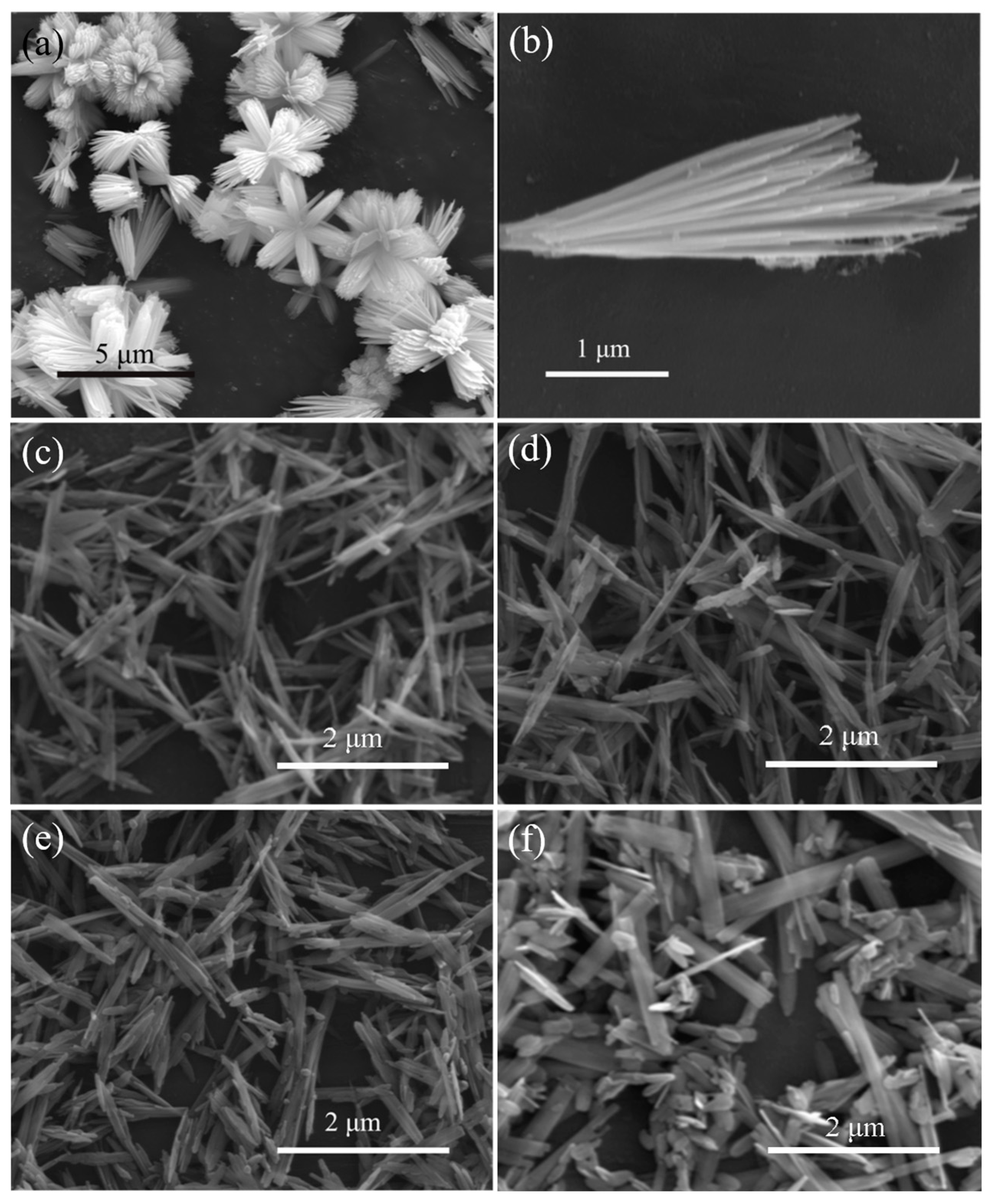
3.1. Characterization

Figure 1 shows the XRD patterns of Al-ZnOHF with different amounts of aluminum ions. The diffraction peaks of each sample could be approximately matched with the standard data of orthorhombic-phase ZnOHF (JCPSD No: 74-1816). After the addition of Al3+, the central position of diffraction peaks changed significantly. It can be seen in the magnified region around the strongest peak (Figure 1b) that the peak trended to a higher angle with the increase in Al3+ concentration at first, indicating that the Al3+ with the size of 0.0535 nm may occupy some positions of the Zn2+ with the size of 0.074 nm. Additionally, then, the peak turned back to the lower angle (1 at.% Al-ZnOHF), indicating that the Al3+ dopes into the interstitial void of the ZnOHF lattice, which results in the broadening of the interplanar crystal spacing.
Figure 2a,b shows the SEM images of pure ZnOHF. The basic structure of pure ZnOHF is nanorods, and the ends of these nanorods are gathered together to constitute flower-like structures with sizes ranging from 3 to 5 μm. SEM graphs of samples with different Al3+ dopings are exhibited in Figure 2c–f. After Al3+ doping, the flower-like structures of pure ZnOHF change into irregular bundles, which also consist of nanorods. With the increase in the amount of Al3+, the length of bundles gradually shortened and thickened, and the morphology gradually changed to microbelts. Additionally, lots of small nanoplates also appeared, and the quantity gradually increased with the increase in Al3+ content. Finally, when the Al3+ doping amount reached 1 at.%, the morphology of the sample was mainly composed of long microbelts and plenty of nanoplates.
To further analyze the microstructure of the obtained samples, TEM and HRTEM images of pure ZnOHF and 0.5 at.% Al-ZnOHF are shown in Figure 3. As shown in Figure 3a,b, pure ZnOHF exhibits a regular nanorods-assembled flower-like structure. The HRTEM image (Figure 3c) shows that the diameter of nanorods is about 14 nm. Additionally, the lattice fringe spacing of pure ZnOHF could be measured as 0.25 nm, corresponding to the (1 1 1) crystal plane of ZnOHF. After Al3+ doping, the morphology of samples changed greatly (Figure 3d,e), which is in accord with the SEM images. Instead of forming a flower-like structure, the nanorods were clustered into bundles and scattered irregularly. The HRTEM image of 0.5 at.% Al-ZnOHF (Figure 3f) exhibits that the diameter of nanorods slightly increased to 25 nm. Its lattice fringe spacing was calculated to be 0.42 nm, a little smaller than that of the (1 1 0) crystal plane. Hence, Al3+ doping changed the exposed surface crystal plane of samples and reduced the crystalline interplanar spacing.
The surface chemical properties of 0.5 at.% Al-ZnOHF were determined via X-ray photoelectron spectroscopy (XPS). As shown in Figure 4a, all 3 main elements (Zn, O, F) can be easily found in the wide XPS spectrum of both pure ZnOHF and 0.5 at.% Al-ZnOHF, and there is also a tiny peak at about 75 eV in the spectra of 0.5 at.% Al-ZnOHF, which can be matched with the Al element. The Zn 2p spectrum in Figure 4b shows that there are 2 peaks centered at 1021.8 eV and 1044.8 eV, corresponding to the Zn2+ orbits, 2p3/2 and 2p1/2, respectively [33]. The peaks of F 2p (Figure 4c) of these 2 samples are centered at about 684.8 eV, matching well with F in the crystal lattice [34]. By comparison, the peaks of Zn and F of these two samples are approximately the same, meaning that Al3+ doping does not change the chemical environment of Zn and F atoms. The difference between these two samples at the high-resolution XPS regions of Al 2p and O 1s is shown in Figure 4d–e. The 0.5 at.% Al-ZnOHF exhibits a significant peak at about 75.2 eV, which can be indexed with Al3+ [35], while pure ZnOHF just shows a gentle background curve in this region (Figure 4d). This difference confirmed that Al ions were successfully doped into ZnOHF. The O 1s peaks of both pure ZnOHF and 0.5 at.% Al-ZnOHF can be deconvoluted into two Gaussian peaks, which are matched with lattice oxygen and adsorbed oxygen species, respectively (Figure 4e) [14,36]. It is significant that the proportion of adsorbed oxygen species of 0.5 at.% Al-ZnOHF is higher than that of pure ZnOHF, confirming that Al3+ doping promotes the surface adsorption of oxygen. In addition, the peaks of the 2 oxygen species exhibit a slight blueshift after Al3+ doping (Olatt. move from 532.0 eV to 531.6 eV and Oads. move from 532.8 eV to 532.2 eV). This phenomenon may be ascribed to the weaker adsorption capacity for electrons of Al3+ than that of Zn2+. When Al3+ substitutes the Zn2+ in the lattice, the electron density increases due to the construction of the Al-O bond [37,38,39]. Therefore, Al3+ doping can decrease the binding energy of electrons of oxygen species and obtain adsorbed oxygen with higher activity.

3.2. Gas-Sensing Properties

The gas-sensing properties of ZnOHF with various amounts of Al3+ doping were tested under different temperatures (Figure 5a). Due to the low thermal stability of ZnOHF at high temperatures (it decomposes into ZnO and HF at over 300 °C) [14], the test temperature should be controlled under 300 °C. Significantly, the best operating temperatures of all samples are 160 °C. The response value of Al3+-doped samples shows various levels of increase compared with pure ZnOHF. Especially, gas sensors based on 0.5 at.% Al-ZnOHF obtained the highest response (49.43) to 10 ppm NO2, 2.42 times higher than that of pure ZnOHF. In addition, the dynamic response curves of sensors based on 0.5 at.% Al-ZnOHF and pure ZnOHF to 10 ppm NO2 are shown in Figure 5b. It is worth noting that the response time of 0.5 at.% Al-ZnOHF (50 s) is a little shorter than that of pure ZnOHF (112 s), but both of them exhibit extremely long recovery times or even cannot recover back to the previous resistance. The resistance of samples at different temperatures is shown in Figure S1a. It is evident that with the increase in the Al3+ doping amount, the resistance of samples rises at first, probably due to the destruction of the regular morphology of pure ZnOHF after ion doping, which is not conducive to electron conduction. Additionally, the resistance reduces when the Al3+ doping amount continuously increases because of the increase in carrier concentration. The extremely high resistance of samples also seriously restricts the gas-sensing test at low temperatures.
To solve this problem, UV-light radiation was utilized in the gas-sensing process. It can be seen in Figure S1b that the resistance of all samples significantly decreases under UV radiation, and the Al3+-doped samples obtained a higher reduction rate with higher UV absorbance. The gas-sensing properties of samples under UV light with the wavelength of 395 nm and strength of 6 mW/cm2 at different temperatures were tested, as illustrated in Figure 5c. It is significant that the best operating temperature of 0.1, 0.2, and 0.5 at.% Al-ZnOHF is 100 °C, while that of 1 at.% Al-ZnOHF is 80 °C, indicating that the operating temperature decreased after UV radiation. Specifically, the sensor based on 0.5 at.% Al-ZnOHF exhibited the highest response, with a value of 110.83 to 10 ppm NO2, which is approximately 4 times higher than that of pure ZnOHF (25.29). Additionally, the best response values of 0.1 at.%, 0.2 at.%, and 1 at.% Al-ZnOHF are 51.32, 64.55, and 35.22, respectively. Therefore, as the concentration of Al3+ goes up, the best operating temperature of samples exhibits a downtrend, and the sensitivity of ZnOHF increases initially and then decreases. Compared with the test results without UV light, the response of 0.5 at.% Al-ZnOHF to 10 ppm NO2 under UV light exhibit over 2-fold promotion, while the responses of other samples only increased a little. As shown in Figure 5d, the response/recovery times of pure ZnOHF and 0.5 at.% Al-ZnOHF were 83 s/128 s and 35 s/96 s, respectively. Both of them were significantly reduced compared with that in the dark environment. Therefore, the 0.5 at.% Al-ZnOHF sample obtains a high response and fast response/recovery speed at 100 °C with UV assistance.
The dynamic sensing curve of 0.5 at.% Al-ZnOHF to NO2 with various concentrations is shown in Figure 6a. With the increase in the gas concentration, the response value continuously rises, and the response/recovery time exhibits no apparent change. It is worth noting that the detection limit of 0.5 at.% Al-ZnOHF is 0.25 ppm, with a response value of 2.18, which is higher than that of pure ZnOHF, as shown in Figure S2a (1.57 to 0.25 ppm NO2). Additionally, the dynamic response curve of these two samples in the dark (Figure S2b,c) exhibits that the detection limits of pure ZnOHF and 0.5 at.% Al-ZnOHF in dark are 0.5 ppm. Therefore, UV light effectively reduces the detection limit of Al-ZnOHF samples. The relationship between NO2 concentration and the response value of pure ZnOHF and 0.5 at.% Al-ZnOHF is shown in Figure 6b, respectively. Significantly, the logarithm of gas concentration and response value of both samples shows an excellent linear relationship with high reliability. Additionally, the slope of the fitted curve of 0.5 at.% Al-ZnOHF is higher than that of pure ZnOHF. This phenomenon means that when exposing a sensor based on 0.5 at.% Al-ZnOHF to NO2; a slight change in gas concentration will lead to a more dramatic change of response value than that of pure ZnOHF, which can be explained by the following equation.
d S d C = S C   d l n S d l n C
Therefore, the gas sensor based on 0.5 at.% Al-ZnOHF can detect NO2 more sensitively and accurately than pure ZnOHF.
Repeatability is also an essential feature of gas sensors. The dynamic sensing curve of 0.5 at.% Al-ZnOHF in 4 cycles was tested. As shown in Figure 6c, the response value and response/recovery time show no discernible difference in these cycles. Therefore, the 0.5 at.% Al-ZnOHF sensor exhibits superior repeatability to NO2 gas at 100 °C under UV assistance. Moreover, the response values of 0.5 at.% Al-ZnOHF within one month remained relatively stable, as shown in Figure 6d, proving its great stability. In order to understand the interference of other gases in practical application, the gas-sensing test of the samples to other gases is shown in Figure 6e. Sensors based on both pure ZnOHF and 0.5 at.% Al-ZnOHF exhibit a high response to NO2, while the response values to other gases are lower than 2, indicating that both samples show excellent selectivity. Compared with many other zinc-based gas-sensing materials shown in Table 1, the Al-ZnOHF sample exhibits a great gas-sensing performance (high sensitivity, short response/recovery time, and low detection limit) at relatively low temperatures.
In practical application, the relative humidity of the gas testing environment is an important factor that affects gas-sensing properties at low operating temperatures. Relative humidity is defined as the ratio of the absolute humidity in the air to the saturated vapor pressure of water under the same condition. Figure 7a shows the sensing curves of the 0.5 at.% Al-ZnOHF sample at various relative humidities at 100 °C. Before the relative humidity reaches 50%, the response value of 0.5 at.% Al-ZnOHF remains relatively stable. When the relative humidity continues to rise, the response value shows a somewhat decline (Figure 7b). However, the response value of the sensor at RH95 was 73.79, still keeping about 67.0% of that at RH20. Figure 7c illustrates that the higher the relative humidity of the test environment is, the faster the response value’s decline. In terms of the response time, with the relative humidity increasing, it rises a little while the recovery time increases to about 250 s (Figure 7c). Thinking of the low operating temperature of 100 °C, Al-ZnOHF exhibits a relatively great water-resistance performance, especially under relative humidity lower than RH50.

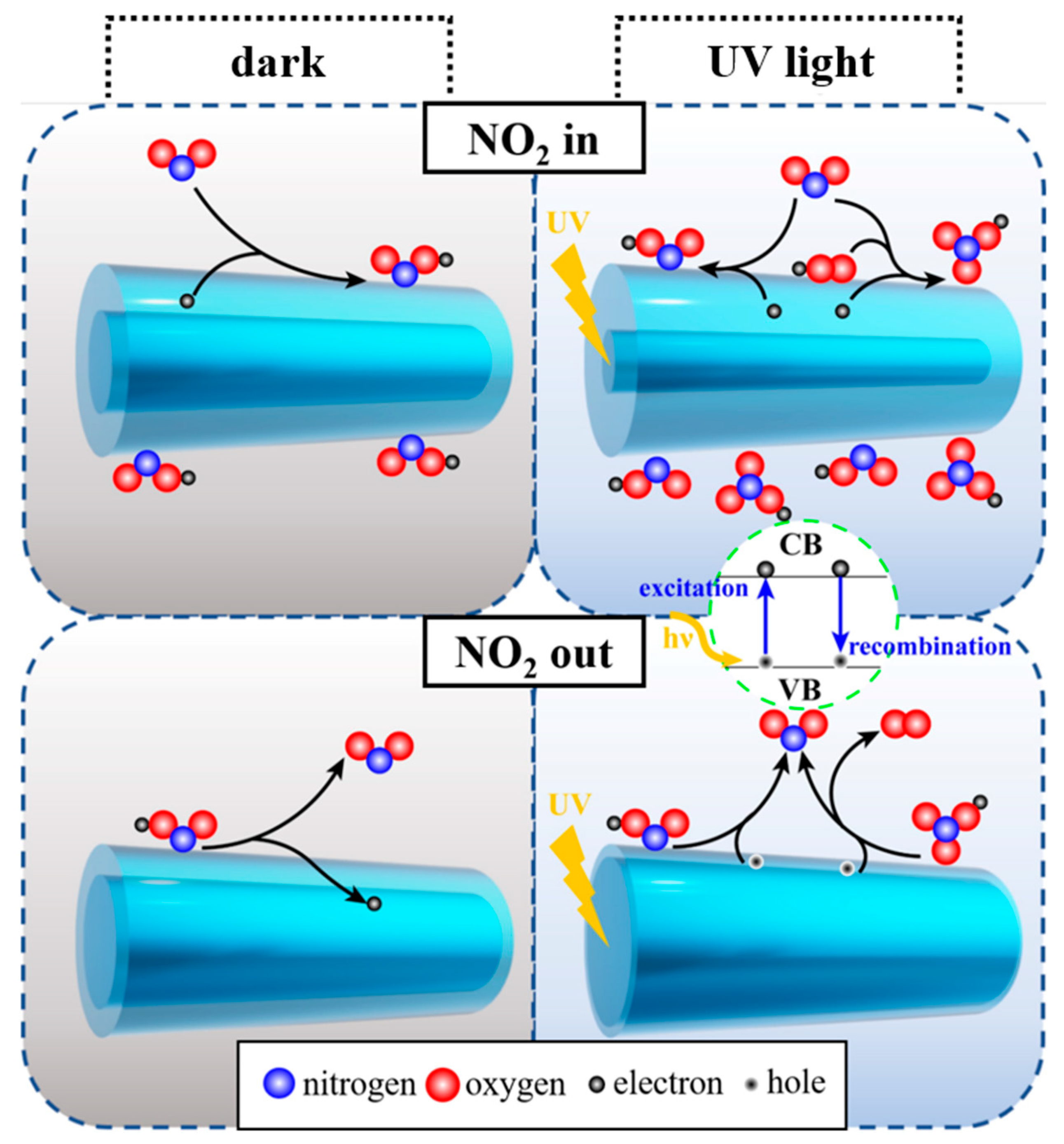
3.3. Gas-Sensing Mechanism

As an n-type semiconductor, the resistance of ZnOHF is mainly controlled by electrons in the conductive band. According to reference research, the resistance of gas-sensing material is mainly determined by the electron depletion layer on the surface, including its resistance and width [45,46]. Under relatively low temperatures, when exposing sensing material to air without photoactivation, some oxygen molecules adsorb on the surface and consume electrons to form oxygen ions ( O 2 a d s , 0–150 °C), as shown in Reaction (1), thereby forming an electron depletion layer with high resistance [47]. When UV light is applied, for one thing, the surface adsorbed oxygen ions react with the photogeneration holes to release oxygen (Reaction (2)). For another, the physically adsorbed oxygen molecules capture photogeneration electrons to form new oxygen species with high activity (Reaction (3)). When these two reactions reach a balance, the surface of the material is coated with photoactivated oxygen ions [48].
O 2 g a s + e O 2 a d s
O 2 a d s + h h ν + O 2 g a s
O 2 g a s + e h ν O 2 h ν
The NO2-sensing process is shown in Figure 8. When exposing Al-ZnOHF sensors to NO2 without UV light, the NO2 molecules are ionized into NO 2 by capturing conductive band electrons, leading to an increase in resistance (Reaction (4)) [49]. In comparison, the UV-activated electrons are more active than those in the dark, making them easier to combine with NO2 molecules (Reaction (5)). In addition, another reaction route will occur under UV light, as shown in Reaction (6). Some NO2 molecules also react with photoactivated oxygen ions and conductive band electrons to create NO 3 [50]. Therefore, more NO2 will be adsorbed and reacted on the surface of Al-ZnOHF, and the sensitivity of the sensors demonstrates enormous improvement.
N O 2 g a s + e N O 2 a d s
N O 2 g a s + e h ν N O 2 h ν
2 N O 2 g a s + O 2 h ν + e h ν 2 N O 3 h ν
When NO2 is pumped out, the surface-adsorbed ions are desorbed and release electrons to the conductive band of materials. The difference is that materials under UV light have a lot of photogenerated holes with high activity, which can efficiently promote the oxidation of NO 2 and NO 3 (Reactions (7) and (8)), as well as their desorption. Hence, UV light assistance can effectively accelerate the recovery process and reduce the recovery time [51,52].
N O 2 h ν + h h ν + N O 2 g a s
2 N O 3 h ν + 2 h h ν + 2 N O 2 g a s + O 2 g a s
In order to figure out the bandgap and UV light absorption of different samples, the UV-visible diffuse reflectance spectra of Al-ZnOHF with various Al3+ concentrations were tested (Figure 9a). Significantly, Al-ZnOHF exhibits a higher absorbance than pure ZnOHF, especially 0.5 at.% Al-ZnOHF. Using the T-plots of (αhν)2 vs. hν of samples (Figure 9b), the band gaps can be calculated as 3.31 eV for pure ZnOHF, 3.29 eV for 0.1 at.% Al- ZnOHF, 3.25 eV for 0.2 at.% Al- ZnOHF, 3.20 eV for 0.5 at.% Al-ZnOHF, and 3.40 eV for 1 at.% Al-ZnOHF. Hence, the introduction of a low content of Al3+ can significantly narrow the bandgap of ZnOHF, and the 0.5 at.% Al-ZnOHF sample has the minimum bandgap. The UV absorption and bandgap are the main influencing factors of the generation of photoactivated electrons and holes, thereby affecting the gas-sensing process [37,53]. Therefore, compared with other samples, 0.5 at.% Al-ZnOHF exhibits the highest increase in the response value rate before and after UV light radiation.

4. Conclusions

In this work, Al-ZnOHF nanorods were synthesized via a simple one-step hydrothermal method for highly efficient NO2-sensing detection. Under UV light assistance, the gas sensitivity of 0.5 at.% Al-ZnOHF was further enhanced, and the response/recovery time was also significantly shortened. The sample preserved good anti-humidity performance, especially under RH50. The improvement in the gas-sensing performance of Al-ZnOHF can be ascribed to the reduced bandgap and increased electron concentration. UV irradiation generated electrons and holes with high activity, which means that they can react with target gas effectively and efficiently. The new reaction route during the adsorption and desorption process also promotes the enhancement in g as-sensing performance. Therefore, this work prepares an effective gas sensor based on 0.5 at.% Al-ZnOHF for NO2 detection and provides new insights into modifying ZnOHF-based sensing material with superior gas-sensing performance.

Supplementary Materials

The following supporting information can be downloaded at: https://www.mdpi.com/article/10.3390/ma16093577/s1, Figure S1: The resistance of samples at different temperatures (a) without and (b) with UV radiation; Figure S2: The dynamic response curve of (a) pure ZnOHF with UV assistance, 100 °C, (b) pure ZnOHF in dark, 160 °C and (c) 0.5 at.% Al-ZnOHF in dark, 160 °C.

Author Contributions

X.Y.: Methodology, Investigation, Writing—original draft. R.W.: Data curation, Formal analysis. L.W.: Conceptualization, Project administration, Resources. H.S.: Formal analysis. J.Z.: Funding acquisition and formal analysis. F.L.: Data curation. K.F.: Writing—review and editing. Z.W.: Writing—review and editing. F.W.: Writing—review and editing. J.L.: Visualization and editing. All authors have read and agreed to the published version of the manuscript.

Funding

This work was supported by the Natural Foundation of Shandong Province (ZR2022MF311); the National Natural Science Foundation of China (No. 51702188); the Natural Science and Development Foundation of Shenzhen (JCYJ20190807093205660); and the Project Innovation Team of Jinan (2021GXRC036).

Institutional Review Board Statement

Not applicable.

Informed Consent Statement

Not applicable.

Data Availability Statement

The data presented in this study are available on request from the corresponding author. The data are not publicly available due to privacy restrictions.

Conflicts of Interest

The authors declare no conflict of interest.

References

  1. Hu, Y.; Li, T.; Zhang, J.; Guo, J.; Wang, W.; Zhang, D. High-sensitive NO2 sensor based on p-NiCo2O4/n-WO3 heterojunctions. Sens. Actuators B Chem. 2022, 352, 130912. [Google Scholar] [CrossRef]
  2. Choi, M.S.; Kim, M.Y.; Mirzaei, A.; Kim, H.; Kim, S.; Baek, S.; Chun, D.W.; Jin, C.; Lee, K.H. Selective, sensitive, and stable NO2 gas sensor based on porous ZnO nanosheets. Appl. Surf. Sci. 2021, 568, 150910. [Google Scholar] [CrossRef]
  3. Yang, Z.; Zhang, D.; Chen, H. MOF-derived indium oxide hollow microtubes/MoS2 nanoparticles for NO2 gas sensing. Sens. Actuators B Chem. 2019, 300, 127037. [Google Scholar] [CrossRef]
  4. Li, W.; Zhang, Y.; Long, X.; Cao, J.; Xin, X.; Guan, X.; Peng, J.; Zheng, X. Gas sensors based on mechanically exfoliated MoS2 nanosheets for room-temperature NO2 detection. Sensors 2019, 19, 2123. [Google Scholar] [CrossRef] [PubMed]
  5. Du, W.; Wu, N.; Wang, Z.; Liu, J.; Xu, D.; Liu, W. High response and selectivity of platinum modified tin oxide porous spheres for nitrogen dioxide gas sensing at low temperature. Sens. Actuators B Chem. 2018, 257, 427–435. [Google Scholar] [CrossRef]
  6. Nam, B.; Ko, T.; Hyun, S.; Lee, C. NO2 sensing properties of WO3-decorated In2O3 nanorods and In2O3-decorated WO3 nanorods. Nano Converg. 2019, 6, 40. [Google Scholar] [CrossRef]
  7. Mokrushin, A.; Averin, A.; Gorobtsov, P.; Simonenko, N.; Simonenko, E.; Kuznetsov, N. Obtaining of ZnO/Fe2O3 thin nanostructured films by AACVD for detection of ppb-concentrations of NO2 as a biomarker of lung infections. Biosensors 2023, 13, 445. [Google Scholar] [CrossRef]
  8. Wang, M.; Jin, Z.; Liu, M.; Jiang, G.; Lu, H.; Zhang, Q.; Ju, J.; Tang, Y. Nanoplate-assembled hierarchical cake-like ZnO microstructures: Solvothermal synthesis, characterization and photocatalytic properties. RSC Adv. 2017, 7, 32528–32535. [Google Scholar] [CrossRef]
  9. Wang, M.; Sun, T.; Tang, Y.; Jiang, G.; Shi, Y. Template-free synthesis and photocatalytic properties of flower-like ZnOHF nanostructures. Mater. Lett. 2015, 160, 150–153. [Google Scholar] [CrossRef]
  10. Mirzaei, A.; Haghighat, F.; Chen, Z.; Yerushalmi, L. Sonocatalytic removal of ampicillin by Zn(OH)F: Effect of operating parameters, toxicological evaluation and by-products identification. J. Hazard. Mater. 2019, 375, 86–95. [Google Scholar] [CrossRef]
  11. Chen, H.; Zhu, L.; Hou, Q.; Liang, W.; Liu, H.; Li, W. ZnOHF nanostructure-based quantum dots-sensitized solar cells. J. Mater. Chem. 2012, 22, 23344. [Google Scholar] [CrossRef]
  12. Tian, H.; Wang, Y.; Zhang, J.; Ma, Y.; Cui, H.; Cui, Q.; Ma, Y. Compression behavior of copper hydroxyfluoride CuOHF as a case study of the high-pressure responses of the hydrogen-bonded two-dimensional layered materials. J. Phys. Chem. C 2019, 123, 25492–25500. [Google Scholar] [CrossRef]
  13. Zhang, G.; Wang, B.; Li, L.; Yang, S. Phosphorus and yttrium co-doped Co(OH)F nanoarray as highly efficient and bifunctional electrocatalysts for overall water splitting. Small 2019, 15, 1904105. [Google Scholar] [CrossRef] [PubMed]
  14. Yao, X.; Zhao, J.; Jin, Z.; Jiang, Z.; Xu, D.; Wang, F.; Zhang, X.; Song, H.; Pan, D.; Chen, Y.; et al. Flower-like hydroxyfluoride-sensing platform toward NO2 detection. ACS Appl. Mater. Interfaces 2021, 13, 26278–26287. [Google Scholar] [CrossRef] [PubMed]
  15. Mohan, V.; Marappan, G.; Chidambaram, D.; Rajendran, K.; Surya, V.J.; Venugopal, G.; Sivalingam, Y. Photo-enhanced acetone adsorption on delta-MnO2 nanoparticles: A step towards non-invasive detection of diabetes mellitus. Mater. Lett. 2022, 306, 130944. [Google Scholar] [CrossRef]
  16. Chang, J.; Deng, Z.; Li, M.; Wang, S.; Mi, L.; Sun, Q.; Horprathum, M.; He, Y.; Kong, F.; Fang, X.; et al. Visible light boosting hydrophobic ZnO/(Sr0.6Bi0.305)2Bi2O7 chemiresistor toward ambient trimethylamine. Sens. Actuators B Chem. 2022, 352, 131076. [Google Scholar] [CrossRef]
  17. Wang, M.; Zhu, Y.; Luo, Q.; Ge, C.; Liu, G.; Qiao, G.; Kim, E.J. Below-room-temperature solution-grown ZnO porous nanosheet arrays with ppb-level NO2 sensitivity under intermittent UV irradiation. Appl. Surf. Sci. 2021, 566, 150750. [Google Scholar] [CrossRef]
  18. Zhang, D.; Yu, S.; Wang, X.; Huang, J.; Pan, W.; Zhang, J.; Meteku, B.E.; Zeng, J. UV illumination-enhanced ultrasensitive ammonia gas sensor based on (001) TiO2/MXene heterostructure for food spoilage detection. J. Hazard. Mater. 2022, 423, 127160. [Google Scholar] [CrossRef]
  19. Anbalagan, A.K.; Gupta, S.; Kumar, R.R.; Tripathy, A.R.; Chaudhary, M.; Haw, S.; Murugesan, T.; Lin, H.; Chueh, Y.; Tai, N.; et al. Gamma-ray engineered surface defects on zinc oxide nanorods towards enhanced NO2 gas sensing performance at room temperature. Sens. Actuators B Chem. 2022, 369, 132255. [Google Scholar] [CrossRef]
  20. Bang, J.H.; Kwon, Y.J.; Lee, J.; Mirzaei, A.; Lee, H.Y.; Choi, H.; Kim, S.S.; Jeong, Y.K.; Kim, H.W. Proton-beam engineered surface-point defects for highly sensitive and reliable NO2 sensing under humid environments. J. Hazard. Mater. 2021, 416, 125841. [Google Scholar] [CrossRef]
  21. Wang, G.; He, Z.; Shi, G.; Wang, H.; Zhang, Q.; Li, Y. Controllable construction of Titanium dioxide-Zirconium dioxide@Zinc hydroxyfluoride networks in micro-capillaries for bio-analysis. J. Colloid Interface Sci. 2015, 446, 290–297. [Google Scholar] [CrossRef] [PubMed]
  22. Peng, Y.; Zhou, H.; Wang, Z. Synthesis, characterization and photocatalytic activity of Zn(OH)F hierarchical nanofibers prepared by a simple solution-based method. CrystEngComm 2012, 14, 2812–2816. [Google Scholar] [CrossRef]
  23. Eadi, S.B.; Shin, H.; Kumar, P.S.; Song, K.; Yuvakkumar, R.; Lee, H. Fluorine-implanted indium-gallium-zinc oxide (IGZO) chemiresistor sensor for high-response NO2 detection. Chemosphere 2021, 284, 131287. [Google Scholar] [CrossRef] [PubMed]
  24. Li, X.; Wang, Y.; Cheng, P.; Liu, Y.; Lei, Z.; Liu, M.; Yao, X.; Yan, H.; Weng, Z. Multicore-shell structured Ce-In2O3 for acetone detection by spray pyrolysis using NaCl as a high temperature auxiliary agent. Mater. Chem. Front. 2021, 6, 213–224. [Google Scholar] [CrossRef]
  25. Mokoena, T.P.; Swart, H.C.; Hillie, K.T.; Motaung, D.E. Engineering of rare-earth Eu3+ ions doping on p-type NiO for selective detection of toluene gas sensing and luminescence properties. Sens. Actuators B Chem. 2021, 347, 130530. [Google Scholar] [CrossRef]
  26. Qin, C.; Wang, B.; Wang, Y. Metal-organic frameworks-derived Mn-doped Co3O4 porous nanosheets and enhanced CO sensing performance. Sens. Actuators B Chem. 2022, 351, 130943. [Google Scholar] [CrossRef]
  27. Mokrushin, A.S.; Nagornov, I.A.; Simonenko, T.L.; Simonenko, N.P.; Gorobtsov, P.Y.; Arkhipushkin, I.A.; Simonenko, E.P.; Sevastyanov, V.G.; Kuznetsov, N.T. Gas-sensitive nanostructured ZnO films praseodymium and europium doped: Electrical conductivity, selectivity, influence of UV irradiation and humidity. Appl. Surf. Sci. 2022, 589, 152974. [Google Scholar] [CrossRef]
  28. Sun, J.; Wang, Y.; Song, P.; Yang, Z.; Wang, Q. Metal-organic framework-derived Cr-doped hollow In2O3 nanoboxes with excellent gas-sensing performance toward ammonia. J. Alloys Compd. 2021, 879, 160472. [Google Scholar] [CrossRef]
  29. Nagarjuna, Y.; Lin, J.; Wang, S.; Hsiao, W.; Hsiao, Y. AZO-based ZnO nanosheet MEMS sensor with different Al concentrations for enhanced H2S gas sensing. Nanomaterials 2021, 11, 3377. [Google Scholar] [CrossRef]
  30. Ramgir, N.S.; Goyal, C.P.; Goyal, D.; Patil, S.J.; Ikeda, H.; Ponnusamy, S.; Muthe, K.P.; Debnath, A.K. NO2 sensor based on Al modified ZnO nanowires. Mater. Sci. Semicond. Process. 2021, 134, 106027. [Google Scholar] [CrossRef]
  31. Jaballah, S.; Alaskar, Y.; AlShunaifi, I.; Ghiloufi, I.; Neri, G.; Bouzidi, C.; Dahman, H.; El Mir, L. Effect of Al and Mg doping on reducing gases detection of ZnO nanoparticles. Chemosensors 2021, 9, 300. [Google Scholar] [CrossRef]
  32. Bulut, F.; Ozturk, O.; Acar, S.; Yildirim, G. Effect of Ni and Al doping on structural, optical, and CO2 gas sensing properties of 1D ZnO nanorods produced by hydrothermal method. Microsc. Res. Tech. 2021, 85, 1502–1517. [Google Scholar] [CrossRef] [PubMed]
  33. Liu, F.; Huang, G.; Wang, X.; Xie, X.; Xu, G.; Lu, G.; He, X.; Tian, J.; Cui, H. High response and selectivity of single crystalline ZnO nanorods modified by In2O3 nanoparticles for n-butanol gas sensing. Sens. Actuators B Chem. 2018, 277, 144–151. [Google Scholar] [CrossRef]
  34. Mu, Y.; Zhou, T.; Li, D.; Liu, W.; Jiang, P.; Chen, L.; Zhou, H.; Ge, G. Highly stable and durable Zn-metal anode coated by bi-functional protective layer suppressing uncontrollable dendrites growth and corrosion. Chem. Eng. J. 2022, 430, 132839. [Google Scholar] [CrossRef]
  35. Tian, X.; Yao, L.; Cui, X.; Zhao, R.; Xiao, X.; Wang, Y. Novel Al-doped CdIn2O4 nanofibers based gas sensor for enhanced low-concentration n-butanol sensing. Sens. Actuators B Chem. 2022, 351, 130946. [Google Scholar] [CrossRef]
  36. Zhu, B.; Liu, Y.; Zhao, H.; Zhang, X.; He, P.; Wu, L.; Liu, Y.; Yang, T. ZnOHF/N-doped carbon hybrids as a novel anode material for enhanced lithium storage. J. Alloys Compd. 2021, 889, 161705. [Google Scholar] [CrossRef]
  37. Du, W.; Si, W.; Du, W.; Ouyang, T.; Wang, F.; Gao, M.; Wu, L.; Liu, J.; Qian, Z.; Liu, W. Unraveling the promoted nitrogen dioxide detection performance of N-doped SnO2 microspheres at low temperature. J. Alloys Compd. 2020, 834, 155209. [Google Scholar] [CrossRef]
  38. Wang, Y.; Liu, B.; Xiao, S.; Wang, X.; Sun, L.; Li, H.; Xie, W.; Li, Q.; Zhang, Q.; Wang, T. Low-temperature H2S detection with hierarchical Cr-doped WO3 microspheres. ACS Appl. Mater. Inter. 2016, 8, 9674–9683. [Google Scholar] [CrossRef]
  39. Zhang, Z.; Haq, M.; Wen, Z.; Ye, Z.; Zhu, L. Ultrasensitive ppb-level NO2 gas sensor based on WO3 hollow nanospheres doped with Fe. Appl. Surf. Sci. 2018, 434, 891–897. [Google Scholar] [CrossRef]
  40. Jiang, T.; Liu, X.; Sun, J. UV-enhanced NO2 sensor using ZnO quantum dots sensitized SnO2 porous nanowires. Nanotechnology 2022, 33, 185501. [Google Scholar] [CrossRef]
  41. Zhang, Q.; Pang, Z.; Hu, W.; Li, J.; Liu, Y.; Liu, Y.; Yu, F.; Zhang, C.; Xu, M. Performance degradation mechanism of the light-activated room temperature NO2 gas sensor based on Ag-ZnO nanoparticles. Appl. Surf. Sci. 2021, 541, 148418. [Google Scholar] [CrossRef]
  42. Paolucci, V.; De Santis, J.; Lozzi, L.; Rigon, M.; Martucci, A.; Cantalini, C. ZnO thin films containing aliovalent ions for NO2 gas sensor activated by visible light. Ceram. Int. 2021, 47, 25017–25028. [Google Scholar] [CrossRef]
  43. Sun, K.; Zhan, G.; Chen, H.; Lin, S. Low-operating-temperature NO2 sensor based on a CeO2/ZnO heterojunction. Sensors 2021, 21, 8269. [Google Scholar] [CrossRef] [PubMed]
  44. Liang, Y.; Hsu, Y. Enhanced sensing ability of brush-like Fe2O3-ZnO nanostructures towards NO2 gas via manipulating material synergistic effect. Int. J. Mol. Sci. 2021, 22, 6884. [Google Scholar] [CrossRef]
  45. Chen, L.; Song, Y.; Liu, W.; Dong, H.; Wang, D.; Liu, J.; Liu, Q.; Chen, X. MOF-based nanoscale Pt catalyst decorated SnO2 porous nanofibers for acetone gas detection. J. Alloys Compd. 2022, 893, 162322. [Google Scholar] [CrossRef]
  46. Liang, J.; Wu, W.; Lou, Q.; Wang, K.; Xuan, C. Room temperature NO2 sensing performance of Ag nanoparticles modified VO2 nanorods. J. Alloys Compd. 2022, 890, 161837. [Google Scholar] [CrossRef]
  47. Navarrete, E.; Guell, F.; Martinez-Alanis, P.R.; Llobet, E. Chemical vapour deposited ZnO nanowires for detecting ethanol and NO2. J. Alloys Compd. 2022, 890, 161923. [Google Scholar] [CrossRef]
  48. Xu, F.; HO, H. Light-activated metal oxide gas sensors: A review. Micromachines 2017, 8, 333. [Google Scholar] [CrossRef]
  49. Benamara, M.; Massoudi, J.; Dahman, H.; Ly, A.; Dhahri, E.; Debliquy, M.; El Mir, L.; Lahem, D. Study of room temperature NO2 sensing performances of ZnO1−x (x = 0, 0.05, 0.10). Appl. Phys. A 2022, 128, 31. [Google Scholar] [CrossRef]
  50. Šetka, M.; Claros, M.; Chmela, O.; Vallejos, S. Photoactivated materials and sensors for NO2 monitoring. J. Mater. Chem. C 2021, 9, 16804–16827. [Google Scholar] [CrossRef]
  51. Chen, X.; Hu, J.; Chen, P.; Yin, M.; Meng, F.; Zhang, Y. UV-light-assisted NO2 gas sensor based on WS2/PbS heterostructures with full recoverability and reliable anti-humidity ability. Sens. Actuators B Chem. 2021, 339, 129902. [Google Scholar] [CrossRef]
  52. Hung, N.M.; Hung, C.M.; Van Duy, N.; Hoa, N.D.; Hong, H.S.; Dang, T.K.; Viet, N.N.; Thing, L.V.; Phuoc, P.H.; Van Hieu, N. Significantly enhanced NO2 gas-sensing performance of nanojunction-networked SnO2 nanowires by pulsed UV-radiation. Sens. Actuators A Phys. 2021, 327, 112759. [Google Scholar] [CrossRef]
  53. Kamble, V.S.; Navale, Y.H.; Patil, V.B.; Desai, N.K.; Vajekar, S.N.; Salunkhe, S.T. Studies on structural, spectral and morphological properties of co-precipitation derived Co-doped ZnO nanocapsules for NO2 sensing applications. J. Mater. Sci. Mater. Electron. 2021, 32, 26503–26519. [Google Scholar] [CrossRef]
Figure 1. (a) The XRD spectra and (b) magnified region of the strongest peak of all Al-ZnOHF samples.
Materials 16 03577 g001
Figure 2. SEM images of (a,b) pure ZnOHF, (c) 0.1 at.% Al-ZnOHF, (d) 0.2 at.% Al-ZnOHF, (e) 0.5 at.% Al-ZnOHF, (f) 1 at.% Al-ZnOHF.
Materials 16 03577 g002
Figure 3. TEM and HRTEM images of (ac) pure ZnOHF, (df) 0.5 at.% Al-ZnOHF.
Materials 16 03577 g003
Figure 4. (a) XPS wide spectrum, (b) Zn 2p region, (c) F 1s region, (d) Al 2p region, and (e) O 1s region XPS spectra of pure ZnOHF and 0.5 at.% Al-ZnOHF. (the dots are test data and the red and blue lines are fixed curves).
Materials 16 03577 g004
Figure 5. (a) Gas responses of all obtained samples to 10 ppm NO2 at different temperatures in the dark. (b) The response–recovery curves of pure ZnOHF and 0.5 at.% Al-ZnOHF to 10 ppm NO2 at 160 °C in dark. (c) Gas responses of all obtained samples to 10 ppm NO2 at different temperatures under UV assistance. (d) The response–recovery curves of pure ZnOHF and 0.5 at.% Al-ZnOHF to 10 ppm NO2 at 100 °C under UV assistance.
Materials 16 03577 g005
Figure 6. Under UV assistance and 100 °C: (a) the dynamic response curve of 0.5 at.% Al-ZnOHF; (b) the linear relationship between the logarithm of response value and the logarithm of gas concentration of pure ZnOHF (blue) and Al-ZnOHF (red); (c) response–recovery curve in 4 cycles of 0.5 at.% ZnOHF; (d) the response change of 0.5 at.% ZnOHF at 1 month, and (e) response value of pure ZnOHF and 0.5 at.% Al-ZnOHF to 10 ppm NO2, SO2, NH3, CO, CH4 and CO2.
Materials 16 03577 g006
Figure 7. (a) The response–recovery curves, (b) the response value change, (c) response/recovery time of 0.5 at.% Al-ZnOHF to 10 ppm NO2 at various relative humidities at 100 °C under UV radiation.
Materials 16 03577 g007
Figure 8. The response and recovery process of Al-ZnOHF in dark and with UV assistance.
Materials 16 03577 g008
Figure 9. (a) The UV-vis diffuse reflectance spectra and (b) T-plots of (αhν)2 vs. hν of all as-prepared samples (the dashed line is the tangent line of curves).
Materials 16 03577 g009
Table 1. The NO2-sensing performance of materials in references and this work.
MaterialsOperating ConditionS (Rg/Ra or Ra/Rg)Tres/Trec (s)Detection LimitRef.
ZnO/SnO2 composite40 °C, UV (395 nm, 34 μW/cm2)25 (1 ppm)251/470100 ppb[40]
Ag-ZnO nanoparticles25 °C, visible light (455 nm, 70 mW/cm2)2.5 (5 ppm)200/1751 ppm[41]
Si-ZnO thin films75 °C, purple-blue (430 nm)19.1 (400 ppb)60/180-[42]
CeO2/ZnO nanorods120 °C190.6% (5 ppm)104/417100 ppb[43]
Fe2O3-ZnO nanostructures300 °C6.34 (10 ppm)26/1851 ppm[44]
3D flower-like ZnOHF 200 °C82.71 (10 ppm)13/350.1 ppm[14]
0.5 at.%Al-ZnOHF100 °C, UV (395 nm, 6 mW/cm2)110.83 (10 ppm)35/960.25 ppmThis work
Disclaimer/Publisher’s Note: The statements, opinions and data contained in all publications are solely those of the individual author(s) and contributor(s) and not of MDPI and/or the editor(s). MDPI and/or the editor(s) disclaim responsibility for any injury to people or property resulting from any ideas, methods, instructions or products referred to in the content.

