Article

Antioxidant and Antimicrobial Properties of Hydrolysed Collagen Nanofibers Loaded with Ginger Essential Oil

by
Mariana Daniela Berechet
1,
Carmen Gaidau
1,
Aleksandra Nešić
2,
Rodica Roxana Constantinescu
1,
Demetra Simion
1,
Olga Niculescu
1,
Maria Daniela Stelescu
1,*,
Irina Sandulache
1 and
Maria Râpă
3,*
1
The National Research & Development Institute for Textiles and Leather, 16 Lucretiu Patrascanu Street, 030508 Bucharest, Romania
2
Faculty of Technology, University of Novi Sad, 21102 Novi Sad, Serbia
3
Faculty of Materials Science and Engineering, Polytechnic University of Bucharest, 060042 Bucharest, Romania
*
Authors to whom correspondence should be addressed.
Materials 2023, 16(4), 1438; https://doi.org/10.3390/ma16041438
Submission received: 27 January 2023 / Revised: 5 February 2023 / Accepted: 6 February 2023 / Published: 8 February 2023

Abstract

:
Hydrolysed collagen obtained from bovine leather by-products were loaded with ginger essential oil and processed by the electrospinning technique for obtaining bioactive nanofibers. Particle size measurements of hydrolysed collagen, GC-MS analysis of ginger essential oil (EO), and structural and SEM examinations of collagen nanofibers loaded with ginger essential oil collected on waxed paper, cotton, and leather supports were performed. Antioxidant and antibacterial activities against Staphylococcus aureus and Escherichia coli and antifungal activity against Candida albicans were also determined. Data show that the hydrolysed collagen nanofibers loaded with ginger EO can be used in the medical, pharmaceutical, cosmetic, or niche fields.

1. Introduction

The approaching of adopting naturally derived polymers in the form of a combination with plant extracts such as essential oils is preferred when it is desired to find the biological properties of the constituent compounds in medicinal products or cosmetics or they are used in the substitution or regrowth of human tissue [1]. The excellent biocompatibility and biodegradability of natural polymers versus that of synthetic polymers is an important topic to consider in the domain of medical, cosmetic, or biomedical engineering products [2].
The mixing together of natural polymers, for example, collagen, with essential oils to take advantage of both materials can lead to the most effective antifungal, antimicrobial, and regeneration results [3,4]. Collagen is a major element in the extracellular matrix (ECM) and, therefore, the largest plentiful protein in human beings [5]. Its important biological features when compared with different polymers lead to a best selection for medical and cosmetic applications [6]. In various research studies, the collagen composite fibres have been used in combination with polycaprolactone (PCL) and the addition of highly toxic solvents, such as hexafluoroisopropanol [4,7,8]. The technological process during the manufacturing and extraction of collagen might have an important effect on solubility, viscosity, and subsequent processability by electrospinning [9,10,11]. Electrospinning is a nano-technique that uses an electrostatic spinning machine as a tool to make nanofibers simply and easily. Thanks to the convenience of nanofibers, such as light fabrication, easy operation, high surface-area-to-volume ratio, and surface bionics of the extracellular matrix for suitability in biomedical engineering, the electrospun nanofiber scaffolds have been extensively investigated in tissue engineering [12,13,14,15,16,17,18], drug delivery [5,19,20], energy science [21,22,23], sensors [24,25,26], catalysts [27,28], filtration [29,30], food science [31,32,33], and environmental science [34]. Electrospinning technology has been tested for the manufacturing of collagen nanofibers extracted in acidic medium from calfskin [35], or collagen from bovine dermis [36], collagen extracted in alkaline medium from fish skin [37], and the extraction in acidic medium from collagen in fish skin in the presence of PCL [38]. Due to the high efficiency of capturing bioactive substances and ideal protection for bioactivity [39], electrospinning nanofibers can be examined for controlled release of bioactive and food compounds [40,41]. Electrospinning could play important roles in maximizing the use of collagen resources extracted from fish skin, cowhide, or rabbit skin for tissue engineering and drug and cosmetic administration [18,21,37]. The use of the electrospinning technique allows the production of nanofiber materials with high ratios between surface and volume as well as the loading of bioactive compounds with applications in medical products, cosmetics, and medicines [12,14,15,16,17,18]. Various polymer types have been used by electrospinning for the fabrication of active materials containing encapsulated active agents. This emerging technology was also employed to deposit matt fibres on cotton to improve its performance [13,16]. They could even be designed to obtain products with regenerative or antioxidant, antimicrobial, or antifungal protective properties used in medicines and cosmetics [42].
The objective of this research is to obtain nanofibers from hydrolysed collagen loaded with ginger essential oil. Through the electrospinning technique, nanofibers with 3D structure and antibacterial and antifungal properties were obtained and collected on waxed paper, cotton cloth, and leather supports. The use of ginger essential oil loaded in hydrolysed collagen can be a natural alternative to the pathogen resistance of antimicrobial agents [43].

2. Materials and Methods

2.1. Chemical Materials

In this study, the following chemical materials were used: ginger (Zingiber officinale) essential oil from Adams Vision SRL (Targu Mures, Romania), sodium carbonate (Na2CO3) from SG GXG Chemicals SRL (Budesti-Racovita, Valcea, Romania), Alcalase 2.4 L (protease from Bacillus licheniformis, 2.4 U/g) from Sigma-Aldrich (St. Louis, MI, USA), hydrated calcium oxide (CaO) purchased from Cristal R Chim SRL (Bucharest, Romania), and pearl sodium hydroxide (NaOH) acquired from Lachner (Neratovice, Czech Republic).

2.2. Obtaining of Hydrolysed Collagen Concentrates

To obtain the hydrolysed collagen, the bovine leather was alkaline hydrolysed in 400% (w/v) water in the presence of 10% CaO (w/w) for 4 h at a temperature of 80 °C and under traditional mechanical stirring. Hydrolysis was continued enzymatically with 0.4% Alcalase (w/w) with pH adjustment at 8–9, with a solution of 20% concentration NaOH (v/v) for 3 h under mechanical stirring at a temperature of 65 °C. The enzyme deactivation occurred at a temperature of 80 °C for 15 min. The collagen hydrolysate thus obtained was decanted and filtered and further concentrated at a ratio of 1:4 in a rotary evaporator at a temperature of 65 °C, 150 rpm, and pressure of 150 mbar. Finally, a concentrated hydrolysed collagen of honey consistency and amber colour was obtained [44].

2.3. Physical–Chemical Characterization of Hydrolysed Collagen

The concentrated hydrolysed collagen was analysed by physical–chemical methods according to the standards in force and methods such as ash content (EN ISO 4047:2008), dry matter (EN ISO 4684:2006), total nitrogen and protein (ISO 5397:1996), pH (STAS 8619/3:1990), amino nitrogen (according to ICPI method), and electrical conductivity (EN ISO 27883:1997). The viscosity of concentrated hydrolysed collagen hydrolysate was determined with a Brookfield AMETEK DV2T TC-550 Viscometer (Brookfield, Toronto, ON, Canada) at a temperature of 25 °C. The particle size and zeta potential of collagen were evaluated by using a Dynamic Light Scattering (DLS) instrument from Malvern (Zetasizer Nano-ZS, Malvern Hills, UK).

2.4. Characterization of Ginger Essential Oil (EO)

2.4.1. GC-MS Analysis

Bioactive compounds from ginger EO were identified using Gas Chromatography coupled with Mass Spectroscopy (DSQ II MS, Thermo Scientific, Waltham, MA, USA), supplied with TR-5 MS non-polar capillary column having dimensions of 60 m × 0.25 μm × 0.25 μm. The technical parameters temperature, heating rate, temperature of split/splitless injector, and pneumatic control system of pressure/carrying gas flow were fixed in the range between room temperature to 350 °C, 0.1 °C/min to 120 °C/min, 50 °C to 375 °C, and 5%, respectively.

2.4.2. Determination of Total Phenolic Content (TPC) and DPPH Free-Radical Scavenging Assay

Determination of total phenolic content (TPC) of ginger EO was carried out based on Folin-Ciocalteu’s method [44]. A concentration of 60 mg mL−1 ginger EO was achieved in ethanol at room temperature, and 50 µL of the ginger EO sample prepared as above was mixed with 0.240 mL Folin-Ciocalteu’s reagent and 3.6 mL distilled water. The obtained solution was maintained for 5 min in a dark place at ambient temperature, and then 0.68 mL 7.5% (w/v) solution of sodium carbonate (Na2CO3·10H2O) was added. This solution was moderately stirred and kept for 30 min in darkness at a temperature of 40 °C. The solution absorbance was read at a wavelength of 740 nm against blank, and the TPC calculated as mg of gallic acid equivalent per g of dry weight (GAE/g dw) was carried out using a calibration curve based on gallic acid.
Antioxidant activity of ginger EO was performed by mixing together 2.5 mL of DPPH (1,1-diphenyl-2-picrylhydrazyl) in an ethanol solution (0.150 mmol/L) with 0.5 mL of 60 mg mL−1 ginger EO. After 30 min standing in a dark place at ambient temperature, the absorbance was read at 517 nm by using a UV/VIS spectrometer (Orion UV-VIS AQUAMATE 8000, Thermo Fisher Scientific, Eindhoven, The Netherlands). Every analysis was conducted in triplicate.

2.5. Obtaining of Nanofibers Based on Hydrolysed Collagen and Ginger EO

The concentrated hydrolysed collagen together with 10% ginger EO was mechanically stirred at 400 rpm for 50 min. Nanofibers based on the above homogenized solution were fabricated by electrospinning process (Fluidnatek equipment, Bioinicia, Valencia, Spain). Nanofibers of hydrolysed collagen were also obtained, as a control. The applied electric potential, the flow rate, and the distance between the injector needle and the storage support fixed on the metal collector were adjusted depending on the support used. The electrospinning processing parameters were as follows: an electrical potential of 9.8 kV applied for waxed paper, 18.5 kV for cotton, and 20.6 kV for leather between the positive injector needle and negative support; a flow rate of 550 µL for waxed paper, 700 µL for cotton, and 1000 µL for leather; and a distance of 13 cm between injector needle to each support type. The experiments were carried out at room temperature and a relative humidity of 32 ± 5%.

2.6. Ginger EO Release

Ginger EO loading efficiency was determined by ultraviolet-visible (UV-VIS) spectroscopy. About 20 mg of electrospun material was solubilized into ethanol (96%). A volume of supernatant liquid was achieved by centrifugation at 6000 rpm for 10 min, then filtration (0.45 μm polytetrafluoroethylene (PTFE) filter, Whatman, Maidstone, UK) and, in the end, a wavelength of 290 nm was examined. The theoretical loading capacity of ginger EO was determined taking into account the essential oil introduced in the electrospinning solution, in other words, 10 wt.% directly proportional to the volume of hydrolysed collagen solution. Calibration curves containing ginger EO concentrations ranging from 0 mg mL−1 to 20 mg mL−1 were used by UV-VIS testing of known concentrations of ginger EO in ethanol.

2.7. Attenuated Total Reflectance (ATR)—Fourier Transform Infrared (FT-IR) Spectroscopy

ATR-FT-IR technique was performed with an FT-IR/ATR spectrometer—Jasco 4200 (Jasco Corporation, Tokyo, Japan)—operating in the wavenumber ranging from 4000 cm−1 to 650 cm−1, which detected the absorbance peaks at 0.5 cm−1 spectral resolution. The device was equipped with a Michelson interferometer with incident angle of 45°, with Digital Signal Processing technology (DSP), cubic angular mirrors with self-aligning mechanisms, and a standard thermostatic DLATGS Peltier detector (Jasco Corporation, Tokyo, Japan).

2.8. Scanning Electron Microscopy (SEM)

The electronic microscopic examinations were created using an FEI Quanta 200 microscope (FEI, Valley City, ND, USA), which operated at high vacuum, 15 kV, in addition to a gaseous secondary electron (GSED) detector.

2.9. Microbiological Analyses

An adapted method for determining antibacterial and antifungal activity was used according to the standard ISO 20743:2013—Determination of antibacterial activity of textile products. This document mentions quantitative test methods for antibacterial activity assessment of all antibacterial textile items, which include nonwovens. Both nanofibers based on collagen (control) and containing ginger EO were analysed towards Escherichia coli (Gram negative bacteria) ATCC 11229, Staphylococcus aureus ATCC 6538 (Gram positive bacteria), and Candida albicans ATCC 10231 yeast. The cell concentration in the inoculum used was of 2.4 × 10⁴ CFU/mL (Colony Forming Units) for Escherichia coli, 3.6 × 10⁴ CFU/mL for Staphylococcus aureus, and 2.8 × 10⁴ CFU/mL for Candida albicans. The initial cell concentration was calculated earlier by decimal dilutions (10−4) using sterile deionized water; then, from the last dilution, a volume of 100 µL was taken for each strain and spread further on Nutrient Agar. The plate counting was recorded at 24 h of incubation, this being kept as a reference for the cellular developments of the control. Antibacterial activity ratio (R) was calculated according to Equation (1).
R % = C t T t C t × 100
where: Ct is the average number of colonies of two control samples after 24 h or the specified incubation period, expressed as CFU/mL; Tt is the average number of colonies of two test samples after 24 h or the specified incubation period, expressed as CFU/mL. The results were also indicated as the logarithmic reduction resistance against bacteria and fungi. The antimicrobial efficacy and degree of microbial and logarithmic reduction for each sample were estimated depending on the initial cell concentration.

3. Results

3.1. Physical–Chemical Parameters of Hydrolysed Collagen
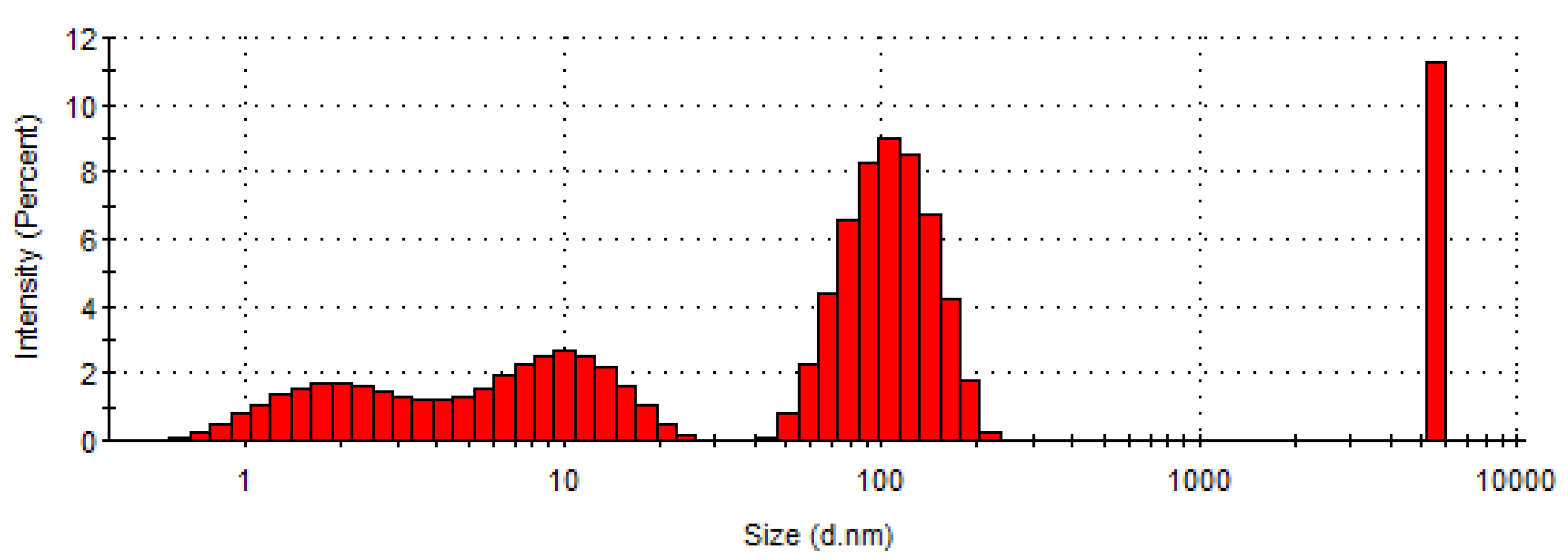
The concentrated hydrolysed collagen was characterized by determining dry matter, ash, total nitrogen, protein content, pH, aminic nitrogen, and electrical conductivity properties. Table 1 shows the obtained values.
The hydrolysed collagen extracted enzymatically shows high contents of protein (82.43%) and dry matter (60.40%) and a viscosity value (1623 cP) suitable for electrospinning.

3.2. DLS Analysis

The size of collagen particles measured possess an average of 1150 nm with majority populations at 109.3 nm (52.3%), 9.7 nm (22.3%), and 1.9 nm (14.3%) (Figure 1) and a zeta potential value of −7.64 mV (Figure 2), as presented in Table 2.
The particle size measurement of hydrolysed collagen showed small particles, with 36.6% of particle sizes below 10 nm and 52.3% of particle sizes at 109.3 nm, meaning an easy penetration of tissues. The high polydispersity of hydrolysed collagen with high particle size and low particle size components is due to the associative properties of proteins in high concentration (60.4%).
Other authors reported a polydisperse solution of hydrolysed collagen, with diverse particle size from 65 nm to 246 nm [45], or from a low value up to around 5000 nm [46]. The largest particle (~5500 nm), which is monodispersed and different from the other three smaller groups with broader distribution, could be due to the insufficient concentration of alkali used during the synthesis process of collagen [46].
The zeta potential value is low corresponding to a non-stable dispersion.

3.3. Gas Chromatography—Mass Spectroscopy (GC-MS)

Table 3 reveals the retention times, percentage of area in which the compounds were found, as well as and names.
The ginger EO obtained by distilling ginger rhizomes is used in aromatherapy due to its invigorating action, in perfumery bringing warm, woody notes, and also in cosmetics, due to its anti-inflammatory, peripheral circulation stimulation, and toning properties [47,48,49,50]. The strong antimicrobial, antifungal, and antioxidant activities of ginger EO are well recognized [51]. Ginger essential oil contains monoterpenic hydrocarbons (α-pinene, camphene, and myrcene), oxygenated compounds (citral, cineol, and citronellol acetate), and sesquiterpenoids (zingiberene) [47,48,52]. The composition of essential oils depends on environmental factors for plant growth: light intensity and duration, altitude, harvest season, soil, and nutrients [49,50].
The majority compounds identified in ginger EO were limonene (21.88%), camphene (21.47%), α-pinene (11.29%), cineole (10.46%), and zingiberene (9.32%) (Table 3).

3.4. Efficiency of Ginger EO in Collagen Nanofibers

It was demonstrated that the amount of ginger EO from hydrolysed collagen nanofibers was 32 mg mL−1, below the theoretical concentration of 100 mg mL−1. Accordingly, the loading efficiency was 32%. Berechet et al. [44] also reported a 29–39% efficiency of encapsulation during the nanoencapsulation of thyme and oregano EO into collagen. It was expected that the efficiency of ginger EO loading was higher immediately after the electrospinning process and it decreased due to the volatility of EO.

3.5. Total Phenolic Content (TPC) and DPPH Free-Radical Scavenging Activity Assessment

Nanofibers loaded with ginger EO indicated a high capacity to prevent the DPPH free radicals of 79.8 ± 7% and 0.88 mg GAE/g dry matter. This outcome is generally in accordance with that obtained by Ali et al. [53]. Thus, the antioxidant activity by DPPH for the extract of ginger rhizome in petroleum ether and chloroform: methanol was 97.47 ± 0.93% and the total phenolic content was 60.34 ± 0.43 mg GAE/g dry matter. In another paper, Wijayanti et al. [54] found 4.85 mg GAE/g dry weight in the case of ginger extraction in methanol. However, it was revealed that the phenolic compounds commonly occur as part of the action to defend against invading pathogens or in plant response to adverse environments, and they are not precisely associated with the growth activity and progress of plant tissue [53].

3.6. Structural Characterization by ATR-FT-IR

The ATR-FT-IR spectra for the ginger EO and nanofibers based on hydrolysed collagen loaded with ginger EO and hydrolysed collagen (control) are shown in Figure 3. The maximum absorption of ginger EO appeared at the characteristic bands (Figure 3a), as follows: 3478 cm−1 (–OH groups from phenols), 2918 cm−1 (asymmetric stretching vibration of –CH2 group), 1680 cm−1 (stretching vibration of –C=O), 1453 cm−1 (–C–H group), 1376–1082 cm−1 (–C–O–C group), 982 cm−1 (C–C group), and 982–883 cm−1 (C–C–H) [50,52].
The characteristic peaks detected for nanofibers based on hydrolysed collagen containing ginger EO appear at 3266 cm−1 (O–H and N–H stretching vibration), 1632 cm−1 (amide I and C=O stretching), 1544 cm−1 (amide II and N–H bending), 1444–1411 cm−1 (CH2 bending), 1239 cm−1 amide III (CN and NH), and 1084 cm−1 (stretching of the C–O group). The spectrum for nanofibers based on hydrolysed collagen containing ginger EO is not different than that for hydrolysed collagen nanofibers. The incorporation of ginger EO did not lead to changes in absorption peaks, but to a high intensity of absorption peaks as compared with control (Figure 3b). This behaviour could be explained by the low amount of ginger EO introduced as well as to the possible volatilization of bioactive compounds in time. A similar effect was reported by Al-Hilifi [55], who incorporated ginger EO into chitosan. Figure 3c shows the main absorption peaks for ginger EO and hydrolysed collagen nanofibers in the fingerprint region of 1800–800 cm−1. The overlapping of characteristic peaks can be observed in the spectrum of nanofibers containing ginger EO, meaning that the interactions between hydrolysed collagen and ginger EO components took place.

3.7. SEM Examination

Figure 4, Figure 5 and Figure 6 reveal the SEM images observed for nanofibers of hydrolysed collagen and hydrolysed collagen with ginger EO collected on waxed paper, cotton, and leather supports at a magnification of 8000×.
The collagen nanofibers loaded with ginger essential oil obtained by electrospinning have a 3D structure with average sizes of 524.3 nm to 665.5 nm, increased by ginger essential oil loading (Figure 4, Figure 5 and Figure 6) as compared to collagen nanofibers with average diameters of 464.2 nm to 531.2 nm on waxed paper, cotton, and leather supports (Table 4).

3.8. Microbiological Evaluation

Table 5 and Table 6 show the antibacterial and antifungal activities performed for hydrolysed collagen nanofibers and hydrolysed collagen nanofibers containing ginger EO towards Escherichia coli, Staphylococcus aureus, and Candida albicans.
Antibacterial tests show better results of collagen nanofibers containing ginger EO with resistance against Escherichia coli and Staphylococcus aureus of 73.33% and 96.8%, respectively, as compared with control collagen nanofibers, which show 70% resistance against Escherichia coli and 96.67% resistance against Staphylococcus aureus. It is common knowledge that essential oils are more efficient against Gram positive bacteria than Gram negative bacteria due to their different membrane structures. Each component of ginger EO has a specific ability to disrupt or penetrate the bacteria structure, directly influencing the antimicrobial activity [56]. Among all compounds of ginger EO identified by GC-MS analysis, Ar-Curcumene has an aromatic nucleus in its structure. The literature describes the antimicrobial activity of ginger EO, which is mainly attributed to the camphene, phellandrene, zingiberene, and zingerone compounds [55,57]. In addition, collagen from nanofiber compositions brings potential antibacterial activity [58,59].
The antifungal tests showed a 95.51% resistance against Candida albicans for hydrolysed collagen nanofibers containing ginger EO. The control (hydrolysed collagen nanofibers) recorded a 76.67% resistance against Candida albicans. The microbiological tests showed that the hydrolysed collagen nanofibers loaded with ginger EO have a resistance against Gram negative bacteria Escherichia coli (73.33%) and a higher resistance against Gram positive bacteria Staphylococcus aureus (96.8%) and Candida albicans fungus (95.51%).
Through these experiments, hydrolysed collagen nanofibers loaded with ginger EO were fabricated through the electrospinning technique and deposited on waxed paper, cotton, and leather supports. Microbiological analyses showed antibacterial and antifungal properties for nanofibers based on hydrolysed collagen containing ginger EO and could recommend its use in the manufacture of linings for gloves, hats, or shoes with a therapeutic purpose, as well as therapeutic stockings or other niche products. It would also be possible to obtain footwear that, when used for a long time and in demanding conditions for the foot (sports shoes or boots), could prevent the development of mycoses [60]. However, other tests that demonstrate the rate of change of collagen fibres under the influence of elevated temperature and moisture as well as removing ginger EO from the environment under the influence of moisture must be undertaken. In this study, the use of natural compounds for producing eco-friendly nanofibers with antifungal and antibacterial effects could be a solution for other products obtained by synthesis [61,62].

4. Conclusions

Hydrolysed collagen was extracted by alkaline-enzymatic hydrolysis and concentrated in a rotary evaporator to a viscosity value of 1623 cP, when it was able to be processed by electrospinning. Nanofibers based on hydrolysed collagen containing 10% ginger EO deposited on leather, textile, and waxed paper supports by the electrospinning process showed slight antimicrobial properties as compared to collagen-based nanofibers (control) against Escherichia coli, and Staphylococcus aureus. A high efficiency to the Candida albicans fungus was observed for the tested nanofibers loaded with ginger EO. The nanofibers fabricated by electrospinning have a well-defined 3D structure with average diameters ranging from 464.4 nm to 665.5 nm. Collagen nanofibers loaded with ginger EO could be used in medicine, pharmaceuticals, cosmetics, or niche products, as well as for natural treatment of cotton or leather products.
For future studies, the investigations of water absorption and the release of bioactive compounds to the environment will be conducted, so that the results of this work can be transferred to other natural polymers loaded with essential oils that are processed by the electrospinning technique.

Author Contributions

Conceptualization, M.D.B. and M.R.; Data curation, M.R. and O.N.; Formal analysis, O.N.; Funding acquisition, C.G., M.D.S. and M.R.; Investigation, A.N., R.R.C. and I.S.; Methodology, M.R.; Project administration, C.G.; Software, D.S.; Supervision, M.D.S.; Validation, M.D.B., C.G. and M.R.; Visualization, M.D.B.; Writing—original draft, M.D.B.; Writing—review and editing, M.R. All authors have read and agreed to the published version of the manuscript.

Funding

This research was funded by grants of the Romanian Ministry for Research, Innovation, and Digitalization, CCCDI-UEFISCDI, project number PN-III-P3-3.5-EUK-2019-0237, Contract 219/2020 (NonActivPans), and project PN 23260302_BIO-LEATHER/2023.

Institutional Review Board Statement

Not applicable.

Informed Consent Statement

Not applicable.

Data Availability Statement

Data supporting reported results can be found from the corresponding authors.

Acknowledgments

M.D.B. acknowledges the Short-Term Scientific Mission (STSM) ECOST-STSM-Request-CA15114-44124 and Faculty of Technology Novi Sad, University of Novi Sad, Serbia, for technical support.

Conflicts of Interest

The authors declare no conflict of interest.

References

  1. Dippold, D.; Cai, A.J.; Hardt, M.; Boccaccini, A.R.; Horch, R.E.; Beier, J.P.; Schubert, D.W. Investigation of the batch-to-batch inconsistencies of Collagen in PCL-Collagen nanofibers. Mater. Sci. Eng. C Mater. Biol. Appl. 2019, 95, 217–225. [Google Scholar] [CrossRef] [PubMed]
  2. Sionkowska, A. Current research on the blends of natural and synthetic polymers as new biomaterials: Review. Prog. Polym. Sci. 2011, 36, 1254–1276. [Google Scholar] [CrossRef]
  3. Heydarkhan-Hagvall, S.; Schenke-Layland, K.; Dhanasopon, A.P.; Rofail, F.; Smith, H.; Wu, B.M.; Shemin, R.; Beygui, R.E.; MacLellan, W.R. Three-dimensional electrospun ECM-based hybrid scaffolds for cardiovascular tissue engineering. Biomaterials 2008, 29, 2907–2914. [Google Scholar] [CrossRef] [PubMed]
  4. Fu, W.; Liu, Z.L.; Feng, B.; Hu, R.J.; He, X.M.; Wang, H.; Yin, M.; Huang, H.M.; Zhang, H.B.; Wang, W. Electrospun gelatin/PCL and collagen/PLCL scaffolds for vascular tissue engineering. Int. J. Nanomed. 2014, 9, 2335–2344. [Google Scholar] [CrossRef]
  5. Choi, J.; Yang, B.J.; Bae, G.N.; Jung, J.H. Herbal Extract Incorporated Nanofiber Fabricated by an Electrospinning Technique and its Application to Antimicrobial Air Filtration. ACS Appl. Mater. Interfaces 2015, 7, 25313–25320. [Google Scholar] [CrossRef]
  6. Krithica, N.; Natarajan, V.; Madhan, B.; Sehgal, P.K.; Mandal, A.B. Type I Collagen Immobilized Poly(caprolactone) Nanofibers: Characterization of Surface Modification and Growth of Fibroblasts. Adv. Eng. Mater. 2012, 14, B149–B154. [Google Scholar] [CrossRef]
  7. Tillman, B.W.; Yazdani, S.K.; Lee, S.J.; Geary, R.L.; Atala, A.; Yoo, J.J. The in vivo stability of electrospun polycaprolactone-collagen scaffolds in vascular reconstruction. Biomaterials 2009, 30, 583–588. [Google Scholar] [CrossRef]
  8. Ekaputra, A.K.; Prestwich, G.D.; Cool, S.M.; Hutmacher, D.W. The three-dimensional vascularization of growth factor-releasing hybrid scaffold of poly (epsilon-caprolactone)/collagen fibers and hyaluronic acid hydrogel. Biomaterials 2011, 32, 8108–8117. [Google Scholar] [CrossRef]
  9. Li, Z.; Wang, C. Effects of Working Parameters on Electrospinning. In One-Dimensional Nanostructures: Electrospinning Technique and Unique Nanofibers; Li, Z., Wang, C., Eds.; Springer: Berlin/Heidelberg, Germany, 2013; pp. 15–28. [Google Scholar]
  10. Nezarati, R.M.; Eifert, M.B.; Cosgriff-Hernandez, E. Effects of Humidity and Solution Viscosity on Electrospun Fiber Morphology. Tissue Eng. Part C Methods 2013, 19, 810–819. [Google Scholar] [CrossRef]
  11. Yu, D.; Chi, C.F.; Wang, B.; Ding, G.F.; Li, Z.R. Characterization of acid- and pepsin-soluble collagens from spines and skulls of skipjack tuna (Katsuwonus pelamis). Chin. J. Nat. Med. 2014, 12, 712–720. [Google Scholar] [CrossRef]
  12. Sun, G.; Wei, D.X.; Liu, X.W.; Chen, Y.Y.; Li, M.; He, D.N.; Zhong, J. Novel biodegradable electrospun nanofibrous P(DLLA-CL) balloons for the treatment of vertebral compression fractures. Nanomed. Nanotechnol. Biol. Med. 2013, 9, 829–838. [Google Scholar] [CrossRef]
  13. Ingavle, G.C.; Leach, J.K. Advancements in Electrospinning of Polymeric Nanofibrous Scaffolds for Tissue Engineering. Tissue Eng. Part B Rev. 2014, 20, 277–293. [Google Scholar] [CrossRef]
  14. Ercolani, E.; Del Gaudio, C.; Bianco, A. Vascular tissue engineering of small-diameter blood vessels: Reviewing the electrospinning approach. J. Tissue Eng. Regen. Med. 2015, 9, 861–888. [Google Scholar] [CrossRef]
  15. Kucinska-Lipka, J.; Gubanska, I.; Janik, H.; Sienkiewicz, M. Fabrication of polyurethane and polyurethane based composite fibres by the electrospinning technique for soft tissue engineering of cardiovascular system. Mater. Sci. Eng. C Mater. Biol. Appl. 2015, 46, 166–176. [Google Scholar] [CrossRef]
  16. Liu, X.W.; Wei, D.X.; Zhong, J.; Ma, M.J.; Zhou, J.; Peng, X.T.; Ye, Y.; Sun, G.; He, D.N. Electrospun Nanofibrous P(DLLA-CL) Balloons as Calcium Phosphate Cement Filled Containers for Bone Repair: In Vitro and in Vivo Studies. ACS Appl. Mater. Interfaces 2015, 7, 18540–18552. [Google Scholar] [CrossRef]
  17. Muerza-Cascante, M.L.; Haylock, D.; Hutmacher, D.W.; Dalton, P.D. Melt Electrospinning and Its Technologization in Tissue Engineering. Tissue Eng. Part B Rev. 2015, 21, 187–202. [Google Scholar] [CrossRef]
  18. Bi, C.H.; Li, X.H.; Xin, Q.; Han, W.; Shi, C.P.; Guo, R.H.; Shi, W.Z.; Qiao, R.R.; Wang, X.C.; Zhong, J. Effect of extraction methods on the preparation of electrospun/electrosprayed microstructures of tilapia skin collagen. J. Biosci. Bioeng. 2019, 128, 234–240. [Google Scholar] [CrossRef]
  19. Ashammakhi, N.; Wimpenny, I.; Nikkola, L.; Yang, Y. Electrospinning: Methods and Development of Biodegradable Nanofibres for Drug Release. J. Biomed. Nanotechnol. 2009, 5, 1–19. [Google Scholar] [CrossRef]
  20. Hu, X.L.; Liu, S.; Zhou, G.Y.; Huang, Y.B.; Xie, Z.G.; Jing, X.B. Electrospinning of polymeric nanofibers for drug delivery applications. J. Control. Release 2014, 185, 12–21. [Google Scholar] [CrossRef]
  21. Dong, B.; Arnoult, O.; Smith, M.E.; Wnek, G.E. Electrospinning of Collagen Nanofiber Scaffolds from Benign Solvents. Macromol. Rapid Commun. 2009, 30, 539–542. [Google Scholar] [CrossRef]
  22. Gong, X.; Yang, J.L.; Jiang, Y.L.; Mu, S.C. Application of Electrospinning Technique in Power Lithium-Ion Batteries. Prog. Chem. 2014, 26, 41–47. [Google Scholar] [CrossRef]
  23. Shi, X.M.; Zhou, W.P.; Ma, D.L.; Ma, Q.; Bridges, D.; Ma, Y.; Hu, A.M. Electrospinning of Nanofibers and Their Applications for Energy Devices. J. Nanomater. 2015, 2015, 122. [Google Scholar] [CrossRef]
  24. Kim, I.D.; Rothschild, A. Nanostructured metal oxide gas sensors prepared by electrospinning. Polym. Adv. Technol. 2011, 22, 318–325. [Google Scholar] [CrossRef]
  25. Su, Z.Q.; Ding, J.W.; Wei, G. Electrospinning: A facile technique for fabricating polymeric nanofibers doped with carbon nanotubes and metallic nanoparticles for sensor applications. RSC Adv. 2014, 4, 52598–52610. [Google Scholar] [CrossRef]
  26. Suo, H.R.; Wang, Z.H.; Dai, G.L.; Fu, J.Z.; Yin, J.; Chang, L.Q. Polyacrylonitrile Nerve Conduits With Inner Longitudinal Grooved Textures to Enhance Neuron Directional Outgrowth. J. Microelectromech. Syst. 2018, 27, 457–463. [Google Scholar] [CrossRef]
  27. Higgins, D.C.; Wang, R.Y.; Hoque, M.A.; Zamani, P.; Abureden, S.; Chen, Z.W. Morphology and composition controlled platinum-cobalt alloy nanowires prepared by electrospinning as oxygen reduction catalyst. Nano Energy 2014, 10, 135–143. [Google Scholar] [CrossRef]
  28. Jiang, C.L.; Nie, J.; Ma, G.P. A polymer/metal core-shell nanofiber membrane by electrospinning with an electric field, and its application for catalyst support. RSC Adv. 2016, 6, 22996–23007. [Google Scholar] [CrossRef]
  29. Shabafrooz, V.; Mozafari, M.; Vashaee, D.; Tayebi, L. Electrospun Nanofibers: From Filtration Membranes to Highly Specialized Tissue Engineering Scaffolds. J. Nanosci. Nanotechnol. 2014, 14, 522–534. [Google Scholar] [CrossRef]
  30. Choi, J.S.; Kim, H.S.; Yoo, H.S. Electrospinning strategies of drug-incorporated nanofibrous mats for wound recovery. Drug Deliv. Transl. Res. 2015, 5, 137–145. [Google Scholar] [CrossRef]
  31. Bhushani, J.A.; Anandharamakrishnan, C. Electrospinning and electrospraying techniques: Potential food based applications. Trends Food Sci. Technol. 2014, 38, 21–33. [Google Scholar] [CrossRef]
  32. Ghorani, B.; Tucker, N. Fundamentals of electrospinning as a novel delivery vehicle for bioactive compounds in food nanotechnology. Food Hydrocoll. 2015, 51, 227–240. [Google Scholar] [CrossRef]
  33. Rezaei, A.; Nasirpour, A.; Fathi, M. Application of Cellulosic Nanofibers in Food Science Using Electrospinning and Its Potential Risk. Compr. Rev. Food Sci. Food Saf. 2015, 14, 269–284. [Google Scholar] [CrossRef]
  34. Zhang, N.; Qiao, R.R.; Su, J.; Yan, J.; Xie, Z.Q.; Qiao, Y.Q.; Wang, X.C.; Zhong, J. Recent Advances of Electrospun Nanofibrous Membranes in the Development of Chemosensors for Heavy Metal Detection. Small 2017, 13, 1604293. [Google Scholar] [CrossRef]
  35. Matthews, J.A.; Wnek, G.E.; Simpson, D.G.; Bowlin, G.L. Electrospinning of collagen nanofibers. Biomacromolecules 2002, 3, 232–238. [Google Scholar] [CrossRef]
  36. Dong, Z.X.; Kennedy, S.J.; Wu, Y.Q. Electrospinning materials for energy-related applications and devices. J. Power Sources 2011, 196, 4886–4904. [Google Scholar] [CrossRef]
  37. Zhou, T.; Wang, N.P.; Xue, Y.; Ding, T.T.; Liu, X.; Mo, X.M.; Sun, J. Electrospun tilapia collagen nanofibers accelerating wound healing via inducing keratinocytes proliferation and differentiation. Colloids Surf. B Biointerfaces 2016, 143, 415–422. [Google Scholar] [CrossRef]
  38. Zhang, Q.; Lv, S.; Lu, J.F.; Jiang, S.T.; Lin, L. Characterization of polycaprolactone/collagen fibrous scaffolds by electrospinning and their bioactivity. Int. J. Biol. Macromol. 2015, 76, 94–101. [Google Scholar] [CrossRef]
  39. Wei, D.X.; Qiao, R.R.; Dao, J.W.; Su, J.; Jiang, C.M.; Wang, X.C.; Gao, M.Y.; Zhong, J. Soybean Lecithin-Mediated Nanoporous PLGA Microspheres with Highly Entrapped and Controlled Released BMP-2 as a Stem Cell Platform. Small 2018, 14, 1800063. [Google Scholar] [CrossRef]
  40. Nath, S.D.; Son, S.; Sadiasa, A.; Min, Y.K.; Lee, B.T. Preparation and characterization of PLGA microspheres by the electrospraying method for delivering simvastatin for bone regeneration. Int. J. Pharm. 2013, 443, 87–94. [Google Scholar] [CrossRef]
  41. Gomez-Estaca, J.; Gavara, R.; Hernandez-Munoz, P. Encapsulation of curcumin in electrosprayed gelatin microspheres enhances its bioaccessibility and widens its uses in food applications. Innov. Food Sci. Emerg. Technol. 2015, 29, 302–307. [Google Scholar] [CrossRef]
  42. Topuz, F.; Uyar, T. Antioxidant, antibacterial and antifungal electrospun nanofibers for food packaging applications. Food Res. Int. 2020, 130, 108927. [Google Scholar] [CrossRef] [PubMed]
  43. Moussa, A.; Noureddine, D.; Hammoudi, S.M.; Saad, A.; Bourabeh, A.; Houari, H. Additive potential of ginger starch on antifungal potency of honey against Candida albicans. Asian Pac. J. Trop. Biomed. 2012, 2, 253–255. [Google Scholar] [CrossRef]
  44. Berechet, M.D.; Gaidau, C.; Miletic, A.; Pilic, B.; Rapa, M.; Stanca, M.; Ditu, L.M.; Constantinescu, R.; Lazea-Stoyanova, A. Bioactive Properties of Nanofibres Based on Concentrated Collagen Hydrolysate Loaded with Thyme and Oregano Essential Oils. Materials 2020, 13, 1618. [Google Scholar] [CrossRef] [PubMed]
  45. Ivanova, S.F.; Petrova, N.N. Polyfunctional Materials Based on Collagen Hydrolysate Obtained from Swim Bladders of Northern Fish Species. Mater. Sci. Forum 2019, 945, 422–427. [Google Scholar] [CrossRef]
  46. Ramalingam, S.; Sreeram, K.J.; Rao, J.R.; Nair, B.U. Hybrid composites: Amalgamation of proteins with polymeric phenols as a multifunctional material for leather processing. RSC Adv. 2015, 5, 33221–33232. [Google Scholar] [CrossRef]
  47. Rafi, M.; Lim, L.W.; Takeuchi, T.; Darusman, L.K. Simultaneous determination of gingerols and shogaol using capillary liquid chromatography and its application in discrimination of three ginger varieties from Indonesia. Talanta 2013, 103, 28–32. [Google Scholar] [CrossRef]
  48. Jacob, J.; Haponiuk, J.T.; Thomas, S.; Peter, G.; Gopi, S. Use of Ginger Nanofibers for the Preparation of Cellulose Nanocomposites and Their Antimicrobial Activities. Fibers 2018, 6, 79. [Google Scholar] [CrossRef]
  49. Jacob, J.; Peter, G.; Thomas, S.; Haponiuk, J.T.; Gopi, S. Chitosan and polyvinyl alcohol nanocomposites with cellulose nanofibers from ginger rhizomes and its antimicrobial activities. Int. J. Biol. Macromol. 2019, 129, 370–376. [Google Scholar] [CrossRef]
  50. Abral, H.; Ariksa, J.; Mahardika, M.; Handayani, D.; Aminah, I.; Sandrawati, N.; Sapuan, S.M.; Ilyas, R.A. Highly transparent and antimicrobial PVA based bionanocomposites reinforced by ginger nanofiber. Polym. Test. 2020, 81, 106186. [Google Scholar] [CrossRef]
  51. Noori, S.; Zeynali, F.; Almasi, H. Antimicrobial and antioxidant efficiency of nanoemulsion-based edible coating containing ginger (Zingiber officinale) essential oil and its effect on safety and quality attributes of chicken breast fillets. Food Control 2018, 84, 312–320. [Google Scholar] [CrossRef]
  52. Kalhoro, M.T.; Zhang, H.; Kalhoro, G.M.; Wang, F.K.; Chen, T.H.; Faqir, Y.; Nabi, F. Fungicidal properties of ginger (Zingiber officinale) essential oils against Phytophthora colocasiae. Sci. Rep. 2022, 12, 2191. [Google Scholar] [CrossRef]
  53. Ali, A.M.A.; El-Nour, M.E.M.; Yagi, S.M. Total phenolic and flavonoid contents and antioxidant activity of ginger (Zingiber officinale Rosc.) rhizome, callus and callus treated with some elicitors. J. Genet. Eng. Biotechnol. 2018, 16, 677–682. [Google Scholar] [CrossRef]
  54. Wijayanti, I.I.; Budiharjo, A.; Pangastuti, A.; Prihapsara, F.; Artanti, A.N. Total phenolic content and antioxidant activity of ginger extract and SNEDDS with eel fish bone oil (Anguilla spp.). Nusant. Biosci. 2018, 10, 164–169. [Google Scholar] [CrossRef]
  55. Al-Hilifi, S.A.; Al-Ali, R.M.; Petkoska, A.T. Ginger Essential Oil as an Active Addition to Composite Chitosan Films: Development and Characterization. Gels 2022, 8, 327. [Google Scholar] [CrossRef]
  56. Bakkali, F.; Averbeck, S.; Averbeck, D.; Waomar, M. Biological effects of essential oils—A review. Food Chem. Toxicol. 2008, 46, 446–475. [Google Scholar] [CrossRef]
  57. Teles, A.M.; dos Santos, B.A.; Gomes Ferreira, C.; Mouchreck, A.N.; da Silva Calabrese, K.; Abreu-Silva, A.L.; Almeida-Souza, F. Ginger (Zingiber officinale) Antimicrobial Potential: A Review. In Ginger Cultivation and Its Antimicrobial and Pharmacological Potentials; Wang, H., Ed.; IntechOpen: London, UK, 2019. [Google Scholar]
  58. Wang, X.; Zhao, D.; Li, Y.; Zhou, X.; ZHui, Z.; Lei, X.; Qiu, L.; Bai, Y.; Wang, C.; Xia, J.; et al. Collagen hydrogel with multiple antimicrobial mechanisms as anti-bacterial wound dressing. Int. J. Biol. Macromol. 2023, 232, 123413. [Google Scholar] [CrossRef]
  59. Perez-Puyana, V.M.; Jiménez-Rosado, M.G.A.; Martínez, I.; Romero, A. Antimicrobial potential of protein-based bioplastics. In Protein-Based Biopolymers from Source to Biomedical Applications; Kalia, S., Sharma, S., Eds.; Woodhead Publishing Series in Biomaterials; Woodhead Publishing: Shaxton, UK, 2023; pp. 313–353. [Google Scholar]
  60. Chirila, C.; Deselnicu, V.; Berechet, M.D. Footwear protection against fungi using thyme essential oil. Leather Footwear J. 2017, 17, 173–178. [Google Scholar] [CrossRef]
  61. Berechet, M.D.; Constantinescu, R.R.; Râpă, M.; Chirilă, C.; Stanca, M.; Simion, D.; Surdu, L.; Gurău, D.F. Antifungal and antibacterial treatments based on natural compounds for lining leather and footwear articles. Leather Footwear J. 2019, 19, 201–216. [Google Scholar] [CrossRef]
  62. Surdu, L.; Stelescu, M.D.; Manaila, E.; Nicula, G.; Iordache, O.; Dinca, L.C.; Berechet, M.-D.; Vamesu, M.; Gurau, D. The Improvement of the Resistance to Candida albicans and Trichophyton interdigitale of Some Woven Fabrics Based on Cotton. Bioinorg. Chem. Appl. 2014, 2014, 763269. [Google Scholar] [CrossRef] [Green Version]
Figure 1. Histogram of particle size distribution in concentrated hydrolysed collagen.
Materials 16 01438 g001
Figure 2. Zeta potential measurement of concentrated hydrolysed collagen.
Materials 16 01438 g002
Figure 3. ATR FT-IR spectra for ginger EO (a), nanofibers of hydrolysed collagen and hydrolysed collagen loaded with ginger EO from 4000–600 cm−1 (b), and nanofibers of hydrolysed collagen and hydrolysed collagen loaded with ginger EO from 1800–800 cm−1 (c).
Materials 16 01438 g003aMaterials 16 01438 g003b
Figure 4. SEM images for hydrolysed collagen nanofibers (a) and hydrolysed collagen. Nanofibers containing ginger EO (b) collected on waxed paper support, at 8000×.
Materials 16 01438 g004
Figure 5. SEM images for hydrolysed collagen nanofibers (a) and hydrolysed collagen. Nanofibers containing ginger EO (b) collected on cotton support, at 8000×.
Materials 16 01438 g005
Figure 6. SEM images for hydrolysed collagen nanofibers (a) and hydrolysed collagen. Nanofibers containing ginger EO (b) collected on leather support, at 8000×.
Materials 16 01438 g006
Table 1. Physical and chemical parameters of the hydrolysed collagen.
Parameters, U.M.
Dry Matter,
%
Ash a,
%
Total Nitrogen a,
%
Protein b,
%
pH,
Units of
pH
Aminic Nitrogen b, %Viscosity,
cP
Electrical Conductivity, µS/cm
60.406.2414.6782.438.541.431623870
a Values reported on a dry matter basis, b value reported on protein basis.
Table 2. Particle sizes and zeta potential measurements.
SampleMain PopulationsAverage, nmPdlZeta Potential, mV
Size, nm%Size, nm%Size, nm%
Hydrolysed collagen 1.914.39.722.3109.352.311500.898−7.64
Table 3. Chemical composition of compounds identified in ginger EO recognized by GC-MS.
No.
Peak
Retention Time,
min.
Name of CompoundsFormulaPercentage of Area, %
112.99TricycleneC10H164.34
213.44α-PineneC10H1611.29
314.27CampheneC10H1621.47
415.78MyrceneC10H162.24
517.92LimoneneC10H1621.88
618.14Cineole (Eucalyptol)C10H18O10.46
725.88IsoborneolC10H18O1.40
830.45NeralC10H16O3.89
932.26CitralC10H16O4.03
1036.31Citronellol acetateC12H22O21.35
1142.95Ar-CurcumeneC15H222.32
1243.53ZingibereneC15H249.32
1344.1γ-MuuroleneC15H243.32
1444.91α-FunebreneC15H242.69
Table 4. Average diameter for hydrolysed nanofibers based on collagen and collagen containing ginger EO deposited on different supports.
SampleAverage Diameter, nm
Paper Waxed SupportCotton SupportLeather Support
Nanofibers of hydrolysed collagen464.2485.2531.2
Nanofibers of hydrolysed collagen containing ginger EO665.5524.3649.7
Table 5. Antibacterial activity of hydrolysed collagen nanofibers and hydrolysed collagen nanofibers loaded with ginger EO against Escherichia coli and Staphylococcus aureus.
0Value, CFU/mLR, %Log10 Red.
Escherichia coli
Inoculum concentrationT0 = 2.4 × 10⁴
Nanofibers of hydrolysed collagenT24 = 7.2 × 102700.52
Nanofibers of hydrolysed collagen loaded with ginger EOT24 = 6.4 × 10273.330.57
Staphylococcus aureus
Inoculum concentrationT0 = 3.6 × 10⁴
Nanofibers of hydrolysed collagenT24 = 1.2 × 10296.671.48
Nanofibers of hydrolysed collagen loaded with ginger EOT24 = 11.6 × 10296.81.49
Table 6. Antifungal activity for nanofibers based on hydrolysed collagen and hydrolysed collagen containing ginger EO against Candida albicans.
SampleValue, CFU/mL R, %Log10 Red.
Inoculum concentrationT0 = 2.8 × 10476.67%0.63
Hydrolysed collagen nanofibersT24 = 5.8 × 10395.51%1.35
Hydrolysed collagen nanofibers loaded with ginger essential oilT24 = 1.25 × 10³
Disclaimer/Publisher’s Note: The statements, opinions and data contained in all publications are solely those of the individual author(s) and contributor(s) and not of MDPI and/or the editor(s). MDPI and/or the editor(s) disclaim responsibility for any injury to people or property resulting from any ideas, methods, instructions or products referred to in the content.

