Article

Dual-Emissive Monoruthenium Complexes of N(CH3)-Bridged Ligand: Synthesis, Characterization, and Substituent Effect

1
School of Medicine, Huaqiao University, Quanzhou 362021, China
2
Beijing National Laboratory for Molecular Sciences, CAS Key Laboratory of Photochemistry, CAS Research/Education Center for Excellence in Molecular Sciences, Institute of Chemistry, Chinese Academy of Sciences, Beijing 100190, China
*
Authors to whom correspondence should be addressed.
Materials 2023, 16(20), 6792; https://doi.org/10.3390/ma16206792
Submission received: 16 September 2023 / Revised: 13 October 2023 / Accepted: 19 October 2023 / Published: 20 October 2023
(This article belongs to the Special Issue Advanced Materials for Luminescent Applications)

Abstract

:
Three monoruthenium complexes 1(PF6)23(PF6)2 bearing an N(CH3)-bridged ligand have been synthesized and characterized. These complexes have a general formula of [Ru(bpy)2(L)](PF6)2, where L is a 2,5-di(N-methyl-N’-(pyrid-2-yl)amino)pyrazine (dapz) derivative with various substituents, and bpy is 2,2′-bipyridine. The photophysical and electrochemical properties of these compounds have been examined. The solid-state structure of complex 3(PF6)2 is studied by single-crystal X-ray analysis. These complexes show two well-separated emission bands centered at 451 and 646 nm (Δλmax = 195 nm) for 1(PF6)2, 465 and 627 nm (Δλmax = 162 nm) for 2(PF6)2, and 455 and 608 nm (Δλmax = 153 nm) for 3(PF6)2 in dilute acetonitrile solution, respectively. The emission maxima of the higher-energy emission bands of these complexes are similar, while the lower-energy emission bands are dependent on the electronic nature of substituents. These complexes display two consecutive redox couples owing to the stepwise oxidation of the N(CH3)-bridged ligand and ruthenium component. Moreover, these experimental observations are analyzed by computational investigation.

1. Introduction

Dual emissions are an interesting fundamental photophysical phenomenon which means that the two excited states of a luminophore with the same or different spin manifold could be emissive simultaneously. Dual emissions of a single-component luminescent material from the excited states with the same spin manifold are not consistent with Kasha’s rule which states that only the lowest energy excited state of a given spin multiplicity is emissive [1,2]. In addition, the dual-emission phenomena of luminescent materials could be affected by unrecognized impurities, isomers, aggregation behavior, or the fluctuation of instruments. However, this kind of intriguing behavior has been found in different kinds of luminescent materials such as organic small molecules, polymers, quantum dots, hybrid lead halides, metal—organic frameworks (MOFs), and transition-metal complexes (TMCs). Related phenomenon can be divided into three categories including dual fluorescence [3,4,5,6,7,8,9,10], dual phosphorescence [11,12,13,14,15,16,17], and dual fluorescence/phosphorescence [18,19,20,21,22,23,24,25]. Compared with luminescent materials with a single emission band, dual-emissive materials have been used in ratiometric sensing (metal ion [26], molecular oxygen [27,28,29,30], protein [31,32], DNA [33,34], and other species [35,36]), white-emitting generation [37,38,39], near-infrared circularly polarized luminescence [40], and multicolor bioimaging [41,42].
Due to the presence of multiple intraligand and charge-transfer excited states, TMCs are good candidates for constructing dual-emissive materials [43,44]. The dual-emissive characteristic of TMCs could be achieved by tuning electronic structures of the ligand [45,46,47,48] or the synthesis of dinuclear bridged complexes with the same or different metal ions [49,50,51]. To date, a number of dual-emissive TMCs have been reported based on monometallic [52,53,54], dimetallic [55,56,57,58], and multimetallic complexes [59,60,61,62]. The dual-emissive multimetallic complexes often demand complicated synthetic procedures with low production yield. Moreover, the existence of an intra/intermolecular interaction or energy transfer process might hinder the observation of dual-emissive properties [63]. In comparison, monometallic complexes possess simpler structures. The structural modification of monometallic complexes may provide a means to design and construct dual-emissive materials with higher production and quantum yields.
In our previous work, a fluorescence/phosphorescence dual-emissive mononuclear ruthenium complex [Ru(bpy)2(dapz)]2+ (22+, Scheme 1) was designed and synthesized, where dapz is 2,5-di(N-methyl-N′-(2-pyridyl)amino)pyrazine, and bpy is 2,2′-bipyridine, respectively [64]. This complex contains an electron-rich bidentate ligand dapz with a large bite angle, and it shows dual emissions as a result of energy-separated excited states. The dual-emissive behavior of 2(PF6)2 could be tuned by solvents, oxygen, and metal ions. To extend our work and understand the effect of substituent on dual-emissive behavior, a series of dual-emissive mononuclear ruthenium complexes 1(PF6)23(PF6)2 have been designed and prepared by changing the electronic nature of the bidentate N(CH3)-bridged ligand (Scheme 1).

2. Experimental Section

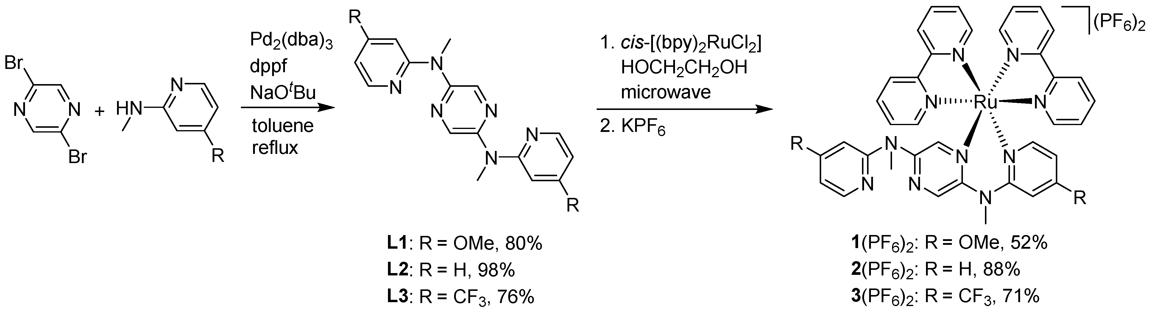
2.1. Synthetic Details

All commercial reagents were used directly without further purification. NMR measurements (1H and 13C NMR) were performed on a Bruker Avance spectrometer in the designated solvents (CD3CN and CDCl3 for complexes and ligands, respectively). Mass spectrometry data were collected by a Thermo Exactive GC and a Bruker Autoflex III MALDI-TOF mass spectrometer. A Flash EA 1112 analyzer was applied to obtain elemental analysis data.

2.1.1. Synthesis of 2,5-di(N-methyl-N′-(4-methoxy-2-pyridyl)amino)pyrazine (L1)

A mixture of 2,5-dibromopyazine (238 mg, 1.0 mmol), N-methyl-4-methoxy-2-pyridinamine (304 mg, 2.2 mmol), tris(dibenzylideneacetone)dipalladium(0) (92 mg, 0.1 mmol), sodium tert-butoxide (384 mg, 4.0 mmol), and 1,1′-ferrocenediyl-bis(diphenylphosphine) (55 mg, 0.1 mmol) was dissolved in toluene (10 mL). The reaction mixture was refluxed at 130 °C for two days under an N2 atmosphere in a sealed pressure tube. After cooling to room temperature, the solvent was evaporated in vacuo, and the crude product was purified by silica gel column chromatography (eluent: petroleum ether/acetic ether = 5/1) to yield 280 mg of L1 as a brown solid (80%). 1H NMR (400 MHz, CDCl3): δ = 3.56 (s, 6H), 3.81 (s, 6H), 6.45 (d, J = 4.0 Hz, 2H), 6.50 (s, 2H), 8.12 (d, J = 4.0 Hz, 2H), 8.44 (s, 2H). 13C NMR (100 MHz, CDCl3): δ = 36.3, 55.4, 97.1, 104.3, 136.4, 148.5, 149.3, 159.2, 167.4. EI-MS (m/z): 352 for [M]+. ESI-HRMS: calcd. for C18H20N6O2 352.1648. Found: 352.1645.

2.1.2. Synthesis of 2,5-di(N-methyl-N′-(4-(trifluoromethyl)-2-pyridyl)amino)pyrazine (L3)

A suspension of 2,5-dibromopyazine (95 mg, 0.4 mmol), N-methyl-4-(trifluoromethyl)-2-pyridinamine (176 mg, 1.0 mmol), tris(dibenzylideneacetone)dipalladium(0) (37 mg, 0.04 mmol), sodium tert-butoxide (153 mg, 1.6 mmol), and 1,1′-ferrocenediyl-bis(diphenylphosphine) (22 mg, 0.04 mmol) was dissolved in toluene (10 mL). The reaction mixture was refluxed at 130 °C for two days under an N2 atmosphere in a sealed pressure tube. After cooling to room temperature, the solvent was evaporated in vacuo, and the crude product was purified by silica gel column chromatography (eluent: petroleum ether/acetic ether = 5/1) to yield 130 mg of L3 as a yellow solid (76%). 1H NMR (400 MHz, CDCl3): δ = 3.65 (s, 6H), 7.03 (d, J = 4.0 Hz, 2H), 7.26 (s, 2H), 8.42 (d, J = 8.0 Hz, 2H), 8.54 (s, 2H). 13C NMR (100 MHz, CDCl3): δ = 36.4, 107.3, 111.8, 127.3 (q, J = 271 Hz), 136.8, 140.7 (q, J = 33 Hz), 148.6, 149.4, 157.7. EI-MS (m/z): 428 for [M]+. ESI-HRMS: calcd. for C18H14N6F6 428.1184. Found: 428.1190.

2.1.3. Synthesis of 1(PF6)2

A suspension of cis-[Ru(bpy)2Cl2] (51.9 mg, 0.1 mmol) and L1 (35.2 mg, 0.1 mmol) in HOCH2CH2OH (5 mL) was heated under microwave irradiation for 30 min. The resulting deep red solution was cooled. A saturated KPF6 solution was added, and the orange precipitate was formed. A flash column chromatography on SiO2 was used to purify the crude product (eluent: CH3CN/KNO3(aq) = 200/1) to give 55 mg 1(PF6)2 as an orange solid in 52% yield. 1H NMR (400 MHz, CD3CN): δ = 3.35 (s, 3H; NCH3), 3.44 (s, 3H; NCH3), 3.82 (s, 3H; OCH3), 3.85 (s, 3H; OCH3), 6.38 (s, 1H; OCH3–pyridine-H), 6.43 (dd, J = 8.0 and 4.0 Hz, 1H; OCH3–pyridine-H), 6.56 (dd, J = 4.0 and 2.0 Hz, 1H; OCH3–pyridine-H), 6.71 (d, J = 4.0 Hz, 1H; OCH3–pyridine-H), 7.15 (t, J = 8.0 Hz, 2H; bipyridine-H), 7.29 (t, J = 4.0 Hz, 1H; OCH3–pyridine-H), 7.56–7.64 (m, 6H; bipyridine-H), 7.87 (t, J = 8.0 Hz, 1H; bipyridine-H), 7.92 (t, J = 8.0 Hz, 1H; bipyridine-H), 8.10 (t, J = 8.0 Hz, 1H; bipyridine-H), 8.15 (t, J = 8.0 Hz, 1H; bipyridine-H), 8.29 (s, 1H; bipyridine-H), 8.33–8.36 (m, 3H; bipyridine-H), 8.40 (d, J = 8.0 Hz, 1H; OCH3–pyridine-H), 8.49 (d, J = 8.0 Hz, 1H; pyrazine-H), 8.58 (d, J = 4.0 Hz, 1H; pyrazine-H). 13C NMR (100 MHz, CD3CN): δ = 41.0, 57.2, 101.4, 106.6, 109.3, 124.9, 125.0, 125.1, 125.4, 127.4, 128.0, 128.3, 135.9, 137.1, 138.3, 138.5, 138.8, 138.9, 152.1, 152.6, 152.8, 153.5, 154.1, 158.0, 158.1, 158.2, 169.2. MALDI-MS: m/z = 910.8 for [M − PF6]+, 765.9 for [M − 2PF6]2+, 609.9 for [M − 2PF6 − bpy]2+. Anal. calcd. for C38H36F12N10O2P2Ru·H2O: C, 42.50; H, 3.57; N, 13.04. Found: C, 42.17; H, 3.11; N, 12.96.

2.1.4. Synthesis of 3(PF6)2

A suspension of cis-[Ru(bpy)2Cl2] (51.9 mg, 0.1 mmol) and L3 (42.8 mg, 0.1 mmol) in HOCH2CH2OH (5 mL) was heated under microwave irradiation for 30 min. The resulting deep red solution was cooled. A saturated KPF6 solution was added, and the orange precipitate was formed. A flash column chromatography on SiO2 was used to purify the crude product (eluent: CH3CN/KNO3(aq) = 300/1) to give 80 mg 3(PF6)2 as an orange solid in 71% yield. 1H NMR (400 MHz, CD3CN): δ = 3.40 (s, 3H; NCH3), 3.57 (s, 3H; NCH3), 7.03 (d, J = 8.0 Hz, 1H; CF3–pyridine-H), 7.13 (d, J = 4.0 Hz, 1H; CF3–pyridine-H), 7.20–7.24 (m, 2H; CF3–pyridine-H), 7.33 (t, J = 8.0 Hz, 1H; bipyridine-H), 7.55–7.67 (m, 6H; bipyridine-H), 7.80 (s, 1H; bipyridine-H), 7.89 (d, J = 4.0 Hz, 1H; CF3–pyridine-H), 7.93 (t, J = 8.0 Hz, 1H; bipyridine-H), 7.98 (t, J = 8.0 Hz, 1H; bipyridine-H), 8.17 (t, J = 8.0 Hz, 1H; bipyridine-H), 8.21 (t, J = 8.0 Hz, 1H; bipyridine-H), 8.36–8.41 (m, 4H; bipyridine-H), 8.48 (d, J = 8.0 Hz, 1H; CF3–pyridine-H), 8.52–8.55 (m, 2H; pyrazine-H). 13C NMR (100 MHz, CD3CN): δ = 36.1, 41.4, 108.9, 112.9, 113.7, 116.4, 122.0, 122.6, 124.7, 125.2, 125.5, 125.6, 127.9, 128.2, 128.3, 128.5, 136.2, 138.8, 138.9, 139.3, 139.4, 140.3 (q, J = 61.6 Hz), 148.1, 149.7, 149.9, 152.6, 152.7, 153.6, 153.7, 153.9, 157.6, 158.0, 158.2, 160.6. MALDI-MS: m/z = 986.7 for [M − PF6]+, 842.8 for [M − 2PF6]2+, 684.8 for [M − 2PF6 − bpy]2+. Anal. calcd. for C38H30F18N10P2Ru: C, 40.33; H, 2.67; N, 12.38. Found: C, 40.23; H, 2.84; N, 12.17.

2.2. X-ray Crystallography

The X-ray diffraction measurements were performed on a Rigaku Saturn 724 diffractometer on a rotating anode (Mo Kα radiation, λ = 0.71073 Å). Olex2 software (SVN Revision No. 5506) was applied to analyze the data and generate the structure graphic. The crystallographic data of 3(PF6)2 (CCDC 2107266) were deposited in The Cambridge Crystallographic Data Centre for free.

2.3. Spectroscopic Measurements

The electronic absorption measurements were performed on a TU-1810DSPC spectrometer in chromatographic grade acetonitrile at room temperature. An F-380 spectrofluorometer was employed to collect the steady-state emission and excitation spectra. The samples of photophysical analysis were prepared by using the 1 cm pathlength quartz cuvettes. The quinine sulfate in 1.0 M aq H2SO4 (Φ = 55%) and [Ru(bpy)3](PF6)2 (Φ = 9.5%) were used to calculate the relative luminescence quantum yields in degassed CH3CN as the reference. The luminescence decays and temperature-dependent emission studies were carried out on the FLS920 and FLS1000 spectrophotometer, respectively.

2.4. Electrochemical Measurements

All electrochemical measurements were carried out on a CHI660D electrochemical station based on a three-electrode system. After bubbling with nitrogen for 5 min, the samples of electrochemical studies were tested at a scan rate of 100 mV/s in 0.1 M Bu4NClO4/CH3CN. The potential of a saturated Ag/AgCl electrode was used to calculate the relative potentials of these compounds as the reference.

2.5. DFT and TDDFT Calculations

The B3LYP exchange-correlation functional and the Gaussian 09 software package were employed for the density functional theory (DFT) calculations. The crystallographic data of 3(PF6)2 were employed to generate the input files. The Los Alamos effective core potential LANL2DZ basis set for Ru and 6-31G* for other atoms were used to optimize the electronic structures of these compounds. All of the calculations were carried out including the solvation effects and using the no symmetry constraints. The DFT-optimized structures were employed for the TDDFT calculations on the same level of theory. In order to make the optimized geometries be local minima, frequency calculations were carried out on the same level of theory. An isovalue of 0.02 e bohr−3 was set for all orbitals on calculations.

2.6. HPLC Analysis

All HPLC measurements were performed on a Shimadzu UFLC system. The analysis data were collected by a Shim-pack XR-ODS column (2.2 μm, 75 mm × 4.6 mm, i.d.). A gradient solvent of CH3CN in water was used to elute these samples (10−90% over 0–10 min, followed by an isocratic elution of 90% CH3CN for 5 min). A 0.1% of trifluoroacetic acid was added in all solvents. The flow rate was set at 1.0 mL/min. The detection wavelengths were set at 450, 451, and 430 nm for 1(PF6)2, 2(PF6)2, and 3(PF6)2, respectively.

3. Results and Discussions

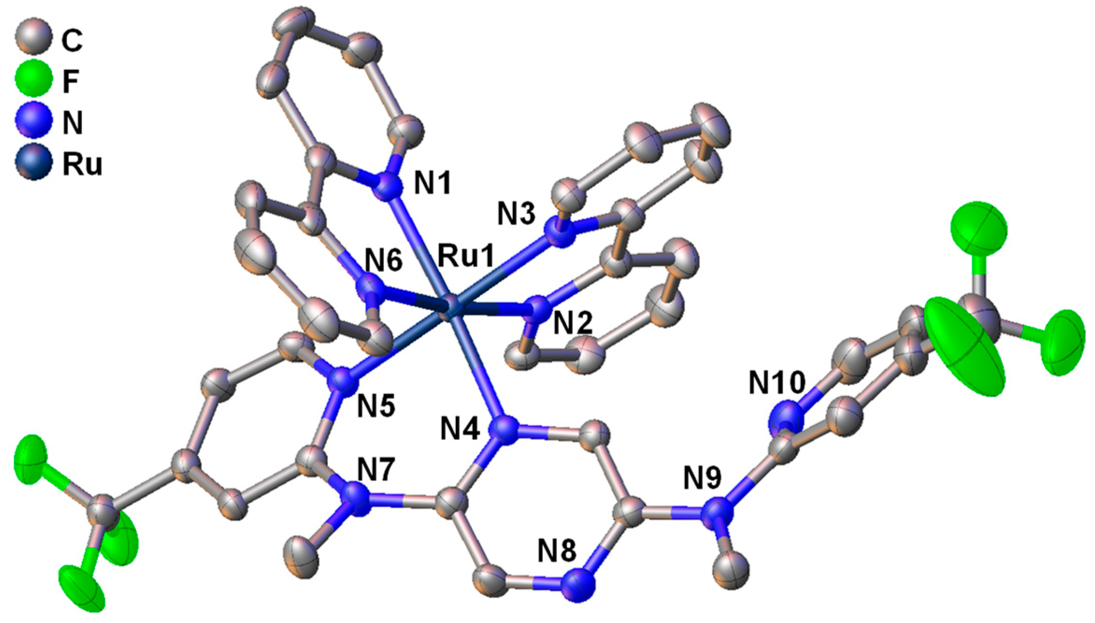
3.1. Studies on Preparation and Single-Crystal X-ray Analysis

Three N(CH3)-bridged ligands L1L3 and corresponding mononuclear ruthenium complexes 1(PF6)23(PF6)2 were synthesized as outlined in Scheme 1. The bidentate ligands L1L3 were obtained through a Pd-catalyzed C-N coupling reaction of 2,5-dibromopyrazine with 2-(N-methylamino)pyridine derivatives in the range of 76−98% yields [64,65]. The reaction of L1, L2, and L3 with 1 equiv. cis-[Ru(bpy)2Cl2] under microwave irradiation, followed by anion exchange using potassium hexafluorophosphate, provided complexes 1(PF6)2, 2(PF6)2, and 3(PF6)2 in a 52%, 88%, and 71% yield, respectively. These new compounds were fully characterized by nuclear magnetic resonance (NMR) (Figures S1–S8), mass spectrometry (Figures S9–S11), and elemental analysis. Furthermore, high performance liquid chromatography (HPLC) analysis results indicate that these complexes have high purities (Figure S12).
The solid-state structure of complex 3(PF6)2 was determined by single-crystal X-ray analysis. Figure 1 shows the ORTEP diagram, and Tables S1 and S2 in the Supporting Information summarize the crystallographic data. A single crystal of complex 3(PF6)2 was acquired by the slow diffusion of (C2H5)O into a solution of the complex in CH3CN. The coordination geometry of the ruthenium atom has a distorted octahedral with bpy and L3. The N–Ru–N bite angle of the bpy (78.86(11)° and 78.68(11)°) is smaller than that of ligand L3 (88.30(10)°). Similar findings have been recorded in our previously reported NN bidentate Ru(II) complexes [64,65]. The Ru–N bond lengths of complex 3(PF6)2 are in the range of 2.056(3)−2.099(3) Å. No distinct length difference is present among the Ru–N bonds associated with bpy and L3. The bidentate ligand bpy has a planar structure. However, ligand L3 shows a severely twisted structure. The torsion angles between the two pyridine planes and the pyrazine plane of L3 are 26.38° and 127.73°, respectively.

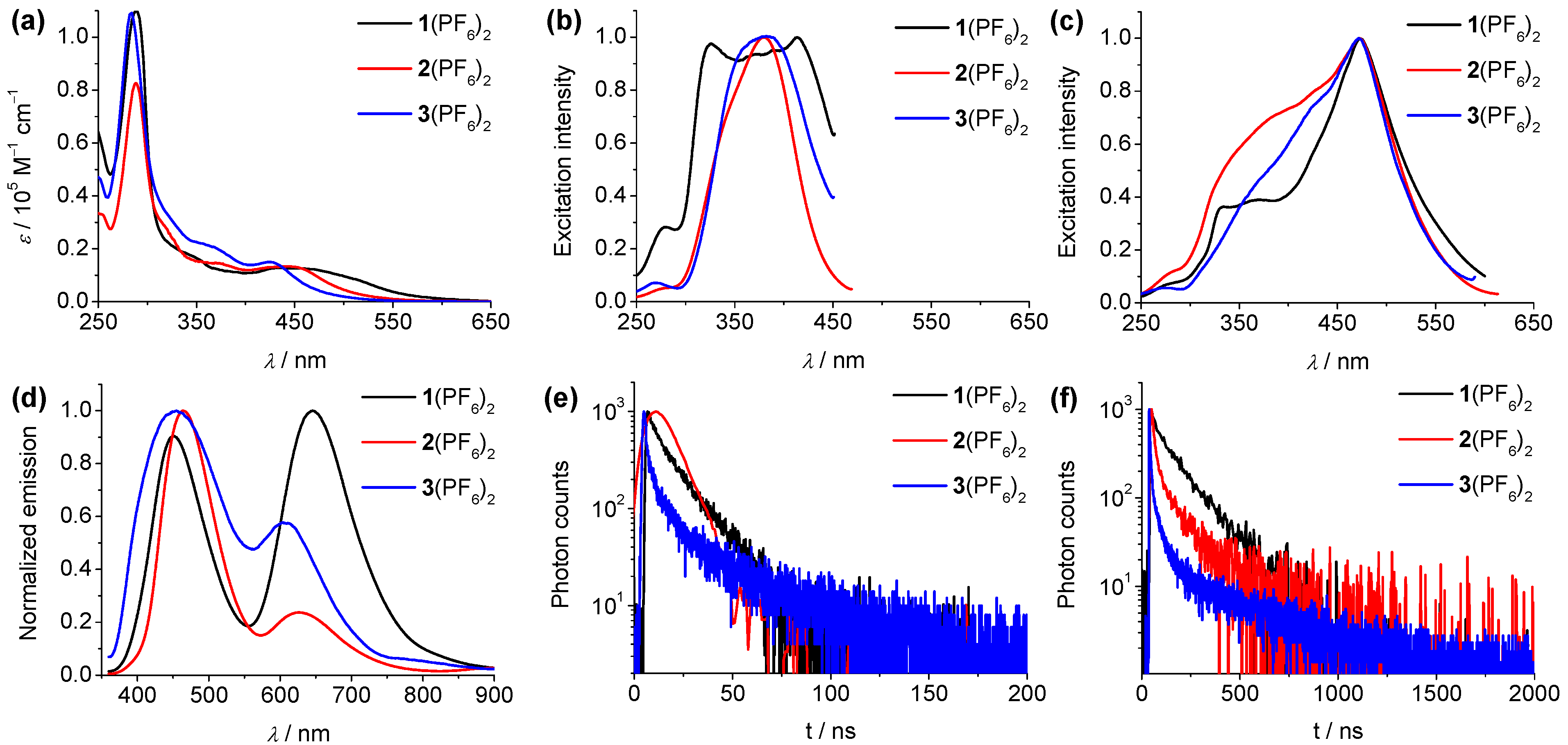
3.2. Spectroscopic Studies

The absorption, excitation, and steady-state emission spectra and the emission decay studies of complexes 1(PF6)23(PF6)2 in acetonitrile are shown in Figure 2, and their photophysical data are summarized in Table 1. Ligands L1L3 display two absorption bands at 310 and 360 nm, 310 and 361 nm, and 295 and 353 nm, respectively (Figure S13). These absorption bands in the UV and visible region are ascribed to the ππ* excitations and the intraligand charge-transfer (ICT) transitions from the amine unit to pyrazine ring of the N(CH3)-bridged ligand, respectively. Under nitrogen saturated conditions, ligands L1L3 display structureless emission bands at 457, 445, and 450 nm with quantum yields of 24%, 43%, and 0.06%, respectively, relative to 55% of quinine sulfate in 1.0 M aq H2SO4 (Figure S13). In the nitrogen saturated solutions, the excited-state lifetimes of these emission bands were determined to be 5.6, 11.5, and 0.4 ns for L1L3 (Table 1), respectively. These results indicate that the substituent effect will increase the non-emissive deactivation pathway of these ligands and thus impair the emission properties. Compared with L3 with electron-withdrawing –CF3 groups, L1 with electron-donating –OMe groups shows better quantum yield and longer excited-state lifetime due to the inductive effect of an alkoxyl substituent [66,67]. These experimental findings indicate that these emission bands of ligands L1L3 are of singlet charge-transfer (1CT) character.
Complexes 1(PF6)23(PF6)2 show intense absorption bands in the UV region at 289, 288, and 284 nm, respectively (Figure 2a), which are assigned to ππ* transitions of ligands. The intense and broad bands in the visible region are observed at 350 and 467 nm, 375 and 448 nm, and 368 and 427 nm for 1(PF6)23(PF6)2, respectively. As indicated by time-dependent density functional theory (TDDFT) calculations below, these higher-energy absorption bands are associated with the ligand-to-ligand charge-transfer (LLCT) transitions from the N(CH3)-bridged ligand to bpy and ICT of the N(CH3)-bridged ligand, while the lower-energy absorption bands are assigned to the metal-to-ligand charge-transfer (MLCT) transitions from the ruthenium component to bpy ligand. By variation of the substituents, 3(PF6)2, with electron-withdrawing –CF3 groups on the pyridine rings shows a 40 nm blue shift of the MLCT absorption band in comparison to 1(PF6)2 containing electron-donating –OMe groups. All three complexes show intriguing dual-emissive behavior in dilute acetonitrile solution under irradiation, with two well-separated emission bands centered at 451 and 646 nm for 1(PF6)2, 465 and 627 nm for 2(PF6)2, and 455 and 608 nm for 3(PF6)2, respectively. The wavelength difference between the dual emission maxima (Δλmax) is 195, 162, and 153 nm for 1(PF6)23(PF6)2, respectively (Figure 2d). The excitation spectra of the higher-energy emission bands of these complexes are associated with the LLCT/ICT absorptions, while the longer-wavelength MLCT absorptions are basically in accordance with the excitation spectra of the lower-energy emission bands with a slight red shift (Figure 2b,c and Figure S14). The emission maxima of the higher-energy emission bands of these complexes have similar emission wavelength, while the lower-energy emission band shows a 38 nm blue shift from 1(PF6)2 to 3(PF6)2 with decreasing electron-donating capabilities of substituents. This is consistent with an ascending order of the energy of the MLCT absorption band from 1(PF6)2 to 3(PF6)2. The solvent effect on the dual-emissive properties of 2(PF6)2 was reported in our previous work [64]. Similar results were observed for 1(PF6)2 and 3(PF6)2. The relative intensities and emission maxima of these two emission bands are also strongly dependent on the solvent polarity. For instance, the lower-energy emission band of 1(PF6)2 became dominant in DMF, while the two emission bands exhibited comparable intensities in other solvents. For complex 3(PF6)2, the higher-energy emission band became dominant in CH3CN, acetone, and CH3OH, while the lower-energy emission band displayed a much higher intensity in DMSO. In DMF, the two emission bands have comparable intensities (Figure S15). The emission spectra in the solid state of 1(PF6)2 and 3(PF6)2 exhibit similar emission maxima (600 and 645 nm for 1(PF6)2 and 3(PF6)2, respectively) with respect to that of the lower-energy emission band in CH3CN (Figure S16). Compared with the lower-energy emission band in CH3CN, the emission spectrum of pure powder of 2(PF6)2 shows a 31 nm red shift most likely due to the aggregates formed by intermolecular interaction. In nitrogen saturated acetonitrile solution, complex 3(PF6)2 has a low quantum yield of 0.53%, relative to 9.5% of [Ru(bpy)3](PF6)2 [68], while complex 1(PF6)2 shows a better quantum yield of 2.1%. Compared with the prototype complex 2(PF6)2, complexes 1(PF6)2 and 3(PF6)2 possess lower emission quantum yields. We conjecture that the introduction of substituents may increase the flexibility of structure and molecular vibrations and enhance the rate constant of the nonradiative process.
In luminescence decay studies, all three complexes 1(PF6)23(PF6)2 exhibit distinct excited-state lifetimes for two emission bands in acetonitrile solution (Figure 2e,f, and Table 1). In air-equilibrated solutions, the excited-state lifetimes of the higher-energy emission bands are 14, 10, and 10 ns, while the lower-energy emission bands have longer lifetimes of 89, 58, and 24 ns for 1(PF6)23(PF6)2, respectively. In nitrogen saturated solutions, the lifetimes of the higher-energy emission bands show little changes with respect to those in air-equilibrated solutions (16, 13, and 15 ns for 1(PF6)23(PF6)2, respectively), while the lifetimes of the lower-energy emission bands are considerably elongated (193, 219, and 189 ns for 1(PF6)23(PF6)2, respectively). Compared with ligands, the substituent effect on excited-state lifetimes of these complexes is negligible. Moreover, the lifetimes of the representative complexes 1(PF6)2 and 2(PF6)2 were measured in glassy CH3CN at 77 K. The higher-energy emission bands still exhibit nanosecond range lifetimes (5.0 ns for both 1(PF6)2 and 2(PF6)2), and those of the lower-energy emission bands significantly increase to a microsecond range (0.53 and 2.5 μs for 1(PF6)2 and 2(PF6)2, respectively). Based on these experimental observations, the higher- and the lower-energy emission bands are ascribed to the admixtures of the singlet (N(CH3)-bridged ligand to bpy 1LLCT and N(CH3)-bridged ligand 1ICT charge transfer) and the triplet 3MLCT (ruthenium component to bpy charge transfer) character, respectively. A number of studies suggest that the dual fluorescence/phosphorescence character of a single complex was found in different luminescent TMCs such as platinum [18,19,20,27,29], osmium [21], copper [22], and iridium [24,30], as well as ruthenium complexes [28,32,34]. Kasha’s rule states that only the lowest energy excited state of a given spin multiplicity is emissive. However, the dual emissions coming from the excited states with the different spin manifold of a single complex are still obeying Kasha’s rule.

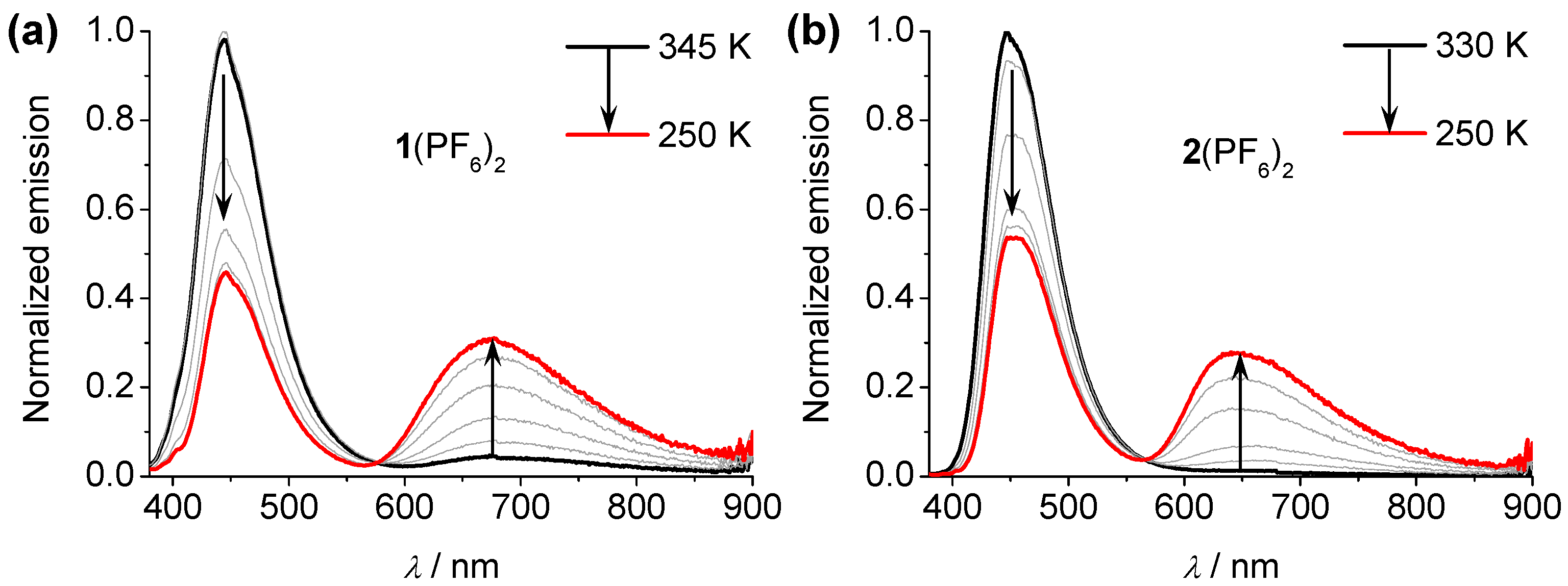
3.3. Temperature-Dependent Emission Spectral Studies

To investigate the changes in dual-emissive properties by temperature stimuli, the temperature-dependent emission spectral studies of the representative complexes 1(PF6)2 and 2(PF6)2 have been measured in CH3CN solution and shown in Figure 3. Both of the complexes display similar emission spectral changes. Upon decreasing the temperature from 345 to 250 K, the higher-energy emission intensities decrease significantly, while those of the lower-energy emissions gradually increase. During the spectral changes process, a well isoluminescence point was recorded at 576 and 566 nm for 1(PF6)2 and 2(PF6)2, respectively. The excited-state lifetimes of the higher-energy emission bands of these complexes still fall in the nanosecond range from 345 to 250 K, excluding a thermally activated process. This suggests that the Franck–Condon transitions of the higher-energy emission are slowed down upon decreasing the temperature and thus facilitate the intersystem crossing from 1LLCT/1ICT to the 3MLCT state [69]. The relative intensity of the two emission bands of 1(PF6)23(PF6)2 vary as a function of the excitation wavelength (Figure S17). When excited at a shorter wavelength (300–360 nm), the higher-energy emission band is much higher than the lower-energy emission band. When a longer excitation wavelength was applied (380–500 nm), the higher-energy emission band almost disappeared, and the lower-energy emission band became dominant.

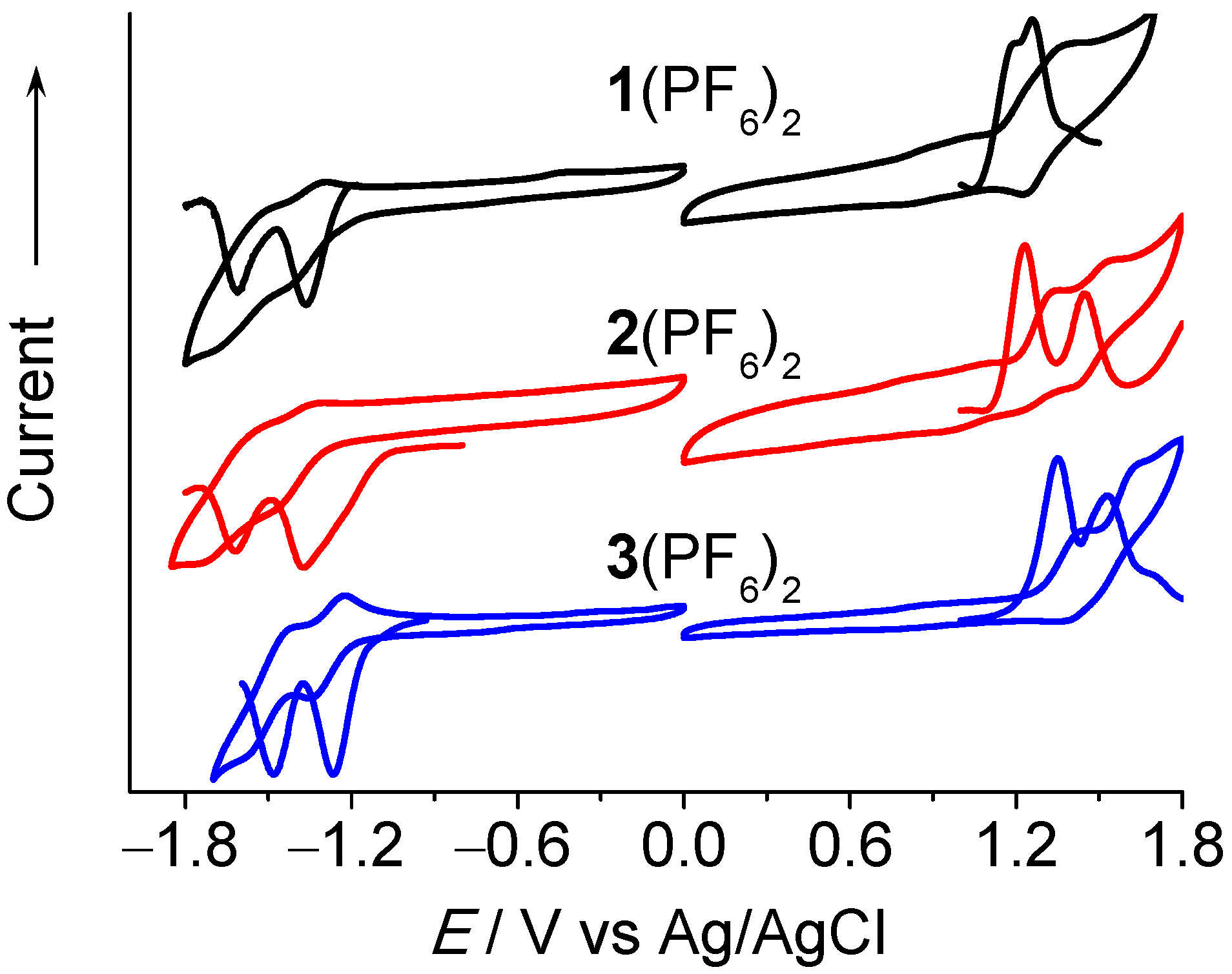
3.4. Electrochemical Studies

Cyclic voltammetry (CV) and differential pulse voltammetry (DPV) were applied to study the electrochemical properties of complexes 1(PF6)23(PF6)2 and ligands L1L3. Figure 4 displays the CV and DPV profiles of 1(PF6)23(PF6)2, and their electrochemical data are collected in Table 1, together with ligands L1L3. In the anodic scan, 1(PF6)23(PF6)2 display an irreversible and a reversible oxidation peak at +1.12 and +1.26 V, +1.23 and +1.44 V, and +1.35 and +1.53 V, versus Ag/AgCl in CH3CN, respectively. The presence of the electron-donating groups –OMe in complex 1(PF6)2 make both oxidation processes more negative in comparison to 2(PF6)2 (110 and 180 mV shift for the first and second oxidation peaks, respectively). In contrast, 120 and 90 mV positive redox shifts are observed for the first and second oxidation peaks of 3(PF6)2 relative to that of 2(PF6)2 due to the introduction of electron-withdrawing –CF3 groups. The first irreversible peaks are assigned to the N(CH3)-bridged ligand oxidation while the second reversible signals are ascribed to the RuII/III process. In the cathodic scan, 1(PF6)23(PF6)2 show two consecutive reversible reduction waves at −1.36 and −1.61 V, −1.37 and −1.62 V, and −1.27 and −1.48 V, versus Ag/AgCl in CH3CN, respectively. These peaks are associated with the reductions of bpy ligands. These assignments are consistent with the electrochemical findings of these ligands (Figure S18). L1L3 display the amine-based irreversible oxidation peaks at +0.84, +0.89, and +1.01 V, versus Ag/AgCl in CH3CN, respectively. No obvious reduction processes were found for these ligands. These assignments are also supported by the DFT calculation results discussed below. The difference between the first anodic and first cathodic electrochemical potential (ΔEechem) of these complexes was employed to calculate the energy gaps which were determined to be 2.48, 2.60, and 2.62 eV for 1(PF6)23(PF6)2, respectively.

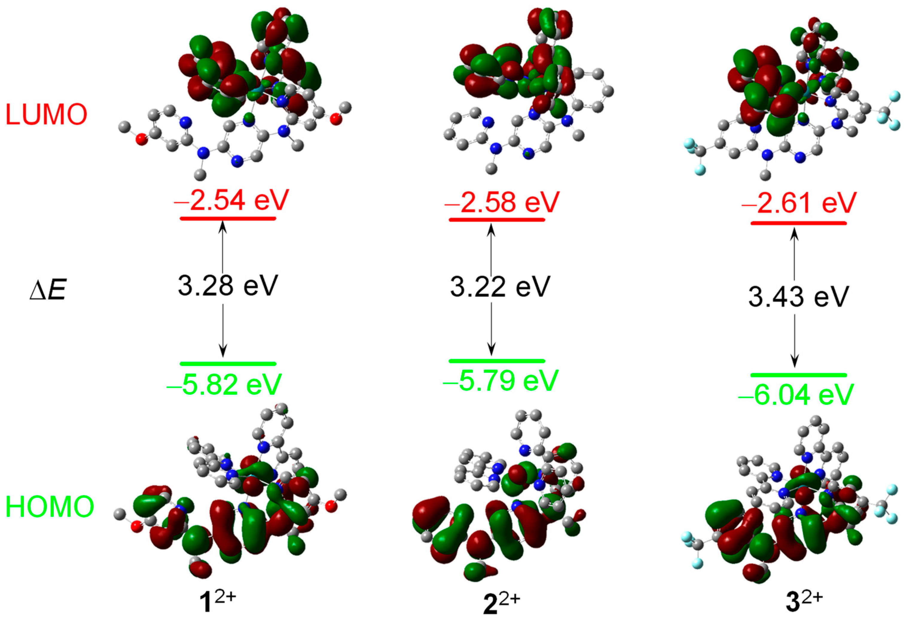
3.5. DFT and TDDFT Calculations

In order to further understand the electronic transitions of complexes 1(PF6)23(PF6)2, DFT calculations have been carried out on complexes 12+, 22+, and 32+. The optimized geometries were generated from the single-crystal structure of 3(PF6)2. Figure 5 displays the calculated energy diagram and isodensity plots of 12+32+. The frontier energy gap of complex 32+ (3.43 eV) is slightly larger with respect to those of 12+ and 22+ (3.28 and 3.22 eV, respectively). This is mainly caused by the stabilization of the highest occupied molecular orbital (HOMO) level of 32+. These calculation findings are in agreement with the above experimental results.
Figures S19 and S20 display the representative frontier orbital isodensity plots of 12+ and 32+, respectively. The HOMOs of these complexes are dominated by the N(CH3)-bridged ligand, with minor contributions from the ruthenium component. The lower occupied orbitals (HOMO–1, HOMO–2, and HOMO–3) are dominated by the ruthenium ion. The lowest unoccupied molecular orbital (LUMO) and LUMO+1 of these complexes have a bpy character, while the LUMO+2 is dominated by the N(CH3)-bridged ligand.
In the same level of theory, TDDFT calculations were performed on the basis of the DFT-optimized structures of these complexes. These predicted electronic transitions are summarized in Table 2 and shown in Figure S21. The predicted S1 excitation of 12+ is associated with the HOMO → LUMO transition, which is associated with the low-energy absorption extending over 650 nm. The lower-energy emission band at 646 nm was assigned to the predicted S2 excitation (HOMO–1 → LUMO+1) which is attributed to the MLbpyCT transitions. The higher-energy S3, S4, S7, and S8 transitions have similar MLbpyCT characters. The predicted S9, S10, S11, S12, S13, and S19 excitations are mainly responsible for the observed absorption bands from 330 to 400 nm. TDDFT calculation results indicate that these excitations are associated with LLCT (amine unit of L1 to bpy), ICT (amine unit to pyrazine ring of L1), and L1-targeted MLCT transitions. These states are perturbed by MLCT states [64], and they are associated with the higher-energy emission band, indicating the admixtures of the LLCT/ICT character of the excited state. A similar situation was observed for 32+. The predicted S1 excitation of 32+ has a very low oscillator strength (f). The lower-energy emission band at 608 nm was assigned to the predicted S2 excitation (HOMO–1 → LUMO+1) which is attributed to the MLbpyCT transitions. The predicted S7, S9, S10, S13, and S16 excitations are mainly responsible for the observed higher-energy charge-transfer absorption bands from 350 to 400 nm. These excitations are attributed to LLCT (amine unit of L3 to bpy), ICT (amine unit to pyrazine ring of L3), and L3-targeted MLCT transitions, and they are responsible for the observed higher-energy emission band at 455 nm.

4. Conclusions

In summary, three monoruthenium complexes 1(PF6)23(PF6)2 with dual fluorescence/phosphorescence are prepared and characterized. These complexes show well-separated dual emissions that are ascribed to the 1LLCT/1ICT and 3MLCT transitions, respectively. The substituent and solvent effects in the dual-emissive properties of these complexes have been examined. The energy gaps of two emissions can be tuned by introducing different substituents to the N(CH3)-bridged ligand, which are decreased by enhancing the electron-withdrawing capabilities of substituents. The relative intensities and emission maxima of two emissions are also strongly dependent on the solvent used. Future work will focus on the design and application of dual-emissive transition-metal complexes as ratiometric photoluminescent probes for the detection of proteins and nucleic acids.

Supplementary Materials

The following supporting information can be downloaded at: https://www.mdpi.com/article/10.3390/ma16206792/s1, Figures S1–S8: 1H and 13C NMR spectra of L1, L3, 1(PF6)2, and 3(PF6)2; Figures S9–S11: MADLI-TOF mass spectra of 1(PF6)23(PF6)2; Figure S12: HPLC spectra of 1(PF6)23(PF6)2; Tables S1 and S2: Single-crystal X-ray data of complex 3(PF6)2; Figure S13: Absorption and emission spectra of ligand L1L3; Figure S14: Absorption, emission, and excitation spectra of 1(PF6)23(PF6)2 in CH3CN; Figure S15: Emission spectra changes of 1(PF6)2 and 3(PF6)2 in different solvents; Figure S16: Emission spectra of 1(PF6)23(PF6)2 in solid state; Figure S17: Emission spectra changes of 1(PF6)23(PF6)2 at different excitation wavelengths; Figure S18: CVs and DPVs of ligands L1L3; Figures S19 and S20: DFT calculation results of 1(PF6)2 and 3(PF6)2; Figure S21: TDDFT calculation results of 1(PF6)23(PF6)2.

Author Contributions

Conceptualization, S.-H.W. and Y.-W.Z.; methodology, S.-H.W., Z.-L.G. and Y.-W.Z.; validation, Z.Z. and R.-H.Z.; formal analysis, Z.Z., R.Y. and R.-H.Z.; investigation, S.-H.W., L.W., R.Y., J.-Y.S. and Z.-L.G.; resources, Z.Z., R.Y. and R.-H.Z.; data curation, Z.Z., J.-Y.S. and R.-H.Z.; writing—original draft preparation, S.-H.W. and Y.-W.Z.; writing—review and editing, S.-H.W., L.W., Z.-L.G., J.-Y.S. and Y.-W.Z.; supervision, S.-H.W.; project administration, Y.-W.Z. and S.-H.W.; funding acquisition, S.-H.W., L.W. and Y.-W.Z. All authors have read and agreed to the published version of the manuscript.

Funding

We acknowledge the financial support from the National Natural Science Foundation of China (22004041, 21925112, 21975264, and 21872154) and the Fujian Province Natural Science Foundation (2023J01129).

Institutional Review Board Statement

Not applicable.

Informed Consent Statement

Not applicable.

Data Availability Statement

Not applicable.

Conflicts of Interest

The authors declare no conflict of interest.

References

  1. Kasha, M. Characterization of electronic transitions in complex molecules. Discuss. Faraday Soc. 1950, 9, 14–19. [Google Scholar] [CrossRef]
  2. Behera, S.K.; Park, S.Y.; Gierschner, J. Dual Emission: Classes, Mechanisms, and Conditions. Angew. Chem. Int. Ed. 2021, 60, 22624–22638. [Google Scholar] [CrossRef] [PubMed]
  3. Lu, J.-S.; Fu, H.; Zhang, Y.; Jakubek, Z.J.; Tao, Y.; Wang, S. A Dual Emissive BODIPY Dye and Its Use in Functionalizing Highly Monodispersed PbS Nanoparticles. Angew. Chem. Int. Ed. 2011, 50, 11658–11662. [Google Scholar] [CrossRef]
  4. Swamy, P.C.A.; Mukherjee, S.; Thilagar, P. Dual emissive borane–BODIPY dyads: Molecular conformation control over electronic properties and fluorescence response towards fluoride ions. Chem. Commun. 2013, 49, 993–995. [Google Scholar] [CrossRef]
  5. Mukherjee, S.; Thilagar, P. Fine-Tuning Dual Emission and Aggregation-Induced Emission Switching in NPI–BODIPY Dyads. Chem. Eur. J. 2014, 20, 9052–9062. [Google Scholar] [CrossRef] [PubMed]
  6. Li, R.; Gong, Z.-L.; Tang, J.-H.; Sun, M.-J.; Shao, J.-Y.; Zhong, Y.-W.; Yao, J. Triarylamines with branched multi-pyridine groups: Modulation of emission properties by structural variation, solvents, and tris(pentafluorophenyl)borane. Sci. China Chem. 2018, 61, 545–556. [Google Scholar] [CrossRef]
  7. Matsuo, K.; Saito, S.; Yamaguchi, S. Photodissociation of B−N Lewis Adducts: A Partially Fused Trinaphthylborane with Dual Fluorescence. J. Am. Chem. Soc. 2014, 136, 12580–12583. [Google Scholar] [CrossRef]
  8. Wu, P.; Hou, X.; Xu, J.-J.; Chen, H.-Y. Ratiometric fluorescence, electrochemiluminescence, and photoelectrochemical chemo/biosensing based on semiconductor quantum dots. Nanoscale 2016, 8, 8427–8442. [Google Scholar] [CrossRef]
  9. Ventura, B.; Durola, F.; Frey, J.; Heitz, V.; Sauvage, J.-P.; Flamigni, L. Near-infrared dual luminescence from an extended zinc porphyrin. Chem. Commun. 2012, 48, 1021–1023. [Google Scholar] [CrossRef]
  10. Sun, X.-Y.; Yue, M.; Jiang, Y.-X.; Zhao, C.-H.; Liao, Y.-Y.; Lei, X.-W.; Yue, C.-Y. Combining Dual-Light Emissions to Achieve Efficient Broadband Yellowish-Green Luminescence in One-Dimensional Hybrid Lead Halides. Inorg. Chem. 2021, 60, 1491–1498. [Google Scholar] [CrossRef]
  11. Blakley, R.L.; Myrick, M.L.; Dearmond, M.K. Interligand and Charge-Transfer Emission from [Ru(bpy)(HDPA)2]2+: A Dual Emitting Ru(II) Complex. J. Am. Chem. Soc. 1986, 108, 7843–7844. [Google Scholar] [CrossRef]
  12. Wang, J.-H.; Li, M.; Zheng, J.; Huang, X.-C.; Li, D. A dual-emitting Cu6–Cu2–Cu6 cluster as a self-calibrated, wide-range luminescent molecular thermometer. Chem. Commun. 2014, 50, 9115–9118. [Google Scholar] [CrossRef] [PubMed]
  13. Glazer, E.C.; Magde, D.; Tor, Y. Ruthenium Complexes That Break the Rules: Structural Features Controlling Dual Emission. J. Am. Chem. Soc. 2007, 129, 8544–8551. [Google Scholar] [CrossRef]
  14. Keyes, T.E.; O’Connor, C.; Vos, J.G. Evidence for the presence of dual emission in a ruthenium(II) polypyridyl mixed ligand complex. Chem. Commun. 1998, 8, 889–890. [Google Scholar] [CrossRef]
  15. Zambrana, J.L.; Ferloni, E.X.; Colis, J.C.; Gafney, H.D. Multiple Charge-Transfer Emissions from Different Metal−Ligand Pairs in Ruthenium Diimines. Inorg. Chem. 2008, 47, 2–4. [Google Scholar] [CrossRef]
  16. Song, L.-Q.; Feng, J.; Wang, X.-S.; Yu, J.-H.; Hou, Y.-J.; Xie, P.-H.; Zhang, B.-W.; Xiang, J.-F.; Ai, X.-C.; Zhang, J.-P. Dual Emission from 3MLCT and 3ILCT Excited States in a New Ru(II) Diimine Complex. Inorg. Chem. 2003, 42, 3393–3395. [Google Scholar] [CrossRef]
  17. Kumar, S.; Hisamatsu, Y.; Tamaki, Y.; Ishitani, O.; Aoki, S. Design and Synthesis of Heteroleptic Cyclometalated Iridium(III) Complexes Containing Quinoline-Type Ligands that Exhibit Dual Phosphorescence. Inorg. Chem. 2016, 55, 3829–3843. [Google Scholar] [CrossRef]
  18. Kozhevnikov, D.N.; Kozhevnikov, V.N.; Shafikov, M.Z.; Prokhorov, A.M.; Bruce, D.W.; Williams, J.A.G. Phosphorescence vs. Fluorescence in Cyclometalated Platinum(II) and Iridium(III) Complexes of (Oligo)thienylpyridines. Inorg. Chem. 2011, 50, 3804–3815. [Google Scholar] [CrossRef]
  19. Cao, Y.; Wolf, M.O.; Patrick, B.O. Dual-Emissive Platinum(II) Metallacycles with Thiophene-Containing Bisacetylide Ligands. Inorg. Chem. 2016, 55, 8985–8993. [Google Scholar] [CrossRef] [PubMed]
  20. Hudson, Z.M.; Zhao, S.-B.; Wang, R.-Y.; Wang, S. Switchable Ambient-Temperature Singlet–Triplet Dual Emission in Nonconjugated Donor–Acceptor Triarylboron–PtII Complexes. Chem. Eur. J. 2009, 15, 6131–6137. [Google Scholar] [CrossRef] [PubMed]
  21. Cheng, Y.-M.; Yeh, Y.-S.; Ho, M.-L.; Chou, P.-T.; Chen, P.-S.; Chi, Y. Dual Room-Temperature Fluorescent and Phosphorescent Emission in 8-Quinolinolate Osmium(II) Carbonyl Complexes: Rationalization and Generalization of Intersystem Crossing Dynamics. Inorg. Chem. 2005, 44, 4594–4603. [Google Scholar] [CrossRef] [PubMed]
  22. Li, J.; Wang, L.; Zhao, Z.; Li, X.; Yu, X.; Huo, P.; Jin, Q.; Liu, Z.; Bian, Z.; Huang, C. Two-Coordinate Copper(I)-NHC Complexes: Novel Dual-Emissive Property and Ultralong Room Temperature Phosphorescence. Angew. Chem. Int. Ed. 2020, 59, 8210–8217. [Google Scholar] [CrossRef] [PubMed]
  23. Shimizu, M.; Nagano, S.; Kinoshita, T. Dual Emission from Precious Metal-Free Luminophores Consisting of C, H, O, Si, and S/P at Room Temperature. Chem. Eur. J. 2020, 26, 5162–5167. [Google Scholar] [CrossRef] [PubMed]
  24. Gitlina, A.Y.; Ivonina, M.V.; Sizov, V.V.; Starova, G.L.; Pushkarev, A.P.; Volyniuk, D.; Tunik, S.P.; Koshevoy, I.O.; Grachova, E.V. A rare example of a compact heteroleptic cyclometalated iridium(III) complex demonstrating well-separated dual emission. Dalton Trans. 2018, 47, 7578–7586. [Google Scholar] [CrossRef] [PubMed]
  25. Sun, C.-J.; Meng, G.; Li, Y.; Wang, N.; Chen, P.; Wang, S.; Yin, X. Millisecond Time-scale Photoluminescence of B−N-doped Tetrathienonaphthalene with Borane/Amine Substituents. Inorg. Chem. 2021, 60, 1099–1106. [Google Scholar] [CrossRef]
  26. You, Y.; Han, Y.; Lee, Y.-M.; Park, S.Y.; Nam, W.; Lippard, S.J. Phosphorescent Sensor for Robust Quantification of Copper(II) Ion. J. Am. Chem. Soc. 2011, 133, 11488–11491. [Google Scholar] [CrossRef]
  27. Liu, Y.; Guo, H.; Zhao, J. Ratiometric luminescent molecular oxygen sensors based on uni-luminophores of C^N Pt(II)(acac) complexes that show intense visible-light absorption and balanced fluorescence/phosphorescence dual emission. Chem. Commun. 2011, 47, 11471–11473. [Google Scholar] [CrossRef]
  28. Martin, A.; Byrne, A.; Dolan, C.; Forster, R.J.; Keyes, T.E. Solvent switchable dual emission from a bichromophoric ruthenium–BODIPY complex. Chem. Commun. 2015, 51, 15839–15841. [Google Scholar] [CrossRef]
  29. Zhao, Q.; Zhou, X.; Cao, T.; Zhang, K.Y.; Yang, L.; Liu, S.; Liang, H.; Yang, H.; Li, F.; Huang, W. Fluorescent/phosphorescent dual-emissive conjugated polymer dots for hypoxia bioimaging. Chem. Sci. 2015, 6, 1825–1831. [Google Scholar] [CrossRef]
  30. Gupta, S.K.; Haridas, A.; Choudhury, J. Remote Terpyridine Integrated NHC–IrIII Luminophores as Potential Dual-Emissive Ratiometric O2 Probes. Chem. Eur. J. 2017, 23, 4770–4773. [Google Scholar] [CrossRef]
  31. Lo, K.K.-W.; Zhang, K.Y.; Leung, S.-K.; Tang, M.-C. Exploitation of the Dual-emissive Properties of Cyclometalated Iridium(III)–Polypyridine Complexes in the Development of Luminescent Biological Probes. Angew. Chem. Int. Ed. 2008, 47, 2213–2216. [Google Scholar] [CrossRef] [PubMed]
  32. Shao, J.-Y.; Wu, S.-H.; Ma, J.; Gong, Z.-L.; Sun, T.-G.; Jin, Y.; Yang, R.; Sun, B.; Zhong, Y.-W. Ratiometric detection of amyloid-β aggregation by a dual-emissive tris-heteroleptic ruthenium complex. Chem. Commun. 2020, 56, 2087–2090. [Google Scholar] [CrossRef] [PubMed]
  33. Walker, M.G.; Ramu, V.; Meijer, A.J.H.M.; Das, A.; Thomas, J.A. A ratiometric sensor for DNA based on a dual emission Ru(dppz) light-switch complex. Dalton Trans. 2017, 46, 6079–6086. [Google Scholar] [CrossRef] [PubMed]
  34. Wu, S.-H.; Yang, R.; Sun, B.; Tang, J.-H.; Gong, Z.-L.; Ma, J.; Wang, L.; Liu, J.; Ma, D.-X.; Shao, J.-Y.; et al. Dual-Emissive Tris-Heteroleptic Ruthenium Complexes: Tuning the DNA-Triggered Ratiometric Emission Response by Ancillary Ligands. Inorg. Chem. 2021, 60, 14810–14819. [Google Scholar] [CrossRef] [PubMed]
  35. Lee, M.H.; Kim, J.S.; Sessler, J.L. Small molecule-based ratiometric fluorescence probes for cations, anions, and biomolecules. Chem. Soc. Rev. 2015, 44, 4185–4191. [Google Scholar] [CrossRef]
  36. Gui, R.; Jin, H.; Bu, X.; Fu, Y.; Wang, Z.; Li, Q. Recent advances in dual-emission ratiometric fluorescence probes forchemo/biosensing and bioimaging of biomarkers. Coord. Chem. Rev. 2019, 383, 82–103. [Google Scholar] [CrossRef]
  37. Chen, Z.; Ho, C.-L.; Wang, L.; Wong, W.-Y. Single-Molecular White-Light Emitters and Their Potential WOLED Applications. Adv. Mater. 2020, 32, 1903269. [Google Scholar] [CrossRef]
  38. Kawashiro, M.; Mori, T.; Ito, M.; Ando, N.; Yamaguchi, S. Photodissociative Modules that Control Dual-Emission Properties in Donor–π–Acceptor Organoborane Fluorophores. Angew. Chem. Int. Ed. 2023, 62, e202303725. [Google Scholar] [CrossRef]
  39. Cheng, X.; Yue, S.; Chen, R.; Yin, J.; Cui, B.-B. White Light-Emitting Diodes Based on One-Dimensional Organic−Inorganic Hybrid Metal Chloride with Dual Emission. Inorg. Chem. 2022, 61, 15475–15483. [Google Scholar] [CrossRef]
  40. Vázquez-Domínguez, P.; Journaud, O.; Vanthuyne, N.; Jacquemin, D.; Favereau, L.; Crassous, J.; Ros, A. Helical donor–acceptor platinum complexes displaying dual luminescence and near-infrared circularly polarized luminescence. Dalton Trans. 2021, 50, 13220–13226. [Google Scholar] [CrossRef]
  41. Huang, X.; Song, J.; Yung, B.C.; Huang, X.; Xiong, Y.; Chen, X. Ratiometric optical nanoprobes enable accurate molecular detection and imaging. Chem. Soc. Rev. 2018, 47, 2873–2920. [Google Scholar] [CrossRef]
  42. Kim, Y.; Kim, H.; Son, J.B.; Filatov, M.; Choi, C.H.; Lee, N.K.; Lee, D. Single-Benzene Dual-Emitters Harness Excited-State Antiaromaticity for White Light Generation and Fluorescence Imaging. Angew. Chem. Int. Ed. 2023, 62, e202302107. [Google Scholar] [CrossRef] [PubMed]
  43. Magde, D.; Magde, M.D., Jr.; Glazer, E.C. So-called “dual emission” for 3MLCT luminescence in ruthenium complex ions: What is really happening? Coord. Chem. Rev. 2016, 306, 447–467. [Google Scholar] [CrossRef]
  44. Wu, S.-H.; Gong, Z.-L.; Shao, J.-Y.; Yang, R.; Zhong, Y.-W. Dual-emissive transition-metal complexes and their applications as ratiometric photoluminescent probes. Sci. Sin. Chim. 2020, 50, 315–323. (In Chinese) [Google Scholar]
  45. Steube, J.; Kruse, A.; Bokareva, O.S.; Reuter, T.; Demeshko, S.; Schoch, R.; Cordero, M.A.A.; Krishna, A.; Hohloch, S.; Meyer, F.; et al. Janus-type emission from a cyclometalated iron(III) complex. Nat. Chem. 2023, 15, 468–474. [Google Scholar] [CrossRef]
  46. Kwak, S.W.; Choi, B.H.; Lee, J.H.; Hwang, H.; Lee, J.; Kwon, H.; Chung, Y.; Lee, K.M.; Park, M.H. Synthesis and Dual-Emission Feature of Salen-Al/Triarylborane Dyads. Inorg. Chem. 2017, 56, 6039–6043. [Google Scholar] [CrossRef]
  47. Zhang, K.Y.; Liu, H.-W.; Tang, M.-C.; Choi, A.W.-T.; Zhu, N.; Wei, X.-G.; Lau, K.-C.; Lo, K.K.-W. Dual-Emissive Cyclometalated Iridium(III) Polypyridine Complexes as Ratiometric Biological Probes and Organelle-Selective Bioimaging Reagents. Inorg. Chem. 2015, 54, 6582–6593. [Google Scholar] [CrossRef] [PubMed]
  48. Zhang, K.Y.; Gao, P.; Sun, G.; Zhang, T.; Li, X.; Liu, S.; Zhao, Q.; Lo, K.K.-W.; Huang, W. Dual-Phosphorescent Iridium(III) Complexes Extending Oxygen Sensing from Hypoxia to Hyperoxia. J. Am. Chem. Soc. 2018, 140, 7827–7834. [Google Scholar] [CrossRef]
  49. Glazer, E.C.; Magde, D.; Tor, Y. Dual Emission from a Family of Conjugated Dinuclear RuII Complexes. J. Am. Chem. Soc. 2005, 127, 4190–4192. [Google Scholar] [CrossRef]
  50. Han, M.; Tian, Y.; Yuan, Z.; Zhu, L.; Ma, B. A Phosphorescent Molecular “Butterfly” that undergoes a Photoinduced Structural Change allowing Temperature Sensing and White Emission. Angew. Chem. Int. Ed. 2014, 53, 10908–10912. [Google Scholar] [CrossRef] [PubMed]
  51. Zhou, C.; Tian, Y.; Yuan, Z.; Han, M.; Wang, J.; Zhu, L.; Tameh, M.S.; Huang, C.; Ma, B. Precise Design of Phosphorescent Molecular Butterflies with Tunable Photoinduced Structural Change and Dual Emission. Angew. Chem. Int. Ed. 2015, 54, 9591–9595. [Google Scholar] [CrossRef]
  52. López-López, J.C.; Bautista, D.; Gonzáez-Herrero, P. Stereoselective Formation of Facial Tris-Cyclometalated PtIV Complexes: Dual Phosphorescence from Heteroleptic Derivatives. Chem. Eur. J. 2020, 26, 11307–11315. [Google Scholar] [CrossRef] [PubMed]
  53. Scattergood, P.A.; Ranieri, A.M.; Charalambou, L.; Comia, A.; Ross, D.A.W.; Rice, C.R.; Hardman, S.J.O.; Heully, J.-L.; Dixon, I.M.; Massi, M.; et al. Unravelling the Mechanism of Excited-State Interligand Energy Transfer and the Engineering of Dual Emission in [Ir(CN)2(NN)]+ Complexes. Inorg. Chem. 2020, 59, 1785–1803. [Google Scholar] [CrossRef]
  54. Kaufmann, M.; Müller, C.; Cullen, A.A.; Brandon, M.P.; Dietzek, B.; Pryce, M.T. Photophysics of Ruthenium(II) Complexes with Thiazole π-Extended Dipyridophenazine Ligands. Inorg. Chem. 2021, 60, 760–773. [Google Scholar] [CrossRef]
  55. Kisel, K.S.; Melnikov, A.S.; Grachova, E.V.; Hirva, P.; Tunik, S.P.; Koshevoy, I.O. Linking ReI and PtII Chromophores with Aminopyridines: A Simple Route to Achieve a Complicated Photophysical Behavior. Chem. Eur. J. 2017, 23, 11301–11311. [Google Scholar] [CrossRef] [PubMed]
  56. Wu, S.-H.; Ma, D.-X.; Gong, Z.-L.; Ma, J.; Shao, J.-Y.; Yang, R.; Zhong, Y.-W. Synthesis, Photophysical, and Computational Studies of a Bridged IrIII-PtII Heterodimetallic Complex. Crystals 2021, 11, 236. [Google Scholar] [CrossRef]
  57. Liu, L.-Y.; Fang, H.; Chen, Q.; Chan, M.H.-Y.; Ng, M.; Wang, K.-N.; Liu, W.; Tian, Z.; Diao, J.; Mao, Z.-W.; et al. Multiple-Color Platinum Complex with Super-Large Stokes Shift for Super-Resolution Imaging of Autolysosome Escape. Angew. Chem. Int. Ed. 2020, 59, 19229–19236. [Google Scholar] [CrossRef] [PubMed]
  58. Fan, Y.; Zhang, L.-Y.; Dai, F.-R.; Shi, L.-X.; Chen, Z.-N. Preparation, Characterization, and Photophysical Properties of Pt-M (M = Ru, Re) Heteronuclear Complexes with 1,10-Phenanthrolineethynyl Ligands. Inorg. Chem. 2008, 47, 2811–2819. [Google Scholar] [CrossRef] [PubMed]
  59. Yao, L.-Y.; Yam, V.W.-W. Dual Emissive Gold(I)−Sulfido Cluster Framework Capable of Benzene−Cyclohexane Separation in the Solid State Accompanied by Luminescence Color Changes. J. Am. Chem. Soc. 2021, 143, 2558–2566. [Google Scholar] [CrossRef]
  60. Zhang, S.-S.; Su, H.-F.; Zhuang, G.-L.; Wang, X.-P.; Tung, C.-H.; Sun, D.; Zheng, L.-S. A hexadecanuclear silver alkynyl cluster based NbO framework with triple emissions from the visible to near-infrared II region. Chem. Commun. 2018, 54, 11905–11908. [Google Scholar] [CrossRef]
  61. Lei, Z.; Guan, Z.-J.; Pei, X.-L.; Yuan, S.-F.; Wan, X.-K.; Zhang, J.-Y.; Wang, Q.-M. An Atomically Precise Au10Ag2 Nanocluster with Red–Near-IR Dual Emission. Chem. Eur. J. 2016, 22, 11156–11160. [Google Scholar] [CrossRef]
  62. Shan, X.-C.; Jiang, F.-L.; Yuan, D.-Q.; Zhang, H.-B.; Wu, M.-Y.; Chen, L.; Wei, J.; Zhang, S.-Q.; Pan, J.; Hong, M.-C. A multi-metal-cluster MOF with Cu4I4 and Cu6S6 as functional groups exhibiting dual emission with both thermochromic and near-IR character. Chem. Sci. 2013, 4, 1484–1489. [Google Scholar] [CrossRef]
  63. Li, G.; Zhu, D.; Wang, X.; Su, Z.; Bryce, M.R. Dinuclear metal complexes: Multifunctional properties and applications. Chem. Soc. Rev. 2020, 49, 765–838. [Google Scholar] [CrossRef]
  64. Wu, S.-H.; Shao, J.-Y.; Gong, Z.-L.; Chen, N.; Zhong, Y.-W. Tuning the dual emissions of a monoruthenium complex with a dangling coordination site by solvents, O2, and metal ions. Dalton Trans. 2018, 47, 292–297. [Google Scholar] [CrossRef]
  65. Wu, S.-H.; Shao, J.-Y.; Dai, X.; Cui, X.; Su, H.; Zhong, Y.-W. Synthesis and Characterization of Trisbidentate Ruthenium Complexes of Di(pyrid-2-yl)-methylamine. Eur. J. Inorg. Chem. 2017, 2017, 3064–3071. [Google Scholar] [CrossRef]
  66. Shao, P.; Li, Y.; Azenkeng, A.; Hoffmann, M.R.; Sun, W. Influence of Alkoxyl Substituent on 4,6-Diphenyl-2,2′-bipyridine Ligand on Photophysics of Cyclometalated Platinum(II) Complexes: Admixing Intraligand Charge Transfer Character in Low-Lying Excited States. Inorg. Chem. 2009, 48, 2407–2419. [Google Scholar] [CrossRef]
  67. Maestri, M.; Armaroli, N.; Balzani, V.; Constable, E.C.; Thompson, A.M.W.C. Complexes of the Ruthenium(II)—2,2′:6′,2′′-Terpyridine Family. Effect of Electron-Accepting and -Donating Substituents on the Photophysical and Electrochemical Properties. Inorg. Chem. 1995, 34, 2759–2767. [Google Scholar] [CrossRef]
  68. Ishida, H.; Tobita, S.; Hasegawa, Y.; Katoh, R.; Nozaki, K. Recent advances in instrumentation for absolute emission quantum yield measurements. Coord. Chem. Rev. 2010, 254, 2449–2458. [Google Scholar] [CrossRef]
  69. Climent, C.; Alam, P.; Pasha, S.S.; Kaur, G.; Choudhury, A.R.; Laskar, I.R.; Alemany, P.; Casanova, D. Dual emission and multi-stimuli-response in iridium(III) complexes with aggregation-induced enhanced emission: Applications for quantitative CO2 detection. J. Mater. Chem. C 2017, 5, 7784–7798. [Google Scholar] [CrossRef]
Scheme 1. Structures and the synthetic route of complexes 1(PF6)23(PF6)2.
Materials 16 06792 sch001
Figure 1. The crystal structure of complex 3(PF6)2. The thermal ellipsoids are set at 50% probability. PF6, solvents, and H atoms excluded for clarity.
Materials 16 06792 g001
Figure 2. (a) UV/vis absorption spectra of complexes 1(PF6)23(PF6)2 in acetonitrile at a concentration of 5 × 10⁻⁵ M. (b,c) Excitation spectra of complexes 1(PF6)23(PF6)2 at higher- and lower-energy emission bands. (d) Emission spectra of complexes 1(PF6)23(PF6)2 in acetonitrile on excitation at 350 nm. (e,f) Emission decay profiles of complexes 1(PF6)23(PF6)2 at higher- and lower-energy emission bands under N2-saturated condition.
Materials 16 06792 g002
Figure 3. Temperature-dependent emission spectra changes of complexes 1(PF6)2 (a) and 2(PF6)2 (b) in CH3CN on excitation at 350 nm.
Materials 16 06792 g003
Figure 4. Cyclic and differential pulse voltammograms of 1(PF6)23(PF6)2 at a scan rate of 100 mV/s in 0.1 M Bu4NClO4/CH3CN.
Materials 16 06792 g004
Figure 5. Isodensity plots and energies of HOMO and LUMO of 12+, 22+, and 32+ (isovalue = 0.02 e bohr−3).
Materials 16 06792 g005
Table 1. Photophysical and electrochemical data a.
Compoundλmax,abs [nm] (ε [105 M−1cm−1]) bλmax,emi
[nm] c
τ [ns] d
(air)
τ [ns]
(N2)
τ [ns]
(77K)
Φ (N2) eE1/2,anodic [V] fE1/2,cathodic [V]
L1310 (0.16), 360 (0.07)457ND5.6ND24%+0.84ND
L2310 (0.13), 361 (0.07)445ND11.5ND43%+0.89ND
L3295 (0.08), 329 (0.07), 353 (0.07)450ND0.4ND0.06%+1.01ND
1(PF6)2289 (1.10), 350 (0.16), 467 (0.12)451/64614/8916/1935.0/5282.1%+1.12, +1.26−1.36, −1.61
2(PF6)2288 (0.83), 375 (0.14), 448 (0.13)465/62710/5813/2195.0/25006.7%+1.23, +1.44−1.37, −1.62
3(PF6)2284 (1.09), 368 (0.21), 427 (0.15)455/60810/2415/189ND0.53%+1.35, +1.53−1.27, −1.48
a All spectral data were collected in a 1.0 cm quartz cell. ND = not determined. b The absorption spectra were measured in acetonitrile. c The excitation wavelength is 360 nm and 350 nm for ligands and complexes, respectively. d The data measured were well simulated by a biexponential decay, the average lifetime τ was determined by τ = [A1(τ1)2 + A2(τ2)2]/(A1τ1 + A2τ2). e Quantum yield was determined by using the quinine sulfate in 1.0 M aq H2SO4 (55%) and [Ru(bpy)3](PF6)2 (9.5%) as the reference for ligands and complexes, respectively. f All data were recorded at a scan rate of 100 mV/s in 0.1 M Bu4NClO4/CH3CN. The E1/2 value was determined by Ag/AgCl as the reference.
Table 2. TDDFT results a.
Comp.SnE [ev]λ [nm]fDominant Transition(s) (% Contribution b)Assignment c
12+12.654680.0111HOMO → LUMO (61)Ldapz-OMeLbpyCT
22.724550.0190HOMO–1 → LUMO+1 (41)MLbpyCT
32.734540.0144HOMO–2 → LUMO (36)MLbpyCT
42.814420.0378HOMO–2 → LUMO+1 (36)MLbpyCT
73.044090.0678HOMO–3 → LUMO (34)MLbpyCT
83.113990.0702HOMO–3 → LUMO+1 (28), HOMO–3 → LUMO (17)MLbpyCT
93.193890.0319HOMO → LUMO+2 (75)ICT
103.333730.0150HOMO–2 → LUMO+2 (59), HOMO–1 → LUMO+2 (22)MLdapz-OMeCT/ICT
113.453590.0596HOMO–1 → LUMO+2 (39)MLdapz-OMeCT/ICT
123.533510.0696HOMO–3 → LUMO+2 (69), HOMO–1 → LUMO+2 (16)MLdapz-OMeCT/ICT
133.563490.0102HOMO → LUMO+3 (18)Ldapz-OMeLbpyCT
193.813250.0183HOMO → LUMO+5 (52)Ldapz-OMeLbpyCT
32+22.814410.0107HOMO–1 → LUMO+1 (35)MLbpyCT
32.834380.0181HOMO–1 → LUMO (23), HOMO–1 → LUMO+1 (27)MLbpyCT
42.934230.0467HOMO → LUMO+1 (40)Ldapz-CF3LbpyCT
73.133960.0930HOMO–3 → LUMO (50)MLbpyCT
93.173910.0555HOMO–2 → LUMO+1 (37)MLbpyCT
103.273790.0406HOMO–1 → LUMO+3 (26), HOMO → LUMO+3 (33)MLdapz-CF3CT/ICT
133.423630.0945HOMO–2 → LUMO+2 (67), HOMO → LUMO+2 (14)MLdapz-CF3CT/ICT
143.523520.1135HOMO–3 → LUMO+2 (38), HOMO–1 → LUMO+2 (13)MLdapz-CF3CT/ICT
163.583470.0388HOMO–1 → LUMO+12 (21)Ldapz-CF3LbpyCT
203.773290.0310HOMO–1 → LUMO+4 (22), HOMO → LUMO+4 (12)MLdapz-CF3CT/Ldapz-CF3LbpyCT
a Calculated on the level of theory of B3LYP/LANL2DZ/CPCM (solvent = CH3CN). b The actual percent contribution = (configuration coefficient)2 × 2 × 100%. c L1 = Ldapz-OMe, L3 = Ldapz-CF3.
Disclaimer/Publisher’s Note: The statements, opinions and data contained in all publications are solely those of the individual author(s) and contributor(s) and not of MDPI and/or the editor(s). MDPI and/or the editor(s) disclaim responsibility for any injury to people or property resulting from any ideas, methods, instructions or products referred to in the content.

