
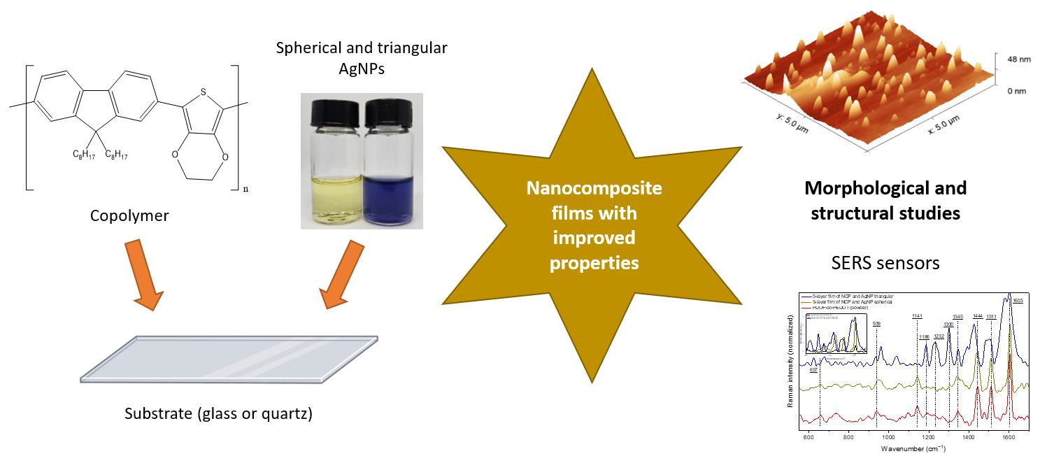
Nanocomposite Films of Silver Nanoparticles and Conjugated Copolymer in Natural and Nano-Form: Structural and Morphological Studies


Abstract
:
1. Introduction
2. Materials and Methods
2.1. Synthesis of the Copolymer and Silver Nanoparticles
2.2. Preparation and Characterization of Nanocopolymer (NCP)
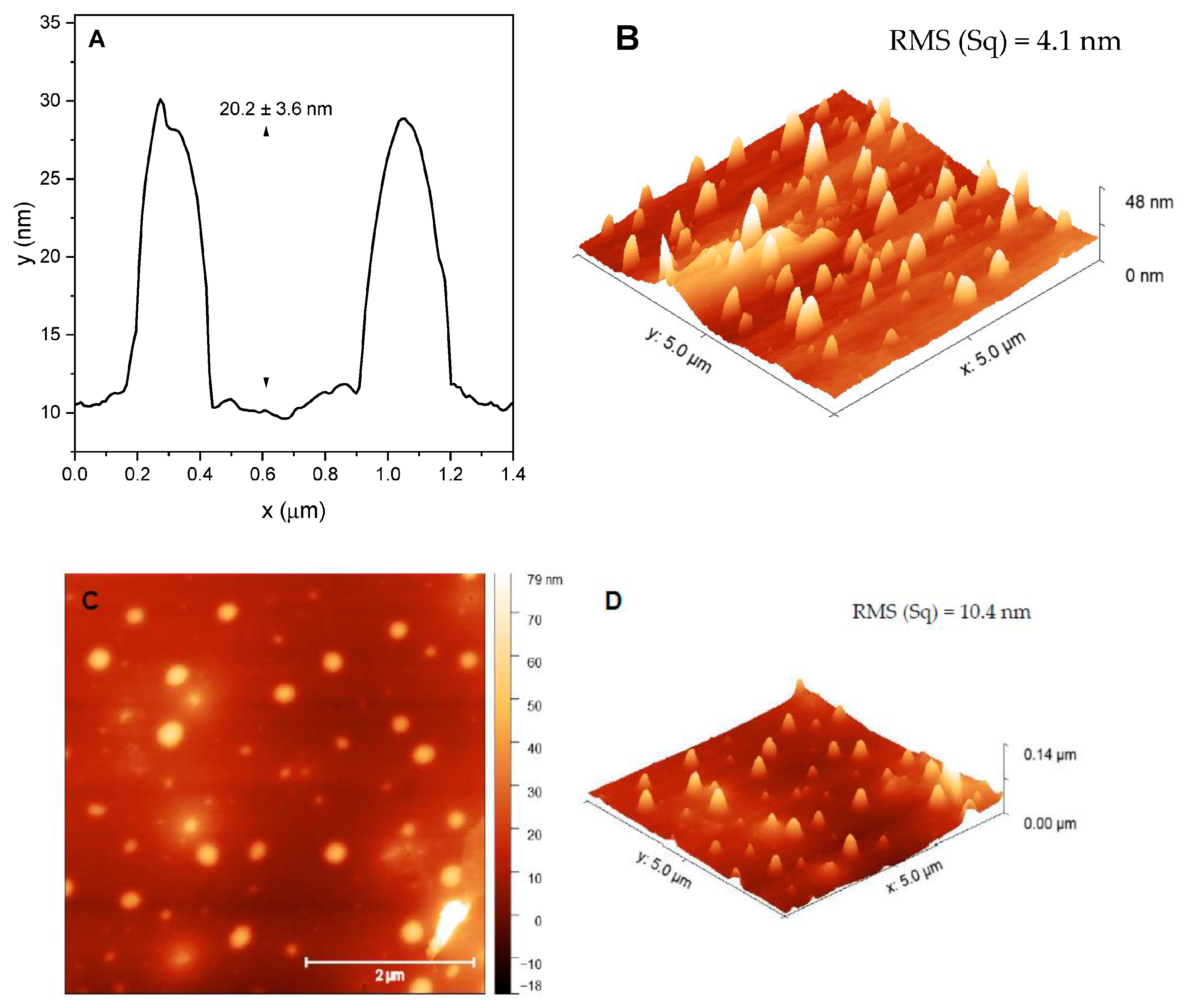
2.3. Preparation and Characterization of Spin Coating and Casting Films
2.4. Characterization Techniques
3. Results and Discussion
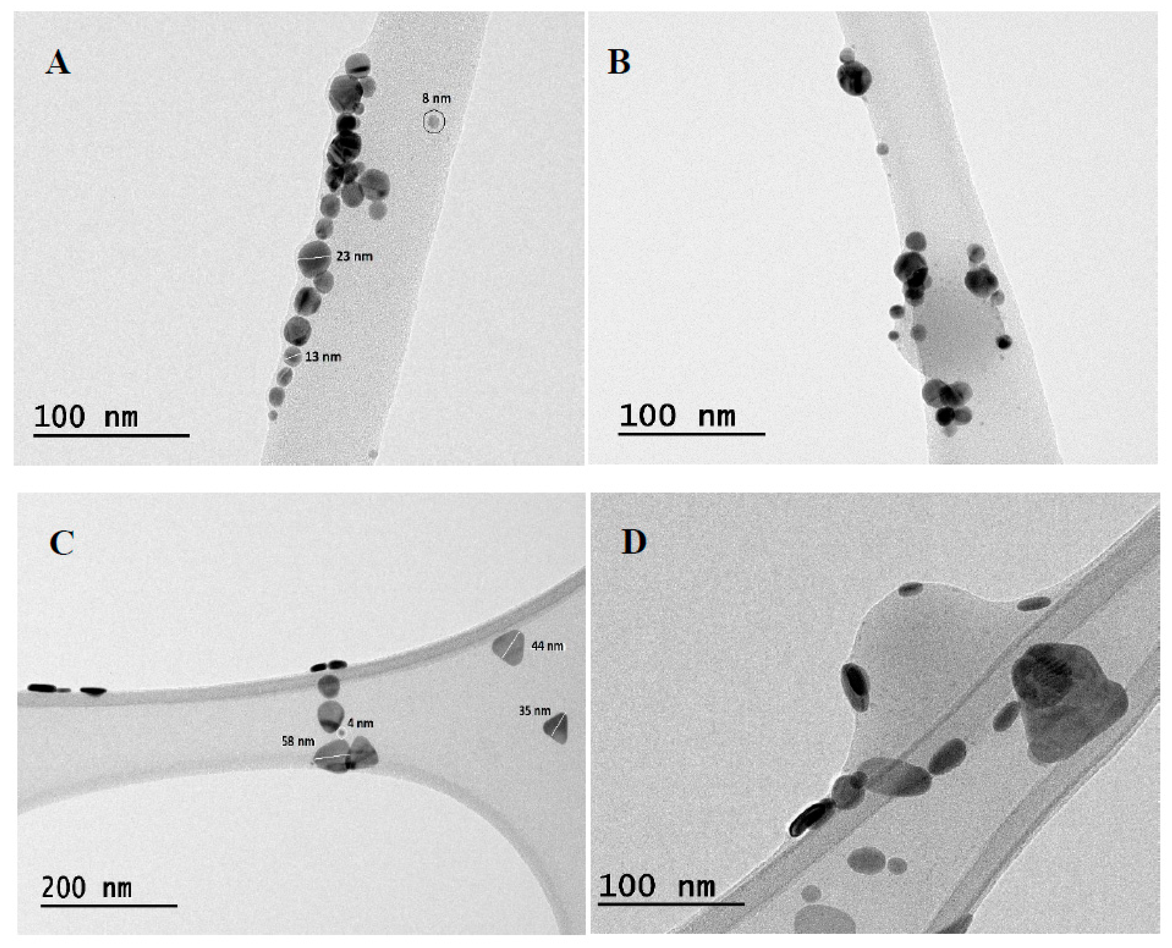
3.1. Characterizations of Pristine Materials and Dispersions of NCP and AgNP
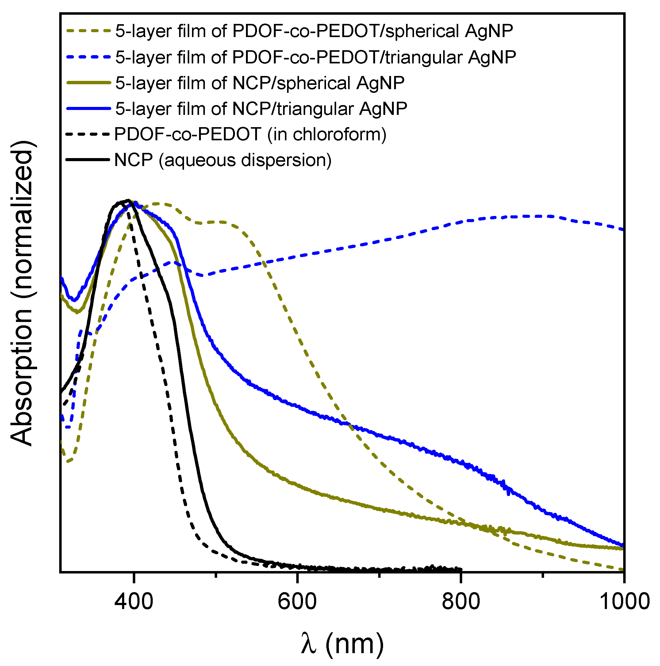
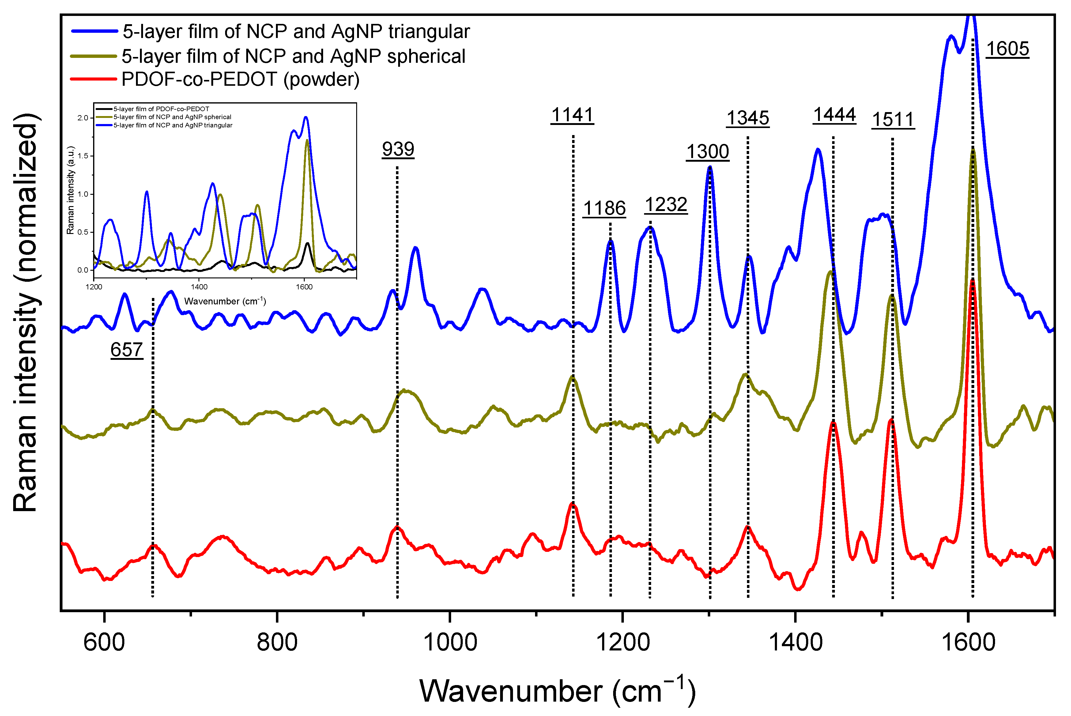
3.2. Films Containing PDOF-co-PEDOT or NCP with AgNP
- (i)
- band shifts, mainly for the film with triangular AgNP;
- (ii)
- increase in the relative intensities of the bands between 1350–1180 cm−1 and the band at 939 cm−1 for the film with triangular AgNP.
4. Conclusions
Supplementary Materials
Author Contributions
Funding
Institutional Review Board Statement
Informed Consent Statement
Data Availability Statement
Acknowledgments
Conflicts of Interest
References
- Da Rocha Rodrigues, R.; da Silva, R.L.C.G.; Caseli, L.; Péres, L.O. Conjugated Polymers as Langmuir and Langmuir-Blodgett Films: Challenges and Applications in Nanostructured Devices. Adv. Colloid Interface Sci. 2020, 285, 102277. [Google Scholar] [CrossRef]
- Salhi, O.; Ez-zine, T.; El Rhazi, M. Hybrid Materials Based on Conducting Polymers for Nitrite Sensing: A Mini Review. Electroanalysis 2021, 33, 1681–1690. [Google Scholar] [CrossRef]
- Tsai, M.H.; Lin, Y.K.; Luo, S.C. Electrochemical SERS for in Situ Monitoring the Redox States of PEDOT and Its Potential Application in Oxidant Detection. ACS Appl. Mater. Interfaces 2019, 11, 1402–1410. [Google Scholar] [CrossRef]
- De Jesus, C.G.; da Rocha Rodrigues, R.; Caseli, L.; Péres, L.O. Conducting Polymers Modulating the Catalytic Activity of Urease in Thin Composite Films. Colloids Surf. A Physicochem. Eng. Asp. 2022, 654, 130136. [Google Scholar] [CrossRef]
- Norrman, K.; Ghanbari-Siahkali, A.; Larsen, N.B. Studies of Spin-Coated Polymer Films. Annu. Reports Prog. Chem.-Sect. C 2005, 101, 174–201. [Google Scholar] [CrossRef]
- Rajamanickam, S.; Mohammad, S.M.; Hassan, Z.; Omar, A.F.; Muhammad, A. Investigations into Ag Nanoparticles–Carbon–Poly(9,9-Di-n-Octylfluorenyl-2,7-Diyl) (PFO) Composite: Morphological, Structural, Optical, and Electrical Characterization. Polym. Bull. 2022, 79, 9111–9130. [Google Scholar] [CrossRef]
- Bober, P.; Stejskal, J.; Trchová, M.; Hromádková, J.; Prokeš, J. Polyaniline-Coated Silver Nanowires. React. Funct. Polym. 2010, 70, 656–662. [Google Scholar] [CrossRef]
- Ayad, M.M.; Prastomo, N.; Matsuda, A.; Stejskal, J. Sensing of Silver Ions by Nanotubular Polyaniline Film Deposited on Quartz-Crystal in a Microbalance. Synth. Met. 2010, 160, 42–46. [Google Scholar] [CrossRef]
- Garai, A.; Chatterjee, S.; Nandi, A.K. Nanocomposites of Silver Nanoparticle and Dinonylnaphthalene Disulfonic Acid-Doped Thermoreversible Polyaniline Gel. Polym. Eng. Sci. 2010, 50, 446–454. [Google Scholar] [CrossRef]
- Liu, L.; Zhang, X.; Yang, L.; Ren, L.; Wang, D.; Ye, J. Metal Nanoparticles Induced Photocatalysis. Natl. Sci. Rev. 2017, 4, 761–780. [Google Scholar] [CrossRef]
- McNamara, K.; Tofail, S.A.M. Nanoparticles in Biomedical Applications. Adv. Phys. X 2017, 2, 54–88. [Google Scholar] [CrossRef]
- Petryayeva, E.; Algar, W.R. Toward Point-of-Care Diagnostics with Consumer Electronic Devices: The Expanding Role of Nanoparticles. RSC Adv. 2015, 5, 22256–22282. [Google Scholar] [CrossRef]
- Mota, D.R.; Martini, W.D.S.; Pellosi, D.S. Influence of Ag Size and Shape in Dye Photodegradation Using Silver Nanoparticle/ZnO Nanohybrids and Polychromatic Light. Environ. Sci. Pollut. Res. 2023, 30, 57667–57682. [Google Scholar] [CrossRef]
- Tong, Q.; Wang, W.; Fan, Y.; Dong, L. Recent Progressive Preparations and Applications of Silver-Based SERS Substrates. Trends Anal. Chem. 2018, 106, 246–258. [Google Scholar] [CrossRef]
- Kang, S.; Yoon, T.W.; Kim, G.Y.; Kang, B. Review of Conjugated Polymer Nanoparticles: From Formulation to Applications. ACS Appl. Nano Mater. 2022, 5, 17436–17460. [Google Scholar] [CrossRef]
- Braeken, Y.; Cheruku, S.; Ethirajan, A.; Maes, W. Conjugated Polymer Nanoparticles for Bioimaging. Materials 2017, 10, 1420. [Google Scholar] [CrossRef]
- Norio, M.; Suzuki, A. Palladium-Catalyzed Cross-Coupling Reactions of Organoboron Compounds. Chem. Rev. 1995, 95, 2457–2483. [Google Scholar]
- Da Rocha Rodrigues, R.; Caseli, L.; Péres, L.O. Langmuir and Langmuir-Blodgett Films of Poly[(9,9-Dioctylfluorene)-Co-(3-Hexylthiophene)] for Immobilization of Phytase: Possible Application as a Phytic Acid Sensor. Langmuir 2020, 36, 10587–10596. [Google Scholar] [CrossRef]
- Silva, M.M.; Mota, D.R.; Silva, C.B.; de Oliveira, H.P.M.; Pellosi, D.S. Synthesis of Pluronic-Based Silver Nanoparticles/Methylene Blue Nanohybrids: Influence of the Metal Shape on Photophysical Properties. Mater. Sci. Eng. C 2020, 114, 110987. [Google Scholar] [CrossRef]
- Yu, C.Y.; Godana, A.S. Conjugated Polymer Nanoparticles Based on Fluorenes, PEGylated Carbazoles and Diphenylamines. Eur. Polym. J. 2018, 99, 165–171. [Google Scholar] [CrossRef]
- Ouyang, M.; Xiang, W.Q.; Xu, Y.; Zhang, Y.J.; Lou, Q.P.; Zhang, C. Tuning the Emission Color of Conjugated Polymers via Oxidation Copolymerization of Fluorene and 3,4-Ethylenedioxythiophene. Polym. Polym. Compos. 2012, 20, 21–26. [Google Scholar] [CrossRef]
- Espinosa-Roa, A.; Cruz-Carrillo, M.D.J.; Ledesma-Juárez, A.; Montoya del Angel, A.; Romero-Borja, D.; Güizado-Rodríguez, M.; Rodríguez, M.; Galindo, R.; Maldonado, J.L.; Barba, V. Synthesis of Polyfluorenes by Oxidative Polymerization, Their Characterization and Implementation in Organic Solar Cells. J. Mater. Sci. Mater. Electron. 2019, 30, 2716–2725. [Google Scholar] [CrossRef]
- Perepichka, I.F.; Perepichka, D.F.; Meng, H.; Wudl, F. Light-Emitting Polythiophenes. Adv. Mater. 2005, 17, 2281–2305. [Google Scholar] [CrossRef]
- Hestand, N.J.; Spano, F.C. Expanded Theory of H- and J-Molecular Aggregates: The Effects of Vibronic Coupling and Intermolecular Charge Transfer. Chem. Rev. 2018, 118, 7069–7163. [Google Scholar] [CrossRef] [PubMed]
- Aggarwal, N.; Patnaik, A. A New Class of Nitroanilinic Dimer, the PNA O-Dimer: Electronic Structure and Emission Characteristics of O-Dimeric Aggregates. J. Phys. Chem. A 2015, 119, 8388–8399. [Google Scholar] [CrossRef]
- Patil, R.B.; Chougale, A.D. On the Shape Based SPR of Silver Nanostructures. Int. J. Nanotechnol. 2021, 18, 1015–1027. [Google Scholar] [CrossRef]
- Zhang, Q.; Li, N.; Goebl, J.; Lu, Z.; Yin, Y. A Systematic Study of the Synthesis of Silver Nanoplates: Is Citrate a “Magic” Reagent? J. Am. Chem. Soc. 2011, 133, 18931–18939. [Google Scholar] [CrossRef] [PubMed]
- Aherne, D.; Cara, M.; Kelly, J.M.; Gun’Ko, Y.K. From Ag Nanoprisms to Triangular Auag Nanoboxes. Adv. Funct. Mater. 2010, 20, 1329–1338. [Google Scholar] [CrossRef]
- Aherne, D.; Ledwith, D.M.; Gara, M.; Kelly, J.M. Optical Properties and Growth Aspects of Silver Nanoprisms Produced by a Highly Reproducible and Rapid Synthesis at Room Temperature. Adv. Funct. Mater. 2008, 18, 2005–2016. [Google Scholar] [CrossRef]
- Yang, J.; Li, H.; Wang, G.; He, B. Excimer Formation in Uniaxially Stretched Polymer Films. J. Appl. Polym. Sci. 2001, 82, 2347–2351. [Google Scholar] [CrossRef]
- Ibnaouf, K.H. Excimer State of a Conjugated Polymer (MEH-PPV) in Thin Films. Opt. Laser Technol. 2013, 48, 401–404. [Google Scholar] [CrossRef]
- Firet, N.J.; Blommaert, M.A.; Burdyny, T.; Venugopal, A.; Bohra, D.; Longo, A.; Smith, W.A. Operando EXAFS Study Reveals Presence of Oxygen in Oxide-Derived Silver Catalysts for Electrochemical CO2 Reduction. J. Mater. Chem. A 2019, 7, 2597–2607. [Google Scholar] [CrossRef]
- Liu, Y.; Zhang, Q.; Yuan, H.; Luo, K.; Li, J.; Hu, W.; Pan, Z.; Xu, M.; Xu, S.; Levchenko, I.; et al. Comparative Study of Photocatalysis and Gas Sensing of ZnO/Ag Nanocomposites Synthesized by One- and Two-Step Polymer-Network Gel Processes. J. Alloys Compd. 2021, 868, 158723. [Google Scholar] [CrossRef]
- Yun, D.J.; Jung, J.; Sung, Y.M.; Ra, H.; Kim, J.M.; Chung, J.G.; Kim, S.Y.; Kim, Y.S.; Heo, S.; Kim, K.H.; et al. In-Situ Photoelectron Spectroscopy Study on the Air Degradation of PEDOT:PSS in Terms of Electrical and Thermoelectric Properties. Adv. Electron. Mater. 2020, 6, 2000620. [Google Scholar] [CrossRef]
- Vargas-Villanueva, S.; Torres-Ceron, D.A.; Amaya-Roncancio, S.; Arellano-Ramírez, I.D.; Riva, J.S.; Restrepo-Parra, E. Study of the Incorporation of S in TiO2/SO4 Coatings Produced by PEO Process through XPS and DFT. Appl. Surf. Sci. 2022, 599, 153811. [Google Scholar] [CrossRef]
- Schultheiss, A.; Gueye, M.; Carella, A.; Benayad, A.; Pouget, S.; Faure-Vincent, J.; Demadrille, R.; Revaux, A.; Simonato, J.P. Insight into the Degradation Mechanisms of Highly Conductive Poly(3,4-Ethylenedioxythiophene) Thin Films. ACS Appl. Polym. Mater. 2020, 2, 2686–2695. [Google Scholar] [CrossRef]
- Gong, X.; Iyer, P.K.; Moses, D.; Bazan, G.C.; Heeger, A.J.; Xiao, S.S. Stabilized Blue Emission from Polyfluorene-Based Light-Emitting Diodes: Elimination of Fluorenone Defects. Adv. Funct. Mater. 2003, 13, 325–330. [Google Scholar] [CrossRef]
- Marciniak, S.; Crispin, X.; Uvdal, K.; Trzcinski, M.; Birgerson, J.; Groenendaal, L.; Louwet, F.; Salaneck, W.R. Light Induced Damage in Poly(3,4-Ethylenedioxythiophene) and Its Derivatives Studied by Photoelectron Spectroscopy. Synth. Met. 2004, 141, 67–73. [Google Scholar] [CrossRef]
- Stavytska-Barba, M.; Kelley, A.M. Surface-Enhanced Raman Study of the Interaction of PEDOT: PSS with Plasmonically Active Nanoparticles. J. Phys. Chem. C 2010, 114, 6822–6830. [Google Scholar] [CrossRef]
- Mohamed, I.M.A.; Yasin, A.S.; Liu, C. Synthesis, Surface Characterization and Electrochemical Performance of ZnO @ Activated Carbon as a Supercapacitor Electrode Material in Acidic and Alkaline Electrolytes. Ceram. Int. 2020, 46, 3912–3920. [Google Scholar] [CrossRef]
- Tan, T.; Lee, P.K.; Marium, M.; Zettsu, N.; Yu, D.Y.W. (3-Aminopropyl)Triethoxysilane as an Electrolyte Additive for Enhancing the Thermal Stability of Silicon Anode in Lithium-Ion Batteries. ACS Appl. Energy Mater. 2022, 5, 11254–11262. [Google Scholar] [CrossRef]
- O’Carroll, D.; Iacopino, D.; O’Riordan, A.; Lovera, P.; O’Connor, É.; O’Brien, G.A.; Redmond, G. Poly(9,9-Dioctylfluorene) Nanowires with Pronounced β-Phase Morphology: Synthesis, Characterization, and Optical Properties. Adv. Mater. 2008, 20, 42–48. [Google Scholar] [CrossRef]
- Palacios, R.; Formentin, P.; Martinez-Ferrero, E.; Pallarès, J.; Marsal, L.F. β-Phase Morphology in Ordered Poly(9,9-Dioctylfluorene) Nanopillars by Template Wetting Method. Nanoscale Res. Lett. 2011, 6, 35. [Google Scholar] [CrossRef] [PubMed]
- Moraes, B.R.; Campos, N.S.; Izumi, C.M.S. Surface-Enhanced Raman Scattering of EDOT and PEDOT on Silver and Gold Nanoparticles. Vib. Spectrosc. 2018, 96, 137–142. [Google Scholar] [CrossRef]
- Sarkar, S.; Bhowal, A.C.; Kandimalla, R.; Kundu, S. Structural and Electrical Behaviours of PEDOT:PSS Thin Films in Presence of Negatively Charged Gold and Silver Nanoparticles: A Green Synthesis Approach. Synth. Met. 2021, 279, 116848. [Google Scholar] [CrossRef]
- He, Q.F.; Zhang, Y.J.; Yang, Z.L.; Dong, J.C.; Lin, X.M.; Li, J.F. Surface-Enhanced Raman Spectroscopy: Principles, Methods, and Applications in Energy Systems. Chin. J. Chem. 2023, 41, 355–369. [Google Scholar] [CrossRef]


| Diameter (nm) | PDI | Zeta Potential (mV) | |
|---|---|---|---|
| Spherical AgNP | 13.37 | 0.61 | −29.50 |
| Triangular AgNP | 28.65 | 0.61 | −37.80 |
| Nanocopolymer (NCP) | 99.96 | 0.11 | 0.02 |
| NCP/spherical AgNP | 93.57 | 0.14 | −49.60 |
| NCP/triangular AgNP | 82.09 | 0.65 | −43.00 |
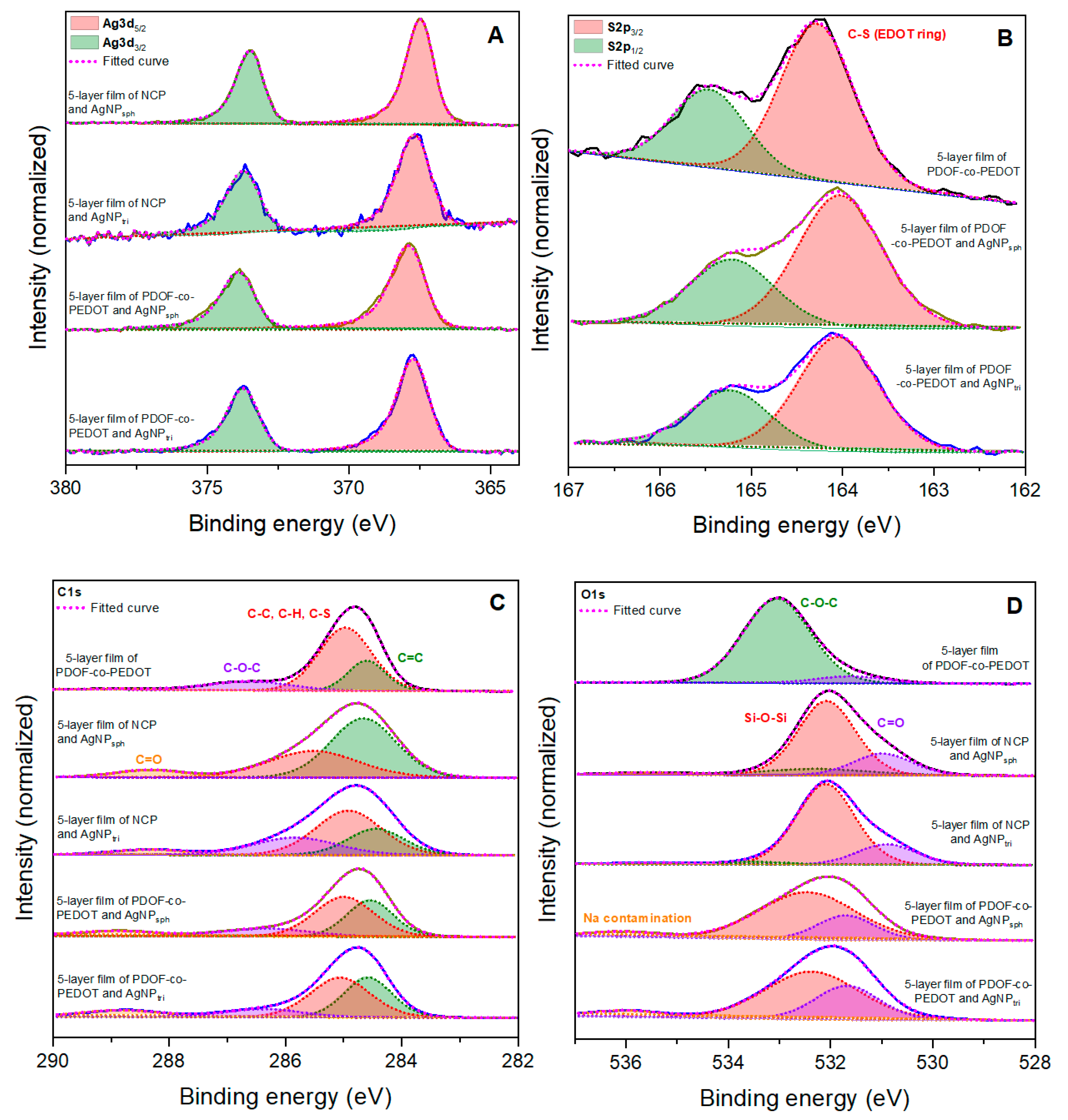
| 5-Layer Film | O1s (% Area) | C1s (% Area) | |
|---|---|---|---|
| C=O | Na Contamination | C=O | |
| NCP/spherical AgNP | 20.04 | 2.38 | 7.62 |
| NCP/triangular AgNP | 19.28 | 1.86 | 6.25 |
| PDOF-co-PEDOT/spherical AgNP | 22.15 | 5.51 | 8.35 |
| PDOF-co-PEDOT/triangular AgNP | 30.41 | 6.48 | 10.04 |
| Raman Signal (cm−1) | Assignments | Literature |
|---|---|---|
| 1511 | asymmetric Cα=Cβ stretch mode of the thiophene ring | [39] |
| 1444 | symmetric Cα=Cβ stretch mode of thiophene | [44] |
| 1345 | Cβ-Cβ stretch mode of inter-ring bonds of thiophene | [44,45] |
| 1141 | C-H bending modes from side chains of fluorene groups | [42] |
| 939 | oxyethylene ring deformation | [44] |
| 657 | C-S-C stretch mode | [45] |
Disclaimer/Publisher’s Note: The statements, opinions and data contained in all publications are solely those of the individual author(s) and contributor(s) and not of MDPI and/or the editor(s). MDPI and/or the editor(s) disclaim responsibility for any injury to people or property resulting from any ideas, methods, instructions or products referred to in the content. |
© 2023 by the authors. Licensee MDPI, Basel, Switzerland. This article is an open access article distributed under the terms and conditions of the Creative Commons Attribution (CC BY) license (https://creativecommons.org/licenses/by/4.0/).
Share and Cite
Rodrigues, R.d.R.; Pellosi, D.S.; Louarn, G.; Péres, L.O. Nanocomposite Films of Silver Nanoparticles and Conjugated Copolymer in Natural and Nano-Form: Structural and Morphological Studies. Materials 2023, 16, 3663. https://doi.org/10.3390/ma16103663
Rodrigues RdR, Pellosi DS, Louarn G, Péres LO. Nanocomposite Films of Silver Nanoparticles and Conjugated Copolymer in Natural and Nano-Form: Structural and Morphological Studies. Materials. 2023; 16(10):3663. https://doi.org/10.3390/ma16103663
Chicago/Turabian StyleRodrigues, Rebeca da Rocha, Diogo Silva Pellosi, Guy Louarn, and Laura Oliveira Péres. 2023. "Nanocomposite Films of Silver Nanoparticles and Conjugated Copolymer in Natural and Nano-Form: Structural and Morphological Studies" Materials 16, no. 10: 3663. https://doi.org/10.3390/ma16103663