
A Label-Free Fluorescent Sensor Based on Si,N-Codoped Carbon Quantum Dots with Enhanced Sensitivity for the Determination of Cr(VI)


Abstract
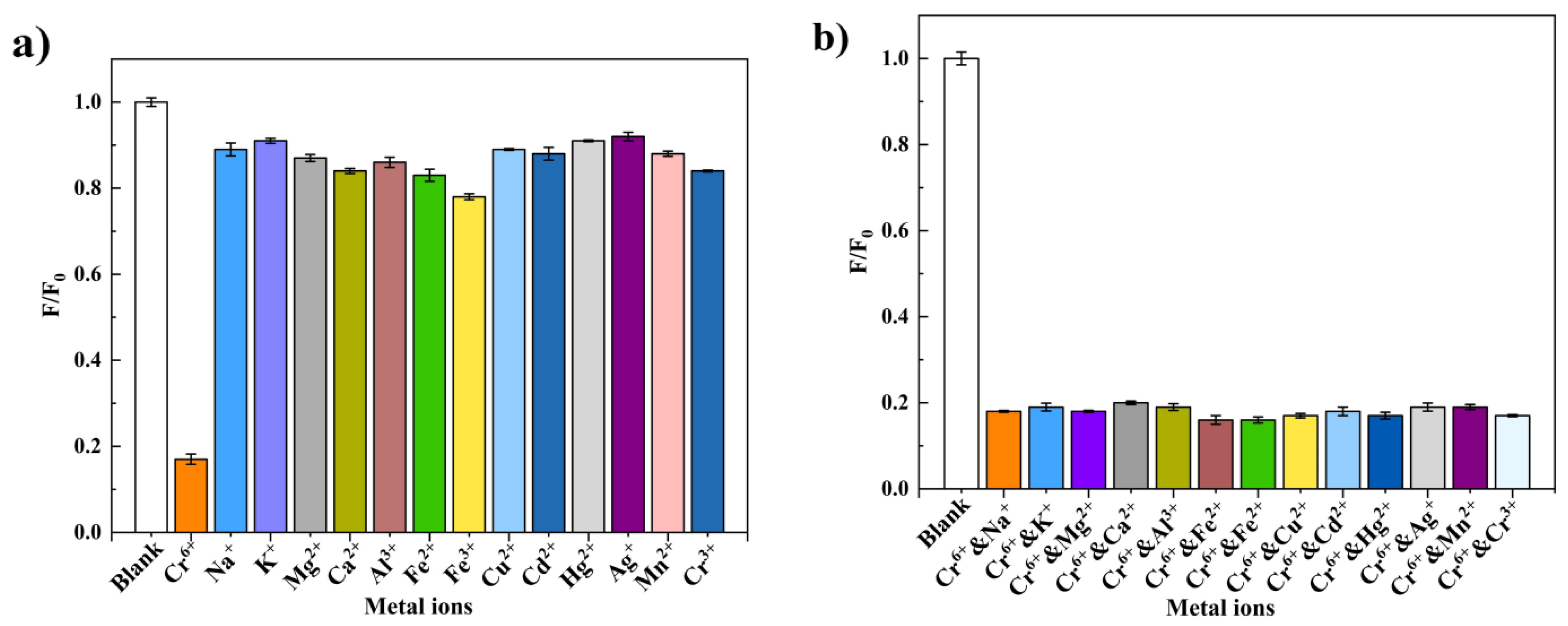
:
1. Introduction
2. Materials and Methods
2.1. Materials
2.2. Preparation of Si,N-CQDs
2.3. Characterization
2.4. Fluorescence Quantum Yield (QY) of Si,N-CQDs
2.5. Detection of Cr(VI)
3. Results and Discussion
3.1. Optical Properties of Si,N-CQDs
3.2. Structural Characterization of Si,N-CQDs
3.3. Detection for Cr(VI)
3.4. Quenching Mechanism
4. Conclusions
Author Contributions
Funding
Institutional Review Board Statement
Informed Consent Statement
Data Availability Statement
Acknowledgments
Conflicts of Interest
References
- Arumugham, T.; Alagumuthu, M.; Amimodu, R.G.; Munusamy, S.; Iyer, S.K. A sustainable synthesis of green carbon quantum dot (CQD) from Catharanthus roseus (white flowering plant) leaves and investigation of its dual fluorescence responsive behavior in multi-ion detection and biological applications. Sustain. Mater. Technol. 2020, 23, e00138. [Google Scholar] [CrossRef]
- Wang, M.; Shi, R.; Gao, M.; Zhang, K.; Deng, L.; Fu, Q.; Wang, L.; Gao, D. Sensitivity fluorescent switching sensor for Cr(VI) and ascorbic acid detection based on orange peels-derived carbon dots modified with EDTA. Food Chem. 2020, 318, 126506. [Google Scholar] [CrossRef]
- Ming, F.; Hou, J.; Hou, C.; Yang, M.; Wang, X.; Li, J.; Huo, D.; He, Q. One-step synthesized fluorescent nitrogen doped carbon dots from thymidine for Cr(VI) detection in water. Spectrochim. Acta Mol. Biomol. Spectrosc. 2019, 222, 117165. [Google Scholar] [CrossRef]
- Liu, M.; Li, T.; Zhang, C.; Zheng, Y.; Wu, C.; Zhang, J.; Zhang, K.; Zhang, Z. Fluorescent carbon dots embedded in mesoporous silica nanospheres: A simple platform for Cr(VI) detection in environmental water. J. Hazard. Mater. 2021, 415, 125699. [Google Scholar] [CrossRef]
- Ravindran, A.; Elavarasi, M.; Prathna, T.C.; Raichur, A.M.; Chandrasekaran, N.; Mukherjee, A. Selective colorimetric detection of nanomolar Cr(VI) in aqueous solutions using unmodified silver nanoparticles. Sens. Actuators B Chem. 2012, 166, 365–371. [Google Scholar] [CrossRef]
- Panichev, N.; Mandiwana, K.; Kataeva, M.; Siebert, S. Determination of Cr(VI) in plants by electrothermal atomic absorption spectrometry after leaching with sodium carbonate. Spectrochim. Acta Part B At. Spectrosc. 2005, 60, 699–703. [Google Scholar] [CrossRef]
- Spanu, D.; Monticelli, D.; Binda, G.; Dossi, C.; Rampazzi, L.; Recchia, S. One-minute highly selective Cr(VI) determination at ultra-trace levels: An ICP-MS method based on the on-line trapping of Cr(III). J. Hazard. Mater. 2021, 412, 125280. [Google Scholar] [CrossRef]
- Frois, S.R.; Grassi, M.T.; de Campos, M.S.; Abate, G. Determination of Cr(VI) in water samples by ICP-OES after separation of Cr(III) by montmorillonite. Anal. Methods 2012, 4, 4389–4394. [Google Scholar] [CrossRef]
- Chen, S.; Zhang, X.; Yu, L.; Wang, L.; Li, H. Simultaneous determination of Cr(III) and Cr(VI) in tannery wastewater using low pressure ion chromatography combined with flow injection spectrophotometry. Spectrochim. Acta Mol. Biomol. Spectrosc. 2012, 88, 49–55. [Google Scholar] [CrossRef]
- Wang, Y.; Hu, X.; Li, W.; Huang, X.; Li, Z.; Zhang, W.; Zhang, X.; Zou, X.; Shi, J. Preparation of boron nitrogen co-doped carbon quantum dots for rapid detection of Cr(VI). Spectrochim. Acta Mol. Biomol. Spectrosc. 2020, 243, 118807. [Google Scholar] [CrossRef]
- Ji, Y.; Zou, X.; Wang, W.; Wang, T.; Zhang, S.; Gong, Z. Co-Doped S, N-Carbon dots and its fluorescent film sensors for rapid detection of Cr(VI) and Ascorbic acid. Microchem. J. 2021, 167, 106284. [Google Scholar] [CrossRef]
- Barman, S.; Sadhukhan, M. Facile bulk production of highly blue fluorescent graphitic carbon nitride quantum dots and their application as highly selective and sensitive sensors for the detection of mercuric and iodide ions in aqueous media. J. Mater. Chem. 2012, 22, 21832–21837. [Google Scholar] [CrossRef]
- Kaur, J.; Sharma, S.; Mehta, S.K.; Kansal, S.K. Highly photoluminescent and pH sensitive nitrogen doped carbon dots (NCQDss) as a fluorescent sensor for the efficient detection of Cr(VI) ions in aqueous media. Spectrochim. Acta Mol. Biomol. Spectrosc. 2020, 227, 117572. [Google Scholar] [CrossRef]
- Ahn, J.; Song, Y.; Kwon, J.E.; Lee, S.H.; Park, K.S.; Kim, S.; Woo, J.; Kim, H. Food waste-driven N-doped carbon dots: Applications for Fe3+ sensing and cell imaging. Mater. Sci. Eng. C 2019, 102, 106–112. [Google Scholar] [CrossRef]
- Rajput, A.; Raj, S.K.; Lebedeva, O.V.; Chesnokova, A.N.; Raskulova, T.V.; Kulshrestha, V. Functionalized carbon dots composite cation exchange membranes: Improved electrochemical performance and salt removal efficiency. Colloids Surf. A Physicochem. Eng. Asp. 2021, 609, 125677. [Google Scholar] [CrossRef]
- Magdy, G.; Hakiem AF, A.; Belal, F.; Abdel-Megied, A.M. Green one-pot synthesis of nitrogen and sulfur co-doped carbon quantum dots as new fluorescent nanosensors for determination of salinomycin and maduramicin in food samples. Food Chem. 2021, 343, 128539. [Google Scholar] [CrossRef]
- Xu, X.; Ray, R.; Gu, Y.; Ploehn, H.J.; Gearheart, L.; Raker, K.; Scrivens, W.A. Electrophoretic analysis and purification of fluorescent single-walled carbon nanotube fragments. J. Am. Chem. Soc. 2004, 126, 12736–12737. [Google Scholar] [CrossRef]
- Sun, Y.-P.; Wang, X.; Lu, F.; Cao, L.; Meziani, M.J.; Luo, P.G.; Gu, L.; Veca, L.M. Doped carbon nanoparticles as a new platform for highly photoluminescent dots. J. Phys. Chem. C 2008, 112, 18295–18298. [Google Scholar] [CrossRef] [Green Version]
- Long, Z.; Fang, D.-C.; Ren, H.; Ouyang, J.; He, L.; Na, N. Excited oxidized-carbon nanodots induced by ozone from low-temperature plasma to initiate strong chemiluminescence for fast discrimination of metal ions. Anal. Chem. 2016, 88, 7660–7666. [Google Scholar] [CrossRef]
- Peng, H.; Travas-Sejdic, J. Simple aqueous solution route to luminescent carbogenic dots from carbohydrates. Chem. Mater. 2009, 21, 5563–5565. [Google Scholar] [CrossRef]
- Ng HK, M.; Lim, G.K.; Leo, C.P. Comparison between hydrothermal and microwave-assisted synthesis of carbon dots from biowaste and chemical for heavy metal detection: A review. Microchem. J. 2021, 165, 106116. [Google Scholar]
- Wang, F.; Pang, S.; Wang, L.; Li, Q.; Kreiter, M.; Liu, C.-Y. One-step synthesis of highly luminescent carbon dots in noncoordinating solvents. Chem. Mater. 2010, 22, 4528–4530. [Google Scholar] [CrossRef]
- Mao, L.-H.; Tang, W.-Q.; Deng, Z.-Y.; Liu, S.-S.; Wang, C.-F.; Chen, S. Facile access to white fluorescent carbon dots toward light-emitting devices. Ind. Eng. Chem. Res. 2014, 53, 6417–6425. [Google Scholar] [CrossRef]
- Zhong, D.; Zhuo, Y.; Feng, Y.; Yang, X. Employing carbon dots modified with vancomycin for assaying Gram-positive bacteria like Staphylococcus aureus. Biosens. Bioelectron. 2015, 74, 546–553. [Google Scholar] [CrossRef]
- Hu, G.; Ge, L.; Li, Y.; Mukhtar, M.; Shen, B.; Yang, D.; Li, J. Carbon dots derived from flax straw for highly sensitive and selective detections of cobalt, chromium, and ascorbic acid. J. Colloid Interface Sci. 2020, 579, 96–108. [Google Scholar] [CrossRef]
- Xie, Z.; Yin, Z.; Wu, Y.; Liu, C.; Hao, X.; Du, Q.; Xu, X. White light-emitting diodes based on individual polymerized carbon nanodots. Sci. Rep. 2017, 7, 12146. [Google Scholar] [CrossRef] [Green Version]
- Laddha, H.; Yadav, P.; Jain, Y.; Sharma, M.; Reza, M.; Agarwal, M.; Gupta, R. One-pot microwave-assisted synthesis of blue emissive multifunctional NSP co-doped carbon dots as a nanoprobe for sequential detection of Cr(VI) and ascorbic acid in real samples, fluorescent ink and logic gate operation. J. Mol. Liq. 2022, 346, 117088. [Google Scholar] [CrossRef]
- Liu, Y.; Mao, L.; Liu, X.; Liu, M.; Xu, D.; Jiang, R.; Deng, F.; Li, Y.; Zhang, X.; Wei, Y. A facile strategy for fabrication of aggregation-induced emission (AIE) active fluorescent polymeric nanoparticles (FPNs) via post modification of synthetic polymers and their cell imaging. Mater. Sci. Eng. C 2017, 79, 590–595. [Google Scholar] [CrossRef]
- Luo, P.G.; Sahu, S.; Yang, S.-T.; Sonkar, S.K.; Wang, J.; Wang, H.; LeCroy, G.E.; Cao, L.; Sun, Y.-P. Carbon “quantum” dots for optical bioimaging. J. Mater. Chem. B 2013, 1, 2116–2127. [Google Scholar] [CrossRef]
- Yang, Z.-C.; Wang, M.; Yong, A.M.; Wong, S.Y.; Zhang, X.-H.; Tan, H.; Chang, A.Y.; Li, X.; Wang, J. Intrinsically fluorescent carbon dots with tunable emission derived from hydrothermal treatment of glucose in the presence of monopotassium phosphate. Chem. Commun. 2011, 47, 11615–11617. [Google Scholar] [CrossRef]
- Sahu, S.; Behera, B.; Maiti, T.K.; Mohapatra, S. Simple one-step synthesis of highly luminescent carbon dots from orange juice: Application as excellent bio-imaging agents. Chem. Commun. 2012, 48, 8835–8837. [Google Scholar] [CrossRef] [PubMed]
- Zhu, S.; Meng, Q.; Wang, L.; Zhang, J.; Song, Y.; Jin, H.; Zhang, K.; Sun, H.; Wang, H.; Yang, B. Highly photoluminescent carbon dots for multicolor patterning, sensors, and bioimaging. Angew. Chem. Int. Ed. 2013, 125, 4045–4049. [Google Scholar] [CrossRef]
- Xu, Q.; Kuang, T.; Liu, Y.; Cai, L.; Peng, X.; Sreeprasad, T.S.; Zhao, P.; Yu, Z.; Li, N. Heteroatom-doped carbon dots: Synthesis, characterization, properties, photoluminescence mechanism and biological applications. J. Mater. Chem. B 2016, 4, 7204–7219. [Google Scholar] [CrossRef] [PubMed]
- Yang, G.; Wu, C.; Luo, X.; Liu, X.; Gao, Y.; Wu, P.; Cai, C.; Saavedra, S.S. Exploring the emissive states of heteroatom-doped graphene quantum dots. J. Phys. Chem. C 2018, 122, 6483–6492. [Google Scholar] [CrossRef]
- Park, S.K.; Lee, H.; Choi, M.S.; Suh, D.H.; Nakhanivej, P.; Park, H.S. Straightforward and controllable synthesis of heteroatom-doped carbon dots and nanoporous carbons for surface-confined energy and chemical storage. Energy Storage Mater. 2018, 12, 331–340. [Google Scholar] [CrossRef]
- Das, P.; Ganguly, S.; Mondal, S.; Bose, M.; Das, A.K.; Banerjee, S.; Das, N.C. Heteroatom doped photoluminescent carbon dots for sensitive detection of acetone in human fluids. Sens. Actuators B Chem. 2018, 266, 583–593. [Google Scholar] [CrossRef]
- Sharma, S.; Ghosh, K.S. Recent advances (2017–2020) in the detection of copper ion by using fluorescence sensors working through transfer of photo-induced electron (PET), excited-state intramolecular proton (ESIPT) and Förster resonance energy (FRET). Spectrochim. Acta Mol. Biomol. Spectrosc. 2021, 254, 119610. [Google Scholar] [CrossRef]
- Vallet-Regí, M.; Colilla, M.; Izquierdo-Barba, I. Bioactive mesoporous silicas as controlled delivery systems: Application in bone tissue regeneration. J. Biomed. Nanotechnol. 2008, 4, 1–15. [Google Scholar]
- Zan, M.; Rao, L.; Huang, H.; Xie, W.; Zhu, D.; Li, L.; Qie, X.; Guo, S.-S.; Zhao, X.-Z.; Liu, W. A strong green fluorescent nanoprobe for highly sensitive and selective detection of nitrite ions based on phosphorus and nitrogen co-doped carbon quantum dots. Sens. Actuators B Chem. 2018, 262, 555–561. [Google Scholar] [CrossRef]
- Jiang, R.; Liu, M.; Li, C.; Huang, Q.; Huang, H.; Wan, Q.; Wen, Y.; Cao, Q.-Y.; Zhang, X.; Wei, Y. Facile fabrication of luminescent polymeric nanoparticles containing dynamic linkages via a one-pot multicomponent reaction: Synthesis, aggregation-induced emission and biological imaging. Mater. Sci. Eng. C 2017, 80, 708–714. [Google Scholar] [CrossRef]
- Cao, Q.-Y.; Jiang, R.; Liu, M.; Wan, Q.; Xu, D.; Tian, J.; Huang, H.; Wen, Y.; Zhang, X.; Wei, Y. Microwave-assisted multicomponent reactions for rapid synthesis of AIE-active fluorescent polymeric nanoparticles by post-polymerization method. Mater. Sci. Eng. C 2017, 80, 578–583. [Google Scholar] [CrossRef] [PubMed]
- Jiang, X.; Huang, J.; Chen, T.; Zhao, Q.; Xu, F.; Zhang, X. Synthesis of hemicellulose/deep eutectic solvent based carbon quantum dots for ultrasensitive detection of Ag+ and L-cysteine with “off-on” pattern. Int. J. Biol. Macromol. 2020, 153, 412–420. [Google Scholar] [CrossRef] [PubMed]
- Ma, Y.; Chen, A.Y.; Huang, Y.Y.; He, X.; Xie, X.F.; He, B.; Yang, J.H.; Wang, X.Y. Off-on fluorescent switching of boron-doped carbon quantum dots for ultrasensitive sensing of catechol and glutathione. Carbon 2020, 162, 234–244. [Google Scholar] [CrossRef]
- Ghanem, A.; Al-Marjeh, R.A.-Q.B.; Atassi, Y. Novel nitrogen-doped carbon dots prepared under microwave-irradiation for highly sensitive detection of mercury ions. Heliyon 2020, 6, e03750. [Google Scholar] [CrossRef]
- Qiu, Y.; Gao, D.; Yin, H.; Zhang, K.; Zeng, J.; Wang, L.; Xia, L.; Zhou, K.; Xia, Z.; Fu, Q. Facile, green and energy-efficient preparation of fluorescent carbon dots from processed traditional Chinese medicine and their applications for on-site semi-quantitative visual detection of Cr(VI). Sens. Actuators B-Chem. 2020, 324, 128722. [Google Scholar] [CrossRef]
- Kaur, N.; Mehta, A.; Mishra, A.; Chaudhary, S.; Rawat, M.; Basu, S. Amphiphilic carbon dots derived by cationic surfactant for selective and sensitive detection of metal ions. Mater. Sci. Eng. C 2019, 95, 72–77. [Google Scholar] [CrossRef]
- Cao, D.; Luo, Y.X.; Liu, W.P.; Li, Y.S.; Gao, X.F. Enzyme-free fluorescence determination of uric acid and trace Hg(II) in serum using Si/N doped carbon dots. Spectrochim. Acta Mol. Biomol. Spectrosc. 2021, 263, 120182. [Google Scholar] [CrossRef]
- Li, X.; Chai, C.; Zhang, Y.; Wang, Y.; Lv, J.; Bian, W.; Choi, M. Microwave synthesis of nitrogen and sulfur co-doped carbon dots for the selective detection of Hg2+ and glutathione. Opt. Mater. 2020, 99, 109559. [Google Scholar] [CrossRef]
- Fang, B.; Lu, X.; Hu, J.; Zhang, G.; Zheng, X.; He, L.; Cao, J.; Gu, J.; Cao, F. pH controlled green luminescent carbon dots derived from benzoxazine monomers for the fluorescence turn-on and turn-off detection. J. Colloid Interface Sci. 2019, 536, 516–525. [Google Scholar] [CrossRef]
- Zhang, W.J.; Liu, S.G.; Han, L.; Luo, H.Q.; Li, N.B. A ratiometric fluorescent and colorimetric dual-signal sensing platform based on N-doped carbon dots for selective and sensitive detection of copper(II) and pyrophosphate ion. Sens. Actuators B Chem. 2019, 283, 215–221. [Google Scholar] [CrossRef]
- Pu, Z.F.; Wen, Q.L.; Yang, Y.J.; Cui, X.M.; Cao, Q.E. Fluorescent carbon quantum dots synthesized using phenylalanine and citric acid for selective detection of Fe3+ ions. Spectrochim. Acta Mol. Biomol. Spectrosc. 2019, 229, 117944. [Google Scholar] [CrossRef] [PubMed]
- He, S.; Qi, S.; Sun, Z.; Zhu, G.; Zhang, K.; Chen, W. Si, N-codoped carbon dots: Preparation and application in iron overload diagnosis. J. Mater. Sci. 2019, 54, 4297–4305. [Google Scholar] [CrossRef]


Publisher’s Note: MDPI stays neutral with regard to jurisdictional claims in published maps and institutional affiliations. |
© 2022 by the authors. Licensee MDPI, Basel, Switzerland. This article is an open access article distributed under the terms and conditions of the Creative Commons Attribution (CC BY) license (https://creativecommons.org/licenses/by/4.0/).
Share and Cite
Zhang, J.; Jing, C.; Wang, B. A Label-Free Fluorescent Sensor Based on Si,N-Codoped Carbon Quantum Dots with Enhanced Sensitivity for the Determination of Cr(VI). Materials 2022, 15, 1733. https://doi.org/10.3390/ma15051733
Zhang J, Jing C, Wang B. A Label-Free Fluorescent Sensor Based on Si,N-Codoped Carbon Quantum Dots with Enhanced Sensitivity for the Determination of Cr(VI). Materials. 2022; 15(5):1733. https://doi.org/10.3390/ma15051733
Chicago/Turabian StyleZhang, Jinyu, Cai Jing, and Binsong Wang. 2022. "A Label-Free Fluorescent Sensor Based on Si,N-Codoped Carbon Quantum Dots with Enhanced Sensitivity for the Determination of Cr(VI)" Materials 15, no. 5: 1733. https://doi.org/10.3390/ma15051733