Article

The Synthesis and Characterisation of the High-Hardness Magnetic Material Mn2N0.86

1
State Key Laboratory of Superhard Materials, College of Physics, Jilin University, Changchun 130012, China
2
Institute of High Pressure Physics, School of Physical Science and Technology, Ningbo University, Ningbo 315211, China
*
Authors to whom correspondence should be addressed.
Materials 2022, 15(21), 7780; https://doi.org/10.3390/ma15217780
Submission received: 2 October 2022 / Revised: 28 October 2022 / Accepted: 1 November 2022 / Published: 4 November 2022

Abstract

:
High-quality P6322 Mn2N0.86 samples were synthesised using a high-pressure metathesis reaction, and the properties of the material were investigated. The measurements revealed that the Vickers hardness was 7.47 GPa, which is less than that predicted by commonly used theoretical models. At low air pressure, Mn2N0.86 and MnO coexist at 500 to 600 °C, and by excluding air, we succeeded in producing Mn4N by heating Mn2N0.86 in nitrogen atmosphere; we carefully studied this process with thermogravimetry and differential scanning calorimetry (TG-DSC). This gives a hint that to control temperature, air pressure and gas concentration might be an effective way to prepare fine Mn-N-O catalysis. Magnetic measurements indicated that ferromagnetism and antiferromagnetism coexist within Mn2N0.86 at room temperature and that these magnetic properties are induced by nitrogen vacancies. Ab intio simulation was used to probe the nature of the magnetism in greater detail. The research contributes to the available data and the understanding of Mn2N0.86 and suggests ways to control the formation of materials based on Mn2N0.86.

1. Introduction

Transition metal nitrides have attracted much attention owing to their excellent electronic and magnetic properties, hardness, and potential applications [1,2,3,4]. Currently, hermetic sintering [5,6], the arc method [7,8,9], and film deposition [10] are commonly used to produce these compounds. However, all of these methods have drawbacks: nitrogen vacancies inevitably arise during hermetic sintering and with the arc method because of the nature of nitrogen bonding and the high temperature required during syntheses. Apart from this, film deposition methods often result in films with properties that differ from those of bulk samples [11]. Therefore, high pressure is an effective way to prevent nitrogen from escaping [12], and high-temperature and high-pressure (HTHP) synthesis is an effective means of preparing fine nitrides [13,14].
Mn is an element with abundant electronic and spin states that can enrich the properties of compounds formed with light elements. Compounds of Mn with N can be classified into four categories: θ (MnN), η (Mn3N2), ε (Mn4N), and ζ (Mn5N2, Mn2N, and Mn2N0.86) [15]. MnN and Mn3N2 are antiferromagnetic with Néel temperatures of 650 K [16] and 927 K [17], respectively. Mn4N has two inequivalent manganese sites in different positions, which causes ferrimagnetism [18]. Mn2N0.86 has been studied both experimentally and theoretically [19,20,21,22,23], although conflicting results were obtained. An in-depth understanding of Mn2N0.86 might help us to understand Mn2N0.86-based catalytic processes [24] and electronic materials [25]. In some studies of compounds consisting of manganese and other light elements [26], the characteristic antiferromagnetic curve was not observed upon the application of an external magnetic field. A study of fine Mn-N was therefore expected to enrich our knowledge and deepen the understanding of the characteristics of these compounds.
In this paper, we briefly describe our methods, present data and results, discuss the results to provide the necessary perspective, and state conclusions.

2. Experimental and Simulation Details

We prepared single-phase Mn-N compounds by mechanically mixing Mn powder (99.95% purity) and NaN3 powder in a 2:1 molar ratio. The thoroughly mixed precursor was placed in a precustomised h-BN tube that was wrapped in pyrophyllite. The assembly was inserted into a China-type SPD6×600T apparatus, with a platinum rhodium electrode to monitor the sample temperature. This equipment enabled us to accurately measure the phase-transition temperatures and temperature range of the phases.
We pulverised the products into powder with an agate mortar and acquired the X-ray diffraction (XRD) patterns (SmartLab SE D/teX Ultra250 diffractometer: power 3 kW, target material Cu, λ = 1.5404 Å, range from 20° to 90°, scanning rate for 2°/min; Rigaku Company). In order to more accurately determine the proportion of nitrogen in the sample, we used an oxygen nitrogen hydrogen analyser (ONH836, LECO, St. Joseph, MI USA) to test the content of nitrogen in the sample for subsequent testing and analysis. The Vickers hardness of the re-sintered samples was measured with a microhardness tester (HV-1000ZDT, China). The surface morphology of the samples was examined with field-emission scanning electron microscopy (SEM) and energy-dispersive spectrometry (EDS) (Magellan 400, Nanolab Technologies, Milpitas, CA, USA), and the crystalline nature of the synthesized specimen was examined with transmission electron microscopy (TEM, JEM-2100F). Magnetisation hysteresis loops were recorded at temperatures of 5, 100, 200, and 300 K by sweeping the magnetic field from −50 kOe to 50 kOe. The variation in the magnetization with temperature was measured in the range 5–350 K, with a magnetic field of 1000 Oe using a SQUID magnetometer (MPMS-XL7). To explore the temperature stability and the approximate nitrogen concentration, we subjected the samples to thermogravimetric analysis and differential scanning calorimetry (TG-DSC, Netzsch STA 449F3). The microstructures and electronic structures of our samples were simulated with spin-polarised DFT using the Perdew−Burke−Ernzerhof (PBE) exchange–correlation function within the Cambridge Serial Total Energy Package (CASTEP) code [27]. The virtual crystal approximation (VCA) calculations based on a weighting of the contribution of the pseudopotentials according to the site occupancies were used [28]. This VCA approach could, in principle, be used to study any composition in a solid solution.
The electron exchange and correlation were treated using the PBE formulation of the generalised gradient approximation (GGA) [29]. The cut-off energy of 520 eV for a reciprocal-space grid was chosen for the plane wave. The Brillouin zones were sampled by 6 × 6 × 6 k-point meshes according to the Monkhorst–Pack scheme for geometric optimisation, which ensured that the total energies per formula unit were well converged. Experiments show that it is magnetic, so in the subsequent calculation, spin polarization was included.

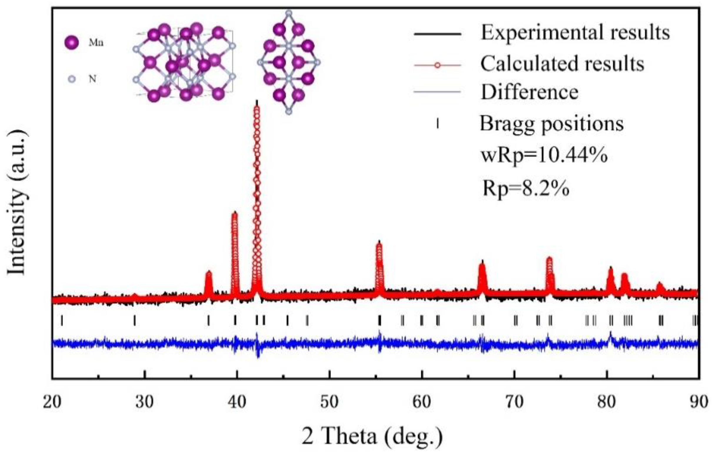
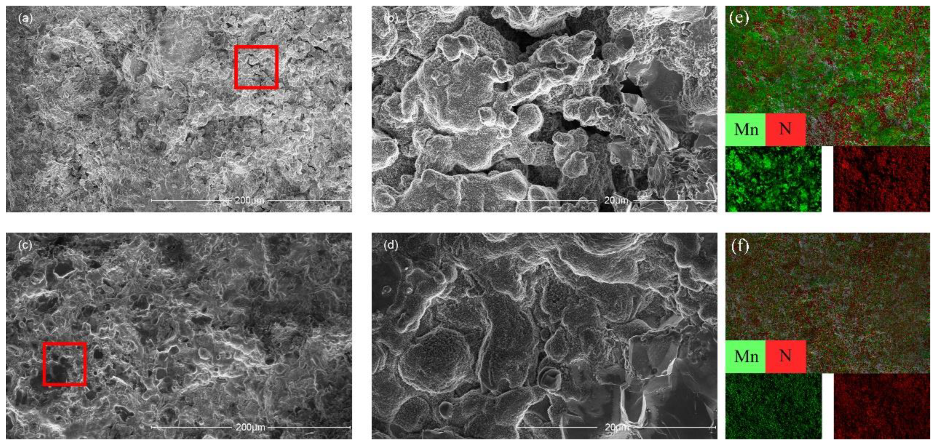
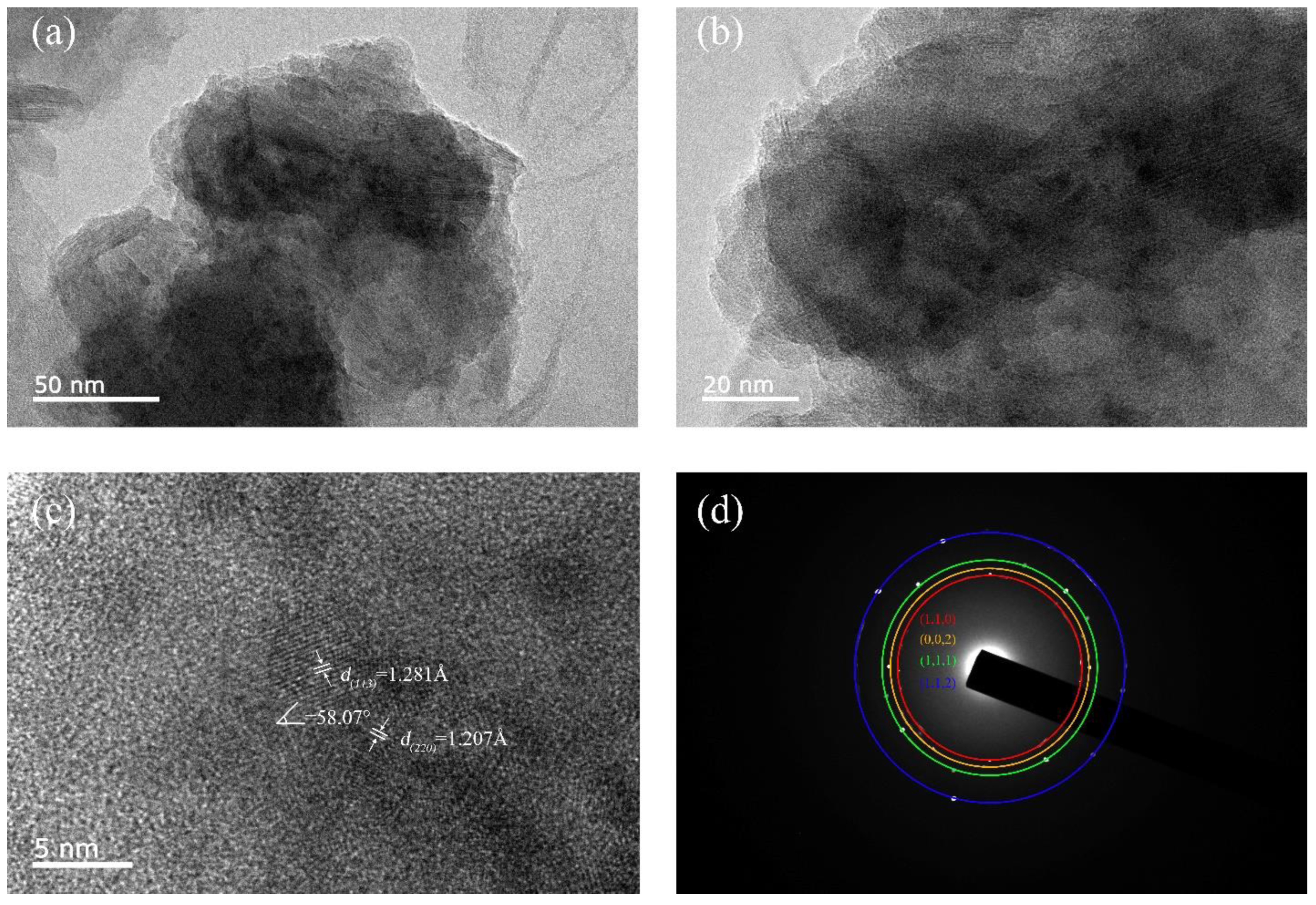
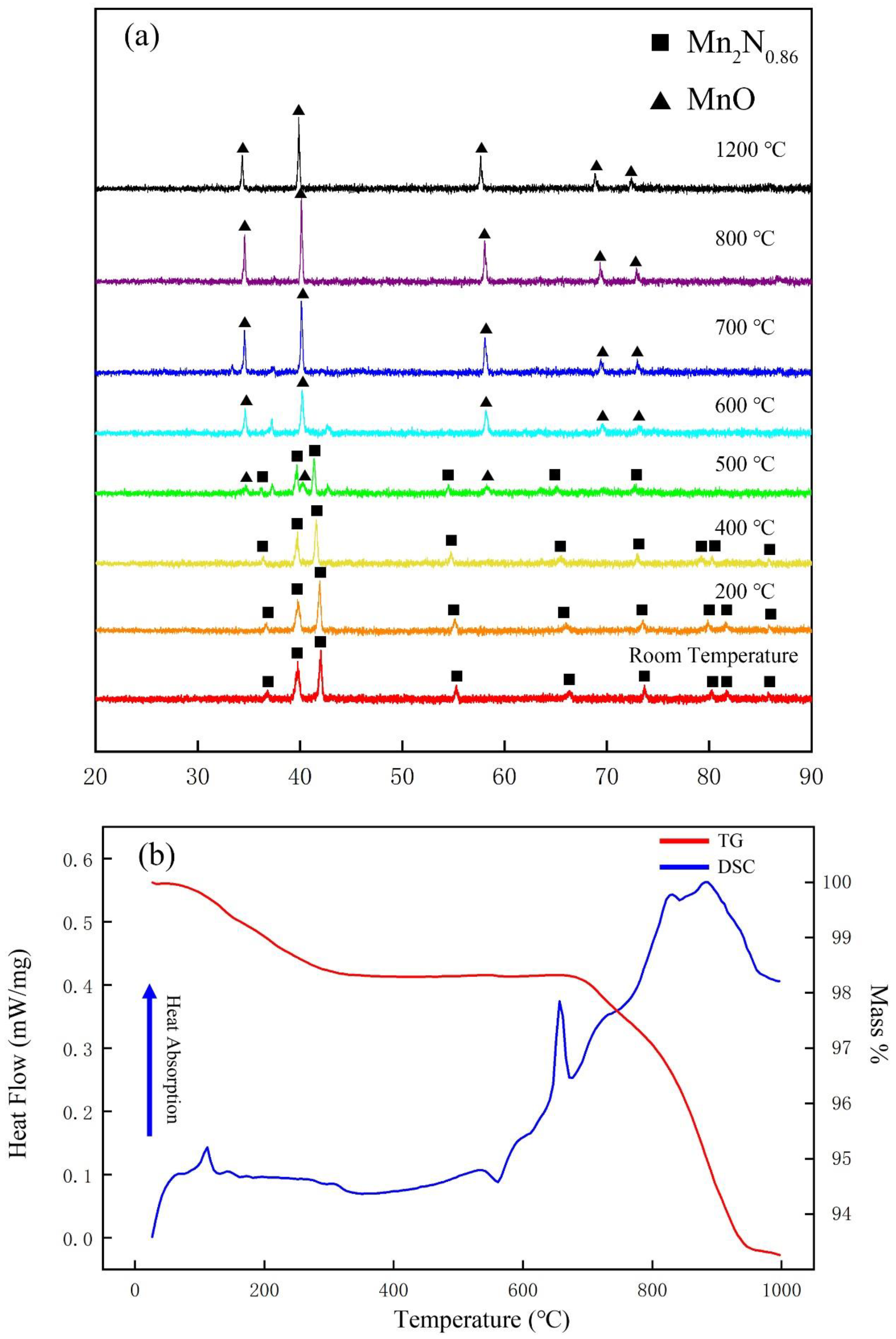
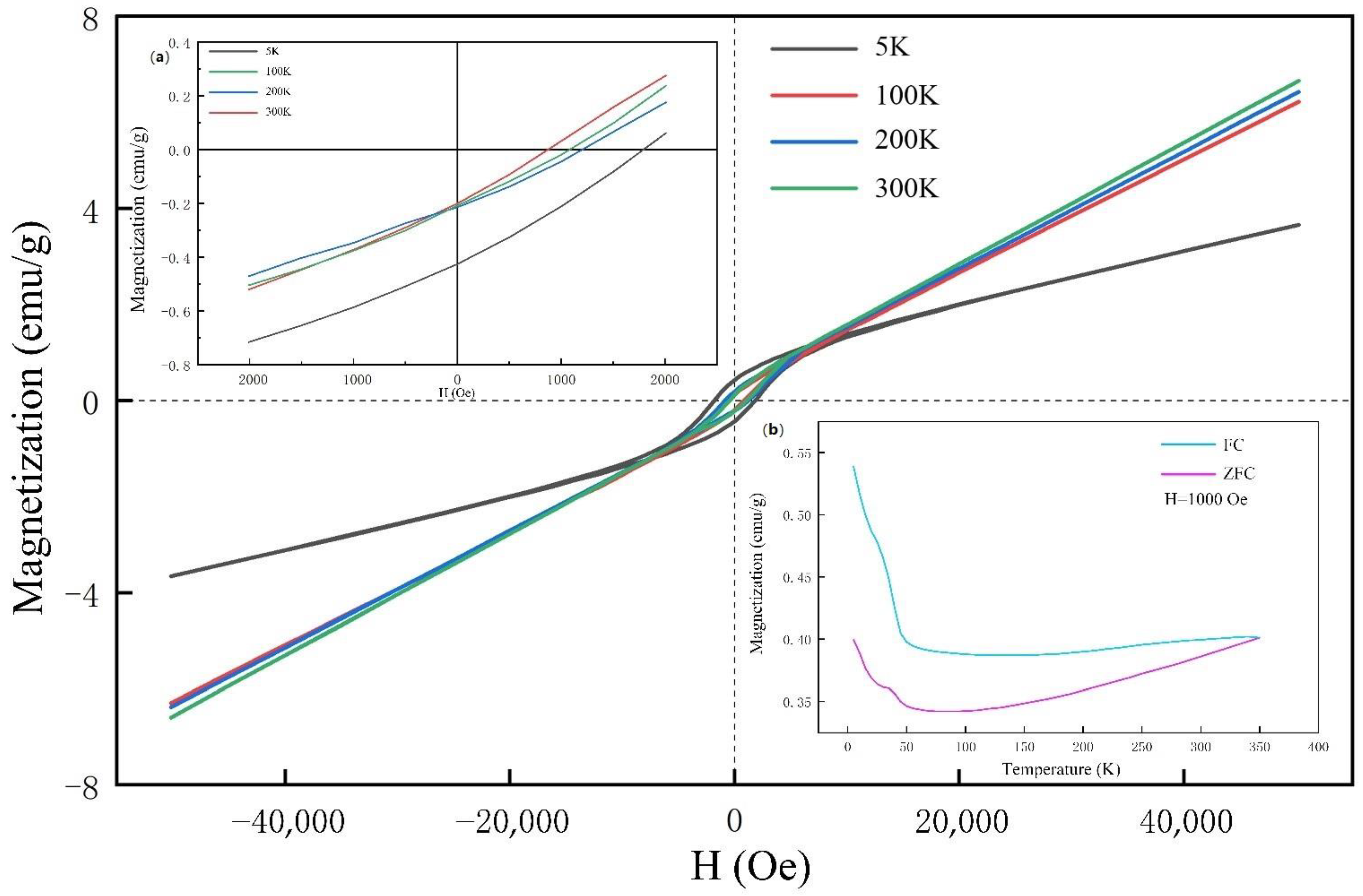
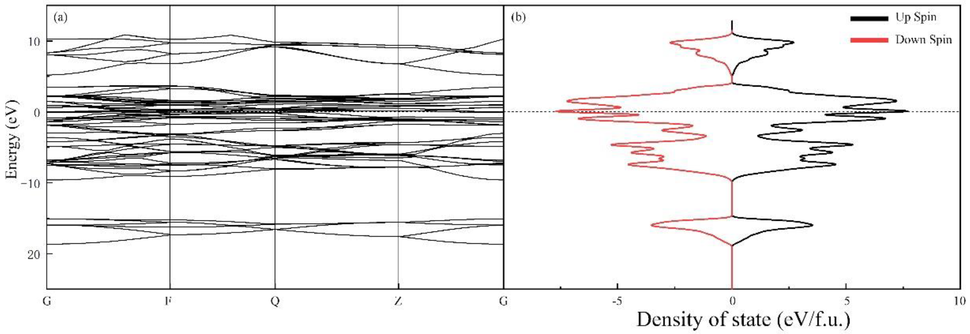
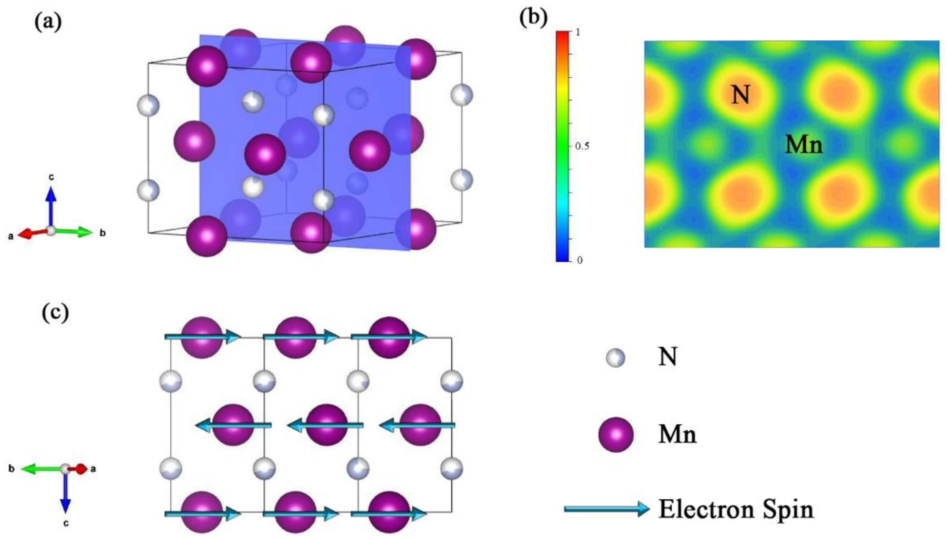
3. Results and Discussion

We prepared initial samples at 5 GPa and 1400 °C with 30 min holding and re-sintered the samples at 500 °C. The re-sintered samples were ground, whereupon their XRD patterns were recorded. The diffraction peaks confirmed that the samples were well crystallised. A comparison of Rietveld’s data [30] with the crystalline data on the standard card indicated that our samples consisted of Mn2N0.86 in the space group P6322, a = 4.8641 (3) Å, c = 4.5269 (2) Å. The XRD pattern and the ball-and-stick structure model are shown in Figure 1. In Mn2N0.86, each N atom is coordinated with six Mn atoms, and each Mn atom is also coordinated with six N atoms. The crystal structure data are summarised in Table 1. The resistance was recorded throughout the synthesis and re-sintering processes to qualitatively monitor the interaction. At the same time, we found that the sample contained 9.8% N. Considering the range of machine error, we thought that there was a certain nitrogen deficiency in the sample. However, according to the results of refinement, we believed that a small amount of N deficiency would not cause lattice distortion or changes to the crystal space group.
The drastic changes in the resistance at 300 °C and 1200 °C might correspond to the violent outgassing of NaN3 and the melting of Mn in the initial raw material. The resistance remained fairly stable at 1400 °C, the optimal temperature at which to synthesise the compound. During the secondary sintering, the resistance increased as the temperature increased to 500 °C, where the resistance reached its most stable point. Although the measurement of the resistance is an elementary approach, it provided an indication of the reactions that were taking place in the chamber.
The surface morphology of the sample and the distributions of Mn and N in the samples were examined with SEM and EDS (Figure 2). The quality of the originally synthesised sample was poor in that it was excessively porous and poor crystallinity, which we ascribed to the thermal decomposition of the raw material NaN3 to form N2. A second sintering process was therefore introduced in an attempt to enhance the quality of the original products. As shown in Figure 2c,d, no obvious pores are visible, and the homogeneity and densification improved. In addition, the EDS analysis of the elemental distribution in the samples after the second sintering indicated that the Mn and N atoms were more evenly distributed, and Na was not detected, as shown in Figure 2e,f. This seemed peculiar considering that NaN3 is an important reactant, and the fate of the Na was not immediately clear. Upon closer inspection with the aid of XRD analysis, we found NaO and NaOH in the BN capsules, which means that the BN capsules absorbed the Na atoms from the reactants during the synthesis. This is quite different from other metathesis reactions, such as the formation of CrN [13,14], VN [13], IrN2 [31], and OsN2 [31]. This difference might be attributed to the N concentration of the product being relatively low and to the fact that the molar ratio of Na is sufficiently low for it to be completely adsorbed by the BN capsules. Therefore, there might be a critical concentration of Na in the metathesis below which the product does not contain any Na. The further characterization of the samples with TEM gave the selected electron area diffraction (SAED) patterns in Figure 3d. The observed circular SAED pattern suggests that the synthesized specimen is of polycrystalline nature. The inter-planar distances calculated from the crystal lattice fringes of high-resolution TEM (HRTEM) shown in Figure 3c are 1.281 Å and 1.207 Å, which correspond to the distances of planes (113) and (220). Meanwhile, the observations of the HRTEM are in line with the measured peaks of the XRD patterns.
We measured the Vickers hardness of the re-sintered sample of Mn2N0.86 and determined its hardness to be 7.47 GPa (Figure 4). As is well known, the hardness is related to the elastic and plastic properties of the material. The relevant mechanical properties were also calculated; for the hexagonal system, the Voigt and Reuss methods were used to evaluate B and G [32]. Several models are available for estimating the hardness of a material [33,34,35,36,37,38], all of which have unique advantages and disadvantages. With Chen’s model [33] of Hv = 2(k2G)0.585 − 3 (1), Hv, G, and B are the hardness (GPa), shear modulus (GPa), and bulk modulus (GPa), respectively. The parameter k is the Pugh’s modulus ratio, namely, k = G/B; our calculated Vickers hardness of Mn2N0.86 is 14.26 GPa, which is close to the previously predicted value of 12.01 GPa [20] but significantly larger than the measured value. From Table 2, we can see that by considering magnetism, its bulk elastic modulus is overestimated compared with the previous results [20], which also leads to its B/G > 3. Pugh proposed that the B/G ratio should represent a measure of the machinable behaviour. The critical value that separates ductile and brittle materials is approximately 1.75 [39], and thus, our results revealed that Mn2N0.86 is still brittle. The predicted Vickers hardness is always higher than the actual hardness for most synthesised transition-metal–light-element compounds [40,41,42,43,44,45,46,47,48,49], regardless of the model that is chosen. We attribute this paradox to the coexistence of high itinerant electrons and the nature of the covalent bonding.
The sample was analysed by variable temperature XRD with the SE X-ray diffractometer. During this measurement, the sample was mounted on a single crystal low-vacancy Pt sheet, which also functioned as the heating carrier. The results, shown in Figure 5a, indicate that the sample remained stable until 400 °C; at 500 °C, peaks of MnO started appearing; and above 600 °C, only MnO remained. A TG-DSC analysis was conducted from room temperature to 1000 °C at a rate of 10 °C per minute to measure the energy changes the samples underwent as a function of temperature (Figure 5b). The conditions under which this analysis was conducted differed from those of the variable temperature XRD test in that measurements were conducted under a nitrogen gas flow of 30 mL/min. The mass loss of the sample increased during the entire heating process and was greater than 6% overall. Combining the XRD results and the weight of the final residue, we concluded that the final product was Mn4N. The TG and DSC curves reveal that obvious weight loss starts at approximately 120 °C; accordingly, an obvious endothermic peak appears on the DSC curve near this temperature. This peak might correspond to the evaporation of the water adsorbed to the sample, which constituted approximately 1.5% of the original mass. The obvious exothermic peak at approximately 550 °C represents an average energy output of approximately 7.795 J/g and might be related to the magnetic phase transition [50]. An intense endothermic peak starts to appear as mass loss commences at 650 °C, which marks the release of nitrogen from the sample. Taking into account the final mass and the XRD results of the residual sample, the heating process from 650 °C to 950 °C was associated with the conversion of Mn2N0.68 to Mn4N. When the heating temperature reached about 900 °C, the N atoms in Mn4N began to escape. So far, we have found a method for preparing Mn4N. More abundant than previous studies on high-temperature synthesis [51,52], these findings suggest that factors such as the gas pressure of oxygen, gas concentrations of N2 and O2, and temperature, all of which would be easy to control on an industrial scale, could be varied to control the percentage of MnO-Mn2N0.86-Mn4N to produce the ideal catalytic material MnO-MnxNy.
We recorded the magnetic hysteresis curves at different temperatures with a field sweep from −50 kOe to 50 kOe (Figure 6, with the inset (b) showing the FC and ZFC curves. The hysteresis decreases with increasing temperature, and the FC curve does not coincide with the ZFC curve. This led us to speculate that ferromagnetism and antiferromagnetism coexisted in the sample simultaneously. This did not correspond with the results from previous studies in which the prepared samples were not single-phased, and the researchers attributed the occurrence of both types of magnetism to impurities [22].
To gain a deeper understanding of the co-existence of ferro- and antiferromagnetism in our sample, we conducted first-principle DFT simulations of Mn2N0.86. The band structure and spin density of states (spin-DOSs), shown in Figure 7, indicate that Mn2N0.86 is metallic because of the overlapping bands at the Fermi level. Moreover, the DOSs for different spins are symmetric, which means Mn2N0.86 should be antiferromagnetic. The ferromagnetism might originate from the N vacancies. XRD and EDS are insufficiently sensitive to minute amounts of N vacancies. We simulated the latent heat of Mn2N from both the antiferromagnetic and ferromagnetic phases to the nonmagnetic phase with the former being approximately 5% of the latter. Then, using these values to fit the exothermic peak in Figure 5b near 600 K, we determined that the Mn2N might be 95% antiferromagnetic and 5% ferromagnetic. Although we do not consider Mn2N and Mn2N0.86 to be equal, the analogy could serve as a helpful indication.
The electronic localisation function (ELF) of the Mn2N0.86 is shown in Figure 8a,b The electrons between the Mn and N atoms are strongly localised, and the ELF is below 0.5, which corresponds with the value of free electrons. The spin direction of Mn atoms is shown in Figure 8c. As an electron-donating element, Mn transfers most of its electrons to proximate N atoms. This reveals the ionic nature of the Mn-N bond, which is supported by the ELF of Mn2N0.86, and explains the partially ferromagnetic nature of our sample. Nitrogen vacancies inevitably occur in transition-metal nitrides, and in the case of Mn2N0.86, N resides at a fractionally occupied position that has the characteristics of a vacancy. The first-principle DFT simulations showed that for Mn2N0.86, the energy of ferromagnetism is approximately 0.6 eV lower than that of antiferromagnetism. In the case of N vacancies, the system has the tendency to spontaneously evolve to ferromagnetism, which has lower energy. According to the Heisenberg model, an exchange takes place between magnetic atoms, and this energy is expressed as E e x = 2 A i j S i S j , where A is the exchange integral, and S i , S j is the spin interaction term for the spin at the i th and j th sites. An exchange correlation exists between Mn atoms at the N vacancies in the crystal, where A > 0. Moreover, the spins between Mn atoms that undergo this exchange are parallel, and thus, S i · S j > 0 . This explains the ferromagnetism in the sample.

4. Conclusions

We synthesised Mn2N0.86 samples using high pressure and high temperature. A second sintering process enhanced the quality of the samples, which met the requirements for many types of measurements. The Vickers hardness of the Mn2N0.86 sample was measured to be 7.47 GPa, and the tested hardness was lower than the simulated one. This difference was considered to be a consequence of the large number of valence electrons and the mixed ionic/metallic bonding. The TG-DSC measurements enabled us to outline the reaction sequence and latent heat during the evolution of Mn2N0.86 to Mn4N under N2 gas protection, which provides a route for the preparation of Mn4N. The melting point of Mn2N0.86 is approximately 650 K, but when heated without the protection of N2, it would be oxidised to MnO at temperatures above 500 °C. The simulation suggested that Mn2N0.86 is antiferromagnetic; however, we determined it to be a combination of chiefly antiferromagnetism and to a lesser extent ferromagnetism owing to N vacancies. This study indicates that by controlling the air pressure, air concentration, and temperature, one could have ideal MnO-MnxNy catalysis and prepare MnN4. The magnetic properties of a transition metal nitride could be affected by a very low percentage of N vacancies, which could be utilised as sensors.

Author Contributions

Data curation, X.Z., Q.T., W.Z., K.B., T.C. and J.C.; Formal analysis, X.W., Y.G., Z.Y., T.M. and S.M.; Funding acquisition, T.C.; Investigation, S.Z.; Resources, P.Z.; Software, C.Z., T.M. and J.Z.; Writing—original draft, S.Z.; Writing—review & editing, S.Z. All authors have read and agreed to the published version of the manuscript.

Funding

This research was funded by the National Key R&D Program of China (2018YFA0703404, 2017YFA0403704, 2016YFB0201204), the National Natural Science Foundation of China (Nos. 11774121, 91745203), and the Program for Changjiang Scholars and Innovative Research Team in University (No. IRT_15R23), Parts of calculations were performed in the High Performance Computing Center (HPCC) of Jilin University.

Institutional Review Board Statement

Not applicable.

Informed Consent Statement

Not applicable.

Data Availability Statement

The data that support the findings of this study are available from the corresponding author upon reasonable request.

Acknowledgments

This work was supported by the National Key R&D Program of China (2018YFA0703404, 2017YFA0403704, 2016YFB0201204), the National Natural Science Foundation of China (Nos. 11774121, 91745203), and the Program for Changjiang Scholars and Innovative Research Team in University (No. IRT_15R23), Parts of calculations were performed in the High Performance Computing Center (HPCC) of Jilin University.

Conflicts of Interest

The authors declare no conflict of interest.

References

  1. Akopov, G.; Yeung, M.T.; Kaner, R.B. Rediscovering the Crystal Chemistry of Borides. Adv. Mater. 2017, 29, 1604506. [Google Scholar] [CrossRef] [PubMed]
  2. Spengler, W.; Kaiser, R.; Christensen, A.N.; Müller-Vogt, G. Raman scattering, superconductivity, and phonon density of states of stoichiometric and nonstoichiometric TiN. Phys. Rev. B 1978, 17, 1095–1101. [Google Scholar] [CrossRef]
  3. Lei, W.W.; Liu, D.; Li, X.F.; Zhang, J.; Zhou, Q.; Hu, J.Z.; Cui, Q.L.; Zou, G.T. High-pressure study of low-compressibility Ta2N. J. Phys. Condens. Matter 2007, 19, 425233. [Google Scholar] [CrossRef]
  4. Zou, Y.; Wang, X.; Chen, T.; Li, X.; Qi, X.; Welch, D.; Zhu, P.; Liu, B.; Cui, T.; Li, B. Hexagonal-structured ε-NbN: Ultra-incompressibility, high shear rigidity and a possible hard superconducting material. Sci. Rep. 2015, 5, 10811. [Google Scholar] [CrossRef] [PubMed]
  5. Rognerud, E.G.; Rom, C.L.; Todd, P.K.; Singstock, N.R.; Bartel, C.J.; Holder, A.M.; Neilson, J.R. Kinetically Controlled Low-Temperature Solid-State Metathesis of Manganese Nitride Mn3N2. Chem. Mater. 2019, 31, 7248–7254. [Google Scholar] [CrossRef]
  6. Mamoru Mekata, J.H.; Takaki, H. Neutron Diffraction Study of Antiferromagnetic Mn2N. J. Phys. Soc. Jpn. 1968, 25, 234–238. [Google Scholar] [CrossRef]
  7. Fu, Q.; Kokalj, D.; Stangier, D.; Kruis, F.E.; Tillmann, W. Aerosol synthesis of titanium nitride nanoparticles by direct current arc discharge method. Adv. Powder Technol. 2020, 31, 4119–4128. [Google Scholar] [CrossRef]
  8. Shen, L.; Cheng, T.; Wu, L.; Li, X.; Cui, Q. Synthesis and optical properties of aluminum nitride nanowires prepared by arc discharge method. J. Alloys Compd. 2008, 465, 562–566. [Google Scholar] [CrossRef]
  9. Shen, L.-H.; Cui, Q.-L. Synthesis of Cubic Chromium Nitride Nanocrystals Powders by Arc Discharge Plasma Method. J. Inorg. Mater. 2010, 25, 411–414. [Google Scholar] [CrossRef]
  10. Zhang, Z.; Mi, W. Progress in ferrimagnetic Mn4N films and its heterostructures for spintronics applications. J. Phys. D Appl. Phys. 2021, 55, 013001. [Google Scholar] [CrossRef]
  11. Guerrero-Sánchez, J.; Takeuchi, N. Structural stability and the electronic and magnetic properties of ferrimagnetic Mn 4 N(0 0 1) surfaces. Appl. Surf. Sci. 2017, 407, 209–212. [Google Scholar] [CrossRef]
  12. Feng, X.; Bao, K.; Tao, Q.; Li, L.; Shao, Z.; Yu, H.; Xu, C.; Ma, S.; Lian, M.; Zhao, X.; et al. Role of TM–TM Connection Induced by Opposite d-Electron States on the Hardness of Transition-Metal (TM = Cr, W) Mononitrides. Inorg. Chem. 2019, 58, 15573–15579. [Google Scholar] [CrossRef]
  13. Wang, S.; Yu, X.; Zhang, J.; Wang, L.; Leinenweber, K.; He, D.; Zhao, Y. Synthesis, Hardness, and Electronic Properties of Stoichiometric VN and CrN. Cryst. Growth Des. 2015, 16, 351–358. [Google Scholar] [CrossRef]
  14. Chen, M.; Wang, S.; Zhang, J.; He, D.; Zhao, Y. Synthesis of Stoichiometric and Bulk CrN through a Solid-State Ion-Exchange Reaction. Chem. A Eur. J. 2012, 18, 15459–15463. [Google Scholar] [CrossRef] [PubMed]
  15. Yang, H.; Al-Brithen, H.; Smith, A.R.; Borchers, J.A.; Cappelletti, R.L.; Vaudin, M.D. Structural and magnetic properties of η-phase manganese nitride films grown by molecular-beam epitaxy. Appl. Phys. Lett. 2001, 78, 3860–3862. [Google Scholar] [CrossRef]
  16. Suzuki, K.; Kaneko, T.; Yoshida, H.; Obi, Y.; Fujimori, H.; Morita, H. Crystal structure and magnetic properties of the compound MnN. J. Alloys Compd. 2000, 306, 66–71. [Google Scholar] [CrossRef]
  17. Leineweber, A.; Niewa, R.; Jacobs, H.; Kockelmann, W. The manganese nitrides η-Mn3N2 and θ-Mn6N5 + x: Nuclear and magnetic structures. J. Mater. Chem. 2000, 10, 2827–2834. [Google Scholar] [CrossRef]
  18. Shen, X.; Chikamatsu, A.; Shigematsu, K.; Hirose, Y.; Fukumura, T.; Hasegawa, T. Metallic transport and large anomalous Hall effect at room temperature in ferrimagnetic Mn4N epitaxial thin film. Appl. Phys. Lett. 2014, 105, 072410. [Google Scholar] [CrossRef]
  19. Wang, G.; Meng, M.; Zhou, W.; Wang, Y.; Wu, S.; Li, S. Magnetic properties of Mn2N0.86 (111) thin film grown on MgO (001) substrate by molecular beam epitaxy. Mater. Lett. 2016, 184, 291–293. [Google Scholar] [CrossRef]
  20. Yu, R.; Chong, X.; Jiang, Y.; Zhou, R.; Yuan, W.; Feng, J. The stability, electronic structure, elastic and metallic properties of manganese nitrides. RSC Adv. 2014, 5, 1620–1627. [Google Scholar] [CrossRef]
  21. Sun, Z.; Song, X. Preparation and magnetic characterization of ultrafine-grained ζ-Mn2N0.86 compound bulk. Mater. Lett. 2009, 63, 2059–2062. [Google Scholar] [CrossRef]
  22. Feng, W.; Sun, N.; Du, J.; Zhang, Q.; Liu, X.; Deng, Y.; Zhang, Z. Structural evolution and magnetic properties of Mn–N compounds. Solid State Commun. 2008, 148, 199–202. [Google Scholar] [CrossRef]
  23. Liu, Y.; Xu, L.; Li, X.; Hu, P.; Li, S. Growth and magnetic property of ζ-phase Mn2N1±x thin films by plasma-assisted molecular beam epitaxy. J. Appl. Phys. 2010, 107, 103914. [Google Scholar] [CrossRef]
  24. Fang, C.; Hu, C.; Li, D.; Chen, J.; Luo, M. Unravelling the efficient catalytic performance of ozone decomposition over nitrogen-doped manganese oxide catalysts under high humidity. New J. Chem. 2020, 44, 17993–17999. [Google Scholar] [CrossRef]
  25. Feng, W.J.; Li, D.; Deng, Y.F.; Zhang, Q.; Zhang, H.H.; Zhang, Z.D. Magnetic and transport properties of Mn3+x Ga1−x N compounds. J. Mater. Sci. 2010, 45, 2770–2774. [Google Scholar] [CrossRef]
  26. Ma, S.; Bao, K.; Tao, Q.; Xu, C.; Feng, X.; Zhao, X.; Ge, Y.; Zhu, P.; Cui, T. Double-zigzag boron chain-enhanced Vickers hardness and manganese bilayers-induced high d-electron mobility in Mn3B4. Phys. Chem. Chem. Phys. 2018, 21, 2697–2705. [Google Scholar] [CrossRef]
  27. Segall, M.D.; Lindan, P.J.D.; Probert, M.J.; Pickard, C.J.; Hasnip, P.J.; Clark, S.J.; Payne, M.C. First-principles simulation: Ideas, illustrations and the CASTEP code. J. Phys. Condens. Matter 2002, 14, 2717–2744. [Google Scholar] [CrossRef]
  28. Bellaiche, L.; Vanderbilt, D. Virtual crystal approximation revisited: Application to dielectric and piezoelectric properties of perovskites. Phys. Rev. B 2000, 61, 7877–7882. [Google Scholar] [CrossRef] [Green Version]
  29. Perdew, J.P.; Burke, K.; Ernzerhof, M. Generalized gradient approximation made simple. Phys. Rev. Lett. 1996, 77, 3865. [Google Scholar] [CrossRef] [Green Version]
  30. Toby, B.H. EXPGUI, a graphical user interface for GSAS. J. Appl. Crystallogr. 2001, 34, 210–213. [Google Scholar] [CrossRef]
  31. Young, A.F.; Sanloup, C.; Gregoryanz, E.; Scandolo, S.; Hemley, R.J.; Mao, H.-K. Synthesis of Novel Transition Metal NitridesIrN2andOsN2. Phys. Rev. Lett. 2006, 96, 155501. [Google Scholar] [CrossRef] [PubMed] [Green Version]
  32. Sun, L.; Gao, Y.; Xiao, B.; Li, Y.; Wang, G. Anisotropic elastic and thermal properties of titanium borides by first-principles calculations. J. Alloys Compd. 2013, 579, 457–467. [Google Scholar] [CrossRef]
  33. Chen, X.-Q.; Niu, H.; Li, D.; Li, Y. Modeling hardness of polycrystalline materials and bulk metallic glasses. Intermetallics 2011, 19, 1275–1281. [Google Scholar] [CrossRef] [Green Version]
  34. Gao, F.; He, J.; Wu, E.; Liu, S.; Yu, D.; Li, D.; Zhang, S.; Tian, Y. Hardness of Covalent Crystals. Phys. Rev. Lett. 2003, 91, 015502. [Google Scholar] [CrossRef] [PubMed]
  35. Guo, X.; Li, L.; Liu, Z.; Yu, D.; He, J.; Liu, R.; Xu, B.; Tian, Y.; Wang, H.-T. Hardness of covalent compounds: Roles of metallic component and d valence electrons. J. Appl. Phys. 2008, 104, 023503. [Google Scholar] [CrossRef]
  36. Šimůnek, A.; Vackář, J. Hardness of Covalent and Ionic Crystals: First-Principle Calculations. Phys. Rev. Lett. 2006, 96, 085501. [Google Scholar] [CrossRef] [Green Version]
  37. Li, K.; Wang, X.; Zhang, F.; Xue, D. Electronegativity Identification of Novel Superhard Materials. Phys. Rev. Lett. 2008, 100, 235504. [Google Scholar] [CrossRef]
  38. Li, Q.; Zhou, D.; Zheng, W.; Ma, Y.; Chen, C. Anomalous Stress Response of UltrahardWBnCompounds. Phys. Rev. Lett. 2015, 115, 185502. [Google Scholar] [CrossRef] [Green Version]
  39. Liu, Y.; Jiang, Y.; Zhou, R.; Feng, J. Mechanical properties and chemical bonding characteristics of WC and W2C compounds. Ceram. Int. 2014, 40, 2891–2899. [Google Scholar] [CrossRef]
  40. Zhao, X.; Li, L.; Bao, K.; Zhu, P.; Tao, Q.; Ma, S.; Liu, B.; Ge, Y.; Li, D.; Cui, T. Synthesis and characterization of a strong ferromagnetic and high hardness intermetallic compound Fe2B. Phys. Chem. Chem. Phys. 2020, 22, 27425–27432. [Google Scholar] [CrossRef]
  41. Ma, S.; Huang, Z.; Xing, J.; Liu, G.; He, Y.; Fu, H.; Wang, Y.; Li, Y.; Yi, D. Effect of crystal orientation on microstructure and properties of bulk Fe2B intermetallic. J. Mater. Res. 2015, 30, 257–265. [Google Scholar] [CrossRef]
  42. Dubrovinskaia, N.; Dubrovinsky, L.; Solozhenko, V.L. Comment on “Synthesis of Ultra-Incompressible Superhard Rhenium Diboride at Ambient Pressure”. Science 2007, 318, 1550. [Google Scholar] [CrossRef] [PubMed] [Green Version]
  43. Levine, J.B.; Tolbert, S.H.; Kaner, R.B. Advancements in the Search for Superhard Ultra-Incompressible Metal Borides. Adv. Funct. Mater. 2009, 19, 3519–3533. [Google Scholar] [CrossRef]
  44. Fu, H.; Peng, W.; Gao, T. Structural and elastic properties of ZrC under high pressure. Mater. Chem. Phys. 2009, 115, 789–794. [Google Scholar] [CrossRef]
  45. Gou, H.; Hou, L.; Zhang, J.; Gao, F. Pressure-induced incompressibility of ReC and effect of metallic bonding on its hardness. Appl. Phys. Lett. 2008, 92, 241901. [Google Scholar] [CrossRef]
  46. Ma, S.; Bao, K.; Tao, Q.; Huang, Y.; Xu, C.; Li, L.; Feng, X.; Zhao, X.; Zhu, P.; Cui, T. Investigation the origin and mechanical properties of unusual rigid diamond-like net analogues in manganese tetraboride. Int. J. Refract. Met. Hard Mater. 2019, 85, 104845. [Google Scholar] [CrossRef]
  47. Gou, H.; Tsirlin, A.A.; Bykova, E.; Abakumov, A.M.; Van Tendeloo, G.; Richter, A.; Ovsyannikov, S.V.; Kurnosov, A.V.; Trots, D.M.; Konôpková, Z.; et al. Peierls distortion, magnetism, and high hardness of manganese tetraboride. Phys. Rev. B 2014, 89, 064108. [Google Scholar] [CrossRef] [Green Version]
  48. Wang, B.; Li, X.; Wang, Y.X.; Tu, Y.F. Phase Stability and Physical Properties of Manganese Borides: A First-Principles Study. J. Phys. Chem. C 2011, 115, 21429–21435. [Google Scholar] [CrossRef]
  49. Knappschneider, A.; Litterscheid, C.; Brgoch, J.; George, N.C.; Henke, S.; Cheetham, A.K.; Hu, J.G.; Seshadri, R.; Albert, B. Manganese Tetraboride, MnB4: High-Temperature Crystal Structure, p-n Transition, 55Mn NMR Spectroscopy, Solid Solutions, and Mechanical Properties. Chem. A Eur. J. 2015, 21, 8177–8181. [Google Scholar] [CrossRef]
  50. Yan, J.; Shi, K.; Deng, S.; Zhao, W.; Lu, H.; Sun, Y.; Chen, Y.; Wang, C. The influence of combination of the first-order and second-order phase transitions on magnetocaloric effects in Mn3Cu1-xFexN. Solid State Commun. 2018, 282, 33–37. [Google Scholar] [CrossRef]
  51. Li, C.; Yang, Y.; Lv, L.; Huang, H.; Wang, Z.; Yang, S. Fabrication and magnetic characteristic of ferrimagnetic bulk Mn4N. J. Alloys Compd. 2008, 457, 57–60. [Google Scholar] [CrossRef]
  52. Tan, S.; Gao, C.; Yuan, H.; Wu, J.; Wang, C.; Cao, R.; Sun, Y. An antiperovskite compound with multifunctional properties: Mn3PdN. J. Solid State Chem. 2021, 302, 122389. [Google Scholar] [CrossRef]
Figure 1. The powder XRD spectrum and the refined structure information of the sample. The black line represents the original experimental data, and the red circles represent the fitting results after Rietveld; ball-and-stick model of the crystal unit cell viewed from different directions.
Materials 15 07780 g001
Figure 2. (a) A full view of a randomly selected area after the initial synthesis of the sample; (b) the enlarged view of the red frame region in (a); (c) the overall view of another randomly selected region of the sample after secondary sintering and annealing; (d) the enlargement of the red frame region of (c); (e) the element distribution diagram of the initial synthesized EDS of the sample; (f) the element distribution diagram of the sample after secondary sintering and annealing by EDS.
Materials 15 07780 g002
Figure 3. (a,b) are the bright-field TEM images of the sample. (c) The HRTEM of the sample, the planes of (113) and (220), and the angle of two crystal planes. (d) The SAED of the sample.
Materials 15 07780 g003
Figure 4. Vickers hardness for Mn2N0.86. The applied load ranged from 0.49 N (low load) to 9.8 N (high load). Typical optical images of the indentation are shown in the inset.
Materials 15 07780 g004
Figure 5. Variable-temperature XRD test data and TG-DSC test data of the samples. (a) Variable-temperature XRD test data: The black rectangles mark the Mn2N0.86 characteristic peaks, and the black triangles mark the MnO characteristic peaks. (b) TG-DSC curve of the sample: The red line is the TG curve, and the blue line is the DSC curve.
Materials 15 07780 g005
Figure 6. Hysteresis loops for Mn2N0.86 at 5 K, 100 K, 200 K, and 300 K. (a) The M-H curves within the magnetic field of 2000 Oe. (b) The temperature dependences of the magnetization for the Mn2N0.86; the applied magnetic field is 1000 Oe.
Materials 15 07780 g006
Figure 7. (a) Energy band diagram of the sample. (b) TDOS diagram of the sample. The Fermi surface has been marked with a dashed line.
Materials 15 07780 g007
Figure 8. (a) The cutting slab of Mn2N0.86 along the (100) plane. (b) ELF of Mn2N0.86 2D display for (100) plane. (c) Magnetic structure of Mn2N0.86 projected.
Materials 15 07780 g008
Table 1. Crystal structure information.
Formula weight60.96
Crystal systemHexagonal
Space-groupP6322
Cell parametersa = 4.8641(3) Å
c = 4.5269(2) Å
Cell ratioa/b = 1.0000
b/c = 1.0745
Cell volume92.76(1) Å3
Particle size139.7 nm
Strain broadening1.745 × 10−5
Table 2. The mechanic parameters of Mn2N0.86.
PhaseC11C12C13C33C44C66
Mn2N0.86265.711428.144191.555429.35897.692189.637
Bulk Modulus
B (GPa)
Young’s Modulus
E (GPa)
Shear Modulus
G (GPa)
Poisson’s Ratio
V
Pugh’s Ratio
(B/G)
Vickers Hardness
(GPa)
Mn2N0.86192.07285.82114.150.251.6814.26
Publisher’s Note: MDPI stays neutral with regard to jurisdictional claims in published maps and institutional affiliations.

