
Titanium Implant Surface Effects on Adherent Macrophage Phenotype: A Systematic Review


Abstract
:1. Introduction
2. Materials and Methods
2.1. Review Question
2.2. Search Strategy
2.3. Inclusion and Exclusion Criteria
2.4. Data Extraction
2.5. Risk of Bias
3. Results
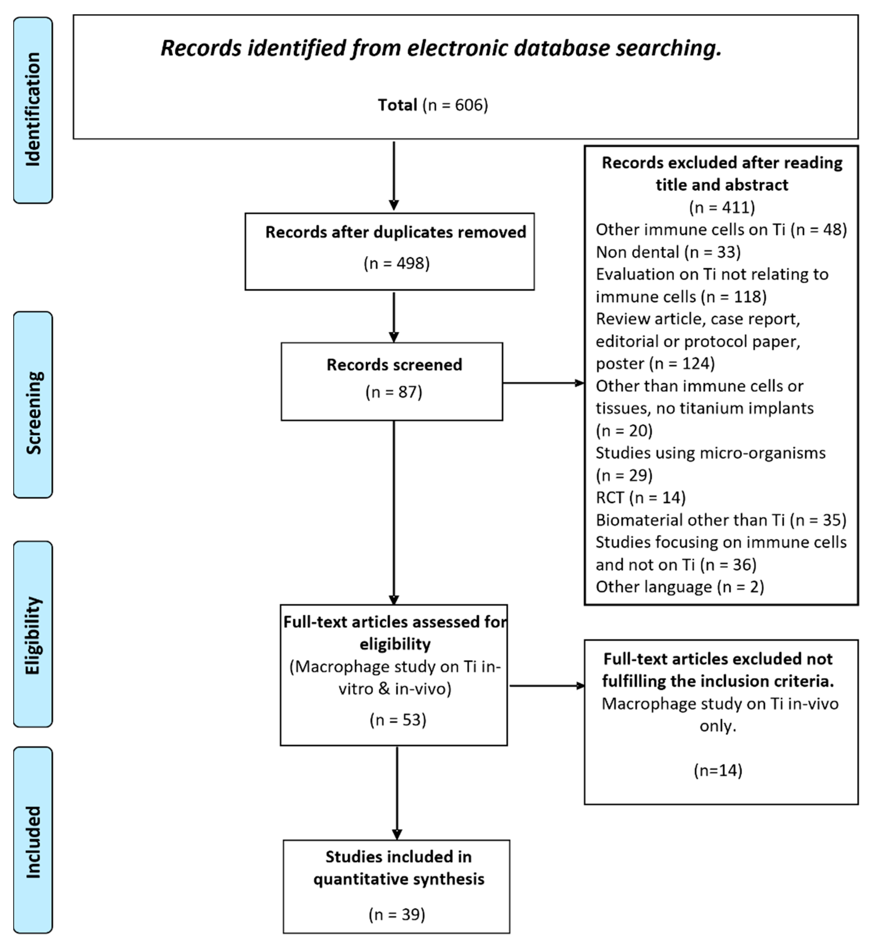
3.1. Data Retrieval
3.2. Surface Topography
3.3. Cell Morphology
3.4. Cellular Response
3.5. Risk of Bias
4. Discussion
5. Conclusions
Supplementary Materials
Author Contributions
Funding
Institutional Review Board Statement
Informed Consent Statement
Data Availability Statement
Conflicts of Interest
References
- Kaur, M.; Singh, K. Review on titanium and titanium based alloys as biomaterials for orthopaedic applications. Mater. Sci. Eng. C Mater. Biol. Appl. 2019, 102, 844–862. [Google Scholar] [CrossRef] [PubMed]
- Donath, K.; Kirsch, A.; Osborn, J.F. Zelluläre Dynamik um enossale Titanimplantate. Fortschr. Zahnärztl. Implantol. 1984, 1, 55–58. [Google Scholar]
- Pajarinen, J.; Kouri, V.-P.; Jämsen, E.; Li, T.-F.; Mandelin, J.; Konttinen, Y.T. The response of macrophages to titanium particles is determined by macrophage polarization. Acta Biomater. 2013, 9, 9229–9240. [Google Scholar] [CrossRef] [PubMed]
- Sridharan, R.; Cameron, A.R.; Kelly, D.J.; Kearney, C.J.; O’Brien, F.J. Biomaterial based modulation of macrophage polarization: A review and suggested design principles. Mater. Today 2015, 18, 313–325. [Google Scholar] [CrossRef]
- Arron, J.R.; Choi, Y. Bone versus immune system. Nature 2000, 408, 535–536. [Google Scholar] [CrossRef] [PubMed]
- Cui, Y.; Li, H.; Li, Y.; Mao, L. Novel insights into nanomaterials for immunomodulatory bone regeneration. Nanoscale Adv. 2021, 4, 334–352. [Google Scholar] [CrossRef]
- Guder, C.; Gravius, S.; Burger, C.; Wirtz, D.C.; Schildberg, F.A. Osteoimmunology: A Current Update of the Interplay between Bone and the Immune System. Front. Immunol. 2020, 11, 58. [Google Scholar] [CrossRef] [Green Version]
- Mills, C.D.; Kincaid, K.; Alt, J.M.; Heilman, M.J.; Hill, A.M. M-1/M-2 Macrophages and the Th1/Th2 Paradigm. J. Immunol. 2000, 164, 6166–6173. [Google Scholar] [CrossRef] [Green Version]
- Mantovani, A.; Sica, A.; Locati, M. Macrophage Polarization Comes of Age. Immunity 2005, 23, 344–346. [Google Scholar] [CrossRef] [Green Version]
- Wennerberg, A.; Albrektsson, T. Effects of titanium surface topography on bone integration: A systematic review. Clin. Oral Implants Res. 2009, 20, 172–184. [Google Scholar] [CrossRef]
- Jennissen, H.P. Ultra-Hydrophilic Transition Metals as Histophilic Biomaterials. Macromol. Symp. 2005, 225, 43–70. [Google Scholar] [CrossRef]
- Schwarz, F.; Wieland, M.; Schwartz, Z.; Zhao, G.; Rupp, F.; Geis-Gerstorfer, J.; Schedle, A.; Broggini, N.; Bornstein, M.M.; Buser, D.; et al. Potential of chemically modified hydrophilic surface characteristics to support tissue integration of titanium dental implants. J. Biomed. Mater. Res. Part B Appl. Biomater. 2008, 88, 544–557. [Google Scholar] [CrossRef] [PubMed]
- Pan, H.; Xie, Y.; Zhang, Z.; Li, K.; Hu, D.; Zheng, X.; Tang, T. Immunomodulation effect of a hierarchical macropore/nanosurface on osteogenesis and angiogenesis. Biomed. Mater. 2017, 12, 45006. [Google Scholar] [CrossRef] [PubMed]
- Richardson, W.S.; Wilson, M.C.; Nishikawa, J.; Hayward, R.S. The well-built clinical question: A key to evidence-based decisions. ACP J. Club 1995, 123, A12–A13. [Google Scholar] [CrossRef]
- Johnson, B.T.; Hennessy, E.A. Systematic reviews and meta-analyses in the health sciences: Best practice methods for research syntheses. Soc. Sci. Med. 2019, 233, 237–251. [Google Scholar] [CrossRef]
- Schneider, K.; Schwarz, M.; Burkholder, I.; Kopp-Schneider, A.; Edler, L.; Kinsner-Ovaskainen, A.; Hartung, T.; Hoffmann, S. “ToxRTool”, a new tool to assess the reliability of toxicological data. Toxicol. Lett. 2009, 189, 138–144. [Google Scholar] [CrossRef]
- Klimisch, H.-J.; Andreae, M.; Tillmann, U. A Systematic Approach for Evaluating the Quality of Experimental Toxicological and Ecotoxicological Data. Regul. Toxicol. Pharmacol. 1997, 25, 1–5. [Google Scholar] [CrossRef] [Green Version]
- Milleret, V.; Tugulu, S.; Schlottig, F.; Hall, H. Alkali treatment of microrough titanium surfaces affects macrophage/monocyte adhesion, platelet activation and architecture of blood clot formation. Eur. Cells Mater. 2011, 21, 430–444. [Google Scholar] [CrossRef]
- Hamlet, S.; Alfarsi, M.; George, R.; Ivanovski, S. The effect of hydrophilic titanium surface modification on macrophage inflammatory cytokine gene expression. Clin. Oral Implants Res. 2011, 23, 584–590. [Google Scholar] [CrossRef]
- Barth, K.A.; Waterfield, J.D.; Brunette, D.M. The effect of surface roughness on RAW 264.7 macrophage phenotype. J. Biomed. Mater. Res. Part A 2013, 101A, 2679–2688. [Google Scholar] [CrossRef]
- Alfarsi, M.A.; Hamlet, S.M.; Ivanovski, S. Titanium surface hydrophilicity modulates the human macrophage inflammatory cytokine response. J. Biomed. Mater. Res. Part A 2013, 102, 60–67. [Google Scholar] [CrossRef] [PubMed]
- Nagasawa, M.; Cooper, L.; Ogino, Y.; Mendonca, G.; Liang, R.; Yang, S.; Uoshima, K. Topography Influences Adherent Cell Regulation of Osteoclastogenesis. J. Dent. Res. 2015, 95, 319–326. [Google Scholar] [CrossRef] [PubMed]
- Hotchkiss, K.M.; Reddy, G.B.; Hyzy, S.L.; Schwartz, Z.; Boyan, B.D.; Olivares-Navarrete, R. Titanium surface characteristics, including topography and wettability, alter macrophage activation. Acta Biomater. 2015, 31, 425–434. [Google Scholar] [CrossRef] [PubMed] [Green Version]
- Eger, M.; Sterer, N.; Liron, T.; Kohavi, D.; Gabet, Y. Scaling of titanium implants entrains inflammation-induced osteolysis. Sci. Rep. 2017, 7, 39612. [Google Scholar] [CrossRef]
- Hotchkiss, K.M.; Ayad, N.B.; Hyzy, S.L.; Boyan, B.D.; Olivares-Navarrete, R. Dental implant surface chemistry and energy alter macrophage activation in vitro. Clin. Oral Implant. Res. 2016, 28, 414–423. [Google Scholar] [CrossRef]
- Kianoush, F.; Nematollahi, M.; Waterfield, J.D.; Brunette, D.M. Regulation of RAW264.7 macrophage polarization on smooth and rough surface topographies by galectin-3. J. Biomed. Mater. Res. Part A 2017, 105, 2499–2509. [Google Scholar] [CrossRef] [PubMed]
- Choi, S.-M.; Park, J.-W. Multifunctional effects of a modification of SLA titanium implant surface with strontium-containing nanostructures on immunoinflammatory and osteogenic cell function. J. Biomed. Mater. Res. Part A 2018, 106, 3009–3020. [Google Scholar] [CrossRef]
- Hotchkiss, K.M.; Clark, N.M.; Olivares-Navarrete, R. Macrophage response to hydrophilic biomaterials regulates MSC recruitment and T-helper cell populations. Biomaterials 2018, 182, 202–215. [Google Scholar] [CrossRef] [PubMed]
- Yang, C.; Sun, Y.; Yu, W.; Yin, X.; Weng, J.; Feng, B. Modulation of macrophage phenotype through controlled release of interleukin-4 from gelatine coatings on titanium surfaces. Eur. Cells Mater. 2021, 36, 15–29. [Google Scholar] [CrossRef] [PubMed]
- Becker, M.; Quabius, S.; Kewitz, T.; Hansen, L.; Becker, G.; Kern, M.; Kersten, H.; Harder, S. In vitro proinflammatory gene expression changes in human whole blood after contact with plasma-treated implant surfaces. J. Cranio-Maxillofac. Surg. 2019, 47, 1255–1261. [Google Scholar] [CrossRef] [PubMed]
- Hamlet, S.M.; Lee, R.S.; Moon, H.; Alfarsi, M.A.; Ivanovski, S. Hydrophilic titanium surface-induced macrophage modulation promotes pro-osteogenic signalling. Clin. Oral Implant. Res. 2019, 30, 1085–1096. [Google Scholar] [CrossRef] [PubMed]
- Hotchkiss, K.M.; Sowers, K.T.; Olivares-Navarrete, R. Novel in vitro comparative model of osteogenic and inflammatory cell response to dental implants. Dent. Mater. 2019, 35, 176–184. [Google Scholar] [CrossRef] [PubMed]
- Zhu, W.-Q.; Shao, S.-Y.; Xu, L.-N.; Chen, W.-Q.; Yu, X.-Y.; Tang, K.-M.; Tang, Z.-H.; Zhang, F.; Qiu, J. Enhanced corrosion resistance of zinc-containing nanowires-modified titanium surface under exposure to oxidizing microenvironment. Nanobiotechnology 2019, 17, 55. [Google Scholar] [CrossRef]
- Ma, Q.-L.; Zhao, L.-Z.; Liu, R.-R.; Jin, B.-Q.; Song, W.; Wang, Y.; Zhang, Y.-S.; Chen, L.-H. Improved implant osseointegration of a nanostructured titanium surface via mediation of macrophage polarization. Biomaterials 2014, 35, 9853–9867. [Google Scholar] [CrossRef]
- Wang, J.; Qian, S.; Liu, X.; Xu, L.; Miao, X.; Xu, Z.; Cao, L.; Wang, H.; Jiang, X. M2 macrophages contribute to osteogenesis and angiogenesis on nanotubular TiO2 surfaces. J. Mater. Chem. B 2017, 5, 3364–3376. [Google Scholar] [CrossRef] [PubMed]
- Wang, J.; Meng, F.; Song, W.; Jin, J.; Ma, Q.; Fei, D.; Fang, L.; Chen, L.; Wang, Q.; Zhang, Y. Nanostructured titanium regulates osseointegration via influencing macrophage polarization in the osteogenic environment. Int. J. Nanomed. 2018, 13, 4029–4043. [Google Scholar] [CrossRef] [PubMed] [Green Version]
- Ma, Q.; Fang, L.; Jiang, N.; Zhang, L.; Wang, Y.; Zhang, Y.-M.; Chen, L.-H. Bone mesenchymal stem cell secretion of sRANKL/OPG/M-CSF in response to macrophage-mediated inflammatory response influences osteogenesis on nanostructured Ti surfaces. Biomaterials 2018, 154, 234–247. [Google Scholar] [CrossRef]
- Chen, B.; You, Y.; Ma, A.; Song, Y.; Jiao, J.; Song, L.; Shi, E.; Zhong, X.; Li, Y.; Li, C. Zn-Incorporated TiO2 Nanotube Surface Improves Osteogenesis Ability Through Influencing Immunomodulatory Function of Macrophages. Int. J. Nanomed. 2020, 15, 2095–2118. [Google Scholar] [CrossRef] [PubMed] [Green Version]
- Li, K.; Liu, S.; Hu, T.; Razanau, I.; Wu, X.; Ao, H.; Huang, L.; Xie, Y.; Zheng, X. Optimized Nanointerface Engineering of Micro/Nanostructured Titanium Implants to Enhance Cell–Nanotopography Interactions and Osseointegration. ACS Biomater. Sci. Eng. 2020, 6, 969–983. [Google Scholar] [CrossRef] [PubMed]
- Bai, L.; Liu, Y.; Du, Z.; Weng, Z.; Yao, W.; Zhang, X.; Huang, X.; Yao, X.; Crawford, R.; Hang, R.; et al. Differential effect of hydroxyapatite nano-particle versus nano-rod decorated titanium micro-surface on osseointegration. Acta Biomater. 2018, 76, 344–358. [Google Scholar] [CrossRef] [PubMed]
- Bai, L.; Du, Z.; Du, J.; Yao, W.; Zhang, J.; Weng, Z.; Liu, S.; Zhao, Y.; Liu, Y.; Zhang, X.; et al. A multifaceted coating on titanium dictates osteoimmunomodulation and osteo/angio-genesis towards ameliorative osseointegration. Biomaterials 2018, 162, 154–169. [Google Scholar] [CrossRef] [PubMed]
- Zhang, R.; Liu, X.; Xiong, Z.; Huang, Q.; Yang, X.; Yan, H.; Ma, J.; Feng, Q.; Shen, Z. The immunomodulatory effects of Zn-incorporated micro/nanostructured coating in inducing osteogenesis. Artif. Cells Nanomed. Biotechnol. 2018, 46, 1123–1130. [Google Scholar] [CrossRef]
- Takebe, J.; Ito, S.; Champagne, C.; Cooper, L.; Ishibashi, K. Anodic oxidation and hydrothermal treatment of commercially pure titanium surfaces increases expression of bone morphogenetic protein-2 in the adherent macrophage cell line J774A.1. J. Biomed. Mater. Res. Part A 2007, 80, 711–718. [Google Scholar] [CrossRef] [PubMed]
- Scislowska-Czarnecka, A.; Menaszek, E.; Szaraniec, B.; Kolaczkowska, E. Ceramic modifications of porous titanium: Effects on macrophage activation. Tissue Cell 2012, 44, 391–400. [Google Scholar] [CrossRef] [PubMed]
- Nayak, S.; Dey, T.; Naskar, D.; Kundu, S.C. The promotion of osseointegration of titanium surfaces by coating with silk protein sericin. Biomaterials 2013, 34, 2855–2864. [Google Scholar] [CrossRef] [PubMed]
- Wu, C.; Chen, Z.; Yi, D.; Chang, J.; Xiao, Y. Multidirectional Effects of Sr-, Mg-, and Si-Containing Bioceramic Coatings with High Bonding Strength on Inflammation, Osteoclastogenesis, and Osteogenesis. ACS Appl. Mater. Interfaces 2014, 6, 4264–4276. [Google Scholar] [CrossRef] [PubMed] [Green Version]
- Wu, C.; Chen, Z.; Wu, Q.; Yi, D.; Friis, T.; Zheng, X.; Chang, J.; Jiang, X.; Xiao, Y. Clinoenstatite coatings have high bonding strength, bioactive ion release, and osteoimmunomodulatory effects that enhance in vivo osseointegration. Biomaterials 2015, 71, 35–47. [Google Scholar] [CrossRef] [PubMed]
- Huang, L.; Luo, Z.; Hu, Y.; Shen, X.; Li, M.; Li, L.; Zhang, Y.; Yang, W.; Liu, P.; Cai, K. Enhancement of local bone remodeling in osteoporotic rabbits by biomimic multilayered structures on Ti6Al4V implants. J. Biomed. Mater. Res. Part A 2016, 104, 1437–1451. [Google Scholar] [CrossRef] [PubMed]
- Rydén, L.; Omar, O.; Johansson, A.; Jimbo, R.; Palmquist, A.; Thomsen, P. Inflammatory cell response to ultra-thin amorphous and crystalline hydroxyapatite surfaces. J. Mater. Sci. Mater. Med. 2016, 28, 9. [Google Scholar] [CrossRef] [PubMed] [Green Version]
- Araújo-Gomes, N.; Romero-Gavilán, F.; Zhang, Y.; Martinez-Ramos, C.; Elortza, F.; Azkargorta, M.; DE Llano, J.J.M.; Gurruchaga, M.; Goñi, I.; Beucken, J.V.D.; et al. Complement proteins regulating macrophage polarisation on biomaterials. Colloids Surfaces B Biointerfaces 2019, 181, 125–133. [Google Scholar] [CrossRef]
- Chen, M.; Huang, L.; Shen, X.; Li, M.; Luo, Z.; Cai, K.; Hu, Y. Construction of multilayered molecular reservoirs on a titanium alloy implant for combinational drug delivery to promote osseointegration in osteoporotic conditions. Acta Biomater. 2020, 105, 304–318. [Google Scholar] [CrossRef] [PubMed]
- Morra, M.; Cassinelli, C.; Bollati, D.; Cascardo, G.; Bellanda, M. Adherent Endotoxin on Dental Implant Surfaces: A Reappraisal. J. Oral Implant. 2015, 41, 10–16. [Google Scholar] [CrossRef] [PubMed]
- Zhang, Y.; Chen, S.E.; Shao, J.; van den Beucken, J.J.J.P. Combinatorial Surface Roughness Effects on Osteoclastogenesis and Osteogenesis. ACS Appl. Mater. Interfaces 2018, 10, 36652–36663. [Google Scholar] [CrossRef] [PubMed] [Green Version]
- He, Y.; Yang, X.; Yuan, Z.; Shen, X.; Xu, K.; Lin, C.; Tao, B.; Li, K.; Chen, M.; Hu, Y.; et al. Regulation of MSC and macrophage functions in bone healing by peptide LL-37-loaded silk fibroin nanoparticles on a titanium surface. Biomater. Sci. 2019, 7, 5492–5505. [Google Scholar] [CrossRef]
- Zhang, W.; Lu, X.; Yuan, Z.; Shen, M.; Song, Y.; Liu, H.; Deng, J.; Zhong, X.; Zhang, X. Establishing an osteoimmunomodulatory coating loaded with aspirin on the surface of titanium primed with phase-transited lysozyme. Int. J. Nanomed. 2019, 14, 977–991. [Google Scholar] [CrossRef] [Green Version]
- McWhorter, F.Y.; Wang, T.; Nguyen, P.; Chung, T.; Liu, W.F. Modulation of macrophage phenotype by cell shape. Proc. Natl. Acad. Sci. USA 2013, 110, 17253–17258. [Google Scholar] [CrossRef] [Green Version]
- Sun, S.J.; Yu, W.Q.; Zhang, Y.L.; Jiang, X.Q.; Zhang, F.Q. Effects of TiO2nanotube layers on RAW 264.7 macrophage behaviour and bone morphogenetic protein-2 expression. Cell Prolif. 2013, 46, 685–694. [Google Scholar] [CrossRef] [PubMed]
- Chamberlain, L.M.; Brammer, K.S.; Johnston, G.W.; Chien, S.; Jin, S. Macrophage Inflammatory Response to TiO2 Nanotube Surfaces. J. Biomater. Nanobiotechnol. 2011, 2, 293–300. [Google Scholar] [CrossRef] [Green Version]
- Campbell, M.; McKenzie, J.E.; Sowden, A.; Katikireddi, S.V.; Brennan, S.E.; Ellis, S.; Hartmann-Boyce, J.; Ryan, R.; Shepperd, S.; Thomas, J.; et al. Synthesis without meta-analysis (SWiM) in systematic reviews: Reporting guideline. BMJ 2020, 368, l6890. [Google Scholar] [CrossRef]


| Search String | |
|---|---|
| 1 | “Titanium” (all) |
| 2 | “Dental implants” (all) |
| 3 | “Immune mediators” (all) OR “Macrophages” (all) OR “Neutrophils” (all) OR “Platelets” (all) OR “Lymphocytes” (all) OR “Cytokines” OR “Complement system” OR “proteins” |
| 4 | “Osseointegration” (mh) |
| 5 | #1 AND #2 AND #3 AND #4 |
| Ref. | Cell Type | Surface Modification(s) | Surface Coating |
|---|---|---|---|
| [13] | RAW 264.7 | Polished (PT); Plasma sprayed (TPS); Nano plasma sprayed (NTPS) | NA |
| [18] | Human monocytes | Blasted and acid etched | NA |
| [19] | RAW 264.7 | Polished; Blasted and acid etched (SLA); Hydrophilic SLA (modSLA) | NA |
| [20] | RAW 264.7 | Polished; Blasted and acid etched | NA |
| [21] | THP-1 | Polished; Blasted and acid etched; Hydrophilic, blasted and acid etched | NA |
| [22] | Rodent bone marrow macrophages | Smooth; Microrough; Nanorough | NA |
| [23] | Murine macrophage | Polished; Oxygen plasma treated; Blasted and Etched; Hydrophilic Blasted and Etched | NA |
| [24] | Bone marrow macrophages | Machined; Blasted; Blasted and acid etched | NA |
| [25] | Murine macrophages | Blasted and acid etched; Coated | Zirconia |
| [26] | RAW264.7 | Polished; Blasted and acid etched; Physical grooves | NA |
| [27] | J774.A1 | Coated; Blasted and acid etched | Strontium (Sr) |
| [28] | Primary macrophages | Smooth; Rough; Rough-hydrophilic | NA |
| [29] | RAW264.7 | Coated | IL4 and Genipin hydrogel |
| [30] | Human whole blood | Blasted and acid etched; Plasma treated | NA |
| [31] | THP-1 and Rodent macrophages | SLA; modSLA | NA |
| [32] | Murine macrophages | Blasted and acid etched; Coated | Calcium phosphate |
| [33] | RAW 264.7 | SLA; Nanowire; Nanowire +Zn | NA |
| [34] | Human monocytes | Polished; Nanotubes (NT5, NT20) | NA |
| [35] | RAW 264.7 | Nanotubes (NT10, NT20) | NA |
| [36] | Murine bone marrow macrophages | Polished; Nanotubes | NA |
| [37] | Human monocytes | Polished (P); Nanotubes (NT5, NT20) | NA |
| [38] | RAW 264.7 | Zn-incorporated TiO2 nanotube | NA |
| [39] | RAW 264.7 | Nanowires (NW); Nanonests (NN); Nanoflakes (NF) | NA |
| [40] | RAW 264.7 | Micro-arc oxidation (MAO); Steam hydrothermal treatment | NA |
| [41] | RAW 264.7 | Micro-arc oxidation. | NA |
| [42] | RAW264.7 | Micro-arc oxidation (MAO); Coated | Zinc acetate |
| [43] | J774.A1 | Polished; Coated | Hydroxyapatite (HA) |
| [44] | RAW 264.7 | Coated | Hydroxyapatite BioGlass Calcium Silicate |
| [45] | RAW 264.7 | Coated | Serisin Sericin-RGD peptide |
| [46] | RAW 264.7 | Coated | Strontium-Mg-Silicone Hydroxyapatite |
| [47] | RAW 264.7 | Coated | HA-Clinoenstatite |
| [48] | RAW264.7 | Coated | Chitosan Calcitonin bone morphogenetic protein 2 |
| [49] | Human mononuclear cells | Coated | amorphous HA crystalline HA |
| [50] | RAW 264.7 | Coated | 3-glycidoxypropyl-trimethoxysilane (GPTMS) |
| [51] | RAW 264.7 | Coated | Chitosan-β-cyclodextrin Calcitonin |
| [52] | J774.A1 | Blasted and acid etched; Coated | LPS endotoxin |
| [53] | RAW 264.7 | Low, Medium and High roughness | NA |
| [54] | RAW 264.7 | Coated | peptide LL-37-loaded silk fibroin nanoparticle |
| [55] | RAW 264.7 | Coated | Sodium hyaluronate and aspirin (ASA) nanoparticles |
| Ref. | Author | SMOOTH (Ra > 100 nm) | ROUGH (Ra > 100 nm) |
|---|---|---|---|
| [13] | Pan 2017 | NO | YES |
| [18] | Milleret 2011 | NO | YES |
| [21] | Alfarsi 2014 | NO | YES |
| [23] | Hotchkiss 2016 | YES | NO |
| [26] | Kianoush 2017 | NO | YES |
| [27] | Choi 2018 | NO | YES |
| [29] | Yang2018 | NO | YES |
| [32] | Hotchkiss 2019 | NO | YES |
| [33] | Zhu 2019 | NO | YES |
| [34] | Ma 2014 | NO | YES |
| [35] | Wang 2017 | NO | YES |
| [37] | Ma 2018 | NO | YES |
| [38] | Chen 2020 | NO | YES |
| [39] | Li 2020 | NO | YES |
| [40] | Bai 2018 | NO | YES |
| [41] | Bai 2018 | NO | YES |
| [42] | Zhang 2018 | NO | YES |
| [43] | Takebe 2007 | NO | YES |
| [44] | Scislowska-Czarneck 2012 | NO | YES |
| [47] | Wu 2015 | YES | YES |
| [48] | Huang 2016 | NO | YES |
| [49] | Rydén 2017 | NO | YES |
| [53] | Zhang 2018 | YES | YES |
| [54] | He 2019 | NO | YES |
| [55] | Zhang 2019 | YES | YES |
| Ref. | Morphological Changes | Proliferation Changes |
|---|---|---|
| [13] | PT–Round shape. TPS–Elongated body. NTPS–multi-directional elongation and spreading and 3D distribution of the cytoskeleton. | Increased human umbilical vein endothelial cells proliferation when incubated with RAW 264.7 cell medium from NTPS surface. |
| [18] | Signifiicant amount of fibrous structures. | Macrophage/ monocyte number similar on both surfaces. |
| [19] | NR | Increased cell attachment at 24 h on modSLA surface. |
| [20] | NR | LPS/Interferon–50% lower cell number after 24 h on Po surface. |
| [21] | Pseudopodia-like extensions from the cell body | NA |
| [22] | NR | Micro < Nano surface at 3 and 7 d and > cells adherent to Nano surface at 14 d. |
| [23] | Macrophage cell number and morphology similar on smooth titanium surface and control surface whereas less elongated on rough surface. No giant cells. | NR |
| [24] | NR | Apoptosis assay–neither LPS nor titanium affected macrophage survival |
| [25] | NR | NR |
| [26] | More spread morphology on rougher grooved surface. | Novel grooved topographies altered cell morphology. |
| [27] | Sr-SLA–branched cell shape with lamellipodial projections and exhibited a larger cell size compared to SLA. | SrSLA displayed higher level of attachment and proliferation. |
| [28] | NR | Increased proliferation of activated T-cells treated with media from macrophages plated on titanium compared to plastic. |
| [29] | 0.7% genipin +IL4–discoid shape. F-actin and pseudopodia appeared. | Higher viability on GG07-I and GG07 than on polished titanium. |
| [30] | NR | NR |
| [31] | NR | NR |
| [32] | Macrophages attached with little or no variation on the surfaces | NR |
| [33] | Zinc-decorated tianium surfaces inhibited the adhesion and proliferation of macrophages and induced M2 state. | Zn-decorated Ti surfaces inhibited the adhesion and proliferation of macrophages. |
| [34] | Polished and NT20–stretched spindle-like shape and no significant difference in the macrophage proportion. NT5–oval shape and enhanced cell spreading but inhibited cell stretching after attachment. | Fewer cells attached to NT5 surface than NT20 or polished surfaces. |
| [35] | NT20–Low density, appeared elongated and increased slowly from 1to 3 days.Titanium and NT10–stretched rapidly and exhibited a similar oval shape. | NT10 and NT20 reduced macrophage adhesion after 24 h. Minimum adherence on NT 20. |
| [36] | NR | NR |
| [37] | NT5 and NT20–cells well stretched. NT–aligned in a consistent direction. P–unordered distribution. | NT5 and 20 conditioned media increased proliferation at days 3 and 7. |
| [38] | Titanium–spherical and clustered morphology. Nanotubes–both round-like cells (M1) and elongated (M2) cells. | Release of Zn decreased cell activity and proliferation but did not increase cell apoptosis. |
| [39] | Ti and NF-Ti–native round morphology. NW-Ti and NN–Ti- multidirectional protrusions and elongated cells with spindle-like morphology. | Surface nanostructures alter cytoskeletal structures in macrophages, although these structures were relatively disorganized compared to those in MSCs. |
| [40] | MAO + steam–number of fully spread lamellipodia interacting with the surfaces. MAO–slight and planar lamellipodia. | MAO-H0.5 induced more robust macrophage adhesion and activation compared to other surfaces. |
| [41] | High temperature annealing enhanced macrophage activation. Considerable number of fully spread and rounded macrophages with greater filopodia. | Macrophage adhesion increased on high temp annealed surfaces (MAO-650). |
| [42] | Day 1–MAO showed more membrane protrusion and larger spreading area than Zn coated. Day 3 and 5–MAO and Zn coatings aggregated into clusters, exhibited spherical morphology. | Fewer cells on MAO c.f. heat-treated surfcae at day1. No difference at days 3 and 5. |
| [43] | Polished–spherical with numerous microvilli HA–more spread out and were characterized by a thin cytoplasmic rim with numerous microvilli adhering to the disk surface. At 72 h–extensive cellular networks. | No significant effect of HA |
| [44] | BG–Less flattened and no clusters. HA and BG–flattened cells and formed clusters. | BG–increased day 3 and 7 CS–reduction both days. HA–decreased day 7 |
| [45] | NR | NA |
| [46] | NR | NR |
| [47] | RAW cells grew equally well on both HA and clinoenstatite surfaces. | Phagocytosis higher on HA-coated surfaces |
| [48] | Greater proportion of cells on uncoated substrates were of larger size and multiple nuclei compared with those cells on coated titanium substrate. | RAW264.7 on different substrates in the presence of M-CSF and RANKL could induce the differentiation of macrophage cells into osteoclasts |
| [49] | More adherent cells on amorphous HA in comparison to crystalline HA. | Higher ratio of adherent cells demonstrated for the amorphous HA |
| [50] | NR | NR |
| [51] | NR | NR |
| [52] | NR | NR |
| [53] | Both large giant cells and smaller, undifferentiated macrophages were visible on all surfaces. | Higher cell numbers for rougher surfaces. |
| [54] | Normal cell morphology, viability and spread | More migration of cells in Ti-SF/LL-37 group. |
| [55] | Titanium ASA– larger polygonal morphology. Titanium–spindle shaped cells. | Effect of ASA on cell proliferation concentration dependent. Combination of COLI and ACS improved migration of MSC’s. |
| Ref. | Osteogenic Activity | Gene and Cytokine Expression Changes |
|---|---|---|
| [13] | Increased BMSCs calcium deposition when incubated with RAW cell medium from NTPS surface. | NTPS surface: M1 (INOS, TNFα, IL6, IL1β and IFNγ) -lowest levels. M2 (ARG, IL4, IL10 and IL1ra) - highest values. |
| [18] | NR | NR |
| [19] | NR | modSLA–down regulation of pro-inflammatory cytokines (TNFα IL1α, IL1β, CCL2) and upregulation of anti-inflammatory cytokines (IL4, IL10, IL11 and IL13). |
| [20] | NR | SLA surfaces did not activate Arg-1 and NOS2 expression, but relative to polished surfaces MCP-1 and MIP-1α were upregulated after 5 days, whereas the secretion of the M1-associated chemokine IP-10 was lowered. |
| [21] | NR | Key pro-inflammatory mediators (CCL-1, 2, 3, 4, 18, 19 and 20, CXCL-1, 5, 8 and12, IL1b, TNF, CCR7, LTB and LTB4R) downregulated on the modSLA surface c.f. SLA at day 3. |
| [22] | ALP mRNA raised at 7d. Decreased OPG with increased surface roughness (S > M > N) | Expression of prominent osteoclast-promoting factors TNFα and MCSF increased by BMSCs cultured on both micro- and nano-scale titanium topographies. |
| [23] | NR | Smooth Ti induced inflammatory macrophage activation (IL1β, IL6, and TNFα). Hydrophilic rough titanium induced anti-inflammatory interleukins IL4 and IL10. |
| [24] | SLA particles increased size and total area of TRAP+ve cells | SB particles induced the most severe inflammatory response (increased IL1β, IL6 and TNFα). Particles from sandblasted/acid-etched discs induced a milder inflammatory response. |
| [25] | NR | Increase in IL1b, IL6 and TNFα on Ti and TiZr surfaces, decrease in IL1b and IL6 on TiZr modSLA compared with TCPS. |
| [26] | NR | Galectin-3 inhibitor (lactose) down-regulated M2 marker (mannose receptor) while M1 marker (iNOS) was up-regulated on smooth and rough surfaces. |
| [27] | β-catenin was increased in cells grown on the Sr-SLA surface at early time points (3 and 7 days, Figure 9A). | SrSLA increased M2 phenotype (arginase 1, MR and CD163). |
| [28] | Highest MSC recruitment with M2 activated and macrophages on rough hydrophilic Ti. | Macrophages on hydrophilic Ti consistently released higher levels of anti-inflammatory factors (IL4, IL10). |
| [29] | NR | IL4 loaded (GG07-I) induced phenotype switch from M1 to M2 at 7 days. Decreased IL1β, IL6, TNFα. Increased IL10 and TGFβ1. |
| [30] | NR | 24 h gene expression of proinflammatory cytokines decreased in control group and increased in test groups. |
| [31] | >2-fold increase TGFß / BMP signalling in OB’s cocultured with macrophages on Ti surfaces (modSLA vs. SLA). | M1 on modSLA increased CD163, Arg1, BMP, TGFβ M2 on SLA increased INOS, IL1β. |
| [32] | rough hydrophilic RXD-SLActive surface increased RUNX2, SP7, BGLAP, ALP, BMP2 and VEGFA from MSCs. | Rough hydrophilic RXD-SLActive surface induced the highest level of anti-inflammatory factors from macrophages with the lowest level of pro-inflammatory factors. Osseospeed and TiUnite implants supported lower levels of osteogenesis and increased secretion of pro-inflammatory factors. |
| [33] | Osteogenic capacity of TiSLA, Ti-NW and Ti-NW-Zn all enhanced. | Macrophages on Ti-SLA, Ti-NW and Ti-NW-Zn tended to be M2 phenotype rather than M1 phenotype (IL6 no change, increased IL10). No difference in M1 polarization. |
| [34] | osteogenic activity NT5 > NT20 > P | Nanotube surfaces enhanced osteogenic gene expression. NT20 surface showed greater osteo-inductive effect compared to the P and NT5 surfaces at all time points. |
| [35] | Osteogenic capacity of MC3T3 in CM from NT 20 was enhanced (NT 20 > NT 10 ≈ cp Ti) | NT 20 induced anti-inflammatory M2 macrophage state with increased IL10 and ARG, while NT 10 was associated with M1 macrophage phenotype with increased IL1β, iNOS and TNFα. |
| [36] | BMSC osteogenic activity was NT100 > NT30 > P. | Increased expression of iNOS and IL6 on NT100 Increased expression of Arg1 and IL10 on NT30. |
| [37] | Increased bMSC ECM mineralization on NT5 surface. Increased multinuclear giant cell and osteoclast formation on NT20. | NT20 - Increased IFNg and IL1b secretion NT-5 - Increased TGFb. Both NT5 and NT20 samples inhibited IL8 secretion. |
| [38] | TNT groups increased transcription levels of osteogenic related genes. | M1 cells: 15VZn and 25VZn moderate inhibition of IL6 and TNFα. M2 cells: TGFβ and HO-1 showed positive promotion. |
| [39] | Macrophage CM effect on BMSC’s NW-Ti > NN-Ti > NF-Ti. | BMSCs on nanostructured surface exerted greater effects on M0 and M1 macrophages, causing them to adopt a less inflammatory macrophage profile characterized by reduced expression of IL6 and Tnfα and concurrent increased expression of IL10 and Arg1. |
| [40] | MAO-H0.5 increased TGFβ1, BMP2, VEGF and decreased TRAP. MAO-H3 and -H6 decreased TGFβ1, BMP2, VEGF and increased TRAP. | MAO-H0.5 downregulated M1 markers IL1b, IL6, IL18, CD11c and CD86 compared to MAO-H3 and -H6. Increased expression of M2 markers, IL10 and CD206 on MAO-H6. |
| [41] | Increased metabolic activity of OB’s grown on MAO-450 and -650. | MAO-650 downregulated IL6, IL1b and TNFα. Facilitated transition to M2 with increased expression of IL10, CD206, and CD163. |
| [42] | MHTZn - high ALP activity, more calcium nodules. | TNFα, IL6, IL4 and IL10 upregulated on MHTZn compared to the MAO. |
| [43] | BMP2 secretion at 24h in HAcpTi cultures | The ratio of BMP2 mRNA was higher on HAcpTi than on ScpTi after 24 h. |
| [44] | NR | HA–TNF decreased on day 3 and 7 while IL6 and IFN was increased on day 3. BG–IL12 and IL10 unchanged while decrease in secretion of TNF and MCP1 on both days of culture. IFN decreased on day 7. CS–enhanced production of IL6 and IL12. |
| [45] | NA | Ti-SS and Ti-SSRGD–low NO production. Pristine Ti – higher NO, TNFα and IL1b in comparison to Ti-SS and Ti-SS-RGD. |
| [46] | NR | SMS–IL1ra upregulated. IL1b, IL6 and OSM expression downregulated. Osteoclast activity genes (TRAP, CTSK, CA2, RANK and MMP9) all significantly downregulated. |
| [47] | NR. | CLT–downregulated inflammatory gene (IL1b, IL6, IFNg and OSM) gene expression. Increased anti-inflammatory IL-10 secretion. Expression of osteoclast activity genes (TRAP, CTSK, CA2, RANK and MMP9 were all significantly downregulated. |
| [48] | The osteoclast-like cells on TC4/LBL/CT and TC4/LBL/CT/BMP2 implants displayed much lower TRAP activity than those cellson bare TC4 or TC4/LBL. | NR |
| [49] | Similar TGFβ1 in all groups. No BMP2 detected in any group. | Higher TNFα with amorphous HA at 24 h. |
| [50] | No difference in ALP activity between the sol-gel materials. | GPTMS (100G) - Increase in TNFα and IL10. |
| [51] | Anti-osteoporotic biofunctionalized Ti increased BMP2, VEGF, decreased MCSF, TRAP. | RAW264.7 cells grown on biofunctionalized Ti showed superior M2 phenotypical differentiation efficiency, but lower MCF/TRAP gene expression levels. |
| [52] | NR | LPS endotoxin stimulated IL1, IL6, TNFa, MCP1, COX2, and MCSF overexpression. IL1 and IL6 expression significantly dampened by full endotoxin-removal. Macrophages express same level of IL transcripts after endotoxin removal, irrespective of surface roughness. |
| [53] | NR | TiLR had significantly higher RANK and MMP9 gene expression than TiMR or TiHR. |
| [54] | No significant difference in ALP expression with Ti-SF and Ti-SF/LL-37 compared to Ti. | Ti-SF/LL-37- highest expressions of TNFα, TGFβ1, IL1β, IL-6, CCR7 and iNOS compared to Ti and Ti-SF. |
| [55] | Low ASA concentration promoted ALP activity in BMSCs. | ASA decreased IL6, TNFα and NaNO2 induced by LPS after 12 h. |
| Ref. | Author | Study Conclusion(s) |
|---|---|---|
| [13] | Pan 2017 | Tension-mediated immunomodulatory properties with shift of M1 to M2 phenotypes enhancing osseointegration. |
| [18] | Milleret 2011 | Alkali-treated SBA Ti surfaces perform better in terms of osseointegration, a continuous and structured layer of blood components on the blood-facing surface supports later tissue integration of an endosseous implant. |
| [19] | Hamlet 2012 | Modulation of the inflammatory response may facilitate the enhanced bone wound healing and osseointegration observed clinically using implants with a microrough hydrophilic surface. |
| [20] | Barth 2013 | Macrophages on the SLA surface adopted elements of an M2-like phenotype, suggesting that when implanted the SLA surfaces may enhance wound repair. |
| [21] | Alfarsi 2014 | Hydrophilic titanium surface can modulate human macrophage pro-inflammatory cytokine gene expression and protein secretion. |
| [22] | Nagasawa 2015 | Difference in surface topography altered BMSC phenotype and influenced BMM osteoclastogenesis. |
| [23] | Hotchkiss 2016 | The combination of hydrophilicity and increased surface roughness interact synergistically to yield a microenvironment suitable for reduced healing times and increased osseointegration. |
| [24] | Eger 2017 | Particles from sandblasted discs induced more osteolysis than those from sandblasted/acid-etched discs. |
| [25] | Hotchkiss 2017 | Increase in surface energy reduced proinflammatory cytokines and increased anti-inflammatory cytokines. |
| [26] | Kianoush 2017 | Skewing of phenotype suggests a role for galectin-3 in macrophage polarization towards the M2 phenotype. |
| [27] | Choi 2018 | Sr-containing nanostructures favourably influence early immunoinflammatory macrophage cell functions and functionality of osteogenesis cells. |
| [28] | Hotchkiss 2018 | First study to show the importance of macrophage response to surface modifications (roughness and hydrophilicity) of metallic biomaterials and modulation of the adaptive immune system. |
| [29] | Yang 2018 | Delayed release of IL4 by GG07-I promoted shift to M2 shift in the simulated inflammatory microenvironment. |
| [30] | Becker 2019 | Surface plasma treatment showed reduction in proinflammatory cytokines during initial contact with human whole blood. |
| [31] | Hamlet 2019 | Used defined macrophage populations to show Ti adherent macrophages modulate their phenotype in response to biomaterial surface cues resulting in the secretion of distinct cytokine profiles able to stimulate osteogenic gene expression in osteoblasts via the TGFß/BMP signalling pathway. |
| [32] | Hotchkiss 2019 | Evaluated differences in cell response to commercially available clinical implants—not all surface modification procedures generate the same cell response. |
| [33] | Zhu 2019 | Ti-NW-Zn surfaces not only provided excellent corrosion resistance properties, but also inhibited the adhesion of macrophages. |
| [34] | Ma 2014 | Dominant role of macrophage-related inflammation in bone healing around implants. Surface nanotopography can have an immune-regulating effect in support of the success of implants. |
| [35] | Wang 2017 | Nanotubular TiO2 surfaces were demonstrated to regulate macrophage polarization. The largest nanotubular dimension surface (NT20) showed the least M1/M2 ratio and the least production of pro-inflammatory cytokines with highest expression levels of TGFb, PDGF and MMP9 which favours an osteo-immunomodulatory microenvironment. |
| [36] | Wang 2018 | TiO2 NTs (80–100 nm) induced M1 while TiO2 NTs (30 nm) induced M2 phenotype. |
| [37] | Ma 2018 | NT surface topography and respective CM acted together to promote osteogenic behavior of bMSCs. Both NT5- and NT20-CM induced similar improvements in the osteogenic behaviour related to bioactive factors secreted by monocytes/macrophages exposed to the different surfaces. |
| [38] | Chen 2020 | Zn-loaded TNT surfaces with a suitable diameter (15VZn group) could stimulate cytokine release by macrophages to act on osteoblasts, thereby inducing the osteoclast/osteogenesis balance developing toward osteogenesis. |
| [39] | Li 2020 | NW-Ti, which has higher hydrophilicity promoted the availability of binding domains on adsorbed Fn, provided more α5β1-integrin-specific instructions to BMSCs and was capable of enhancing spreading and osteogenic differentiation.The combination of integrin α5-induced cell spreading and suppression of the interaction between Fg and the integrin αM subunit may act synergistically to cause the accumulation of M2 macrophages on the NW-Ti surface. |
| [40] | Bai 2018 | HA- nanoparticles (MAO-H0.5) downregulated inflammation (compared to nanorods) and stimulated osteogenic and angiogenic factors that promote favourable osteoimmune environment. |
| [41] | Bai 2018 | Highlights the profound effect of surface physicochemical properties on regulation of osteo / angiogenesis and osteo-immunomodulation. |
| [42] | Zhang 2018 | TiO2/ZnO regulated the polarization of M1 and M2 and enhanced osteogenesis, compared to TiO2. |
| [43] | Takebe 2007 | Macrophages have the capacity to adhere to HA/cpTi endosseous implants and provide a source of osteoinductive cytokines that may play a key role in the process of osseointegration. |
| [44] | Scislowska-Czarneck 2012 | Improved bioactivity of titanium was achieved by the application of the hydroxyapatite and bioglass layers. |
| [45] | Nayak 2013 | Sericin immobilized titanium surfaces are potentially useful bioactive coated materials for titanium-based medical implants. |
| [46] | Wu 2014 | SMS coatings switch the macrophage phenotype into M2. Osteoclastic activities were also inhibited by SMS coatings. |
| [47] | Wu 2015 | CLT coatings released Mg and Si ions, and induced an immunomodulation more conducive for osseointegration, demonstrated by downregulation of pro-inflammatory cytokines, enhancement of osteogenesis, and inhibition of osteoclastogenesis. |
| [48] | Huang 2016 | The results indicated that CT or/and BMP2 embedded multilayer structure was capable of inhibiting the osteoclasts differentiation The results indicated that CT and or BMP2 embedded multilayer structure capable of inhibiting osteoclast differentiation. |
| [49] | Rydén 2017 | Thin HA coatings with similar micro-roughness but a different phase composition, nano-scale roughness and wettability associated with different monocyte responses. |
| [50] | Araujo-Gomes 2019 | Inflammatory potential GPTMS concentration dependent. Greater adsorption of complement proteins can condition macrophage polarization. |
| [51] | Chen 2020 | Anti-osteoporotic biofunctionalized Ti implant effectively regulates the biological functions of osteoclast, osteoblasts, and macrophages to promote remodeling and healing of damaged bone tissues. |
| [52] | Morra 2015 | Expression of proinflammatory genes, IL1 and IL6 is directly and selectively related to the amount of adherent endotoxin, and it is largely independent from surface topography. |
| [53] | Zhang 2018 | Results suggest surface roughness is an important factor in mediating osteoclast−material interactions which determine the osteogenic differentiation of osteoblast progenitor cells and hence the process of osseointegration. |
| [54] | He 2019 | Peptide LL-37-loaded silk fibroin nanoparticles improved cell viability, recruitment and paracrine responses of macrophages. |
| [55] | Zhang 2019 | Ti surface containing ASA not only supported the migration, proliferation and differentiation of BMSCs but also reduced the inflammatory response of macrophages compared with Ti discs without surface modification. |
Publisher’s Note: MDPI stays neutral with regard to jurisdictional claims in published maps and institutional affiliations. |
© 2022 by the authors. Licensee MDPI, Basel, Switzerland. This article is an open access article distributed under the terms and conditions of the Creative Commons Attribution (CC BY) license (https://creativecommons.org/licenses/by/4.0/).
Share and Cite
Pitchai, M.; Ipe, D.; Tadakamadla, S.; Hamlet, S. Titanium Implant Surface Effects on Adherent Macrophage Phenotype: A Systematic Review. Materials 2022, 15, 7314. https://doi.org/10.3390/ma15207314
Pitchai M, Ipe D, Tadakamadla S, Hamlet S. Titanium Implant Surface Effects on Adherent Macrophage Phenotype: A Systematic Review. Materials. 2022; 15(20):7314. https://doi.org/10.3390/ma15207314
Chicago/Turabian StylePitchai, Manju, Deepak Ipe, Santosh Tadakamadla, and Stephen Hamlet. 2022. "Titanium Implant Surface Effects on Adherent Macrophage Phenotype: A Systematic Review" Materials 15, no. 20: 7314. https://doi.org/10.3390/ma15207314