

Structural Aspects and Intermolecular Energy for Some Short Testosterone Esters

Abstract
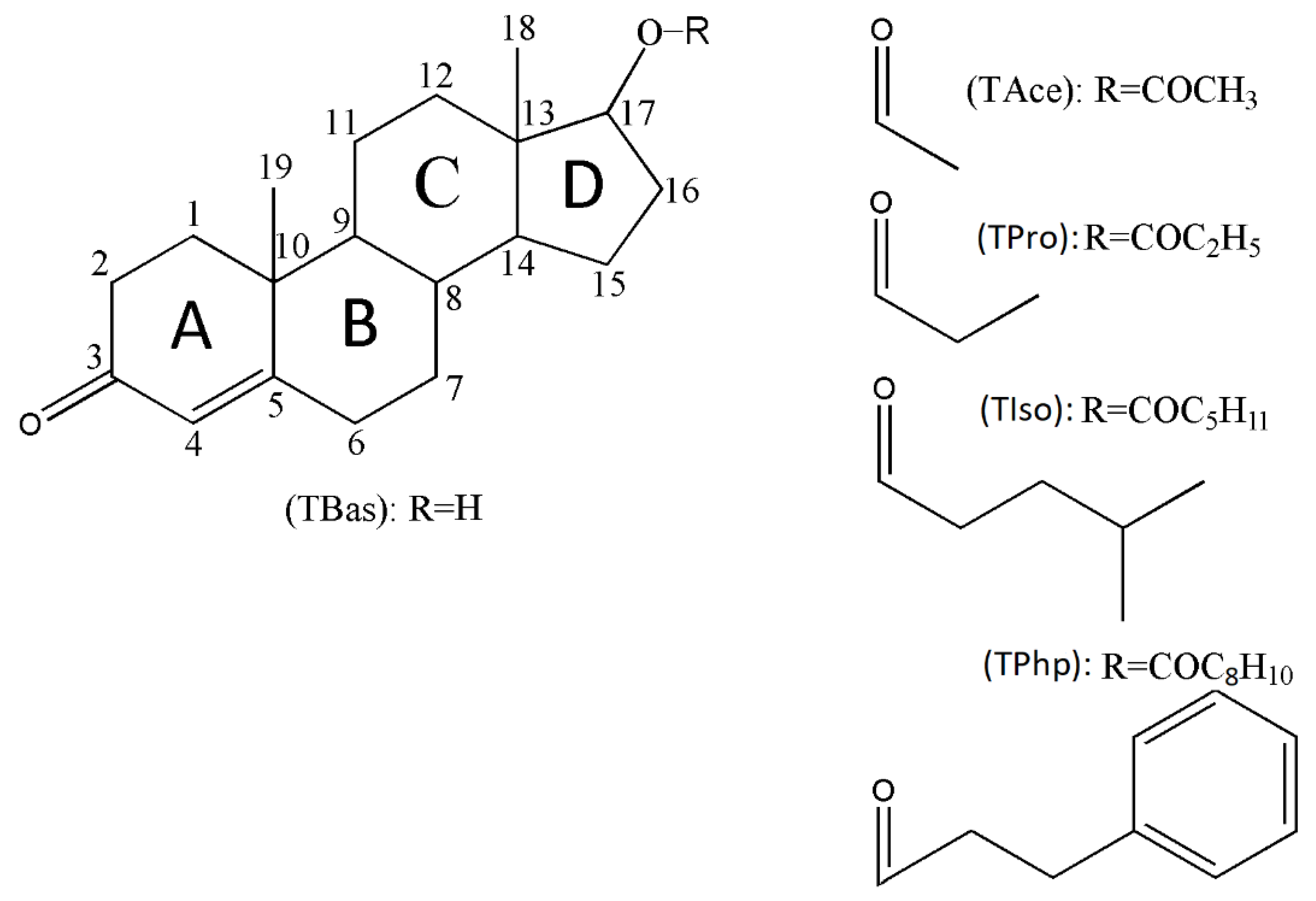
:1. Introduction
- (i)
- Testosterone acetate (Androst-4-en-17β-ol-3-one 17β-acetate; TAce);
- (ii)
- Testosterone propionate (Androst-4-en-17β-ol-3-one 17β-propionate; TPro);
- (iii)
- Testosterone isocaproate (Androst-4-en-17β-ol-3-one 17β-4-methylpentanoate; TIso);
- (iv)
- Testosterone phenylpropionate (Androst-4-en-17β-ol-3-one 17β-phenylpropionate; TPhp).
2. Materials and Methods
2.1. Materials and Crystallisation Experiments
2.2. X-ray Powder Diffraction (XRPD)
2.3. Single-Crystal X-ray Diffraction and Structural Refinement
2.4. Crystal Lattice Energy Computation and Hirshfeld and Fingerprint Plot Analyses
2.5. Solubility Check
3. Results and Discussion
3.1. Crystal Structures and Supramolecular Descriptions
3.1.1. TAce (Testosterone Acetate)
3.1.2. TPro (Testosterone Propionate)
3.1.3. TIso (Testosterone Isocaproate)
3.1.4. TPhp (Testosterone Phenylpropionate)
- (i)
- Asymmetric units are characterised by a single molecule for each ester;
- (ii)
- The formations of supramolecular 3D assemblies are to some extent driven by the C-H•••O interactions, although the dispersion energy has the greatest weight, as will be shown in the crystal energies analysis section; donor–acceptor separation distances show similar values to those of other crystals driven by C-H•••O interactions and belong to the steroid family [30,31,32,33,34];
- (iii)
- The six-membered A rings are found in the intermediate sofa-half-chair geometry, and the B and C rings show chair conformations, while the five-membered D rings adopt intermediate envelope-half-chair geometry. Similar geometries of skeleton rings have been reported in the crystal structure of its C-17 methylated form [35].
3.2. Crystal Energy Analysis
3.3. Hirshfeld and Fingerprint Plot Analysis
- (i)
- Fingerprint plots of esterified forms (Figure 6b) (TAce, TPro, TPhp, and TIso) show symmetry in the spikes, which is a particular feature for the crystals with one molecule in asymmetric units, while the plots of TBas (Figure 6a) are asymmetric due to the different molecular environment in the crystal;
- (ii)
- The diagrams of Tace, TPro, and TIso illustrate protruding H•••O/O•••H spikes, denoting the presence of C-H•••O hydrogen bonds, while for TPhp, the lack of H•••O/O•••H spikes shows that the separation distances of the C-H•••O interactions fall in a range closer to the sum of vdW radii;
- (iii)
- The fingerprint plots of Tbas show more protruding H•••O/O•••H spikes compared with its esterified forms and suggest that strong O-H•••O interactions play more important roles in packing; this feature is seen in the evaluation of crystal energies where the Coulombic energy becomes more significant in TBas due to the presence of strong O-H•••O contacts;
- (iv)
- The quantitative breakdown of fingerprint diagrams (Table 3) in all five crystals reveals a high percentage of H•••H contacts, medium contribution by O•••H/H•••O intercontacts and considerably smaller for C•••H/H•••C, respectively;
- (v)
3.4. Solubility Check
4. Conclusions
Supplementary Materials
Author Contributions
Funding
Institutional Review Board Statement
Informed Consent Statement
Data Availability Statement
Conflicts of Interest
References
- Mooradian, A.D.; Morley, J.E.; Korenman, S.G. Biological actions of androgens. Endocr. Rev. 1987, 8, 1–28. [Google Scholar] [CrossRef] [PubMed]
- Bassil, N.; Alkaade, S.; Morley, J.E. The benefits and risks of testosterone replacement therapy: A review. Ther. Clin. Risk Manag. 2009, 5, 427–448. [Google Scholar] [CrossRef] [PubMed] [Green Version]
- Tuck, S.P.; Francis, R.M. Testosterone, bone and osteoporosis. Front. Horm Res. 2009, 37, 123–132. [Google Scholar] [CrossRef] [PubMed]
- Luetjens, C.M.; Weinbauer, G.F. Chapter 2: Testosterone: Biosynthesis, Transport, Metabolism and (Non-Genomic) Actions, 4th ed.; Cambridge University Press: Cambridge, UK, 2012; pp. 15–32. [Google Scholar]
- Pappas, I.I.; Craig, W.Y.; Spratt, L.V.; Spratt, D.I. Efficacy of Sex Steroid Therapy Without Progestin or GnRH Agonist for Gonadal Suppression in Adult Transgender Patients. J. Clin. Endocrinol. Metab. 2021, 106, E1290–E1300. [Google Scholar] [CrossRef]
- Quigley, C.A.; Bellis, A.D.; Marschke, K.B.; Awady, M.K.; Wilson, E.M.; French, F.S. Androgen receptor defects: Historical, clinical, and molecular perspectives. Endocr. Rev. 1995, 16, 271–321. [Google Scholar] [CrossRef]
- Stanworth, R.D.; Jones, T.H. Testosterone for the aging male; current evidence and recommended practice. Clin. Interv. Aging. 2008, 3, 25–44. [Google Scholar] [CrossRef] [Green Version]
- Institute of Medicine (US). Committee on Assessing the Need for Clinical Trials of Testosterone Replacement Therapy. In Testosterone and Aging: Clinical Research Directions; National Academies Press: Washington, DC, USA, 2004. [Google Scholar]
- Braunstein, G.D. The influence of anabolic steroids on muscular strength. Princ. Med. Biol. 1997, 8, 465–474. [Google Scholar] [CrossRef]
- Vermeulen, A. Longacting steroid preparations. Acta Clin. Belg. 1975, 30, 48–55. [Google Scholar] [CrossRef]
- Forsdahl, G.; Erced, D.; Geisendorfer, T.; Turkalj, M.; Plavec, D.; Thevis, M.; Tretzele, L.; Gmeinera, G. Detection of testosterone esters in blood. Drug Test. Anal. 2015, 7, 983–989. [Google Scholar] [CrossRef]
- Elks, J.; Ganellin, C.R. The Dictionary of Drugs: Chemical Data: Chemical Data, Structures and Bibliographies, 1st ed.; Springer: Easton, PA, USA, 1990; p. 652. [Google Scholar]
- Reisch, J.; Eki-Gucer, N.; Takacs, M.; Henkel, G. Photochemische Studien, 54. Photodimerisierung von Testosteronpropionat in kristallinem Zustand und Kristallstruktur von Testosteronpropionat. Liebigs Ann. Der Chem. 1989, 1989, 595–597. [Google Scholar] [CrossRef]
- Griffiths, P.J.F.; James, K.C.; Rees, M. Crystallographic data for some testosterone esters. Acta Cryst. 1965, 19, 149. [Google Scholar] [CrossRef]
- Alcock, N.W.; Sanders, K.J.; Rodger, A. Potential injectable contraceptive steroids: Testosterone buciclate. Acta Cryst. 2004, E60, 348–349. [Google Scholar] [CrossRef]
- Böcskei, Z.; Gérczei, T.; Bodor, A.; Schwartz, R.; Náray-Szabó, G. Three Testosterone Derivatives. Acta Cryst. 1996, C52, 2899–2903. [Google Scholar] [CrossRef]
- Roberts, P.J.; Pettersen, R.C.; Sheldrick, G.M.; Isaacs, N.W.; Kennard, O. Crystal and molecular structure of 17β-hydroxyandrost-4-en-3-one (testosterone). J. Chem. Soc. Perkin Trans. 1973, 2, 1978–1984. [Google Scholar] [CrossRef]
- Land, L.M.; Li, P.; Baummer, P.M. The Influence of Water Content of Triglyceride Oils on the Solubility. Pharm. Res. 2005, 5, 784–788. [Google Scholar] [CrossRef]
- Remington. The Science and Practice of Pharmacy, 23rd ed.; Adejare, A., Ed.; Elsevier: Philadelphia, PA, USA, 2020. [Google Scholar]
- Rowe, R.C.; Sheskey, P.J.; Quinn, M.E. (Eds.) Handbook of Pharmaceutical Excipients, 5th ed.; Pharmaceutical Press: London, UK; American Pharmacists Association: Washington, DC, USA, 2006. [Google Scholar]
- CrysAlis PRO. Agilent Technologies UK Ltd., Oxford Diffraction; Agilent Technologies UK Ltd.: Yarnton, Oxfordshire, UK, 2015. [Google Scholar]
- Sheldrick, G.M. SHELXT—Integrated space-group and crystal-structure determination. Acta Cryst. 2015, A71, 3–8. [Google Scholar] [CrossRef] [Green Version]
- Sheldrick, G.M. A short history of SHELX. Acta Cryst. 2008, A64, 112–122. [Google Scholar] [CrossRef] [Green Version]
- Sheldrick, G.M. Crystal structure refinement with SHELXL. Acta Cryst. 2015, C71, 3–8. [Google Scholar] [CrossRef] [Green Version]
- Dolomanov, O.V.; Bourhis, L.J.; Gildea, R.J.; Howard, J.A.K.; Puschmann, H. OLEX2: A complete structure solution, refinement and analysis program. J. Appl. Cryst. 2009, 42, 339–341. [Google Scholar] [CrossRef]
- Gavezzotti, A. Efficient computer modeling of organic materials. The atom–atom, Coulomb–London–Pauli (AA-CLP) model for intermolecular electrostatic-polarization, dispersion and repulsion energies. New J. Chem. 2011, 35, 1360–1368. [Google Scholar] [CrossRef]
- Turner, M.J.; McKinnon, J.J.; Wolff, S.K.; Grimwood, D.J.; Spackman, P.R.; Jayatilaka, D.; Spackman, M.A. CrystalExplorer17; University of Western Australia: The Nedlands, Australia, 2017. [Google Scholar]
- Spackman, M.A.; McKinnon, J.J. Fingerprinting intermolecular interactions in molecular crystals. CrystEngComm 2002, 4, 378–392. [Google Scholar] [CrossRef]
- Alvarez, S. A cartography of the van der Waals territories. Dalton Trans. 2013, 42, 8617–8636. [Google Scholar] [CrossRef] [PubMed] [Green Version]
- Ohrt, J.; Haner, B.A.; Norton, D.A. Crystal data (II) for some androstanes. Acta Cryst. 1965, 19, 479. [Google Scholar] [CrossRef]
- Turza, A.; Borodi, G.; Pop, M.M.; Ulici, A. Polymorphism and β-cyclodextrin complexation of methyldrostanolone. J. Mol. Struct. 2022, 1250, 131852. [Google Scholar] [CrossRef]
- Turza, A.; Ulici, A.; Pop, M.M.; Borodi, G. Solid forms and β-cyclodextrin complexation of turinabol. Acta Cryst. 2022, C78, 305–313. [Google Scholar] [CrossRef]
- Rajnikant, V.; Dinesh, D.; Aziz, N.; Gupta, B.D. Analysis of C–H•••O Intermolecular Interactions in 4-Androstene-3,17-dione. J. Chem. Crystallogr. 2009, 39, 24–27. [Google Scholar] [CrossRef]
- Rajnikant, V.; Gupta, V.K.; Khan, E.H.; Shafi, S.; Hashmi, S.; Shafiullah, B. Varghese, Dinesh, Crystal structure of cholest-4-ene-3,6-dione: A steroid. Crystallogr. Rep. 2001, 46, 963–966. [Google Scholar] [CrossRef]
- Gaedecki, Z. Structure of 17-α-methyl-testosterone semihydrate C20H30O2 H2O. J. Crystallogr. Spectrosc. Res. 1989, 19, 577–587. [Google Scholar] [CrossRef]
- Turza, A.; Miclaus, M.O.; Pop, A.; Borodi, G. Crystal and molecular structures of boldenone and four boldenone steroid esters. Z. Kristallogr. Cryst. Mater. 2019, 234, 671–683. [Google Scholar] [CrossRef]
- Borodi, G.; Turza, A.; Camarasan, P.A.; Ulici, A. Structural studies of Trenbolone, Trenbolone Acetate, Hexahydrobenzylcarbonate and Enanthate esters. J. Mol. Struct. 2020, 1212, 128127. [Google Scholar] [CrossRef]
- Borodi, G.; Turza, A.; Bende, A. Exploring the Polymorphism of Drostanolone Propionate. Molecules 2020, 25, 1436. [Google Scholar] [CrossRef] [PubMed] [Green Version]
- Turza, A.; Borodi, G.; Pop, A.; David, M. Structural studies of some androstane based prodrugs. J. Mol. Struct. 2022, 1248, 131440. [Google Scholar] [CrossRef]
- Caplette, J.; Frigo, T.; Jozwiakowski, M.; Shea, H.; Mirmehrabi, M.; Müller, P. Characterization of new crystalline forms of hydroxyprogesterone caproate. Int. J. Pharm. 2017, 527, 42–51. [Google Scholar] [CrossRef] [PubMed]
- Molcomson, C.; Lawrence, M.J. A comparison of incorporation of model steroids into non-ionic micellar and microemulsion systems. J. Pharm. Pharmacol. 1993, 45, 141–143. [Google Scholar] [CrossRef]


| Identification Code | TPro (Testosterone Propionate) | TIso (Testosterone Isocaproate) | TPhp (Testosterone Phenylpropionate) |
|---|---|---|---|
| Empirical formula | C22H32O3 | C25H38O3 | C28H36O3 |
| Formula weight | 344.47 | 386.57 | 420.57 |
| Temperature/K | 293(2) | 293(2) | 293(2) |
| Crystal system | orthorhombic | monoclinic | monoclinic |
| Space group | P212121 | P21 | P21 |
| a/Å | 7.57470(16) | 7.2877(3) | 13.4097(7) |
| b/Å | 12.6768(2) | 12.3741(5) | 5.9105(3) |
| c/Å | 20.4038(4) | 13.1272(6) | 15.4054(8) |
| α/° | 90 | 90 | 90 |
| β/° | 90 | 103.305(4) | 95.073(5) |
| γ/° | 90 | 90 | 90 |
| Volume/Å3 | 1959.23(6) | 1152.02(9) | 1216.22(11) |
| Z | 4 | 2 | 2 |
| ρcalcg/cm3 | 1.168 | 1.106 | 1.148 |
| μ/mm−1 | 0.594 | 0.552 | 0.568 |
| F(000) | 752.0 | 418.0 | 456.0 |
| Crystal size/mm | 0.09 × 0.08 × 0.07 | 0.1 × 0.03 × 0.01 | 0.09 × 0.09 × 0.01 |
| Radiation | CuKα (λ = 1.54184) | CuKα (λ = 1.54184) | CuKα (λ = 1.54184) |
| 2Θ range/° | 8.212 to 141.254 | 9.952 to 141.104 | 5.76 to 141.522 |
| Index ranges | −8 ≤ h ≤ 9, −15 ≤ k ≤ 15, −24 ≤ l ≤ 24 | −8 ≤ h ≤ 8, −15 ≤ k ≤ 15, −15 ≤ l ≤ 16 | −16 ≤ h ≤ 16, −7 ≤ k ≤ 7, −18 ≤ l ≤ 18 |
| Reflections collected | 28,102 | 15,810 | 14,176 |
| Independent reflections | 3723 [Rint = 0.0243, Rsigma = 0.0119] | 4322 [Rint = 0.0246, Rsigma = 0.0185] | 4529 [Rint = 0.0796, Rsigma = 0.0508] |
| Data/restraints/ parameters | 3723/0/229 | 4322/1/257 | 4529/1/282 |
| Goodness-of-fit on F2 | 1.044 | 1.076 | 1.078 |
| Final R indexes [I ≥ 2σ (I)] | R1 = 0.0421, wR2 = 0.1187 | R1 = 0.0621, wR2 = 0.1739 | R1 = 0.0762, wR2 = 0.1784 |
| Final R indexes [all data] | R1 = 0.0440, wR2 = 0.1217 | R1 = 0.0740, wR2 = 0.1900 | R1 = 0.1102, wR2 = 0.1959 |
| Largest diff. peak/hole/e Å−3 | 0.17/−0.17 | 0.33/−0.21 | 0.25/−0.21 |
| Flack parameter | 0.04(6) | 0.06(8) | −0.4(3) |
| Structure | Molar Mass g/mol | Ecoul (kJ/mol) | Epol (kJ/mol) | Edisp (kJ/mol) | Erep (kJ/mol) | Elatt (kJ/mol) |
|---|---|---|---|---|---|---|
| TBas | 288.43 | −33.3 | −47.5 | −130.9 | 60.9 | −150.8 |
| TAce | 330.46 | −15.5 | −55.0 | −126.1 | 37.1 | −159.5 |
| TPro | 344.49 | −18.3 | −55.5 | −126.4 | 33.9 | −166.3 |
| TIso | 386.57 | −21.3 | −57.7 | −141.9 | 52.7 | −168.2 |
| TPhp | 420.59 | −19.3 | −52.3 | −149.0 | 36.1 | −184.5 |
| Structure | H•••H | O•••H/H•••O | C•••H/H•••C | C•••O/O•••C | O•••O | C•••C |
|---|---|---|---|---|---|---|
| TBas Mol A | 78.8% | 17.2% | 4.0% | - | - | - |
| TBas Mol B | 76.4% | 19.7% | 3.9% | - | - | - |
| TAce | 75.4% | 20.2% | 2.8% | 1.2% | - | 0.4% |
| TPro | 76.5% | 19.4% | 4.0% | - | 0.1% | - |
| TIso | 80.4% | 16.3% | 3.3% | - | - | - |
| TPhp | 72.7% | 15.2% | 12.1% | - | - | - |
Publisher’s Note: MDPI stays neutral with regard to jurisdictional claims in published maps and institutional affiliations. |
© 2022 by the authors. Licensee MDPI, Basel, Switzerland. This article is an open access article distributed under the terms and conditions of the Creative Commons Attribution (CC BY) license (https://creativecommons.org/licenses/by/4.0/).
Share and Cite
Turza, A.; Popescu, V.; Mare, L.; Borodi, G. Structural Aspects and Intermolecular Energy for Some Short Testosterone Esters. Materials 2022, 15, 7245. https://doi.org/10.3390/ma15207245
Turza A, Popescu V, Mare L, Borodi G. Structural Aspects and Intermolecular Energy for Some Short Testosterone Esters. Materials. 2022; 15(20):7245. https://doi.org/10.3390/ma15207245
Chicago/Turabian StyleTurza, Alexandru, Violeta Popescu, Liviu Mare, and Gheorghe Borodi. 2022. "Structural Aspects and Intermolecular Energy for Some Short Testosterone Esters" Materials 15, no. 20: 7245. https://doi.org/10.3390/ma15207245