
Investigation of the Structure and Corrosion Resistance of Novel High-Entropy Alloys for Potential Biomedical Applications

 , , , and
Abstract
:1. Introduction
2. Materials and Methods
2.1. Materials
2.2. Sample Preparation
2.3. Characterization Methods
3. Results and Discussion
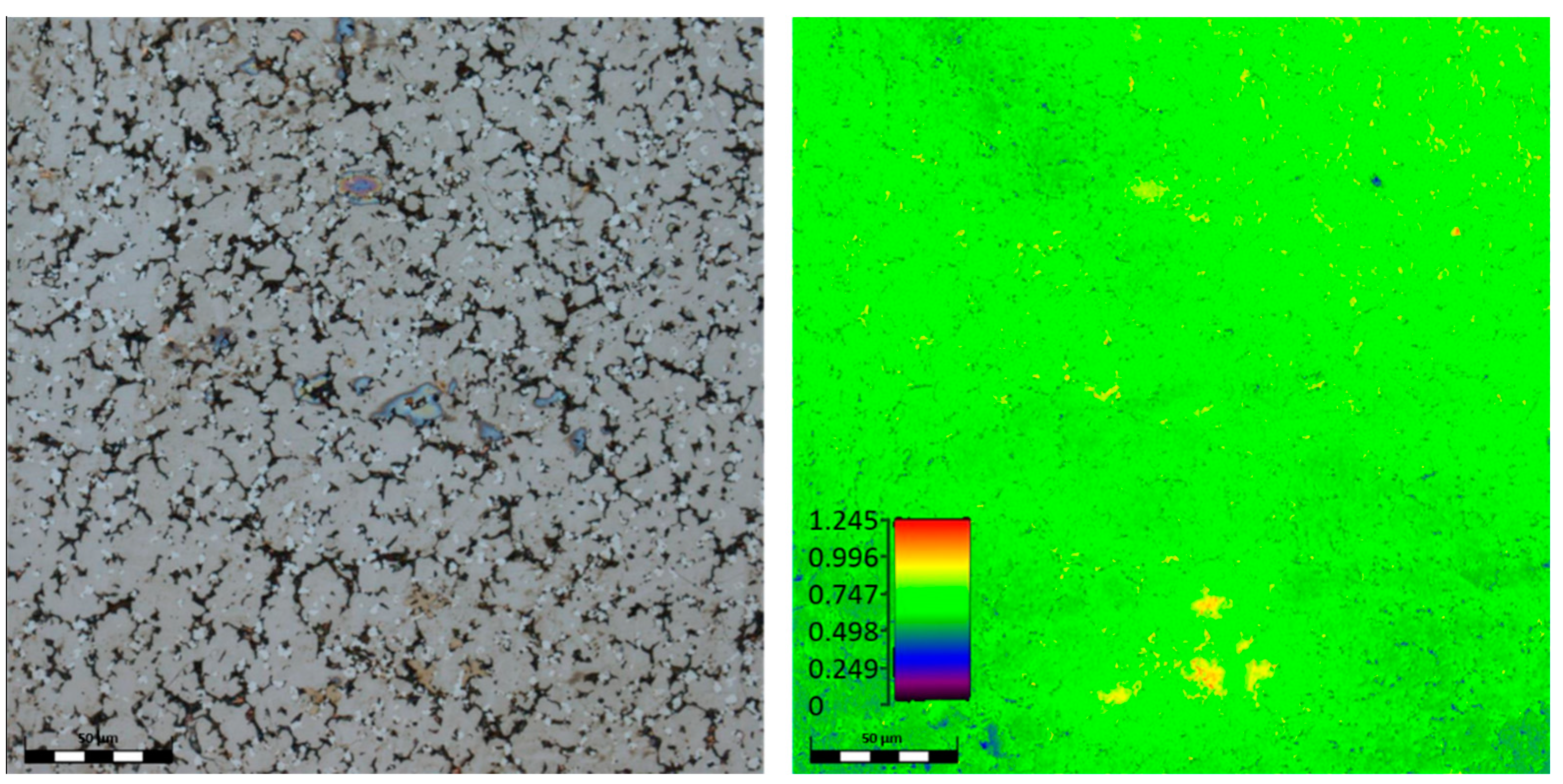
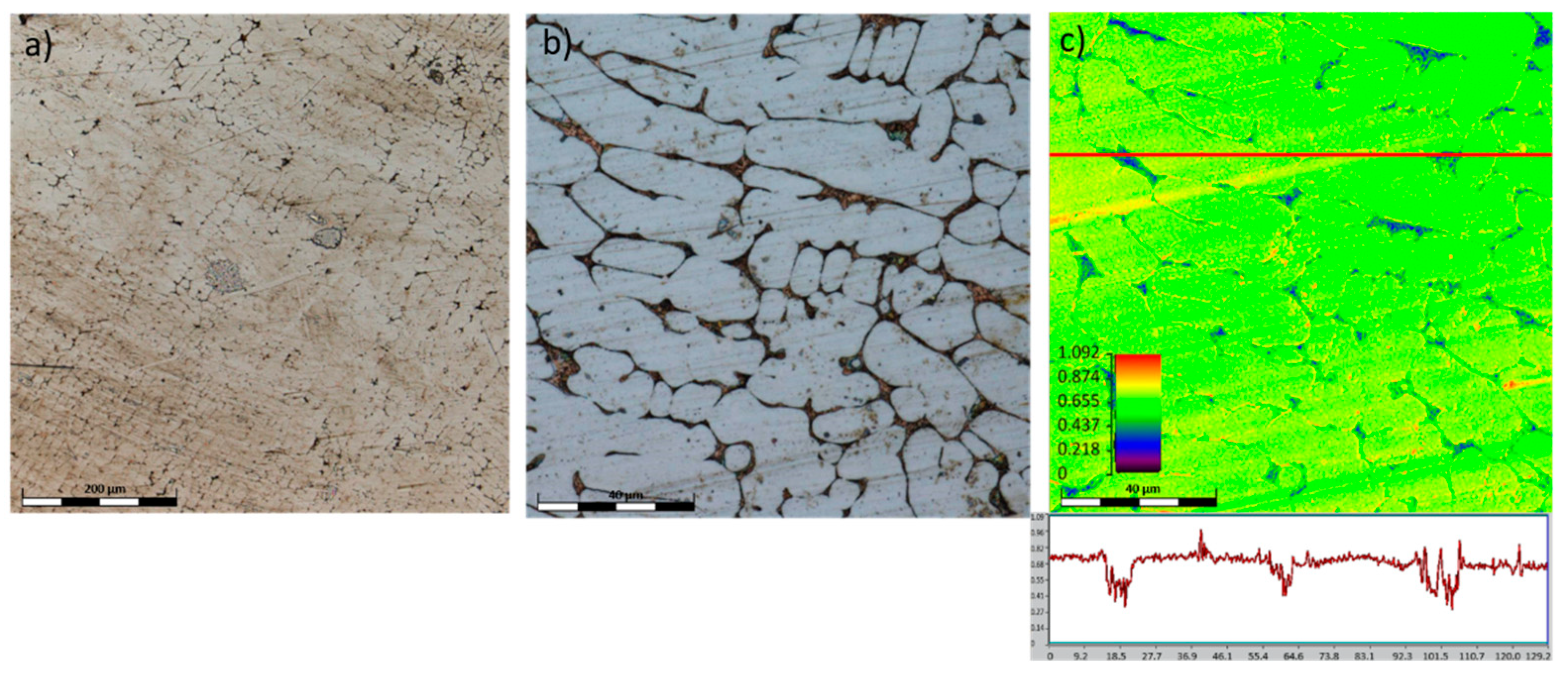
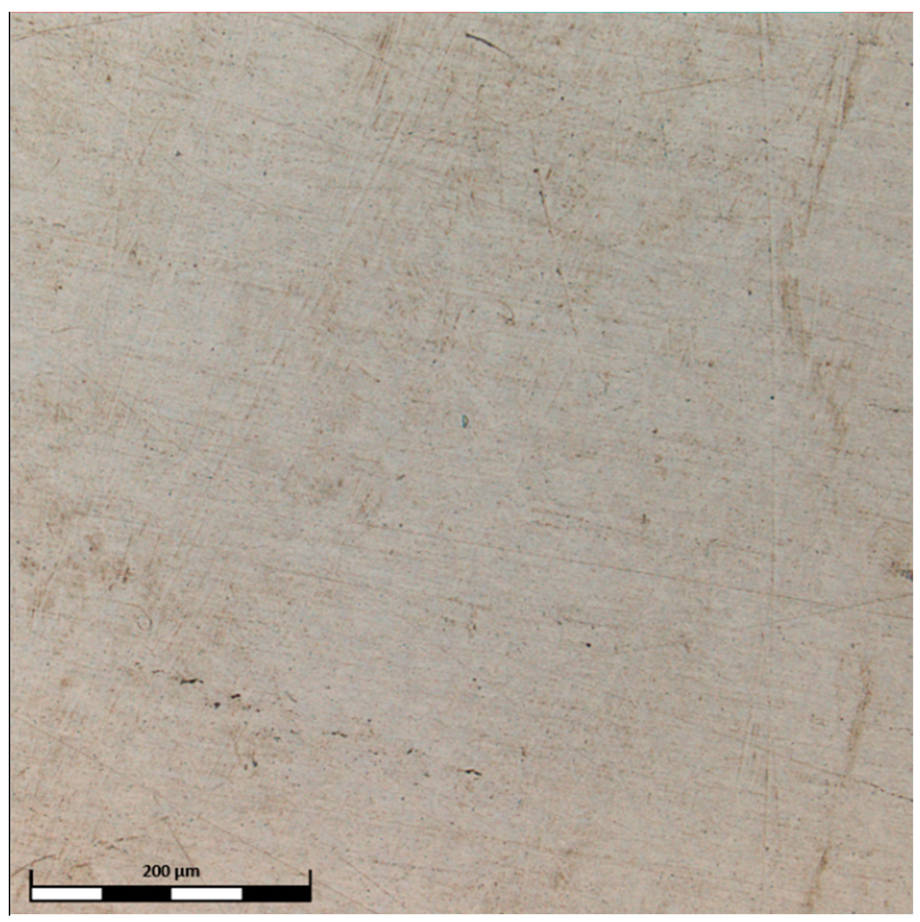
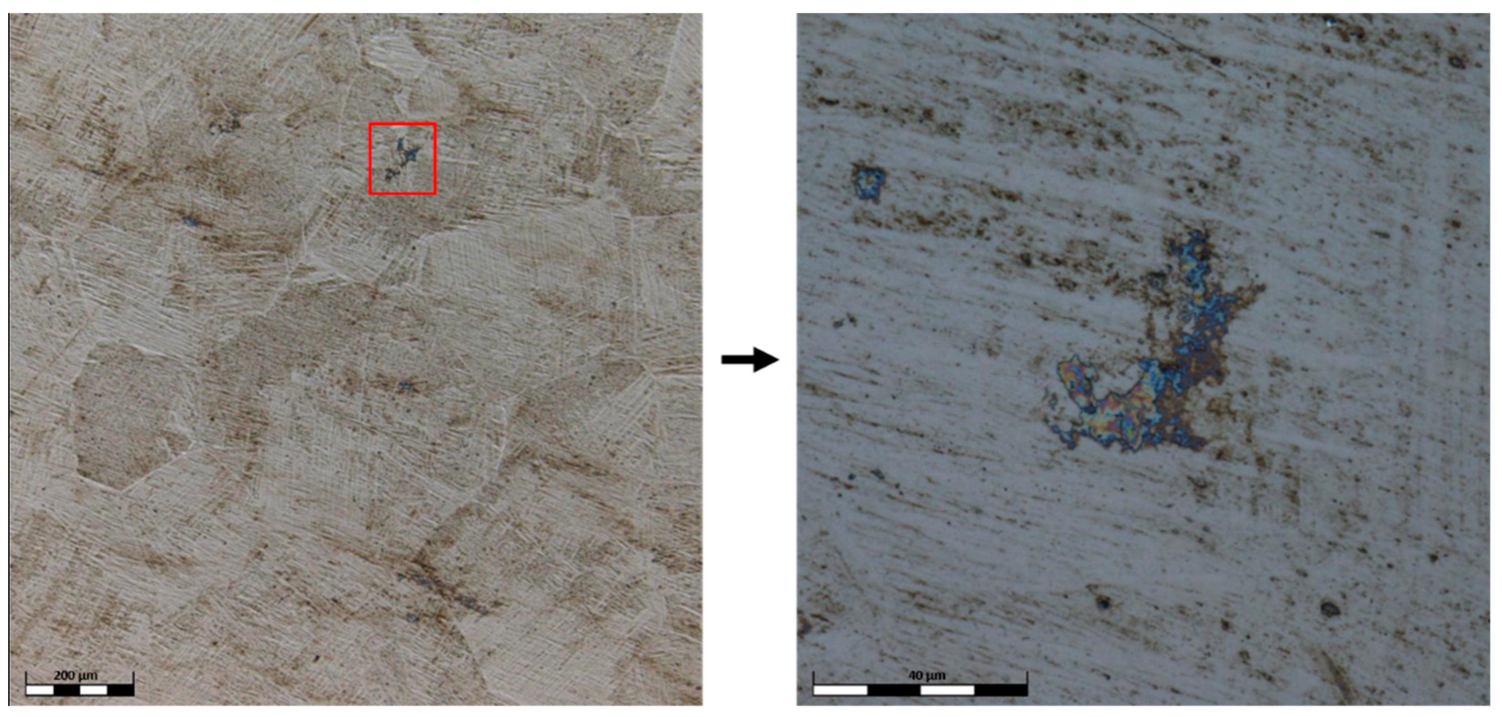
3.1. Microstructure
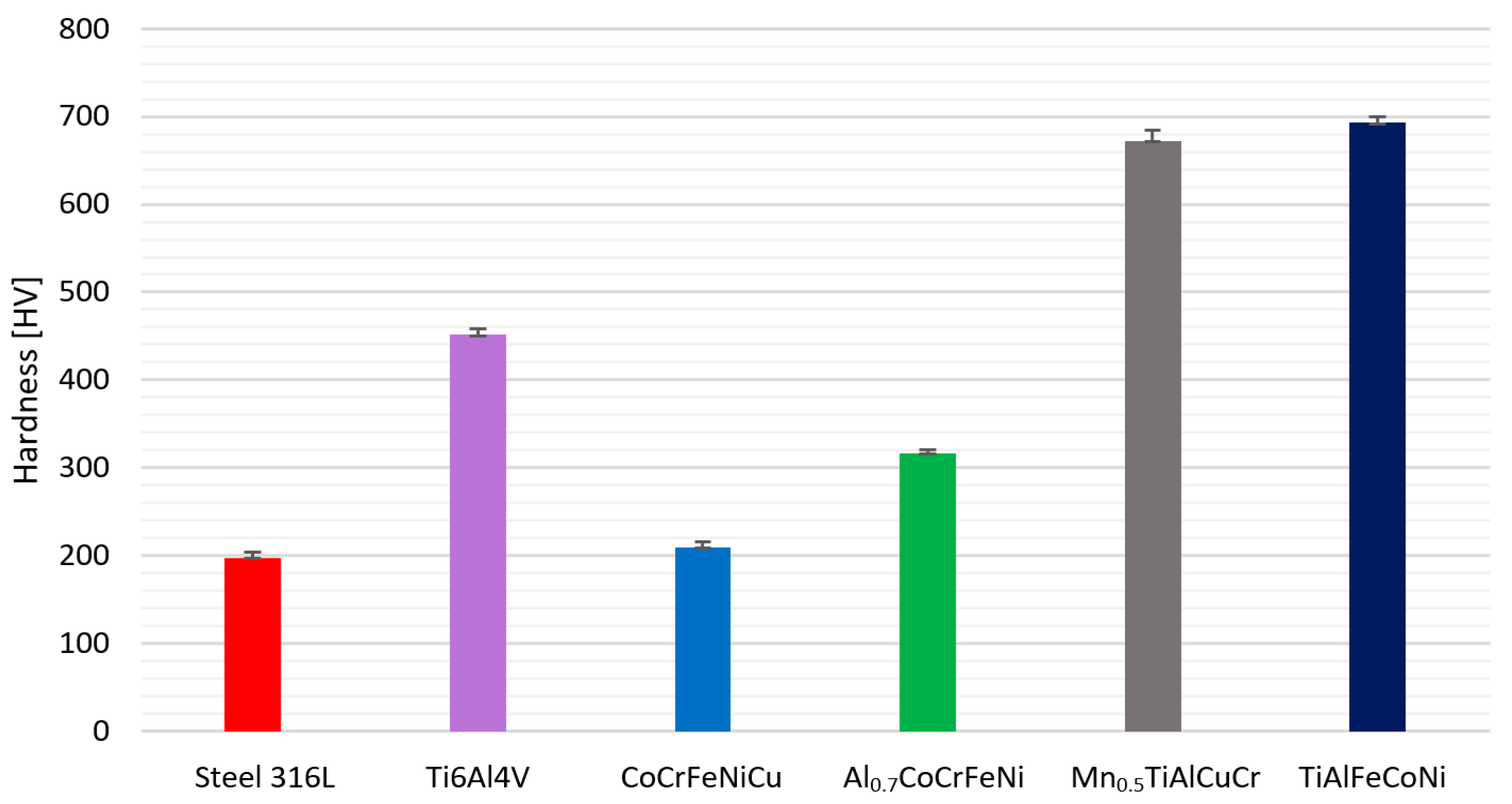
3.2. Hardness
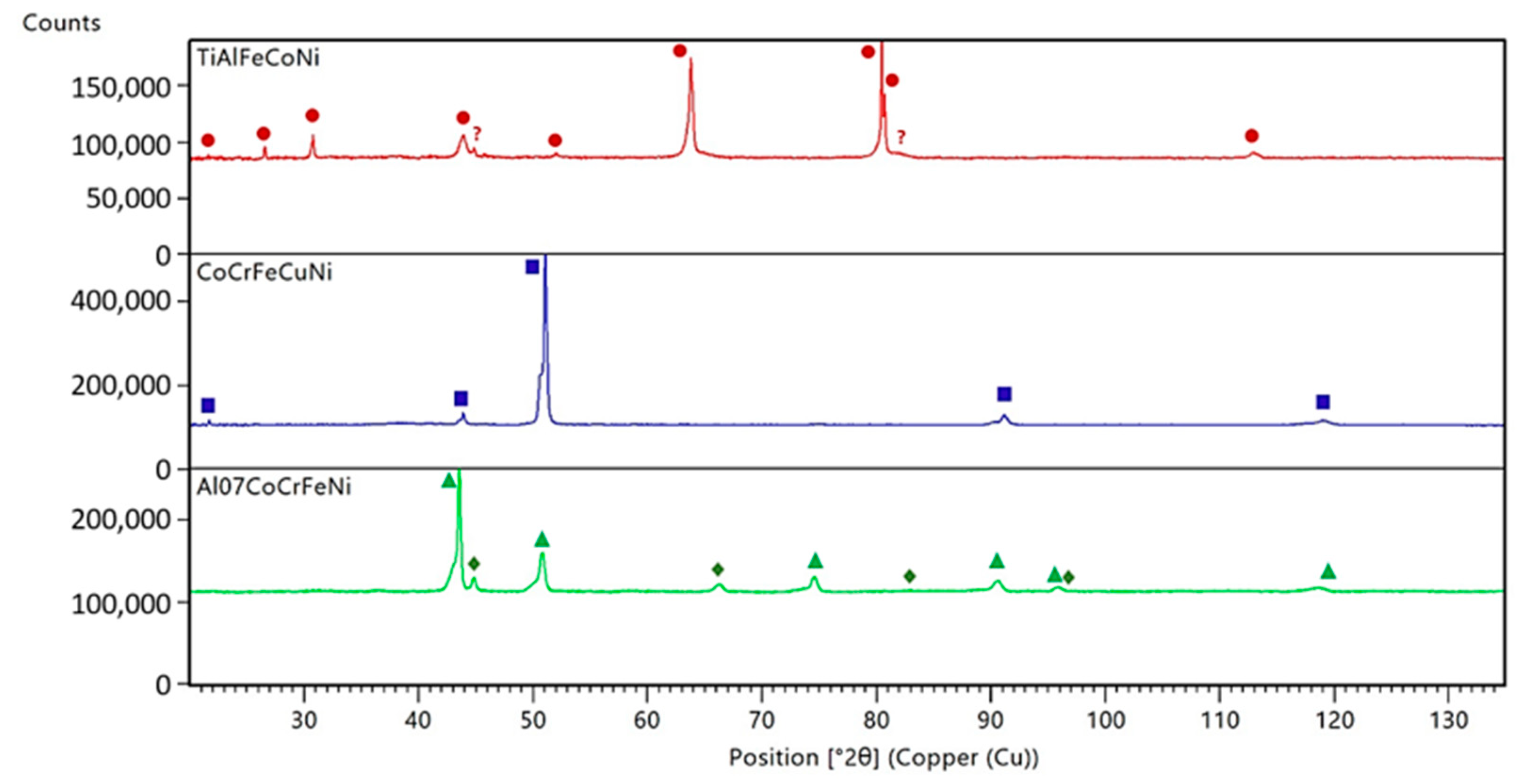
3.3. X-ray Diffraction Analysis
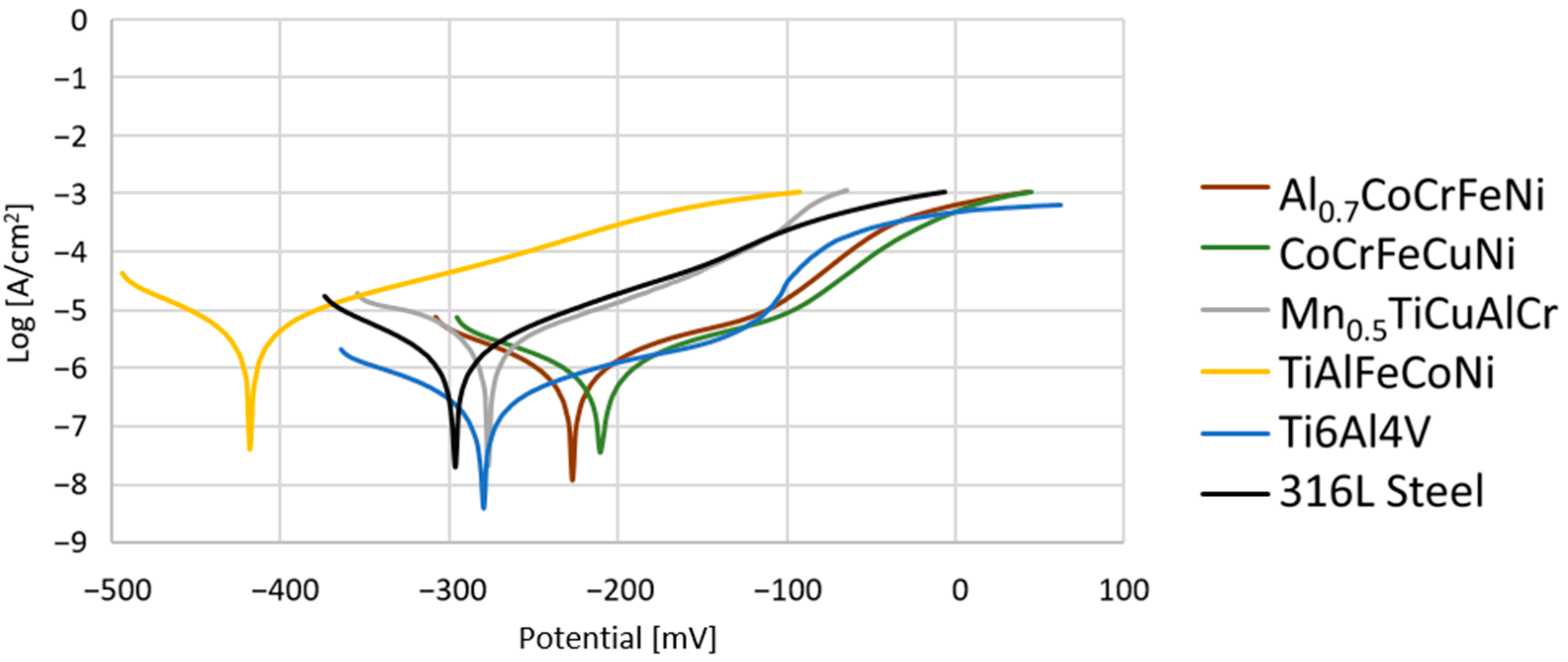
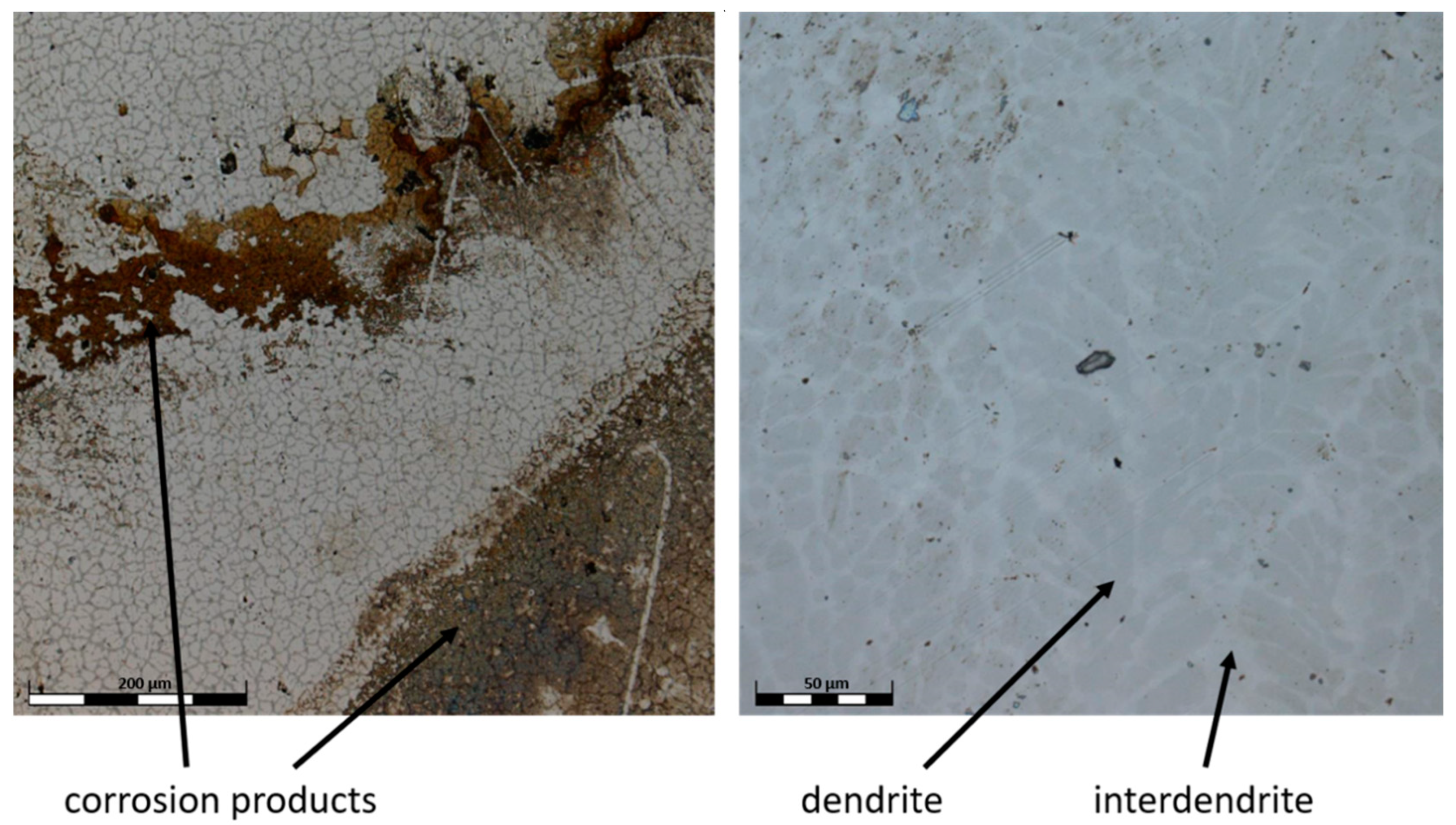
3.4. Corossion Properties
4. Conclusions
- The studied high-entropy alloys crystallize mainly as solid solutions with simple crystal structures of high symmetry. Both TiAlFeCoNi and CoCrFeNiCu samples seem to be disordered single phases of an fcc type structure, but disclose the different types of space group. The neglectable contribution of two unrecognized peaks visible on the TiAlFeCoNi diagram does not influence the quality of refinements. The bi-phase Al0.7CoCrFeNi sample has the predominant contribution of an ordered fcc phase and a neglectable amount of ordered bcc structure.
- Three out of four selected high-entropy alloys showed good corrosion properties. CoCrFeNiCu and Al0.7CoCrFeNi exhibited lower values for corrosion potentials and lower corrosion current density, as well as significantly lower corrosion rates, which may suggest a better corrosion resistance than the reference implant alloys (steel 316L and Ti6Al4V). These results indicate that these alloys can be considered as potential alloys for biomedical applications. This, of course, still requires many more studies to confirm their biocompatibility.
- The presence of titanium in the high-entropy alloys (TiAlFeCoNi, Mn0.5TiCuAlCr) resulted in a significant strengthening effect on the materials, which could potentially result in good wear resistance. However, out of all samples tested, when compared with all investigated alloys, the TiAlFeCoNi alloy exhibited the worst corrosion properties, which excludes it as a material for biomedical applications. On the other hand, Mn0.5TiAlCuCr showed corrosion characteristics similar to Ti6Al4V, which combined with a high hardness, may determine its application as a friction element for biomedical implants. Nevertheless, in the long run, the precipitates present in these alloys may contribute to a decrease in their anti-corrosion properties.
Author Contributions
Funding
Institutional Review Board Statement
Informed Consent Statement
Data Availability Statement
Acknowledgments
Conflicts of Interest
References
- Zhang, Y.; Zuo, T.T.; Tang, Z.; Gao, M.C.; Dahmen, K.A.; Liaw, P.K.; Lu, Z.P. Microstructures and Properties of High-Entropy Alloys. Prog. Mater. Sci. 2014, 61, 1–93. [Google Scholar] [CrossRef]
- Yeh, J. Recent Progress in High-Entropy Alloys. Ann. De Chim. Sci. Des Mater. 2006, 31, 633–648. [Google Scholar] [CrossRef]
- Yin, X.; Xu, S. Properties and Preparation of High Entropy Alloys. In Proceedings of the MATEC Web of Conferences, Qingdao, China, 25–27 August 2017; EDP Sciences: Les Ulis, France, 2018; Volume 142. [Google Scholar]
- Yeh, J.-W.; Chen, S.-K.; Lin, S.-J.; Gan, J.-Y.; Chin, T.-S.; Shun, T.-T.; Tsau, C.-H.; Chang, S.-Y. Nanostructured High-Entropy Alloys with Multiple Principal Elements: Novel Alloy Design Concepts and Outcomes. Adv. Eng. Mater. 2004, 6, 299–303. [Google Scholar] [CrossRef]
- Huang, P.-K.; Yeh, J.-W.; Shun, T.-T.; Chen, S.-K. Multi-Principal-Element Alloys with Improved Oxidation and Wear Resistance for Thermal Spray Coating. Adv. Eng. Mater. 2004, 6, 74–78. [Google Scholar] [CrossRef]
- Miracle, D.; Miller, J.; Senkov, O.; Woodward, C.; Uchic, M.; Tiley, J. Exploration and Development of High Entropy Alloys for Structural Applications. Entropy 2014, 16, 494–525. [Google Scholar] [CrossRef]
- Lee, C.P.; Chen, Y.Y.; Hsu, C.Y.; Yeh, J.W.; Shih, H.C. The Effect of Boron on the Corrosion Resistance of the High Entropy Alloys Al[Sub 0.5]CoCrCuFeNiB[Sub x]. J. Electrochem. Soc. 2007, 154, C424. [Google Scholar] [CrossRef]
- Zhu, J.M.; Fu, H.M.; Zhang, H.F.; Wang, A.M.; Li, H.; Hu, Z.Q. Synthesis and Properties of Multiprincipal Component AlCoCrFeNiSix Alloys. Mater. Sci. Eng. A 2010, 527, 7210–7214. [Google Scholar] [CrossRef]
- Hsu, C.Y.; Juan, C.C.; Wang, W.R.; Sheu, T.S.; Yeh, J.W.; Chen, S.K. On the Superior Hot Hardness and Softening Resistance of AlCoCrxFeMo0.5Ni High-Entropy Alloys. Mater. Sci. Eng. A 2011, 528, 3581–3588. [Google Scholar] [CrossRef]
- Miracle, D.B.; Senkov, O.N. A Critical Review of High Entropy Alloys and Related Concepts. Acta Mater. 2017, 122, 448–511. [Google Scholar] [CrossRef] [Green Version]
- Chen, J.; Zhou, X.; Wang, W.; Liu, B.; Lv, Y.; Yang, W.; Xu, D.; Liu, Y. A Review on Fundamental of High Entropy Alloys with Promising High–Temperature Properties. J. Alloys Compd. 2018, 760, 15–30. [Google Scholar] [CrossRef]
- Tokarewicz, M.; Grądzka-Dahlke, M. Review of Recent Research on AlCoCrFeNi High-Entropy Alloy. Metals 2021, 11, 1302. [Google Scholar] [CrossRef]
- Tsai, M.-H.; Yeh, J.-W. High-Entropy Alloys: A Critical Review. Mater. Res. Lett. 2014, 2, 107–123. [Google Scholar] [CrossRef]
- Wang, S.-P.; Xu, J. TiZrNbTaMo High-Entropy Alloy Designed for Orthopedic Implants: As-Cast Microstructure and Mechanical Properties. Mater. Sci. Eng. C 2017, 73, 80–89. [Google Scholar] [CrossRef] [PubMed]
- Navi, A.S.; Haghighi, S.E.; Haghpanahi, M.; Momeni, A. Investigation of Microstructure and Corrosion of TiNbTaZrMo High-Entropy Alloy in the Simulated Body Fluid. J. Bionic. Eng. 2021, 18, 118–127. [Google Scholar] [CrossRef]
- Hua, N.; Wang, W.; Wang, Q.; Ye, Y.; Lin, S.; Zhang, L.; Guo, Q.; Brechtl, J.; Liaw, P.K. Mechanical, Corrosion, and Wear Properties of Biomedical Ti–Zr–Nb–Ta–Mo High Entropy Alloys. J. Alloys Compd. 2021, 861, 157997. [Google Scholar] [CrossRef]
- Todai, M.; Nagase, T.; Hori, T.; Matsugaki, A.; Sekita, A.; Nakano, T. Novel TiNbTaZrMo High-Entropy Alloys for Metallic Biomaterials. Scr. Mater. 2017, 129, 65–68. [Google Scholar] [CrossRef] [Green Version]
- Ishimoto, T.; Ozasa, R.; Nakano, K.; Weinmann, M.; Schnitter, C.; Stenzel, M.; Matsugaki, A.; Nagase, T.; Matsuzaka, T.; Todai, M.; et al. Development of TiNbTaZrMo Bio-High Entropy Alloy (BioHEA) Super-Solid Solution by Selective Laser Melting, and Its Improved Mechanical Property and Biocompatibility. Scr. Mater. 2021, 194, 113658. [Google Scholar] [CrossRef]
- Yuan, Y.; Wu, Y.; Yang, Z.; Liang, X.; Lei, Z.; Huang, H.; Wang, H.; Liu, X.; An, K.; Wu, W.; et al. Formation, Structure and Properties of Biocompatible TiZrHfNbTa High-Entropy Alloys. Mater. Res. Lett. 2019, 7, 225–231. [Google Scholar] [CrossRef] [Green Version]
- González-Masís, J.; Cubero-Sesin, J.M.; Campos-Quirós, A.; Edalati, K. Synthesis of Biocompatible High-Entropy Alloy TiNbZrTaHf by High-Pressure Torsion. Mater. Sci. Eng. A 2021, 825, 141869. [Google Scholar] [CrossRef]
- Codescu, M.M.; Vladescu, A.; Geanta, V.; Voiculescu, I.; Pana, I.; Dinu, M.; Kiss, A.E.; Braic, V.; Patroi, D.; Marinescu, V.E.; et al. Bio-Functionalization of a Novel Biocompatible High Entropy Alloy Used For Bone Implants. Preprints 2021, 2021040259. [Google Scholar] [CrossRef]
- Popescu, G.; Ghiban, B.; Popescu, C.A.; Rosu, L.; Truscă, R.; Carcea, I.; Soare, V.; Dumitrescu, D.; Constantin, I.; Olaru, M.T.; et al. New TiZrNbTaFe High Entropy Alloy Used for Medical Applications. IOP Conf. Ser. Mater. Sci. Eng. 2018, 400, 022049. [Google Scholar] [CrossRef] [Green Version]
- Edalati, P.; Mohammadi, A.; Tang, Y.; Floriano, R.; Fuji, M.; Edalati, K. Phase Transformation and Microstructure Evolution in Ultrahard Carbon-Doped AlTiFeCoNi High-Entropy Alloy by High-Pressure Torsion. Mater. Lett. 2021, 302, 130368. [Google Scholar] [CrossRef]
- Edalati, P.; Floriano, R.; Tang, Y.; Mohammadi, A.; Pereira, K.; Luchessi, A.; Edalati, K. Ultrahigh Hardness and Biocompatibility of High-Entropy Alloy TiAlFeCoNi Processed by High-Pressure Torsion. Mater. Sci. Eng. C 2020, 112, 110908. [Google Scholar] [CrossRef]
- Gwalani, B.; Wang, T.; Jagetia, A.; Gangireddy, S.; Muskeri, S.; Mukherjee, S.; Lloyd, J.T.; Banerjee, R.; Mishra, R.S. Dynamic Shear Deformation of a Precipitation Hardened Al0.7CoCrFeNi Eutectic High-Entropy Alloy Using Hat-Shaped Specimen Geometry. Entropy 2020, 22, 431. [Google Scholar] [CrossRef] [PubMed] [Green Version]
- Liu, G.; Liu, L.; Liu, X.; Wang, Z.; Han, Z.; Zhang, G.; Kostka, A. Microstructure and Mechanical Properties of Al0.7CoCrFeNi High-Entropy-Alloy Prepared by Directional Solidification. Intermetallics 2018, 93, 93–100. [Google Scholar] [CrossRef]
- Wang, W.-R.; Wang, W.-L.; Wang, S.-C.; Tsai, Y.-C.; Lai, C.-H.; Yeh, J.-W. Effects of Al Addition on the Microstructure and Mechanical Property of AlxCoCrFeNi High-Entropy Alloys. Intermetallics 2012, 26, 44–51. [Google Scholar] [CrossRef]
- Zhang, Y.; Xing, Q. High Entropy Alloys: Manufacturing Routes. In Reference Module in Materials Science and Materials Engineering; Elsevier: Amsterdam, The Netherlands, 2020; ISBN 978-0-12-803581-8. [Google Scholar]
- Degen, T.; Sadki, M.; Bron, E.; König, U.; Nénert, G. The HighScore Suite. Powder Diffr. 2014, 29, S13–S18. [Google Scholar] [CrossRef] [Green Version]
- Klekotka, M.; Zielińska, K.; Stankiewicz, A.; Kuciej, M. Tribological and Anticorrosion Performance of Electroplated Zinc Based Nanocomposite Coatings. Coatings 2020, 10, 594. [Google Scholar] [CrossRef]
- Burduhos-Nergis, D.-P.; Vizureanu, P.; Sandu, A.V.; Bejinariu, C. Phosphate Surface Treatment for Improving the Corrosion Resistance of the C45 Carbon Steel Used in Carabiners Manufacturing. Materials 2020, 13, 3410. [Google Scholar] [CrossRef]
- Guo, S.; Liu, C.T. Phase Stability in High Entropy Alloys: Formation of Solid-Solution Phase or Amorphous Phase. Prog. Nat. Sci. Mater. Int. 2011, 21, 433–446. [Google Scholar] [CrossRef] [Green Version]
- Chiu, T.-M.; Mahmoudi, M.; Dai, W.; Elwany, A.; Liang, H.; Castaneda, H. Corrosion Assessment of Ti-6Al-4V Fabricated Using Laser Powder-Bed Fusion Additive Manufacturing. Electrochim. Acta 2018, 279, 143–151. [Google Scholar] [CrossRef]
- Pathote, D.; Jaiswal, D.; Singh, V.; Behera, C.K. Optimization of Electrochemical Corrosion Behavior of 316L Stainless Steel as an Effective Biomaterial for Orthopedic Applications. Mater. Today Proc. 2022, 57, 265–269. [Google Scholar] [CrossRef]


| Series No. | Alloy | Composition [at.%] | |||||||||
|---|---|---|---|---|---|---|---|---|---|---|---|
| Al | Co | Cr | Fe | Ni | Ti | Mn | Cu | Mo | V | ||
| 1 | Al0.7CoCrFeNi | 13.82 | 21.86 | 21.40 | 22.13 | 20.79 | - | - | - | - | - |
| 2 | CoCrFeNiCu | - | 20.66 | 19.83 | 19.98 | 19.40 | - | - | 20.14 | - | - |
| 3 | TiAlFeCoNi | 18.85 | 19.83 | - | 20.19 | 20.24 | 20.89 | - | - | - | - |
| 4 | Mn0.5TiCuAlCr | 21.56 | - | 21.38 | - | - | 24.38 | 11.16 | 21.53 | - | - |
| 5 | Ti6Al4V | 10.88 | - | - | - | - | 85.06 | - | - | - | 4.06 |
| 6 | 316L | - | - | 18.99 | 68.28 | 11.52 | - | - | - | 1.22 | - |
| Alloy | Area | Element (at.%) | ||||
|---|---|---|---|---|---|---|
| Al | Co | Cr | Fe | Ni | ||
| Al0.7CoCrFeNi | global | 13.82 | 21.86 | 21.40 | 22.13 | 20.79 |
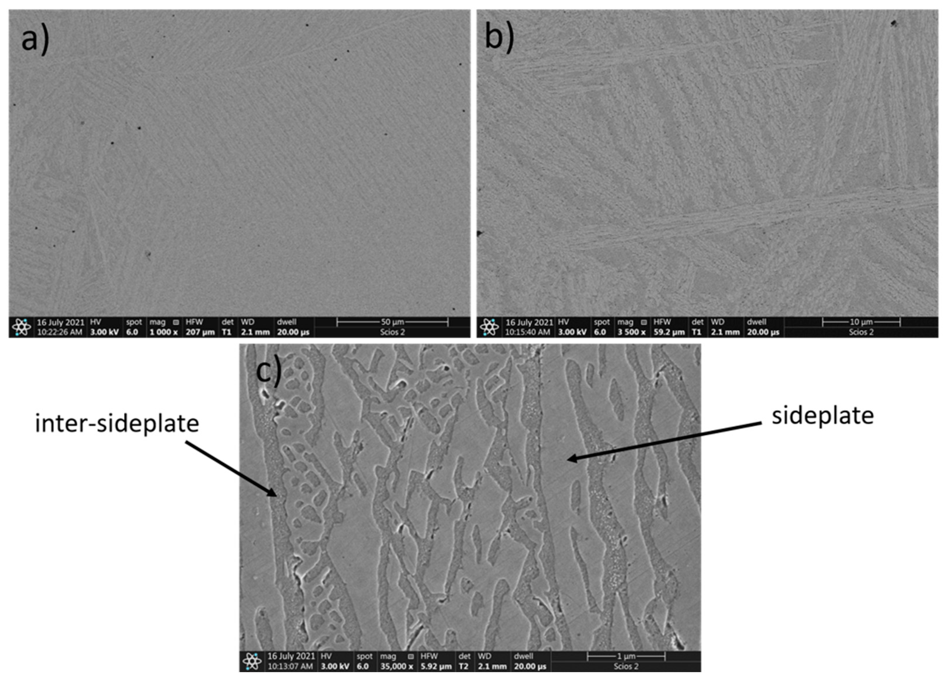
| sideplate | 10.45 | 22.89 | 22.11 | 23.85 | 20.71 | |
| inter-sideplate | 16.25 | 19.75 | 23.28 | 21.17 | 19.55 | |
| Alloy | Area | Element (at.%) | ||||
|---|---|---|---|---|---|---|
| Co | Cr | Fe | Ni | Cu | ||
| CoCrFeNiCu | global | 20.66 | 19.83 | 19.98 | 19.40 | 20.14 |
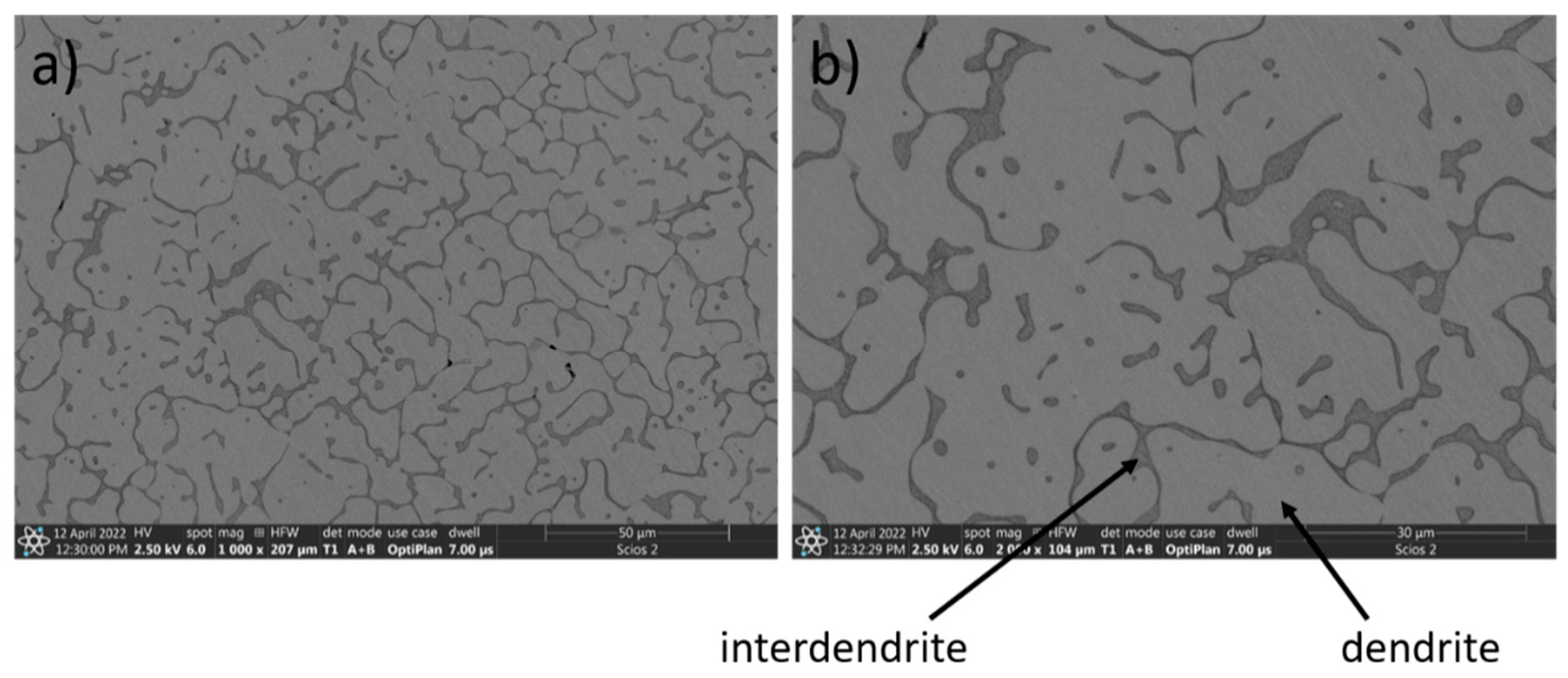
| dendrite | 22.94 | 22.26 | 22.63 | 22.43 | 9.75 | |
| interdendrite | 3.82 | 2.77 | 4.00 | 9.17 | 80.23 | |
| Alloy | Area | Element (at.%) | ||||
|---|---|---|---|---|---|---|
| Ti | Al | Fe | Co | Ni | ||
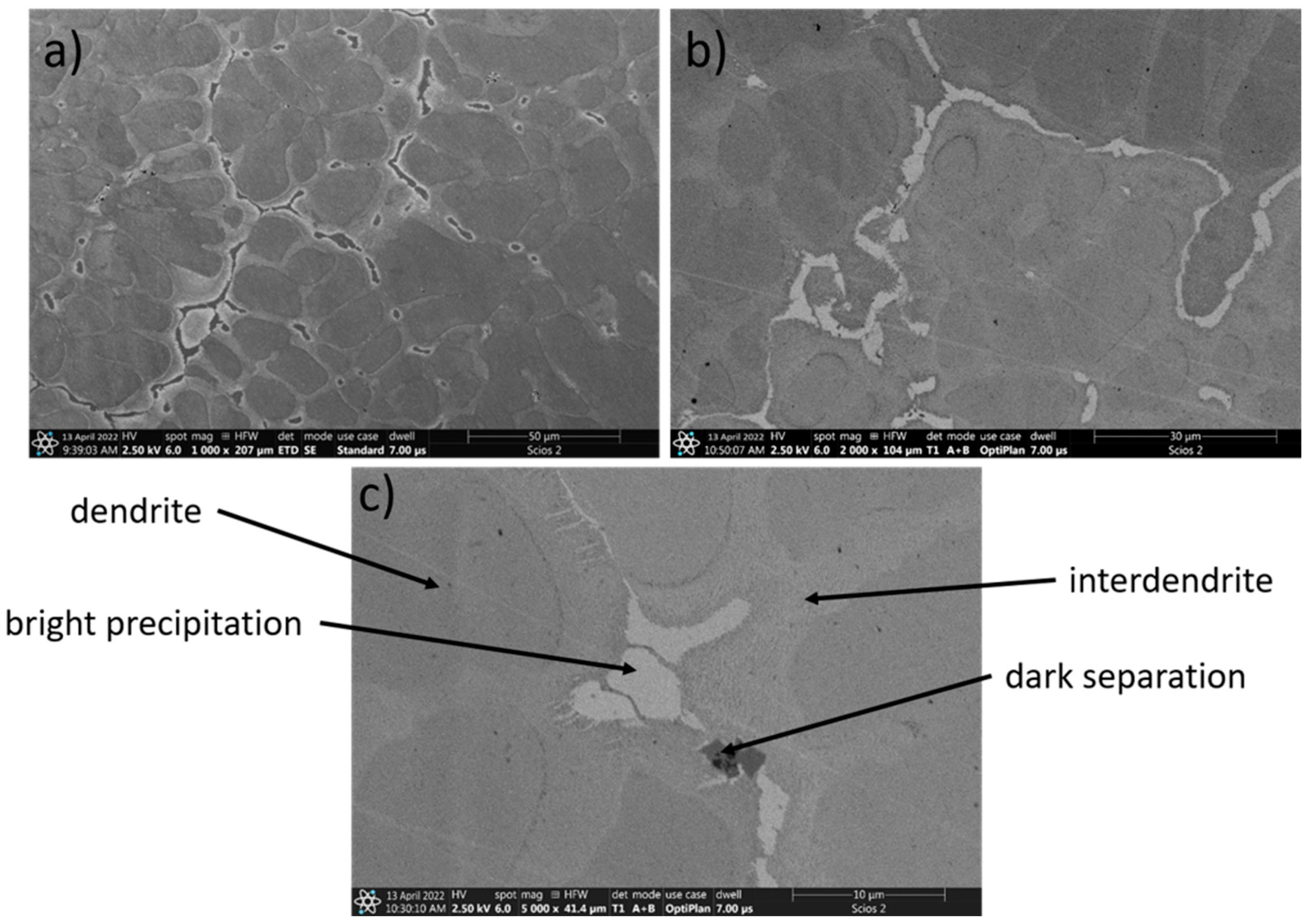
| TiAlFeCoNi | global | 20.89 | 18.85 | 20.19 | 19.83 | 20.24 |
| dendrite | 21.62 | 23.89 | 10.74 | 22.19 | 21.56 | |
| interdendrite | 23.55 | 3.55 | 41.49 | 17.42 | 13.99 | |
| dark separation | 48.15 | 33.19 | 9.28 | 4.77 | 4.61 | |
| Alloy | Area | Element (at.%) | ||||
|---|---|---|---|---|---|---|
| Mn | Ti | Cu | Al | Cr | ||
| Mn0.5TiCuAlCr | global | 11.16 | 24.38 | 21.53 | 21.56 | 21.38 |
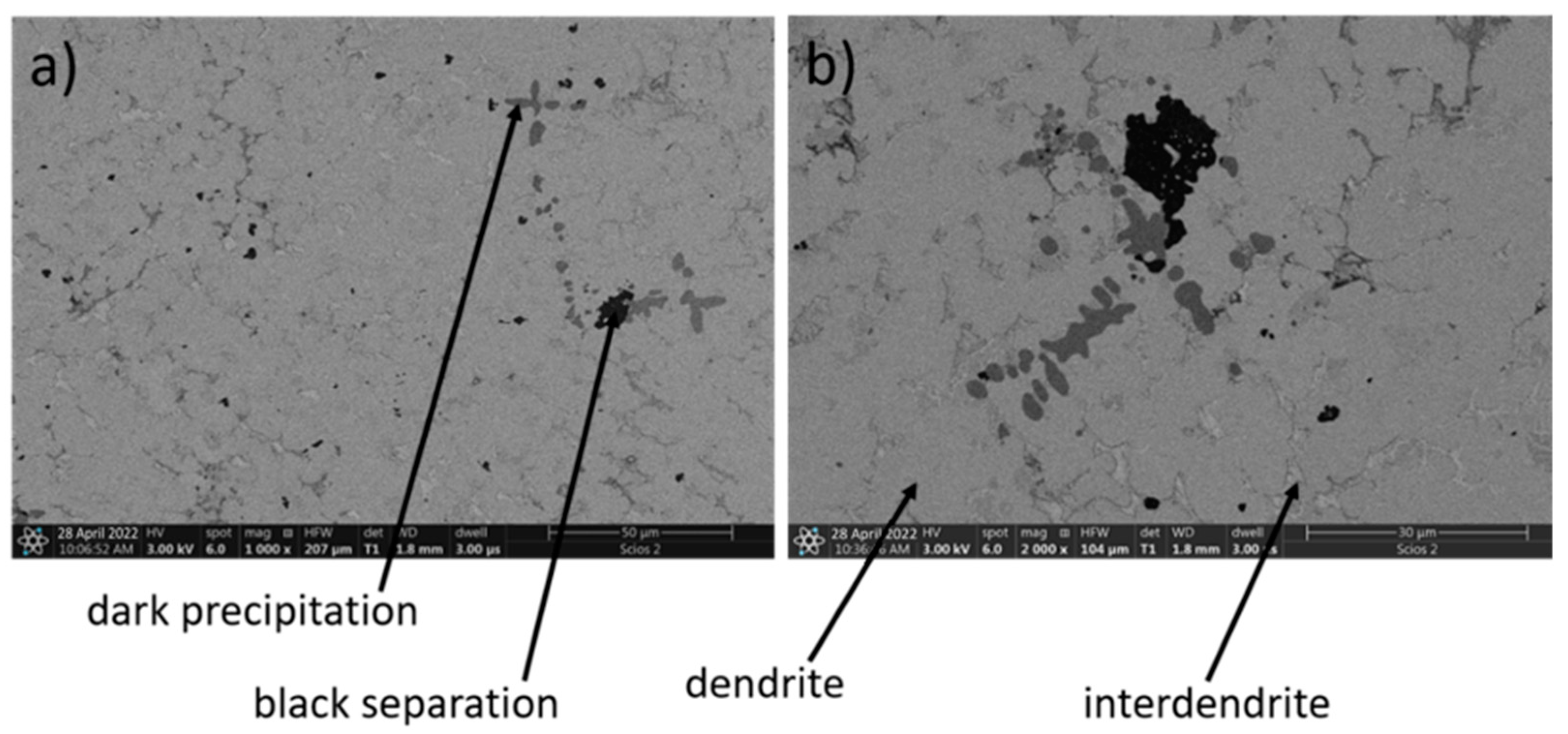
| dendrite | 20.66 | 30.13 | 12.40 | 20.66 | 24.60 | |
| interdendrite | 7.28 | 0.86 | 68.96 | 21.95 | 0.95 | |
| dark precipitation | 0.90 | 94.78 | 2.04 | 1.16 | 1.11 | |
| black separation | 1.49 | 4.15 | 5.33 | 87.07 | 1.97 | |
| Sample | Crystal Structure Space Group; Unit Cell Parameter [Å] | Vol. [%] | Order State; Graphic Symbol of the Phase |
|---|---|---|---|
| TiAlFeCoNi | Pm-3m (no.221); 5.8262 ± 0.0002 | 100 | disordered fcc; red circle |
| Fm-3m (no.225); 5.8271 ± 0.0008 | 100 | ordered fcc; red question mark | |
| CoCrFeNiCu | Pm-3m (no.221); 3.5745 ± 0.0001 | 100 | disordered fcc; blue square |
| Al0.7CoCrFeNi | Fm-3m (no.225); 3.5948 ± 0.0001 | 94 | ordered fcc; green triangle |
| Im-3m (no.229); 2.8584 ± 0.0005 | 6 | ordered bcc; olive diamond |
| Alloy | Ecorr [mV] | icorr [μA/cm2] | vcor [μA/Year] | Rp [kΩ∙cm2] |
|---|---|---|---|---|
| Al0.7FeCrCoNi | −224 | 0.9 | 11.7 | 17.7 |
| CoCrFeNiCu | −210 | 1.1 | 14.1 | 20.4 |
| TiAlFeCoNi | −435 | 4.6 | 83.2 | 3.4 |
| Mn0.5TiCuAlCr | −253 | 1.3 | 26.1 | 8.8 |
| 316L | −291 | 2.3 | 33.5 | 19.5 |
| Ti6Al4V | −268 | 1 | 51.4 | 25.3 |
Publisher’s Note: MDPI stays neutral with regard to jurisdictional claims in published maps and institutional affiliations. |
© 2022 by the authors. Licensee MDPI, Basel, Switzerland. This article is an open access article distributed under the terms and conditions of the Creative Commons Attribution (CC BY) license (https://creativecommons.org/licenses/by/4.0/).
Share and Cite
Tokarewicz, M.; Grądzka-Dahlke, M.; Rećko, K.; Łępicka, M.; Czajkowska, K. Investigation of the Structure and Corrosion Resistance of Novel High-Entropy Alloys for Potential Biomedical Applications. Materials 2022, 15, 3938. https://doi.org/10.3390/ma15113938
Tokarewicz M, Grądzka-Dahlke M, Rećko K, Łępicka M, Czajkowska K. Investigation of the Structure and Corrosion Resistance of Novel High-Entropy Alloys for Potential Biomedical Applications. Materials. 2022; 15(11):3938. https://doi.org/10.3390/ma15113938
Chicago/Turabian StyleTokarewicz, Marzena, Małgorzata Grądzka-Dahlke, Katarzyna Rećko, Magdalena Łępicka, and Kamila Czajkowska. 2022. "Investigation of the Structure and Corrosion Resistance of Novel High-Entropy Alloys for Potential Biomedical Applications" Materials 15, no. 11: 3938. https://doi.org/10.3390/ma15113938